Article

Screening and Comparative Characterization of Microorganisms from Iranian Soil Samples Showing ω-Transaminase Activity toward a Plethora of Substrates

1
Department of Biotechnology, Faculty of Biology Science, Alzahra University, 1993893973 Tehran, Iran
2
BLT_II: Technical Biology, Karlsruhe Institute of Technology (KIT), Fritz-Haber-Weg 4, 76131 Karlsruhe, Germany
3
Department of Chemistry, School of Sciences, Alzahra University, 1993891176 Tehran, Iran
4
Higher Education Complex of Shirvan, 9468194477 Shirvan, Iran
*
Authors to whom correspondence should be addressed.
Catalysts 2019, 9(10), 874; https://doi.org/10.3390/catal9100874
Submission received: 26 September 2019 / Revised: 15 October 2019 / Accepted: 18 October 2019 / Published: 22 October 2019
(This article belongs to the Special Issue Novel Enzyme and Whole-Cell Biocatalysts)

Abstract

:
In this study, soil microorganisms from Iran were screened for ω-transaminase (ω-TA) activity based on growth on minimal media containing (rac)-α-methylbenzylamine (rac-α-MBA) as a sole nitrogen source. Then, for the selection of strains with high enzyme activity, a colorimetric o-xylylendiamine assay was conducted. The most promising strains were identified by 16S rDNA sequencing. Five microorganisms showing high ω-TA activity were subjected to determine optimal conditions for ω-TA activity, including pH, temperature, co-solvent, and the specificity of the ω-TA toward different amine donors and acceptors. Among the five screened microorganisms, Bacillus halotolerans turned out to be the most promising strain: Its cell-free extract showed a highly versatile amino donor spectrum toward aliphatic, aromatic chiral amines and a broad range of pH activity. Transaminase activity also exhibited excellent solvent tolerance, with maximum turnover in the presence of 30% (v/v) DMSO.

Graphical Abstract

1. Introduction

Chiral amines as building blocks are prevalent in pharmaceuticals and chemical industries [1]. For instance, chiral amines are present in roughly 40% of FDA approved pharmaceuticals [2,3]. Furthermore, chiral β-amino acids (β-aas) have attracted significant attention recently [4]. They are critical building blocks in highly important medicines and natural compounds such as maraviroc (HIV remedy) [5,6], taxol (cytostatic drug against breast and ovarian cancer), jaspamide (insecticidal and antifungal agent), theopalauamide (antifungal compound), and dolastatin (antitumor agent) [7]. β-aas are also employed in the production of peptidomimetics that are stable against degradation by proteolytic enzymes [4,8].
The significant challenge in industry is to use optically pure amines and amino acids from cost-effective production [1,9]. For the mass production of such compounds, nowadays biocatalysts are frequently used to comply with green chemistry as well be economically feasible [1,5]. Among various enzymatic methods for producing chiral amine compounds, ω-transaminases (ω-TAs) are promising enzymes to synthesize chiral amines. They show high substrate spectra, enantioselectivity, and no requirement to regenerate external cofactors [10,11,12,13]. They belong to pyridoxal-5′–phosphate-(PLP)-dependent enzymes that transfer an amino group from an amino donor to amino accepters such as prochiral ketones or aldehydes [14,15]. In addition, ω-TAs can be used for providing optically enriched chiral amino acids via the kinetic resolution of racemic amines or asymmetric synthesis from prochiral ketones [12].
A restricted substrate scope for large molecules [16], as well as rather low enzyme stability under extreme reaction conditions associated with pH, temperature [17], and organic solvents [5,13] have been major hurdles of using ω-TAs. Thus, these limitations should be circumvented by additional screening approaches to fill the toolbox of industrially applicable enzymes.
Basically, there are different approaches to discover new ω-TAs: metagenomic screenings [18,19], site-directed mutagenesis of well-established enzymes [20], and screening new biocatalysts by identifying novel microorganisms from as-yet unexplored screening sites [21,22,23].
Results of several studies indicated that Iranian landscapes contain highly diverse microbial storages due to considerable variety in geographical features, including mountain range peaks with volcano activity, sandy deserts (Dasht-e Lut is one of the hottest points of the Earth), rivers [24], forest areas, hot springs, glaciers, and frequent snowing. Iran also has two coastlines to its north and south. Besides the extremely versatile environments in Iran, industries such as petroleum (oil well) and petrochemical units releasing chemical substances cause influences on the microorganism populations of the above-mentioned country. With such a variety of environments, Iran’s soil can be considered a rich source for screening microorganisms, which are able to produce special extremophile biocatalysts [25,26,27,28].
The purpose of this study is to identify novel ω-TA from Iranian soil microorganisms to be examined as a catalyst in the synthesis of enantiopure β-amino acid (β-aa) and other chiral amino compounds. Relevant properties of this novel ω-TA, such as the effect of temperature, pH, and solvent and substrate spectra were also investigated.

2. Results and Discussion

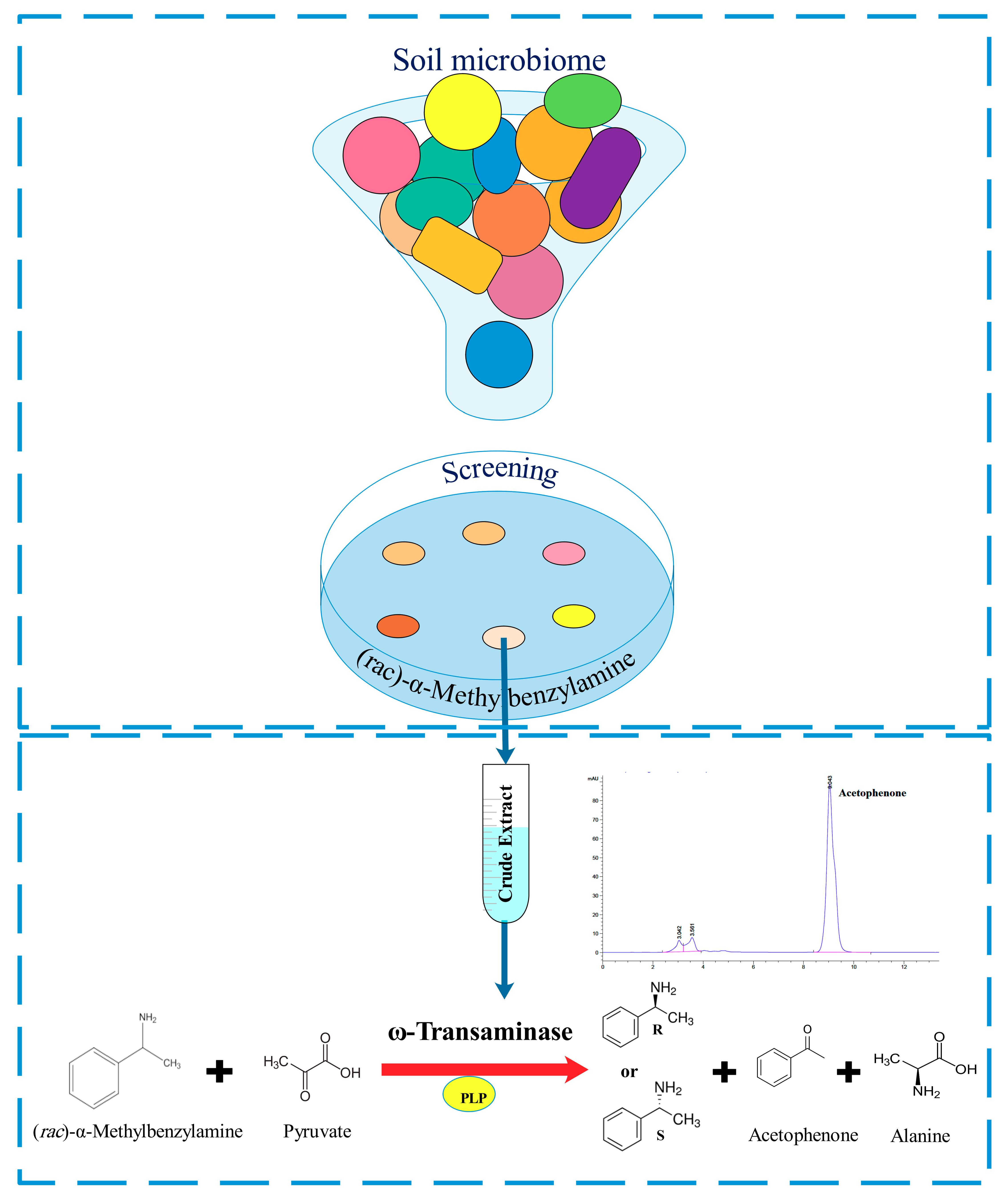
2.1. Microorganisms Screening and Identification

In this study, 42 strains were isolated by enrichment culture against (rac)-α-methylbenzylamine (rac-MBA) as a sole nitrogen source. Induction and amount of ω-TA is affected by nitrogen source in media. In complex media such as Luria-Bertani (LB) medium, some of the microorganisms produce enzymes with lower activity [29]. Therefore, minimal media (MIM) containing (rac)-α-MBA as a model amine compound [22,23] was used in the experiment.
They were subsequently validated through o-xylylenediamine (o-XDA) assay, with the color changing from yellow to black after 4 h. o-XDA after deamination undergoes spontaneous cyclization, tautomerization, and irreversible polymerization to form a black product [1]. Five strains showing ω-TA activity were found to be promising. Four of these strains were identified by 16S rDNA sequencing to be members of the genus Bacillus close to Bacillus halotolerans (BaH) (99.93% similarity) and Bacillus endophyticus (BaE) (99.76% similarity). Two subspecies belong to Bacillus subtilis with 99.92% and 99.93% similarity to stercoris (BaS) and inaquosorum (BaI), respectively. The fifth bacterium is associated to Rhizobium radiobacter (RhZ) with 99.77% similarity (Table 1). All of the strains except BaE were isolated from first method of screening.

2.2. Enzymatic Properties and Substrate Specificity

Appropriate temperature, pH, and organic solvents play important roles in ω-TA activity [5,30]. To survey such effects, the optimum reaction condition against these three factors as investigated in the screened bacteria. Several studies have been performed by applying cell and crude extract to convert different substrates [2,18,21,29,31,32,33], so instead of using high-priced purified protein, we applied cell-free extracts to optimize reaction parameters and to investigate substrate scope.
The relative values of ω-TA activity were calculated based on producing acetophenone as a product of (S)-MBA deamination. The highest activity was considered as 100%.

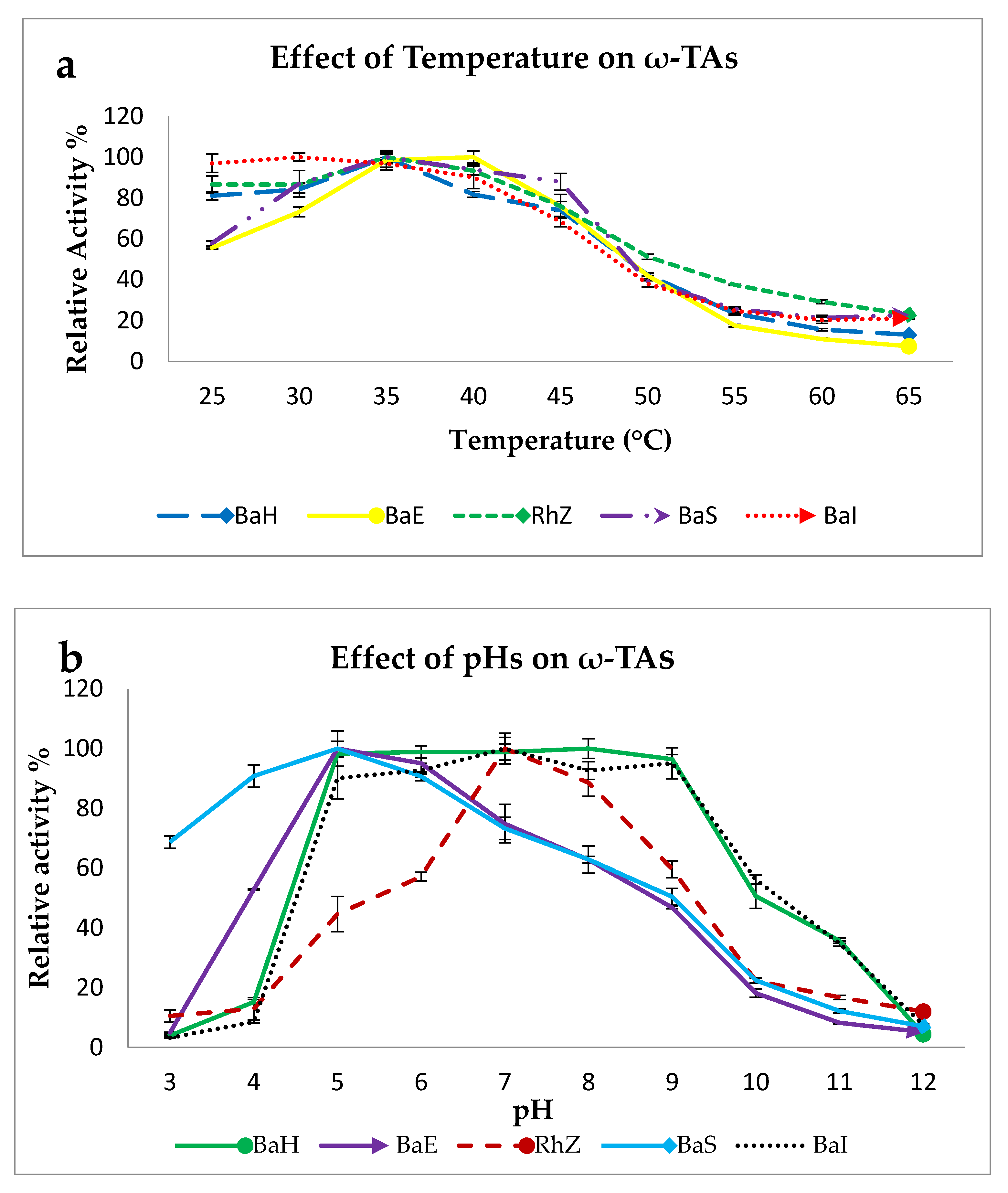
2.2.1. Effect of Temperature on ω-TA Activity

The cell-free extracts were incubated at defined temperatures (25–65 °C) for one hour. The crude extracts of BaH, RhZ, and BaS shared similar expected mesophilic temperature profiles between 30 and 65 °C, with highest activity at 35 °C, which is reported for other ω-TAs [21,34]. They exhibited reasonable activity of around 70% up to 45 °C. Among all of them, 40–50% of activity remained at 50 °C, which decreased to approximately 10–20% at temperatures above 60 °C (Figure 1a).
For ω-TABaE and ω-TABaI, the optimum reaction temperature was 40 °C and 30 °C, respectively. No significant change of activity was observed at 35 °C. Thus, for surveying the effect of pH and solvent on enzyme activity on all crude extracts, the temperature was maintained at 35 °C. Additionally, ω-TABaI showed approximately the same activity at temperatures between 20 and 30 °C (Table S1).

2.2.2. Effect of pH on ω-TA Activity

In contrast to other ω-TAs that usually show their maximum activity at slightly alkaline pH [12,21,31], ω-TABaI and ω-TABaH exhibited an almost constant activity in the pH range of 5 to 9. Surprisingly, for ω-TABaS and ω-TABaE, the highest enzyme activity was observed under acidic conditions (pH 5). Notably, 70% of ω-TABaS activity remained at pH 3, Furthermore, ω-TARhz preferred neutral conditions (Figure 1b). No enzyme activity was observed at pH 12. In the literature data, there is no report concerning ω-TA activity in acidic conditions. ω-TA activity at different pHs was mostly tested in pH range from 6 to 11 [4,17,18].

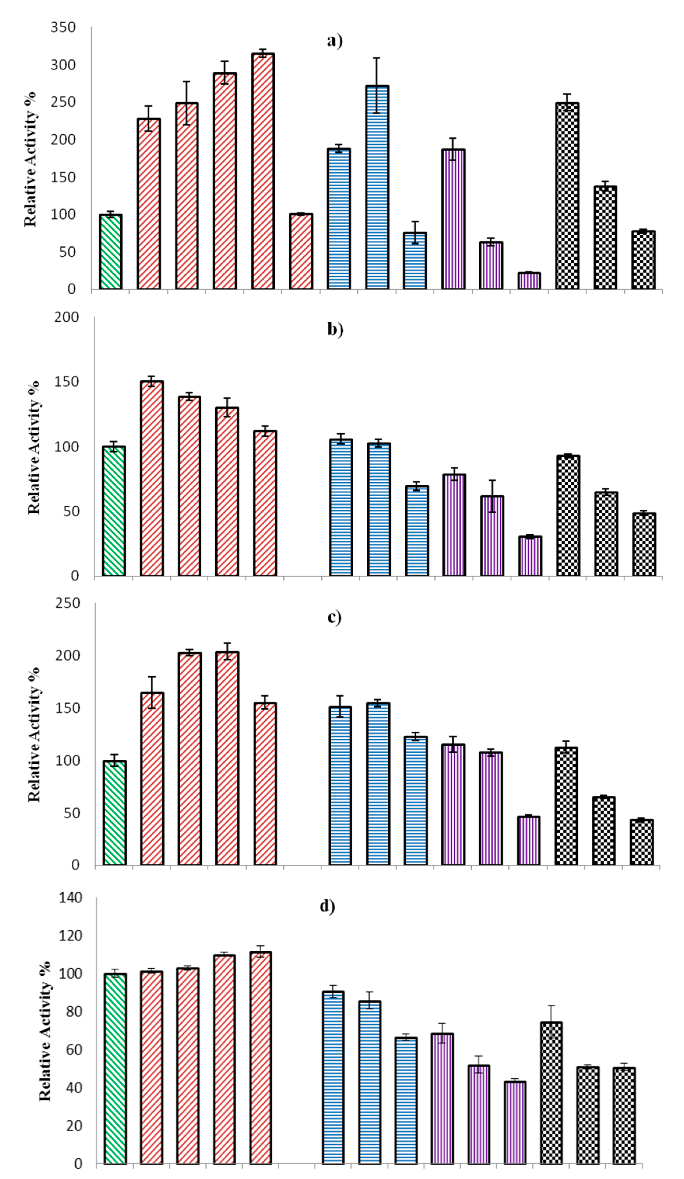
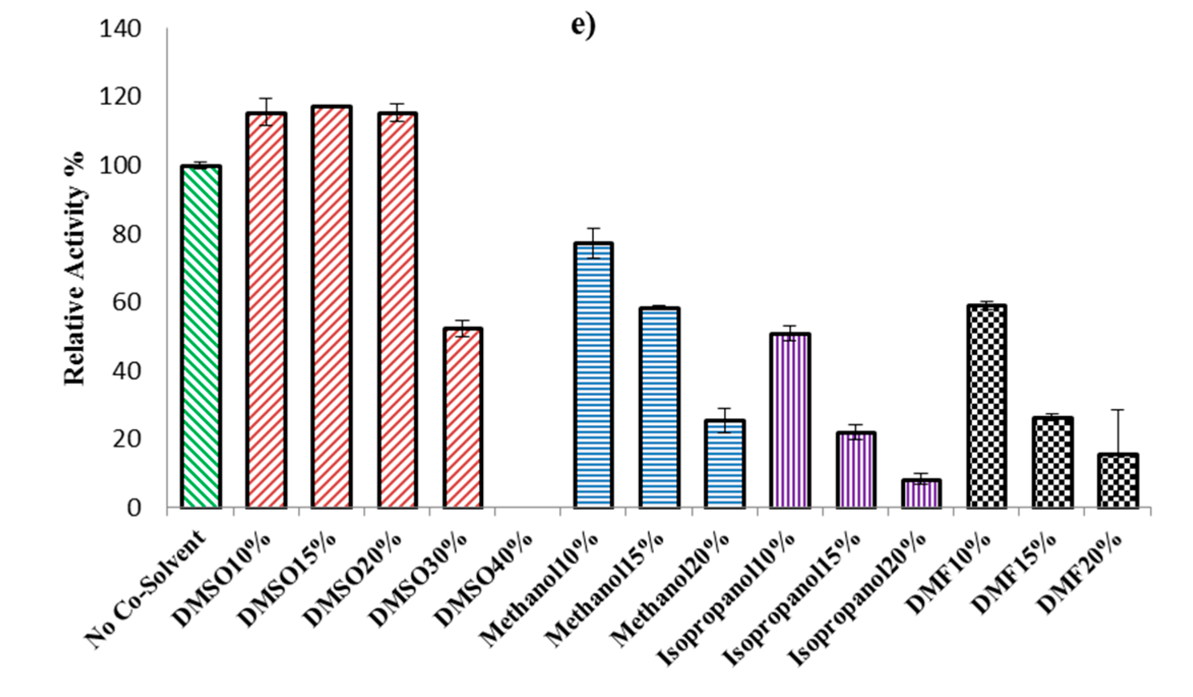
2.2.3. Effect of Organic Solvents on ω-TA Activity

Organic solvents are often added to ω-TA reactions to increase substrate solubility. However, this mostly causes negative effects on enzyme activity [10]. Therefore, the examination of the tolerance of the applied enzyme to different solvents is highly required [5]. To analyze whether solvents affect enzyme activity, four different solvents with a 0%–20% with the exception of DMSO which increased up to 40% v/v concentration were tested. The ω-TA activity in aqueous buffer without addition of organic solvents was set as 100%. The results were expressed as relative activity in %.
Dimethyl sulfoxide (DMSO) is a common solvent that was used in concentrations ranging from 5%–20% v/v in different projects [5]. DMSO as co-solvent led to the highest activity, followed by methanol, N,N-Dimethylformamide (DMF), and isopropanol. In some cases (BaI and Rhz), the reactions without any co-solvent showed better activity than methanol. It is remarkable to mention that ω-TABaH activity was enhanced threefold with a concentration of DMSO up to 30%. The results again suggest that DMSO is the most suitable organic solvent for catalytic activation of ω-TAs. However, methanol in some cases also increased enzyme activity (Figure 2). Methanol was mentioned as a second co-solvent after DMSO for enhancing ω-TA activity in Pawer et al.’s experiment [2].

2.2.4. Substrate Specificity and Enantioselectivity

The substrate spectrum of all strains was investigated in order to find the most suitable amino donor and acceptor.
Regarding amino donors, 28 substrates, including aliphatic and aromatic amino compounds were reacted to pyruvate as an amino acceptor to investigate amino donor specificity, and alanine generation was monitored by the HPLC technique. Racemates as well as the pure, (S) and (R) enantiomers (if available) of amino donors were chosen to explore the enantioselectivity of ω-TA. The results of the substrate screening are illustrated in Table 2. HPLC analysis proved that all enzymes show predominant formation of an (S)-configuration. This is in accordance with most previously reported publications in the literature indicating that (R)-stereoselectivity are rather rare in wild-type microorganisms [35]. All cell-free extracts showed high activity against (S)- α-methylbenzylamine (S-MBA) 1s.
BaH showed considerable activity toward a wide range of substrates, including (S), (R), and racemates of aromatic and aliphatic amino donors. Although this cell-free extract also showed activity toward some tested (R)-enantiomers (1r, 2r, 4r, 5r, 7r, 8r, 13r), (S)-selectivity appeared to be not so strict in this strain. Notably, it is difficult to conclude that BaH showed (R)-selectivity since in this study, crude extract was applied. There is a possibility that (R) converted to (S)-enantiomer via enzymatic racemization [36], and then the latter was subjected to ω-TA activity. On the other hand, (R)-preference towards 13r in compare to 13s leads to another hypothesis, in which both R and S transaminases are present in crude extract.
While the cell-free extract of BaH revealed varying enantiopreference toward all tested amino donors to different degrees, the substrate specificity was found to resemble what has previously been reported by Mathew et al. [5,30], as a preference to react with aromatic β-amino acids is clearly visible [5]. (S)-MBA 1s is converted with the highest activity, which therefore was set as 100%. Although a few ω-TAs are described with activity toward aromatic β-amino acids (56), it is remarkable that BaH uses β-phenylalanine (13, 13s, 13r) and (rac)-β-homophenylalanine (14) as an amino donor; both of these were inert for crude extraction from other bacteria in this experiment, which implies the presence of a broad binding pocket in the active site of ω-TABaH. ω-TA activity against 14 has been reported by Mathew et al. for ω-TABG, with 50% relative activity in comparison with 13 [30]. A recent study by Buß et al. explained the lack of ω-TA activity against 14 (by an ω-TA that was highly active against 13) by sterice hindrance caused by the additional carbon atom located between the amino group and phenyl group [36]. Several substituted derivatives of β-phenylalanine are converted as well.
BaE presented only (S)-selectivity in relation to tested substrates. Similar to BaH, the strain’s crude extract exhibited activity toward 1s, almost double that of 1. BaE and BaH share the same activity toward 12, while BaI showed almost double the activity in comparison with those. The relative activity of ω-TA of BaI toward (rac)-MBA 1 exceeded 60%, which might be related to the presence of racemase in the crude extract of bacteria [37,38].
Crude extracts of RhZ and BaS displayed (S)-selectivity toward the tested amino donors with similar preference profiles. RhZ showed more activity toward 17s as an aliphatic substrate than MBA 1s, which was not observed for other screened strains.
Amino acceptors play a critical role in ω-TA activity [30]. Eight ketones were reacted with (S)-MBA to explore the amino acceptor specificities by HPLC detection of acetophenone production (Table 3). Aliphatic ketones such as sodium pyruvate 7b and α-ketoglutaric acid 8b showed high conversion, whereas most aromatic ketones were not accepted by the investigated enzymes.
Cell-free extract of BaH, BaS, and BaI exhibited some activity toward bulkier substrates, including 1b or 2b. It is supposed that β-amino acid esters were produced by using these acceptors, but this needs to be investigated in further experiments. BaH, BaS, and BaI showed activity toward 1b as amino acceptor. BaI was the only extract to show activity toward acceptor 2b.
The substrate scope of ω-TA is determined by the active site, which consists of a large and a small binding pocket. The former can accommodate bulky residues, including aryl groups, whereas the latter is loaded by a small group such as a methyl substituent [14]. So most ω-TAs have a limitation in accepting bulky substrates without applying protein engineering. Voss et al. reported that after double mutation on ω-TA 3FCR from Ruegeria sp. TM1040, this enzyme gained activity toward bulkier substrates such as phenylpropylamine and phenylbutylamine [16]. Strain BaH showed a good ability to convert bulky substrates and showed different substrate spectra with known ω-TAs [15,35]. Purification and further investigation of this enzyme is intended for further studies.

3. Materials and Methods

3.1. Chemicals

All solvents and chemicals used in this study were purchased from Sigma-Aldrich (St Louis, MO, USA), Carl Roth GmbH (Karlsruhe, Germany), and Peptech (Burlington, MA, USA).

3.2. Screening and Identification

3.2.1. Enrichment of Microorganisms on (rac)-α-MBA as Sole Nitrogen Source

Due to the role of transaminase in nitrogen metabolism [29], areas enriched with nitrogen compounds were chosen as potential sites for screening novel ω-transaminase. Soil samples (2 g) from a petrochemical site, oil well, and an agriculture field in Iran were separately suspended in 200 mL sterile minimal medium (MIM) in 500 mL Erlenmeyer flasks containing 10 mM (rac)-α-MBA, 100 mM glycerol, 1 g/L MgSO4 .7H2O, 0.02 mg/L H3BO3, 0.2 mM CaCl2, 0.1 mg/L MnSO4. 4H2O, 0.1 mg/L CuSO4 .5H2O, 0.1 mg/L NiSO4. 6H2O, 2.0 mg/L NaMoO4, 0.05 mg/L CoCl2, 0.1 mg/L ZnCl2, 4 mg/L FeSO4. 7H2O, and potassium phosphate buffer (50 mM, pH 7.0) [39]. FeSO4 and (rac)-α-MBA were separately sterile filtrated and added to the medium. Autoclaving causes precipitation of ferric hydrate [40]. Each sample was prepared in duplicate. Enrichment went through two strategies:
In the first method, Erlenmeyer flasks were incubated for 24 h at 180 rpm at 35 °C (depending on the original temperature of the soil samples). After 24 h of incubation, a 100 µL sample was transferred to 20 mL of fresh minimal medium and incubated for 24 h under the same conditions as described before. This serial dilution was repeated 3 times. After that, the culture was diluted with PBS buffer (10: 90 µL) and poured to MIM agar plates (mineral medium, 10 mM (rac)-α-MBA with 1.5% agar) and incubated at 35 °C. As soon as colonies were visible, single colonies were streaked on new MIM agar plates to obtain uniform colonies under the same temperature.
In the second method, Erlenmeyer flasks were transferred to a dark place, without shaking, at 35 °C. During incubation, 1 mL of concentrated MIM (5X), 0.5% v/v glycerol, and 10 mM (rac)-α-MBA were added to the flasks every week. After 3 months, the culture broth was centrifuged (1000 g, 2 min) and 100 µL of supernatant was spread on MIM agar plates. The colonies were isolated after 3 days of incubation at 35 °C. This was continued until the isolation of single colonies was achieved.

3.2.2. Selection of the Most Promising Strains

To identify whether isolated strains use (rac)-α-MBA as a sole nitrogen source or atmospheric nitrogen, o-Xylylenediamine was applied as a smart amino donor. Each colony was cultivated in a shake flask containing 20 mL MIM medium with 12 mM (rac)-α-MBA for three days at 35 °C at 120 rpm. The cells were harvested by centrifugation at 6000× g for 10 min at 4 °C and resuspended in HEPES buffer (50 mM, pH 7.5). The cell concentration was adjusted to around 20 mg/mL by measuring dry cell mass based on Buß et al. [36]. The cells were frozen at −80 °C and thawed at room temperature for cell disruption.
Reaction was started by adding 50 µL of whole cells to 150 µL reaction solution containing 7.5 mM o-Xylylenediamine, 5 mM pyruvate, 1 mM PLP, and 10% DMSO in HEPES buffer (50 mM, pH 7.5) [41] in a 96-well plate. Each reaction was conducted in triplicate. The plate was incubated overnight at 35 °C, 150 rpm.

3.2.3. Identification of Bacteria

The positive strains were grown separately overnight in LB broth at 35 °C, 150 rpm. DNA was extracted by ZR soil Microbe DNA KitTM. PCR was done with a Q5 high-fidelity PCR kit (NEB, Germany). Universal Primers (27F and 1492R) were applied to amplify 1.5 kb 16S rDNA fragments. PCR was conducted following the manufacturer’s instructions for Q5 polymerase. Gene sequencing was performed by Eurofins Company (Ebersberg, Germany). The resulting 16S rRNA gene sequences were compared with available gene sequences in the EZ bioCloud database [42].

3.3. Enzymatic Properties and Substrate Specificity

3.3.1. Preparation of Crude Extract

The inoculum (3 mL) was prepared by picking a single colony and cultivating it for three days in MIM with 12 mM (rac)-α-MBA. The medium (400 mL) was inoculated by the addition of 1% (v/v) of the latter culture into 1 L shaking flasks containing MIM with 0.5 g/L yeast extract and 12 mM (rac)-α-MBA overnight at 150 rpm and 35 °C. Cells were harvested before reaching stationary phase by centrifugation at 6000× g for 10 min at 4 °C. After washing the pellet with sodium phosphate buffer (50 mM, pH 7), cells were resuspended in 10 mL lysis buffer (sodium phosphate (50 mM, pH 7), 0.1 µM PLP, 100µg/mL lysozyme]. Afterwards, they were incubated for 1 h at room temperature, followed by sonification (3 cycles: 30 s pulse, 20 s pause; 60% amplitude) on ice. Subsequently, the cell debris was removed by centrifugation (20,000 rpm, 4 °C, 15 min) in a JA-30–50 rotor (Coulter–Beckman centrifuge). The protein concentration of crude extract was determined by using the Roti®-Quant universal kit (Carl Roth, Karlsruhe, Germany) following the manufacturer’s instructions. Eventually, they were mixed with 15% glycerol and preserved in −80 °C for further analysis.

3.3.2. Effect of Temperature, pH, and Co-Solvent

The effects of temperature and pH on enzyme activity were examined at different temperatures (25–65 °C at pH7) and various pHs (3–12 at 35 °C) by using Britton–Robinson buffer (0.04 M H3BO3, 0.04 M H3PO4, 0.04 M CH3COOH) containing an amino donor (10 mM), amino acceptor (10 mM), PLP (0.1 mM), and DMSO (10%). The total reaction volume was set to 0.25 mL. The reactions were initiated by adding crude extract of enzyme (~1 mg/mL).
To study the effect of solvent on ω-TA activity, reactions were carried out applying various organic solvents with 0–20% (v/v) (for DMSO 0–40%). (rac)-α-MBA (10 mM) was used as amino donor and pyruvate (10 mM) as amino acceptor in the presence of PLP (0.1 mM) and cell-free extract (~1 mg/mL). The reactions were carried out in Britton–Robinson buffer (pH 7) at 35 °C as described above.
Each reaction was conducted in triplicate and incubated (ThermoMixer, Eppendorf) at 600 rpm for 1 h. As a control, the reaction was conducted without adding enzyme. Furthermore, the amount of acetophenone in crude extract of enzyme was also evaluated. Reactions were stopped by heating to 95 °C for 5 min. After centrifugation, the supernatant was analyzed by HPLC to detect produced acetophenone according to Section 3.4. Results are shown as relative activity.

3.3.3. Substrate Specificity and Enantioselectivity

The reactions were carried out according to Section 3.3.2. Various amino donors (10 mM) listed in Table 1 were tested using sodium pyruvate (10 mM) as amino acceptor in the Britton–Robinson buffer (pH 7) and 15% (v/v) DMSO. The reaction was carried out at 35 °C, 600 rpm, for 20 h (ThermoMixer, Eppendorf). The produced alanine was analyzed by HPLC according to Section 3.4.
Different amino acceptors (10 mM) were tested with (S)-α-MBA as amino donor (10 mM) in Britton–Robinson buffer, pH 7, at 35 °C, 600 rpm, for 20 h. The production of acetophenone was analyzed by HPLC according to Section 3.4.
Each reaction was conducted in triplicate, and the average of three independent reactions was used for evaluation. Enzyme inactivation was applied by heating to 95 °C for 5 min. The supernatant was used for analysis after centrifugation. In addition, the amounts of alanine and acetophenone were measured in enzyme crude extracts as blanks.

3.4. HPLC Analytics

All samples were analyzed by Agilent 1100 series HPLC system (Santa Clara, CA, USA). For the analysis of alanine, derivatization by using ortho-phthalaldehyde was carried out according to Brucher et al. [43] and Buß et al. [44], with an automated precolumn derivatization. A reversed-phase C18 column (150 × 4.6 mm HyperClone 5 µm ODS, Phenomenex, Germany) with isocratic elution with 35% (v/v) methanol and 65% (v/v) sodium phosphate buffer (40 mM, pH 6.5) at a flow rate of 0.8 mL min−1 and detection at 338 nm at 25 °C was used.
The acetophenone concentration in the samples was determined chromatographically by isocratic elution with acetonitrile/water (50/50, v/v) at flow rate of 0.6 mL min−1 with UV detection at 254 nm [45], at 25 °C applying a C18 Hypersil-keystone column (250 × 4.6 mm 5 µ Hypersil). The injection volume was adjusted to 1 µL.

4. Conclusions

Our motivation for screening novel microorganisms exhibiting transaminase activity resulted from several challenges for the industrial application of ω-TAs, e.g., lacking activity at acidic pH, high temperature, unnatural substrates, (R)-configured molecules, and elevated concentrations of organic solvents (needed due to low substrate solubility in aqueous systems). We succeeded in finding promising new strains from a high variety of Iranian soil samples by enrichment culture using (rac)-α-methylbenzylamine (α-MBA) as a sole nitrogen source.
In particular, a Bacillus halotolerans (BaH) strain was isolated from a petroleum refinery, exhibiting ω-TA activity using a broad spectrum of amino donors over a pH range of 5–9 at elevated concentrations of DMSO and other organic solvents up to 30% (v/v). Two other strains isolated from an agricultural field (Bacillus endophyticus, BaE) and an oilfield (Bacillus subtilis, BaS) showed the highest ω-TA activity against α-MBA at pH 5 with 70% remaining activity at pH 3 (BaS), whereas ω-TAs are usually described to prefer slightly alkaline conditions. The enzymes dedicated to these extraordinary activities will be purified and subjected to in-depth studies for application-technical characterization.

Supplementary Materials

The following are available online at https://www.mdpi.com/2073-4344/9/10/874/s1, Table S1: The Effect of temperature on the amination of pyruvate and (S)-MBA as amino donor using 1 mg/mL cell-free extracts of BaI strain. Reaction conditions are given in the method section. Formation of acetophenone was detected by HPLC at 254 nm. Every reaction was conducted in three replicates.

Author Contributions

Conceptualization, J.F. and J.R., methodology, N.G.N., M.Z.M., and U.E., validation, J.R., M.M.H., and U.E., formal analysis, N.G.N., investigation, N.G.N., data curation, N.G.N., writing—original draft preparation, N.G.N., writing—review and editing, N.G.N., J.F., J.R., U.E., M.M.H., M.Z.M., and C.S., visualization, M.Z.M., supervision, J.F., J.R., and C.S., project administration, J.F.

Funding

This research received no external funding.

Acknowledgments

This study was financially supported by grant No: 970201 of the Biotechnology Development Council of the Islamic Republic of Iran. The publication of this article was funded by Karlsruhe Institute of Technology (Helmholtz).

Conflicts of Interest

The authors declare that there is no conflict of interest.

References

  1. Gomm, A.; Lewis, W.; Green, A.P.; O’Reilly, E. A New Generation of Smart Amine Donors for Transaminase-Mediated Biotransformations. Chem. A Eur. J. 2016, 22, 12692–12695. [Google Scholar] [CrossRef] [Green Version]
  2. Pawar, S.V.; Hallam, S.J.; Yadav, V.G. Metagenomic Discovery of a Novel Transaminase for Valorization of Monoaromatic Compounds. RSC Adv. 2018, 8, 22490–22497. [Google Scholar] [CrossRef]
  3. Aleku, G.A.; France, S.P.; Man, H.; Mangas-Sanchez, J.; Montgomery, S.L.; Sharma, M.; Leipold, F.; Hussain, S.; Grogan, G.; Turner, N.J. A Reductive Aminase from Aspergillus Oryzae. Nat. Chem. 2017, 9, 961. [Google Scholar] [CrossRef]
  4. Kim, G.H.; Jeon, H.; Khobragade, T.P.; Patil, M.D.; Sung, S.; Yoon, S.; Won, Y.; Choi, I.S.; Yun, H. Enzymatic Synthesis of Sitagliptin Intermediate Using a Novel ω-Transaminase. Enzym. Microb. Technol. 2019, 120, 52–60. [Google Scholar] [CrossRef]
  5. Mathew, S.; Nadarajan, S.P.; Chung, T.; Park, H.H.; Yun, H. Biochemical Characterization of Thermostable ω-Transaminase from Sphaerobacter Thermophilus and its Application for Producing Aromatic β-and γ-Amino Acids. Enzym. Microb. Technol. 2016, 87, 52–60. [Google Scholar] [CrossRef]
  6. Haycock-Lewandowski, S.J.; Wilder, A.; Åhman, J. Development of a Bulk Enabling Route to Maraviroc (UK-427,857), a CCR-5 Receptor Antagonist. Org. Process. Res. Dev. 2008, 12, 1094–1103. [Google Scholar] [CrossRef]
  7. Slomka, C.; Zhong, S.; Fellinger, A.; Engel, U.; Syldatk, C.; Bräse, S.; Rudat, J. Chemical Synthesis and Enzymatic, Stereoselective Hydrolysis of a Functionalized Dihydropyrimidine for the Synthesis of β-Amino Acids. AMB Express 2015, 5, 85. [Google Scholar] [CrossRef]
  8. Weiner, B.; Szymański, W.; Janssen, D.B.; Minnaard, A.J.; Feringa, B.L. Recent Advances in the Catalytic Asymmetric Synthesis of β-Amino Acids. Chem. Soc. Rev. 2010, 39, 1656–1691. [Google Scholar] [CrossRef]
  9. Turner, N.J.; Truppo, M.D. Biocatalytic Routes to Nonracemic Chiral Amines. In Chiral Amine Synthesis: Methods, Developments and Applications; Nugent, T.C., Ed.; Wiley-VCH: Weinheim, Germany, 2010; pp. 431–459. [Google Scholar]
  10. Mathew, S.; Jeong, S.S.; Chung, T.; Lee, S.H.; Yun, H. Asymmetric Synthesis of Aaromatic β-Amino Acids Using ω-Transaminase: Optimizing the Lipase Concentration to Obtain Thermodynamically Unstable β-Keto Acids. Biotechnol. J. 2016, 11, 185–190. [Google Scholar] [CrossRef]
  11. Malik, M.S.; Park, E.S.; Shin, J.S. Features and Technical Applications of ω-Transaminases. Appl. Microbiol. Biotechnol. 2012, 94, 1163–1171. [Google Scholar] [CrossRef]
  12. Jiang, J.; Chen, X.; Feng, J.; Wu, Q.; Zhu, D. Substrate Profile of an ω-Transaminase from Burkholderia Vietnamiensis and its Potential for the Production of Optically Pure Amines and Unnatural Amino Acids. J. Mol. Catal. B Enzym. 2014, 100, 32–39. [Google Scholar] [CrossRef]
  13. Cerioli, L.; Planchestainer, M.; Cassidy, J.; Tessaro, D.; Paradisi, F. Characterization of a Novel Amine Transaminase from Halomonas Elongata. J. Mol. Catal. B Enzym. 2015, 120, 141–150. [Google Scholar] [CrossRef]
  14. Voss, M.; Das, D.; Genz, M.; Kumar, A.; Kulkarni, N.; Kustosz, J.; Kumar, P.; Bornscheuer, U.T.; Höhne, M. In Silico Based Engineering Approach to Improve Transaminases for the Conversion of Bulky Substrates. ACS Catal. 2018, 8, 11524–11533. [Google Scholar] [CrossRef]
  15. Rudat, J.; Brucher, B.R.; Syldatk, C. Transaminases for the Synthesis of Enantiopure Beta-Amino Acids. AMB Express 2012, 2, 11. [Google Scholar] [CrossRef]
  16. Pavlidis, I.V.; Weiß, M.S.; Genz, M.; Spurr, P.; Hanlon, S.P.; Wirz, B.; Iding, H.; Bornscheuer, U.T. Identification of (S)-Selective Transaminases for the Asymmetric Synthesis of Bulky Chiral Amines. Nat. Chem. 2016, 8, 1076–1082. [Google Scholar] [CrossRef]
  17. Kelly, S.A.; Magill, D.J.; Megaw, J.; Skvortsov, T.; Allers, T.; McGrath, J.W.; Allen, C.C.; Moody, T.S.; Gilmore, B.F. Characterisation of a Solvent-Tolerant Haloarchaeal (R)-Selective Transaminase Isolated from a Triassic Period Salt Mine. Appl. Microbiol. Biotechnol. 2019, 103, 5727–5737. [Google Scholar] [CrossRef]
  18. Leipold, L.; Dobrijevic, D.; Jeffries, J.W.; Bawn, M.; Moody, T.S.; Ward, J.M.; Hailes, H.C. The Identification and Use of Robust Transaminases from a Domestic Drain Metagenome. Green Chem. 2019, 21, 75–86. [Google Scholar] [CrossRef]
  19. Ferrandi, E.E.; Previdi, A.; Bassanini, I.; Riva, S.; Peng, X.; Monti, D. Novel Thermostable Amine Transferases from Hot Spring Metagenomes. Appl. Microbiol. Biotechnol. 2017, 101, 4963–4979. [Google Scholar] [CrossRef]
  20. Genz, M.; Vickers, C.; van den Bergh, T.; Joosten, H.-J.; Dörr, M.; Höhne, M.; Bornscheuer, U. Alteration of the Donor/Acceptor Spectrum of the (S)-Amine Transaminase from Vibrio Fluvialis. Int. J. Mol. Sci. 2015, 16, 26953–26963. [Google Scholar] [CrossRef]
  21. Kelly, S.A.; Megaw, J.; Caswell, J.; Scott, C.J.; Allen, C.C.; Moody, T.S.; Gilmore, B.F. Isolation and Characterisation of a Halotolerant ω-Transaminase from a Triassic Period Salt Mine and Its Application to Biocatalysis. ChemistrySelect 2017, 2, 9783–9791. [Google Scholar] [CrossRef]
  22. Shin, J.S.; Kim, B.G. Kinetic Resolution of α-Methylbenzylamine with o-Transaminase Screened from Soil Microorganisms: Application of a Biphasic System to Overcome Product Inhibition. Biotechnol. Bioeng. 1997, 55, 348–358. [Google Scholar] [CrossRef]
  23. Pavkov-Keller, T.; Strohmeier, G.A.; Diepold, M.; Peeters, W.; Smeets, N.; Schürmann, M.; Gruber, K.; Schwab, H.; Steiner, K. Discovery and Structural Characterisation of New Fold Type IV-Transaminases Exemplify the Diversity of This Eenzyme Fold. Sci. Rep. 2016, 6, 38183. [Google Scholar] [CrossRef] [PubMed]
  24. Yazdi, A.; Emami, M.H.; Shafiee, S.M. Dasht-E Lut in Iran, the Most Complete Collection of Beautiful Geomorphological Phenomena of Desert. Open J. Geol. 2014, 4, 249–261. [Google Scholar] [CrossRef]
  25. Ataee, N.; Fooladi, J.; Namaei, M.H.; Rezadoost, H.; Mirzajani, F. Biocatalysts Screening of Papaver Bracteatum Flora for Thebaine Transformation to Codeine and Morphine. Biocatal. Agric. Biotechnol. 2017, 9, 127–133. [Google Scholar] [CrossRef]
  26. Ghasemi, Y.; Rasoul-Amini, S.; Ebrahiminezhad, A.; Kazemi, A.; Shahbazi, M.; Talebnia, N. Screening and Isolation of Extracellular Protease Producing Bacteria from the Maharloo Salt Lake. Iran. J. Pharm. Sci. 2011, 7, 175–180. [Google Scholar]
  27. Alghabpoor, S.S.; Panosyan, H.; Trchounian, A.; Popov, Y. Purification and Characterization of a Novel Thermostable and Acid Stable α-Amylase from Bacillus Sp. Iranian S1. Int. J. Eng. Trans. B Appl. 2013, 26, 815–820. [Google Scholar] [CrossRef]
  28. Shirsalimian, M.; Amoozegar, M.; Sepahy, A.A.; Kalantar, S.; Dabbagh, R. Isolation of Extremely Halophilic Archaea from a Saline River in the Lut Desert of Iran, Moderately Resistant to Desiccation and Gamma Radiation. Microbiology 2017, 86, 403–411. [Google Scholar] [CrossRef]
  29. Shin, J.S.; Kim, B.G. Comparison of the ω-Transaminases from Different Microorganisms and Application to Production of Chiral Amines. Biosci. Biotechnol. Biochem. 2001, 65, 1782–1788. [Google Scholar] [CrossRef]
  30. Mathew, S.; Bea, H.; Nadarajan, S.P.; Chung, T.; Yun, H. Production of Chiral β-Amino Acids Using ω-Transaminase from Burkholderia Graminis. J. Biotechnol. 2015, 196, 1–8. [Google Scholar] [CrossRef]
  31. Guo, F.; Berglund, P. Transaminase Biocatalysis: Optimization and Application. Green Chem. 2017, 19, 333–360. [Google Scholar] [CrossRef]
  32. Dreßen, A.; Hilberath, T.; Mackfeld, U.; Billmeier, A.; Rudat, J.; Pohl, M. Phenylalanine Ammonia Lyase from Arabidopsis Thaliana (AtPAL2): A Potent MIO-Enzyme for the Synthesis of Non-Canonical Aromatic Alpha-Amino Acids: Part I: Comparative Characterization to the Enzymes from Petroselinum Crispum (PcPAL1) and Rhodosporidium Toruloides (RtPAL). J. Biotechnol. 2017, 258, 148–157. [Google Scholar]
  33. Schätzle, S.; Höhne, M.; Robins, K.; Bornscheuer, U.T. Conductometric Method for the Rapid Characterization of the Substrate Specificity of Amine-Transaminases. Anal. Chem. 2010, 82, 2082–2086. [Google Scholar] [CrossRef]
  34. Schätzle, S.; Höhne, M.; Redestad, E.; Robins, K.; Bornscheuer, U.T. Rapid and Sensitive Kinetic Assay for Characterization of ω-Transaminases. Anal. Chem. 2009, 81, 8244–8248. [Google Scholar] [CrossRef] [PubMed]
  35. Koszelewski, D.; Tauber, K.; Faber, K.; Kroutil, W. ω-Transaminases for the Synthesis of Non-Racemic α-Chiral Primary Amines. Trends Biotechnol. 2010, 28, 324–332. [Google Scholar] [CrossRef] [PubMed]
  36. Buß, O.; Dold, S.M.; Obermeier, P.; Litty, D.; Muller, D.; Grüninger, J.; Rudat, J. Enantiomer Discrimination in β-Phenylalanine Degradation by a Newly Isolated Paraburkholderia Strain BS115 and Type Strain PsJN. AMB Express 2018, 8, 149. [Google Scholar] [CrossRef] [PubMed]
  37. Conti, E.; Stachelhaus, T.; Marahiel, M.A.; Brick, P. Structural Basis for the Activation of Phenylalanine in the Non-Ribosomal Biosynthesis of Gramicidin S. EMBO J. 1997, 16, 4174–4183. [Google Scholar] [CrossRef] [PubMed]
  38. Yamada, M.; Kurahashi, K. Further Purification and Properties of Adenosine Triphosphate-Dependent Phenylalanine Racemase of Bacillus Brevis Nagano. J. Biochem. 1969, 66, 529–540. [Google Scholar] [CrossRef]
  39. Yun, H.; Lim, S.; Cho, B.K.; Kim, B.G. ω-Amino Acid: Pyruvate Transaminase from Alcaligenes Denitrificans Y2k-2: A New Catalyst for Kinetic Resolution of β-Amino Acids and Amines. Appl. Environ. Microbiol. 2004, 70, 2529–2534. [Google Scholar] [CrossRef]
  40. Temple, K.L.; Colmer, A.R. The Autotrophic Oxidation of Iron by a New Bacterium: Thiobacillus Ferrooxidans. J. Bacteriol. 1951, 62, 605. [Google Scholar]
  41. Buß, O.; Voss, M.; Delavault, A.; Gorenflo, P.; Syldatk, C.; Bornscheuer, U.; Rudat, J. β-Phenylalanine Ester Synthesis from Stable β-Keto Ester Substrate Using Engineered ω-Transaminases. Molecules 2018, 23, 1211. [Google Scholar] [CrossRef]
  42. Yoon, S.H.; Ha, S.M.; Kwon, S.; Lim, J.; Kim, Y.; Seo, H.; Chun, J. Introducing EzBioCloud: A taxonomically united database of 16S rRNA gene sequences and whole-genome assemblies. Int. J. Syst. Evol. Microbiol. 2017, 67, 1613. [Google Scholar] [PubMed]
  43. Brucher, B.; Rudat, J.; Syldatk, C.; Vielhauer, O. Enantioseparation of Aromatic β³-Amino Acid by Precolumn Derivatization with o-Phthaldialdehyde and N-Isobutyryl-l-Cysteine. Chromatographia 2010, 71, 1063–1067. [Google Scholar] [CrossRef]
  44. Buß, O.; Muller, D.; Jager, S.; Rudat, J.; Rabe, K.S. Improvement in the Thermostability of a β-Amino Acid Converting ω-Transaminase by Using FoldX. ChemBioChem 2018, 19, 379–387. [Google Scholar] [CrossRef] [PubMed]
  45. Gao, S.; Su, Y.; Zhao, L.; Li, G.; Zheng, G. Characterization of a (R)-Selective Amine Transaminase from Fusarium Oxysporum. Process Biochem. 2017, 63, 130–136. [Google Scholar] [CrossRef]
Figure 1. The effect of temperature (a) and pH (b) on the amination of pyruvate and (S)-MBA as amino donor using 1 mg/mL of cell-free extracts of screened strains: Bacillus halotolerans (BaH), Bacillus endophyticus (BaE), Rhizobium radiobacter (RhZ), Bacillus subtilis subsp. stercoris (BaS) and Bacillus subtilis subsp. inaquosorum (BaI).Reaction conditions are given in the method section. Formation of acetophenone was detected by HPLC at 254 nm. The value of 100% corresponded to the highest activity. Error bars represent ± standard deviation.
Catalysts 09 00874 g001
Figure 2. Effect of co-solvent on ω-TA activity of screened strains. (a) BaH; (b) BaE; (c) BaS; (d) RhZ; (e) BaI. The value of 100% corresponds to enzyme activity in the no co-solvent condition. 1 mg/mL cell-free extract was added to react under the following conditions: (S)-MBA (10 mM), pyruvate (10 mM), PLP (0.1 mM), and co-solvent (0–30% v/v) in Britton–Robinson buffer (pH 7) at 35 °C. Formation of acetophenone was detected by HPLC at 254 nm. Error bars represent ± standard deviation.
Catalysts 09 00874 g002aCatalysts 09 00874 g002b
Table 1. Results of 16SrDNA sequencing.
IsolateClosest Relative in GenBank% Similarity/Sequence Length (bp)
BaHBacillus halotolerans (LPVF01000003)99.9/1388
BaEBacillus endophyticus (AF295302)99.7/1273
BaSBacillus subtilis subsp. stercoris (JHCA01000027)99/1385
BaIBacillus subtilis subsp. inaquosorum (AMXN01000021)99.9/1406
RhZRhizobium radiobacter (AJ389904)99.4/1338
Number in brackets is GenBank accession number.
Table 2. Amino donor screened assays were carried out in Britton–Robinson buffer, pH 7 with amino donor (10 mM), pyruvate (10 mM), PLP (0.1 mM), and 15% (v/v) DMSO. The reaction was initiated by adding crude extract to a final concentration of 1 mg protein per ml. Reactions were performed at 35 °C, 600 rpm for 20 h. Relative activity was measured by HPLC detection of alanine formation at 338 nm. All reactions were conducted in triplicate, and each cell-free extract was separately analyzed for alanine content as a control.
Amino DonorRelative Activity%
BaHBaEBaSBaIRhZ
1(rac)α-methylbenzylamine Catalysts 09 00874 i0015348456036
1s(S)100100100100100
1r(R)230060
2(rac)3-amino-3-(4-chlorophenyl) propionic acid Catalysts 09 00874 i002190000
2s(S)200000
2r(R)160000
3(rac)3-amino-3-(3,4-dichloro-phenyl)propionic acid Catalysts 09 00874 i003200000
4(rac)3-amino-3-(4-nitrophenyl)propionic acid Catalysts 09 00874 i004510000
4r(R)190000
5r(R)(R)3-amino-3-(2-nitro-phenyl)-propionic acid Catalysts 09 00874 i005160000
6(rac)3-amino-3-(4-methoxyphenyl) propionic acid Catalysts 09 00874 i006210000
6s(S)170000
7s(S)3-amino-3-(4-hydroxy-phenyl)propionic acid Catalysts 09 00874 i007180000
7r(R)150000
8(rac)3-amino-3-(4-bromophenyl)-propionic acid Catalysts 09 00874 i008210000
8r(R)200000
9(rac)3-amino-3-(4-fluorophenyl)propionic acid Catalysts 09 00874 i009430000
9s(S)160000
10(rac)Methyl(-3-amino-3-phenyl propanoate hydrochloride) Catalysts 09 00874 i010110000
11(rac)3-amino-3-(4-isopropylphenyl) propionic acid Catalysts 09 00874 i011200000
12(rac)1-aminocyclopropane-1-carboxylic acid Catalysts 09 00874 i01217170300
13(rac)β-phenylalanine Catalysts 09 00874 i013170000
13s(S)160000
13r(R)220000
14(rac)β-Homophenylalanine Catalysts 09 00874 i014160000
15 Isopropylamine Catalysts 09 00874 i0152000210
16 Sec-butylamine Catalysts 09 00874 i0162570270
17s(S)3-amino butyric acid Catalysts 09 00874 i01791595420135
The activity against (S)-MBA was set as 100. An amount under 1% was considered as zero activity.
Table 3. Amino acceptor screened assays were carried out in Britton–Robinson buffer, pH 7 with (S)-MBA (10 mM), amino acceptor (10 mM), PLP (0.1 mM), and 15% (v/v) DMSO. The reaction was started by the addition of crude extract to a final concentration of 1 mg/mL in the following conditions: 35 °C, 600 rpm for 20 h. Relative activity was measured by HPLC detection of acetophenone formation at 254 nm. All reactions were conducted in triplicate, and each cell-free extract was separately analyzed as a control.
Amino AcceptorsRelative Activity%
BaHBaEBaSBaIRhZ
1bEthyl benzoylacetate Catalysts 09 00874 i01840.95020.8414.870
2bEthyl 3-(4-methoxyphenyl)-3-oxopropanoate Catalysts 09 00874 i01900060.480
3bSodium 3-oxo-3-phenylpropanoate Catalysts 09 00874 i02000000
4bEthyl(4-flurobenzoyl)acetate Catalysts 09 00874 i02100000
5bEthyl(4-chlorobenzoyl)acetate Catalysts 09 00874 i02200000
6bEthyl 4-nitrobenzoylacetate Catalysts 09 00874 i02300000
7bPyruvate Catalysts 09 00874 i02410010043.7310036.39
8bα-ketoglutarate Catalysts 09 00874 i02517.097.311000100
In each column, the highest activity was defined as 100%. Activity less than 1% was considered zero.

