Article

Hydrolysis of Glycosyl Thioimidates by Glycoside Hydrolase Requires Remote Activation for Efficient Activity

1
Institut de Chimie Organique et Analytique, ICOA, Université d’Orléans, CNRS, UMR 7311, BP6759 Rue de Chartres, F-45067 Orléans CEDEX 2, France
2
Department of Chemistry and Biochemistry, University of Missouri-St. Louis, One University Boulevard, St. Louis, MO 63121, USA
*
Author to whom correspondence should be addressed.
Catalysts 2019, 9(10), 826; https://doi.org/10.3390/catal9100826
Submission received: 18 September 2019 / Revised: 27 September 2019 / Accepted: 30 September 2019 / Published: 1 October 2019
(This article belongs to the Special Issue Novel Enzyme and Whole-Cell Biocatalysts)

Abstract

:
Chemoenzymatic synthesis of glycosides relies on efficient glycosyl donor substrates able to react rapidly and efficiently, yet with increased stability towards chemical or enzymatic hydrolysis. In this context, glycosyl thioimidates have previously been used as efficient donors, in the case of hydrolysis or thioglycoligation. In both cases, the release of the thioimidoyl aglycone was remotely activated through a protonation driven by a carboxylic residue in the active site of the corresponding enzymes. A recombinant glucosidase (DtGly) from Dictyoglomus themophilum, previously used in biocatalysis, was also able to use such glycosyl thioimidates as substrates. Yet, enzymatic kinetic values analysis, coupled to mutagenesis and in silico modelling of DtGly/substrate complexes demonstrated that the release of the thioimidoyl moiety during catalysis is only driven by its leaving group ability, without the activation of a remote protonation. In the search of efficient glycosyl donors, glycosyl thioimidates are attractive and efficient. Their utility, however, is limited to enzymes able to promote leaving group release by remote activation.

Graphical Abstract

1. Introduction

Enzymes proved to be efficient synthetic tools for the eco-compatible synthesis of many classes of compounds. Non-organic solvents, mild experimental conditions and high regio- or stereo- specificity inherent to biocatalyzed reactions have increased the added value of enzymes in transformation processes, from the laboratory bench to the industrial scale [1]. Moreover, genetic modifications of recombinant enzymes are now powerful tools to easily alter versatility and properties of the engineered proteins. Rational mutagenesis, directed evolution, or even de novo design have dramatically broadened the applicability of enzymes in biocatalysis [2].
In the glycochemistry field, a vast array of carbohydrate-metabolizing enzymes (CAZYmes), including glycoside hydrolases (GH) or glycosyltransferases (GT), has been engineered and used for the chemo-enzymatic synthesis of glycosides [3]. The corresponding methodologies have proved useful in numerous applications ranging from glycosylated natural products to pharmaceuticals [4,5]. However, only few examples in the literature have been describing the use of CAZYmes for the preparation of synthetic thioglycosides that exhibit a sulphur atom linking the glycone and aglycone counterparts instead of more conventional oxygen or nitrogen atoms [6]. Interestingly, when compared to the corresponding O-glycosides, S-glycosides are highly stable towards enzymatic and acidic hydrolyses. As a result, thioglycosides have been used as substrate analogues or inhibitors of O-GH involved in many diseases including cancer, lysosomal storage disorder, viral and bacterial infections [7,8].
Activated glycosyl donors have been used for a long time, especially in chemoenzymatic synthesis of oligosaccharides [9,10,11]. In retaining GH, where the stereochemistry of the anomeric carbon is conserved, these activated donors are of high interest because they enable the formation of the glycosyl-enzyme intermediate through the release of the leaving group (Figure 1). This first step is common to all enzymatic activities (hydrolase [12], transglycosidase [13], halogenase [12] and thioligase [14]) because the final outcome of the reaction only depends on the nature of the nucleophile that will attack the glycosyl-enzyme intermediate in the second step. Depending on the reaction and the substrate employed, this step can be rate-determining.
In addition to the well characterized O-glycosides bearing a potent leaving group, some activated S-glycosides have been reported as efficient substrates for thioligases [15] or glycoside hydrolases [16]. This latter hydrolytic activity is peculiar as very few examples of S-glycosides hydrolysis by glycoside hydrolases have been reported in literature [16,17,18,19,20,21,22,23,24,25,26]. Among those examples, putting aside GlcNAcase, GH4 and myrosinase that do not operate through the canonical GH mechanism, only almond β-glucosidase GH1, Aspergillus niger GH3 [16,22,27], Micromonospora viridifaciens sialidase [21], Caldocellum saccharolyticum glucosidase [24] and Oryza sativa Os4BGlu12 [23] have been isolated and identified as thioglycoside hydrolases (Table 1).
In most cases, S-substrate hydrolysis is much less efficient than the rate observed for the corresponding O-substrate. Indeed, thioglycosides are less efficient substrates because no general acid/base catalysis is available [28]. Yet, a new class of reactive thioglucosides (Figure 2, Table 1) bearing a thioimidoyl moiety was reported, which were efficiently hydrolysed by almond GH1, as well as A. niger GH3 [16]. In both cases, the authors demonstrated that benzoxazolyl 1-thio-β-D-glucopyranoside (GlcSBox) and benzimidazolyl 1-thio-β-D-glucopyranoside (GlcSBiz) hydrolyses were catalysed by remote activation of the C-S bond through protonation of the ring nitrogen in the aglycone. Such remote activation was also described in the case of Araf51 [15], which was able to use similar arabinofuranosyl thioimidates as glycosyl donors in thioglycoligation reaction [29,30,31,32,33,34,35]. In the context of chemoenzymatic synthesis of glycosides, these substrates are attractive because of their high stability towards chemical hydrolysis in aqueous solutions, as well as efficient leaving group ability [15].
In this work, we demonstrated that DtGly, a GH previously used in chemoenzymatic synthesis of O-glycosides, was able to hydrolyse these glycosyl thioimidates. Combined in vitro enzymatic analysis with in silico modelling of the enzyme-substrate interaction have helped us to decipher the molecular mechanism of this rare hydrolysis.

2. Results

2.1. DtGly Can Hydrolyze Thioglycosides

DtGly (uniprot B5YCI2_DICT6) is encoded by dicth_0359 gene in the thermophile Dictyoglomus thermophilum genome. We have recently reported the cloning, expression and purification of this protein [36]. As many other D. thermophilum proteins [37,38,39,40,41], DtGly was found to be thermostable and also exhibited a wide substrate specificity, as it is able to hydrolyse pNP β-D-glucoside, pNP β-D-galactoside and pNP β-D-fucoside. Moreover, our previous study demonstrated that DtGly could be used in chemoenzymatic synthesis of glycosides, thereby serving as an attractive biocatalyst that needed to be assayed for other substrates [36].
In this context, we have focused on thioglycoside hydrolysis, as few examples of S-GH are available in literature. Three S-containing substrates were tested, namely GlcSBiz, GlcSBox and GlcSTaz that bear benzimidazolyl, benzoxazolyl and thiazolinyl aglycones, respectively (Figure 2).
Unlike pNP-Glc, wherein the hydrolysis can be easily monitored by quantification of the released pNP group, hydrolysis rates of the S-containing substrates were determined by quantification of the released glucose. This was achieved by monitoring o-dianisidine oxidation enzymatically coupled to glucose production [42]. This methodology applied to pNP-Glc hydrolysis gave similar kinetic values to those previously reported using pNP quantification (data not shown).
All three S-containing substrates were hydrolysed by DtGly (Table 2), with KM values higher but in the same order of magnitude, as those observed for pNP-Glc (2- to 5-fold increase). However, the catalytic rate kcat was decreased by one order of magnitude, indicating that the reaction is dramatically slowed in the case of S-containing substrates. Therefore, the catalytic efficiencies of GlcSBiz, GlcSBox and GlcSTaz were found to be 20 to 40 times lower than the value determined for pNP-Glc.
GlcSBiz, GlcSBox and GlcSTaz have previously been used as substrates for sweet almond and A. niger β-glucosidases [16], yet with a much different behaviour. GlcSBiz was hydrolysed by this enzyme as efficiently as pNP-Glc. Kinetics analysis proved that GlcSBiz was efficiently hydrolysed by those glucosidases thanks to the remote protonation of the imidazole ring nitrogen. A much lower activity was observed for GlcSBox and no activity could be observed for GlcSTaz.
To better understand the chemistry underlying the thioglucoside hydrolysis by DtGly, we first investigated whether these substrates were efficiently binding in the active site, because low GH activities can arise from a second binding mode of substrates, as already reported [43]. Inhibition of pNP-Glc hydrolysis by GlcSBiz demonstrated that the latter is a competitor in the active site to pNP-Glc (Figure 3). Moreover, it efficiently binds into the active site, as an inhibitory constant Ki of 177 ± 11 µM was calculated from the inhibition curves.

2.2. Identification of Residues Surrounding the Thioglycoside Substrates in DtGly Active Site

Structural analysis of DtGly was carried out to identify potential residues that might be involved in S-containing substrate hydrolysis mechanism. Despite our efforts to crystallize DtGly, no diffracting crystal could be obtained, thus we decided to build a homology model of the enzyme. To do so, a 3D structure of β-glycosidase from Pyrococcus horikoshii was chosen because of its high sequence identity (resp. homology) with DtGly of 45% (resp. 63%) [44].
An initial model of residues 2-416 was built using a ModWeb server (ModPipe Protein Quality Score of 1.6, considered as reliable); missing residues were then added and the overall model was equilibrated by several cycles of energy minimization and molecular dynamics (Figure 4A).
In order to evaluate potential roles of active site residues in sulphur-containing substrate hydrolysis, modelling of substrate-bound DtGly were done by molecular docking. Using the conformation of glucosides in other closely related GH1 x-ray structures (β-glucosidase from Thermotoga maritima PDB 1OIM and 1OIF [45]), GlcSBiz, GlcSBox and GlcSTaz were independently docked into the DtGly active site. Figure 4B depicts the residues surrounding GlcSBiz, as well as the network of H-bonds between the sugar moiety and several polar residues (Gln20, Glu159, Glu324, Asn367, Glu369). An additional H-π interaction between glucose and Trp362 is also visible, as already seen for other GH [40]. The same interactions were found for other substrates or conformations (see the Supplementary Materials).
In the context of identification of potential residues involved in the S-glycoside activation during hydrolysis this model confirms that no acidic residue except Glu159 was close enough to remotely protonate aglycone moieties of substrates, as expected considering in vitro assays.

2.3. DtGly Hydrolysis of S-Glycosides Does Not Involve General Acid/Base Catalysis
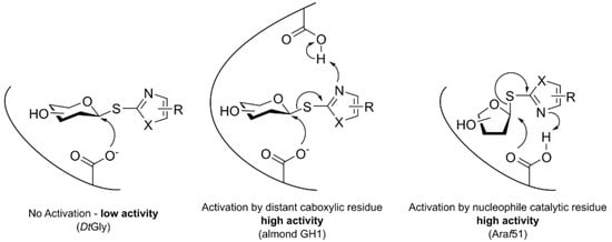
In our model, the catalytic glutamate Glu159, which acts as the acid/base residue in retaining GH mechanism [46], is the only one close enough to activate thioglycoside hydrolysis. Although direct protonation on sulphur cannot occur in the case of thiogycosides [28], examples of distant protonation of the aglycone by a catalytic residue have been reported [15]. We have thus generated two mutants, namely Glu159Ala and Glu159Gln to assess the potential role of this residue in the thioglycoside hydrolysis. Unlike Glu159Gln that could be purified to homogeneity, Glu159Ala mutant was not soluble after cell lysis and thus could not be purified. This mutant was left aside for further experiments.
Glu159Gln mutation led to a dramatic decrease of catalytic efficiency for pNPGlc, as shown in Table 2. KM values for this substrate are lower but in the same order of magnitude (200 µM vs. 460 µM), which can be explained by conservation of the active site structure in the mutant and decreased kcat value by a factor of 150 because KM is related to kcat. This loss of hydrolytic activity upon acid/base mutation is usual, as reported in many other studies, especially those concerning thioligase generation [14,40].
When using GlcSBox as a substrate, DtGly Glu159Gln exhibits a reduced kcat value (0.38 to 0.06 s−1), as expected because nucleophile water attack is not activated by deprotonation. However, second order rate constant remains unchanged, indicating that the first step of the reaction is not compromised by the removal of the glutamate residue. Thus, the release of the thiol leaving group is not activated by Glu159 and is only dictated by its leaving capability (i.e., pKa).

3. Discussion

We have previously used D. thermophilum DtGly as a versatile tool for synthesis of glycosides and looked for alternate substrates for this enzyme. Thioimidate glycosyl donors have been used for a long time in organic synthesis to generate a wide range of glycosides and glycans [34,47,48,49]. In this context, we tested previously reported glycosyl thioimidates as substrates for almond GH1 and A. niger GH3 [16]. Examples of cloning and characterization of thioglycoside hydrolases are scarcely available in literature and even fewer studies on mechanism underlying the thioglycoside hydrolysis by GH have been published.
DtGly is able to hydrolyse S-glycosides, with lower activities than those observed for O-glycosides. This hydrolytic activity is rate-limited by release of the thiol-containing leaving group and not water nucleophilic attack, unlike generally accepted mechanism for the O-glycoside hydrolysis [19,28]. The modelling of substrate-DtGly complexes as well as mutagenesis of the acid/base residue also demonstrated that no residue was able to remotely protonate the benzimidazole group nor the sulphur atom. If DtGly is able to hydrolyse S-containing substrates without general acid catalysis, the hydrolysis rate is limited by the leaving group capability, as no remote activation is possible. The pKa of leaving groups 2-mercaptobenzimidazole (for GlcSBiz) and 2-mercaptobenzoxazole (for GlcSBox) have been experimentally determined at 5.8 [50] and 6.58 [51]. To our knowledge, no value is available for 2-mercaptothiazoline (for GlcSTaz).
In the case of almond and A. niger glycosidases, a remote protonation occurring on a nitrogen atom of the benzimidazole moiety of GlcSBiz was shown to accelerate the leaving group release, thus increasing the catalytic rate to a value close to those observed for O-glycosides. Another GH exhibiting thioglycosidase activity on 2’-thio-benzimidazolyl arabinosides activated by remote deprotonation, namely Araf51 [15], was also reported. The modelling of Araf51/substrate complex demonstrated that the nucleophile catalytic residue was responsible for the remote protonation on imidazole nitrogen, mostly because a furanosyl ring is much more flexible than a pyranosyl ring and allows the nucleophile residue to interact with the aglycone.

4. Materials and Methods

4.1. Materials

para-Nitrophenyl β-D-glucopyranoside (pNP-Glc) was purchased from Carbosynth (Oxford, UK). 2-benzoxazolyl 1-thio-β-D-glucopyranoside (GlcSBox) [52], 2-benzimidazolyl 1-thio-β-D-glucopyranoside (GlcSBiz) [16] and 2-thiazolinyl 1-thio-β-D-glucopyranoside (GlcSTaz) [49] were prepared as previously described. Otherwise specified, all other chemicals were purchased from Thermo Fisher Scientific (Waltham, MA, USA) and were of purest quality available. Mutagenic primers were purchased from Eurofins Genomics (Ebersberg, Germany) and WT DtGly coding expression plasmid (pET28a-dtgly) was prepared as previously described [36].

4.2. Production of WT and E159Q DtGly

pET28a-dtgly was used as a template for mutagenic PCR in the Quikchange Site-directed mutagenesis kit (Agilent, Les Ulis, France). Primers containing desired mutation on acid/base residue position (E159) were constituted of a pair of complement oligonucleotides designed using Agilent tools website (www.genomics.agilent.com, mutated codons are highlighted in bold): DtGly E159A: 5’-gaattactggatgactataaatgcgcccaatgcttatgccttt-3’ and DtGly E159Q: 5’-atcttgtgaattactggatgactataaatcagcccaatgcttatg-3’. Mutagenesis procedure was performed according to the kit procedure. Sequences of pET28a-dtglyE159A and pET28a-dtglyE159Q were verified by Sanger sequencing at Eurofins Genomics (Ebersberg, Germany).
Production and purification of DtGly variants was done as previously reported [36]. Briefly, Escherishia coli Rosetta(DE3) transformed with expression plasmids were grown in LB medium supplemented with chloramphenicol (34 µg/mL) and kanamycin (30 µg/mL) at 37 °C until OD600 reached 0.6. Induction was then done by addition of 1 mM IPTG and incubated overnight a 25 °C. Cells were harvested, lyzed by freeze-thaw cycles and sonication and supernatant was clarified by heat treatment for 15 min at 70 °C before centrifugation. Finally, supernatant was loaded on a Nickel column (HisPure, Thermo Scientific) and purified by elution with lysis buffer containing 500 mM imidazole.

4.3. pNP Release Quantification Assay

DtGly variants (WT or mutants) activity towards pNP-Glc hydrolysis was determined at 37 °C in a 200 µL incubation containing 5 ng of the enzyme, 0.01–10 mM pNP-Glc, Citrate-Phosphate buffer (20 mM, pH 6) and 0.1–1 mM GlcBox for inhibition studies. After 20 min of incubation, 100 µL of Na2CO3 1 M was added and released pNP was quantified at 405 nm (ε405 = 19,500 cm−1.M−1). Prism 4 (GraphPad) was used to fit data according to Michaelis-Menten model, or competitive inhibition model and retrieve kinetic parameters.

4.4. Glucose Release Assay

To determine GlcSBox, GlcSTaz and GlcSBiz hydrolysis rate by DtGly variants, produced glucose was quantified using a continuous coupled enzyme assay [42]. Incubations were similar as those for pNP-Glc hydrolysis with the addition of glucose oxidase from Aspergillus niger (Sigma-Aldrich, Saint Louis, MO, USA, 0.4 u), horseradish peroxidase (Sigma-Aldrich, 0.4 u) and o-dianisidine (Sigma-Aldrich, 100 µM). Dianisidine oxidation coupled to glucose production was monitored at 442 nm during 30 min. Prism 4 (GraphPad) was used to fit data according to Michaelis-Menten model and retrieve kinetic parameters.

4.5. Computational Studies

The structure of β-glycosidase from Pyrococcus horikoshii [44] (PDB 1VFF, 45%/63% sequence identity/homology) was used as a template for homology model building using a ModWeb server from the A. Sali Laboratory (https://modbase.compbio.ucsf.edu/modweb/). The resulting model was prepared with AmberTools [53] and equilibrated using NAMD software [54] and Amber fb15 force field [55] (3 cycles of 10,000 minimization steps and 0.5 ns dynamics at 100 K).
Docking of GlcSBox, GlcSTaz and GlcSBiz substrates into DtGly active site model was done by firstly applying AM1-BCC charges on ligands [56]. Then each substrate was placed 10 Å away, facing the active site (according to PDB 1VFF). DtGly-substrate complexes were formed using steered molecular dynamics [57] at 100 K using the structural alignment of glucose moiety in its binding pocket as the final orientation according to closest structures bearing a ligand in their active site (β-glucosidase from Thermotoga maritima PDB 1OIM, 1OIF and 1OIF) [45]. DtGly backbone was kept constrained during the whole procedure. Finally, protein-ligand complexes models were equilibrated by releasing substrate constraints and applying several cycles of energy minimization (10,000 steps, steepest descent) followed by molecular dynamics (100 K, 1 ns). Final complex models were obtained by a final energy minimization. For each substrate, several initial conformations were tested, mostly by rotation of the glycosidic bond. All structural figures were drawn using the PyMOL Molecular Graphics system 1.8 (www.pymol.org).

5. Conclusions

This study demonstrates that glycosyl thioimidates are not universal glycosyl donors for chemoenzymatic syntheses. While the above examples of efficient enzymatic activities using such substrates were reported in literature, they rely on an activation by protonation of the aglycone moiety, either with a distant carboxylic acid residue (almond GH1) or the catalytic nucleophile (Araf51, Figure 5). DtGly seems to be the paradigm of the general case of an enzyme that can use those substrates without acid catalysis, yet with a much lower activity. This study paves the way for broadening DtGly applications in biocatalysis. Identification of efficient substrates and mutation into a thioligase are currently under further investigation.

Supplementary Materials

The following are available online at https://www.mdpi.com/2073-4344/9/10/826/s1, Figure S1: Model of docked GlcSBox and GlcSTaz in DtGly active site.

Author Contributions

Conceptualization, R.D. and A.V.D.; Enzymatic studies, L.G., P.L. and Z.A.; data curation and modelling, P.L.; chemical synthesis of substrates, S.G.P. writing—original draft preparation, P.L.; writing—review and editing, P.L., R.D. and A.V.D.; project administration, R.D.; funding acquisition, R.D. and A.V.D.

Funding

L.G., P.L. and R.D. thank the Région Centre Val de Loire for funding (APR IR GLYCOPEPS). A.V.D. thanks the NIGMS for generous support of this work (GM GM111835).

Conflicts of Interest

The authors declare no conflict of interest. The funders had no role in the design of the study; in the collection, analyses, or interpretation of data; in the writing of the manuscript, or in the decision to publish the results.

References

  1. Sheldon, R.A.; Woodley, J.M. Role of Biocatalysis in Sustainable Chemistry. Chem. Rev. 2018, 118, 801–838. [Google Scholar] [CrossRef] [PubMed]
  2. Quin, M.B.; Schmidt-Dannert, C. Engineering of Biocatalysts: From Evolution to Creation. ACS Catal. 2011, 1, 1017–1021. [Google Scholar] [CrossRef] [PubMed]
  3. Hancock, S.M.; Vaughan, M.D.; Withers, S.G. Engineering of glycosidases and glycosyltransferases. Curr. Opin. Chem. Biol. 2006, 10, 509–519. [Google Scholar] [CrossRef] [PubMed]
  4. Li, T.-L.; Liu, Y.-C.; Lyu, S.-Y. Combining biocatalysis and chemoselective chemistries for glycopeptide antibiotics modification. Curr. Opin. Chem. Biol. 2012, 16, 170–178. [Google Scholar] [CrossRef] [PubMed]
  5. Ati, J.; Lafite, P.; Daniellou, R. Enzymatic synthesis of glycosides: From natural O- and N-glycosides to rare C- and S-glycosides. Beilstein J. Org. Chem. 2017, 13, 1857–1865. [Google Scholar] [CrossRef]
  6. Guillotin, L.; Lafite, P.; Daniellou, R. Chapter 10 Enzymatic thioglycosylation: Current knowledge and challenges. In Carbohydrate Chemistry; The Royal Society of Chemistry: London, UK, 2014; Volume 40, pp. 178–194. ISBN 978-1-84973-965-8. [Google Scholar]
  7. Driguez, H. Thiooligosaccharides as Tools for Structural Biology. Chembiochem 2001, 2, 311–318. [Google Scholar] [CrossRef]
  8. Wardrop, D.J.; Waidyarachchi, S.L. Synthesis and biological activity of naturally occurring α-glucosidase inhibitors. Nat. Prod. Rep. 2010, 27, 1431–1468. [Google Scholar] [CrossRef]
  9. O’Neill, E.C.; Field, R.A. Enzymatic synthesis using glycoside phosphorylases. Carbohydr. Res. 2015, 403, 23–37. [Google Scholar] [CrossRef]
  10. Lairson, L.L.; Henrissat, B.; Davies, G.J.; Withers, S.G. Glycosyltransferases: Structures, Functions and Mechanisms. Annu. Rev. Biochem. 2008, 77, 521–555. [Google Scholar] [CrossRef]
  11. De Bruyn, F.; Maertens, J.; Beauprez, J.; Soetaert, W.; De Mey, M. Biotechnological advances in UDP-sugar based glycosylation of small molecules. Biotechnol. Adv. 2015, 33, 288–302. [Google Scholar] [CrossRef]
  12. Rye, C.S.; Withers, S.G. Glycosidase mechanisms. Curr. Opin. Chem. Biol. 2000, 4, 573–580. [Google Scholar] [CrossRef]
  13. Wang, L.-X.; Huang, W. Enzymatic transglycosylation for glycoconjugate synthesis. Curr. Opin. Chem. Biol. 2009, 13, 592–600. [Google Scholar] [CrossRef] [PubMed] [Green Version]
  14. Jahn, M.; Marles, J.; Warren, R.A.J.; Withers, S.G. Thioglycoligases: Mutant Glycosidases for Thioglycoside Synthesis. Angew. Chem. Int. Ed. 2003, 42, 352–354. [Google Scholar] [CrossRef] [PubMed]
  15. Almendros, M.; Danalev, D.; François-Heude, M.; Loyer, P.; Legentil, L.; Nugier-Chauvin, C.; Daniellou, R.; Ferrières, V. Exploring the synthetic potency of the first furanothioglycoligase through original remote activation. Org. Biomol. Chem. 2011, 9, 8371–8378. [Google Scholar] [CrossRef] [Green Version]
  16. Avegno, E.A.-B.; Hasty, S.J.; Parameswar, A.R.; Howarth, G.S.; Demchenko, A.V.; Byers, L.D. Reactive thioglucoside substrates for β-glucosidase. Arch. Biochem. Biophys. 2013, 537, 1–4. [Google Scholar] [CrossRef] [PubMed]
  17. Goodman, I.; Fouts, J.R.; Bresnick, E.; Menegas, R.; Hitchings, G.H. A Mammalian Thioglycosidase. Science 1959, 130, 450–451. [Google Scholar] [CrossRef]
  18. Meulenbeld, G.H.; Hartmans, S. Thioglucosidase activity from Sphingobacterium sp. strain OTG1. Appl. Microbiol. Biotechnol. 2001, 56, 700–706. [Google Scholar] [CrossRef] [PubMed]
  19. Macauley, M.S.; Stubbs, K.A.; Vocadlo, D.J. O-GlcNAcase catalyzes cleavage of thioglycosides without general acid catalysis. J. Am. Chem. Soc. 2005, 127, 17202–17203. [Google Scholar] [CrossRef] [PubMed]
  20. Cetinbaş, N.; Macauley, M.S.; Stubbs, K.A.; Drapala, R.; Vocadlo, D.J. Identification of Asp174 and Asp175 as the key catalytic residues of human O-GlcNAcase by functional analysis of site-directed mutants. Biochemistry 2006, 45, 3835–3844. [Google Scholar] [CrossRef]
  21. Narine, A.A.; Watson, J.N.; Bennet, A.J. Mechanistic requirements for the efficient enzyme-catalyzed hydrolysis of thiosialosides. Biochemistry 2006, 45, 9319–9326. [Google Scholar] [CrossRef]
  22. Shen, H.; Byers, L.D. Thioglycoside hydrolysis catalyzed by beta-glucosidase. Biochem. Biophys. Res. Commun. 2007, 362, 717–720. [Google Scholar] [CrossRef] [PubMed]
  23. Sansenya, S.; Opassiri, R.; Kuaprasert, B.; Chen, C.J.; Cairns, J.R.K. The crystal structure of rice (Oryza sativa L.) Os4BGlu12, an oligosaccharide and tuberonic acid glucoside-hydrolyzing beta-glucosidase with significant thioglucohydrolase activity. Arch. Biochem. Biophys. 2011, 510, 62–72. [Google Scholar] [CrossRef] [PubMed]
  24. Wathelet, J.-P.; Iori, R.; Leoni, O.; Rollin, P.; Mabon, N.; Marlier, M.; Palmieri, S. A recombinant β-O-glucosidase from Caldocellum saccharolyticum to hydrolyse desulfo-glucosinolates. Biotechnol. Lett. 2001, 23, 443–446. [Google Scholar] [CrossRef]
  25. Reese, E.T.; Clapp, R.C.; Mandels, M. A thioglucosidase in fungi. Arch. Biochem. Biophys. 1958, 75, 228–242. [Google Scholar] [CrossRef]
  26. Yip, V.L.Y.; Withers, S.G. Family 4 glycosidases carry out efficient hydrolysis of thioglycosides by an alpha,beta-elimination mechanism. Angew. Chem. Int. Ed. 2006, 45, 6179–6182. [Google Scholar] [CrossRef] [PubMed]
  27. Niemec-Cyganek, A.; Szeja, W. Heteroaryl thioglycosides, a new class of substrated for glycosidases. Pol. J. Chem. 2003, 77, 969–973. [Google Scholar]
  28. Jensen, J.L.; Jencks, W.P. Hydrolysis of benzaldehyde O,S-acetals. J. Am. Chem. Soc. 1979, 101, 1476–1488. [Google Scholar] [CrossRef]
  29. Hanessian, S.; Bacquet, C.; Lehong, N. Chemistry of the glycosidic linkage. Exceptionally fast and efficient formation of glycosides by remote activation. Carbohydr. Res. 1980, 80, C17–C22. [Google Scholar] [CrossRef]
  30. Ferrières, V.; Blanchard, S.; Fischer, D.; Plusquellec, D. A novel synthesis of D-galactofuranosyl, D-glucofuranosyl and D-mannofuranosyl 1-phosphates based on remote activation of new and free hexofuranosyl donors. Bioorg. Med. Chem. Lett. 2002, 12, 3515–3518. [Google Scholar] [CrossRef]
  31. Demchenko, A.V.; Malysheva, N.N.; De Meo, C. S-Benzoxazolyl (SBox) Glycosides as Novel, Versatile Glycosyl Donors for Stereoselective 1,2-Cis Glycosylation. Org. Lett. 2003, 5, 455–458. [Google Scholar] [CrossRef]
  32. Demchenko, A.V.; Pornsuriyasak, P.; De Meo, C.; Malysheva, N.N. Potent, Versatile and Stable: Thiazolyl Thioglycosides as Glycosyl Donors. Angew. Chem. Int. Ed. 2004, 43, 3069–3072. [Google Scholar] [CrossRef] [PubMed]
  33. Euzen, R.; Guégan, J.-P.; Ferrières, V.; Plusquellec, D. First O-Glycosylation from Unprotected 1-Thioimidoyl Hexofuranosides Assisted by Divalent Cations. J. Org. Chem. 2007, 72, 5743–5747. [Google Scholar] [CrossRef] [PubMed]
  34. Kamat, M.N.; De Meo, C.; Demchenko, A.V. S-Benzoxazolyl as a Stable Protecting Moiety and a Potent Anomeric Leaving Group in Oligosaccharide Synthesis. J. Org. Chem. 2007, 72, 6947–6955. [Google Scholar] [CrossRef] [PubMed] [Green Version]
  35. Hasty, S.J.; Demchenko, A.V. Glycosyl Thioimidates as Versatile Building Blocks for Organic Synthesis. Chem. Heterocycl. Compd. 2012, 48, 220–240. [Google Scholar] [CrossRef] [PubMed]
  36. Guillotin, L.; Cancellieri, P.; Lafite, P.; Landemarre, L.; Daniellou, R. Chemo-enzymatic synthesis of 3-O-(β-d-glycopyranosyl)-sn-glycerols and their evaluation as preservative in cosmetics. Pure Appl. Chem. 2017, 89, 11302. [Google Scholar] [CrossRef]
  37. Fukusumi, S.; Kamizono, A.; Horinouchi, S.; Beppu, T. Cloning and nucleotide sequence of a heat-stable amylase gene from an anaerobic thermophile, Dictyoglomus thermophilum. Eur. J. Biochem. 1988, 174, 15–21. [Google Scholar] [CrossRef] [PubMed]
  38. Shi, R.; Li, Z.; Ye, Q.; Xu, J.; Liu, Y. Heterologous expression and characterization of a novel thermo-halotolerant endoglucanase Cel5H from Dictyoglomus thermophilum. Bioresour. Technol. 2013, 142, 338–344. [Google Scholar] [CrossRef] [PubMed]
  39. Nakajima, M.; Imamura, H.; Shoun, H.; Horinouchi, S.; Wakagi, T. Transglycosylation Activity of Dictyoglomus thermophilum Amylase A. Biosci. Biotechnol. Biochem. 2004, 68, 2369–2373. [Google Scholar] [CrossRef]
  40. Guillotin, L.; Richet, N.; Lafite, P.; Daniellou, R. Is the acid/base catalytic residue mutation in β-d-mannosidase Dt Man from Dictyoglomus thermophilum sufficient enough to provide thioglycoligase activity? Biochimie 2017, 137, 190–196. [Google Scholar] [CrossRef] [PubMed]
  41. Guillotin, L.; Kim, H.; Traore, Y.; Moreau, P.; Lafite, P.; Coquoin, V.; Nuccio, S.; De Vaumas, R.; Daniellou, R. Biochemical Characterization of the α-L-Rhamnosidase Dt Rha from Dictyoglomus thermophilum: Application to the Selective Derhamnosylation of Natural Flavonoids. ACS Omega 2019, 4, 1916–1922. [Google Scholar] [CrossRef]
  42. Huggett, A.S.G.; Nixon, D.A. Use of glucose oxidase, peroxidase and o-dianisidine in determination of blood and urinary glucose. Lancet 1957, 270, 368–370. [Google Scholar] [CrossRef]
  43. Guillotin, L.; Lafite, P.; Daniellou, R. Unraveling the Substrate Recognition Mechanism and Specificity of the Unusual Glycosyl Hydrolase Family 29 BT2192 from Bacteroides thetaiotaomicron. Biochemistry 2014, 53, 1447–1455. [Google Scholar] [CrossRef] [PubMed]
  44. Akiba, T.; Nishio, M.; Matsui, I.; Harata, K. X-ray structure of a membrane-bound β-glycosidase from the hyperthermophilic archaeon Pyrococcus horikoshii. Proteins Struct. Funct. Bioinform. 2004, 57, 422–431. [Google Scholar] [CrossRef] [PubMed]
  45. Zechel, D.L.; Boraston, A.B.; Gloster, T.; Boraston, C.M.; Macdonald, J.M.; Tilbrook, D.M.G.; Stick, R.V.; Davies, G.J. Iminosugar Glycosidase Inhibitors: Structural and Thermodynamic Dissection of the Binding of Isofagomine and 1-Deoxynojirimycin to β-Glucosidases. J. Am. Chem. Soc. 2003, 125, 14313–14323. [Google Scholar] [CrossRef] [PubMed]
  46. Zechel, D.L.; Withers, S.G. Dissection of nucleophilic and acid-base catalysis in glycosidases. Curr. Opin. Chem. Biol. 2001, 5, 643–649. [Google Scholar] [CrossRef]
  47. Zhu, X.; Schmidt, R.R. New Principles for Glycoside-Bond Formation. Angew. Chem. Int. Ed. 2009, 48, 1900–1934. [Google Scholar] [CrossRef] [PubMed]
  48. Codée, J.D.C.; Litjens, R.E.J.N.; Van den Bos, L.J.; Overkleeft, H.S.; Van der Marel, G.A. Thioglycosides in sequential glycosylation strategies. Chem. Soc. Rev. 2005, 34, 769–782. [Google Scholar] [CrossRef] [PubMed]
  49. Pornsuriyasak, P.; Demchenko, A.V. S-Thiazolinyl (STaz) Glycosides as Versatile Building Blocks for Convergent Selective, Chemoselective and Orthogonal Oligosaccharide Synthesis. Chem.-Eur J. 2006, 12, 6630–6646. [Google Scholar] [CrossRef] [PubMed]
  50. Jerez, G.; Kaufman, G.; Prystai, M.; Schenkeveld, S.; Donkor, K.K. Determination of thermodynamic p K a values of benzimidazole and benzimidazole derivatives by capillary electrophoresis. J. Sep. Sci. 2009, 32, 1087–1095. [Google Scholar] [CrossRef]
  51. Akhond, M.; Ghaedi, M.; Tashkhourian, J. Development of a New Copper(II) Ion-selective Poly(vinyl chloride) Membrane Electrode Based on 2-Mercaptobenzoxazole. Bull. Korean Chem. Soc. 2005, 26, 882–886. [Google Scholar] [Green Version]
  52. Kamat, M.N.; Rath, N.P.; Demchenko, A. V Versatile Synthesis and Mechanism of Activation of S-Benzoxazolyl Glycosides. J. Org. Chem. 2007, 72, 6938–6946. [Google Scholar] [CrossRef] [PubMed]
  53. Case, D.A.; Darden, T.A.; III, T.E.C.; Simmerling, C.L.; Wang, J.; Duke, R.E.; Luo, R.; Walker, R.C.; Zhang, W.; Merz, K.M.; et al. AMBER 12 2012; University of California: San Francisco, CA, USA, 2012. [Google Scholar]
  54. Phillips, J.C.; Braun, R.; Wang, W.; Gumbart, J.; Tajkhorshid, E.; Villa, E.; Chipot, C.; Skeel, R.D.; Kale, L.; Schulten, K. Scalable molecular dynamics with NAMD. J. Comput. Chem. 2005, 26, 1781–1802. [Google Scholar] [CrossRef] [PubMed] [Green Version]
  55. Wang, L.-P.; McKiernan, K.A.; Gomes, J.; Beauchamp, K.A.; Head-Gordon, T.; Rice, J.E.; Swope, W.C.; Martínez, T.J.; Pande, V.S. Building a More Predictive Protein Force Field: A Systematic and Reproducible Route to AMBER-FB15. J. Phys. Chem. B 2017, 121, 4023–4039. [Google Scholar] [CrossRef] [PubMed]
  56. Jakalian, A.; Jack, D.B.; Bayly, C.I. Fast, efficient generation of high-quality atomic charges. AM1-BCC model: II. Parameterization and validation. J. Comput. Chem. 2002, 23, 1623–1641. [Google Scholar] [CrossRef] [PubMed]
  57. Lafite, P.; André, F.; Zeldin, D.C.; Dansette, P.M.; Mansuy, D. Unusual Regioselectivity and Active Site Topology of Human Cytochrome P450 2J2. Biochemistry 2007, 46, 10237–10247. [Google Scholar] [CrossRef] [PubMed] [Green Version]
Figure 1. Schematic mechanism of the first step involving the glycosyl-enzyme intermediate formation in retaining GH. The leaving group (LG) release can also be catalysed through another catalytic residue according to its nature. Depending on the nucleophile (Nu) attacking the intermediate, three reactions can take place—hydrolysis, transglycosylation or thioligation.
Catalysts 09 00826 g001
Figure 2. Substrates used in this study.
Catalysts 09 00826 g002
Figure 3. Lineweaver plot of wild-type DtGly inhibition with increasing concentrations of GlcSBiz. Data are expressed as mean ± SD from three independent experiments. Inhibitor concentrations are respectively depicted as crosses (0 µM), circles (100 µM), triangles (250 µM), diamonds (500 µM) and squares (1000 µM). Inset: 2X zoom on axes origin highlighting the intersection of fitted lines on y-axis.
Catalysts 09 00826 g003
Figure 4. (A) Overall representation of DtGly model. Helices and sheets are respectively coloured in blue and orange. (B) Model of docked GlcSBiz in DtGly active site. Residues surrounding the ligand binding pocket are depicted as sticks. For clarity purposes, hydrogens are not represented. Catalytic residues Glu159 (acid/base) and Glu324 (nucleophile) are highlighted in bold. H-bonds are indicated as dashed lines.
Catalysts 09 00826 g004
Figure 5. Glycosyl thioimidates require remote activation to promote the leaving group release.
Catalysts 09 00826 g005
Table 1. Comparison of S- and O-glycoside hydrolysis by GH.
EnzymeOrganismSubstrates Tested Relative Activity S-vs. O-(%)Ref
β-D-GlucosidaseSweet almondpNPSGlc
pNPSGal
pNPSFuc
GlcSBiz
GlcS(N-Me)Biz
GlcSBox
0.13 a
0.07 a
0.06 a
80 a
10 a
5 a
[22]
 
 
[16]
β-D-GlucosidaseA. nigerGlcSBiz
GlcS(N-Me)Biz
5 a
1 a
[16]
Sialidase M. viridifaciensSubstituted pNPSNeuAc0.01–60[21]
Os4BGlu12O. sativapNPSGlc
OctylSGlc
0.5 a
0.1 b
[23]
a ratio of kcat/KM for thioglycoside substrate vs. corresponding para-nitrophenyl glycoside. b ratio of kcat/KM for octyl S-glucoside vs. octyl O-glucoside.
Table 2. Kinetic parameters of WT and acid/base E159Q mutant of DtGly. pNP-Glc hydrolysis activity was measured by pNP release quantification. Other substrate hydrolysis activities were determined by quantification of the released glucose. All experiments were done in three independent replicates and are expressed as Mean ± SD.
EnzymeSubstrateKM (µM)kcat (s−1)kcat/KM (s−1.mM−1)
WTpNP-Glc a460 ± 4031 ± 0.767
WTGlcSBiz 1533 ± 1140.23 ± 0.010.15
WTGlcSBox2246 ± 2890.38 ± 0.030.17
WTGlcSTaz880 ± 520.31 ± 0.010.35
E159QpNP-Glc200 ± 200.20 ± 0.011.0
E159QGlcSBox445 ± 400.06 ± 0.010.13
a Previously reported data [36].

