Communication

Characterization and Structural Insights of a Novel Arylsulfatase from Pseudoalteromonas atlantica T6c

1
Provincial University Key Laboratory of Cellular Stress Response and Metabolic Regulation, College of Life Science, Fujian Normal University, Fuzhou 350117, China
2
Fujian Metrology Institute, Fuzhou 350003, China
*
Author to whom correspondence should be addressed.
These authors contributed equally to this work.
Catalysts 2023, 13(4), 759; https://doi.org/10.3390/catal13040759
Submission received: 23 March 2023 / Revised: 11 April 2023 / Accepted: 12 April 2023 / Published: 15 April 2023
(This article belongs to the Section Biocatalysis)

Abstract

:
Arylsulfatases exhibit great potential in industry for desulfation applications, but less is known about the metallo-β-lactamase (MBL) fold arylsulfatases. To learn more about them, an MBL fold arylsulfatase from Pseudoalteromonas atlantica T6c (PaAst) was identified and characterized, and its structure was elaborated in this study. PaAst was sequence analyzed, heterologously expressed in E. coli, purified by Ni2+-NTA resin affinity chromatography and size-exclusion chromatography, functionally studied by p-nitrophenyl sulfate (pNPS), and crystallized for structure determination. The MBL fold arylsulfatase was identified by sequence analysis and confirmed by enzymatic assay on pNPS with Km 1.00 mM and Vmax 60.80 U/mg at 50 °C and pH 7.5. Furthermore, its crystals were obtained in 0.2 M sodium thiocyamate, 20% PEG3350, and its structure was determined at 2.0 Å that formed a dimer with MBL fold. Our work highlighted the MBL fold arylsulfatases from structural insights and could be the theoretical foundation for investigations into their catalytic mechanism.

1. Introduction

Sulfatase (EC 3.1.6.X) is a large family of hydrolytic enzymes found in varies species, ranging from bacteria to humans, catalyzing the hydrolysis of sulfate ester bonds [1,2,3]. It has been classified into three distinct types, with the best studied, type I, identified as the Cα-formylglycine-dependent arylsulfatases characterized by a unique formylglycine (FGly) posttranslationally modified from an active-site serine or cysteine [2,4,5]. The sulfatases (type II) consist of the Fe2+ α-ketoglutarate-dependent deoxygenate superfamily that require α-ketoglutarate as a cosubstrate [6,7,8], while the type III sulfatases were not reported until Hagelueken et al., in 2006, determined the crystal structure of SdsA1, a metallo-β-lactamase (MBL)-related alkylsulfatase from Pseudomonas aeruginosa [4]. Since then, a variety of MBL fold alkylsulfatases had been classified into sulfatases (type III), for example, SdsA from Pseudomonas sp. ATCC19151 [9], SdsAP from Pseudomonas sp. S9 [2,10], and Pisa1 from Pseudomonas sp. DSM6611 [11]. However, evidence also exists for the arylsulfatases sharing MBL fold, implicating AtsA from Pseudoalteromonas carrageenovora and a series of AtsA-like arylsulfatases [3,12,13,14]. Until now, this type of arylsulfatases could not be classified.
MBL fold sulfatases regulate the sulfation states of substrates, and it is suggested that sulfatases (type III) can degrade sodium dodecyl sulfate (SDS), with SdsA1 allowing its hosts to use SDS as a sole carbon or sulfur source [2,4,10]. SDS has been extensively used as an anionic surfactant in industries and daily life, which may cause secondary pollution to the environment [2,15]. Agar and its important component, agarose, have also been widely used in food, cosmetic, biological and pharmaceutical industries, and the removal of their sulfate groups by arylsulfatases could improve the gelling strength and purity greatly [13,16]. AtsA and its homologous arylsulfatase, PfAST from Pyrococcus furiosus DSM3638, have been proven to exhibit activities towards an artificial aromatic sulfate, p-nitrophenyl sulfate (pNPS), and sulfated marine polysaccharide, agar [14]. Therefore, microbial MBL fold sulfatases are of great industrial interest due to their potential in the biodegradation of SDS and desulfation of marine sulfonated polysaccharides that could be environmental friendly and cost saving with less chemical solvents and reduced waste discharge [10,13,14,17].
Several investigations have been done to determine the structures of type III alkylsulfatases, and the MBL fold proteins exhibit diverse features [10,18]. SdsA1, SdsAP, and Pisa1 share a similar overall structure consisting of an MBL fold domain, a dimerization domain, and a sterol carrier protein type 2 (SCP-2)-like fold domain; however, SdsA1 and SdsAP are primary alkylsulfatases capable of hydrolyzing the primary alkyl sulfates, for example, SDS, while Pisa1 is a secondary alkylsulfatase hydrolyzing the secondary alkyl sulfates, such as rac-2-octyl sulfate [4,10,11]. The difference could be attributed to the structure of their active site regions. Studies on MBL fold arylsulfatases have focused on their enzymatic characteristics and engineering in the past decades, for example AtsA [3,12,13,14], but limited structural information was found. Structural analyses of them were based on structure homology modelling. Furthermore, it has long been assumed that all arylsulfatases belong to the type I arylsulfatases containing a conserved FGly motif with few exceptions [12,19], ignoring the AtsA-like arylsulfatases. The catalytic mechanism of these arylsulfatases was uncertain.
For better knowledge and application of arylsulfatases, further investigations were necessary from genetic, biochemical, and, more importantly, structural aspects. In this study, we identified a novel MBL fold arylsulfatase from a deep-sea agar-degrading bacterium, Pseudoalteromonas atlantica T6c, designated as PaAst, and its functional and structural analyses were conducted preliminarily.

2. Results and Discussion

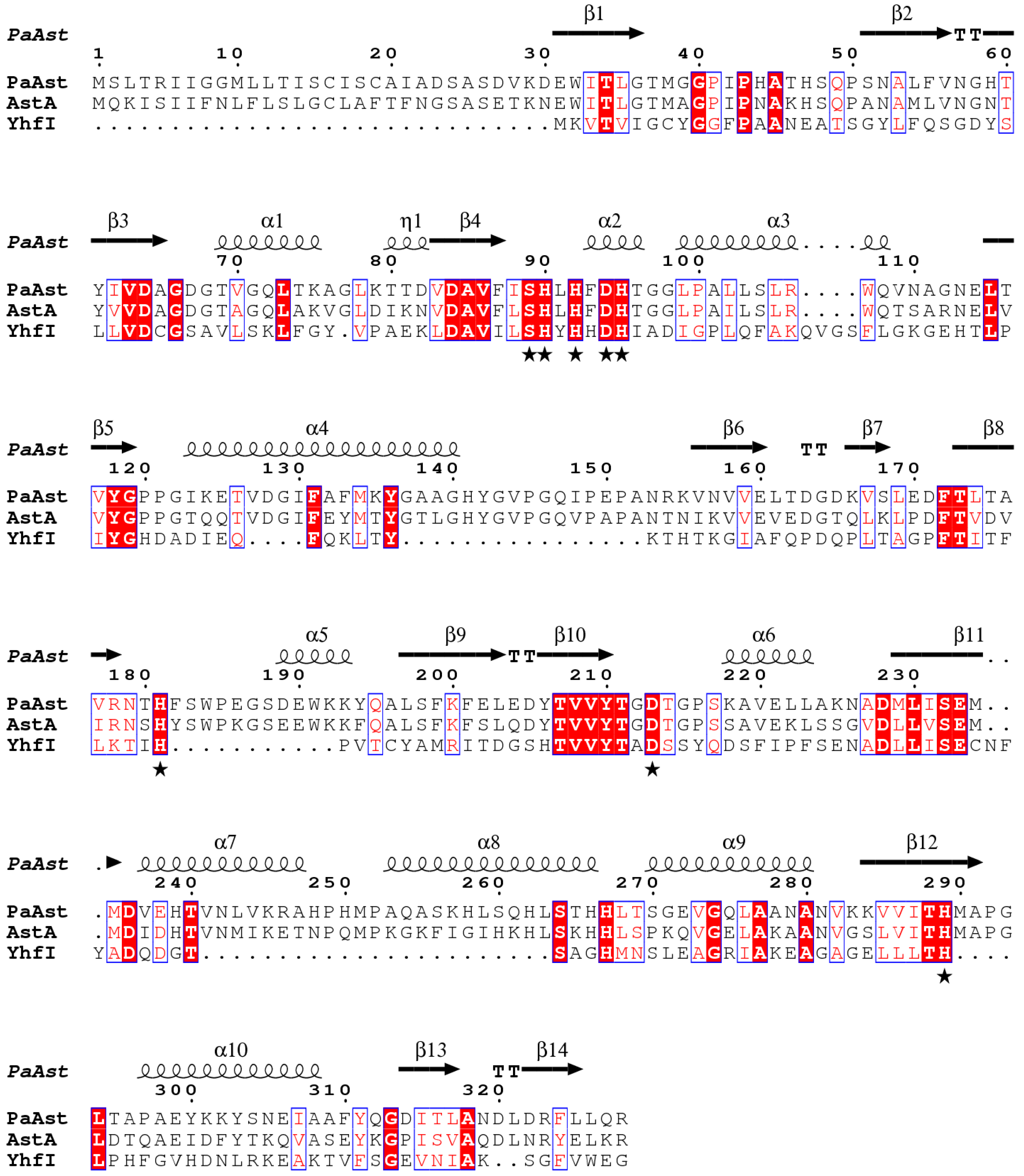
2.1. Sequence Analysis of PaAst

Pseudoalteromonas atlantica T6c (GenBank: CP000388.1) is an agar-degrading bacterium isolated from marine environments and is likely to harbor arylsulfatase genes. In its complete genome, a 987 bp gene was predicted to encode a β-lactamase-like protein of 328 amino acids, with an estimated molecular weight of 35.6 kDa and a theoretical pI of 5.77 [20]. The protein was predicted to possess an N-terminal signal peptide of 22 amino acids with the cleavage site between residues Ala22 and Asp23 [21]. The sequence alignment through BLAST revealed relatively high identities between PaAst and its homologues; 14 MBL-fold metallo-hydrolases and 1 arylsulfatase exhibited identities higher than 60%. The highest identity, 70.68%, was carried out by an MBL fold metallo-hydrolase from Shewanella sp. UCD-KL21 (Sequence ID: WP_083698034.1, E value: 1e-160, coverage: 93%), and PaAst showed identities of 67.07%, 66.77%, and 65.55% to similar hydrolases in Alteromonas stellipolaris (Sequence ID: WP_062086863.1, E value: 2e-167), Alteromonas sp. MMG017 (Sequence ID: WP_211069552.1, E value: 1e-160), and Paraglaciecola hydrolytica (Sequence ID: WP_082769068.1, E value: 9e-168) with 100% coverage, respectively. Moreover, it is a homologue of the arylsulfatase AtsA.
PaAst is a member of the metallo-hydrolase-like MBL-fold superfamily that is highly homologous to AtsA (sequence identity: 60.06%), as well as the starting model for structure determination, YhfI, from Bacillus subtilis (Figure 1). The homologues harbor conserved Zn2+-binding motifs, as “SH × H × DH-H181-D213-H289” in PaAst. In addition to its close identity with AtsA, its only domain, from Glu31 to Ser232, is a conserved arylsulfatase AtsA-like MBL-fold domain (Accession no. cd07719) [22,23]. Accordingly, although the conserved FGly motif (C/S × A/P × R) of arylsulfatases could not be found in its sequence, PaAst has a definite possibility of functioning as the arylsulfatases AtsA and PfAST, which could be confirmed in the following enzymatic assays.

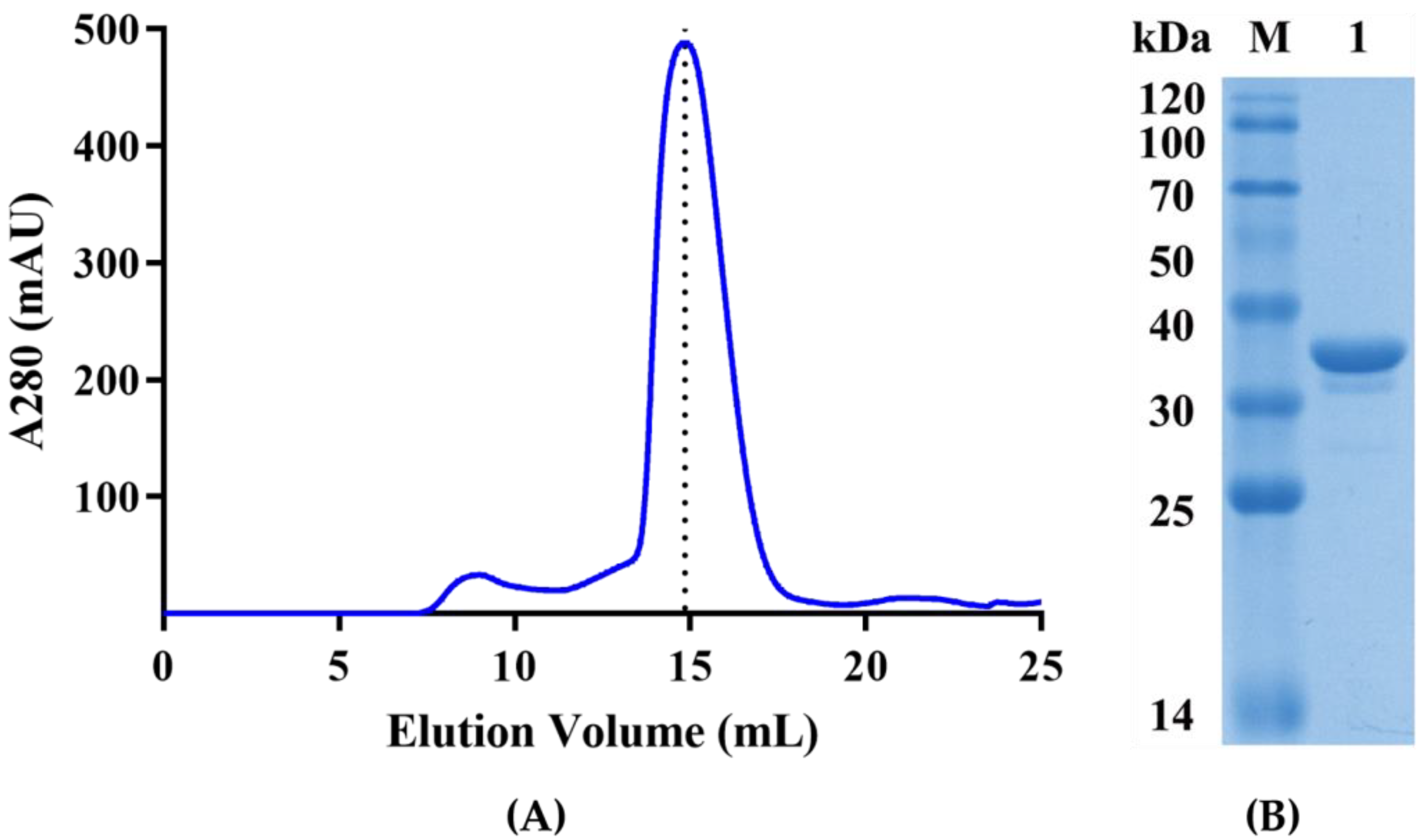
2.2. Expression, Purification, and Characterization of Recombinant PaAst

Recombinant PaAst was constructed, heterologously expressed, and purified for the confirmation of its enzymatic activity against pNPS and subsequent crystallization. The codon-optimized and truncated PaAst gene was inserted into the pET32a expression system, transformed into E. coli BL21 (DE3), and expressed as a PaAst-6*His-Trx fusion protein at a high level. To maintain its bioactivity, the purification mainly consisted of two rounds of Ni2+ affinity chromatography, and the size-exclusion chromatography, was conducted at low temperatures. The calculated molecular weight should have been 34.2 kDa for the purified recombinant enzyme after the 6*His-Trx tag removed, which was consistent with the SDS-PAGE analysis shown in Figure 2. However, in the size-exclusion chromatography, elution volume for the recombinant PaAst was 14.85 mL, indicating a molecular weight close to 75 kDa, appropriately twice the molecular weight of the monomer, indicating that PaAst is a dimer in solution.
Recombinant PaAst was enzymatically active towards pNPS, a common artificial substrate to evaluate enzymatic activities of the arylsulfatases [12]. The purification yields of PaAst are shown in Table 1 after procedures of crude extraction, Ni2+ affinity chromatography, and size-exclusion chromatography. After the steps of Ni2+ affinity chromatography, dialysis, and a secondary Ni2+ affinity chromatography, the total protein decreased from 220.32 mg to 22.93 mg, but the specific activity increased by three folds. The size-exclusion chromatography had less effect on the activity than the former steps, and the final yield for purification was 14.76%. The final yield and purification factor for PaAst were higher than AtsA heterologous expressed in E. coli performed with the same procedures in the same conditions (Table A2). The specific activity of purified PaAst on pNPS was 39.45 U/mg at 50 °C. The kinetic parameters were also obtained at 50 °C by exhibiting a Michaelis–Menten plot with specific activities on different concentrations of pNPS (Figure 3). The purified recombinant PaAst was observed with Km and Vmax of 1.00 mM and 60.80 U/mg, respectively. The results demonstrated that PaAst could hydrolyze pNPS efficiently, indicating its belonging to the arylsulfatase family.

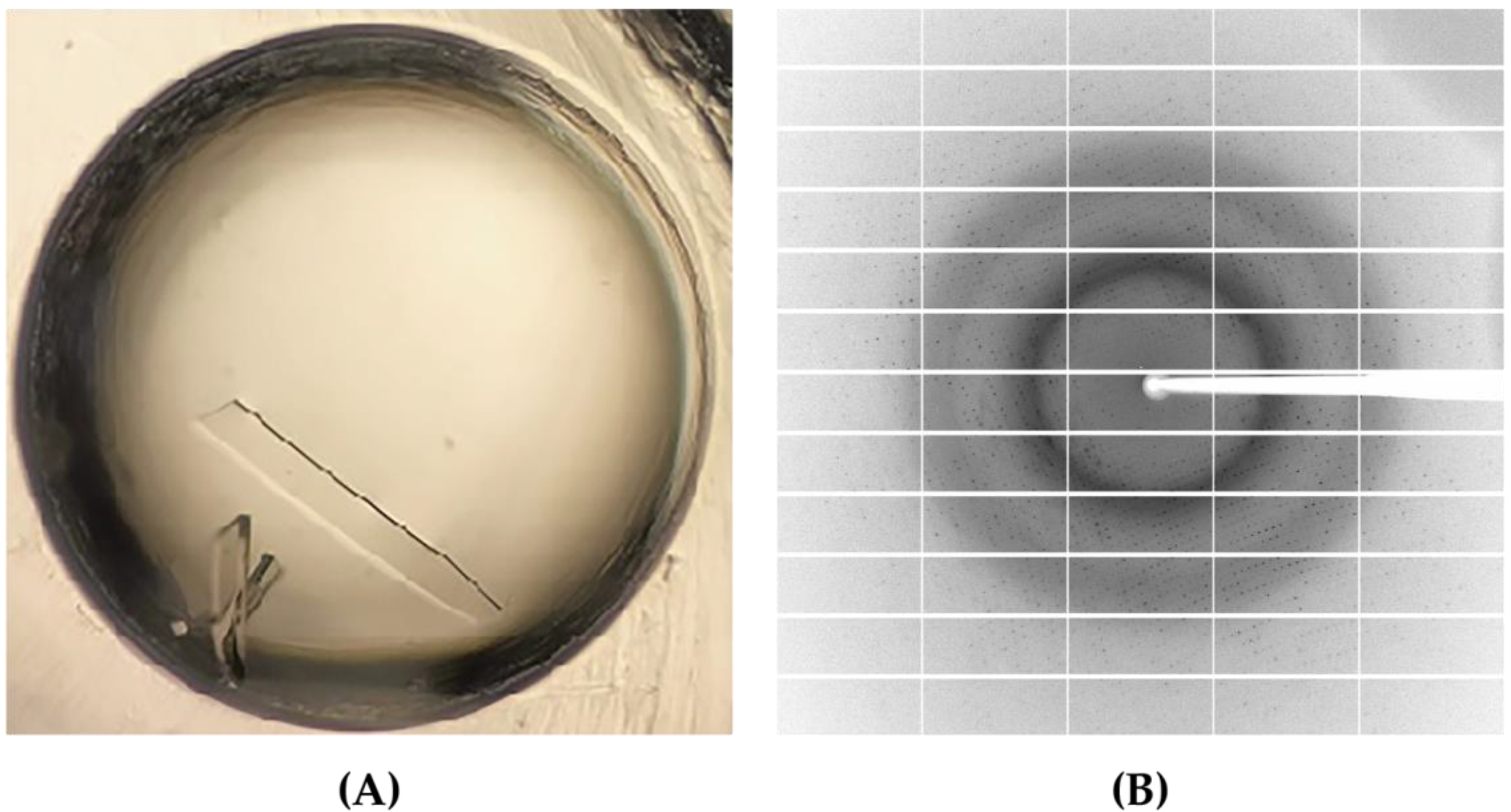
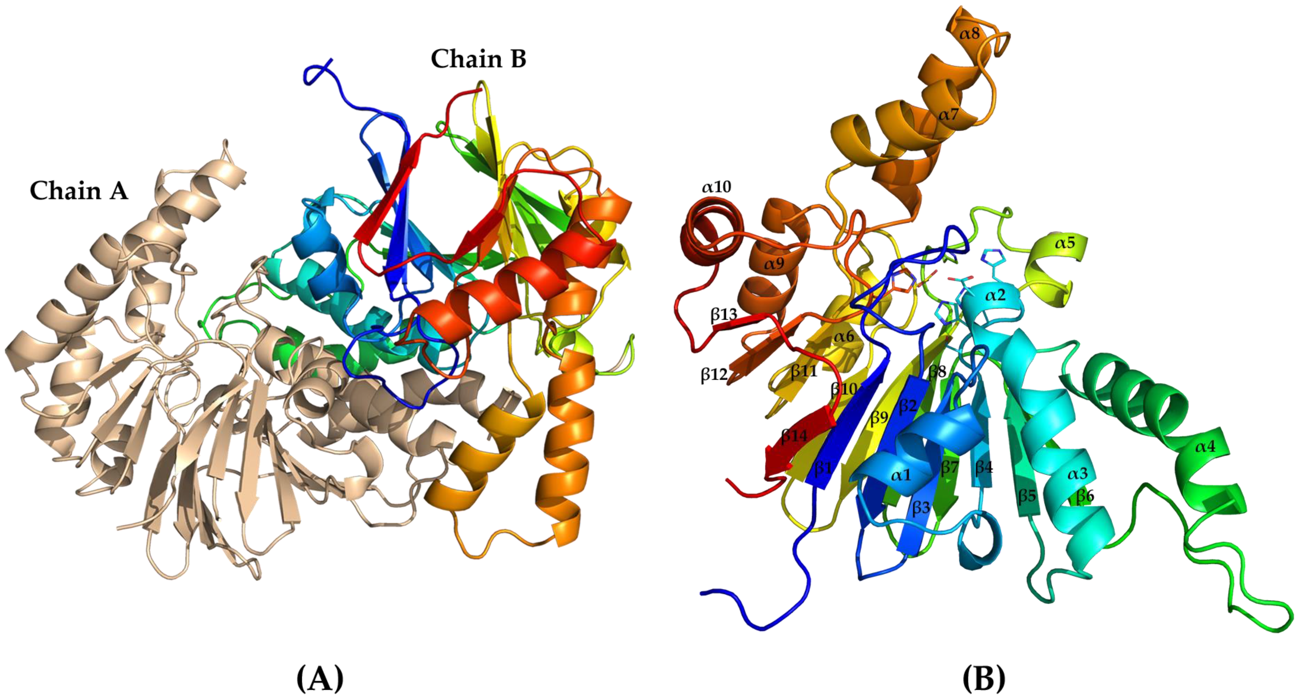
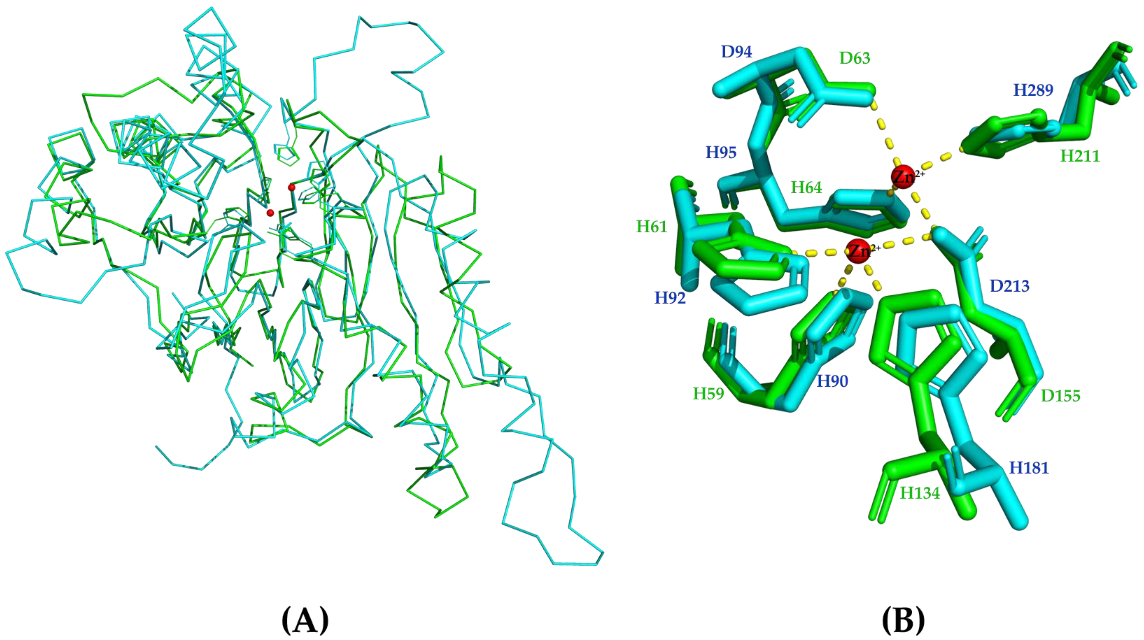
2.3. Crystal Structure of PaAst

Purified recombinant PaAst was crystallized in various conditions, but the majority obtained were needle-like crystals. Fortunately, slice-like diffraction-quality crystals grew in 0.2 M sodium thiocyamate and 20% PEG3350 at 16 °C (Figure 4), and PEG400 was selected as the cryoprotectant. PaAst was crystallized in space group P 1 21 1 with its unit cell parameters a = 55.17 Å, b = 61.38 Å, c = 89.60 Å, α = 90°, β = 97.88°, and γ = 90°, and the diffraction and refinement statistics are summarized in Table A1. The X-ray diffraction data were collected in a resolution range of 38.61–1.55 Å, and the final structure was determined at 2.0 Å with Rwork of 20.04% and Rfree of 24.35% by molecular replacement. A comparison of protein structures through the Dali server indicated that the closest homologue of PaAst was chain A of Bacillus subtilis Ribonuclease Z (RNase Z, PDB: 4GCW), a member of the β-lactamase family, sharing a Z-score of 32.9 with a sequence identity of 27% and an RMSD of 2.1 Å [24,25]. The starting model, YhfI, also exhibited an extensive homology to PaAst, with a Z-score of 29.1, sequence identity of 21%, and RMSD of 1.9 Å.
Each asymmetric unit contained two chains of PaAst sharing RMSD of 0.18 Å over Cα, and the chains formed a dimer with extensive contacts (Figure 5A). According to the calculation of the PDBe PISA Server (https://www.ebi.cu.uk/msd-srv/prot_int/cgi-bin/piserver, accessed on 19 September 2022), the dimer interface had a buried solvent accessible area of 2245.3 Å2, which accounted for 16.5% of the total solvent accessible area. Therefore, the dimerization could be observed both in solution, proved by size-exclusion chromatography, and in the crystal structure.
Residues of the truncated recombinant PaAst were well defined in the electron density, with Chain B primarily presented hereafter for structural analysis (Figure 5B). The structure formed an MBL fold consisting of 14 β-strands (β1-β14) and 10 α-helices (α1-α10). The MBL fold exhibited a typical β-sandwich structure, and the two central β-sheets of it were constituted by 7 β-strands, respectively, β7-β13 and β1-β6 with β14. The α-helices were decorated on either side of the β-sheets: α1, α2, α3, and α4 on one side, and α5 to α10 on the other.
Two neighboring high electron density peaks were identified at the core-facing end of the β-sandwich that modeled as metal ions; however, they were not determined as Zn ions corresponding to those in YhfI. The residues nearby could constitute the metal ion-binding motifs strictly conserved in the MBL-fold proteins with one metal ion coordinated by His90, His92, His181, and Asp213, and the other coordinated by Asp94, His95, D213, and His289, according to the alignment with YhfI (Figure 6). The metal-coordinating residues play critical roles as enzymatic active centers in MBL-fold hydrolases [4,18]. Attempts are underway to determine the metal ions by crystallization and enzymatic assays in the presence of metal ions, and site-directed mutations will be performed to further analyze its active center.
Although several MBL-fold arylsulfatases have been reported in recent decades, limited knowledge could be obtained from their structures. In this study, our sequence and functional analyses revealed that PaAst is an arylsulfatase, and the specific activity, Km and Vmax of purified recombinant PaAst, was observed by 39.45 U/mg, 1.00 mM, and 60.80 U/mg at 50 °C, respectively. Its structure was determined at 2.0 Å and formed a dimer with MBL fold, which could be the first MBL-fold arylsulfatase reported with its structure determined. The identification and characterization of AtsA-like arylsulfatases had indicated the existence of MBL-fold arylsulfatases for years, and the structure of PaAst supported the classification of MBL-fold arylsulfatases as a novel type directly. This work provided insights into the MBL-fold arylsulfatases and may be the theoretical foundation for investigations into their catalytic mechanism.

3. Materials and Methods

3.1. Strains, Plasmids, and Reagents

PaAst (GenBank: ABG39403.1) was codon-optimized and synthesized by Sangon (Shanghai, China) with its N-terminal signal peptide truncated. The recombinant protein was cloned into a modified pET32a vector and expressed in E. coli BL21 (DE3) as described previously, ligated by an N-terminal tobacco etch virus (TEV) cleavage site with the thioredoxin (Trx) and 6*His-tag for better solubility and purification [26,27].
PrimeSTAR® Max DNA Polymerase, T4 DNA Ligase, and the restriction enzymes were purchased from Takara Bio Inc. The Gel Extraction Kits and Plasmid Mini Kits were purchased from Omega Bio-tek (Norcross, GA, USA). All chemicals were in analytical grade and obtained commercially; for example, ampicillin sodium salt, isopropyl β-D-thiogalactoside (IPTG), imidazole, ethylenediaminetetraacetic acid (EDTA), and NiSO4·6H2O were purchased from Sangon, pNPS and p-nitrophenol (pNP) from Sigma-Aldrich (St. Louis, MO, USA), and the protein crystallization screening kits from Hampton Research (Aliso Viejo, CA, USA).

3.2. Sequence Analysis

Protein sequences were aligned by BLAST (https://blast.ncbi.nlm.nih.gov/Blast.cgi, accessed on 25 February 2023) and ESPript 3.0 (http://espript.ibcp.fr/ESPript/ESPript/, accessed on 25 February 2023) [28]. The signal peptides and conserved domains were identified by SignalP-5.0 (https://services.healthtech.dtu.dk/service.php?SignalP-5.0, accessed on 13 September 2022) and NCBI Conserved Domain Search Service (CD Search) (https://www.ncbi.nlm.nih.gov/Structure/cdd/wrpsb.cgi, accessed on 15 September 2022), respectively [22,23]. The physical and chemical parameters of the protein were computed by ProtParam tool (https://web.expasy.org/protparam/, accessed on 24 March 2021).

3.3. Protein Expression and Purification

The recombinant PaAst-pET32a construct consisting of residues 23–328 was heterologously expressed in E. coli and purified by Ni2+-NTA resin affinity chromatography (GE Healthcare, Stockholm, Sweden) and size-exclusion chromatography (Superdex 200 increase 300 GL; GE Healthcare) successively [10,26,27]. The recombinant E. coli BL21 (DE3) cells containing PaAst gene were incubated at 37 °C and induced by IPTG (0.3 mM) at 16 °C for 12 h in Luria−Bertani medium. The protein was harvested via ultrasonication on ice in lysis buffer containing 50 mM Tris-HCl (pH 7.5) and 300 mM NaCl and centrifuged at 15,000 rpm for 15 min at 4 °C (Beckman Coulter, Brea, CA, USA).
Supernatant containing the fusion protein was loaded on a Ni2+-NTA resin affinity column to remove the non-specially bound proteins by elution buffer, which is lysis buffer with low concentrations of imidazole (20 mM and 40 mM), while the target protein could be eluted in a higher concentration of imidazole (300 mM). Digestion and dialysis were conducted at the same time with TEV Protease at 4 °C overnight for the cleavage of Trx-6*His-tag and removal of imidazole. Then a secondary Ni2+ affinity chromatography and size-exclusion chromatography were used to further purify the recombinant protein in buffer containing 50 mM Tris-HCl (pH 7.5) and 150 mM NaCl. The purification was evaluated by 15% SDS-PAGE, and the concentration of purified PaAst was determined using NanoDrop One (Thermo Scientific, Waltham, MA, USA).

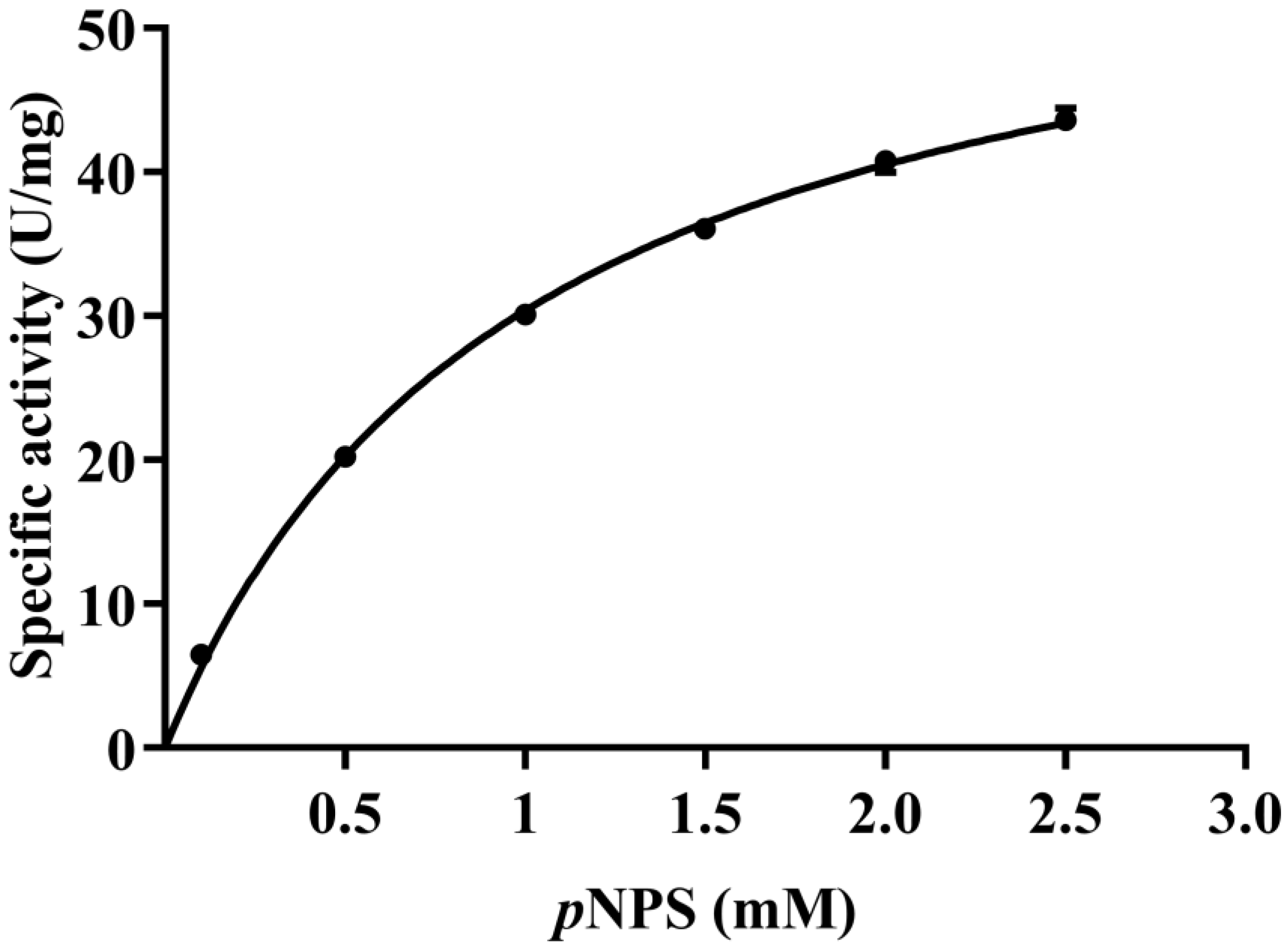
3.4. Enzymatic Activity Assay

The enzymatic activity of PaAst was determined by the artificial substrate pNPS, which could be hydrolyzed into pNP and sulfate by arylsulfatases, as described [3,12,14]. A solution of both enzyme and substrate was made with 50 mM Tris-HCl (pH 7.5) buffer, and the reaction consisted of 20 µL enzyme solution (0.23 µg) and 80 µL substrate solution. Moreover, the kinetic parameters were measured with pNPS in different concentrations (0.1, 0.5, 1, 1.5, 2, and 2.5 mM). After incubation at 50 °C for 8 min, the reaction was terminated by 20 µL 1 M NaOH, and its absorbance at 410 nm was measured by a spectrophotometer to quantify the concentration of pNP and accordingly calculate the reacted pNPS. One unit of enzyme activity (U) was defined as the amount of enzyme required to degrade 1 µM pNPS per min. The kinetic parameters of Km and Vmax were obtained by creating a Michaelis–Menten plot of reaction rate versus substrate concentrations using GraphPad Prism 8.2.1 [29].

3.5. Crystallization

The purified recombinant PaAst was concentrated to 10–15 mg/mL for crystallization, and the crystallization conditions were preliminary screened by the sitting-drop vapor-diffusion method in 96-well plates at 16 °C with more than ten commercial crystallization screening kits, for example, JCSG Core Suites (I to IV, and +, Molecular Dimensions) [10,26]. Crystals of the protein could be found in more than 70 reservoirs, composed of equal volumes (0.6 µL) of protein solution and reservoir solutions, within 1 day. The diffraction-quality crystals were flash-frozen and kept in liquid nitrogen with cryoprotectant.

3.6. Structure Determination and Refinement

X-ray diffraction data sets for PaAst were collected at beamline BL18U1 of the Shanghai Synchrotron Radiation Facility (SSRF, Shanghai, China), and each contained 360 frames (oscillation range 1°). A complete data set was collected at 100 K from a single crystal of the protein obtained in 0.2 M sodium thiocyamate and 20% (v/v) PEG3350, and it was processed by HKL-2000 and XDS [30,31,32]. The structure of PaAst was determined by the molecular replacement method using Phaser with the crystal structure of metallo-β-lactamase fold protein YhfI from Bacillus subtilis (PDB: 6KNT) acting as the starting model [18,33]. Model building and refinement of the structure were executed by Coot and Phenix [34,35], respectively, and the final model was deposited in the Protein Data Bank with structure factors under an accession code of 8GYG (Table A1). The PyMOL Molecular Graphics System was used to visualize and analyze the protein structures.

Author Contributions

Conceptualization, P.D.; Formal analysis, W.Y., L.S. and H.Z.; Investigation, P.D., J.Y., L.W. and L.C.; Methodology, L.S.; Software, D.J.; Supervision, Y.W.; Visualization, W.Y.; Writing—original draft, P.D.; Writing—review and editing, W.Y. All authors have read and agreed to the published version of the manuscript.

Funding

This research was funded by the National Thousand Talents Program of China, Natural Science Foundation of Fujian Province (2019J01280 and 2021J01171), and Special Projects of the Central Government Guiding Local Science and Technology Development (2020L3008).

Data Availability Statement

Data of this study are available in the article.

Conflicts of Interest

The authors declare no conflict of interest.

Appendix A

Table A1. Diffraction data collection and refinement statistics of PaAst structure.
ParameterValue(s)
Data collection
BeamlineSSRF BL18U1
Wavelength (Å)0.97915
Space groupP 1 21 1
Unit cell parameters
a, b, c (Å)55.17, 61.38, 89.60
α, β, γ (°)90, 97.88, 90
Resolution range (Å)38.61–1.55 (1.59–1.55)
Completeness (%)99.7
No. of unique reflections measured85,833
Redundancy6.1 (6.5)
Rmerge0.117 (1.077)
<I/σ(I)>8.3 (1.8)
CC1/20.995 (0.528)
Refinement
Resolution range (Å)29.59–2.0
Completeness (%)99.5
No. of unique reflections refined40,070
Rwork/Rfree (%)20.04/24.35
No. of molecules per asymmetric unit2
No. of residuesChain A: 291
Chain B: 305
No. of atoms5202
Ramachandran Plot (%)
Favored regions97.63
Allowed regions2.03
Outliers0.34
Mean B value, overall (Å2)23.9
PDB ID8GYG
Values in parentheses refer to the highest resolution shell.
Table A2. Purification yields of recombinant AtsA.
Purification ProceduresTotal Protein (mg)Total Activity (U)Specific Activity (U/mg)Purification FactorYield (%)
Crude extract136.075000.5736.751100
Affinity chromatography48.882099.6742.961.1741.98
Size-exclusion chromatography6.39500.1578.322.1310.00
Arylsulfatase AtsA was codon-optimized, synthesized, and heterologously expressed in E. coli. Enzymatic activities were determined by pNPS in 50 mM Tris-HCl (pH 7.5) buffer at 50 °C, the same as the conditions for PaAst.

References

  1. Pogorevc, M.; Faber, K. Purification and characterization of an inverting stereo- and enantioselective sec-alkylsulfatase from the gram-positive bacterium Rhodococcus ruber DSM 44541. Appl. Environ. Microbiol. 2003, 69, 2810–2815. [Google Scholar] [CrossRef] [PubMed] [Green Version]
  2. Long, M.; Ruan, L.; Li, F.; Yu, Z.; Xu, X. Heterologous expression and characterization of a recombinant thermostable alkylsulfatase (sdsAP). Extremophiles 2011, 15, 293–301. [Google Scholar] [CrossRef] [PubMed]
  3. Zhu, Y.; Liu, H.; Qiao, C.; Li, L.; Jiang, Z.; Xiao, A.; Ni, H. Characterization of an arylsulfatase from a mutant library of Pseudoalteromonas carrageenovora arylsulfatase. Int. J. Biol. Macromol. 2017, 96, 370–376. [Google Scholar] [CrossRef]
  4. Hagelueken, G.; Adams, T.M.; Wiehlmann, L.; Widow, U.; Kolmar, H.; Tummler, B.; Heinz, D.W.; Schubert, W.D. The crystal structure of SdsA1, an alkylsulfatase from Pseudomonas aeruginosa, defines a third class of sulfatases. Proc. Natl. Acad. Sci. USA 2006, 103, 7631–7636. [Google Scholar] [CrossRef] [Green Version]
  5. Berteau, O.; Guillot, A.; Benjdia, A.; Rabot, S. A new type of bacterial sulfatase reveals a novel maturation pathway in prokaryotes. J. Biol. Chem. 2006, 281, 22464–22470. [Google Scholar] [CrossRef] [Green Version]
  6. Sogi, K.M.; Gartner, Z.J.; Breidenbach, M.A.; Appel, M.J.; Schelle, M.W.; Bertozzi, C.R. Mycobacterium tuberculosis Rv3406 is a type II alkyl sulfatase capable of sulfate scavenging. PLoS ONE 2013, 8, e65080. [Google Scholar] [CrossRef]
  7. Muller, I.; Kahnert, A.; Pape, T.; Sheldrick, G.M.; Meyer-Klaucke, W.; Dierks, T.; Kertesz, M.; Uson, I. Crystal structure of the alkylsulfatase AtsK: Insights into the catalytic mechanism of the Fe(II) alpha-ketoglutarate-dependent dioxygenase superfamily. Biochemistry 2004, 43, 3075–3088. [Google Scholar] [CrossRef] [PubMed] [Green Version]
  8. Kahnert, A.; Kertesz, M.A. Characterization of a sulfur-regulated oxygenative alkylsulfatase from Pseudomonas putida S-313. J. Biol. Chem. 2000, 275, 31661–31667. [Google Scholar] [CrossRef] [Green Version]
  9. Davison, J.; Brunel, F.; Phanopoulos, A.; Prozzi, D.; Terpstra, P. Cloning and sequencing of Pseudomonas genes determining sodium dodecyl sulfate biodegradation. Gene 1992, 114, 19–24. [Google Scholar] [CrossRef]
  10. Sun, L.; Chen, P.; Su, Y.; Cai, Z.; Ruan, L.; Xu, X.; Wu, Y. Crystal structure of thermostable alkylsulfatase SdsAP from Pseudomonas sp. S9. Biosci. Rep. 2017, 37, BSR20170001. [Google Scholar] [CrossRef] [Green Version]
  11. Knaus, T.; Schober, M.; Kepplinger, B.; Faccinelli, M.; Pitzer, J.; Faber, K.; Macheroux, P.; Wagner, U. Structure and mechanism of an inverting alkylsulfatase from Pseudomonas sp. DSM6611 specific for secondary alkyl sulfates. FEBS J. 2012, 279, 4374–4384. [Google Scholar] [CrossRef] [PubMed]
  12. Kim, D.E.; Kim, K.H.; Bae, Y.J.; Lee, J.H.; Jang, Y.H.; Nam, S.W. Purification and characterization of the recombinant arylsulfatase cloned from Pseudoalteromonas carrageenovora. Protein Expr. Purif. 2005, 39, 107–115. [Google Scholar] [CrossRef] [PubMed]
  13. Zhu, Y.; Liang, M.; Li, H.; Ni, H.; Li, L.; Li, Q.; Jiang, Z. A mutant of Pseudoalteromonas carrageenovora arylsulfatase with enhanced enzyme activity and its potential application in improvement of the agar quality. Food Chem. 2020, 320, 126652. [Google Scholar] [CrossRef]
  14. Jung, K.T.; Kim, H.W.; You, D.J.; Nam, S.W.; Kim, B.W.; Jeon, S.J. Identification of the first archaeal arylsulfatase from Pyrococcus furiosus and its application to desulfatation of agar. Biotechnol. Bioprocess Eng. 2013, 17, 1140–1146. [Google Scholar] [CrossRef]
  15. Shahbazi, R.; Kasra-Kermanshahi, R.; Gharavi, S.; Moosavi-Nejad, Z.; Borzooee, F. Screening of SDS-degrading bacteria from car wash wastewater and study of the alkylsulfatase enzyme activity. Iran. J. Microbiol. 2013, 5, 153–158. [Google Scholar] [PubMed]
  16. Zhang, C.; An, D.; Xiao, Q.; Chen, F.Q.; Zhang, Y.H.; Weng, H.F.; Xiao, A.F. Convenient Agarose Preparation with Hydrogen Peroxide and Desulfation Process Analysis. Mar. Drugs 2021, 19, 297. [Google Scholar] [CrossRef] [PubMed]
  17. Xu, X.; Deng, X.; Lin, J.; Yang, J. Characterization and substrate-accelerated thermal inactivation kinetics of a new serine-type arylsulfatase. Enzyme Microb. Technol. 2022, 154, 109961. [Google Scholar] [CrossRef] [PubMed]
  18. Na, H.W.; Namgung, B.; Song, W.S.; Yoon, S.I. Structural and biochemical analyses of the metallo-beta-lactamase fold protein YhfI from Bacillus subtilis. Biochem. Biophys. Res. Commun. 2019, 519, 35–40. [Google Scholar] [CrossRef]
  19. Kertesz, M.A. Riding the sulfur cycle--metabolism of sulfonates and sulfate esters in gram-negative bacteria. FEMS Microbiol. Rev. 2000, 24, 135–175. [Google Scholar] [CrossRef] [Green Version]
  20. Duvaud, S.; Gabella, C.; Lisacek, F.; Stockinger, H.; Ioannidis, V.; Durinx, C. Expasy, the Swiss Bioinformatics Resource Portal, as designed by its users. Nucleic Acids Res. 2021, 49, W216–W227. [Google Scholar] [CrossRef]
  21. Almagro Armenteros, J.J.; Tsirigos, K.D.; Sonderby, C.K.; Petersen, T.N.; Winther, O.; Brunak, S.; von Heijne, G.; Nielsen, H. SignalP 5.0 improves signal peptide predictions using deep neural networks. Nat. Biotechnol. 2019, 37, 420–423. [Google Scholar] [CrossRef] [PubMed] [Green Version]
  22. Marchler-Bauer, A.; Bo, Y.; Han, L.; He, J.; Lanczycki, C.J.; Lu, S.; Chitsaz, F.; Derbyshire, M.K.; Geer, R.C.; Gonzales, N.R.; et al. CDD/SPARCLE: Functional classification of proteins via subfamily domain architectures. Nucleic Acids Res. 2017, 45, D200–D203. [Google Scholar] [CrossRef] [PubMed] [Green Version]
  23. Lu, S.; Wang, J.; Chitsaz, F.; Derbyshire, M.K.; Geer, R.C.; Gonzales, N.R.; Gwadz, M.; Hurwitz, D.I.; Marchler, G.H.; Song, J.S.; et al. CDD/SPARCLE: The conserved domain database in 2020. Nucleic Acids Res. 2020, 48, D265–D268. [Google Scholar] [CrossRef] [Green Version]
  24. Holm, L. Dali server: Structural unification of protein families. Nucleic Acids Res. 2022, 50, W210–W215. [Google Scholar] [CrossRef] [PubMed]
  25. Pellegrini, O.; Li de la Sierra-Gallay, I.; Piton, J.; Gilet, L.; Condon, C. Activation of tRNA maturation by downstream uracil residues in B. subtilis. Structure 2012, 20, 1769–1777. [Google Scholar] [CrossRef] [Green Version]
  26. Zhu, C.; Chen, Y.; Isupov, M.N.; Littlechild, J.A.; Sun, L.; Liu, X.; Wang, Q.; Gong, H.; Dong, P.; Zhang, N.; et al. Structural Insights into a Novel Esterase from the East Pacific Rise and Its Improved Thermostability by a Semirational Design. J. Agric. Food Chem. 2021, 69, 1079–1090. [Google Scholar] [CrossRef]
  27. Yang, W.; Sun, L.; Dong, P.; Chen, Y.; Zhang, H.; Huang, X.; Wu, L.; Chen, L.; Jing, D.; Wu, Y. Structure-guided rational design of the Geobacillus thermoglucosidasius feruloyl esterase GthFAE to improve its thermostability. Biochem. Biophys. Res. Commun. 2022, 600, 117–122. [Google Scholar] [CrossRef]
  28. Robert, X.; Gouet, P. Deciphering key features in protein structures with the new ENDscript server. Nucleic Acids Res. 2014, 42, W320–W324. [Google Scholar] [CrossRef] [Green Version]
  29. Mitteer, D.R.; Greer, B.D.; Randall, K.R.; Briggs, A.M. Further Evaluation of Teaching Behavior Technicians to Input Data and Graph Using GraphPad Prism. Behav. Anal. 2020, 20, 81–93. [Google Scholar] [CrossRef]
  30. Minor, W.; Cymborowski, M.; Borek, D.; Cooper, D.R.; Chruszcz, M.; Otwinowski, Z. Optimal structure determination from sub-optimal diffraction data. Protein Sci. 2022, 31, 259–268. [Google Scholar] [CrossRef]
  31. Otwinowski, Z.; Minor, W. Processing of X-ray diffraction data collected in oscillation mode. Methods Enzymol. 1997, 276, 307–326. [Google Scholar] [CrossRef]
  32. Kabsch, W. Xds. Acta Crystallogr. D Biol. Crystallogr. 2010, 66, 125–132. [Google Scholar] [CrossRef] [PubMed] [Green Version]
  33. McCoy, A.J.; Grosse-Kunstleve, R.W.; Adams, P.D.; Winn, M.D.; Storoni, L.C.; Read, R.J. Phaser crystallographic software. J. Appl. Crystallogr. 2007, 40, 658–674. [Google Scholar] [CrossRef] [PubMed] [Green Version]
  34. Emsley, P.; Cowtan, K. Coot: Model-building tools for molecular graphics. Acta Crystallogr. D Biol. Crystallogr. 2004, 60, 2126–2132. [Google Scholar] [CrossRef] [PubMed] [Green Version]
  35. Adams, P.D.; Afonine, P.V.; Bunkoczi, G.; Chen, V.B.; Davis, I.W.; Echols, N.; Headd, J.J.; Hung, L.W.; Kapral, G.J.; Grosse-Kunstleve, R.W.; et al. PHENIX: A comprehensive Python-based system for macromolecular structure solution. Acta Crystallogr. D Biol. Crystallogr. 2010, 66, 213–221. [Google Scholar] [CrossRef] [PubMed] [Green Version]
Figure 1. Sequence alignment of PaAst (GenBank: ABG39403.1), AtsA (GenBank: CAA46628.1), and YhfI (GenBank: ADM37089.1). Identical residues are highlighted in red, similar residues are framed in blue, and residues in the conserved Zn2+-binding motifs are labeled with stars.
Catalysts 13 00759 g001
Figure 2. Purification of PaAst. (A) Size-exclusion chromatography of recombinant PaAst. The elution volume was 14.85 mL, between standard samples ovalbumin (44 KD) and conalbumin (75 KD), and closer to conalbumin. (B) SDS-PAGE analysis of the purified recombinant PaAst. Lane M, protein marker; Lane 1, purified recombinant PaAst.
Catalysts 13 00759 g002
Figure 3. Specific activity of PaAst towards pNPS. Some error bars could not be seen.
Catalysts 13 00759 g003
Figure 4. Crystallization and X-ray diffraction of PaAst. (A) Crystals observed in 0.2 M sodium thiocyamate, 20% PEG3350. (B) Representative diffraction pattern.
Catalysts 13 00759 g004
Figure 5. Cartoon representation of PaAst. (A) Dimer of PaAst. Chain A is colored in wheat and Chain B in rainbow color. (B) PaAst monomer. Side chains of the conserved Zn2+-binding motifs are shown with lines.
Catalysts 13 00759 g005
Figure 6. Structural alignment of PaAst and YhfI (PDB: 6KNT, chain A). (A) Ribbon representation of PaAst and YhfI. PaAst is in blue, YhfI is in green, and Zn2+ is in red, and the side chains of metal ion binding sites are shown by lines. (B) Comparison of the metal ion binding sites. The metal ion binding site of PaAst is in blue, YhfI is in green, and Zn2+ is in red.
Catalysts 13 00759 g006
Table 1. Purification yields of recombinant PaAst.
Purification ProceduresTotal Protein (mg)Total Activity (U)Specific Activity (U/mg)Purification FactorYield (%)
Crude extract220.322613.0011.861100
Affinity chromatography22.93840.2136.643.0832.16
Size-exclusion chromatography9.77385.6239.453.3314.76
Enzymatic activities were determined by pNPS in 50 mM Tris-HCl (pH 7.5) buffer at 50 °C.
Disclaimer/Publisher’s Note: The statements, opinions and data contained in all publications are solely those of the individual author(s) and contributor(s) and not of MDPI and/or the editor(s). MDPI and/or the editor(s) disclaim responsibility for any injury to people or property resulting from any ideas, methods, instructions or products referred to in the content.

