
Degradation of Sulfamethoxazole in Aqueous Solution by Low-Energy X-ray Irradiation
 and
and 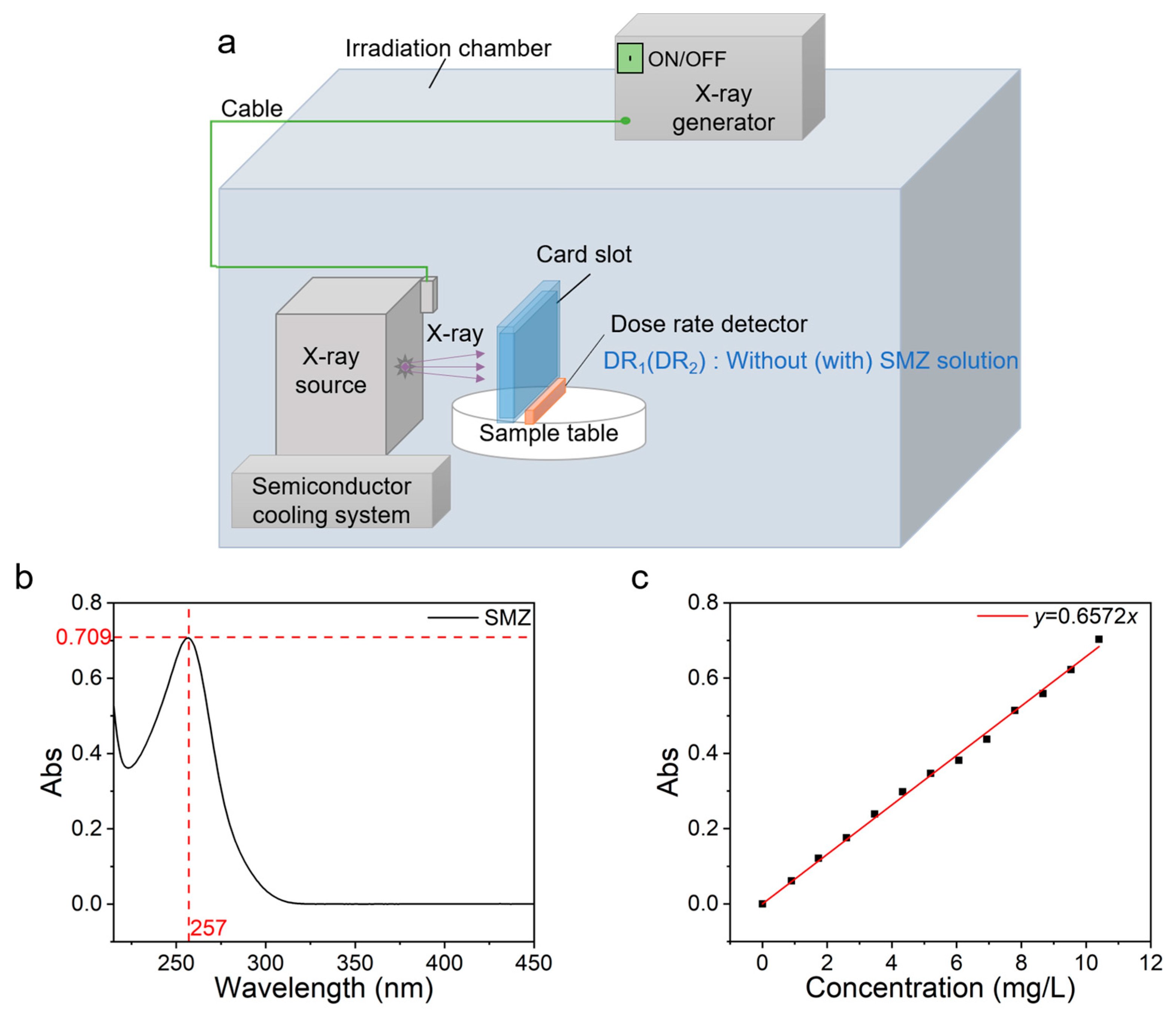
Abstract
:1. Introduction
2. Experimental Design
2.1. Irradiation
2.2. Concentration Calibration of SMZ Solution
3. Results and Discussion
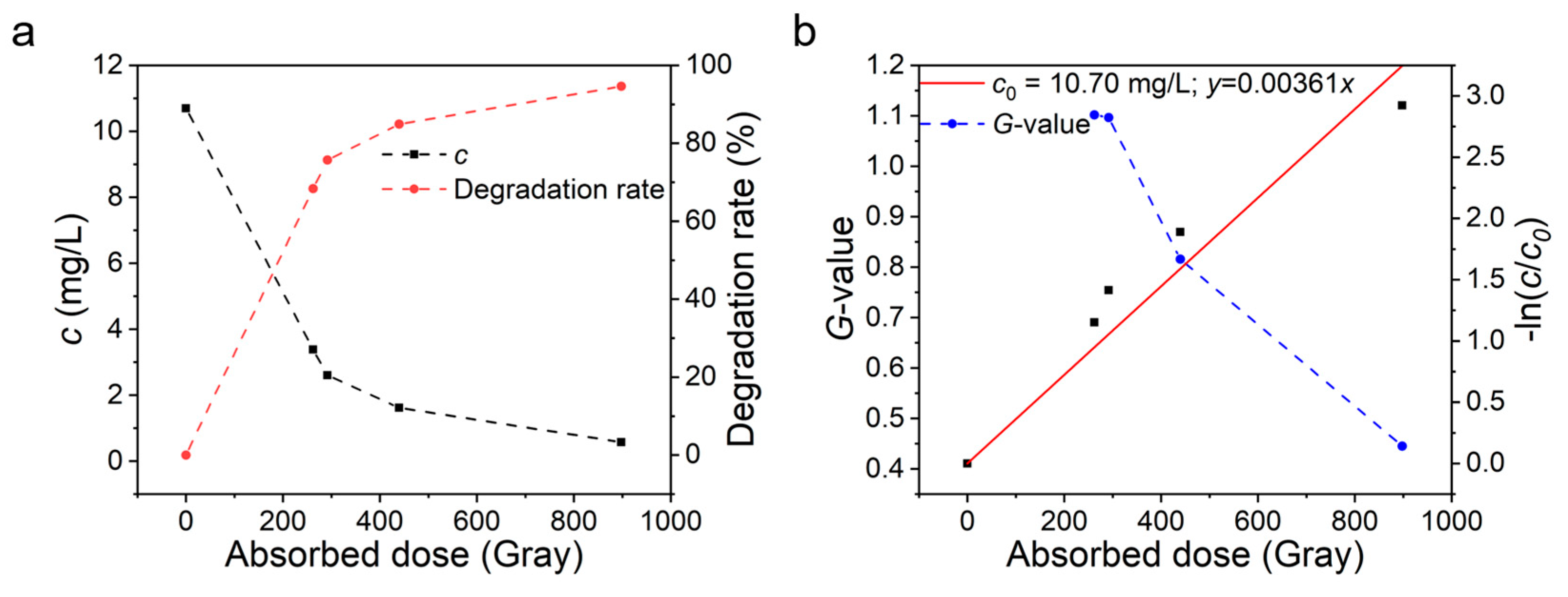
3.1. Effect of Absorbed Dose on SMZ Degradation
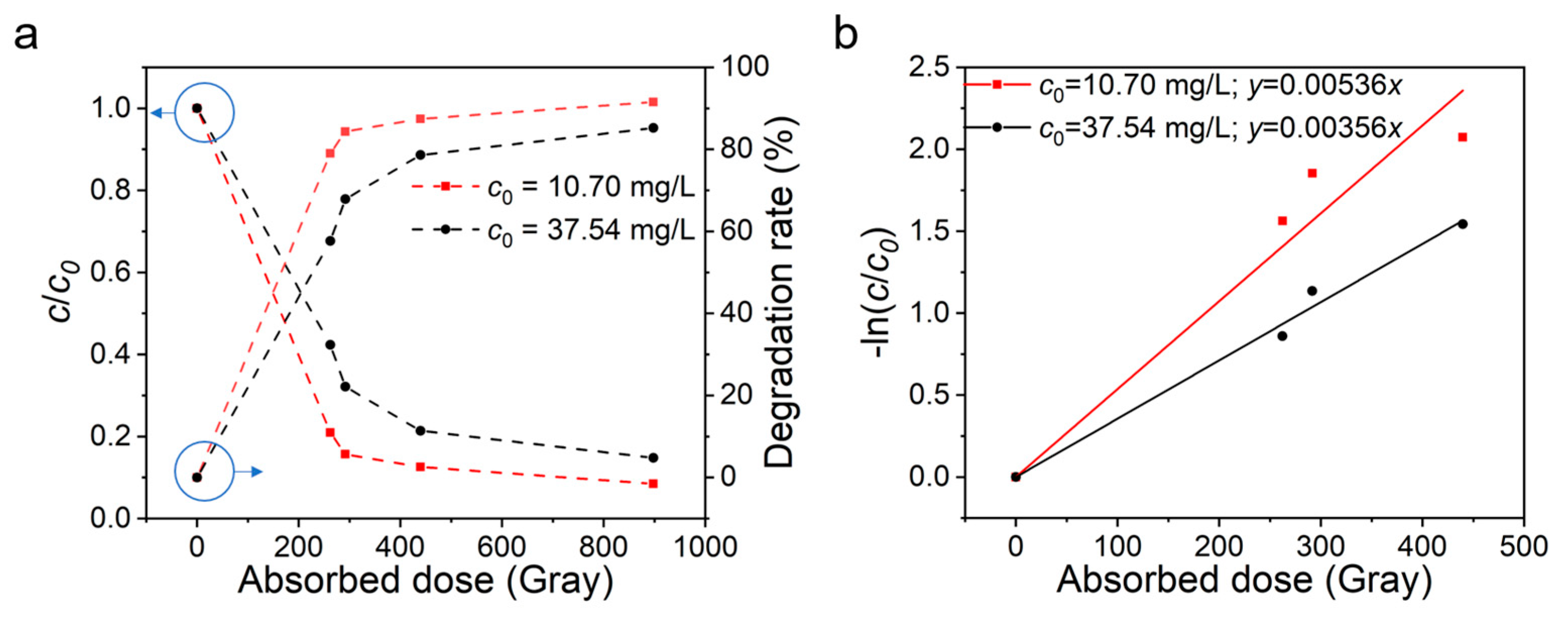
3.2. Effect of Initial Concentration on Degradation of SMZ
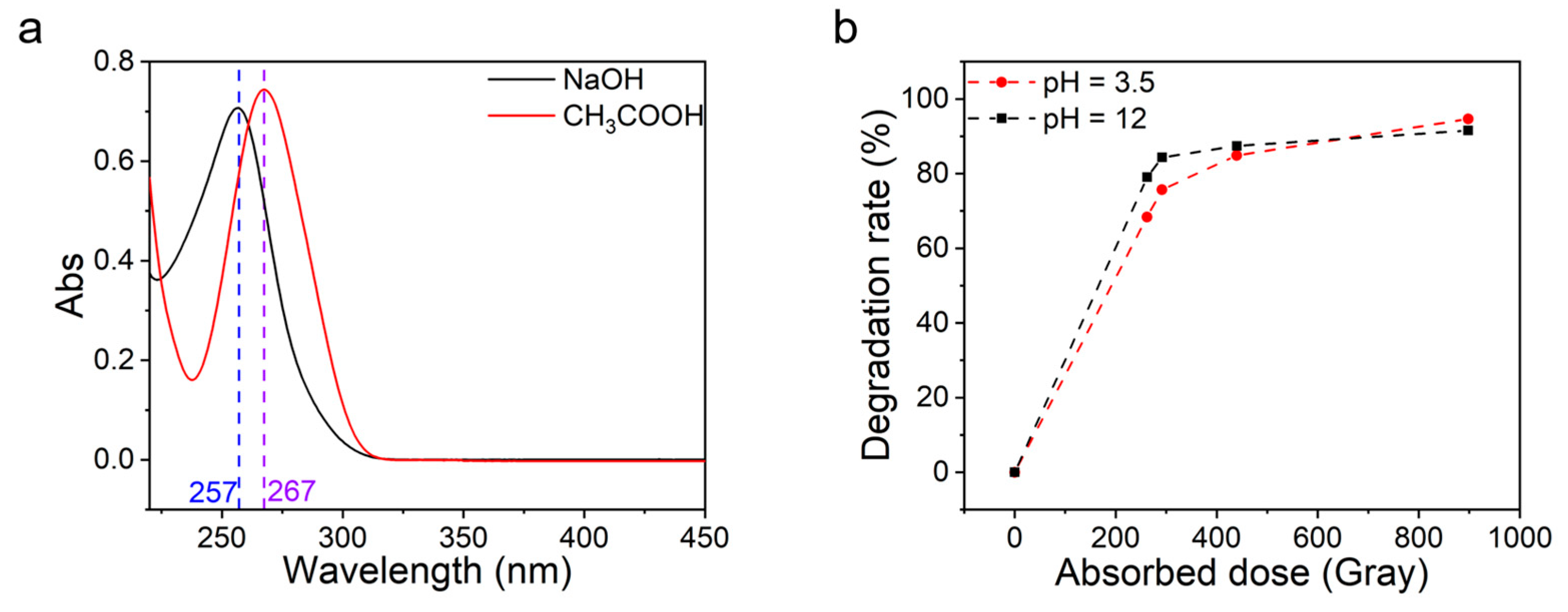
3.3. Effect of pH Value on Degradation of SMZ
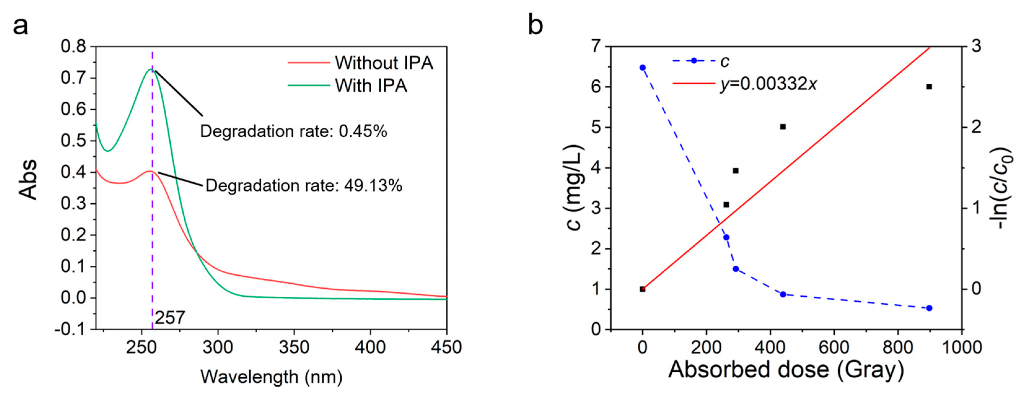
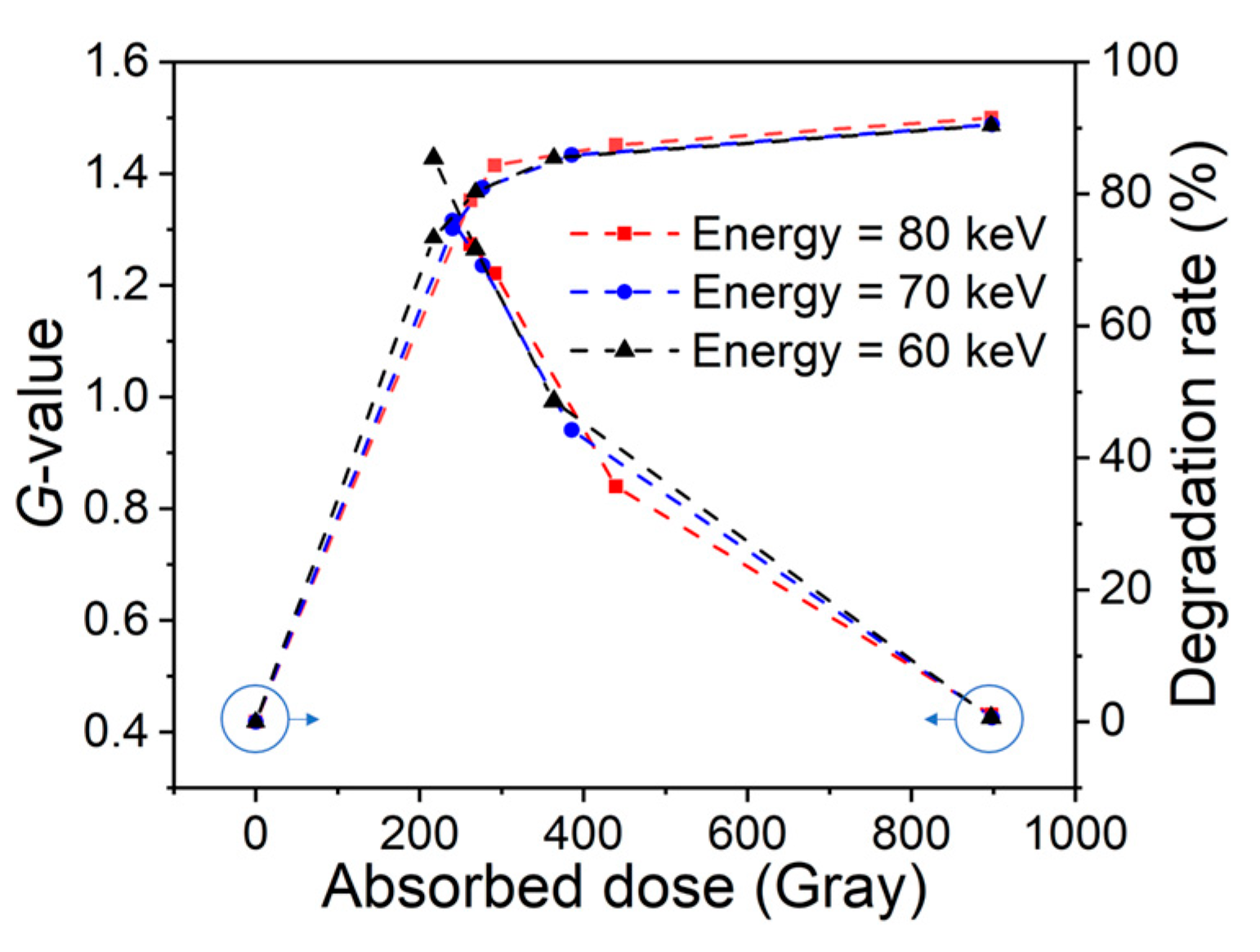
3.4. Effect of Irradiation Energy on SMZ
4. Conclusions
Author Contributions
Funding
Institutional Review Board Statement
Informed Consent Statement
Data Availability Statement
Conflicts of Interest
References
- World Health Organization. Global Antimicrobial Resistance Surveillance System (GLASS) Report: Early Implementation 2020; World Health Organization: Geneva, Switzerland, 2020. [Google Scholar]
- World Health Organization. Global Antimicrobial Resistance and Use Surveillance System (GLASS) Report: 2022; World Health Organization: Geneva, Switzerland, 2022. [Google Scholar]
- Huang, C.-H.; Renew, J.E.; Smeby, K.L.; Pinkston, K.; Sedlak, D.L. Assessment of potential antibiotic contaminants in water and preliminary occurrence analysis. J. Contemp. Water Res. Educ. 2011, 120, 4. [Google Scholar]
- Yang, W.; Fulidong, F.; Ma, J.; Cui, Y. Research progress of antibiotic pollution and treatment technologies in China. In Proceedings of the E3S Web of Conferences, Shanghai, China, 18–20 September 2020; p. 04004. [Google Scholar]
- Sodhi, K.K.; Kumar, M.; Balan, B.; Dhaulaniya, A.S.; Shree, P.; Sharma, N.; Singh, D.K. Perspectives on the antibiotic contamination, resistance, metabolomics, and systemic remediation. SN Appl. Sci. 2021, 3, 269. [Google Scholar] [CrossRef]
- Li, S.-Z.; Li, X.-Y.; Wang, D.-Z. Membrane (RO-UF) filtration for antibiotic wastewater treatment and recovery of antibiotics. Sep. Purif. Technol. 2004, 34, 109–114. [Google Scholar] [CrossRef]
- Back, J.O.; Obholzer, T.; Winkler, K.; Jabornig, S.; Rupprich, M. Combining ultrafiltration and non-thermal plasma for low energy degradation of pharmaceuticals from conventionally treated wastewater. J. Environ. Chem. Eng. 2018, 6, 7377–7385. [Google Scholar] [CrossRef]
- Back, J.O.; Stadlmayr, W.; Jabornig, S.; Winkler, F.; Winkler, K.; Rupprich, M. Removal of arsenic from water with non-thermal plasma (NTP), coagulation and membrane filtration. Water 2018, 10, 1385. [Google Scholar] [CrossRef] [Green Version]
- Wallace, J.S.; Garner, E.; Pruden, A.; Aga, D.S. Occurrence and transformation of veterinary antibiotics and antibiotic resistance genes in dairy manure treated by advanced anaerobic digestion and conventional treatment methods. Environ. Pollut. 2018, 236, 764–772. [Google Scholar] [CrossRef] [PubMed]
- Homem, V.; Santos, L. Degradation and removal methods of antibiotics from aqueous matrices–a review. J. Environ. Manag. 2011, 92, 2304–2347. [Google Scholar] [CrossRef] [PubMed]
- Buxton, G.V.; Greenstock, C.L.; Helman, W.P.; Ross, A.B. Critical Review of rate constants for reactions of hydrated electrons, hydrogen atoms and hydroxyl radicals (⋅ OH/⋅ O− in Aqueous Solution. J. Phys. Chem. Ref. Data 1988, 17, 513–886. [Google Scholar] [CrossRef] [Green Version]
- Bielski, B.H.; Cabelli, D.E.; Arudi, R.L.; Ross, A.B. Reactivity of HO2/O−2 radicals in aqueous solution. J. Phys. Chem. Ref. Data 1985, 14, 1041–1100. [Google Scholar] [CrossRef]
- Alsager, O.A.; Alnajrani, M.N.; Alhazzaa, O. Decomposition of antibiotics by gamma irradiation: Kinetics, antimicrobial activity, and real application in food matrices. Chem. Eng. J. 2018, 338, 548–556. [Google Scholar] [CrossRef]
- Sayed, M.; Khan, J.A.; Shah, L.A.; Shah, N.S.; Khan, H.M.; Rehman, F.; Khan, A.R.; Khan, A.M. Degradation of quinolone antibiotic, norfloxacin, in aqueous solution using gamma-ray irradiation. Environ. Sci. Pollut. Res. 2016, 23, 13155–13168. [Google Scholar] [CrossRef]
- Wang, M.; Zhang, L.; Zhang, G.; Pang, T.; Zhang, X.; Cai, D.; Wu, Z. In situ degradation of antibiotic residues in medical intravenous infusion bottles using high energy electron beam irradiation. Scientific reports 2017, 7, 39928. [Google Scholar] [CrossRef] [PubMed] [Green Version]
- Cho, J.Y.; Chung, B.Y.; Hwang, S.A. Detoxification of the veterinary antibiotic chloramphenicol using electron beam irradiation. Environ. Sci. Pollut. Res. 2015, 22, 9637–9645. [Google Scholar] [CrossRef] [PubMed]
- Silindir, M.; Özer, A.Y. Sterilization methods and the comparison of E-beam sterilization with gamma radiation sterilization. Fabad J. Pharm. Sci. 2009, 34, 43. [Google Scholar]
- Fan, X.; Niemira, B.A. Gamma ray, electron beam, and X-ray irradiation. Food Saf. Eng. 2020, 179, 471–492. [Google Scholar]
- Carvalho, I.T.; Santos, L. Antibiotics in the aquatic environments: A review of the European scenario. Environ. Int. 2016, 94, 736–757. [Google Scholar] [CrossRef]
- Zeng, H.; Li, J.; Zhao, W.; Xu, J.; Xu, H.; Li, D.; Zhang, J. The Current Status and Prevention of Antibiotic Pollution in Groundwater in China. Int. J. Environ. Res. Public Health 2022, 19, 11256. [Google Scholar] [CrossRef]
- Garoma, T.; Umamaheshwar, S.K.; Mumper, A. Removal of sulfadiazine, sulfamethizole, sulfamethoxazole, and sulfathiazole from aqueous solution by ozonation. Chemosphere 2010, 79, 814–820. [Google Scholar] [CrossRef]
- Li, W.; Lan, M.; Peng, X. Removal of antibiotics from swine wastewater by UV/H2O2 combined oxidation. Environ. Poll. Control 2011, 33, 25–28. [Google Scholar]
- Trovó, A.G.; Nogueira, R.F.; Agüera, A.; Fernandez-Alba, A.R.; Sirtori, C.; Malato, S. Degradation of sulfamethoxazole in water by solar photo-Fenton. Chemical and toxicological evaluation. Water Res. 2009, 43, 3922–3931. [Google Scholar] [PubMed]
- Abellán, M.; Bayarri, B.; Giménez, J.; Costa, J. Photocatalytic degradation of sulfamethoxazole in aqueous suspension of TiO2. Appl. Catal. B Environ. 2007, 74, 233–241. [Google Scholar] [CrossRef]
- Oyekunle, D.T.; Gendy, E.A.; Ifthikar, J.; Chen, Z. Heterogeneous activation of persulfate by metal and non-metal catalyst for the degradation of sulfamethoxazole: A review. Chem. Eng. J. 2022, 437, 135277. [Google Scholar] [CrossRef]
- Kim, H.Y.; Kim, T.-H.; Cha, S.M.; Yu, S. Degradation of sulfamethoxazole by ionizing radiation: Identification and characterization of radiolytic products. Chem. Eng. J. 2017, 313, 556–566. [Google Scholar] [CrossRef]
- Wang, J.; Guo, Z.; Shen, X.; Guo, Q.; Zhao, Y.; Zhu, S.; Guo, Z. Gamma irradiation-induced decomposition of sulfamethoxazole in aqueous solution: The influence of additives, biological inhibitory, and degradation mechanisms. Environ. Sci. Pollut. Res. 2017, 24, 23658–23665. [Google Scholar] [CrossRef]
- Wang, S.; Wang, J. Radiation-induced degradation of sulfamethoxazole in the presence of various inorganic anions. Chem. Eng. J. 2018, 351, 688–696. [Google Scholar] [CrossRef]
- Zhuan, R.; Wang, J. Degradation of sulfamethoxazole by ionizing radiation: Kinetics and implications of additives. Sci. Total Environ. 2019, 668, 67–73. [Google Scholar] [CrossRef]
- Kim, T.-H.; Kim, S.D.; Kim, H.Y.; Lim, S.J.; Lee, M.; Yu, S. Degradation and toxicity assessment of sulfamethoxazole and chlortetracycline using electron beam, ozone and UV. J. Hazard. Mater. 2012, 227, 237–242. [Google Scholar] [CrossRef] [PubMed]
- Mäntele, W.; Deniz, E. UV–VIS absorption spectroscopy: Lambert-Beer reloaded. Spectrochim. Acta Part A Mol. Biomol. Spectrosc. 2017, 173, 965–968. [Google Scholar] [CrossRef] [PubMed]
- Liu, Y.; Wang, J.; Zhou, Z.; Zheng, X.; Zhao, L.; Yu, A. The degradation, biodegradability and toxicity evaluation of sulfamethazine antibiotics by gamma radiation. Open Chem. 2020, 18, 1188–1194. [Google Scholar] [CrossRef]
- Sági, G.; Bezsenyi, A.; Kovács, K.; Klátyik, S.; Darvas, B.; Székács, A.; Mohácsi-Farkas, C.; Takács, E.; Wojnárovits, L. Radiolysis of sulfonamide antibiotics in aqueous solution: Degradation efficiency and assessment of antibacterial activity, toxicity and biodegradability of products. Sci. Total Environ. 2018, 622, 1009–1015. [Google Scholar] [CrossRef] [PubMed]
- Espenson, J.H. Chemical Kinetics and Reaction Mechanisms; Citeseer: Princeton, NJ, USA, 1995; Volume 102. [Google Scholar]
- Khan, J.A.; Shah, N.S.; Khan, H.M. Decomposition of atrazine by ionizing radiation: Kinetics, degradation pathways and influence of radical scavengers. Sep. Purif. Technol. 2015, 156, 140–147. [Google Scholar] [CrossRef]
- Basfar, A.; Mohamed, K.; Al-Abduly, A.; Al-Shahrani, A. Radiolytic degradation of atrazine aqueous solution containing humic substances. Ecotoxicol. Environ. Saf. 2009, 72, 948–953. [Google Scholar] [CrossRef] [PubMed]
- Cooper, W.J.; Cadavid, E.; Nickelsen, M.G.; Lin, K.; Kurucz, C.N.; Waite, T.D. Removing THMs from drinking water using high-energy electron-beam irradiation. J. -Am. Water Work. Assoc. 1993, 85, 106–112. [Google Scholar] [CrossRef]
- Sánchez-Polo, M.; López-Peñalver, J.; Prados-Joya, G.; Ferro-García, M.A.; Rivera-Utrilla, J. Gamma irradiation of pharmaceutical compounds, nitroimidazoles, as a new alternative for water treatment. Water Res. 2009, 43, 4028–4036. [Google Scholar] [CrossRef]
- Shuibo, X.; Ting, C.; Zhiping, L.; Yuqi, W.; Yingjiu, L.; Hui, L. Amoxicillin degradation in aqueous solution by gamma-ray irradiation. Chin. J. Environ. Eng. 2015, 9, 73–78. [Google Scholar]


Disclaimer/Publisher’s Note: The statements, opinions and data contained in all publications are solely those of the individual author(s) and contributor(s) and not of MDPI and/or the editor(s). MDPI and/or the editor(s) disclaim responsibility for any injury to people or property resulting from any ideas, methods, instructions or products referred to in the content. |
© 2023 by the authors. Licensee MDPI, Basel, Switzerland. This article is an open access article distributed under the terms and conditions of the Creative Commons Attribution (CC BY) license (https://creativecommons.org/licenses/by/4.0/).
Share and Cite
Yao, J.; Rao, W.; Kong, H.; Sun, W.; Guo, D.; Li, Z.; Wei, X. Degradation of Sulfamethoxazole in Aqueous Solution by Low-Energy X-ray Irradiation. Catalysts 2023, 13, 714. https://doi.org/10.3390/catal13040714
Yao J, Rao W, Kong H, Sun W, Guo D, Li Z, Wei X. Degradation of Sulfamethoxazole in Aqueous Solution by Low-Energy X-ray Irradiation. Catalysts. 2023; 13(4):714. https://doi.org/10.3390/catal13040714
Chicago/Turabian StyleYao, Jun, Weidong Rao, Hua Kong, Wentao Sun, Dengzhu Guo, Zhiwei Li, and Xianlong Wei. 2023. "Degradation of Sulfamethoxazole in Aqueous Solution by Low-Energy X-ray Irradiation" Catalysts 13, no. 4: 714. https://doi.org/10.3390/catal13040714