Article

Influence of Lanthanum Doping on the Photocatalytic and Antibacterial Capacities of Mg0.33Ni0.33Co0.33Fe2O4 Nanoparticles

1
Department of Chemistry, Faculty of Science, Beirut Arab University, Beirut P.O. Box 11-5020, Lebanon
2
Department of Biological Sciences, Faculty of Science, Beirut Arab University, Beirut P.O. Box 11-5020, Lebanon
3
Inorganic and Organometallic Coordination Chemistry Laboratory, Faculty of Sciences I, Lebanese University, Hadath, Beirut P.O. Box 6573/14, Lebanon
4
Molecular Biology Unit, Department of Zoology, Faculty of Science, Alexandria University, Alexandria 21568, Egypt
5
Department of Physics, Faculty of Science, Beirut Arab University, Beirut P.O. Box 11-5020, Lebanon
6
Department of Physics, Faculty of Science, Alexandria University, Alexandria 21568, Egypt
7
Physics Department, University College at Al-Jamom, Umm Al-Qura University, Mecca 24243, Saudi Arabia
*
Authors to whom correspondence should be addressed.
Catalysts 2023, 13(4), 693; https://doi.org/10.3390/catal13040693
Submission received: 8 March 2023 / Revised: 27 March 2023 / Accepted: 30 March 2023 / Published: 2 April 2023

Abstract

:
The increase in environmental pollution, especially water pollution, has intensified the requirement for new strategies for the treatment of water sources. Furthermore, the improved properties of nano-ferrites permit their usage in wastewater treatment. In this regard, novel Mg0.33Ni0.33Co0.33LaxFe2−xO4 nanoparticles (NPs), where 0.00≤x≤0.08, were synthesized to test their photocatalytic, antibacterial and antibiofilm activities. The structural and optical properties of the prepared NPs were investigated by X-ray powder diffraction (XRD), transmission electron microscopy (TEM), UV-Vis spectroscopy and photoluminescence (PL) analysis. As La content increases, the bandgap energy increases, whereas the particle size decreases. The photocatalytic activity of the prepared NPs is evaluated by the degradation of methylene blue (MB) dye under sunlight irradiation. Superior activity is exhibited by Mg0.33Ni0.33Co0.33La0.01Fe1.99O4 NPs. The influence of catalyst dosage, pH, temperature and addition of graphene (Gr) on the photodegradation reaction was studied. Increasing the pH and temperature improved the rate of the photodegradation reaction. The antibacterial and antibiofilm activities of the NPs were assessed against Escherichia coli, Leclercia adecarboxylata, Staphylococcus aureus and Enterococcus faecium. Mg0.33Ni0.33Co0.33Fe2O4 NPs inhibited bacterial growth. They had bacteriostatic activity on all isolates, with a greater effect on Gram-positive bacteria. All tested nano-ferrites had significant antibiofilm activities against some biofilms.

Graphical Abstract

1. Introduction

The most fundamental component for the survival of living things is water. A healthier life is facilitated by the usage of clean and safe water. However, large amounts of dye-containing effluents are discharged untreated into the water, which, in turn, causes water pollution [1]. Dyes, characterized by their cheap production and stability, are widely used in textiles, paper, plastics, leather, printing, cosmetics and coatings. Dye-containing water affects human and animal health and is visible in water even at very low concentrations ( < 1 ppm) [2]. Furthermore, microbiological water pollution is recognized as one of the major problems across the world [3]. Consequently, removing pollutants such as bacteria and dyes from wastewater is highly desirable.
Magnetic NPs are characterized by their unique properties that are not observed in bulk materials. These characteristics can be attributed to the finite size effects, high surface-to-volume ratio and distinct crystal structures. Numerous physical, chemical and biological techniques can be employed to synthesize these NPs. However, choosing the preparation technique depends on the targeted morphology, size, properties and application of the NPs [4]. The co-precipitation method is recognized for its simplicity, affordability and wide size distribution. Furthermore, it gives rise to NPs that can be functionalized in several applications, including catalysis, gas sensing and antibacterial applications [5,6]. Owing to their size, structures and unique optical and magnetic properties, NPs have been used in a variety of fields, including the electrical and electronic industries and the biomedicine field [7]. Various types and structures have been investigated, including the hexagonal and spinel ferrites. Studies on ferrites are fast-moving, owing to their exponentially growing usage in magnetic biosensors, magnetic shielding, information storage, magnetic recording devices, electronic devices, mobile communication, pollution control, medical devices, transformers and catalysis [8].
Concerning the role of nano-ferrites as a pollutant control, they stand out among the most efficient advanced materials in terms of their potential to remove pollutants [9]. This is due to the economic profile and efficiency of nano-ferrites, which permit their usage in the removal of various pollutants such as pesticides, dyes, medicines and hazardous chemicals. In addition, the improvement of the properties of ferrites can be done by modifying the synthesis parameters and the doping or formation of nano-composites [10]. The doping of rare earth metals, such as Samarium (Sm) and Lanthanum (La), improves the surface-to-volume ratio and enhances the optical, electrical and magnetic properties of host materials [11]. For example, doping CoFe2O4 with La improved the magnetic properties, reduced the bandgap energy and increased the surface area [12]. The improved properties permit their usage in environmental applications. Furthermore, La-doped BiFeO3 NPs exhibited superior catalytic performance in the photodegradation of Rhodamine B dye compared to that of pure BiFeO3 NPs [13]. As for the antibacterial activity, magnetic NPs were shown to exhibit a significant antibacterial activity, depending on their shape, size and density of oxygen vacancies [14]. In addition, rare earth metals were shown to exert unique antibacterial advantages, especially against Escherichia coli and Staphylococcus aureus. They act by penetrating the bacterial cells and changing the cytoplasmic composition, thus leading to the destruction of the bacterial cells [15]. Regarding water treatment, some studies reported that La-doped NPs can remove pollutants and microbes, such as Klebsiella pneumonia and other pathogens, during the treatment process. This activity is inversely proportional to the size, as the antibacterial activity can be enhanced by smaller-sized NPs [16].
Among several spinel ferrites, MgFe2O4, NiFe2O4 and CoFe2O4 NPs have attracted the attention of scientists and are useful due to their unique characteristics. It is known that MgFe2O4 is a soft magnetic semiconducting material with a normal spinel structure [17]. NiFe2O4, having an inverse spinel structure, is a soft magnetic semiconducting material [18]. Whereas CoFe2O4, characterized by its inverse spinel structure, is classified as a semi-hard material [19]. Thus, it is interesting to investigate the properties of Mg0.33Ni0.33Co0.33Fe2O4 NPs. As mentioned before, doping ferrites with rare earth metals improves their properties. In this work, La-doped Mg0.33Ni0.33Co0.33LaxFe2−xO4 spinel ferrites where 0.00 ≤ x ≤ 0.08 were synthesized using the co-precipitation technique. In addition, the prepared NPs were characterized by XRD, TEM, UV and PL techniques. Finally, La-doped Mg0.33Ni0.33Co0.33LaxFe2−xO
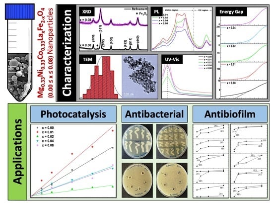
4 NPs were used to disinfect water from methylene blue (MB) dye and bacteria.

2. Results and Discussion

2.1. Characterization of Mg0.33Ni0.33Co0.33LaxFe2−xO4 NPs

Figure 1 displays the refined and raw XRD patterns of Mg0.33Ni0.33Co0.33LaxFe2−xO4 NPs, where 0.00 ≤ x ≤ 0.08. The XRD pattern of pure Mg0.33Ni0.33Co0.33Fe2O4 NPs (x = 0.00) reveals peaks that are located at 30.1°, 35.6°, 36.7°, 43.2°, 53.5°, 57.1° and 62.3°, corresponding to (220), (311), (222), (400), (422), (511) and (440) crystal planes, respectively. The peaks are matched with the standard JCPDS card no. 36-0398 and indexed to the cubic spinel phase with space group Fd3m [20]. Furthermore, the presence of hematite (Fe2O3) as a secondary phase is revealed from the presence of an extra peak located at 2θ = 32.9°. Though, upon doping the NPs with La, the intensity of the peak—referring to the impurity phase—disappears. The absence of any extra peak in the XRD patterns of La-doped NPs ensures the purity of the prepared samples. It is worth mentioning that the values of the lattice parameter (a) of all doped NPs, listed in Table 1, are greater than that of pure NPs. This is mainly attributed to the substitution of Fe3+ with La3+, knowing that the ionic radius of La3+ (1.06 Å) is greater than that of Fe3+ (0.65 Å). Similar behavior was reported in a previous study where the lattice parameter (a) increased upon doping Ni0.3Zn0.5Co0.2LaxFe1.98-xO4 ferrite with La [21].
The particle size distribution and microstructure of Mg0.33Ni0.33Co0.33LaxFe2−xO4 NPs, where 0.00 ≤ x ≤ 0.08, were examined by TEM analysis and represented in Figure 2. The results indicate the synthesis of nano-sized particles with rounded-cube morphology. Similar morphology of CoFe2O4, MgFe2O4 and NiFe2O4 is reported in previously published studies [22,23,24]. Moreover, as La content increases from 0.00 up to 0.08, the particle size decreases from 28.72 to 20.20 nm, as shown in Figure 2 and Table 1. The reduction in the particle size, upon doping Mg0.33Ni0.33Co0.33Fe2O4 with La, is attributed to the settling of the La3+ ions at grain boundaries, which inhibits the growth of the grains. Similar behavior is reported in a previous study, where the particle size of Ni0.3Zn0.5Co0.2Fe2O4 NPs was reduced upon doping with Gd3+ and La3+ ions [25]. Furthermore, the aggregation of the synthesized NPs might be attributed to the magnetic dipole interaction among them [12].
The UV-Vis absorption spectra of Mg0.33Ni0.33Co0.33LaxFe2−xO4 NPs where 0.00 ≤ x ≤ 0.08, in the range of 300–700 nm, are represented in Figure 3. A noticeable peak appears at 335 nm in the absorption spectra of all the prepared NPs. Comparable UV spectra of ferrite NPs, mainly Zn0.5Mg0.5-xLi2xFe2O4 and Ni1-xMgxFe2O4, are stated in previous studies [26,27]. The prepared NPs with various La content exhibited stronger UV absorption, as revealed by the increased absorption intensity. Similar behavior also was reported by Lemziouka et al. [28] upon increasing La content in ZnFe2−xLaxO4 NPs. To better understand the change in optical properties caused by the doping of NPs with La, the optical bandgap energy was calculated. The bandgap energy (Eg) was estimated from Tauc’s plot, which is represented in the following equation:
(αhυ)2 = B (hυ − Eg),
where α is the absorption coefficient, hυ represents the incident photon energy and B denotes the transition probability constant. Thus, the bandgap energy (Eg) was estimated by plotting (αhυ)2 vs. hυ, as displayed in Figure 4 and Table 1. As La content increases from x = 0.00 to 0.08, Eg increases from 2.92 to 3.25 eV. The increase in Eg may result from intrinsic absorption and optical scattering at grain boundaries [29]. In addition, the increase in the bandgap energy accompanied by the decrease in the particle size upon increasing the La content is owed to the quantum size effect [29,30].
It is obvious that when a semiconductor gets exposed to light that has a wavelength equal to or higher than its bandgap energy, it absorbs the light. This causes the excitation of electrons from the valence band to the conduction band, creating a positive hole in the valence band. Thus, an electron-hole pair is generated. The electron-hole pair can either recombine or move from the semiconductor’s surface to the solvent. It is worth mentioning that the photocatalytic activity of the semiconductor is subsequently decreased as a result of the recombination of electron-hole pairs. Furthermore, the recombination rate of the electron-hole pair can be measured by the intensity of PL emission. Thus, the PL investigation was carried out at room temperature by applying an excitation wavelength λext = 330 nm, and the results are displayed in Figure 5a,b. Numerous peaks are detected in UV and visible ranges. Due to the presence of oxygen vacancies and intrinsic imperfections, peaks appear in the visible region. However, the peak located in the UV region at 3.7 eV is attributed to the recombination of the electron-hole pair. Upon doping Mg0.33Ni0.33Co0.33Fe2O4 with La, the intensity of this peak is reduced. Thus, the recombination rate becomes slower. In addition, NPs with La content of 0.01 and 0.04 exhibited the slowest recombination rate of the electron-hole pair.

2.2. Photocatalytic Activity of Mg0.33Ni0.33Co0.33LaxFe2−xO4 NPs

The photocatalytic properties of Mg0.33Ni0.33Co0.33LaxFe2−xO4 NPs where 0 x 0.08 were evaluated by the degradation of MB dye under direct sunlight. The linear plot of ln C0/Ct vs. time (Figure 6 and Table 2) revealed, from high R2 values, the first-order kinetics. So, the rate law for the MB photodegradation reaction is given as follows [31]:
ln (C0/Ct) = kt,
where C0 is the concentration of MB at time t = 0 and Ct is the concentration at any time t and k is the degradation rate constant. As listed in Table 2, the rate constant k increases from 29 × 10−4 to 37 × 10−4 min−1 as x increases from 0 to 0.01; whereas, as x increases to reach 0.08, rate constant k decreases to reach 17 × 10−4 min−1. Among the prepared doped samples, an improved photocatalytic activity is exhibited in the presence of Mg0.33Ni0.33Co0.33LaxFe2−xO4 with x = 0.01.
Mg0.33Ni0.33Co0.33La0.01Fe1.99O4 NPs revealed the lowest PL peak intensity of the peak located at 3.7 eV, showing the slowest recombination rate of the photogenerated electron-hole pair. Among the doped samples, Mg0.33Ni0.33Co0.33La0.01Fe1.99O4 NPs exhibited the highest PL peak intensity of the peaks located in the visible region, revealing the presence of increased oxygen vacancies. The presence of oxygen vacancies plays an important role in enhancing the photocatalytic performance of NPs [32,33]. Thus, the photocatalytic performance of NPs is in good accordance with the PL results.
Whenever the mixture of MB and NPs is placed under sunlight, light absorption by NPs will occur. Consequently, electron-hole pairs are generated. Since Mg0.33Ni0.33Co0.33La0.01Fe1.99O4 NPs revealed the slowest recombination rate of the photogenerated electron-hole pair, the electron-hole pair will migrate to the NPs’ surface. It is worth mentioning that the uncombined electron-hole pair interacts with the O2 and H2O and produces reactive species, such as hydroxyl radicals (OH·), superoxide radicals (O2·−) and H2O2. Finally, the produced reactive species are responsible for the degradation of MB dye through a direct oxidation process [34,35].

2.2.1. Effect of Photocatalyst Dosage

After demonstrating that the Mg0.33Ni0.33Co0.33La0.01Fe1.99O4 exhibited an enhanced photocatalytic activity, the effect of photocatalyst dosage was studied by performing the photodegradation reaction of MB in the presence of different amounts of photocatalyst (0.04, 0.06, 0.08 and 0.1 g). As shown in Figure 7, as the Mg0.33Ni0.33Co0.33La0.01Fe1.99O4 amount increases from 0.04 to 0.06 g, the rate of the reaction increases from 29 × 10−4 to 35 × 10−4 min −1. This is because the active sites of the catalyst increase with increasing the amount of photocatalyst [36]. However, with a further increase in the catalyst amount to 0.1 g, the reaction rate decreases to reach 15 × 10−4 min−1. The presence of an excess amount of catalyst (more than 0.06 g) scatters the sunlight and thus blocks its penetration into the reaction. Therefore, the rate of the reaction starts decreasing. Furthermore, additional amounts of NPs lead to particle agglomeration, which, in turn, reduces photon absorption that takes place on the surface [37]. Thus, 0.06 g is the recommended dosage of the catalyst.

2.2.2. Effect of pH

One of the key variables in the photocatalytic property of substances is the solution’s pH. This is explained by the fact that the pH affects the adsorption behavior of the pollutants as well as the chemical characteristics of the photocatalyst. So, at pH values ranging from 2.47 to 10.73 (acidic, neutral and basic pH values), the effect of pH on the photocatalytic degradation of MB in the presence of Mg0.33Ni0.33Co0.33La0.01Fe1.99O4 NPs was investigated. As shown in Table 3, the rate constant increases from 7 × 10⁻⁴ to 517 × 10−4 min−1 when the pH rises from 2.47 to 10.73. Thus, the rate of the photodegradation reaction of MB is 73.8 and 14.7 times higher in the basic medium (pH = 10.73) compared to that studied in the acidic (pH = 2.47) and neutral (pH = 6.27) medium, respectively. So, the solution’s ideal pH is basic. As reported in previous studies, the point of zero charge (PZC) of MgFe2O4, NiFe2O4 and CoFe2O4 NPs was 8.4, 6.4 and 7.2, respectively [38,39,40]. If the pH is less than the PZC, the surface of NPs will be positively charged; when the pH is greater than the PZC, the negative form of the NPs is more likely to exist [37]. Being a cationic dye, MB is positively charged in the solution. Consequently, the MB dye is attracted to the negatively charged catalyst surface due to opposite charges. Thus, increasing the pH of the solution improves the photocatalytic degradation of MB. Identical results were reported in a previous study, where the pH = 12 solution was the optimal medium for the photodegradation of MB by biosynthesized ZnFe2O4 NPs [41].

2.2.3. Effect of Temperature

To investigate the effect of temperature, the photodegradation reaction was carried out at 313, 323, 328 and 333 K. The obtained results are displayed in Figure 8. As the reaction temperature increases from 313 to 333 K, the rate constant increases from 11 × 10−4 to 58 × 10−4 min−1. Thus, increasing the reaction temperature boosts the rate of the photodegradation reaction of MB. This is attributed to the generation of electron-hole pairs that can be produced more quickly at higher temperatures, causing improved photocatalytic activity. In other words, as reaction temperature increases, more electrons are transferred from the valence band to the conduction band. Consequently, excessive electron-hole pairs are generated. This, in turn, increases the production of reactive radicals that are responsible for the degradation of MB dye. A similar trend has been detected for the degradation of MB in the presence of the Fe2O3/graphene/CuO photocatalyst [42].

2.2.4. Thermodynamic Parameters

The thermodynamic parameters can be estimated by performing the photodegradation reaction of the MB dye at various temperatures. Figure 9 and Table 4 display the results. The activation energy (Ea) and the frequency factor (A) were calculated from the slope and intercept of the logarithmic form of the following Arrhenius equation [43]:
ln k = E a RT   +   ln A
where k is the rate constant, R is the universal gas constant, which is equal to 8.314 J.mol−1. K−1, and T is the temperature. Ea and A were calculated from the slope and intercept of the linear plot of ln k vs. 1/T. Additionally, the slope and intercept of the plot of ln (k/T) vs (1/T) were used to estimate the enthalpy (∆H) and entropy change (∆S), as shown in the following equation [43]:
ln   k T =   Δ H RT + Δ S R
Additionally, the following relationship was used to determine the Gibbs free energy change (∆G):
ΔG = ΔH − TΔS

2.2.5. Influence of Graphene Addition

Since electron-hole recombination severely restricts photocatalytic activity, several techniques are employed to boost the charge carrier separation effectiveness and, consequently, the photocatalytic performance of the photocatalyst. The combination of graphene (Gr) with the photocatalyst constitutes a novel approach in this situation [44]. The impact on photocatalysts of mixing various weight percentages of Gr with 0.06 g Mg0.33Ni0.33Co0.33La0.01Fe1.99O4 was examined. The obtained results are shown in Figure 10a. Furthermore, the degradation %, displayed in Figure 10b, was calculated using the following equation:
Degradation   % = C 0 C t C 0 × 100
The rate of the reaction decreases to 25 × 10−4 min−1 with the addition of Gr up to 10 wt %. However, with a further increase in the Gr content of up to 20 wt.%, the rate of the degradation reaction increases. The photodegradation of the MB is greatly improved when 20 wt.% of Gr is combined with Mg0.33Ni0.33Co0.33La0.01Fe1.99O4 NPs. Moreover, 77.9% of MB dye was degraded after sunlight exposure time up to 240 min in the presence of 20 wt.% Gr/Mg0.33Ni0.33Co0.33La0.01Fe1.99O4 nanocomposites (NCs). This might be ascribed to the effective charge transfer encouraged by Gr addition, which enhances the separation of the photogenerated electron-hole pair [45]. Therefore, the combination of NPs with Gr significantly enhanced the photocatalytic activity of Mg0.33Ni0.33Co0.33La0.01Fe1.99O4 NPs in the MB degradation, and 20 wt.% of Gr is the ideal Gr content. To confirm that the formation of heterojunction between Gr and NPs is the reason behind the improvement of catalytic activity, the photodegradation of MB was evaluated in the presence of pure Gr. As shown in Figure 11, the degradation reaction of MB proceeds 1.24 and 1.77 times faster in the presence of 20 wt.% Gr/Mg0.33Ni0.33Co0.33La0.01Fe1.99O4 NCs compared to that in the presence of pure Gr and Mg0.33Ni0.33Co0.33La0.01Fe1.99O4 NPs, respectively.

2.2.6. Optimal Experimental Conditions

The reaction’s rate was studied by applying the optimal experimental conditions. To do this, the pH of the MB dye solution was adjusted to 10.73 before being combined with 0.06 g of 20 wt.% Gr/Mg0.33Ni0.33Co0.33La0.01Fe1.99O4 NCs. According to Figure 12, when exposure time increases, the intensity of the MB peak—which is positioned around 675 nm—decreases. Furthermore, after applying the ideal conditions, it is important to note that the rate constant of the degradation reaction of MB is boosted to reach 0.0619 min−1 and 96.8% is degraded after 60 min. The results obtained from this study were compared with published reports for the degradation of MB under sunlight radiation in the presence of various nano-ferrites, as listed in Table 5. Superior photocatalytic performance was exhibited by NPs prepared in this study in the degradation of MB dye.

2.3. The Antibacterial Activity of the Mg0.33Ni0.33Co0.33LaxFe2−xO4 NPs

The increase in the discard of wastes in the environment leads to an increase in pollution rates, especially water pollution. To decrease this pollution, researchers suggested the incorporation of NPs, especially nano-ferrites, in this field. This is due to their significant effects on decreasing the heavy metals and inhibiting the growth of microorganisms [51]. In this regard, this study reported the effect of Mg0.33Ni0.33Co0.33LaxFe2−xO4 NPs where 0.00 ≤ x ≤ 0.08 on four bacteria (Escherichia coli, Leclercia adecarboxylata, Staphylococcus aureus and Enterococcus faecium) isolated from wastewater.

2.3.1. MICs and MBCs of the Mg0.33Ni0.33Co0.33LaxFe2−xO4 NPs where 0.00 ≤ x ≤ 0.08

Bacteria were incubated with increasing concentrations (0.375–3 mg/mL) of each of the NPs. At the used concentration range of NPs, the only bacteriostatic effect that was detected was that of the Mg0.33Ni0.33Co0.33La0.00Fe2O4 NPs. It had the best inhibitory activity against Leclercia adecarboxylata and Enterococcus faecium. In contrast, Escherichia coli was the most resistant. The MIC and MBC assays didn’t detect any inhibitory activity for the Mg0.33Ni0.33Co0.33LaxFe2−xO4 NPs where 0.01 ≤ x ≤ 0.08 against any of the tested bacterial isolates (Staphylococcus aureus, Enterococcus faecium, Escherichia coli and Leclercia adecarboxylata). The results are shown in Table 6 and Figure A1 and Figure A4. All results were significant, with p-values < 0.05. Previous studies showed that La-doped NPs exert good antibacterial activity. For example, Anitha et al. reported that La-doped nano-oxides had an effectual action against microorganisms due to the high density of oxygen vacancies, which leads to an increase in the reactive oxygen species (ROS) production [51]. In addition, spinel ferrites were shown to inhibit bacterial growth, especially methicillin-resistant Staphylococcus aureus (MRSA). This is caused by damaging the cell walls and membranes of the bacterial cells [52]. However, the observed results in this work could be attributed to the difference in the composition of the Mg0.33Ni0.33Co0.33LaxFe2−xO4 NPs, in which the inhibitory action depends mainly on the structure of the NP as well as the tested bacterium. The characterization profiles revealed an increase in the bandgap energy upon doping of the nano-ferrites. This bandgap energy decreases the antibacterial activity of the NPs, as reported by Ansari et al. [52], who worked on nano-spinel ferrites and showed a decrease in antibacterial activity, especially against Staphylococcus aureus. In addition, this weak inhibitory effect may be due to the decrease in the oxygen vacancies upon doping. This also affects the antibacterial activity of the NPs, due to the decrease in the production of reactive oxygen species (ROS). This is consistent with previous studies that showed that antibacterial activity is enhanced by the production of ROS [53,54].

2.3.2. Agar Well Diffusion Results of the Mg0.33Ni0.33Co0.33LaxFe2−xO4 NPs where 0.00 ≤ x ≤ 0.08

The agar well diffusion results of the Mg0.33Ni0.33Co0.33LaxFe2−xO4 NPs where 0.00 ≤ x ≤ 0.08 were in accordance with the results of the MIC and MBC assays. The Mg0.33Ni0.33Co0.33LaxFe2−xO4 NP where x = 0.00 had significant antibacterial activity, especially against Leclercia adecarboxylata and Enterococcus faecium. None of the doped NPs showed any zone of inhibition (ZOI) against any of the tested bacterial isolates. The results are shown in Table 7 and Figure A5. All results were significant, with p-values < 0.05, and are shown in Table A1. The observed results may be explained by the TEM observations, which showed that the tested NPs are spherical in shape. Previous studies showed that spherical nanoparticles are less effective than sharp-edged NPs [55,56,57]. Moreover, the agglomeration of the NPs—revealed by the XRD results—also may affect the antibacterial activity. Agglomeration increases the size of NPs, which decreases their capability to penetrate bacterial cells. Li et al. reported that agglomeration should be treated in the NPs before their use in water treatment because it decreases the effectivity of the NPs [56,58,59].

2.3.3. Antibiofilm Activity of Mg0.33Ni0.33Co0.33LaxFe2−xO4 NPs where 0.00 ≤ x ≤ 0.08

Surprisingly, the La-doped NPs had a good inhibitory effect against some of the bacterial biofilms, especially against Enterococcus faecium. The other biofilms were more resistant, especially Escherichia coli. Similarly, good eradication activity was recorded, especially against Leclercia adecarboxylta and Enterococcus faecium. On the other hand, Escherichia coli and Staphylococcus aureus were more resistant. Both the biofilm inhibition and eradication were similar after 24 and 48 h of incubation. The results of biofilm inhibition are shown in Table 8 and Figure A2, and those of biofilm eradication are shown in Table 9 and Figure A3. All results were significant, with a p-value < 0.05. This surprising result, knowing that the doped NPs had no MIC and MBC, could be explained by the ability of the small-sized NPs to penetrate the cells of the biofilm, leading to their dispersion and preventing their attachment. In addition, these results are similar to a previous study, which reported a significant anti-biofilm activity of nano-ferrites, especially against Staphylococcus aureus and Enterococcus columbae [60]. This action is attributed to the interaction of the NPs with the exopolysaccharides and the proteins exerted by the biofilms for attachment, thus preventing the formation of the biofilm [52,60]. In accordance with the use of the same concentrations of the nano-ferrites on the bacterial biofilms in the current study, morphological changes in the bacterial cells were observed, followed by complete lysis of the cells, thus destroying the pre-formed biofilms [61].

3. Materials and Methods

3.1. Synthesis of the Mg0.33Ni0.33Co0.33LaxFe2−xO4 NPs Where 0.00 ≤ x ≤ 0.08

The La-doped magnesium-nickel-cobalt ferrite NPs were prepared by the co-precipitation method. The required masses of magnesium chloride hexahydrate (MgCl2.6H2O), nickel (II) chloride hexahydrate (NiCl2.6H2O), cobalt (II) chloride hydrate (CoCl2.H2O), iron (III) chloride hexahydrate (FeCl3.6H2O) and lanthanum (III) chloride hexahydrate (LaCl3.6H2O) were dissolved in deionized water and stirred for 30 min. The mixture was titrated by careful addition of 3 M sodium hydroxide (NaOH) solution until the pH reached 12. Then, the obtained solution was heated at a temperature of 80 °C, with constant magnetic stirring for 2 h. After that, the solution was kept at room temperature for 10 min to cool down. This was followed by filtration and washing of the obtained precipitate with a solution of 75% deionized water and 25% ethanol until the pH dropped to 7. The obtained precipitates were then dried at 100 °C for 18 h and finally annealed at 550 °C for 4 h.

3.2. Characterization Techniques

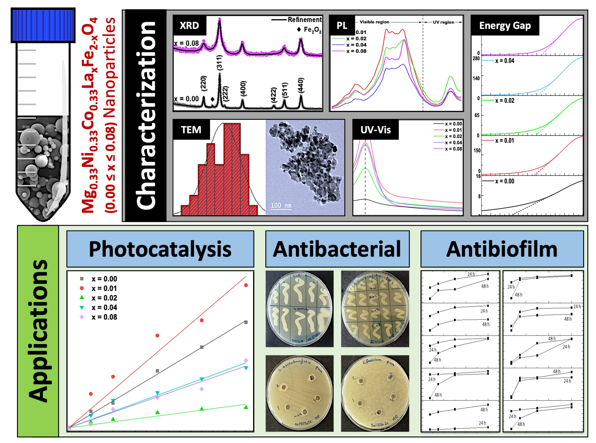
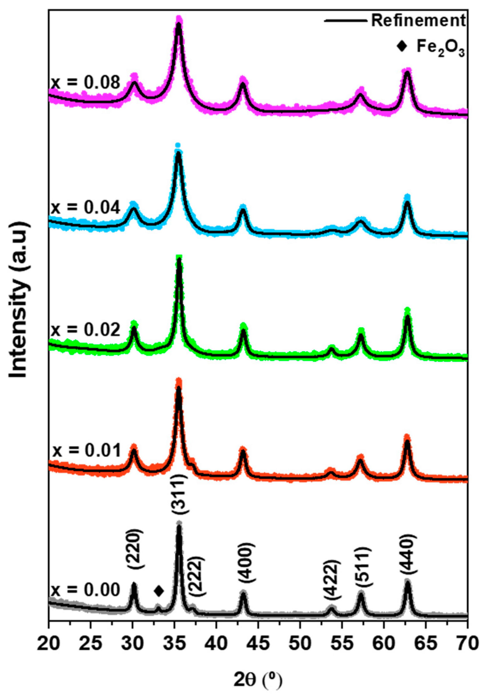
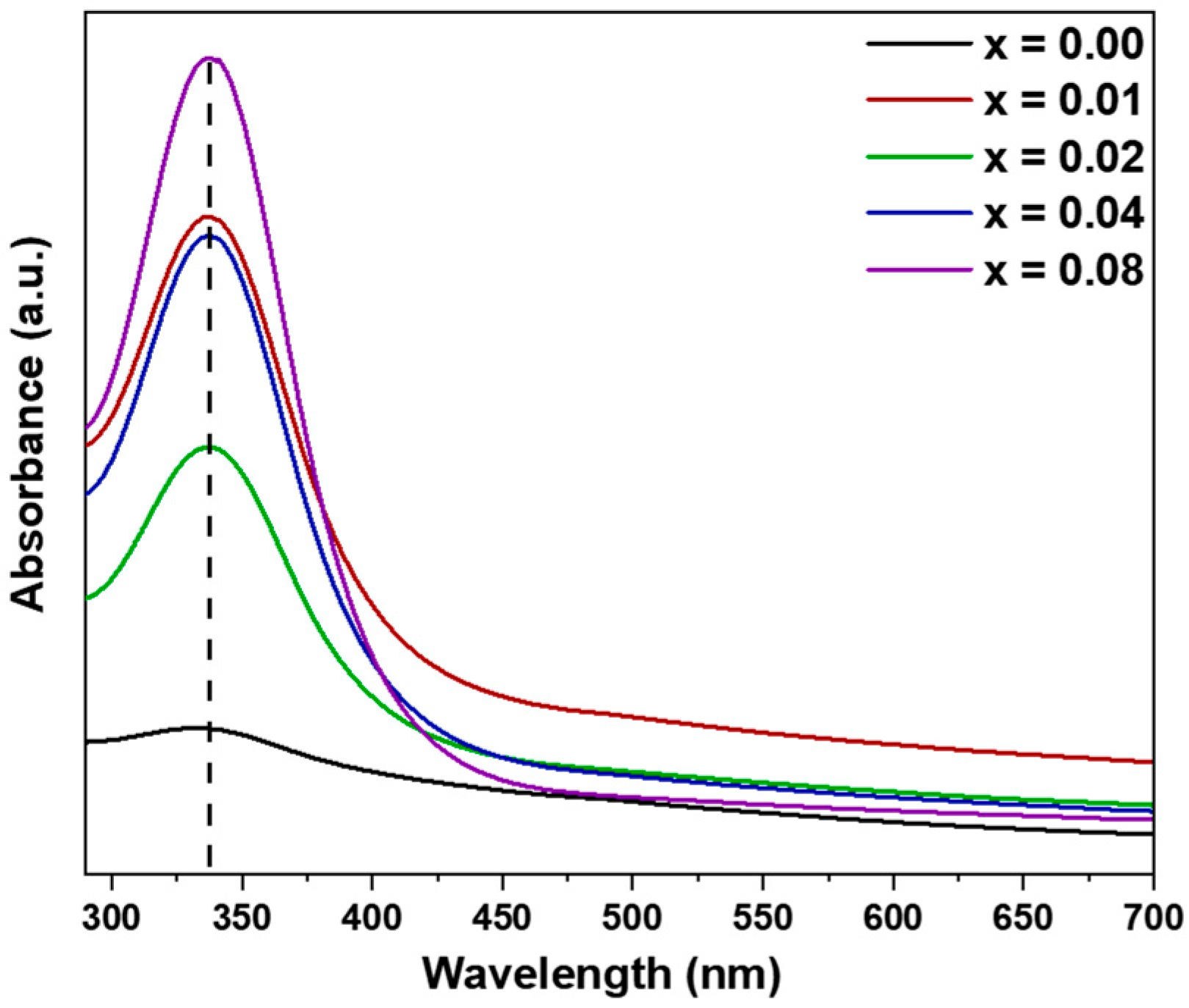
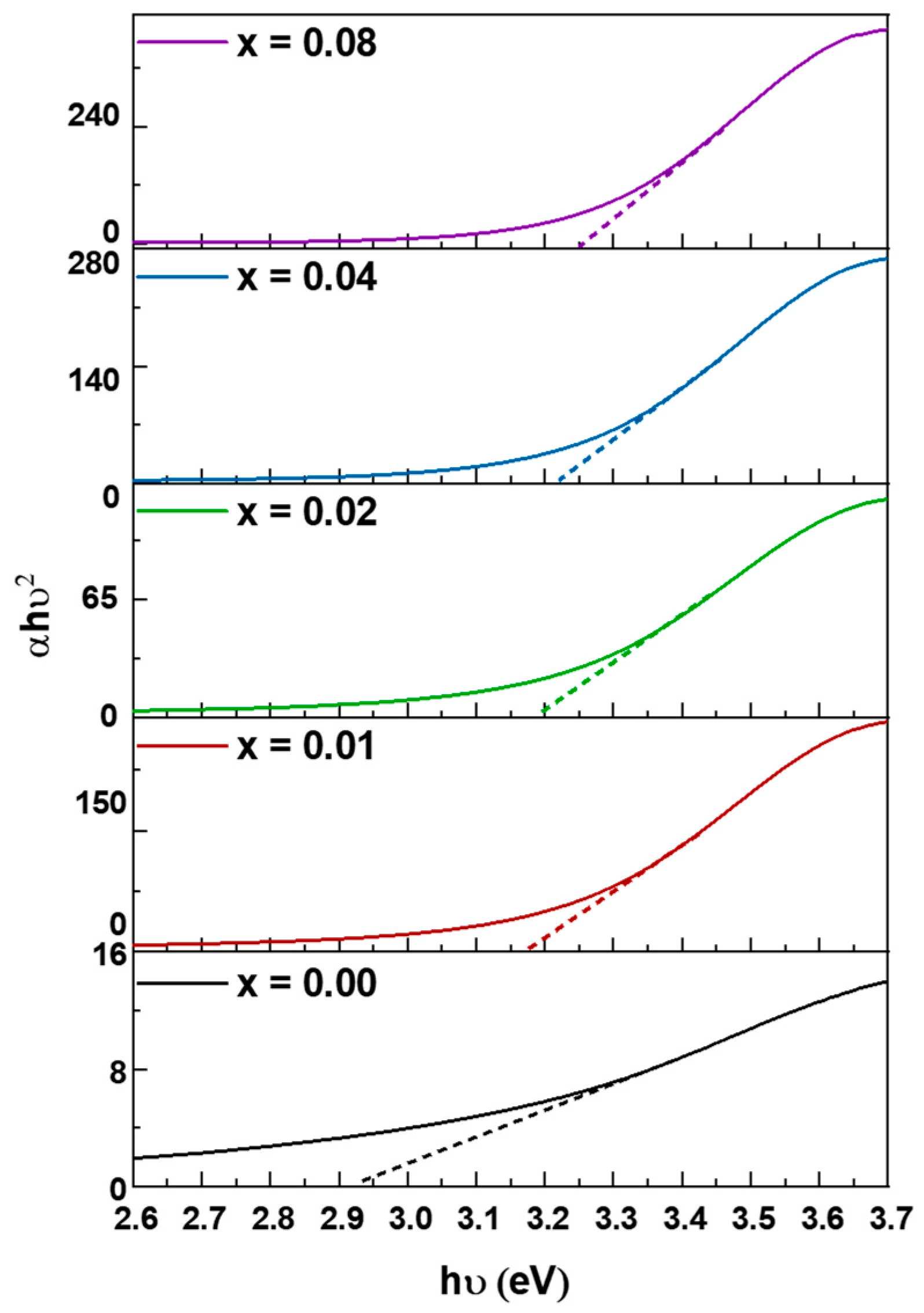
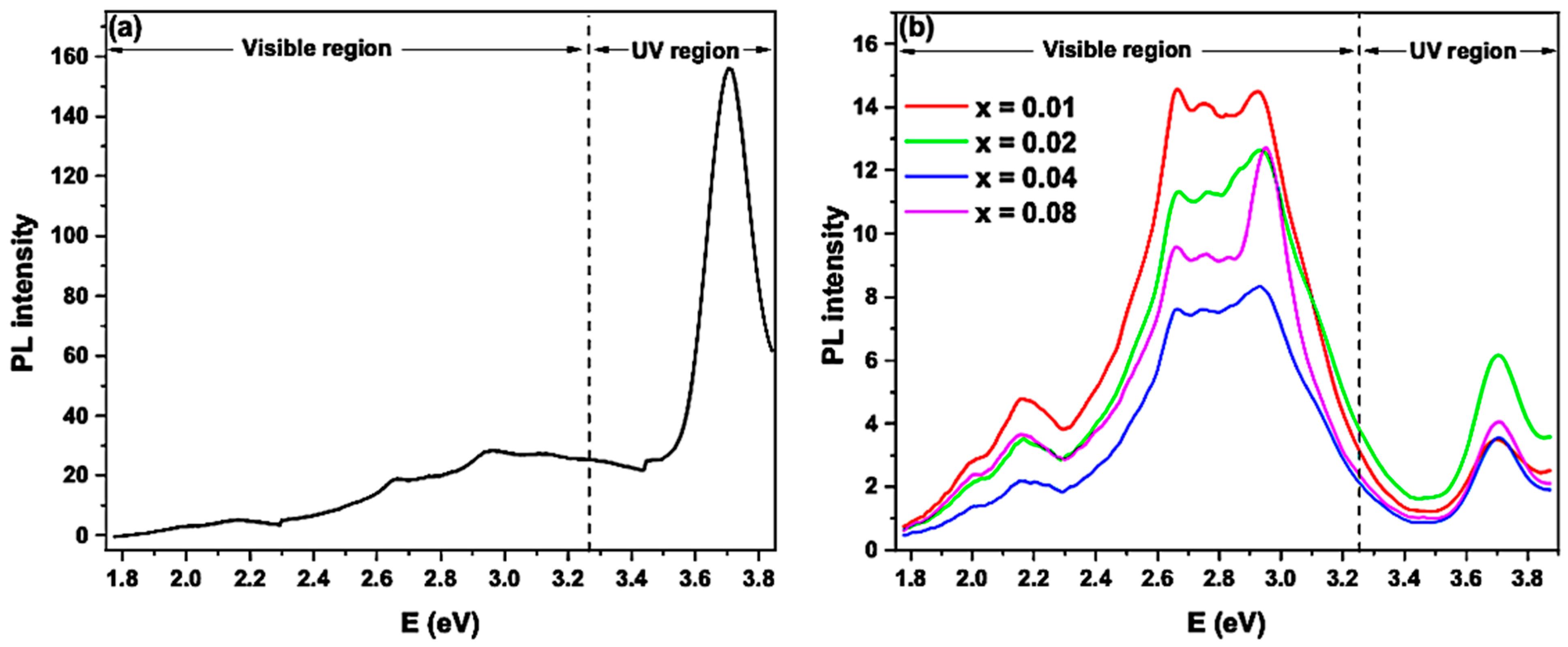
XRD patterns were recorded in the range of 20° ≤ 2θ ≤ 80° using Cu-kα radiation source ( λ   = 1.54056 Å) by X ray diffractometer (D8 Advance, Bruker, Billerica, United states). The morphology and size of the prepared samples were evaluated by TEM using JEM-1400 Plus (JEOL, Tokyo, Japan). To perform the UV test, 0.01 g of each of the prepared NPs was dissolved in 50 mL of 1 M HCl solution. Afterward, the mixtures were sonicated for 5 min. Subsequently, the optical properties of the samples were estimated by ultraviolet-visible (UV–Vis) spectroscopic examinations that were performed at room temperature in the range of 300–700 nm using UV-Vis spectrophotometer (V-670, JASCO, Tokyo, Japan). The PL spectra were recorded at room temperature between 1.8 and 3.9 eV at an excitation wavelength of 330 nm via a fluorescence spectrometer (FP8300, JASCO, Tokyo, Japan).

3.3. Photocatalytic Activity of the Mg0.33Ni0.33Co0.33LaxFe2−xO4 NPs

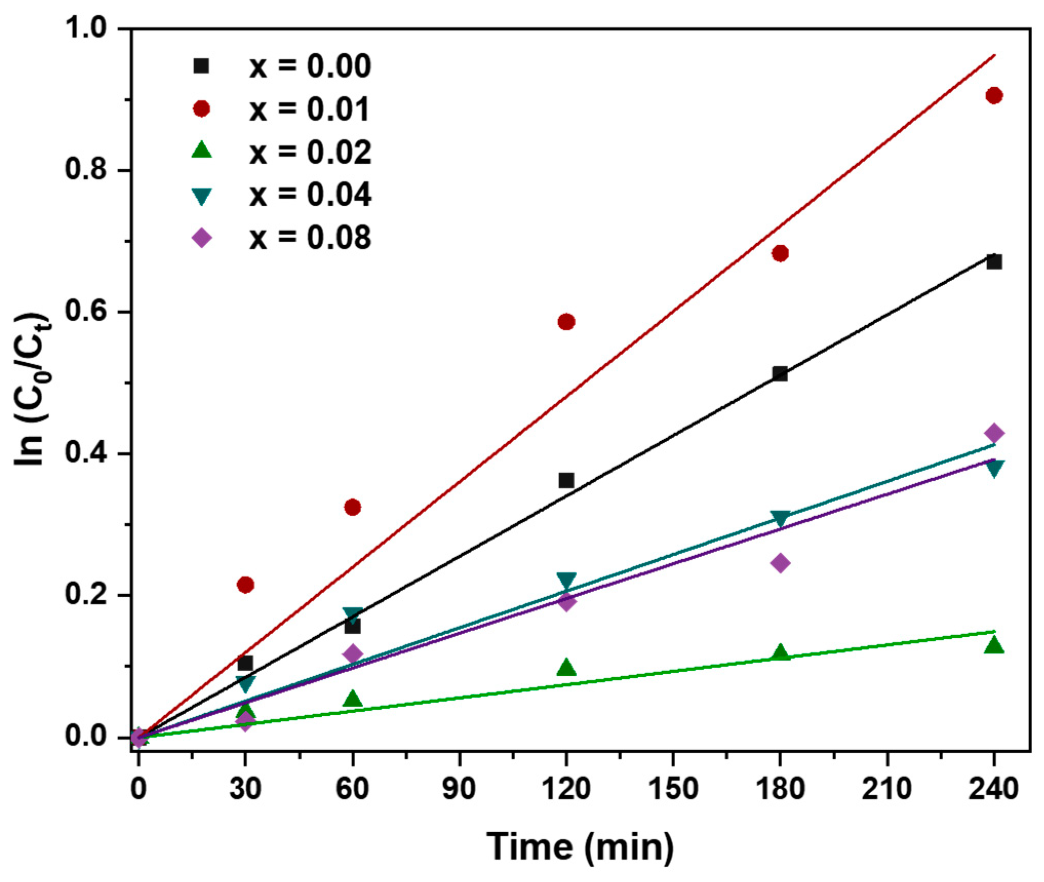
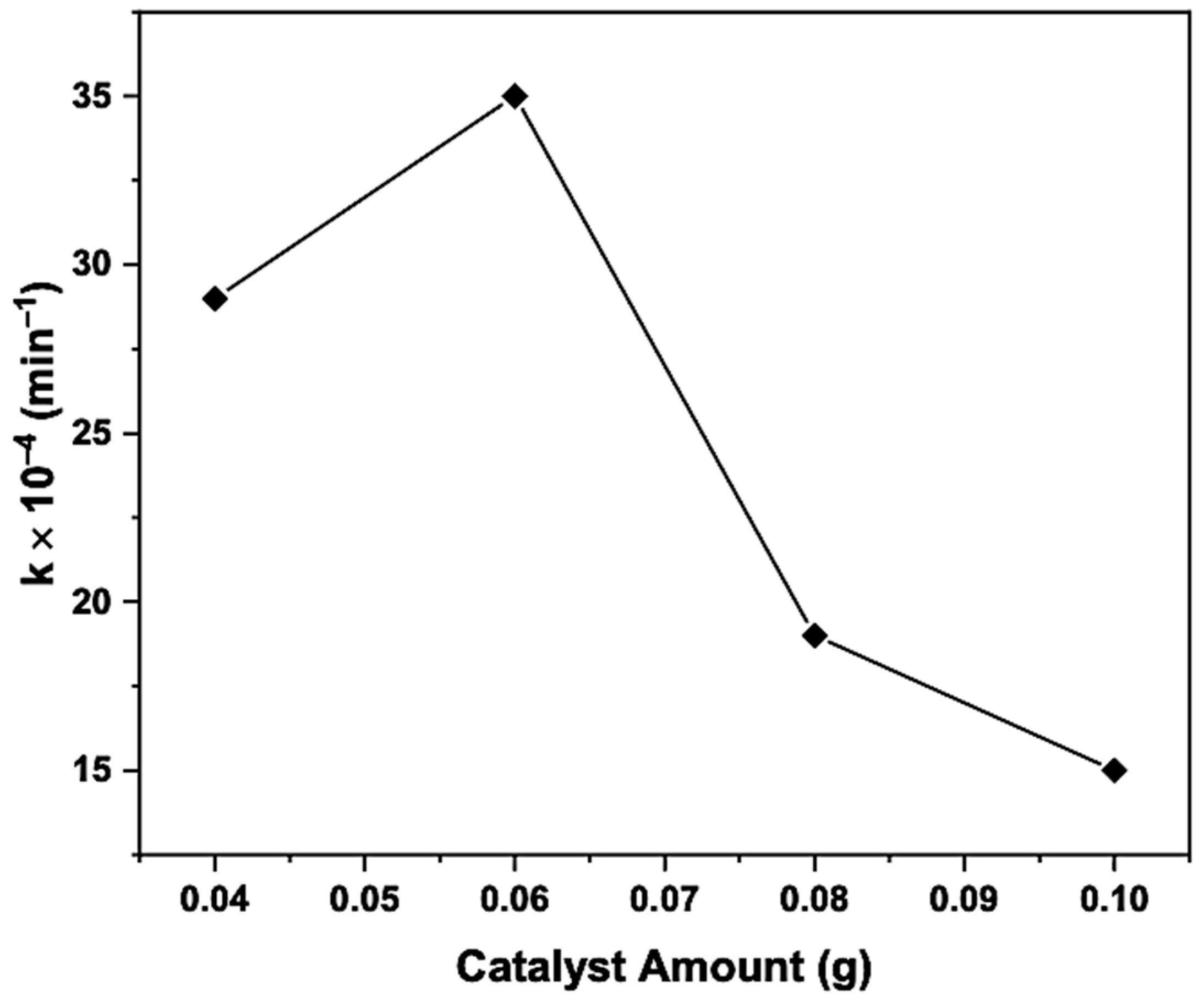
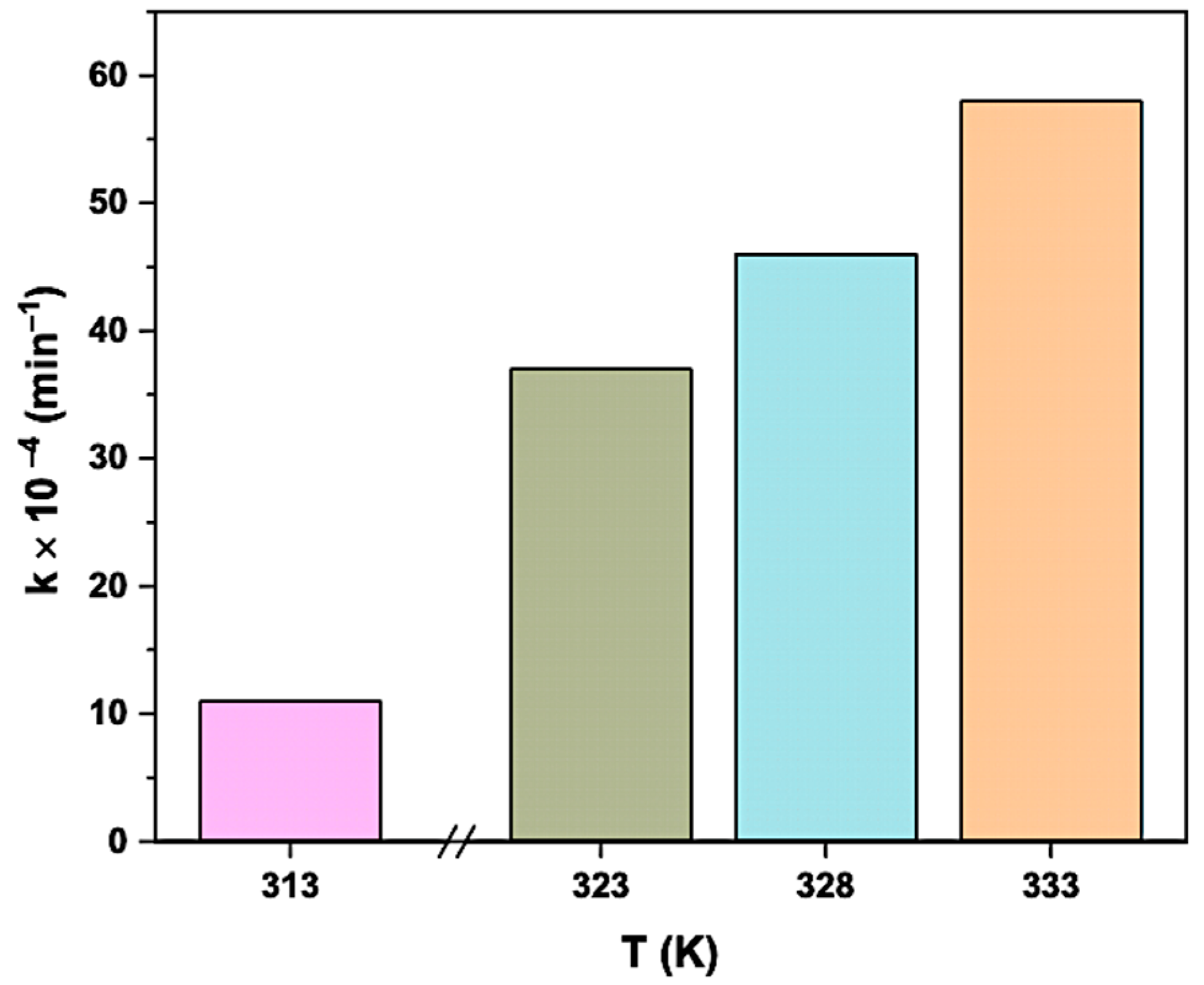
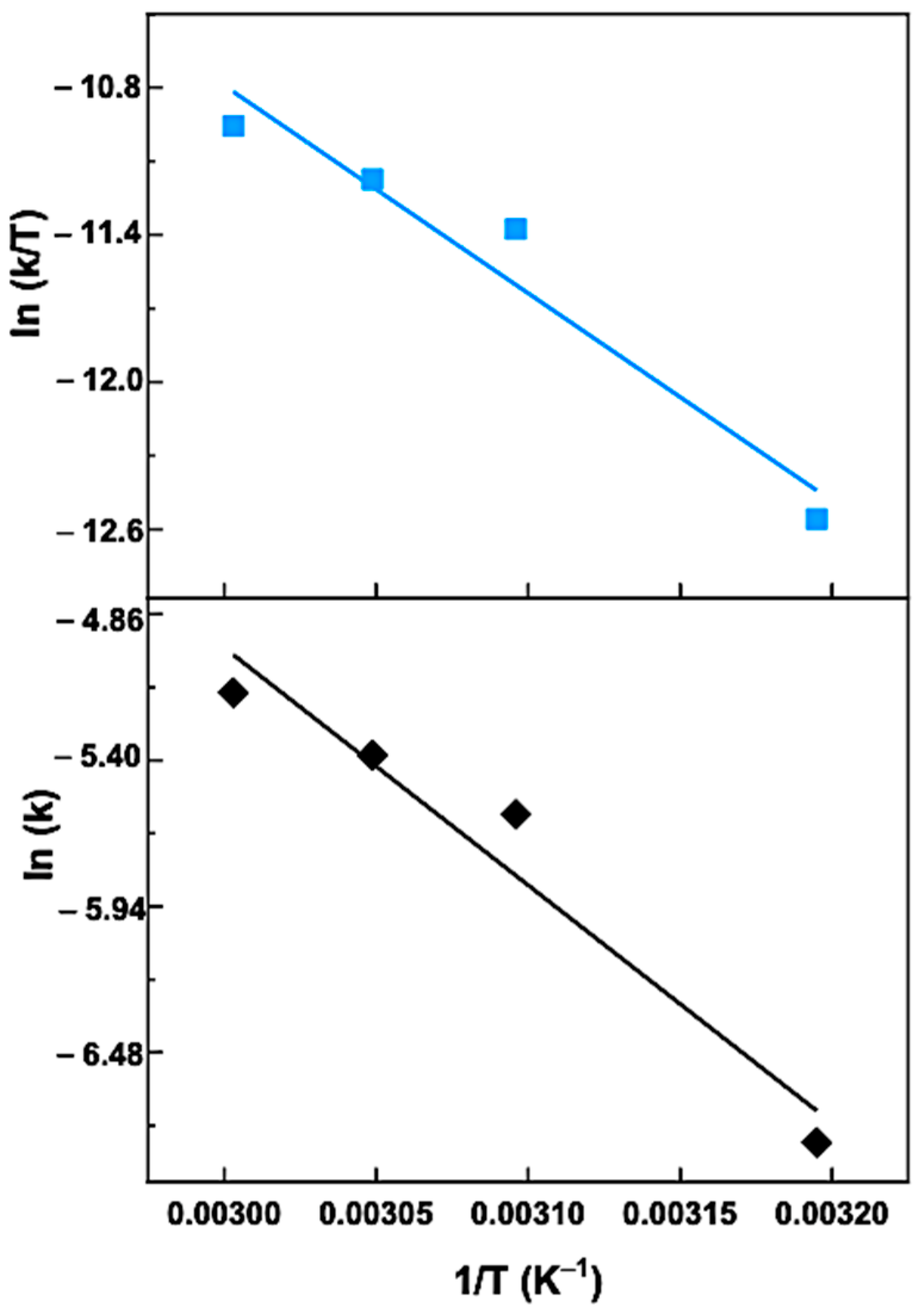
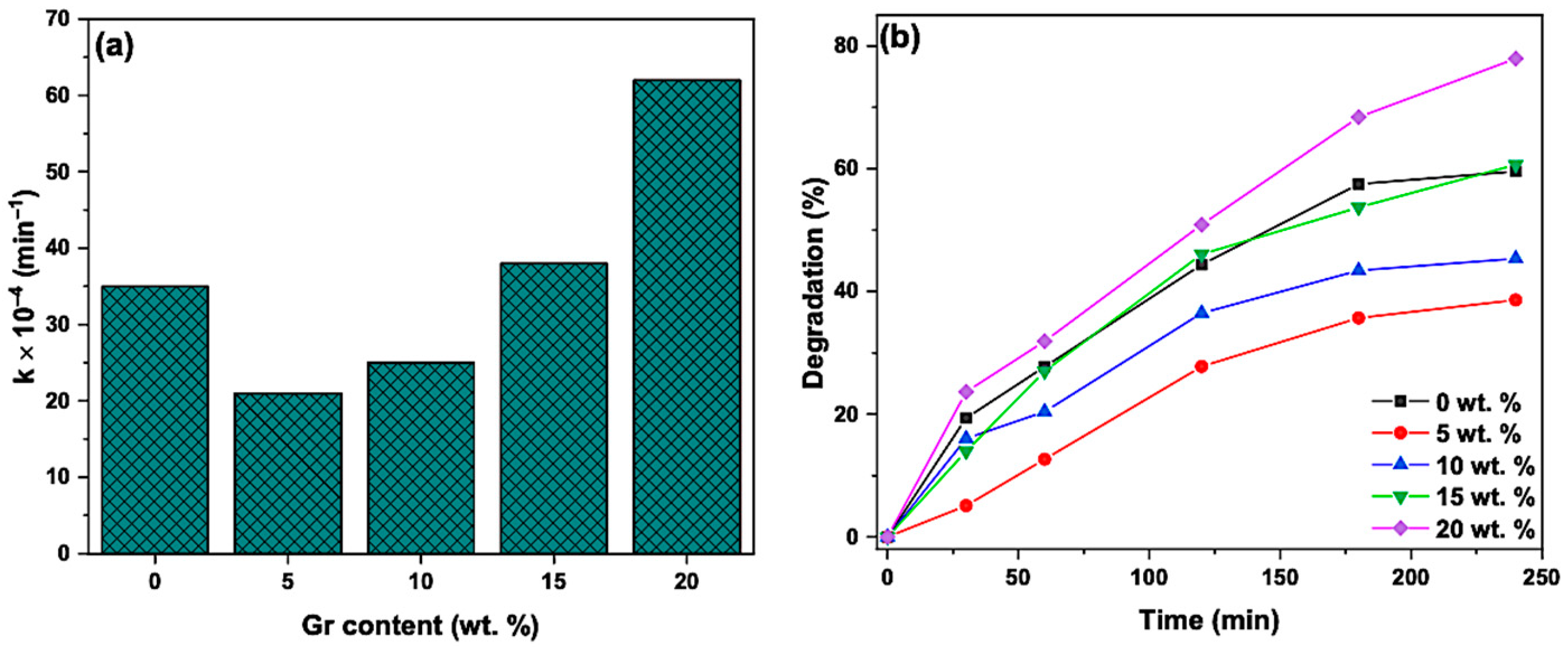
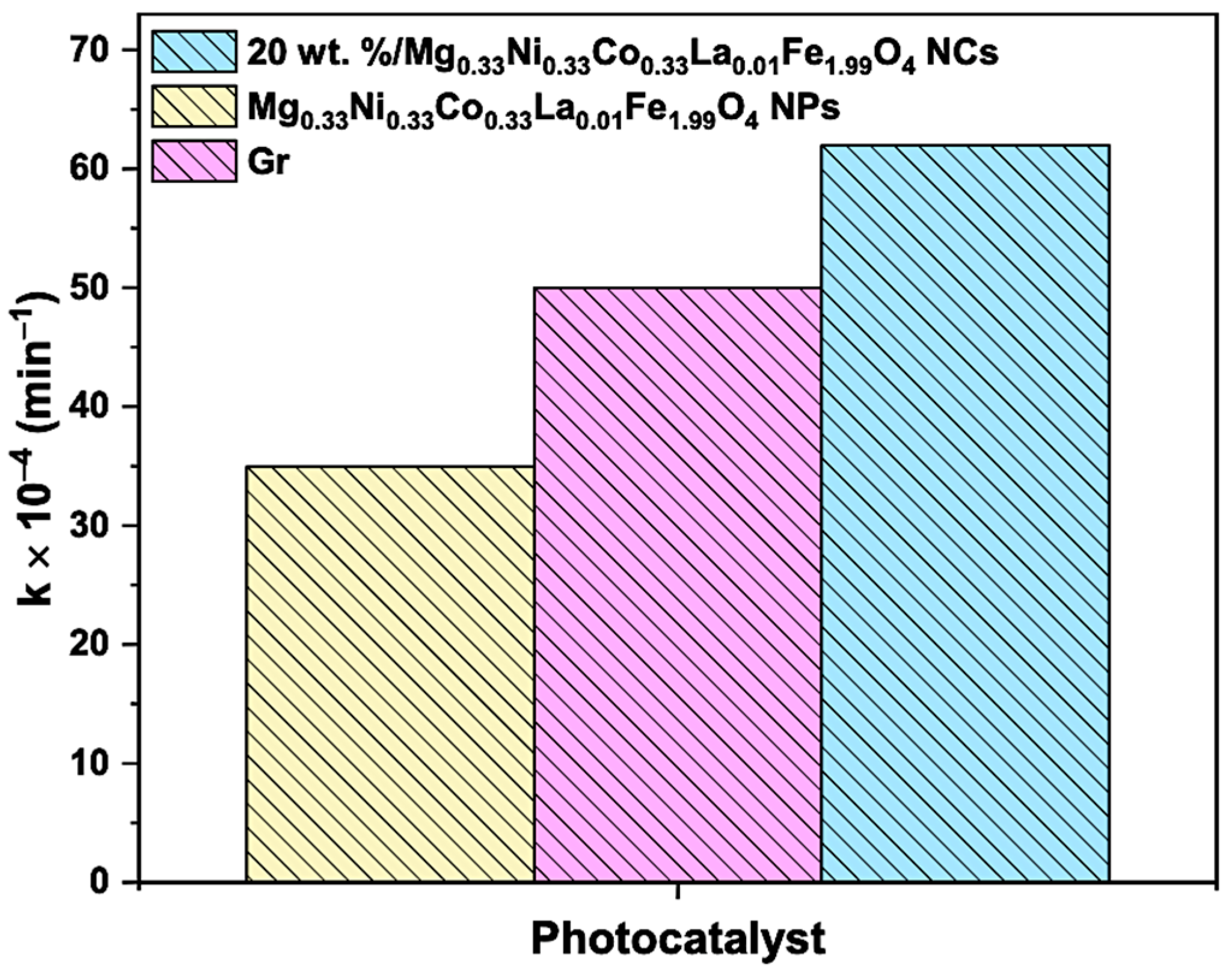
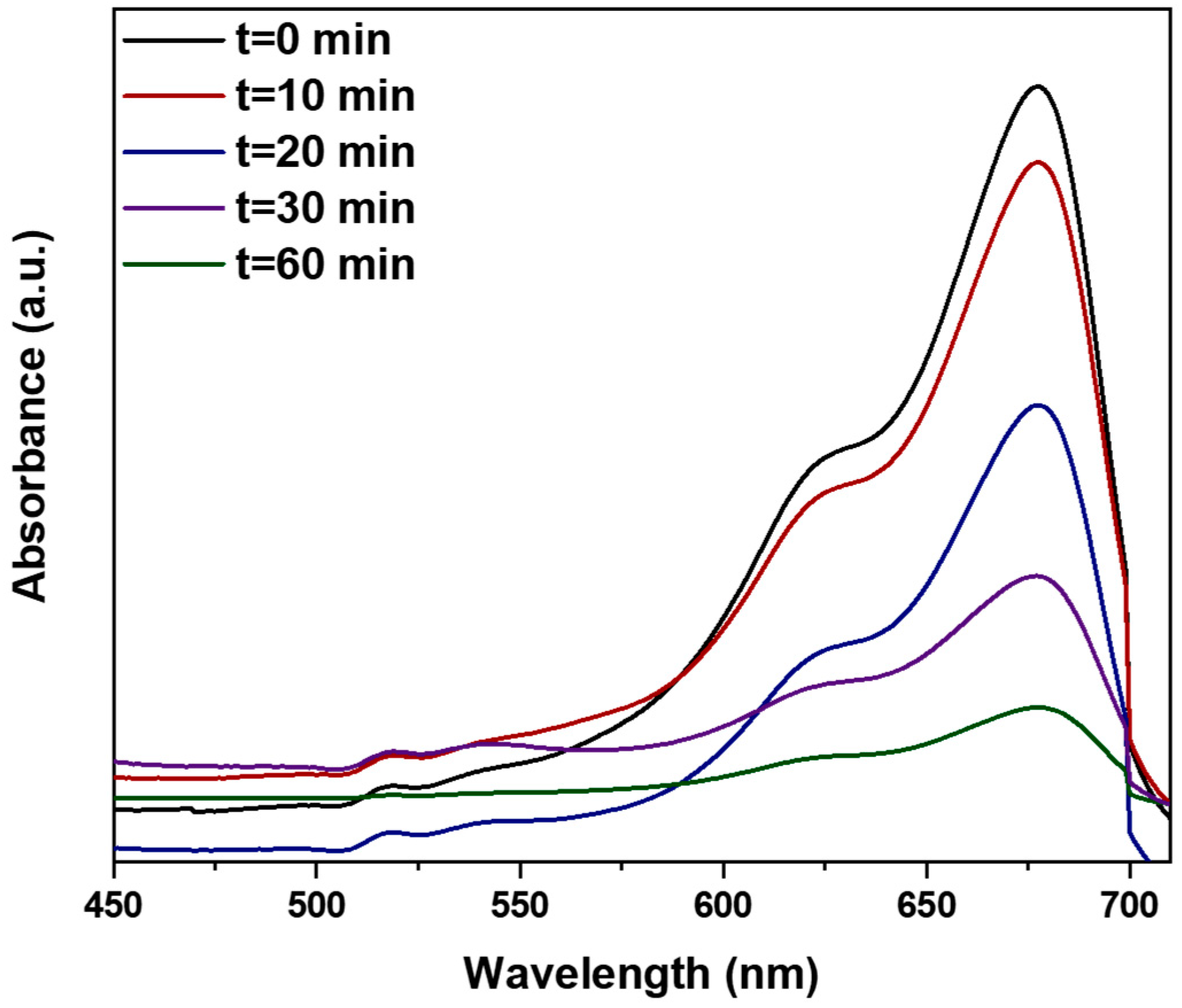
The photocatalytic activity of the Mg0.33Ni0.33Co0.33LaxFe2−xO4 NPs, where 0.00 ≤ x ≤ 0.08, was examined by mixing 0.06 g of each catalyst with 100 mL of 3 ppm methylene blue (MB) solution. The obtained solutions were then stirred for 30 min in the darkness to achieve the adsorption-desorption equilibrium. Then, the mixture was placed under direct sunlight between 11 am and 2 pm. The photodegradation reactions were performed in August. The effect of the catalyst dose, temperature, pH and graphene (Gr) addition was investigated. To study the effect of pH on the photodegradation reaction, a few drops of 0.5 M HCl and NaOH were added to adjust the pH of MB solution between 2 and 11. Furthermore, to prepare nanocomposites with 5, 10, 15 and 20 wt.% Gr, 0.003, 0.006, 0.009 and 0.012 g of Gr, respectively, were mixed with 0.06 g of the most efficient catalyst using 10 mL ethanol solution. Afterward, the mixture was sonicated for 20 min and placed in the oven at 70 °C to remove the ethanol. 3 mL of the MB solution was taken at different time intervals and analyzed using a UV-Vis spectrophotometer (SPECORD 200, Analytik Jena, Thuringia, Germany).

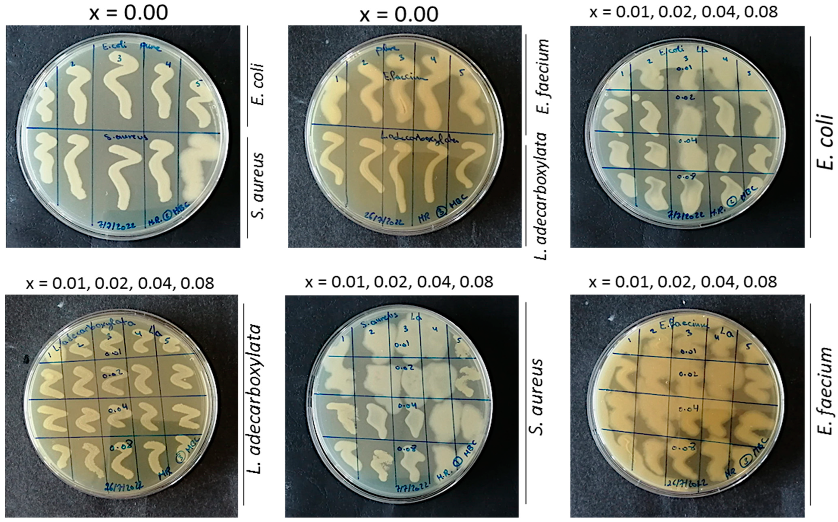
3.4. Antibacterial Activity of the Mg0.33Ni0.33Co0.33LaxFe2−xO4 NPs

3.4.1. Bacterial Isolation and Identification

Bacteria were isolated from wastewater samples. 100 µL of the isolated bacteria were spread on agar plates containing different selective media. The plates were then incubated at 37 °C for 24 h. Then, the bacterial isolates were Gram stained for differentiating between Gram-positive and Gram-negative bacteria prior to VITEK (VITEK 2 Automated Systems, bioMérieux Inc., Massachusetts, United states) for further identification of the bacterial species [62].

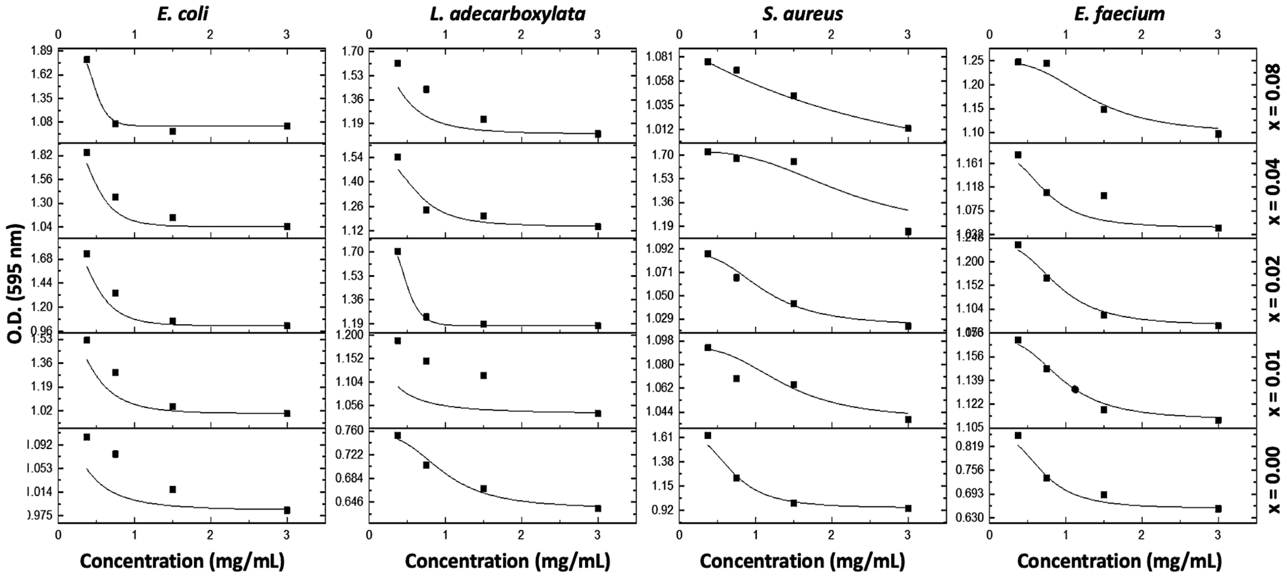
3.4.2. Determination of the Minimum Inhibitory Concentration (MIC) and Minimum Bactericidal Concentration (MBC) by the Microdilution Method

The MICs of Mg0.33Ni0.33Co0.33LaxFe2−xO4 NPs where 0.00 ≤ x ≤ 0.08 were determined by the micro-well dilution assay. The assay was done by dispensing 90 µL of nutrient broth (NB) into the wells of sterile 96-well microplates. Following that, 10 µL of previously prepared bacterial suspensions adjusted to 0.5 McFarland were added to the wells. Then, 100 µL of each NP of different concentrations (0.375–3 mg/mL) were added to the wells. The microplates were then incubated for 24 h at 37 °C. After incubation, the optical density (O.D.) was measured at 595 nm by an ELISA microtiter plate reader (SN 357-912359, Thermo Fisher Scientific, Shanghai, China). The MIC was recorded as the lowest concentration of the NPs that inhibited visible growth of the bacteria. Following the MIC recording, 10 µL of the solutions in the clear wells were spread on plates containing Muller Hinton agar (MHA) and incubated for 24 h at 37 °C for detecting the MBC [63]. All experiments were repeated at least three times.

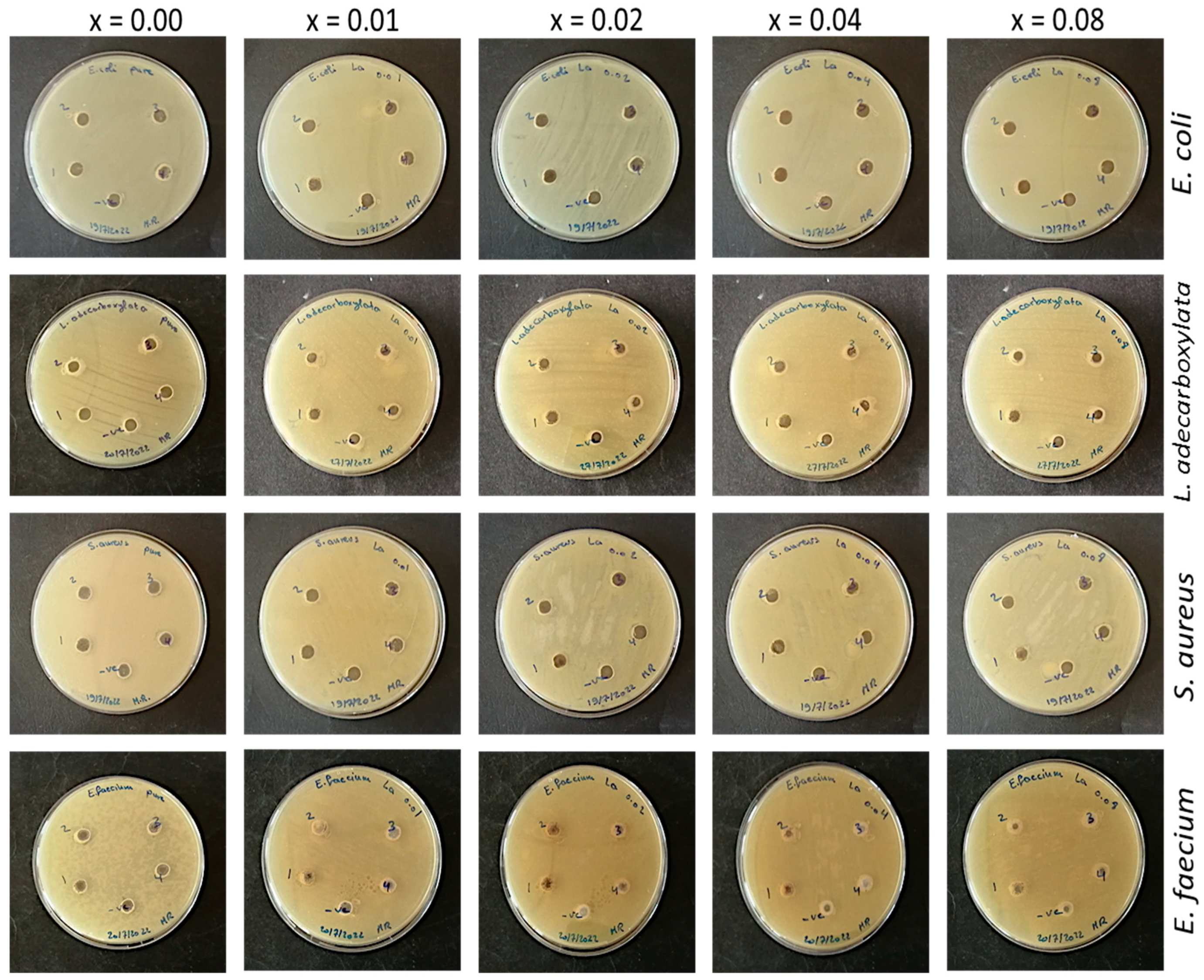
3.4.3. Determination of the Zones of Inhibition (ZOI) by the Agar Well Diffusion Assay

The agar well diffusion assay was performed by evenly spreading 100 µL of the isolated bacterial suspensions (0.5 McFarland) over the surface of the MHA plates. The plates were then punched with a 6 mm cork-borer to create wells. Then, 100 μL of increasing concentrations of each NP (0.375–3 mg/mL) were added to the wells. The plates were then incubated for 24 h at 37 °C. After incubation, the diameter of the ZOI was measured, in which ZOI > 7 mm was considered effective [64,65]. All experiments were repeated at least three times.

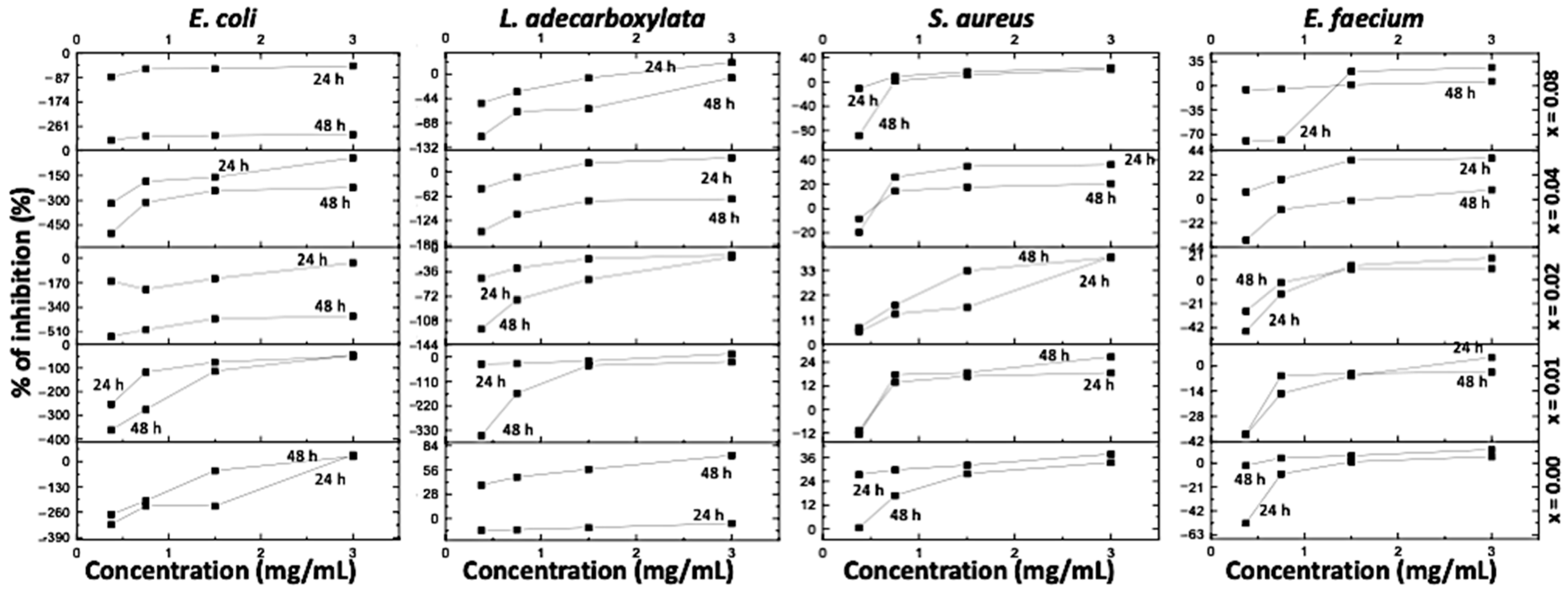
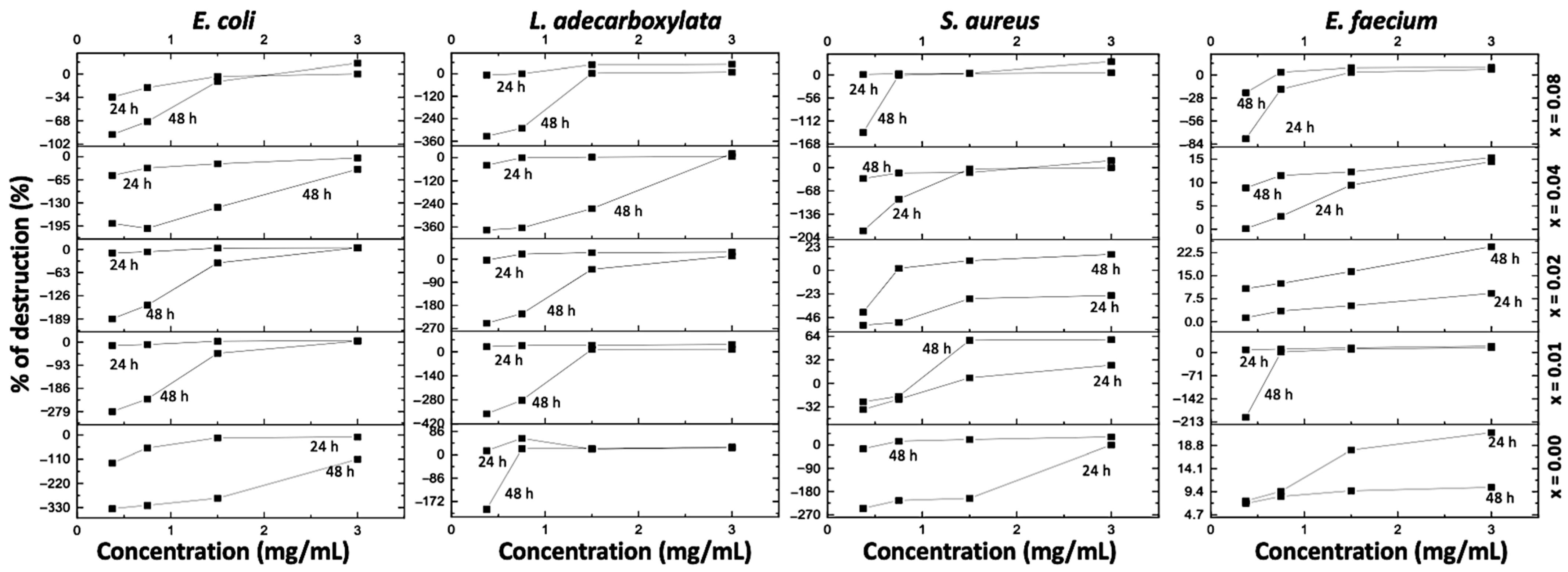
3.4.4. Antibiofilm Activity of the Mg0.33Ni0.33Co0.33LaxFe2−xO4 NPs

  • Inhibition of bacterial biofilm formation
The biofilm inhibition assay was done to determine the potential of Mg0.33Ni0.33Co0.33LaxFe2−xO4 NPs where 0.00 ≤ x ≤ 0.08 to prevent the formation of biofilms. The assay was done by adding 100 µL of bacterial cultures into 96-well microtiter plates and incubating for 4 h at 37 °C to allow the attachment/formation of the biofilms. Then, 100 µL of increasing concentrations of each NP (0.375–3 mg/mL) were added to the wells. A culture medium without any inoculum was used as a negative control. The plates were incubated at 37 °C for 24 and 48 h. The biomass was quantified by the crystal violet (CV) staining technique. Briefly, following the incubation period, the plates were washed with sterile distilled water 5 times, then air-dried and oven-dried for 15 min at 60 °C. Then, 100 µL of 1% CV was added to the wells and incubated for 15 min at room temperature. To remove the unabsorbed stain, the plates were washed with sterile distilled water 5 times and the biofilms were observed in the form of purple rings. To de-stain the wells, 100 µL of 95% ethanol was then added. Finally, the O.D. was measured at 595 nm using an ELISA microplate reader (SN 357-912359, Thermo Fisher Scientific, Shanghai, China) [66]. All experiments were repeated at least three times.
The percentage of inhibition of the formation of the bacterial biofilms was determined using the following equation:
%   Inhibition = O . D .   ( n e g a t i v e   c o n t r o l ) O . D . ( t r e a t e d ) O . D .   ( n e g a t i v e   c o n t r o l )   × 100
  • Eradication of pre-formed bacterial biofilms
The potential of the NPs to eradicate the pre-formed bacterial biofilms was tested by adding 100 µL of the standard cultures of the bacterial isolates into 96-well microtiter plates and incubating for 30 h at 37 °C to form the biofilms. After incubation, 100 µL of increasing concentrations of each NP (0.375–3 mg/mL) were added into the wells and the plates were incubated at 37 °C for 24 and 48 h. A culture medium without any inoculum was considered the negative control. The biomass of each biofilm was detected by CV staining, and the percentage of eradication of the pre-formed bacterial biofilms was determined as mentioned above in the inhibition of the formation of biofilms [66]. All experiments were repeated at least three times.

3.4.5. Statistical Analyses

The statistical tests were done using Excel software. The graphs were drawn using Origin software. The statistical significance was determined by t-Test.

4. Conclusions

Mg0.33Ni0.33Co0.33LaxFe2−xO4 NPs where 0.00 ≤ x ≤ 0.08 were successively synthesized by the co-precipitation method. As x increases from 0.00 to 0.08, Eg increases from 2.92 to 3.25 eV, whereas DTEM decreases from 28.72 to 20.20 nm. Doping Mg0.33Ni0.33Co0.33LaxFe2−xO4 NPs with La enhanced their photocatalytic performance. Among the prepared NPs, enhanced photocatalytic activity was exhibited by NPs where x = 0.01. This was attributed to the slow recombination rate of the electron-hole pair, as revealed by PL analysis. In addition, the optimal catalyst amount was 0.06 g. Furthermore, the incorporation of 20 wt.% of graphene (Gr) improved the photocatalytic activity of Mg0.33Ni0.33Co0.33La0.01Fe1.99O4 NPs. Among the tested NPs, the Mg0.33Ni0.33Co0.33La0.00Fe2O4 NPs had antibacterial activity. An antibiofilm action was observed for some of the doped NPs, especially those where x = 0.01, 0.02 and 0.04, mainly against biofilms of Gram-positive bacteria. Due to their high antibiofilm activity, the Mg0.33Ni0.33Co0.33La0.00Fe2−xO4 NPs are applicable in the biomedical field, especially against bacterial pathogens.

Author Contributions

Conceptualization, D.N., M.I.K., R.A. and W.A.; Data curation, M.R., M.M. and A.A.; Formal analysis, M.R., M.M., A.A. and M.I.K.; Investigation, M.R., M.M., A.A., D.N., M.I.K., R.A. and W.A.; Methodology, M.R., M.M., A.A., D.N., M.I.K. and R.A.; Resources, D.N., M.I.K., R.A. and W.A.; Software, M.M. and A.A.; Supervision, D.N., M.I.K., R.A. and W.A.; Validation, M.I.K. and R.A.; Writing—original draft, M.R., M.M. and A.A.; Writing—review & editing, M.R., M.M., A.A., D.N., M.I.K., R.A. and W.A. All authors have read and agreed to the published version of the manuscript.

Funding

This research received no external funding.

Institutional Review Board Statement

Not applicable.

Informed Consent Statement

Not applicable.

Data Availability Statement

The data supporting the reported results are available with the corresponding author and will be provided upon request.

Conflicts of Interest

The authors declare no conflict of interest.

Appendix A

Figure A1. MICs of the Mg0.33Ni0.33Co0.33LaxFe2−xO4 NPs where 0.00 ≤ x ≤ 0.08.
Catalysts 13 00693 g0a1
Figure A2. Inhibition of the formation of bacterial biofilms by the Mg0.33Ni0.33Co0.33LaxFe2−xO4 NPs where 0.00 ≤ x ≤ 0.08.
Catalysts 13 00693 g0a2
Figure A3. Eradication of the pre-formed bacterial biofilms by the Mg0.33Ni0.33Co0.33LaxFe2−xO4 NPs where 0.00 ≤ x ≤ 0.08.
Catalysts 13 00693 g0a3
Figure A4. MBCs of the Mg0.33Ni0.33Co0.33LaxFe2−xO4 NPs where 0.00 ≤ x ≤ 0.08.
Catalysts 13 00693 g0a4
Figure A5. Agar well diffusion results of the Mg0.33Ni0.33Co0.33LaxFe2−xO4 NPs where 0.00 ≤ x ≤ 0.08.
Catalysts 13 00693 g0a5
Table A1. p-values and significance levels of the agar well diffusion results of the Mg0.33Ni0.33Co0.33LaxFe2−xO4 NPs where 0.00 ≤ x ≤ 0.08.
NanoparticlesBacterial Isolates
Gram-Negative BacteriaGram-Positive Bacteria
Escherichia coliSL. eclerciaadecarboxylataSStaphylococcus aureusSEnterococcus faeciumS
SampleConcentration (mg/mL)p-Values (vs. Water)
x = 0.000.3760.005**0.002**0.003**0.002**
0.750.003**0.001***0.002**0.002**
1.50.002**0.001***0.001***0.001***
3<0.001***<0.001***<0.001***0.001***
x = 0.010.3750.05*0.002**0.001***0.02*
0.750.03**0.003**0.002**0.002**
1.50.02**0.001***0.001***0.01**
30.01**0.001***0.03*0.001***
x = 0.020.3750.005**0.002**0.003**0.002**
0.750.003**0.001***0.002**0.03*
1.50.02*0.001***0.001***0.001***
3<0.001***0.001***<0.001***0.01**
x = 0.040.3750.005**0.002**0.003**0.002**
0.750.003**0.001***0.002**0.002**
1.50.003**0.001***0.01**0.05*
3<0.001***<0.001***0.001***0.001***
x = 0.080.3750.005**0.002**0.003**0.003**
0.750.02*<0.001***0.002**0.02*
1.50.002**0.001***<0.001***0.001***
3<0.001***<0.001***0.01**0.01**
S: significance, p-values were calculated such that: * p < 0.05, ** p < 0.01, *** p < 0.001.

References

  1. Elgarahy, A.; Elwakeel, K.; Mohammad, S.; Elshoubaky, G. A critical review of biosorption of dyes, heavy metals and metalloids from wastewater as an efficient and green process. Clean. Eng. Technol. 2021, 4, 100209. [Google Scholar]
  2. Rafiq, A.; Ikram, M.; Ali, S.; Niaz, F.; Khan, M.; Khan, Q.; Maqbool, M. Photocatalytic degradation of dyes using semiconductor photocatalysts to clean industrial water pollution. J. Ind. Eng. Chem. 2021, 97, 111–128. [Google Scholar]
  3. Some, S.; Mondal, R.; Mitra, D.; Jain, D.; Verma, D.; Das, S. Microbial pollution of water with special reference to coliform bacteria and their nexus with environment. Energy Nexus 2021, 1, 100008. [Google Scholar]
  4. Wallyn, J.; Anton, N.; Vandamme, T.F. Synthesis, principles, and properties of magnetite nanoparticles for in vivo imaging applications—A review. Pharmaceutics 2019, 11, 601. [Google Scholar] [CrossRef] [Green Version]
  5. Astruc, D. Introduction: Nanoparticles in Catalysis; ACS Publications: Washington, DC, USA, 2020; Volume 120, pp. 461–463. [Google Scholar]
  6. Menazea, A.; Ahmed, M. Silver and copper oxide nanoparticles-decorated graphene oxide via pulsed laser ablation technique: Preparation, characterization, and photoactivated antibacterial activity. Nano-Struct. Nano-Objects 2020, 22, 100464. [Google Scholar]
  7. Hajalilou, A.; Mazlan, S.A. A review on preparation techniques for synthesis of nanocrystalline soft magnetic ferrites and investigation on the effects of microstructure features on magnetic properties. Appl. Phys. A 2016, 122, 1–15. [Google Scholar]
  8. Vedrtnam, A.; Kalauni, K.; Dubey, S.; Kumar, A. A comprehensive study on structure, properties, synthesis and characterization of ferrites. AIMS Mater. Sci. 2020, 7, 800–835. [Google Scholar] [CrossRef]
  9. Soufi, A.; Hajjaoui, H.; Elmoubarki, R.; Abdennouri, M.; Qourzal, S.; Barka, N. Spinel ferrites nanoparticles: Synthesis methods and application in heterogeneous fenton oxidation of organic pollutants—A review. Appl. Surf. Sci. Adv. 2021, 6, 100145. [Google Scholar]
  10. Aridi, A.; Naoufal, D.; El-Rassy, H.; Awad, R. Photocatalytic activity of znfe2o4/nio nanocomposites carried out under uv irradiation. Ceram. Int. 2022, 48, 30905–30916. [Google Scholar]
  11. Keerthana, S.; Yuvakkumar, R.; Kumar, P.S.; Ravi, G.; Velauthapillai, D. Rare earth metal (sm) doped zinc ferrite (znfe2o4) for improved photocatalytic elimination of toxic dye from aquatic system. Environ. Res. 2021, 197, 111047. [Google Scholar]
  12. Mariosi, F.R.; Venturini, J.; da Cas Viegas, A.; Bergmann, C.P. Lanthanum-doped spinel cobalt ferrite (cofe2o4) nanoparticles for environmental applications. Ceram. Int. 2020, 46, 2772–2779. [Google Scholar]
  13. Reddy, B.P.; Sekhar, M.C.; Prakash, B.P.; Suh, Y.; Park, S.-H. Photocatalytic, magnetic, and electrochemical properties of la doped bifeo3 nanoparticles. Ceram. Int. 2018, 44, 19512–19521. [Google Scholar]
  14. Anitha, S.; Muthukumaran, S. Structural, optical and antibacterial investigation of la, cu dual doped zno nanoparticles prepared by co-precipitation method. Mater. Sci. Eng. C 2020, 108, 110387. [Google Scholar]
  15. Wang, M.; Su, Y.; Liu, Y.; Liang, Y.; Wu, S.; Zhou, N.; Shen, J. Antibacterial fluorescent nano-sized lanthanum-doped carbon quantum dot embedded polyvinyl alcohol for accelerated wound healing. J. Colloid Interface Sci. 2022, 608, 973–983. [Google Scholar]
  16. De, D.; Mandal, S.M.; Gauri, S.S.; Bhattacharya, R.; Ram, S.; Roy, S.K. Antibacterial effect of lanthanum calcium manganate (la0. 67ca0. 33mno3) nanoparticles against pseudomonas aeruginosa atcc 27853. J. Biomed. Nanotechnol. 2010, 6, 138–144. [Google Scholar] [CrossRef]
  17. Zeng, X.; Hou, Z.; Ju, J.; Gao, L.; Zhang, J.; Peng, Y. The cation distributions of zn-doped normal spinel mgfe2o4 ferrite and its magnetic properties. Materials 2022, 15, 2422. [Google Scholar] [CrossRef]
  18. Naveed-Ul-Haq, M.; Hussain, S. Pressure-induced structural, electronic, and magnetic evolution in cubic inverse spinel nife2o4, an ab-initio study. Appl. Phys. A 2022, 128, 30. [Google Scholar]
  19. Monisha, P.; Priyadharshini, P.; Gomathi, S.; Pushpanathan, K. Influence of mn dopant on the crystallite size, optical and magnetic behaviour of cofe2o4 magnetic nanoparticles. J. Phys. Chem. Solids 2021, 148, 109654. [Google Scholar]
  20. Ajeesha, T.; Ashwini, A.; George, M.; Manikandan, A.; Mary, J.A.; Slimani, Y.; Almessiere, M.; Baykal, A. Nickel substituted mgfe2o4 nanoparticles via co-precipitation method for photocatalytic applications. Phys. B Condens. Matter 2021, 606, 412660. [Google Scholar]
  21. Xueyun, Z.; Dongsheng, Y.; Liling, Z. Improved cut-off frequency in gd/la doped niznco ferrites. Mater. Sci. Eng. B 2021, 272, 115334. [Google Scholar]
  22. de Medeiros, F.; Madigou, V.; Lopes-Moriyama, A.; de Souza, C.P.; Leroux, C. Synthesis of cofe2o4 nanocubes. Nano-Struct. Nano-Objects 2020, 21, 100422. [Google Scholar]
  23. Geetha, K.; Udhayakumar, R.; Manikandan, A. Enhanced magnetic and photocatalytic characteristics of cerium substituted spinel mgfe2o4 ferrite nanoparticles. Phys. B Condens. Matter 2021, 615, 413083. [Google Scholar]
  24. Hariharasuthan, R.; Chitradevi, S.; Radha, K.; Chithambaram, V. Characterization of nife2o4 (nickel ferrite) nanoparticles with very low magnetic saturation synthesized via co-precipitation method. Appl. Phys. A 2022, 128, 1045. [Google Scholar] [CrossRef]
  25. Zhou, X.; Zhou, Y.; Zhou, L.; Wei, J.; Wu, J.; Yao, D. Effect of gd and la doping on the structure, optical and magnetic properties of niznco ferrites. Ceram. Int. 2019, 45, 6236–6242. [Google Scholar]
  26. Chavan, P.; Naik, L. Investigation of energy band gap and conduction mechanism of magnesium substituted nickel ferrite nanoparticles. Phys. Status Solidi A 2017, 214, 1700077. [Google Scholar] [CrossRef]
  27. Elthair, N.A.; Mustafa, E.M.; Elbadawi, A.A. The electrical and optical properties of zn0. 5li2xmg0. 5-xfe2o4 lithium doped nanoparticle prepared by coprecipitation method. Open J. Appl. Sci. 2020, 10, 551–560. [Google Scholar]
  28. Lemziouka, H.; Boutahar, A.; Moubah, R.; Omari, L.; Bahhar, S.; Abid, M.; Lassri, H. Synthesis, structural, optical and dispersion parameters of la-doped spinel zinc ferrites znfe2-xlaxo4 (x = 0.00, 0.001, 0.005, 0.01 and 0.015). Vacuum 2020, 182, 109780. [Google Scholar] [CrossRef]
  29. Roni, M.M.; Hoque, K.; Paul, T.C.; Khan, M.; Hossain, M.E. Synthesis of la-doped mn0· 6zn0. 4laxfe2-xo4 and the study of its structural, electrical and magnetic properties for high frequency applications. Results Mater. 2021, 11, 100215. [Google Scholar] [CrossRef]
  30. Palaniappan, P.; Lenin, N.; Uvarani, R. Copper substitution effect on the structural, electrical, and magnetic properties of manganese and lanthanum (mn1− xcuxla0. 1fe1. 9o4) nanoferrites. J. Alloy. Compd. 2022, 925, 166717. [Google Scholar] [CrossRef]
  31. Alkaykh, S.; Mbarek, A.; Ali-Shattle, E.E. Photocatalytic degradation of methylene blue dye in aqueous solution by mntio3 nanoparticles under sunlight irradiation. Heliyon 2020, 6, e03663. [Google Scholar] [CrossRef]
  32. Wang, C.; Wu, D.; Wang, P.; Ao, Y.; Hou, J.; Qian, J. Effect of oxygen vacancy on enhanced photocatalytic activity of reduced zno nanorod arrays. Appl. Surf. Sci. 2015, 325, 112–116. [Google Scholar]
  33. Nie, J.; Zhu, G.; Zhang, W.; Gao, J.; Zhong, P.; Xie, X.; Huang, Y.; Hojamberdiev, M. Oxygen vacancy defects-boosted deep oxidation of no by β-bi2o3/ceo2-δ pn heterojunction photocatalyst in situ synthesized from bi/ce (co3)(oh) precursor. Chem. Eng. J. 2021, 424, 130327. [Google Scholar]
  34. Bharathi, P.; Harish, S.; Archana, J.; Navaneethan, M.; Ponnusamy, S.; Muthamizhchelvan, C.; Shimomura, M.; Hayakawa, Y. Enhanced charge transfer and separation of hierarchical cuo/zno composites: The synergistic effect of photocatalysis for the mineralization of organic pollutant in water. Appl. Surf. Sci. 2019, 484, 884–891. [Google Scholar]
  35. Khan, I.; Saeed, K.; Zekker, I.; Zhang, B.; Hendi, A.H.; Ahmad, A.; Ahmad, S.; Zada, N.; Ahmad, H.; Shah, L.A. Review on methylene blue: Its properties, uses, toxicity and photodegradation. Water 2022, 14, 242. [Google Scholar]
  36. Nagaraja, R.; Kottam, N.; Girija, C.; Nagabhushana, B. Photocatalytic degradation of rhodamine b dye under uv/solar light using zno nanopowder synthesized by solution combustion route. Powder Technol. 2012, 215, 91–97. [Google Scholar] [CrossRef]
  37. Aridi, A.; Naoufal, D.; El-Rassy, H.; Awad, R. Preparation and characterization of znfe2o4/mn2o3 nanocatalysts for the degradation of nitrobenzene. Chem. Afr. 2023, 1–14. [Google Scholar] [CrossRef]
  38. Hoijang, S.; Wangkarn, S.; Ieamviteevanich, P.; Pinitsoontorn, S.; Ananta, S.; Lee, T.R.; Srisombat, L. Silica-coated magnesium ferrite nanoadsorbent for selective removal of methylene blue. Colloids Surf. A Physicochem. Eng. Asp. 2020, 606, 125483. [Google Scholar]
  39. Fathy, M.A.; Kamel, A.H.; Hassan, S.S. Novel magnetic nickel ferrite nanoparticles modified with poly (aniline-co-o-toluidine) for the removal of hazardous 2, 4-dichlorophenol pollutant from aqueous solutions. RSC Adv. 2022, 12, 7433–7445. [Google Scholar]
  40. Nassar, M.Y.; Mohamed, T.Y.; Ahmed, I.S.; Mohamed, N.M.; Khatab, M. Hydrothermally synthesized co 3 o 4, α-fe 2 o 3, and cofe 2 o 4 nanostructures: Efficient nano-adsorbents for the removal of orange g textile dye from aqueous media. J. Inorg. Organomet. Polym. Mater. 2017, 27, 1526–1537. [Google Scholar]
  41. Makofane, A.; Motaung, D.E.; Hintsho-Mbita, N.C. Photocatalytic degradation of methylene blue and sulfisoxazole from water using biosynthesized zinc ferrite nanoparticles. Ceram. Int. 2021, 47, 22615–22626. [Google Scholar]
  42. Nuengmatcha, P.; Porrawatkul, P.; Chanthai, S.; Sricharoen, P.; Limchoowong, N. Enhanced photocatalytic degradation of methylene blue using fe2o3/graphene/cuo nanocomposites under visible light. J. Environ. Chem. Eng. 2019, 7, 103438. [Google Scholar]
  43. Shelar, S.G.; Mahajan, V.K.; Patil, S.P.; Sonawane, G.H. Effect of doping parameters on photocatalytic degradation of methylene blue using ag doped zno nanocatalyst. SN Appl. Sci. 2020, 2, 1–10. [Google Scholar]
  44. Ahmed, M.A.; Mohamed, A.A. Recent progress in semiconductor/graphene photocatalysts: Synthesis, photocatalytic applications, and challenges. RSC Adv. 2023, 13, 421–439. [Google Scholar]
  45. Khan, M.; Assal, M.E.; Tahir, M.N.; Khan, M.; Ashraf, M.; Hatshan, M.R.; Khan, M.; Varala, R.; Badawi, N.M.; Adil, S.F. Graphene/inorganic nanocomposites: Evolving photocatalysts for solar energy conversion for environmental remediation. J. Saudi Chem. Soc. 2022, 26, 101544. [Google Scholar]
  46. Gaikwad, P.; Kamble, R.; Mane-Gavade, S.; Sabale, S.; Kamble, P. Magneto-structural properties and photocatalytic performance of sol-gel synthesized cobalt substituted nicu ferrites for degradation of methylene blue under sunlight. Phys. B Condens. Matter 2019, 554, 79–85. [Google Scholar]
  47. Satar, N.S.A.; Adnan, R.; Lee, H.L.; Hall, S.R.; Kobayashi, T.; Kassim, M.H.M.; Kaus, N.H.M. Facile green synthesis of ytrium-doped bifeo3 with highly efficient photocatalytic degradation towards methylene blue. Ceram. Int. 2019, 45, 15964–15973. [Google Scholar] [CrossRef]
  48. Irshad, A.; Zulfiqar, M.; Ali, H.M.; Shahzadi, N.; Abd El-Gawad, H.H.; Chokejaroenrat, C.; Sakulthaew, C.; Anjum, F.; Suleman, M. Co-substituted mg–zn spinel nanocrystalline ferrites: Synthesis, characterization and evaluation of catalytic degradation efficiency for colored and colorless compounds. Ceram. Int. 2022, 48, 29805–29815. [Google Scholar] [CrossRef]
  49. Din, S.H.U.; Arshed, M.H.; Ullah, S.; Agboola, P.O.; Shakir, I.; Irshad, A.; Shahid, M. Ag-doped nickel ferrites and their composite with rgo: Synthesis, characterization, and solar light induced degradation of coloured and colourless effluents. Ceram. Int. 2022, 48, 15629–15639. [Google Scholar]
  50. Alhashmialameer, D.; Ullah, S.; Irshad, A.; Alsafari, I.A.; Abd El-Gawad, H.H.; Elsheikh, M.A.A.; Liu, X.; Bashir, S. Copper-doped magnesium ferrite and its composite with rgo: Synthesis, characterization, and degradation of organic effluents and antibacterial study. Ceram. Int. 2022, 48, 24100–24113. [Google Scholar]
  51. Shawky, A.; Tashkandi, N.Y. Visible-light photooxidation of ciprofloxacin utilizing metal oxide incorporated sol-gel processed la-doped natao3 nanoparticles: A comparative study. Environ. Res. 2022, 213, 113718. [Google Scholar]
  52. Ansari, M.A.; Akhtar, S.; Rauf, M.A.; Alomary, M.N.; AlYahya, S.; Alghamdi, S.; Almessiere, M.; Baykal, A.; Khan, F.; Adil, S.F. Sol–gel synthesis of dy-substituted ni0. 4cu0. 2zn0. 4 (fe2-xdyx) o4 nano spinel ferrites and evaluation of their antibacterial, antifungal, antibiofilm and anticancer potentialities for biomedical application. Int. J. Nanomed. 2021, 16, 5633. [Google Scholar]
  53. Khalid, A.; Ahmad, P.; Alharthi, A.I.; Muhammad, S.; Khandaker, M.U.; Rehman, M.; Faruque, M.R.I.; Din, I.U.; Alotaibi, M.A.; Alzimami, K. Structural, optical, and antibacterial efficacy of pure and zinc-doped copper oxide against pathogenic bacteria. Nanomaterials 2021, 11, 451. [Google Scholar] [CrossRef] [PubMed]
  54. Yoon, B.K.; Jackman, J.A.; Valle-González, E.R.; Cho, N.-J. Antibacterial free fatty acids and monoglycerides: Biological activities, experimental testing, and therapeutic applications. Int. J. Mol. Sci. 2018, 19, 1114. [Google Scholar] [CrossRef] [PubMed] [Green Version]
  55. Mohammadi, F.; Gholami, A.; Omidifar, N.; Amini, A.; Kianpour, S.; Taghizadeh, S.-M. The potential of surface nano-engineering in characteristics of cobalt-based nanoparticles and biointerface interaction with prokaryotic and human cells. Colloids Surf. B Biointerfaces 2022, 215, 112485. [Google Scholar] [CrossRef] [PubMed]
  56. Mehta, S.; Kumar, S.; Chaudhary, S.; Bhasin, K. Effect of cationic surfactant head groups on synthesis, growth and agglomeration behavior of zns nanoparticles. Nanoscale Res. Lett. 2009, 4, 1197–1208. [Google Scholar] [CrossRef] [PubMed] [Green Version]
  57. Al Bitar, M.; Khalil, M.; Awad, R. Effect of la3+ and ce3+ dopant ions on structural, optical, magnetic, and antibacterial activity of zno nanoparticles. Mater. Today Commun. 2022, 33, 104683. [Google Scholar] [CrossRef]
  58. Li, G.; Lv, L.; Fan, H.; Ma, J.; Li, Y.; Wan, Y.; Zhao, X. Effect of the agglomeration of tio2 nanoparticles on their photocatalytic performance in the aqueous phase. J. Colloid Interface Sci. 2010, 348, 342–347. [Google Scholar] [CrossRef]
  59. Al Bitar, M.; Khalil, M.; Awad, R. Pure and lanthanum-doped zinc oxide nanoparticles: Synthesis, characterization, and antibacterial activity. Appl. Phys. A 2022, 128, 818. [Google Scholar] [CrossRef]
  60. Maksoud, M.A.; El-Sayyad, G.S.; Ashour, A.; El-Batal, A.I.; Elsayed, M.A.; Gobara, M.; El-Khawaga, A.M.; Abdel-Khalek, E.; El-Okr, M. Antibacterial, antibiofilm, and photocatalytic activities of metals-substituted spinel cobalt ferrite nanoparticles. Microb. Pathog. 2019, 127, 144–158. [Google Scholar] [CrossRef]
  61. Abdel-Rafei, M.K.; Thabet, N.M.; Abdel Maksoud, M.; Abd Elkodous, M.; Kawamura, G.; Matsuda, A.; Ashour, A.; El-Batal, A.I.; El-Sayyad, G.S. Influence of ce3+ substitution on antimicrobial and antibiofilm properties of zncexfe2− xo4 nanoparticles (x= 0.0, 0.02, 0.04, 0.06, and 0.08) conjugated with ebselen and its role subsidised with γ-radiation in mitigating human tnbc and colorectal adenocarcinoma proliferation in vitro. Int. J. Mol. Sci. 2021, 22, 10171. [Google Scholar]
  62. Mezher M, E.H.R.; Khalil, M. Investigating the antimicrobial activity of essential oils against pathogens isolated from sewage sludge of southern lebanese villages. GERMS 2022, 12, 1–19. [Google Scholar]
  63. Iseppi, R.; Tardugno, R.; Brighenti, V.; Benvenuti, S.; Sabia, C.; Pellati, F.; Messi, P. Phytochemical composition and in vitro antimicrobial activity of essential oils from the lamiaceae family against streptococcus agalactiae and candida albicans biofilms. Antibiotics 2020, 9, 592. [Google Scholar] [CrossRef] [PubMed]
  64. Vasireddy, L.; Bingle, L.E.; Davies, M.S. Antimicrobial activity of essential oils against multidrug-resistant clinical isolates of the burkholderia cepacia complex. PLoS ONE 2018, 13, e0201835. [Google Scholar] [CrossRef] [PubMed] [Green Version]
  65. Wang, W.; Li, D.; Huang, X.; Yang, H.; Qiu, Z.; Zou, L.; Liang, Q.; Shi, Y.; Wu, Y.; Wu, S. Study on antibacterial and quorum-sensing inhibition activities of cinnamomum camphora leaf essential oil. Molecules 2019, 24, 3792. [Google Scholar] [CrossRef] [PubMed] [Green Version]
  66. Famuyide, I.M.; Aro, A.O.; Fasina, F.O.; Eloff, J.N.; McGaw, L.J. Antibacterial and antibiofilm activity of acetone leaf extracts of nine under-investigated south african eugenia and syzygium (myrtaceae) species and their selectivity indices. BMC Complement. Altern. Med. 2019, 19, 1–13. [Google Scholar] [CrossRef] [PubMed] [Green Version]
Figure 1. Raw and refined XRD patterns of Mg0.33Ni0.33Co0.33LaxFe2−xO4 NPs where 0.00 x 0.08.
Catalysts 13 00693 g001
Figure 2. Particle size distribution and TEM images of Mg0.33Ni0.33Co0.33LaxFe2−xO4 NPs where (a) x = 0.00, (b) x = 0.01, (c) x = 0.02, (d) x = 0.04 and (e) x = 0.08.
Catalysts 13 00693 g002
Figure 3. UV-Vis spectra of Mg0.33Ni0.33Co0.33LaxFe2−xO4 NPs where 0.00 x 0.08.
Catalysts 13 00693 g003
Figure 4. Tauc’s plot of Mg0.33Ni0.33Co0.33LaxFe2−xO4 NPs where 0.00 x 0.08.
Catalysts 13 00693 g004
Figure 5. PL spectra of (a) pure Mg0.33Ni0.33Co0.33Fe2O4 (x = 0.00) and (b) Mg0.33Ni0.33Co0.33LaxFe2−xO4 NPs where 0.01 x 0.08.
Catalysts 13 00693 g005
Figure 6. ln (C0/Ct) vs. sunlight irradiation time for the photodegradation of MB dye in the presence of Mg0.33Ni0.33Co0.33LaxFe2−xO4 NPs where 0.00 ≤ x ≤ 0.08.
Catalysts 13 00693 g006
Figure 7. The variation of rate constant (k) for the photodegradation of MB dye in the presence of different amounts of Mg0.33Ni0.33Co0.33La0.01Fe1.99O4 NPs.
Catalysts 13 00693 g007
Figure 8. Variation of the rate constant (k) for the photodegradation reaction of MB dye performed at different temperatures in the presence of Mg0.33Ni0.33Co0.33La0.01Fe1.99O4 NPs.
Catalysts 13 00693 g008
Figure 9. Plots of ln (k) and ln (k/T) vs. (1/T).
Catalysts 13 00693 g009
Figure 10. (a) The rate constant (k) and (b) degradation % of the photodegradation of MB dye in the presence of Mg0.33Ni0.33Co0.33La0.01Fe1.99O4 NPs combined with different wt.% of Gr.
Catalysts 13 00693 g010
Figure 11. Rate constant (k) for the photodegradation of MB dye in the presence of Gr, Mg0.33Ni0.33Co0.33La0.01Fe1.99O4 NPs and 20 wt.% Gr/Mg0.33Ni0.33Co0.33La0.01Fe1.99O4 NCs.
Catalysts 13 00693 g011
Figure 12. UV-Vis spectra of MB dye solution extracted at different times from the reaction.
Catalysts 13 00693 g012
Table 1. The values of lattice parameter (a), particle size (DTEM) and bandgap energy (Eg) of Mg0.33Ni0.33Co0.33LaxFe2−xO4 NPs where 0.00 x 0.08.
xa (Å)DTEM (nm)Eg (eV)
0.008.351(3)28.722.92
0.018.366(5)25.983.18
0.028.354(4)25.093.19
0.048.363(1)22.793.22
0.088.372(5)20.203.25
Table 2. Rate constant (k) and coefficient of determination (R2) for the photodegradation of MB dye in the presence of Mg0.33Ni0.33Co0.33LaxFe2−xO4 NPs where 0.00 ≤ x ≤ 0.08.
xk × 10−4 (min−1)R2
0.00290.997
0.01370.972
0.0250.944
0.04160.965
0.08170.953
Table 3. Rate constant (k) for the photodegradation of MB dye in the presence of Mg0.33Ni0.33Co0.33La0.01Fe1.99O4 NPs in different media.
pHk × 10−4 (min−1)
2.477
4.0523
6.2735
9.7539
10.73517
Table 4. Thermodynamic parameters of the photodegradation reaction of MB dye.
T (K)Ea (kJ.mol−1)A (min−1)∆H (kJ.mol−1)∆S (J.mol−1.K−1)∆G (kJ.mol−1)
31373.0219.02 × 10870.34121.332.38
32331.16
32830.56
33329.95
Table 5. Comparison with published reports for the degradation of MB under sunlight radiation in the presence of various nano-ferrites.
PhotocatalystAmount (mg)k (min−1)Ref.
Ni0.5Cu0.5Fe2O4500.032[46]
Ni0.3Co0.2Cu0.5Fe2O40.037
Co0.5Cu0.5Fe2O40.045
5% Y–BiFeO31000.0163[47]
Mg0.5Zn0.5Fe2O4-0.00236[48]
NiFe2O4200.00408[49]
Ag-doped NiFe2O4 @ rGO0.00859
Cu doped MgFe2O4200.00316[50]
MgFe2O4 @ rGO0.00994
Mg0.33Ni0.33Co0.33La0.01Fe1.99O4 NPs600.0035Current study
20 wt.%Gr/Mg0.33Ni0.33Co0.33La0.01Fe1.99O4 NCs0.0619
Table 6. The MICs and MBCs of the Mg0.33Ni0.33Co0.33LaxFe2−xO4 NPs where 0.00 ≤ x ≤ 0.08.
xMICs and MBCs (mg/mL)Gram-Negative BacteriaGram-Positive Bacteria
Escherichia coliLeclercia adecarboxylataStaphylococcus aureusEnterococcus faecium
0.00MIC30.3751.50.375
MBC>3>3>3>3
MBC/MIC>1>8>2>8
0.01MIC>3>3>3>3
MBC>3>3>3>3
MBC/MIC>1>1>1>1
0.02MIC>3>3>3>3
MBC>3>3>3>3
MBC/MIC>1>1>1>1
0.04MIC >3>3>3>3
MBC>3>3>3>3
MBC/MIC>1>1>1>1
0.08MIC>3>3>3>3
MBC>3>3>3>3
MBC/MIC>1>1>1>1
MIC: minimum inhibitory concentration, MBC: minimum bactericidal concentration.
Table 7. Agar well diffusion of the Mg0.33Ni0.33Co0.33La0.00Fe2−xO4 NPs.
NanoparticlesBacterial Isolates
Gram-Negative BacteriaGram-Positive Bacteria
Escherichia coliLeclercia adecarboxylataStaphylococcus aureusEnterococcus faecium
SampleConcentration (mg/mL)ZOI ± SEM (mm)
x = 0.000.3754.6 ± 0.167.3 ± 0.135.6 ± 0.237.3 ± 0.08
0.755.6 ± 0.428.6 ± 0.326.6 ± 0.257.3 ± 0.13
1.56.6 ± 0.029.6 ± 0.2410.3 ± 0.368.6 ± 0.24
310.6 ± 0.3810.6 ± 0.2911.3 ± 0.309.6 ± 0.24
None of the doped nanoparticles showed any zone of inhibition (ZOI) against any of the tested bacterial isolates. SEM: standard error of the mean.
Table 8. Inhibition of the formation of bacterial biofilms by the Mg0.33Ni0.33Co0.33LaxFe2−xO4 NPs where 0.00 ≤ x ≤ 0.08.
NanoparticlesIncubation Time (Hours)Bacterial Isolates and Concentration of Inhibition (mg/mL)
Gram-Negative BacteriaGram-Positive Bacteria
Escherichia coliLeclercia adecarboxylataStaphylococcus aureusEnterococcus faecium
x = 0.0024--0.375-
48-0.3750.753
x = 0.0124-30.75-
48--0.75-
x = 0.0224--0.751.5
48--0.75-
x = 0.0424-1.50.750.75
48--0.75-
x = 0.0824-31.50.75
48--1.5-
Table 9. Eradication of the pre-formed biofilms by the Mg0.33Ni0.33Co0.33LaxFe2−xO4 NPs where 0.00 ≤ x ≤ 0.08.
NanoparticlesIncubation Time
(Hours)
Bacterial Isolates and Concentration of Inhibition (mg/mL)
Gram-Negative BacteriaGram-Positive Bacteria
Escherichia coliLeclercia adecarboxylataStaphylococcus aureusEnterococcus faecium
x = 0.0024-0.75-1.5
48-0.750.753
x = 0.0124-0.375-0.75
48-1.531.5
x = 0.0224-0.75--
48-3-0.375
x = 0.0424---3
48-330.75
x = 0.0824-1.5--
48--3-
Disclaimer/Publisher’s Note: The statements, opinions and data contained in all publications are solely those of the individual author(s) and contributor(s) and not of MDPI and/or the editor(s). MDPI and/or the editor(s) disclaim responsibility for any injury to people or property resulting from any ideas, methods, instructions or products referred to in the content.

