Review

Environmentally Benign Nanoparticles for the Photocatalytic Degradation of Pharmaceutical Drugs

1
Department of Chemistry, Quaid-i-Azam University, Islamabad 45320, Pakistan
2
NUTECH School of Applied Science & Humanities, National University of Technology, Islamabad 44000, Pakistan
3
Department of Environmental Sciences, COMSATS University Islamabad, Vehari Campus, Vehari 61100, Pakistan
*
Author to whom correspondence should be addressed.
Catalysts 2023, 13(3), 511; https://doi.org/10.3390/catal13030511
Submission received: 2 February 2023 / Revised: 27 February 2023 / Accepted: 28 February 2023 / Published: 2 March 2023
(This article belongs to the Special Issue Recent Advances Utilized in the Recycling of Catalysts II)

Abstract

:
A rapid rise in industrialization has led to the release of pharmaceutical pollutants into water bodies, rendering water inappropriate for consumption by humans and animals, challenging our efforts to achieve the clean water sustainable development goal. These pharmaceutical pollutants include antibiotics, anticancer drugs, antidepressants, etc., which are highly stable and persistent in water, in addition to being harmful to life. At times, the secondary pollutant that is formed after degradation is more potent than the parent drug. Conventional water purification methods cannot completely remove these pollutants. Hence, efficient and robust methods are required to degrade pharmaceutical waste. Photocatalytic degradation of drugs is deemed an efficient and effective method for environmental remediation, along with recovery of photocatalysts, which are important for recycling and sustainable use. Herein, we present the synthesis of nanoparticles (NPs) and their application for photocatalytic degradation of pharmaceutical waste as a preferred water treatment method. Additionally, green synthesis of photocatalytic nanomaterials offers the benefit of avoiding secondary pollution. The green synthesis of NPs is employed by using plant extracts that offer a number of metabolites as reducing agents or capping agents, as well as the use of microbes as green nanofactories to tackle the issue of water cleanliness with respect to pharmaceutical waste. Despite regulations concerning drug disposal, some underdeveloped countries do not enforce and practice these guidelines in letter and spirit. Hence, the current work presenting a promising water cleanliness method is expected to contribute to the assurance of strict policy compliance and enforcement, resulting in the resolution of the health concerns with respect to hazardous pharmaceutical waste disposal in water bodies.

1. Introduction

A rapid rise in population, together with an escalated growth in the industrial sector, has led to serious environmental threats. In this situation, maintaining the standard health level of mankind has become a challenge. The release of pharmaceutical waste into the aquatic ecosystem poses a serious risk to human and aquatic life. Although pharmaceuticals are required for improved public health, their implications for the environment are severely damaging and call for immediate action. Moreover, the metabolites of some drugs degraded by biotic or abiotic processes in water reservoirs are more harmful than their parent drugs, compelling the scientific community to adopt preventive measures to reverse their toxic effects on the environment. Unfortunately, owing to their expansive use and continuous discharge, these pharmaceuticals have become ubiquitous in the aquatic environment. This is because conventional wastewater treatment plants lack the ability to effectively remove pharmaceuticals originating from different sources, such as domestic sources, hospital effluents, and agricultural and pharmaceutical sectors [1]. Industrial units mostly release their effluents directly into the sewage network without treatment and proper handling, [2]. As a result, this nuisance challenges water ecosystems globally [3].
Pharmaceutical compounds have different chemical stability and solubility in water, which are important parameters to monitor their contamination level. Despite their presence in minute concentrations, they are prone to bioaccumulate and enter the food chain. Some of them are persistent and accumulate in water bodies over time, for example, antiepileptic carbamazepine, whereas others may be pseudo-persistent and degraded over time, although their continuous input in water disturbs its ecology [4]. The stability of drugs can be greatly influenced by various environmental factors, such as heat intensity, temperature, humidity, etc. [5]. A matter of grave concern is that the annual consumption and discharge of drugs have exponentially increased due to a booming population, as well as the recent pandemic, which has significantly increased the drug load and their degraded products into water bodies, contributing to water pollution. The monitoring of these drugs is complicated by the fact that their influence on living organisms is diverse, in addition to the ecotoxicology of the drugs, and studies on the synergetic impact of multiple drugs are scarce. These organic and inorganic moieties contaminate water and make it unfit for drinking and other purposes, possibly leading to chronic health effects [6]. Aquatic life is disturbed by the presence of toxic substances, causing structural changes in the internal organs (such as kidneys and intestines) of fish, affecting their reproduction and growth patterns [7]. To rectify the rise in water pollution, different conventional methods such as adsorption, filtration, screening, sedimentation, coagulation, and flocculation are employed; however, these methods are not considered suitable for the removal of pharmaceutical effluents due to the production of secondary pollutants in water [8,9]. Moreover, simple biological treatments such as active sludge treatment and biodegradation by microbes are not significantly effective to degrade these pharmaceutical drugs as new degradation products are formed [10]. Some advancements have been adopted for conventional water treatment procedures to improve their efficiency, such as ultrafiltration, nanofiltration, and the use of adsorbents (e.g., charcoal) to remove the contaminants [11,12].
The use of advanced oxidation processes for drug degradation is another emerging technique that can be applied to eliminate pharmaceutical pollutants from water. These oxidation processes include photocatalysis, ozonation, UV/hydrogen peroxide oxidation, and photo-Fenton oxidation. Some other emerging techniques for drug degradation are non-thermal plasma and sonolysis. The former technique uses oxidizing agents such as H2O2, OH radicals, and ozone generated by non-thermal plasma [13,14,15,16]. In sonolysis, the oxidizing agents are produced by ultrasonic radiations. Photocatalytic degradation of drugs is, to date, the most popular method, with its own significance owing to the reuse of the photocatalyst, making it cost-effective and environmentally friendly [17]. Among these, nano-based photocatalysts are simple to synthesize and can result in effective breakdown of the pollutants. The most common and effective nanophotocatalysts that are employed in this regard include ZnO, TiO2, and CeO2 nanoparticles (NPs) [18]. Photocatalytic degradation with photocatalysts involves absorption of light, leading to excitation of electrons from the valence band (VB) to the conduction band (CB). This produces holes in VB that perform oxidation reactions at the surface of photocatalysts that produce hydroxyl radicals. At the same time, the charge separation between the two bands promotes electrons in the conduction band to execute reduction reactions at the surface. The efficiency of a photocatalyst requires reasonable surface-active sites, a suitable band edge position, a narrow bandgap energy, a low rate of charge recombination, boosted charge separation, and charge transfer. Furthermore, at the nanoscale level, a high surface-to-volume ratio with high concentration of atoms at the surface due to the small size of NPs affects their optical and electrical properties. Hence, when a particle approaches a very small size, the atoms at the surface become weakly bound to the bulk atoms and chemically active, which allows them to partake in chemical reactions, acting as catalyst due to the manifestation of high surface energy. These changes are attributed to the electronic structure of the material, leading to a quantum size effect. This also has an effect on their optical properties [19]. Bandgap is an important property that reflects the photoactivity, as well conductance, of the nanomaterial. TiO2 NPs are the most explored photocatalysts, with a bandgap energy of 3.4 eV, while ZnO NPs with a smaller bandgap of 3.2 eV suffer from photocorrosion and a fast rate of recombination of photoexcitons. CeO2 NPs have a bandgap energy of 3.1 eV. Hence, bandgap needs to be narrowed for visible light absorption. This is solved by compositing NPs with noble metals and doping. Hence, TiO2/ZnO offers synergistic properties, as do CuO/ZnO nanocomposites, resulting in enhanced photoactivity [20]. Additionally, the rapid separation and recovery of catalysts are considered important, in accordance with the principles of green chemistry, contributing to the circular economy and requiring efficient retrieval of catalysts from the product for recycling. Green synthesis invokes green chemistry by using bio sources such as plant extracts, fungi, vitamins, etc., to synthesize products with environmentally benign properties without the production of any hazardous substances. Physical and chemical methods for the synthesis of nanomaterials are energy-intensive, as they use UV radiation and temperature for thermal decomposition and pressure. They also require a high concentration of reducing and capping agents to stabilize the NPs and organic solvents, along with the requirement for expensive and complex equipment and tedious solution preparation. The chemicals used in this process are detrimental to the environment and human health. Hence, there is a need to replace these methods with environmentally friendly methods whereby extracts from a bio source can be used as both a reductant and a capping agent, requiring the economical use of chemicals to reduce metal salts to NPs. This not only reduces the use of chemicals but also helps to sustain the environment. It has also been noticed that the NPs formed during green synthesis can be much smaller than those produced by wet chemical methods, such as Fe3O4 NPs [21]. Copious references with respect to green synthesis and its prospects in the future can be found in [19].
This review article endeavors to discuss the various types of pharmaceutical pollutants and the types, role, and synthesis methods of nano-based photocatalysts for the degradation of pharmaceutical drugs and their removal efficiency without contributing to secondary pollution by employing green synthesis. We emphasize the mechanistic details of the degradation process of the active photocatalyst achieved by environmentally friendly methods. This work is important from not only from an environmental point of view but also concerns public health and addresses both life in water and life on land.

2. Types of Pharmaceutical Pollutants

Pharmaceutical pollutants can be classified based on their chemical nature, source (biological or synthetic), the target or site of action, mode of action, and physical/chemical effects on the human body. The forthcoming sections describe some important categories of pharmaceutical compounds based mainly on their chemical effects in the human body.

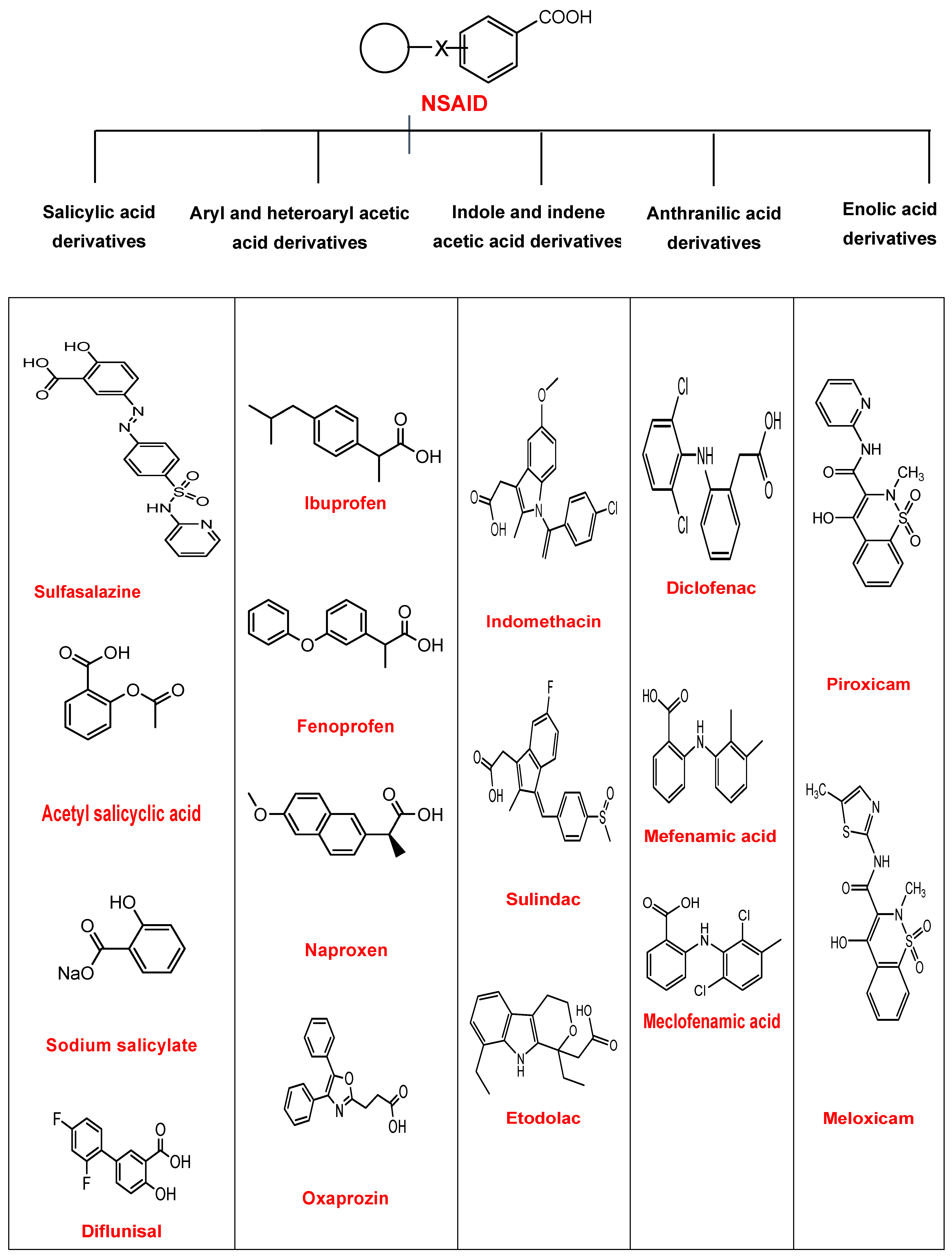
2.1. Non-Steroidal Anti-Inflammatory Drugs (NSAIDs)

Non-steroidal anti-inflammatory drugs are an important class of pharmaceutical drugs that are used to treat pain and inflammation [22]. Apart from their anti-inflammatory effect, NSAIDs also possess antipyretic and analgesic characteristics [23]. About 5–10% of all medications prescribed each year consist of NSAIDs [24]. Some common examples of NSAIDs are ibuprofen, naproxen, piroxicam, meloxicam, flurbiprofen, etc., as illustrated in Scheme 1.
NSAIDs have high bioavailability and are tightly bound to plasma proteins. These compounds are absorbed in the gastrointestinal tract, while the metabolic process is mostly carried out in the liver. The compounds are then excreted in the form of urine. Their life cycles may vary depending on the nature of the drug [26,27]. NSAIDs are classified into further categories depending on the type of derivatives, such as acetic acid, salicylic acid, enolic acid, anthranilic, acid and propionic acid [28]. NSAIDs can be added into water bodies by excretion either directly or via the sewage system. Interaction, metabolism, and degradation of these drugs may occur inside the water environment, where these products may be converted into other products [29]. Scheme 2 depicts the possible degraded products formed by catalytic degradation of ibuprofen by zinc oxide and titania that are activated by photoexcitation of an electron to the conduction band, producing radicals for degradation of pollutants under specific pH conditions, as shown in [30], demonstrating hydroxyibuprofen as the main product by introducing a hydroxyl group followed by decarboxylation and mineralization of the drug in the presence of a catalyst. It is argued that with longer irradiation times, hydroxylation is the primary reaction and may result in a number of other byproducts (not shown here) [31].
Drug manufacturing units are also a major source of introduction of these drugs in water bodies. Pharmaceutical effluents from industry are directly discharged into water bodies, resulting in contamination [32]. Various forms of NSAIDs, such as ibuprofen, naproxen, and diclofenac, have been found to degrade by using UV/persulphate for complete oxidation. The rate of degradation is fast compared to hydrogen peroxide. Their mechanism of degradation was determined to be of first-order kinetics. Removal of water (dehydration) and removal of carbon dioxide (decarboxylation) are the major steps during the degradation of NSAIDs. Removal of chlorine (dechlorination) is also observed during the degradation of NSAIDs. Finally, cleavage in the rings occurs, which leads to the formation of various metabolites [33]. Scheme 3 represents the degradation pathway followed by naproxen [10]. Here, Kolbe’s decarboxylation is identified as the principal process for its degradation via photolysis, followed by further oxidation to form 1-(6-methoxynaphtalene-2-yl)ethylhydroperoxide as a major product. 2-ethyl-6-methoxynaphtalene is formed as a fragment after decarboxylation, which, after hydroxylation and oxidation, forms 1-(6-methoxynaphtalene-2-yl)ethanone, which can dehydrate to substituted ethanol. These products are confirmed by the UHPLC-DAD-MS technique. The ultimate step in photocatalysis is malic acid, which completely mineralizes to CO2, H2O, and non-oxidizable products.
Titania and zinc oxide NPs are most widely studied photocatalysts for the degradation of NSAIDs. Some transition metals are also added as a dopant to increase their catalytic activity. Reduced graphene oxide and other porous materials are also effective in enhancing the photocatalytic activity of these metal oxide nanoparticles [34]. In recent studies, it was illustrated that naproxen is completely degraded by employing g-C3N4 (graphite carbon nitride) as a photocatalyst. The drug was completely degraded within 50–70 min. Some toxic metabolites and intermediates with carcinogenic effects were also detected [35].

2.2. Anticancer Drugs

One of the leading causes of deaths is cancer, which is invariably fatal. Cancer and tumors are characterized by uncontrollable growth of cells. Various treatments are applied to treat cancer, such as chemotherapy and radiotherapy [36]. In chemotherapy, specific tumor cells are killed, and their growth is ceased using potent, low-molecular-weight drugs. Several side effects have been associated with chemotherapy treatment, such as hair loss, nausea, ulcers of the digestive tract, and bone marrow clampdown. These side effects occur due to the action of anticancer drugs on cancerous, as well as healthy cells [37]. Chemotherapy was first used in the 1940s, with nitrogen mustard employed as an alkylating agent that, through the nitrogen lone pair, resulted in an intermediate that prevented replication of DNA of tumor and healthy cells alike. Later, many anticancer drugs such as doxorubicin, mitomycin C, and mitoxantrone were developed for the treatment of malignant and tumor cells, offering higher efficacy and targeted delivery to the tumor. These anticancer drugs are cytotoxic, i.e., they can damage normal cells; studies have shown that not only these drugs but also their degraded products (initially transformed intermediates) are toxic to human and aquatic life [38].
Titania is a useful photocatalyst for the degradation of anticancer drugs such as doxorubicin and methotrexate. These drugs were found to be partially degraded into a hydroxylated product after one hour of irradiation [39], resulting in a final product after fragmentation of sugar moiety.

2.3. Antibiotic Drugs

The word antibiotic originated from the word “antibiosis”, which means “against life”. In the past, it was thought that antibiotics were organic in nature and synthesized by microorganisms that can kill another microorganism [40]. However, antibiotics can also be synthesized by synthetic means. Even a very minute concentration is enough to cease the growth of other microbes. Some antibiotics can kill microbes, for example, bactericidal antibiotics can kill bacteria, while some others can only inhibit the growth of bacteria, such as bacteriostatic antibiotics [41]. Penicillin was the first antibiotic discovered by an English microbiologist, Alexander Fleming, in 1928. This antibiotic was extracted from a fungus, penicillium notatum, which is mostly found in soil. Later, this antibiotic was applied to clinical findings in 1940. Penicillin was first used to treat different bacterial infections [42]. Different types of antibiotics include tetracycline, quinolones, aminoglycosides, sulfonamides, glycopeptides, etc. [43]. Hollow-structured Ag-doped TiO2 NPs were employed as a photocatalyst under visible light for degradation of metronidazole, an antibiotic, and showed high photodegradation efficiency as compared to undoped TiO2, with only a 10% reduction in its degradation efficiency after six cycles [44]. Nanocomposites of needle-like SnO2 NPs loaded onto g-C3N4 nanosheets showed good photocatalytic activity against the degradation of tetracycline in aqueous medium as a result of synergism. Moreover, the recyclability of the nanocomposite-based photocatalyst showed only a minor reduction in its photocatalytic activity, proving the stability of the catalyst and its potential to be applied to other pharmaceutical drugs with the same active moieties [45].
Since antibiotics are among the most prescribed medications annually, the addition of these antibiotics lead to serious pollution to water bodies and are therefore a threat to life [46]. Antibiotics are mainly classified into antibacterial and antiviral drugs. The expected degradation pathway followed by an antibiotic is illustrated in Scheme 4, proposing two common initial degradation pathways. Hydroxylation is the main route for degradation of sulfa drugs, forming mono- and n-hydroxylated intermediates, while S-N cracking resulting from the addition of H+ is the secondary degradation route. The formation of RNH2 is evidence of a photocatalytic process, followed by its complete mineralization with the loss of sulfaniline [47].

2.3.1. Antibacterial Drugs

Antibacterial drugs are chemical substances with biological origin used to stop and inhibit the growth of bacteria or destroy the cell structure of bacteria. These drugs show a complex mode of action depending on the site of the target, mainly attacking the cell wall of bacterial species, and disturbing the synthesis of enzymes, leading to the destruction of the bacterial cell. Some antibacterial drugs may attack the cell membrane of bacterial species, leading to the leakage of intracellular material, while others may cause the complete inhibition of protein synthesis. Nucleic acid synthesis may also be disturbed by the application of certain antibacterial drugs. Moreover, the functioning of certain biochemical reactions is also affected by these drugs, which results in an inadequate production of essential cell components. Depending on the specificity of the target, antibacterial drugs are categorized into two main types, i.e., broad-spectrum and narrow-spectrum antibacterial drugs. A broad range of diseases is treated by the action of broad-spectrum bacteria. Narrow-spectrum antibacterial drugs are used for the treatment of a few specific bacterial diseases [48].
Antibacterial drugs are also responsible for water contamination and are prescribed widely for bacterial infections. A number of photocatalysts have been reported for the removal of these drugs from water bodies. Recently, researchers studied sulfamethazine in water. For this purpose, greigite (Iron sulfide mineral) was used, along with peroxymonosulfate (PMS) and peroxydisulfate (PDM). A degradation rate of about 96.7% was observed within one hour. PMS and PDM generated free radicals (active species) that resulted in an increased rate of degradation [49]. Photocatalysis has attracted considerable attention because of its powerful oxidation-reduction reactions. Semiconductor nanomaterials (such as TiO2 and ZnO) and their nanocomposites are the most effective photocatalysts for the degradation of these pharmaceutical products [50]. In 2016, it was observed that both ZnO and TiO2 can be used to degrade amoxicillin, erythromycin, and streptomycin in the presence of sunlight. Both these photocatalysts were efficient in 100% degradation of drugs due to increased mobility of photogenerated charges. However, ZnO required more time to degrade these drugs completely. It was also observed that the rates of degradation of erythromycin and streptomycin were slower compared to that of amoxicillin. Furthermore, ZnO was reported to be safer and less toxic than TiO2 [51]. Research has shown that doping of semiconductor materials and the formation of hybrid nanocomposites leads to the lowering of the bandgap, which accelerates the percentage of degradation by enhancing photon absorption [52]. A reusable, magnetically separable Fe3O4-functionalized rGO supported on TiO2 showed increased photocatalytic efficiency towards tetracycline as a hybrid photocatalyst due to synergism of the photo-Fenton reaction and the conducting properties of rGO [53].

2.3.2. Antiviral Drugs

Antiviral drugs are widely used to treat infections caused by viruses in the body of the host, which take control of the host’s DNA and start producing viral proteins. First, the virus attaches itself to the surface of the host and starts penetrating inside the host by secreting certain hydrolyzing enzymes. Then, it reaches the host genome and takes complete control. Replication of the viral genome leads to the production of many viral components inside the host body. Ultimately, the newly synthesized viral entities are released outside by budding of the cell membrane and by rupturing of the host cell [54,55]. To control viral activity, antiviral drugs are employed. The first antiviral drug was discovered in 1963. This discovery opened the door for the synthesis of other antiviral drugs. These antiviral drugs have been used to treat millions of patients worldwide [56]. These antiviral drugs are target-specific and are selectively used to treat a particular type of viral infection. These drugs cannot destroy the complete structure of the attacking entity; only growth and other viral activities are stopped. Similarly, in 1990, a viral drug was synthesized to treat HIV (human immunodeficiency virus) [57,58]. Some examples of antiviral drugs are acyclovir, trifluridine, ganciclovir, zidovudine, etc.
Antiviral drugs can pollute water, even at very low concentrations, affecting the water quality. Their degraded metabolites are more persistent than the original products [59]. TiO2 is a very efficient photocatalyst for the removal of Tamiflu (antiviral drug), resulting in 98% degradation with pseudo-first-order reaction kinetics [60]. In recent years, it has been reported that the addition of H2O2 and peroxymonosulfate (PMS) leads to fast degradation in a helical-baffle reactor. In another study, remdesivir was degraded by up to 99% within 5–10 min [61]. A recent paper reported that a ZnO NP-supported photocatalyst with a TiO2 nanorod MXene heterostructure achieved 99.4% efficiency in degrading ceftriaxone sodium [58]. A proposed photocatalytic degradation mechanism of lamivudine by TiO2 is shown in Scheme 5 [62]. It shows two pathways where a major intermediate is formed by attack of an OH· radical, while a secondary product is formed by a photohole h+ attack. The h+ cracks the N6 site, further oxidizing it, leading to aromatic ring opening. Lamivudine and its intermediates completely mineralize within 6 h by 83%, providing the intermediates ample time to decompose, along with the parent compound.

2.4. Antidepressant Drugs

Antidepressant drugs are included in the category of front-line drugs responsible for the treatment of chronic neuropathic pain. They are psychiatric drugs that are used to treat mental illness [63]. Antidepressants are considered the least famous type of painkillers with analgesic effects. The results of a survey suggest that antidepressants account for only 3% of total prescribed analgesics [64]. However, studies show that usage of these antidepressants to minimize chronic pain may lead to the onset of other sensory and emotional actions. Some other side effects are also linked with the use of these antidepressants, and medicinal agencies have reduced the use of these drugs for particular neuropathic pain due to associated limitations [65]. It has been established by different studies that antidepressants exhibit central and periphery analgesic mechanisms of action (see Figure 1) [66,67].
Antidepressants also lead to the enhancement and transmission of adenosines, resulting in an escalated level of extracellular adenosine concentration [68]. Depression is also a consequence of persistent chronic pain. Studies have shown that 30–55% of patients have these interrelated symptoms [69]. On the other hand, pain is the most common symptom found in patients suffering from depression and anxiety [70].
The addition of these drugs to the aquatic environment leads to the contamination of water. Small amounts of these antidepressants are also deposited inside the internal organs of aquatic creatures such as fish and mollusks [71]. Different methods for degradation of antidepressant drugs are applied. According to one report, 90% degradation of sertraline was observed in activated sludge within 0.25 h [72]. More than 99% degradation of venlafaxine was achieved within 3 h by using UV/H2O2 [73]. According to recent research, metal organic frameworks and their composites are used to achieve complete degradation of antidepressants, with a 100% degradation rate reported after 120 min. The generation of free radicals such as OH and SO4∙− is the main driving force for catalytic degradation of these drugs [74]. For venlafaxine, the main mechanism of degradation involves hydroxylation, dehydration, demethylation, and tertiary amine substitution. During the degradation process, the removal of alkyl groups and some other nucleophilic substitution reactions may occur. Not only these antidepressants but also their metabolites and degraded byproducts retain some toxic effects [75].

3. Toxicity of Nanomaterials

Undoubtedly, nanomaterials possess excellent features, and a wide range of applications are associated with them; however, environmental and health hazards limit their use for drug degradation. It has been established by research that nanomaterials that remain in water bodies due to their small size can penetrate through body parts and can damage internal tissues and organs. As many nanomaterials are synthesized due to their multiple applications, a significant amount is added to the aquatic environment. These toxic nanomaterials are then ingested by aquatic creatures such as fish, leading to changes in their internal tissues and organs. The circulatory and respiratory systems of marine species may be disturbed by the toxicity of these nanomaterials [20]. Absorption of nanoparticles by aquatic creatures also leads to various behavioral and physiological changes. Moreover, their fertility rate is decreased, and the death rate is increased due to long-term ingestion of these materials [76]. However, different strategies can be adopted to reduce the toxic effects of nanomaterials, such as replacement of toxic compounds with less toxic compounds by adding chelating agents to modify their surface chemistry, by constructing shell around the nanomaterials, and controlling their shape [77]. The effective separation of NPs from water bodies using filtration membranes may also reduce their exposure to aquatic life. In addition to the various studies reporting toxicological effects of NPs, a few reports discuss the reduction in the cytotoxic effects of nanoparticles by surface coating. Nobuhiro et al. evaluated the reduction in t toxicological effects by coating CuO NPs with a cyclic peptide (cyclic-SCATPFSPQVCS) [78]. Cytotoxicity studies conducted in the presence of CuO NPs with a particle size between 25 and 50 nm in peripheral blood mononuclear cells demonstrated that low concentrations (1 µg/mL) did not affect the cell viability, while concentrations equal to or greater than 10 µg/mL decreased the cell viability [79]. Peptide-coated NPs inhibited cytotoxicity against many microbes and human embryonic kidney cell line HEK293. The author suggested that peptide-coated NPs may have safe applications for industry. Singh et al. demonstrated that CuO NPs produced by surfactant-assisted synthesis have more toxicological effects than NPs synthesized without a surfactant [80]. The particle size, which depends on the synthesis procedure and experimental conditions, also plays a major role in modulating cytotoxic properties. In general, a literature survey reveals that most of studies report that smaller NPs have greater toxicological effects on human cell lines; however, dermal effects are not significant. Further studies are required to establish the synthesis conditions necessary to prepare NPs with fewer toxicological properties. Therefore, greener approaches for the synthesis of nanomaterials without the need to adding any toxic chemical or solvent as a capping and reducing agent may reduce the risks of toxicity caused by NPs [81].

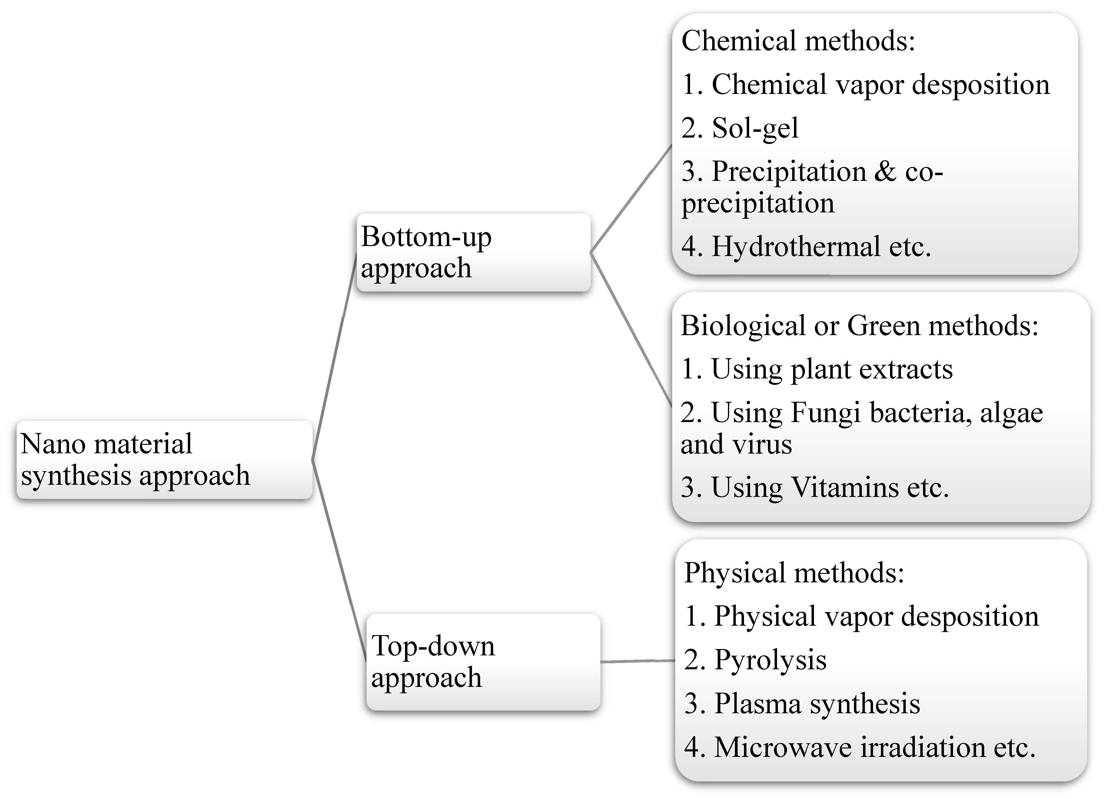
4. Green Synthesis Methods of Nano-Based Photocatalysts

The term “nanotechnology” was first introduced by Richard P. Feynman in 1959. He introduced this concept during his famous lecture, “The plenty of room at the bottom” [82]. He professed that matter at a very small scale (i.e., the atomic and molecular levels) can be employed to construct structures with controlled and precise morphology. Using nanotechnology, matter is reconstructed and restructured at the nano scale (less than 100 nm in diameter) to change its properties. Nanomaterials possess excellent characteristics in the nanometer range as compared to their bulk counterparts [83], such as high optical activity, enhanced catalytic and thermal performance, and excellent mechanical properties due to a high surface-to-volume ratio [84]. The chemical and physical properties of nanomaterials are greatly influenced by decreasing the grain size. Melting and boiling points are also changed as a result of a reduction in size to the nanometer scale [85]. Nanomaterials also exhibit various applications in the medicinal industry. Antimicrobial activities have also been investigated for NPs [86]. Researchers have now switched from chemical to green routes of synthesis, considering the application of these materials in food, cosmetics, electronics, space, and chemical industries [87].
Nanomaterial-based photocatalysts for drug degradation can be synthesized by chemical and physical techniques. Physical methods include plasma synthesis, microwave irradiation, pulse laser methods, sonochemical methods, gamma irradiation, etc., whereas well-known chemical methods include sol–gel, precipitation and coprecipitation and, and electrochemical deposition methods [88]. Green synthesis methods use fewer chemicals and are more environmentally friendly. Chemical synthesis may produce toxic chemical species on the surface of NPs [89,90]. Moreover, it sometimes requires toxic and harmful reducing agents, which release noxious byproducts such as hydrazine. These methods can also be categorized as top-down or bottom-up approaches. In the top-down approach, a large chunk of material is selected and crushed into smaller materials up to the nanometer range, usually by physical methods. This approach is disadvantageous in that it is time-consuming and costly. In addition, the top-down approach does not lead to the production of materials on a large scale [91]. A bottom-up approach refers to the synthesis of nanomaterial by self-assembly of smaller building blocks. This is atom-by-atom and molecule-by-molecule self-assembly of smaller subunits results in the formation of a new material with a diameter ranging from 1 nm to 100 nm. New nuclei are formed, and growth occurs until the desired material is achieved [92]. Such chemical and biological methods are categorized as bottom-up approaches for synthesis of NPs, followed by green synthesis (see Figure 2).

Morphological Dependence of Green Synthesized Nanostructures

The green synthesis method is an environmentally friendly process that invokes green chemistry to ensure reduced used of chemicals, the elimination of the production of toxic waste, and reduced consumption of energy. By using plants and microbes as nanofactories, NPs can be synthesized with different morphologies and properties. Morphology is important for explaining the optical and electrical properties of NPs. Thus, various morphologies of NPs have been reported by employing plant extracts. The presence of capping agents controls the morphologies as well, as the growth and nucleation patterns, of synthesized nanostructures. Morphologies of synthesized NPs can be characterized by employing various techniques, such as scanning electron microscopy (SEM), transmission electron microscopy (TEM), and X-ray diffraction analysis. Furthermore, the presence of the organic functionalities present in synthesized nanomaterials can be analyzed by FT-IR spectroscopy. The morphologies and size of synthesized NPs depend on a number of factors that include pH, temperature, biomass and substrate concentration, exposure time, etc. [93]. Nanoflowers of silver were previously prepared using Kalanchoe Daigremontiana extract, exhibiting enhancement in the photocatalytic performance as compared to spherical nanostructures. The surface area is large in the case of silver nanoflowers, which leads to fast electron transfer, resulting in the degradation of various organic contaminates by employing green synthesized nanoflowers [94]. Capping agents play a crucial role by anchoring to facets of growing nanocrystals to control their morphology. In addition, AgNPs are synthesized by adopting both green and chemical methods; their phytotoxicity and efficiency were compared using various analytical tools. It was concluded that the nanostructures prepared by adopting a green route are environmentally benign and toxin-free. They also possess better catalytic and antibacterial properties [95]. However, it is not as straightforward to control the size and morphology when synthesized by bio sources, as it involves a number of biochemicals such a flavonoids, polyphenols, fatty acids, etc., which are difficult to control in a reaction. Similarly, in another report, Andean cabbage was used as a stabilizing agent to produce AgNPs with an average size of 15–80 nm, while NPs of 20.5 nm were synthesized from Mortiño berry to reduce silver to AgNPs. To this end, NPs were coated with polyphenolics and anthocyanins, leading to stability of the NPs [96,97]. Similarly, AgNPs were synthesized by different sources, such as Allium cepa (onion), resulting in a spherical shape with an average size of 50–100 nm [98], Capparis petiolaris fruit with spherical NPs with diameters of 10–30 nm [99] and Terminalia cuneate bark with NPs with an average diameter of 25–40 nm [100]. It is noted that changing the capping agent by using different plants/extracts results in different dimensions.
ZnO NPs are known to induce reactive oxygen species, which aid in the degradation of pollutants. Waste fruit peels of Punica granatum and Musa acuminate were used to synthesize biogenic ZnO and Ag NPs, which were characterized by different techniques. SEM micrographs confirmed a nanosphere morphology of chemically synthesized Ag NPs, with a nanoflake morphology for ZnO NPs. At the same time, nanospheres were confirmed for both biosynthesized Ag and ZnO NPs. It was also contended that biologically synthesized NPs showed higher antimicrobial activity than chemically synthesized NPs. The average size of chemically synthesized Ag NPs was 9.2 ± 2.28 nm, while biologically synthesized NPs ranged from 7 to 11 nm. Similarly, chemically synthesized ZnO NPs demonstrated a mean average size of 6.4 ± 3.29 nm, whereas biologically synthesized NPs from M. acuminate and P. granatum had average sizes in the range of 10–14 nm [101].
Au NPs were obtained using Persea americana (avocado) oil, as confirmed by UV-Vis spectrophotometer, showing a spherical morphology between electromagnetic radiations of 300–1100 nm under direct sunlight, with an average particle size of 20–100 nm. A weak band at 1000 nm suggests the formation of triangular AuNPs, which were stable for two months, indicating the coexistence of differently shaped NPs [102]. Furthermore, plant extracts such as Cinnamomum zeylanicum leaf, edible mushroom, berries, sugar beets, etc., have materialized as ecofriendly and non-hazardous alternatives to wet chemical methods [103]. Garcinia indica Choissy produce biogenic AuNPs of 20–30 nm in diameter with a spherical shape and around 89% photocatalytic efficiency for organic pollutants [104]. Mussaenda glabrata leaf results in reduction of Au3+ to Au0 and Ag1+ to Ag0 in the presence of phytochemicals. AuNPs showed a mean diameter of 10.59 nm with spherical and triangular NPs, while AgNPs showed an average size of 51.32 with a spherical geometry. An FCC crystal structure was confirmed via XRD. In the presence of NaHB4, silver and gold NPs demonstrated reasonable photocatalytic activity [105]. Under the optimum reaction conditions, AuNPs with different shapes were synthesized instead of spherical NPs, which can be used for different applications. Vegetable waste results in AuNPs with an average size of 10–70 nm with excellent antibacterial activity [106].
α-Fe2O3 NPs with a bandgap of 2.2 eV are promising catalysts [107]. Thus, Peltophorum pterocarpum leaf extract was used to form rod-shaped γ- and α-Fe2O3 NPs with an average size of 16.99 nm [108]. Camellia sinensis tea leaf extract comprising polyphenols resulted in nZVI/Fe0 NPs with a size of 5–15 nm and a spherical shape [109]. It has also been noticed that the NPs formed via green synthesis can are much smaller (2–80 nm) than those produced by wet chemical methods (87–400 nm), such as Fe3O4 NPs [21].
Similarly, biodegradable waste extracts are used to synthesize CuFe2O4, with applications as catalysts and bactericides for water remediation [110]. Methanolic extract of peel agrowaste from fruits such as plum, kiwi, and peach was used to synthesize nanocrystalline TiO2 NPs. O–H, C=O, C–O, and C–H groups found in peel were responsible for the synthesis of TiO2 NPs, the antibactericidal and catalytic properties of which were dose- and time-dependent [111]. Similarly, bacterial cellulose waste (BC) from Achromobactin sp. M15 was employed to synthesize TiO2 NPs with a size range of 5–10 nm, showing innovative properties as compared to NPs prepared by a chemically employed sol–gel method [112]. Copious references for the green synthesis of NPs future prospects can be found in [19].

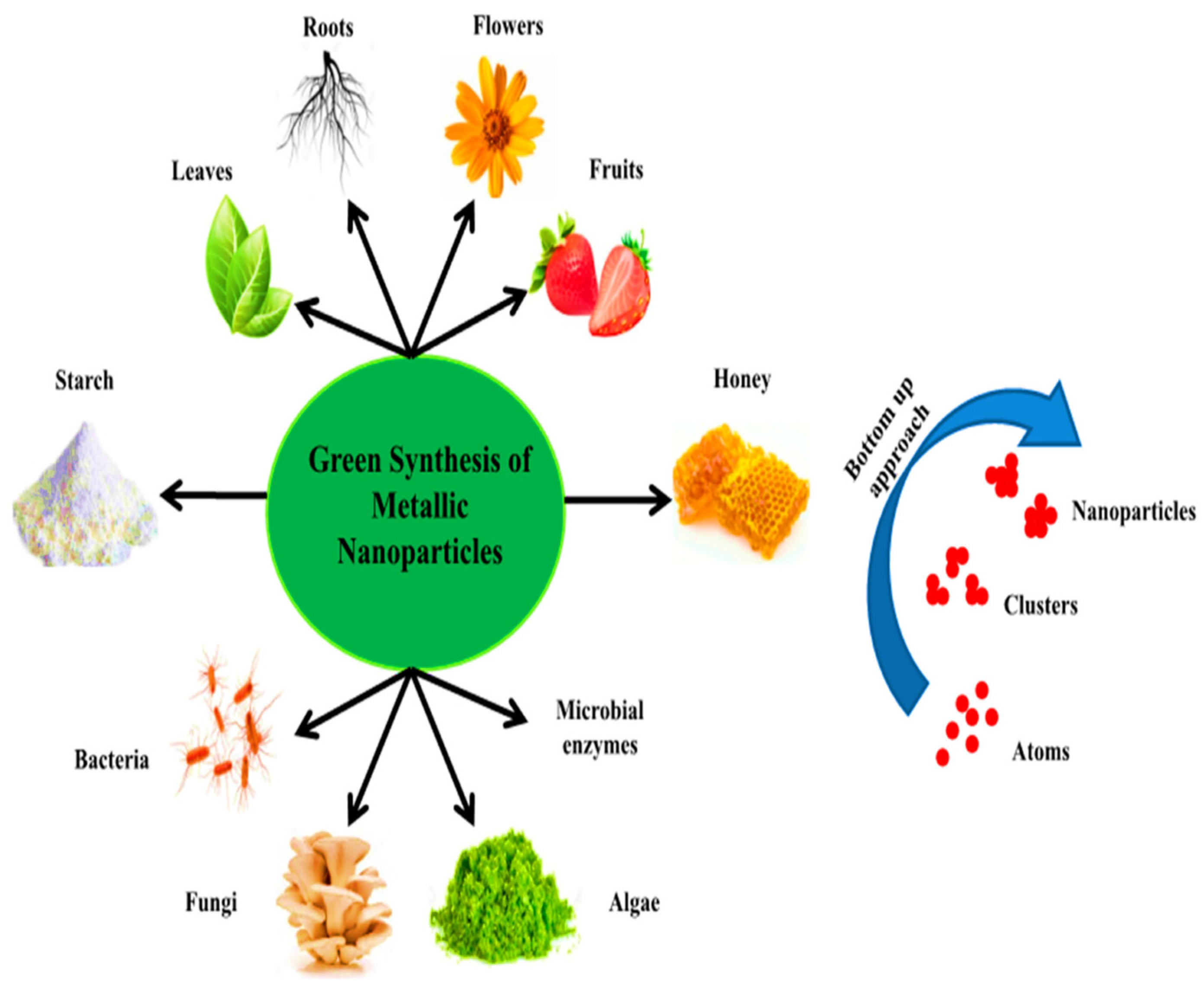
5. Biogenic Sources of Synthesis of NPs

The concepts and principles of green chemistry were first presented by two scientists named Anastas and Warner [113]. For green synthesis of NPs, the selection of solvent and reducing/stabilizing agent is of great significance and often governs the morphologies and size of NPs. Different types of green synthesis methods can be used depending upon the nature of the source of reducing or stabilizing agents, such as plants, algae, vitamins, etc., (as illustrated in Figure 3), from which extracts can be collected. The forthcoming sections discuss the various routes utilized for green synthesis of photocatalysts employed in drug degradation.

5.1. Green Synthesis Using Plant Extracts

Synthesis of NPs using plant extracts is the easiest route, as these are cost-effective and easily available [115]. Medicinal plants contain a large number of metabolites and reducing agents, which help in the reduction of many metal ions to NPs. Some plants also contain a significant quantity of metals that are incorporated into NPs during synthesis. Plants do not require much time for the reduction of metal ions compared to fungi and bacteria. NPs of different sizes and morphologies can be achieved in a short duration [116]. Different parts of plants can be used for green syntheses, such as leaves, flowers, fruits, peels, and stems, owing to the presence of phytochemicals [117]. Plant leaves and flowers should be subjected to drying at room temperature after washing with tap water or distilled water. After complete drying, they are finely crushed into a smooth powder. Extract can be prepared by mixing a weighted amount of powder in distilled water, whereas complete mixing is achieved by heating and stirring. Afterwards, the cooled solution is filtered, and the filtrate is employed for the synthesis of NPs [118]. Some plants produce a large amount of hydrogen ions during the glycolysis process, which play a critical role in the reduction and stabilization of NPs [119]. Furthermore, the nature and type of NPs is the key parameter determining their photocatalytic activity. NPs can be obtained in the form of nanospheres, nanotubes, nanorods, nanofilms, etc.
Studies show that the NPs synthesized from plant extract (such as Convolvulus arvensis) can be efficiently used for the degradation of environmental pollutants such as azo dyes and drugs. These dyes are used as additives in pharmaceuticals for aesthetics and for practical reasons to help patients in distinguishing between different colors of medicine. Thus, silver NPs with well-defined morphology and catalytic activity show the best environmental response [120]. Leaf extract of Vernonia amygdalina was used to synthesize Nb-doped ZnO NPs by using a green approach to photocatalytically degrade tetracycline (TC), which is an important broad-spectrum antibiotic used to treat infectious diseases. The photocatalytic activity under visible light was shown to be 93.2% in 2 h and only reduced to 6% after five recovery cycles [121]. Sensitized ZnO NPs synthesized by chemical precipitation method were found to photocatalytically degrade TC to >80% under visible light for 5 h [122]. Employing a nanocomposite Au0.1Ag0.9/TiO2 in a citric acid membrane resulted in an efficiency of 90% under visible light [123]. Similarly, extract of hydrophyte species such as Persicaria salicifolia is used for the green synthesis of Ag-doped ZnO supported on a carbon-rich support such as biochar for photocatalytic degradation of TC. The photocatalyst showed reasonable recyclability after six successive cycles of use, with biochar facilitating the regeneration of the NPs [124]. Carbon quantum dots (CQDs) synthesized from waste of Citrus limetta modified with Au/Ag boosted the surface plasmon resonance effect when decorated on TiO2 nanofibers. The nanocomposite was able to photocatalytically degrade erythromycin. The photocatalyst exhibited recyclability with reduced photocatalytic activities after several cycles [125]. Plant-assisted synthesis of ZnO used as a photocatalyst has led to the removal of an antibiotic (ciprofloxacin). ZnO NPs were synthesized using extract obtained from Citrus aurantifolia (Lemon peels). A removal efficiency of 90% was obtained after 160 min. A variety of phytochemicals such as proteins, amines, alkaloids, cellulose, hemicellulose, and other aromatic components are present inside the lemon peel extract, resulting in stabilization of ZnO NPs. These green NPs produce no secondary pollutants. Complete degradation resulted in the formation of water and carbon dioxide [126]. The degradation of ciprofloxacin has also been reported by employing ZnO NPs synthesized using the chemical precipitation method under UV light, showing only around 50% degradation after 60 min and proving to be less efficient than green synthesized NPs [127]. Upon exposure to sunlight, excitation of electrons occurs from valence to the conduction band of ZnO NPs, leading to the generation of electrons and holes, which further react with water to generate free radicals. These free radicals are responsible for the breakage of drug structures. However, bandgap modulation in ZnO NPs (3.3 eV) can be accomplished by the addition of certain transition metals (silver, etc.) as a dopant. Narrowing of the bandgap results in shifting of the absorption peaks towards longer wavelengths and low-energy regions. These impurities (dopants) result in the enhancement of photocatalytic activity of ZnO, making it slightly active in response to visible radiation. Similarly, the reduction in the bandgap of titania is also ensured by adding dopants such as nitrogen. These impurities result in the enhancement of optical properties of TiO2 [128]. Thus, chemically synthesized γ-Fe2O3@ZnO showed improved photocatalytic efficiency for ciprofloxacin, from 11.5% for pure γ-Fe2O3 to 92.5% for functionalized NPs that require additional use of chemicals.

5.2. Green Synthesis Using Vitamins

Different vitamins contain a variety of reducing and capping agents. Nanorods and nanospheres can be synthesized by using vitamin B2 as a reducing and capping agent. Gold and platinum NPs of specific shape were prepared using riboflavin (vitamin B2) by reacting the salt precursors of gold and platinum with different solvents. Vitamin B2 is an efficient reducing and capping agent as compared to other chemical compounds, such as sodium borohydride (NaBH4), and appears to be perfect for preparation of NPs [129]. The morphologies and sizes of NPs can be controlled by varying the type of solvents used; density plays an important role in the formation of self-assembled structures and different shapes. The reaction of metal salt with vitamin B2 results in reduction of the metal salt and simultaneous oxidation and capping of the vitamin. Similarly, uniformly sized NPs can be synthesized using ascorbic acid (vitamin C) as a capping and reducing agent. The interaction of radiation with the collective oscillations of electrons in the conduction band result in extinction properties for Au and Ag NPs. This could be circumvented by efforts to tune size and shape by using a different synthesis method. Hence, Au and Ag NPs have been obtained utilizing vitamin C, resulting in a fast and efficient one-pot synthesis method. The size of synthesized NPs is tuned by pH changes, varying the metal or vitamin C solution. This method also allows for changes in the surface chemistry by using different surfactants and can be employed for a vast array of applications [130]. Green synthesis of silver NPs has been reported using vitamin-C-enriched Phyllanthus emblica extract. Here, vitamin C is used as a reducing agent, and the synthesis of AgNPs is affected by the reaction time, temperature, and concentration of metal salt and reducing agent in the solution matrix. Under optimum conditions, AgNPs of 41.2 nm are obtained. Ag NPs have enhanced photocatalytic efficiency for the removal of toxic moieties from water bodies [131,132]. Furthermore, although this area of research is in its incipient stage, core shell noble metal NPs (Fe-Cu) with Pt, Au, and Pd shells have been obtained using vitamin C, resulting in different morphologies that can be further explored for different applications, such as catalysts, nanodevices, etc. vitamin C was reported to reduce Fe and Cu NPs resulting in a core shell structure by the addition of Pt, Au, or Pd [133].

5.3. Green Synthesis Using Algae

A broad range of biological activities is associated with algae, such as antibacterial and antimicrobial applications [134]. These are marine species and are extensively used for the synthesis of metal NPs. Chlorella vulgaris (unicellular algae) can be used to synthesize metallic NPs such as gold NPs with particle sizes of 9–20 nm. Different shapes, sizes, and morphologies of metallic NPs can be achieved using various algal species and reaction conditions [135]. Gold NPs are synthesized using extract from aquatic algae, i.e., Galaxaura elongate, with excellent antibacterial properties [136]. Chlorella ellipsoidea extract is used for the synthesis of silver NPs with enhanced photocatalytic activities for the removal of water contaminants [137].
Gold (I) sulfide NPs can be synthesized using cyanobacteria in aqueous solution [138]. Furthermore, NPs of different sizes, such as gold and silver NPs, ranging from 8 to 12 nm can be obtained using marine algae [139]. Algae lack proper structure such as stems, roots, and shoot systems, but they possess a variety of pigments that play an important role in the synthesis of NPs [140]. Particle size is a key parameter in determining the properties of NPs, which, in turn, is affected by several factors, including pH, temperature, solvent, and chemical concentration [141]. The mechanism of synthesis of nanoparticles using algae is not fully understood, as different algal species react differently with metal precursors. Many species produce inorganic compounds either intra- or extracellularly and have different mechanisms of action to produce NPs [142]. The intracellular mechanism of the synthesis of NPs involves transportation of metal ions to the microbial cell, where positively charged metal ions are attracted towards the negatively charged cell wall. The metal ions are, in turn, reduced by the enzymes released from the microbial cell wall. The extracellular mechanism involves the secretion of enzymes, which reduce the metal ions for the synthesis of NPs [143].

5.4. Green Synthesis Using Fungi

Extracellular and intracellular synthesis of NPs using fungi such as Fusarium oxysporum, Colletotrichum sp., Trichothecium sp., A. fumigatus, Coriolus versicolor, Aspergillus niger, Cladosporium cladosporioides, and others are used widely worldwide, owing to several advantages lacking in other microbes, e.g., tolerance to high flow pressure in bioreactors and easy growth [144,145]. A variety of particle sizes can be obtained by using different fungal strains. Aspergillus flavus has been employed to produce monodisperse Ag NPs with particle sizes of 8.92 ± 1.61 nm. The stability of NPs was also found to be high due to the secretion of fungus, making NPs stable for 3 months [145]. Cultural conditions also impact the synthesis of NPs. Non-agitated culture leads to the production of extracellular metallic NPs, while agitated culture leads to the synthesis of intracellular NPs. It has been observed that the secretion of enzymes and proteins is considerably enhanced in the case of stationary culture as compared to agitated culture conditions [146].
It has been reported that gold NPs possessing biomedical and anticancerous properties can be synthesized by using fungus extract i.e., Fusarium solani [147]. A variety of metallic NPs, such as zinc, copper, silver, gold, iron etc., have been synthesized using fungus extract for the photocatalytic degradation of water pollutants [148]. Fungal extract contains multiple phytochemicals that are useful in the synthesis of nanoparticles such as spherically shaped iron NPs prepared from extract obtained from endophytic fungi Penicillium oxalicum. This extract exhibits expanded surface area for the reduction of metals. These fungus-mediated iron NPs were employed for photocatalysis of colored water contaminants, demonstrating a removal efficiency rate of 99.17%. Furthermore, morphologically controlled synthesis of silver NPs is also achieved by using fungal extract, which can be used for the catalytic degradation of water pollutants due to enhanced catalytic properties [149]. Fungus-assisted production of a nitrogen-doped Co3O4 nanocatalyst was also ensured by using Fusarium oxysporum, which exhibited excellent catalytic efficiency of 87% for toxic moieties [150].

5.5. Synthesis by Bacteria

Green synthesized NPs are formed when metal ions are trapped by microbes, whether at the surface or inside the microbes, which are then reduced in the presence of enzymes and act as electron acceptor moieties for anaerobic respiration. These microbes that include bacteria, viruses, and algae are known to either accumulate these NPs or secrete them to the environment in the presence of stabilizing and capping agents secreted intracellularly and are also known as green nanofactories. Thus, this method is a proven route for bioremediation or synthesis of materials. Many bacteria have been employed in the green synthesis of Au and Ag NPs, such as Bacillus subtilis, Escherichia coli, Rhodobacter capsulatus, Corynebacterium sp., and Lactobacillus sp. CdS NPs have also been synthesized using Escherichia coli, Klebsiella aerogenes, Rhodopseudomonas palustris, and Gluconoacetobacter xylinus [144]. ZnS NP synthesis was reported using Rhodobacter sphaeroides with a particle size of 8 nm [151]. CdS with particle sizes of 3.2–44.9 nm were formed from Escherichia coli and Klebsiella pneumoniae [152]. Copious references on the synthesis of NPs through green nanofactories can be found in [153]. Biogenic nano-TiO2 is formed by employing Bacillus subtilis, which produces NPs with a spherical morphology and a size of 10–30 nm. TiO2 NPs have proven to be a good photocatalyst that act with the help of H2O2 [154]. CuO NPs with an average particle size of 6–7.8 nm were prepared using Cystoseira trinodis extracts from brown alga and showed improved photocatalytic activity [155]. Bacteria are also employed in the synthesis of well-known ZnO NPs with excellent photocatalytic performance. ZnO nanoflowers can easily be synthesized using B. licheniformis, which is an effective and environmentally friendly approach. Nanoflowers have a large surface area with enhanced photocatalytic activities. These nanoflowers were found to be 40 nm in width and 400 nm in height. Absorption of light by the photocatalyst leads to the generation of free radicals (active species). These active species facilitate the process of degradation of organic contaminants. Hence, these photocatalysts help in bioremediation [156].

6. Conclusions

A lack of awareness regarding unregulated disposal of pharmaceutical waste and its hazardous implications has resulted in unchecked environmental degradation that needs to be urgently addressed. Hence, methods that are sustainable and environmentally friendly and are relevant to the aquatic ecosystems and required. These pharmaceutical drug pollutants have been accumulating in water bodies due to a rapid rise in industrialization, lack of awareness of the general public about proper disposal of pharmaceutical waste, and poor sanitation, especially in underdeveloped countries. Furthermore, at the governmental level in developing countries, strict adherence to regulation and guidelines on proper disposal of these drugs and their transformations is almost non-existent. Pharmaceutical waste contains many compounds, such as antibiotics, anticancer drugs, antidepressants etc., which are highly stable and persistent in water. Some of these compounds are pseudo-persistent, but their continuous input in water bodies imbalances their natural removal. The degradation of drugs often results in more harmful secondary pollution, which is also a cause of nuisance. Conventional water treatment procedures fall short of the desired rapid removal level, as evidenced by the presence of ever-increasing pharmaceutical drug pollutants in drinking water. Extensive research is being carried out to degrade drugs through advanced oxidation processes. Photocatalysis is one of the most explored oxidation processes in this regard and is often performed using metal or semiconductor oxide-based NPs synthesized by physical, chemical, or biological methods. Among these methods, biological methods of synthesis of NPs are garnering popularity, owing to their benefits of being greener, simple, and cost-effective. Green synthesis can be carried out using plant-based extracts, microbes (bacteria, fungi, or algae) and vitamins, as they provide reducing/capping agents required for the reduction of metal ions during the synthesis of NPs. The properties and efficiency of NPs in drug degradation depends on the reaction conditions, as well as the source of the reducing/capping agent. Despite the advantages of the biological synthesis of NPs, the cytotoxicity of nanomaterials must be tested, as minute quantities of NPs that remain in the water after remediation can be consumed by humans and animals and may cause adverse health effects. Additionally, intensive research with a strong triple helix platform involving collaboration between academia, industry, and government is necessary to mitigate the risks associated with its disposal and curtail waste in the production cycle with enhanced cooperation across borders in terms of implementing regulations and setting standards for acceptable limits. Thus, study and regulation of the leakage of pharmaceutical waste into water bodies, their fate and risks to non-target organisms, and its solution by employing zero waste and cost-effective technologies is incumbent. More importantly, consistent efforts are required to educate the public to cut down on excessive use and misuse of pharmaceuticals. Although these steps may reduce the waste to some extent, more stringent polices need to be put in place and adhered to in order to tackle severe environmental consequences endangering the sustainability of our planet.

Author Contributions

This manuscript was written through contributions of all authors. The relevant credit roles are: F.Q.: Investigation, methodology, and/writing—original draft A.S.: conceptualization, supervision, project management, resources, and visualization F.J.I.: validation, writing—review and editing, formal analysis; N.S.S. and A.H.: methodology, investigation, writing—review and editing. All authors have read and agreed to the published version of the manuscript.

Funding

This research received no external funding.

Data Availability Statement

No new data were created or analyzed in this study. Data sharing is not applicable to this article.

Acknowledgments

We acknowledge the support of Quaid-i-Azam University, Islamabad, Pakistan.

Conflicts of Interest

The authors declare no conflict of interest.

References

  1. Zuccato, E.; Calamari, D.; Natangelo, M.; Fanelli, R. Presence of therapeutic drugs in the environment. Lancet 2000, 355, 1789–1790. [Google Scholar] [CrossRef] [PubMed]
  2. Rehman, M.S.U.; Rashid, N.; Ashfaq, M.; Saif, A.; Ahmad, N.; Han, J.-I. Global risk of pharmaceutical contamination from highly populated developing countries. Chemosphere 2015, 138, 1045–1055. [Google Scholar] [CrossRef] [PubMed]
  3. Hejna, M.; Kapuścińska, D.; Aksmann, A. Pharmaceuticals in the aquatic environment: A review on eco-toxicology and the remediation potential of algae. Int. J. Environ. Res. Public Health 2022, 19, 7717. [Google Scholar] [CrossRef] [PubMed]
  4. González-González, R.B.; Sharma, P.; Singh, S.P.; Américo-Pinheiro, J.H.P.; Parra-Saldívar, R.; Bilal, M.; Iqbal, H.M. Persistence, environmental hazards, and mitigation of pharmaceutically active residual contaminants from water matrices. Sci. Total Environ. 2022, 821, 153329. [Google Scholar] [CrossRef]
  5. Bavumiragira, J.P.; Yin, H. Fate and transport of pharmaceuticals in water systems: A processes review. Sci. Total Environ. 2022, 823, 153635. [Google Scholar] [CrossRef]
  6. Majumder, S.; Chatterjee, S.; Basnet, P.; Mukherjee, J. ZnO based nanomaterials for photocatalytic degradation of aqueous pharmaceutical waste solutions–A contemporary review. Environ. Nanotechnol. Monit. Manag. 2020, 14, 100386. [Google Scholar] [CrossRef]
  7. Quesada, H.B.; Baptista, A.T.A.; Cusioli, L.F.; Seibert, D.; de Oliveira Bezerra, C.; Bergamasco, R. Surface water pollution by pharmaceuticals and an alternative of removal by low-cost adsorbents: A review. Chemosphere 2019, 222, 766–780. [Google Scholar] [CrossRef]
  8. Majumder, A.; Gupta, B.; Gupta, A.K. Pharmaceutically active compounds in aqueous environment: A status, toxicity and insights of remediation. Environ. Res. 2019, 176, 108542. [Google Scholar] [CrossRef]
  9. Akerdi, A.G.; Bahrami, S.H. Application of heterogeneous nano-semiconductors for photocatalytic advanced oxidation of organic compounds: A review. J. Environ. Chem. Eng. 2019, 7, 103283. [Google Scholar] [CrossRef]
  10. Patel, M.; Kumar, R.; Kishor, K.; Mlsna, T.; Pittman, C.U., Jr.; Mohan, D. Pharmaceuticals of emerging concern in aquatic systems: Chemistry, occurrence, effects, and removal methods. Chem. Rev. 2019, 119, 3510–3673. [Google Scholar] [CrossRef] [Green Version]
  11. de Souza, D.I.; Dottein, E.M.; Giacobbo, A.; Rodrigues, M.A.S.; de Pinho, M.N.; Bernardes, A.M. Nanofiltration for the removal of norfloxacin from pharmaceutical effluent. J. Environ. Chem. Eng. 2018, 6, 6147–6153. [Google Scholar] [CrossRef]
  12. Jaria, G.; Lourenco, M.A.; Silva, C.P.; Ferreira, P.; Otero, M.; Calisto, V.; Esteves, V.I. Effect of the surface functionalization of a waste-derived activated carbon on pharmaceuticals’ adsorption from water. J. Mol. Liq. 2020, 299, 112098. [Google Scholar] [CrossRef]
  13. Magureanu, M.; Mandache, N.B.; Parvulescu, V.I. Degradation of pharmaceutical compounds in water by non-thermal plasma treatment. Water Res. 2015, 81, 124–136. [Google Scholar] [CrossRef] [PubMed]
  14. Bruggeman, P.; Schram, D.; González, M.Á.; Rego, R.; Kong, M.G.; Leys, C. Characterization of a direct dc-excited discharge in water by optical emission spectroscopy. Plasma Sources Sci. Technol. 2009, 18, 025017. [Google Scholar] [CrossRef]
  15. Kanazawa, S.; Kawano, H.; Watanabe, S.; Furuki, T.; Akamine, S.; Ichiki, R.; Ohkubo, T.; Kocik, M.; Mizeraczyk, J. Observation of OH radicals produced by pulsed discharges on the surface of a liquid. Plasma Sources Sci. Technol. 2011, 20, 034010. [Google Scholar] [CrossRef]
  16. Bruggeman, P.; Schram, D.C. On OH production in water containing atmospheric pressure plasmas. Plasma Sources Sci. Technol. 2010, 19, 045025. [Google Scholar] [CrossRef]
  17. Papagiannis, I.; Koutsikou, G.; Frontistis, Z.; Konstantinou, I.; Avgouropoulos, G.; Mantzavinos, D.; Lianos, P.J.C. Photoelectrocatalytic vs. photocatalytic degradation of organic water born pollutants. Catalysts 2018, 8, 455. [Google Scholar] [CrossRef] [Green Version]
  18. Basavarajappa, P.S.; Patil, S.B.; Ganganagappa, N.; Reddy, K.R.; Raghu, A.V.; Reddy, C.V. Recent progress in metal-doped TiO2, non-metal doped/codoped TiO2 and TiO2 nanostructured hybrids for enhanced photocatalysis. Int. J. Hydrog. Energy 2020, 45, 7764–7778. [Google Scholar] [CrossRef]
  19. Guan, Z.; Ying, S.; Ofoegbu, P.C.; Clubb, P.; Rico, C.; He, F.; Hong, J. Green synthesis of nanoparticles: Current developments and limitations. Environ. Technol. Innov. 2022, 26, 102336. [Google Scholar]
  20. Khan, I.; Saeed, K.; Khan, I. Nanoparticles: Properties, applications and toxicities. Arab. J. Chem. 2019, 12, 908–931. [Google Scholar] [CrossRef]
  21. Gokila, V.; Perarasu, V.; Rufina, R. Qualitative comparison of chemical and green synthesized Fe3O4 nanoparticles. Adv. Nano Res. 2021, 10, 71–76. [Google Scholar]
  22. Abdulla, A.; Adams, N.; Bone, M.; Elliott, A.M.; Gaffin, J.; Jones, D.; Knaggs, R.; Martin, D.; Sampson, L.; Schofield, P. Guidance on the management of pain in older people. Age Ageing 2013, 42, 1–57. [Google Scholar] [CrossRef] [PubMed] [Green Version]
  23. Harirforoosh, S.; Asghar, W.; Jamali, F. Adverse effects of nonsteroidal antiinflammatory drugs: An update of gastrointestinal, cardiovascular and renal complications. J. Pharm. Pharm. Sci. 2013, 16, 821–847. [Google Scholar] [CrossRef] [PubMed] [Green Version]
  24. Onder, G.; Pellicciotti, F.; Gambassi, G.; Bernabei, R. NSAID-related psychiatric adverse events. Drugs 2004, 64, 2619–2627. [Google Scholar] [CrossRef] [PubMed]
  25. Bindu, S.; Mazumder, S.; Bandyopadhyay, U. Non-steroidal anti-inflammatory drugs (NSAIDs) and organ damage: A current perspective. Biochem. Pharmacol. 2020, 180, 114147. [Google Scholar] [CrossRef] [PubMed]
  26. Awtry, E.H.; Loscalzo, J. Aspirin. Circulation 2000, 101, 1206–1218. [Google Scholar] [CrossRef]
  27. Verbeeck, R.; Richardson, C.; Blocka, K.L. Clinical pharmacokinetics of piroxicam. J. Rheumatol. 1986, 13, 789–796. [Google Scholar]
  28. Gupta, A.; Bah, M. NSAIDs in the treatment of postoperative pain. Curr. Pain Headache Rep. 2016, 20, 1–14. [Google Scholar] [CrossRef]
  29. Mulkiewicz, E.; Wolecki, D.; Świacka, K.; Kumirska, J.; Stepnowski, P.; Caban, M. Metabolism of non-steroidal anti-inflammatory drugs by non-target wild-living organisms. Sci. Total Environ. 2021, 791, 148251. [Google Scholar] [CrossRef]
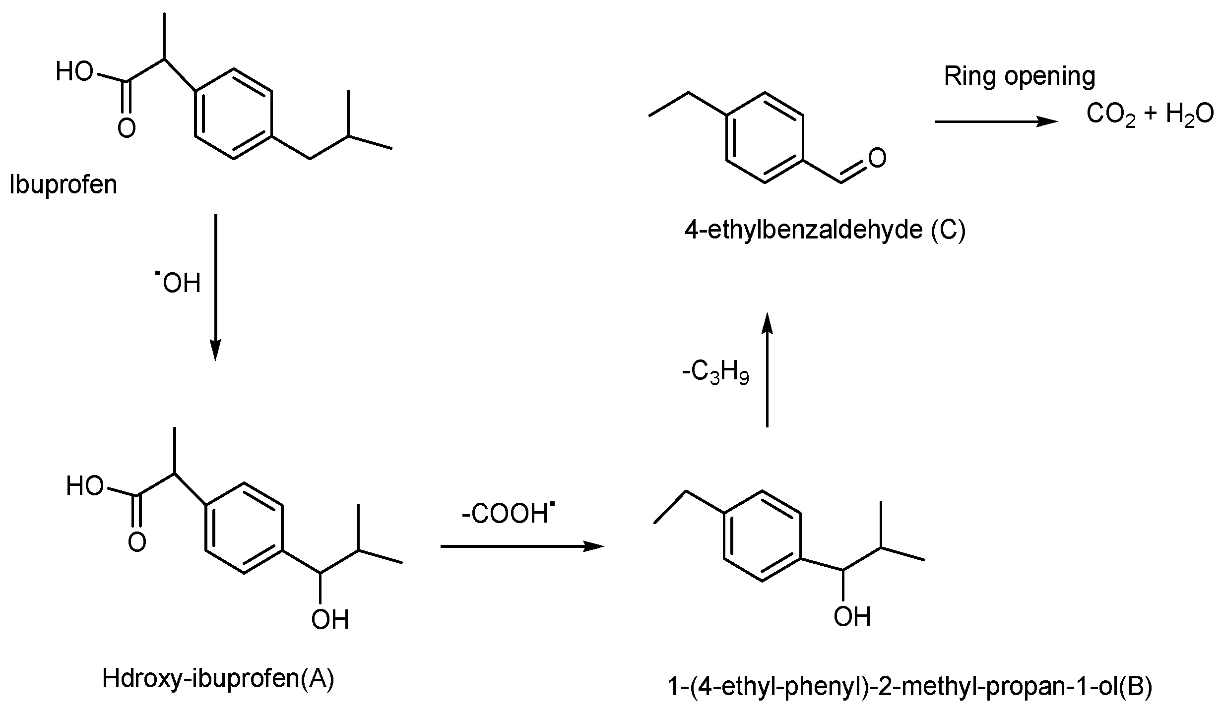
  30. Tanveer, M.; Guyer, G.T.; Abbas, G. Photocatalytic degradation of ibuprofen in water using TiO2 and ZnO under artificial UV and solar irradiation. Water Environ. Res. 2019, 91, 822–829. [Google Scholar] [CrossRef]
  31. Méndez-Arriaga, F.; Esplugas, S.; Giménez, J. Photocatalytic degradation of non-steroidal anti-inflammatory drugs with TiO2 and simulated solar irradiation. Water Res. 2008, 42, 585–594. [Google Scholar] [CrossRef] [PubMed]
  32. Paiga, P.; Santos, L.H.; Ramos, S.; Jorge, S.; Silva, J.G.; Delerue-Matos, C. Presence of pharmaceuticals in the Lis river (Portugal): Sources, fate and seasonal variation. Sci. Total Environ. 2016, 573, 164–177. [Google Scholar] [CrossRef]
  33. Fu, Y.; Gao, X.; Geng, J.; Li, S.; Wu, G.; Ren, H. Degradation of three nonsteroidal anti-inflammatory drugs by UV/persulfate: Degradation mechanisms, efficiency in effluents disposal. Chem. Eng. J. 2019, 356, 1032–1041. [Google Scholar] [CrossRef]
  34. Jiménez-Salcedo, M.; Monge, M.; Tena, M.T. Photocatalytic degradation of ibuprofen in water using TiO2/UV and g-C3N4/visible light: Study of intermediate degradation products by liquid chromatography coupled to high-resolution mass spectrometry. Chemosphere 2019, 215, 605–618. [Google Scholar] [CrossRef] [PubMed]
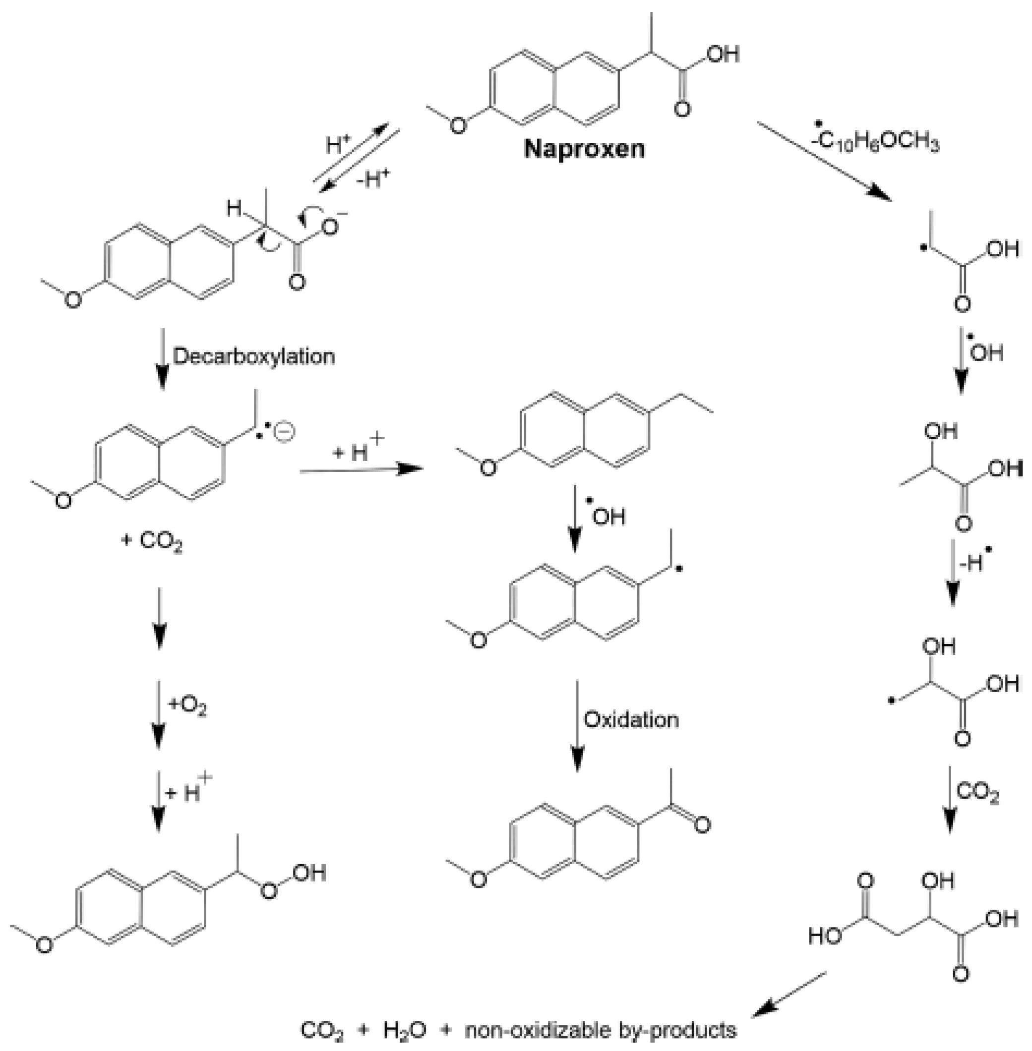
  35. Jiménez-Salcedo, M.; Monge, M.; Tena, M.T. The photocatalytic degradation of naproxen with g-C3N4 and visible light: Identification of primary by-products and mechanism in tap water and ultrapure water. J. Environ. Chem. Eng. 2022, 10, 106964. [Google Scholar] [CrossRef]
  36. Shewach, D.S.; Kuchta, R.D. Introduction to cancer chemotherapeutics. Chem. Rev. 2009, 109, 2859–2861. [Google Scholar] [CrossRef] [Green Version]
  37. Thurston, D.E.; Pysz, I. Chemistry and Pharmacology of Anticancer Drugs; CRC Press: Boca Raton, FL, USA, 2021. [Google Scholar]
  38. Hoppe-Tichy, T. Current challenges in European oncology pharmacy practice. J. Oncol. Pharm. Pract. 2010, 16, 9–18. [Google Scholar] [CrossRef]
  39. Calza, P.; Medana, C.; Sarro, M.; Rosato, V.; Aigotti, R.; Baiocchi, C.; Minero, C. Photocatalytic degradation of selected anticancer drugs and identification of their transformation products in water by liquid chromatography–high resolution mass spectrometry. J. Chromatogr. 2014, 1362, 135–144. [Google Scholar] [CrossRef]
  40. Russell, A.D. Types of antibiotics and synthetic antimicrobial agents. In Hugo and Russell’s Pharmaceutical Microbiology, 7th ed.; Blackwell Scientific Ltd.: Hoboken, NJ, USA, 2004; pp. 152–186. [Google Scholar]
  41. Walsh, C. Antibiotics: Actions, Origins, Resistance; American Society for Microbiology (ASM): Washington, DC, USA, 2003. [Google Scholar]
  42. Etebu, E.; Arikekpar, I. Antibiotics: Classification and mechanisms of action with emphasis on molecular perspectives. Int. J. Appl. Microbiol. Biotechnol. Res. 2016, 4, 90–101. [Google Scholar]
  43. Frank, U.; Tacconelli, E. The Daschner Guide to In-Hospital Antibiotic Therapy: European Standards; Springer Science & Business Media: Berlin/Heidelberg, Germany, 2012. [Google Scholar]
  44. Boxi, S.S.; Paria, S. Visible light induced enhanced photocatalytic degradation of organic pollutants in aqueous media using Ag doped hollow TiO2 nanospheres. RSC Adv. 2015, 5, 37657–37668. [Google Scholar] [CrossRef]
  45. Oluwole, A.O.; Olatunji, O.S. Photocatalytic degradation of tetracycline in aqueous systems under visible light irridiation using needle-like SnO2 nanoparticles anchored on exfoliated gC3N4. Environ. Sci. Eur. 2022, 34, 1–14. [Google Scholar] [CrossRef]
  46. Bilal, M.; Ashraf, S.S.; Barceló, D.; Iqbal, H.M. Biocatalytic degradation/redefining “removal” fate of pharmaceutically active compounds and antibiotics in the aquatic environment. Sci. Total Environ. 2019, 691, 1190–1211. [Google Scholar] [CrossRef] [PubMed]
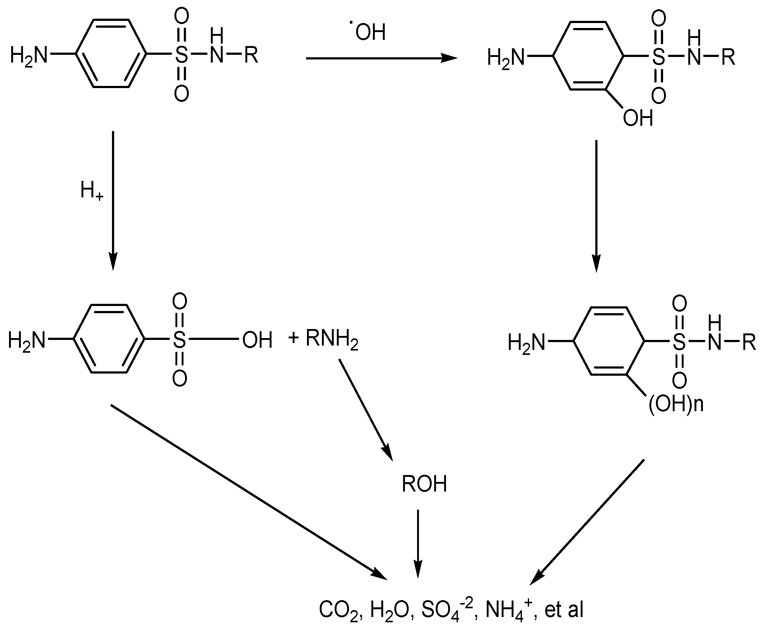
  47. Yang, H.; Li, G.; An, T.; Gao, Y.; Fu, J. Photocatalytic degradation kinetics and mechanism of environmental pharmaceuticals in aqueous suspension of TiO2: A case of sulfa drugs. Catal. Today 2010, 153, 200–207. [Google Scholar] [CrossRef]
  48. Barzic, A.I.; Ioan, S. Antibacterial Drugs—From Basic Concepts to Complex Therapeutic Mechanisms of Polymer Systems; IntechOpen: London, UK, 2015; Volume 2015. [Google Scholar]
  49. Xiang, S.; Dong, H.; Li, Y.; Xiao, J.; Dong, Q.; Hou, X.; Chu, D. A comparative study of activation of peroxymonosulfate and peroxydisulfate by greigite (Fe3S4) for the degradation of sulfamethazine in water. Sep. Purif. Technol. 2022, 290, 120873. [Google Scholar] [CrossRef]
  50. Rokesh, K.; Sakar, M.; Do, T.-O. Emerging hybrid nanocomposite photocatalysts for the degradation of antibiotics: Insights into their designs and mechanisms. Nanomaterials 2021, 11, 572. [Google Scholar] [CrossRef]
  51. Ambrosetti, B.; Campanella, L.; Palmisano, R. Degradation of antibiotics in aqueous solution by photocatalytic process: Comparing the efficiency in the use of ZnO or TiO2. Nanomaterials 2015, 4, 273–281. [Google Scholar] [CrossRef] [Green Version]
  52. Calvete, M.J.; Piccirillo, G.; Vinagreiro, C.S.; Pereira, M. Hybrid materials for heterogeneous photocatalytic degradation of antibiotics. Coord. Chem. Rev. 2019, 395, 63–85. [Google Scholar] [CrossRef]
  53. Wang, W.; Xiao, K.; Zhu, L.; Yin, Y.; Wang, Z. Graphene oxide supported titanium dioxide & ferroferric oxide hybrid, a magnetically separable photocatalyst with enhanced photocatalytic activity for tetracycline hydrochloride degradation. RSC Adv. 2017, 7, 21287–21297. [Google Scholar]
  54. Ryu, W.-S. Virus life cycle. In Molecular Virology of Human Pathogenic Viruses; Academic Press: Cambridge, MA, USA, 2017; pp. 31–45. [Google Scholar]
  55. Connolly, S.A.; Jackson, J.O.; Jardetzky, T.S.; Longnecker, R. Fusing structure and function: A structural view of the herpesvirus entry machinery. Nat. Rev. Microbiol. 2011, 9, 369–381. [Google Scholar] [CrossRef]
  56. De Clercq, E.; Li, G. Approved antiviral drugs over the past 50 years. Am. Soc. Microbiol. J. 2016, 29, 695–747. [Google Scholar] [CrossRef] [Green Version]
  57. He, H. Vaccines and antiviral agents. Front. Immunol. 2013, 2013, 239–250. [Google Scholar]
  58. Parks, J.M.; Smith, J.C. How to discover antiviral drugs quickly. N. Engl. J. Med. 2020, 382, 2261–2264. [Google Scholar] [CrossRef] [PubMed]
  59. Eryildiz, B.; Ozgun, H.; Ersahin, M.E.; Koyuncu, I. Antiviral drugs against influenza: Treatment methods, environmental risk assessment and analytical determination. J. Environ. Manag. 2022, 318, 115523. [Google Scholar] [CrossRef] [PubMed]
  60. Qi, J.; Zhang, W.; Xiang, R.; Liu, K.; Wang, H.Y.; Chen, M.; Han, Y.; Cao, R. Porous nickel–iron oxide as a highly efficient electrocatalyst for oxygen evolution reaction. Adv. Sci. 2015, 2, 1500199. [Google Scholar] [CrossRef] [PubMed]
  61. Mohammadi, S.; Moussavi, G.; Kiyanmehr, K.; Shekoohiyan, S.; Heidari, M.; Naddafi, K.; Giannakis, S. Degradation of the antiviral remdesivir by a novel, continuous-flow, helical-baffle incorporating VUV/UVC photoreactor: Performance assessment and enhancement by inorganic peroxides. Sep. Purif. Technol. 2022, 298, 121665. [Google Scholar] [CrossRef]
  62. Abbas, K.K.; Abdulkadhim Al-Ghaban, A.M.; Rdewi, E.H. Synthesis of a novel ZnO/TiO2-nanorod MXene heterostructured nanophotocatalyst for the removal pharmaceutical ceftriaxone sodium from aqueous solution under simulated sunlight. J. Environ. Chem. Eng. 2022, 10, 108111. [Google Scholar] [CrossRef]
  63. Thi, L.-A.P.; Panchangam, S.C.; Do, H.-T.; Nguyen, V.-H. Prospects and challenges of photocatalysis for degradation and mineralization of antiviral drugs. Nanostructured Photocatal. 2021, 489–517. [Google Scholar]
  64. Wang, Z.; Gao, S.; Dai, Q.; Zhao, M.; Yang, F. Occurrence and risk assessment of psychoactive substances in tap water from China. Environ. Pollut. 2020, 261, 114163. [Google Scholar] [CrossRef]
  65. Breivik, H.; Collett, B.; Ventafridda, V.; Cohen, R.; Gallacher, D. Survey of chronic pain in Europe: Prevalence, impact on daily life, and treatment. Eur. J. Pain 2006, 10, 287–333. [Google Scholar] [CrossRef]
  66. Goldstein, D.J.; Lu, Y.; Detke, M.J.; Lee, T.C.; Iyengar, S. Duloxetine vs. placebo in patients with painful diabetic neuropathy. Pain 2005, 116, 109–118. [Google Scholar] [CrossRef]
  67. Sawynok, J. Topical and peripherally acting analgesics. Pharmacol. Rev. 2003, 55, 1–20. [Google Scholar] [CrossRef] [PubMed] [Green Version]
  68. Micó, J.A.; Ardid, D.; Berrocoso, E.; Eschalier, A. Antidepressants and pain. Trends Pharmacol. Sci. 2006, 27, 348–354. [Google Scholar] [CrossRef] [PubMed]
  69. Sawynok, J.; Reid, A.R.; Esser, M. Peripheral antinociceptive action of amitriptyline in the rat formalin test: Involvement of adenosine. Pain 1999, 80, 45–55. [Google Scholar] [CrossRef] [PubMed]
  70. Banks, S.M.; Kerns, R.D. Explaining high rates of depression in chronic pain: A diathesis-stress framework. Psychol. Bull. 1996, 119, 95. [Google Scholar] [CrossRef]
  71. Leo, R. Chronic pain and comorbid depression. Front. Psychiatry 2005, 7, 403–412. [Google Scholar] [CrossRef] [PubMed]
  72. Castillo-Zacarías, C.; Barocio, M.E.; Hidalgo-Vázquez, E.; Sosa-Hernández, J.E.; Parra-Arroyo, L.; López-Pacheco, I.Y.; Barceló, D.; Iqbal, H.N.; Parra-Saldívar, R. Antidepressant drugs as emerging contaminants: Occurrence in urban and non-urban waters and analytical methods for their detection. Sci. Total Environ. 2021, 757, 143722. [Google Scholar] [CrossRef] [PubMed]
  73. Gornik, T.; Kovacic, A.; Heath, E.; Hollender, J.; Kosjek, T. Biotransformation study of antidepressant sertraline and its removal during biological wastewater treatment. Water Res. 2020, 181, 115864. [Google Scholar] [CrossRef]
  74. Hollman, J.; Dominic, J.A.; Achari, G.; Langford, C.H.; Tay, J.-H. Effect of UV dose on degradation of venlafaxine using UV/H2O2: Perspective of augmenting UV units in wastewater treatment. Environ. Technol. 2018, 41, 1107–1116. [Google Scholar] [CrossRef]
  75. Luo, J.; Dai, Y.; Xu, X.; Liu, Y.; Yang, S.; He, H.; Sun, C.; Xian, Q. Green and efficient synthesis of Co-MOF-based/g-C3N4 composite catalysts to activate peroxymonosulfate for degradation of the antidepressant venlafaxine. J. Colloid Interface Sci. 2022, 610, 280–294. [Google Scholar] [CrossRef]
  76. Krysanov, E.Y.; Pavlov, D.; Demidova, T.; Dgebuadze, Y. Effect of nanoparticles on aquatic organisms. Biol. Bull. 2010, 37, 406–412. [Google Scholar] [CrossRef]
  77. Buchman, J.T.; Hudson-Smith, N.V.; Landy, K.M.; Haynes, C.L. Understanding nanoparticle toxicity mechanisms to inform redesign strategies to reduce environmental impact. Acc. Chem. Res. 2019, 52, 1632–1642. [Google Scholar] [CrossRef] [PubMed]
  78. Ishida, N.; Hosokawa, Y.; Imaeda, T.; Hatanaka, T. Reduction of the cytotoxicity of copper (II) oxide nanoparticles by coating with a surface-binding peptide. Appl. Biochem. Biotechnol. 2020, 190, 645–659. [Google Scholar] [CrossRef] [PubMed]
  79. Zivari Fard, M.; Fatholahi, M.; Abyadeh, M.; Bakhtiarian, A.; Mousavi, S.E.; Falahati, M. The Investigation of the Cytotoxicity of Copper Oxide Nanoparticles on Peripheral Blood Mononuclear Cells. Nanomed. Res. J. 2020, 5, 364–368. [Google Scholar]
  80. Singh, Z.; Singh, I. CTAB surfactant assisted and high pH nano-formulations of CuO nanoparticles pose greater cytotoxic and genotoxic effects. Sci. Rep. 2019, 9, 5880. [Google Scholar] [CrossRef] [Green Version]
  81. Varma, R.S. Greener approach to nanomaterials and their sustainable applications. Curr. Opin. Chem. Eng. 2012, 1, 123–128. [Google Scholar] [CrossRef]
  82. Pan, C.; Zhu, F.; Wu, M.; Jiang, L.; Zhao, X.; Yang, M. Degradation and toxicity of the antidepressant fluoxetine in an aqueous system by UV irradiation. Chemosphere 2022, 287, 132434. [Google Scholar] [CrossRef]
  83. Sobolev, K.; Gutiérrez, M.F. How nanotechnology can change the concrete world. Structures 2005, 84, 14. [Google Scholar]
  84. Roco, M.C. National nanotechnology initiative-past, present, future. Nanosci. Eng. Technol. 2007, 2, 3.1–3.26. [Google Scholar]
  85. Khan, S.T.; Musarrat, J.; Al-Khedhairy, A.A. Countering drug resistance, infectious diseases, and sepsis using metal and metal oxides nanoparticles: Current status. Colloids Surf. B Biointerfaces 2016, 146, 70–83. [Google Scholar] [CrossRef]
  86. Ali, A.S. Application of nanomaterials in environmental improvement. In Nanotechnology and the Environment; IntechOpen: London, UK, 2020. [Google Scholar]
  87. Ahmad, A.; Mukherjee, P.; Senapati, S.; Mandal, D.; Khan, M.I.; Kumar, R.; Sastry, M. Extracellular biosynthesis of silver nanoparticles using the fungus Fusarium oxysporum. Colloids Surf. B Biointerfaces 2003, 28, 313–318. [Google Scholar] [CrossRef]
  88. Rao, M.D.; Gautam, P. Synthesis and characterization of ZnO nanoflowers using C hlamydomonas reinhardtii: A green approach. Environ. Prog. Sustain. Energy 2016, 35, 1020–1026. [Google Scholar] [CrossRef]
  89. Cele, T. Preparation of Nanoparticles in Engineered Nanomaterials: Health and Safety; Avramescu, S.M., Fierascu, I., Akhtar, K., Khan, S.B., Ali, F., Asiri, A., Eds.; IntechOpen: London, UK, 2020; pp. 15–28. [Google Scholar]
  90. Malhotra, S.P.K.; Alghuthaymi, M.A. Biomolecule-assisted biogenic synthesis of metallic nanoparticles. In Agri-Waste and Microbes for Production of Sustainable Nanomaterials, A Volume in Nanobiotechnology for Plant Protection; Abd-Elsalam, K.A., Periakaruppan, R., Rajeshkumar, S., Eds.; Elsevier: Amsterdam, The Netherlands, 2022; pp. 139–163. [Google Scholar]
  91. Abdelghany, T.; Al-Rajhi, A.M.; Al Abboud, M.A.; Alawlaqi, M.; Ganash Magdah, A.; Helmy, E.A.; Mabrouk, A.S. Recent advances in green synthesis of silver nanoparticles and their applications: About future directions: A review. Bionanoscience 2018, 8, 5–16. [Google Scholar] [CrossRef]
  92. Thirumalai Arasu, V.; Prabhu, D.; Soniya, M. Stable silver nanoparticle synthesizing methods and its applications. J. Biosci. Res. 2010, 1, 259–270. [Google Scholar]
  93. Iravani, S.; Korbekandi, H.; Mirmohammadi, S.V.; Zolfaghari, B. Synthesis of silver nanoparticles: Chemical, physical and biological methods. Res. Pharm. Sci. 2014, 9, 385. [Google Scholar] [PubMed]
  94. Molina, G.A.; Esparza, R.; López-Miranda, J.L.; Hernández-Martínez, A.R.; España-Sánchez, B.L.; Elizalde-Peña, E.A.; Estevez, M. Green synthesis of Ag nanoflowers using Kalanchoe Daigremontiana extract for enhanced photocatalytic and antibacterial activities. Colloids Surf. B Biointerfaces 2019, 180, 141–149. [Google Scholar] [CrossRef] [PubMed]
  95. Amooaghaie, R.; Saeri, M.R.; Azizi, M. Synthesis, characterization and biocompatibility of silver nanoparticles synthesized from Nigella sativa leaf extract in comparison with chemical silver nanoparticles. Ecotoxicol. Environ. Saf. 2015, 120, 400–408. [Google Scholar] [CrossRef]
  96. Kumar, B.; Smita, K.; Cumbal, L.; Debut, A. Phytosynthesis of silver nanoparticles using andean cabbage: Structural characterization and its application. Mater. Today Proc. 2020, 21, 2079–2086. [Google Scholar] [CrossRef]
  97. Kumar, B.; Vizuete, K.S.; Sharma, V.; Debut, A.; Cumbal, L. Ecofriendly synthesis of monodispersed silver nanoparticles using Andean Mortiño berry as reductant and its photocatalytic activity. Vacuum 2019, 160, 272–278. [Google Scholar] [CrossRef]
  98. Sharma, P.; Pant, S.; Rai, S.; Yadav, R.B.; Dave, V. Green synthesis of silver nanoparticle capped with Allium cepa and their catalytic reduction of textile dyes: An ecofriendly approach. J. Polym. Environ. 2018, 26, 1795–1803. [Google Scholar] [CrossRef]
  99. Vizuete, K.S.; Kumar, B.; Guzmán, K.; Debut, A.; Cumbal, L. Shora (Capparis petiolaris) fruit mediated green synthesis and application of silver nanoparticles. Green Process. Synth. 2017, 6, 23–30. [Google Scholar] [CrossRef]
  100. Edison, T.N.J.I.; Lee, Y.R.; Sethuraman, M.G. Green synthesis of silver nanoparticles using Terminalia cuneata and its catalytic action in reduction of direct yellow-12 dye. Spectrochim. Acta Part A Mol. Biomol. Spectrosc. 2016, 161, 122–129. [Google Scholar] [CrossRef] [PubMed]
  101. Saffar, R.; Athira, P.; Kalita, K.; Manuel, S.G.; Pradeep, N. Nanoparticle synthesis from biowaste and its potential as an antimicrobial agent. Res. Sq. 2021, 1–16. [Google Scholar]
  102. Kumar, B.; Smita, K.; Debut, A.; Cumbal, L. Utilization of Persea americana (Avocado) oil for the synthesis of gold nanoparticles in sunlight and evaluation of antioxidant and photocatalytic activities. Environ. Nanotechnol. Monit. Manag. 2018, 10, 231–237. [Google Scholar] [CrossRef]
  103. Kumar, B.; Smita, K.; Cumbal, L.; Debut, A. Extracellular biofabrication of gold nanoparticles by using Lantana camara berry extract. Inorg. Nano-Met. Chem. 2017, 47, 138–142. [Google Scholar] [CrossRef]
  104. Desai, M.P.; Sangaokar, G.M.; Pawar, K.D. Kokum fruit mediated biogenic gold nanoparticles with photoluminescent, photocatalytic and antioxidant activities. Process Biochem. 2018, 70, 188–197. [Google Scholar] [CrossRef]
  105. Francis, S.; Joseph, S.; Koshy, E.P.; Mathew, B. Green synthesis and characterization of gold and silver nanoparticles using Mussaenda glabrata leaf extract and their environmental applications to dye degradation. Environ. Sci. Pollut. Res. 2017, 24, 17347–17357. [Google Scholar] [CrossRef] [PubMed]
  106. Mythili, R.; Selvankumar, T.; Srinivasan, P.; Sengottaiyan, A.; Sabastinraj, J.; Ameen, F.; Al-Sabri, A.; Kamala-Kannan, S.; Govarthanan, M.; Kim, H. Biogenic synthesis, characterization and antibacterial activity of gold nanoparticles synthesised from vegetable waste. J. Mol. Liq. 2018, 262, 318–321. [Google Scholar] [CrossRef]
  107. Nagajyothi, P.C.; Prabhakar Vattikuti, S.V.; Devarayapalli, K.C.; Yoo, K.; Shim, J.; Sreekanth, T.V.M. Green synthesis: Photocatalytic degradation of textile dyes using metal and metal oxide nanoparticles-latest trends and advancements. Crit. Rev. Environ. Sci. Technol. 2020, 50, 2617–2723. [Google Scholar] [CrossRef]
  108. Anchan, S.; Pai, S.; Sridevi, H.; Varadavenkatesan, T.; Vinayagam, R.; Selvaraj, R. Biogenic synthesis of ferric oxide nanoparticles using the leaf extract of Peltophorum pterocarpum and their catalytic dye degradation potential. Biocatal. Agric. Biotechnol. 2019, 20, 101251. [Google Scholar] [CrossRef]
  109. Hoag, G.E.; Collins, J.B.; Holcomb, J.L.; Hoag, J.R.; Nadagouda, M.N.; Varma, R.S. Degradation of bromothymol blue by ‘greener’nano-scale zero-valent iron synthesized using tea polyphenols. J. Mater. Chem. 2009, 19, 8671–8677. [Google Scholar] [CrossRef]
  110. Zhang, Y.; Chen, Y.; Kang, Z.-W.; Gao, X.; Zeng, X.; Liu, M.; Yang, D.-P. Waste eggshell membrane-assisted synthesis of magnetic CuFe2O4 nanomaterials with multifunctional properties (adsorptive, catalytic, antibacterial) for water remediation. Colloids Surf. A Physicochem. Eng. Asp. 2021, 612, 125874. [Google Scholar] [CrossRef]
  111. Ajmal, N.; Saraswat, K.; Bakht, M.A.; Riadi, Y.; Ahsan, M.J.; Noushad, M. Cost-effective and eco-friendly synthesis of titanium dioxide (TiO2) nanoparticles using fruit’s peel agro-waste extracts: Characterization, in vitro antibacterial, antioxidant activities. Green Chem. Lett. Rev. 2019, 12, 244–254. [Google Scholar] [CrossRef] [Green Version]
  112. Farag, S.; Amr, A.; El-Shafei, A.; Asker, M.S.; Ibrahim, H.M. Green synthesis of titanium dioxide nanoparticles via bacterial cellulose (BC) produced from agricultural wastes. Cellulose 2021, 28, 7619–7632. [Google Scholar] [CrossRef]
  113. Agarwal, H.; Kumar, S.V.; Rajeshkumar, S. A review on green synthesis of zinc oxide nanoparticles–An eco-friendly approach. Resour. Effic. Technol. 2017, 3, 406–413. [Google Scholar] [CrossRef]
  114. Jamarani, R.; Erythropel, H.C.; Nicell, J.A.; Leask, R.L.; Marić, M. How green is your plasticizer? Polymers 2018, 10, 834. [Google Scholar] [CrossRef] [PubMed] [Green Version]
  115. Kumar, H.; Bhardwaj, K.; Kuča, K.; Kalia, A.; Nepovimova, E.; Verma, R.; Kumar, D. Flower-based green synthesis of metallic nanoparticles: Applications beyond fragrance. Nanomaterials 2020, 10, 766. [Google Scholar] [CrossRef] [Green Version]
  116. Gardea-Torresdey, J.L.; Parsons, J.; Gomez, E.; Peralta-Videa, J.; Troiani, H.; Santiago, P.; Yacaman, M. Formation and growth of Au nanoparticles inside live alfalfa plants. Am. Chem. Soc. 2002, 2, 397–401. [Google Scholar] [CrossRef]
  117. Bali, R.; Razak, N.; Lumb, A.; Harris, A. The synthesis of metallic nanoparticles inside live plants. In Proceedings of the 2006 International Conference on Nanoscience and Nanotechnology, Brisbane, Australia, 3–7 July 2006. [Google Scholar]
  118. Iravani, S. Green synthesis of metal nanoparticles using plants. Green Chem. 2011, 13, 2638–2650. [Google Scholar] [CrossRef]
  119. Wang, Y.; O’Connor, D.; Shen, Z.; Lo, I.M.; Tsang, D.C.; Pehkonen, S.; Pu, S.; Hou, D. Green synthesis of nanoparticles for the remediation of contaminated waters and soils: Constituents, synthesizing methods, and influencing factors. Nanotechnol. Rev. 2019, 226, 540–549. [Google Scholar] [CrossRef]
  120. Ahmad, N.; Sharma, S.; Singh, V.; Shamsi, S.; Fatma, A.; Mehta, B. Biosynthesis of silver nanoparticles from Desmodium triflorum: A novel approach towards weed utilization. Biotechnol. Res. Int. 2011, 2011, 1–8. [Google Scholar] [CrossRef] [Green Version]
  121. Rasheed, T.; Bilal, M.; Li, C.; Nabeel, F.; Khalid, M.; Iqbal, H.M. Catalytic potential of bio-synthesized silver nanoparticles using Convolvulus arvensis extract for the degradation of environmental pollutants. J. Photochem. Photobiol. 2018, 181, 44–52. [Google Scholar] [CrossRef] [PubMed]
  122. Ahadi, M.; Saber Tehrani, M.; Aberoomand Azar, P.; Waqif Husain, S. Novel preparation of sensitized ZnS nanoparticles and its use in photocatalytic degradation of tetracycline. Int. J. Environ. Sci. Technol. 2016, 13, 2797–2804. [Google Scholar] [CrossRef]
  123. Ahuja, T.; Prakash, J. Recent advancements in designing Au/Ag based plasmonic photocatalysts for efficient photocatalytic degradation. In Gold and Silver Nanoparticles; Elsevier: Amsterdam, The Netherlands, 2023; pp. 379–410. [Google Scholar]
  124. Nguyen, T.H.A.; Doan, V.-D.; Tran, A.V.; Nguyen, V.C.; Nguyen, A.-T.; Vasseghian, Y. Green synthesis of Nb-doped ZnO nanocomposite for photocatalytic degradation of tetracycline antibiotic under visible light. Mater. Lett. 2022, 308, 131129. [Google Scholar] [CrossRef]
  125. Hosny, M.; Fawzy, M.; Eltaweil, A.S. Green synthesis of bimetallic Ag/ZnO@ Biohar nanocomposite for photocatalytic degradation of tetracycline, antibacterial and antioxidant activities. Sci. Rep. 2022, 12, 7316. [Google Scholar] [CrossRef] [PubMed]
  126. Batterjee, M.G.; Nabi, A.; Kamli, M.R.; Alzahrani, K.A.; Danish, E.Y.; Malik, M.A. Green Hydrothermal Synthesis of Zinc Oxide Nanoparticles for UV-Light-Induced Photocatalytic Degradation of Ciprofloxacin Antibiotic in an Aqueous Environment. Catalysts 2022, 12, 1347. [Google Scholar] [CrossRef]
  127. El-Kemary, M.; El-Shamy, H.; El-Mehasseb, I. Photocatalytic degradation of ciprofloxacin drug in water using ZnO nanoparticles. J. Lumin. 2010, 130, 2327–2331. [Google Scholar] [CrossRef]
  128. Emeline, A.V.; Kuznetsov, V.N.; Rybchuk, V.K.; Serpone, N. Visible-light-active titania photocatalysts: The case of N-doped s—Properties and some fundamental issues. Int. J. Photoenergy 2008, 2008, 258394. [Google Scholar] [CrossRef] [Green Version]
  129. Thakur, A.; Kumar, P.; Kaur, D.; Devunuri, N.; Sinha, R.; Devi, P. TiO2 nanofibres decorated with green-synthesized P Au/Ag@ CQDs for the efficient photocatalytic degradation of organic dyes and pharmaceutical drugs. RSC Adv. 2020, 10, 8941–8948. [Google Scholar] [CrossRef] [Green Version]
  130. Malassis, L.; Dreyfus, R.; Murphy, R.J.; Hough, L.A.; Donnio, B.; Murray, C.B. One-step green synthesis of gold and silver nanoparticles with ascorbic acid and their versatile surface post-functionalization. RSC Adv. 2016, 6, 33092–33100. [Google Scholar] [CrossRef]
  131. Nadagouda, M.N.; Varma, R.S. Green and controlled synthesis of gold and platinum nanomaterials using vitamin B2: Density-assisted self-assembly of nanospheres, wires and rods. Green Chem. 2006, 8, 516–518. [Google Scholar] [CrossRef]
  132. Mookriang, S.; Jimtaisong, A.; Saewan, N.; Kittigowittana, K.; Rachtanapun, P.; Pathawinthranond, V.; Sarakornsri, T. Green synthesis of silver nanoparticles using a vitamin C rich Phyllanthus emblica extract. Adv. Mater. Res. 2013, 662, 864–868. [Google Scholar] [CrossRef]
  133. Nadagouda, M.N.; Varma, R.S. A greener synthesis of core (Fe, Cu)-shell (Au, Pt, Pd, and Ag) nanocrystals using aqueous vitamin C. Cryst. Growth Des. 2007, 7, 2582–2587. [Google Scholar] [CrossRef]
  134. Francis, S.; Nair, K.M.; Paul, N.; Koshy, E.P.; Mathew, B. Catalytic activities of green synthesized silver and gold nanoparticles. Bioprocess Biosyst. Eng. 2019, 9, 97–104. [Google Scholar] [CrossRef]
  135. Selvin, J.; Huxley, A.; Lipton, A. Immunomodulatory potential of marine secondary metabolites against bacterial diseases of shrimp. Aquaculture 2004, 230, 241–248. [Google Scholar] [CrossRef] [Green Version]
  136. Luangpipat, T.; Beattie, I.R.; Chisti, Y.; Haverkamp, R.G. Gold nanoparticles produced in a microalga. J. Nanoparticle Res. 2011, 13, 6439–6445. [Google Scholar] [CrossRef]
  137. Abdel-Raouf, N.; Al-Enazi, N.M.; Ibraheem, I.B. Green biosynthesis of gold nanoparticles using Galaxaura elongata and characterization of their antibacterial activity. Arab. J. Chem. 2017, 10, S3029–S3039. [Google Scholar] [CrossRef] [Green Version]
  138. Borah, D.; Das, N.; Das, N.; Bhattacharjee, A.; Sarmah, P.; Ghosh, K.; Chandel, M.; Rout, J.; Pandey, P.; Ghosh, N. Alga-mediated facile green synthesis of silver nanoparticles: Photophysical, catalytic and antibacterial activity. Appl. Organomet. Chem. 2020, 34, 5597. [Google Scholar] [CrossRef]
  139. Lengke, M.F.; Fleet, M.E.; Southam, G. Morphology of gold nanoparticles synthesized by filamentous cyanobacteria from gold (I)−thiosulfate and gold (III)−chloride complexes. Langmuir 2006, 22, 2780–2787. [Google Scholar] [CrossRef]
  140. Singaravelu, G.; Arockiamary, J.; Kumar, V.G.; Govindaraju, K. A novel extracellular synthesis of monodisperse gold nanoparticles using marine alga, Sargassum wightii Greville. Colloids Surf. B Biointerfaces 2007, 57, 97–101. [Google Scholar] [CrossRef]
  141. Thema, F.; Manikandan, E.; Dhlamini, M.; Maaza, M. Green synthesis of ZnO nanoparticles via Agathosma betulina natural extract. Mater. Lett. 2015, 161, 124–127. [Google Scholar] [CrossRef]
  142. Hulkoti, N.I.; Taranath, T. Biosynthesis of nanoparticles using microbes—A review. Colloids Surf. B Biointerfaces 2014, 121, 474–483. [Google Scholar] [CrossRef] [PubMed]
  143. Anil Kumar, S.; Abyaneh, M.K.; Gosavi, S.; Kulkarni, S.K.; Pasricha, R.; Ahmad, A.; Khan, M. Nitrate reductase-mediated synthesis of silver nanoparticles from AgNO3. Biotechnol. Lett. 2007, 29, 439–445. [Google Scholar] [CrossRef] [PubMed]
  144. Narayanan, K.B.; Sakthivel, N. Biological synthesis of metal nanoparticles by microbes. Adv. Colloid Interface Sci. 2010, 156, 1–13. [Google Scholar] [CrossRef] [PubMed]
  145. Vigneshwaran, N.; Ashtaputre, N.; Varadarajan, P.; Nachane, R.; Paralikar, K.; Balasubramanya, R. Biological synthesis of silver nanoparticles using the fungus Aspergillus flavus. Mater. Lett. 2007, 61, 1413–1418. [Google Scholar] [CrossRef]
  146. Ahmad, A.; Senapati, S.; Khan, M.I.; Kumar, R.; Sastry, M. Extra-/intracellular biosynthesis of gold nanoparticles by an alkalotolerant fungus, Trichothecium sp. J. Biomed. Nanotechnol. 2005, 1, 47–53. [Google Scholar] [CrossRef]
  147. Clarance, P.; Luvankar, B.; Sales, J.; Khusro, A.; Agastian, P.; Tack, J.-C.; Al Khulaifi, M.M.; Al-Shwaiman, H.A.; Elgorban, A.M.; Syed, A. Green synthesis and characterization of gold nanoparticles using endophytic fungi Fusarium solani and its in-vitro anticancer and biomedical applications. Saudi J. Biol. Sci. 2020, 27, 706–712. [Google Scholar] [CrossRef]
  148. Mittal, S.; Roy, A. Fungus and plant-mediated synthesis of metallic nanoparticles and their application in degradation of dyes. In Photocatalytic Degradation of Dyes; Elsevier: Amsterdam, The Netherlands, 2021; pp. 287–308. [Google Scholar]
  149. Zhao, X.; Zhou, L.; Riaz Rajoka, M.S.; Yan, L.; Jiang, C.; Shao, D.; Zhu, J.; Shi, J.; Huang, Q.; Yang, H. Fungal silver nanoparticles: Synthesis, application and challenges. Crit. Rev. Biotechnol. 2018, 38, 817–835. [Google Scholar] [CrossRef]
  150. Islam, S.N.; Naqvi, S.M.A.; Parveen, S.; Ahmad, A. Endophytic fungus-assisted biosynthesis, characterization and solar photocatalytic activity evaluation of nitrogen-doped Co3O4 nanoparticles. Appl. Nanosci. 2021, 11, 1651–1659. [Google Scholar] [CrossRef]
  151. Bai, H.-J.; Zhang, Z.-M.; Gong, J. Biological synthesis of semiconductor zinc sulfide nanoparticles by immobilized Rhodobacter sphaeroides. Biotechnol. Lett. 2006, 28, 1135–1139. [Google Scholar] [CrossRef]
  152. Abd Elsalam, S.S.; Taha, R.H.; Tawfeik, A.M.; El-Monem, A.; Mohamed, O.; Mahmoud, H.A. Antimicrobial activity of bio and chemical synthesized cadmium sulfide nanoparticles. Egypt. J. Hosp. Med. 2018, 70, 1494–1507. [Google Scholar] [CrossRef] [Green Version]
  153. Ghosh, S.; Ahmad, R.; Zeyaullah, M.; Khare, S.K. Microbial nano-factories: Synthesis and biomedical applications. Front. Chem. 2021, 9, 626834. [Google Scholar] [CrossRef] [PubMed]
  154. Dhandapani, P.; Maruthamuthu, S.; Rajagopal, G. Bio-mediated synthesis of TiO2 nanoparticles and its photocatalytic effect on aquatic biofilm. J. Photochem. Photobiol. B Biol. 2012, 110, 43–49. [Google Scholar] [CrossRef] [PubMed]
  155. Gu, H.; Chen, X.; Chen, F.; Zhou, X.; Parsaee, Z. Ultrasound-assisted biosynthesis of CuO-NPs using brown alga Cystoseira trinodis: Characterization, photocatalytic AOP, DPPH scavenging and antibacterial investigations. Ultrason. Sonochem. 2018, 41, 109–119. [Google Scholar] [CrossRef] [PubMed]
  156. Raliya, R.; Tarafdar, J.C. ZnO nanoparticle biosynthesis and its effect on phosphorous-mobilizing enzyme secretion and gum contents in Clusterbean (Cyamopsis tetragonoloba L.). Agric. Res. 2013, 2, 48–57. [Google Scholar] [CrossRef] [Green Version]
Scheme 1. Structure-based classification of NSAIDs. Reprinted from reference Bindu et al., 2020 with permission of Biochemical Pharmacology, 2020. Copyright (2022), Elsevier, Amsterdam, The Netherlands [25].
Catalysts 13 00511 sch001
Scheme 2. Proposed degradation mechanism of Ibuprofen by titania and zinc oxide as photocatalysts. Reprinted from Tanveer M. et al., 2019 with permission from Water Environment Research, 2019. Copyright © 1999–2023 John Wiley & Sons, Inc., New Jersey, United States [30].
Catalysts 13 00511 sch002
Scheme 3. Degradation mechanism of Naproxen. Reprinted from Patel M. et al., 2019 with permission from Chemosphere 2019. Copyright © 2023 American Chemical Society, Washington, United States [10].
Catalysts 13 00511 sch003
Scheme 4. Degradation pathway of sulfa pharmaceuticals (antibiotics). Reprinted from Yang H. et al., 2010 with permission from Catalysis Today, 2010. Copyright © 2010 Elsevier B.V., Amsterdam, The Netherlands [47].
Catalysts 13 00511 sch004
Scheme 5. Proposed photocatalytic degradation mechanism of lamivudine by TiO2. Reprinted from Thi L-AP. et al., 2021 with permission from Nanostructured Photocatalysts, 2021. Copyright © 2021 Elsevier B.V., Amsterdam, The Netherlands [62].
Catalysts 13 00511 sch005
Figure 1. The mechanism of action of antidepressants. Reprinted from Micó et al., 2006 with permission from the Trends in Pharmacological Sciences, 2006. Copyright (2022), Elsevier, Amsterdam, The Netherlands [67].
Catalysts 13 00511 g001
Figure 2. Synthesis approaches for nanomaterials.
Catalysts 13 00511 g002
Figure 3. Different sources for green synthesis of metal NPs. Reprinted from Kumar et al., 2020 with permission of International Journal of Molecular Sciences, 2020. Copyright 1996–2022 MDPI (Basel, Switzerland) [114].
Catalysts 13 00511 g003
Disclaimer/Publisher’s Note: The statements, opinions and data contained in all publications are solely those of the individual author(s) and contributor(s) and not of MDPI and/or the editor(s). MDPI and/or the editor(s) disclaim responsibility for any injury to people or property resulting from any ideas, methods, instructions or products referred to in the content.

