Article

Enhanced Photocatalytic Performance of Visible-Light-Driven BiVO4 Nanoparticles through W and Mo Substituting

1
Department of Materials Science and Engineering, Feng Chia University, Taichung 40724, Taiwan
2
Department of Chemical Engineering, Feng Chia University, Taichung 40724, Taiwan
3
Department of Environmental Engineering and Science, Feng Chia University, Taichung 40724, Taiwan
*
Author to whom correspondence should be addressed.
Catalysts 2023, 13(3), 475; https://doi.org/10.3390/catal13030475
Submission received: 30 December 2022 / Revised: 19 February 2023 / Accepted: 21 February 2023 / Published: 26 February 2023
(This article belongs to the Special Issue From Design to Application of Nanomaterials in Catalysis)

Abstract

:
Bismuth vanadate (BiVO4), W-doped BiVO4 (BiVO4:W), and Mo-doped BiVO4 (BiVO4:Mo) nanoparticles were synthesized at pH = 4 using a green hydrothermal method. The effects of 2 at% W or Mo doping on the microstructural and optical characteristics of as-prepared BiVO4 nanoparticles and the effect of combining particle morphology modification and impurity dopant incorporation on the visible-light-derived photocatalytic degradation of dilute Rhodamine B (RhB) solution are studied. XRD examination revealed that these obtained BiVO4-based nanoparticles had a highly crystalline and single monoclinic phase. SEM and TEM observations showed that impurity doping could modify the surface morphology, change the particle shape, and reduce the particle diameter to enlarge their specific surface area, increasing the reactive sites of the photocatalytic process. XPS and FL measurements indicated that W- and Mo-doped nanoparticles possessed higher concentrations of oxygen vacancies, which could promote the n-type semiconductor property. It was found that the BiVO4:W and BiVO4:Mo powder samples exhibited better photocatalytic activity for efficient RhB removal than that shown by pristine BiVO4 powder samples under visible light illumination. That feature can be ascribed to the larger surface area and improved concentration of photogenerated charge carriers of the former.

1. Introduction

Degradation of harmful substrates, specifically those sourced from industrial wastes, using semiconductor photocatalysts to remove organic pollutants from sewage remains an important issue [1,2,3]. The photocatalysis degradation approach using functional oxide semiconductors is believed to be an efficient, eco-friendly, and clean strategy for the removal of organic pollutants without any harmful byproduct [4,5]. Past studies have not only involved the selective synthesis of new visible-light-driven photocatalysts, but also focused on the improvement of photocatalytic degradation efficiency for organic contaminants, as well as investigating the effect factors and photocatalytic reaction mechanisms [6,7,8]. Bismuth vanadate (BiVO4) is a non-titania-based semiconductor photocatalyst and shows promise for application in renewable energy production systems (such as green fuel production from sunlight and water) and the resolution of environmental pollution (such as water remediation and degradation of organic pollutants) [9].
BiVO4 nanoparticles possess a monoclinic scheelite structure. They are a promising and extensively n-type semiconducting photocatalytic nanomaterial with an optical bandgap energy of approximately 2.4–2.5 eV, and they can be used to harvest solar energy [2,10]. This material has a low production cost, is non-toxic, and possesses considerable chemical stability, as well as exhibiting high photocatalytic activity under visible light irradiation [1]. To improve the photocatalytic activity, it is important to enlarge the specific surface area and improve the density of the photogenerated charge carriers of photocatalysts [7]. Theoretical electronic structural calculations have revealed that both tungsten (W) and molybdenum (Mo) are shallow donors in monoclinic BiVO4 crystals [11,12]. Substitution of W or Mo ions for some V5+ sites in BiVO4 can enhance its n-type characteristics [9,13]. Previous reports have well demonstrated that doping BiVO4 with W and Mo can extensively improve its photocatalytic performance [14,15,16,17]. Thus, appropriate amounts of W or Mo doping could increase the concentration of photogenerated charge carriers and possibly enhance the photocatalytic and photoelectrochemical performance significantly. Parmar and colleagues have reported that BiVO4 doped with 2 at% W and Mo can dramatically enhance the water photo-oxidation activity [11]. Tian et al. reported that hydrothermal Mo-doped BiVO4 microcrystals (doping levels of ~2 to 13 mol%) had a better photocurrent density than that of pure BiVO4 microcrystals [18]. Thalluri et al. found that the optimum surface atomic percentages of dopants for hydrothermally synthesized BiVO4 powders were 0.9% and 1.2% for W and Mo for sun-derived water oxidation [9].
Several wet-chemical approaches, such as the solution combustion method, sol-gel process, co-precipitation method, and hydrothermal method, can be used for fabricating low-dimensional crystalline oxide nanostructures [8,19]. The hydrothermal synthesis method is convenient and environmentally friendly, and this feasible, low-temperature process allows shape and size control [19]. It is commonly used to prepare BiVO4-based nanostructures [20]. It is well agreed that variations of the pH value of the hydrothermal reaction solution can strongly affect the morphology, particle diameter, specific surface area, and surface charge of synthesized BiVO4-based products [8,21]. Dong et al. reported that monoclinic phase BiVO4 crystalline particles synthesized in acidic solutions (pH < 5) using a hydrothermal method exhibited good adsorption capacity and excellent photocatalytic degradation performance [10]. Li et al. [3] and Ressnig et al. [22] found that BiVO4 nanoparticles synthesized at pH = 4 had excellent photocatalytic activity in organic dye degradation due to the effect of morphological features. In this study, three kinds of BiVO4-based nanoparticles were synthesized at pH = 4.0 using a hydrothermal method for the photodegradation of Rhodamine B (RhB) solution. Herein, the effects of the impurity dopant (2 at% W or Mo) on the microstructural features and optical properties of as-synthesized BiVO4 nano-sized products and the improvement of photocatalytic degradation of RhB in aqueous solution through W and Mo doping under visible light illumination at room temperature were investigated.

2. Results and Discussion

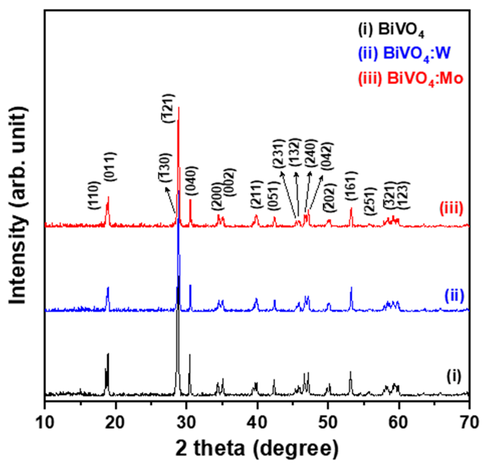
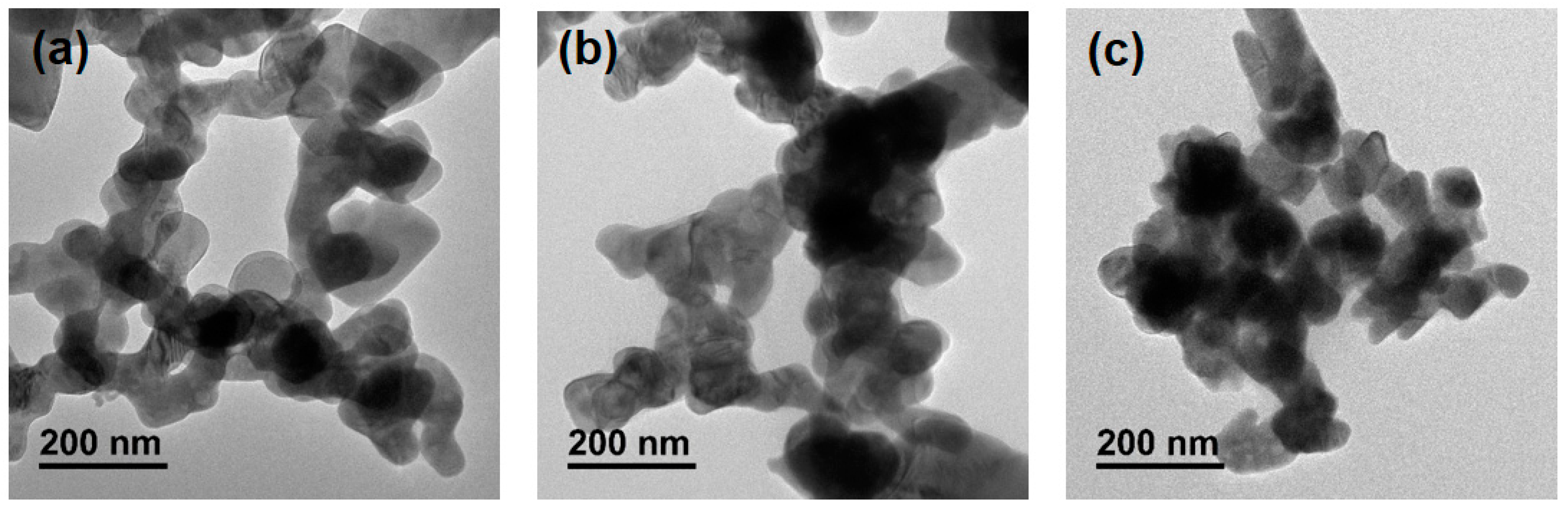
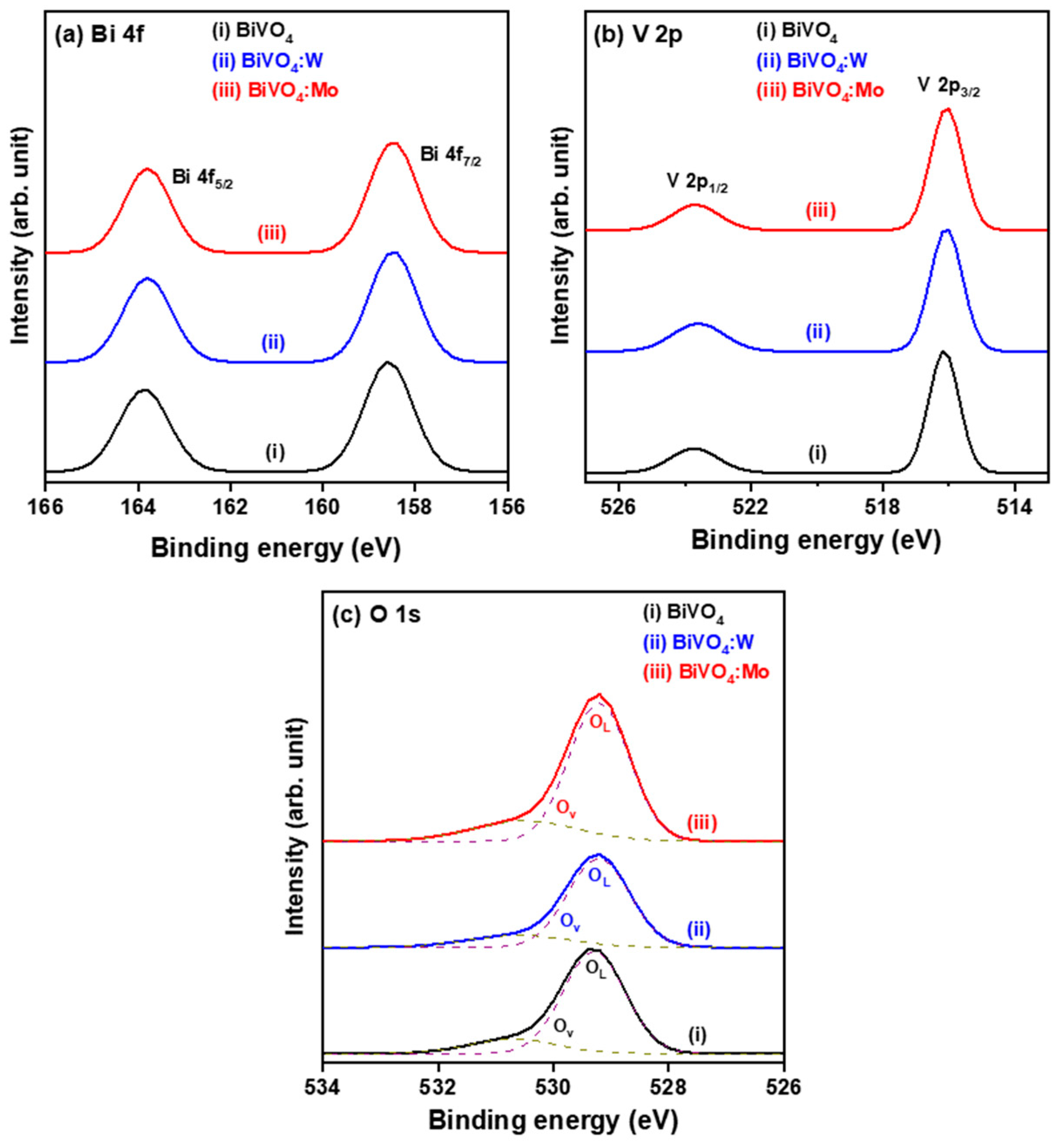
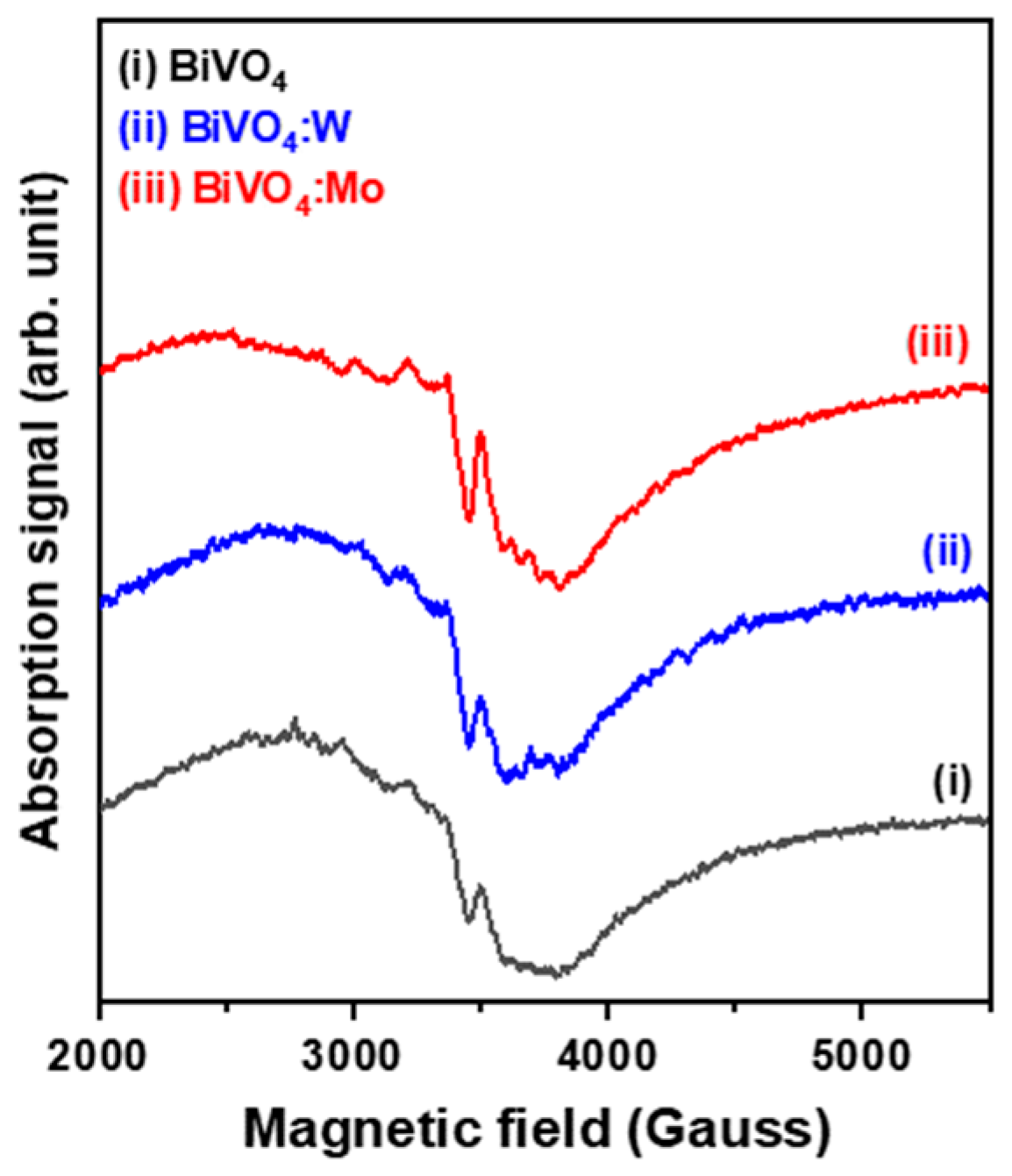
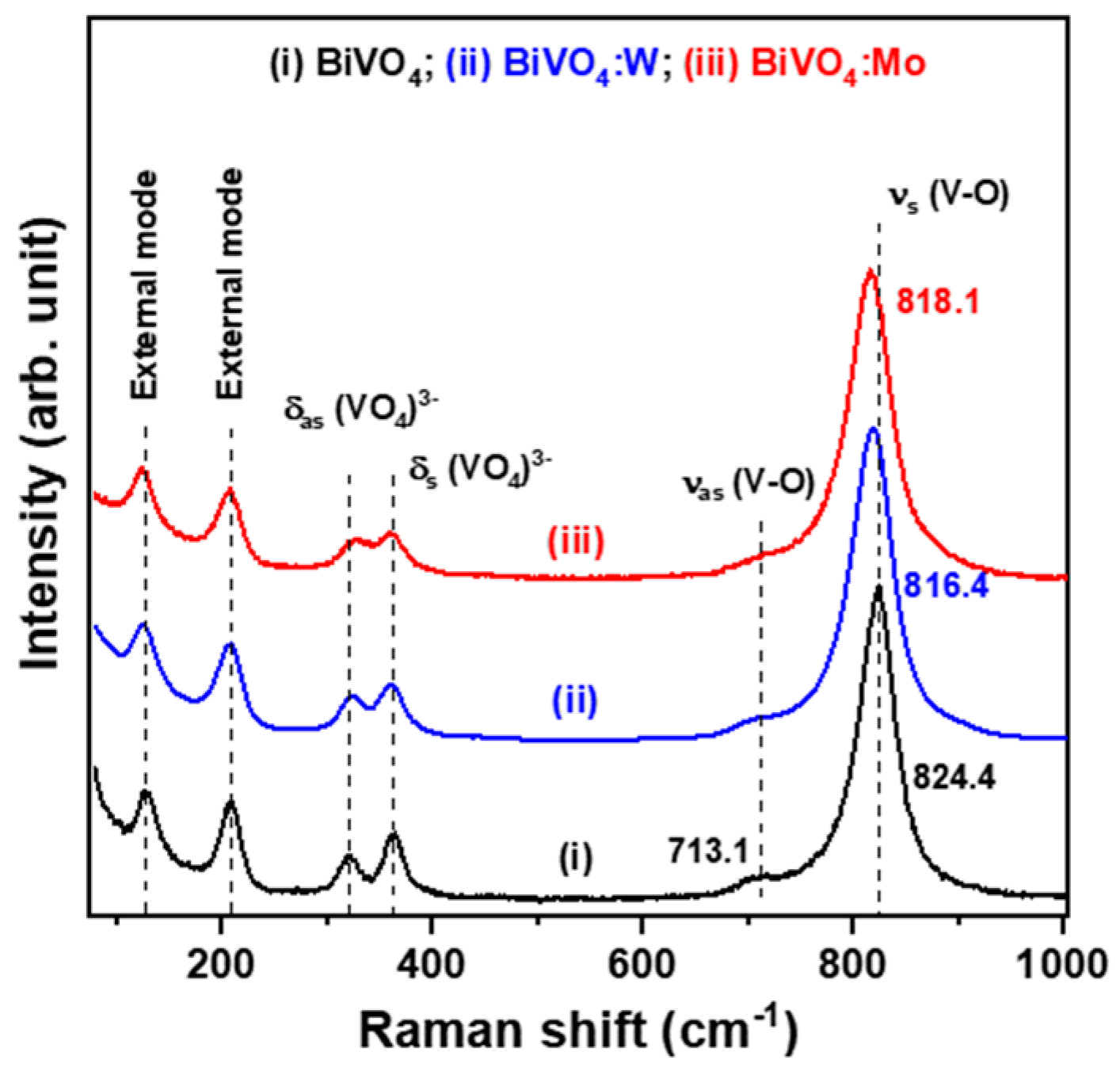
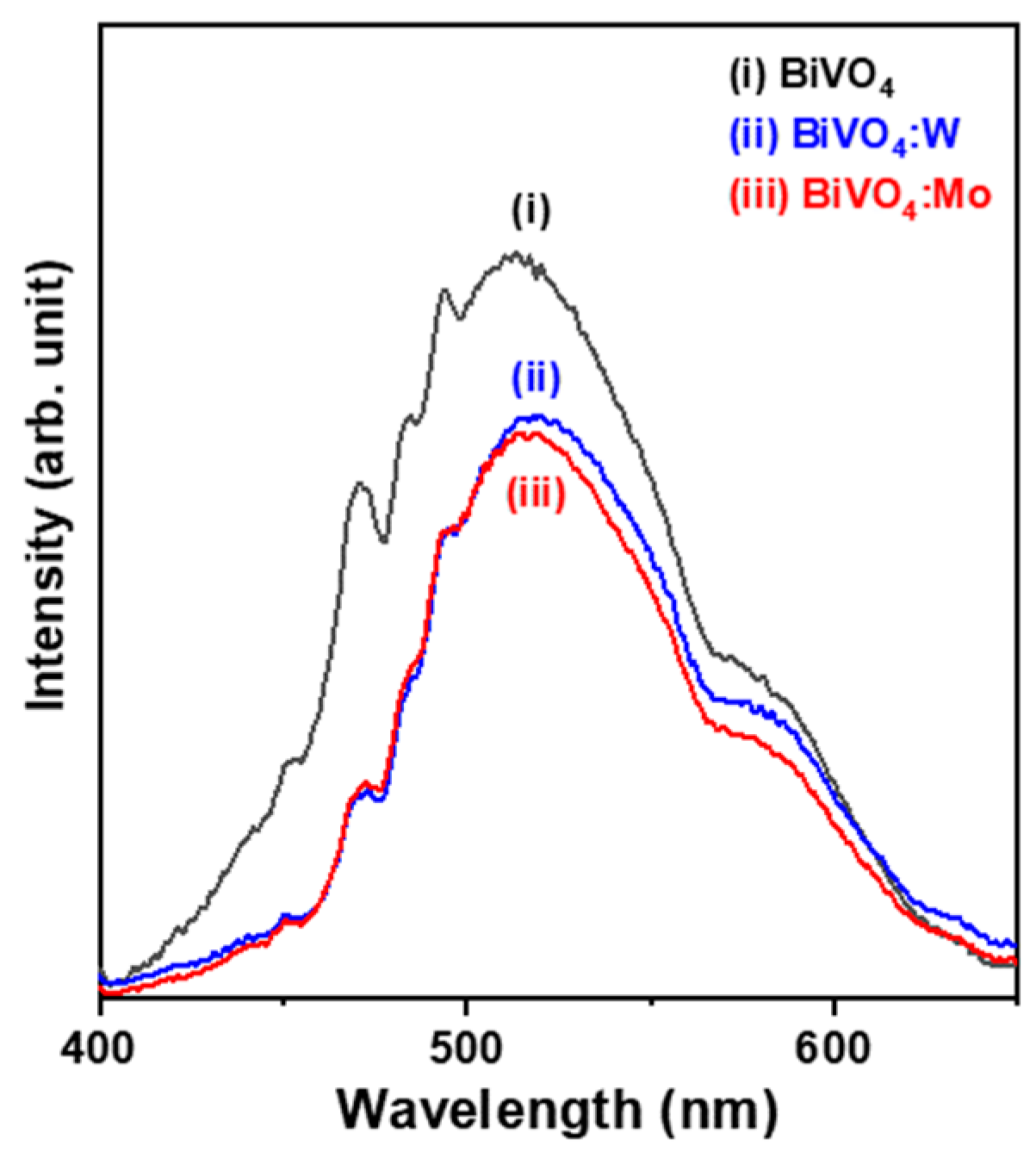
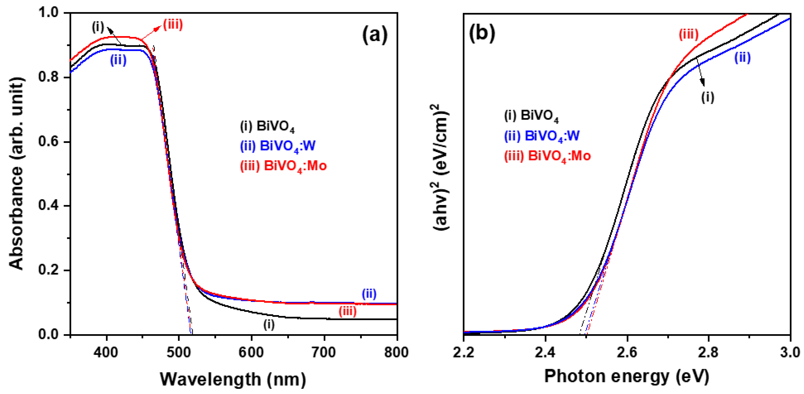
2.1. Physical Characteristics of Hydrothermally Synthesized BiVO4-Based Nanoparticles

Figure 1 presents the XRD patterns of pure BiVO4, W-doped BiVO4 (BiVO4:W), and Mo-doped BiVO4 (BiVO4:Mo) powder samples prepared at pH = 4 using a hydrothermal method. The diffraction peaks of the three XRD patterns were identified and in good agreement with the standard data for the monoclinic scheelite BiVO4 (JCPDS file No. 14-0668); in addition, no diffraction peaks other than those of the mBiVO4 phase, such as Bi2O3, appeared in the XRD patterns of these powder samples. These results indicated that pure monoclinic phase BiVO4 crystals were obtained. The two theta (2θ) angular positions of the three relative intensity peaks at 18.88°, 28.83°, and 30.55° identified the (011), (−121), and (040) lattice planes. In addition, sharp diffraction peaks were observed from the three BiVO4-based samples, revealing that the synthesized oxide powders had good crystallinity.
These hydrothermally synthesized oxide powders had a monoclinic structure and exhibited preferential growth along the (−121) plane. It is worth noting that the relative intensities of the diffraction peaks of I(011)/I(−121) and I(040)/I(−121) for the BiVO4:W and BiVO4:Mo powder samples were 32% to 28% lower than those of the BiVO4 powder sample (I(011)/I(−121) = 0.356 and I(040)/I(−121) = 0.304), indicating that the substitution of W and Mo into mBiVO4 lattices led to enhancement of the growth rate of the (−121) plane and may have caused changes in the particle morphology and diameter. In addition, we also found that the 2θ angles of the four major diffraction peaks, including (011), (−121), (040), and (161), slightly shifted toward the high 2θ angle side when BiVO4 was doped with W or Mo, implying the incorporation of W and Mo ions into mBiVO4 crystals.
Figure S1 shows three SEM micrographs of the hydrothermally synthesized BiVO4-based nanoparticles. It was found that W or Mo doping significantly influenced the powder morphology. As shown in Figure S1a, both irregular granular and plate-like small particles clustered into micro-sized aggregates. When BiVO4 was doped with W, the morphology of the as-synthesized powder sample observably changed to aggregates of nano-sized crystals along with acicular-like and slice-like particles (seen in Figure S1b). For the BiVO4 doped with Mo, ultra-fine aggregated particles were found, as shown in Figure S1c. The measured specific surface areas of the three obtained oxide powder samples are listed in Table 1. The impurity-doped BiVO4 powder samples exhibited a higher specific surface area (>7.07 m2g−1) than that of the undoped BiVO4 powder sample (~4.71 m2g−1), and the BiVO4:Mo powder sample (8.21 m2g−1) had the highest specific surface area. The increased surface area of oxide nanoparticles could provide more active reactive sites during the photocatalytic degradation process [6].
TEM micrographs of the three BiVO4-based powder samples are presented in Figure 2, showing the significantly granular morphology of the nanoparticles. The particle size of the sampled nanoparticles was estimated from the corresponding TEM images in ImageJ software, and the average particle sizes of the undoped, W-doped, and Mo-doped BiVO4 powder samples were determined to be 164, 137, and 135 nm, respectively (Table 1). Iwase et al. have reported that 1 mol% Mo- or W-doped BiVO4 particles prepared by an aqueous route possessed finer crystallinity than un-doped BiVO4 [16]. It was recognized that the as-synthesized BiVO4-based powders comprised nano-sized crystallites, and the average particle sizes of the two impurity-doped samples were finer than that of the undoped sample because the extrinsic doping caused lattice distortion and caused weak internal stress, which apparently led to inhibition of the crystal growth and thus reduced the growth rate.
The presence of the constituent elements of Bi, V, and O in these as-synthesized BiVO4-based nanoparticles, as well as the W and Mo in the two impurity-doped powder samples, were confirmed by wide-scan XPS analysis (data not shown). The core-level XPS spectra of the three BiVO4-based powder samples taken at the Bi 4f, V 2p, and O 1s binding energy regions are shown in Figure 3. In addition, the W 4f and Mo 3d core-level XPS spectra of the BiVO4:W and BiVO4:Mo samples are provided in the Supplementary Materials (Figure S2). The fine-scan XPS examination both identified the chemical bonding states of the elements in these oxide powder samples and confirmed that the Bi 4f, V 2p, W 4f, and Mo 3d orbitals split into two characteristic peaks owing to the spin–orbit interaction, which was consistent with the XPS results of previous reports [11].
In the Bi 4f XPS spectra shown in Figure 3a, the two characteristic peaks located at binding energies of 158.6 eV and 163.8 eV corresponded to the Bi 4f7/2 and Bi 4f5/2 states, respectively, indicating that the oxidation state of Bi ions was +3 in the obtained oxide powders [13,23]. A strong V 2p3/2 peak and a weak V 2p1/2 peak were located at binding energies of approximately 516.2 eV and 523.8 eV (Figure 3b), representing the oxidation state of V ions of +5 [13,23]. Moreover, it was found that the characteristic peaks of Bi 4f7/2 and V 2p3/2 of the W- and Mo-doped BiVO4 samples slightly shifted toward the low binding energy region due to the impurity doping effect. As shown in Figure S2, the peaks of the W 4f XPS spectrum positioned at 34.9 eV and 36.9 eV were assigned to the W 4f7/2 and W 4f5/2 states (Figure S2a), and the characteristic peaks located at 231.9 eV and 235.1 eV exhibited in the Mo 2d XPS spectrum were consistent with the Mo 2d5/2 and Mo 2d3/2 states (Figure S2b) [11].
The O 1s XPS spectra are shown in Figure 3c. Each XPS characteristic curve was divided into two distinct sub-peaks, which were located at 529.4 eV and 530.8 eV for the undoped powder sample. They corresponded to lattice oxygen in mBiVO4 crystals (OL) and surface oxygen vacancies (Ov). The area ratios of Ov to OL sub-peaks for the BiVO4, BiVO4:W, and BiVO4:Mo powder samples were 0.197, 0.222, and 0.255, respectively. These calculated results revealed significant increases in the oxygen vacancy concentrations of the two impurity-doped powder samples. The Ov formation in oxide semiconductors could promote the generation of shallow donors, enhance the absorption of visible light, and improve the photocatalytic activity [24,25]. Moreover, the binding energies of the OL and Ov sub-peaks of the W- and Mo-doped powder samples showed slight shifts of 0.2 eV toward the lower binding energy side; these were related to changes in the strength of the metal–oxygen bond energy resulting from the substitution of W and Mo ions for V ion sites in BiVO4 nanocrystals.
Electron spin resonance (ESR) measurement explores the energy absorption behavior of asymmetric electron orbitals under variations in the magnetic field and specific electromagnetic wave frequency. Figure 4 shows three ESR spectra obtained from the undoped and impurity-doped powder samples, revealing significant anisotropy owing to the nature of the crystal-field asymmetry [26]. It was found that the resonance field (Hr = 3384 Oe) and resonance linewidth (ΔH = 300 Oe) of the BiVO4:Mo sample were higher than those of the BiVO4 and BiVO4:W samples (Hr ~ 3372 Oe and ΔH ≤ 1000 Oe), indicating that the former possessed a higher magnitude of lattice distortion. The calculated results of the Landé g-factor for BiVO4, BiVO4:W, and BiVO4:Mo were 2.082, 2.083, and 2.074, respectively. The g-factor of the BiVO4:Mo sample was close to the g-factor values (2.00) in published reports [16].
The chemical structures of the BiVO4-based nanoparticles were identified by the Raman analytical technique, and three Raman spectra are presented in Figure 5. The intense band centered at 824.4 cm−1 correlated with the symmetric V-O stretching mode (νs), and a weak signal located at the low wavenumber side shoulder (713.1 cm−1) was assigned to the anti-symmetric V-O stretching mode (νas) for the pristine crystalline BiVO4 particles [27]. The spectra related to the symmetric and anti-symmetric bending modes (δs and δas) of the vanadate anion (VO43−) were found at 363.4 cm−1 and 321.3 cm−1, respectively. In addition, two external modes attributed to rotation and translation were detected at 207.0 cm−1 and 121.7 cm−1, individually. It is well known that the Raman band position, namely the peak wavenumber of the Raman band, is strongly related to the short-range order, and also that the width of the Raman band, namely the full width at half maximum (FWHM), is sensitive to lattice defects and structural disorder, crystallinity, and particle size [8]. The peak position of the most intensive Raman band of the two impurity-doped powder samples shifted toward the low wavenumber side as compared with that of the un-doped powder sample (Figure 5 and Table 1), indicating that the short-range symmetric stretching mode of the VO4 tetrahedra in the structure of monoclinic BiVO4 turned into a more regular state [28]. The FWHMs of the most intensive Raman bands for the BiVO4, BiVO4:W, and BiVO4:Mo powder samples were 44.07, 48.37, and 48.87, respectively. That feature revealed that the value of FWHM significantly increased when a small amount of impurity dopant was incorporated into the BiVO4 crystals and caused slight lattice distortion.
The fluorescence (FL) spectra of the three powder samples were obtained to characterize and explore the recombination rate of photogenerated electrons and holes [29]. As shown in Figure 6, the emission spectra exhibited a strong emission intensity at approximately 514–518 nm when excited by the near-UV light wavelength of 350 nm. The BiVO4:W and BiVO4:Mo powder samples had relatively lower emission intensities (spectra (ii) and (iii)), whereas the pristine BiVO4 powder sample possessed the strongest emission intensity (spectrum (i)). It is well agreed that a stronger FL emission intensity contributes to a higher recombination rate of photogenerated carriers [30]. Therefore, a lower FL intensity implies higher photogenerated electron–hole pair separation efficiency and thus higher photocatalytic activity [31].
The BiVO4-based powder samples exhibited similar absorption spectra in the wavelength range of 300–800 nm, as shown in Figure 7a. Each absorption curve showed low absorption in the wavelength range of 600–800 nm; displayed a steep shape absorption feature, representing the absorption edge, within the range of 450–520 nm; and showed a strong absorption ability in the measured wavelength region shorter than 450 nm. The nature of the absorption edge is attributed to the band-to-band transition of direct bandgap semiconductor materials, whereas the absorption bands contained a slight tail extending to the right-sides until approximately 550 nm. This feature resulted from defects in the BiVO4-based crystals. In addition, it was found that the absorbance magnitudes of the impurity-doped powder samples (curves (ii) and (iii)) were twice as high as that of the un-doped powder sample (curve (i)) in the 600–800 nm region due to the former exhibiting an irregular particle morphology and possessing a much larger specific surface area.
Figure 7b depicts the curves of absorption energy (αhν) versus photon energy (hν), which were transferred from the recorded absorbance data. It was found that the absorption edges of the two impurity-doped powder samples slightly shifted toward the high photon energy side (i.e., blue-shift behavior) relative to that of the undoped powder sample. The width of the optical bandgap (Eg) was estimated from Tauc plots based on the relationship (αhν)2 ∝ (hν − Eg), where hν is the photon energy. The optical bandgap energies were determined to be 2.48, 2.49, and 2.50 eV for the BiVO4, BiVO4:W, and BiVO4:Mo powder samples (Table 1) by extrapolation of the onset of the dropping part to the x-axis (photon energy, eV) in the Tauc plots (as shown in Figure 7b by the dotted lines). The determined data are close to the results reported by Lei et al. [21] and Sharifi et al. [23].
Oshikiri and colleagues reported that the conduction band (CB) bottom of pristine BiVO4 crystals is dominated by V 3d orbitals, and the valence band (VB) top is contributed by hybridized O 2p states and Bi 6s orbitals, according to the calculated results of BiVO4 electronic structures based on the density-functional theory [32]. The hybridization of the O 2p and Bi 6s orbitals extends the top of VB, increases the mobility of photo-excited holes, and favors the photocatalytic degradation rate of organic dyes and pollutants [8]. This nature plays an important role in photocatalytic activity applications and the improvement of the photodegradation efficiency of Bi-based oxide photocatalysts. The obtained monoclinic BiVO4-based nanoparticles had an optical bandgap width of approximately 2.48–2.5 eV, which led to a characteristic of visible light absorption, confirming their applicability in visible light-driven photocatalysis.

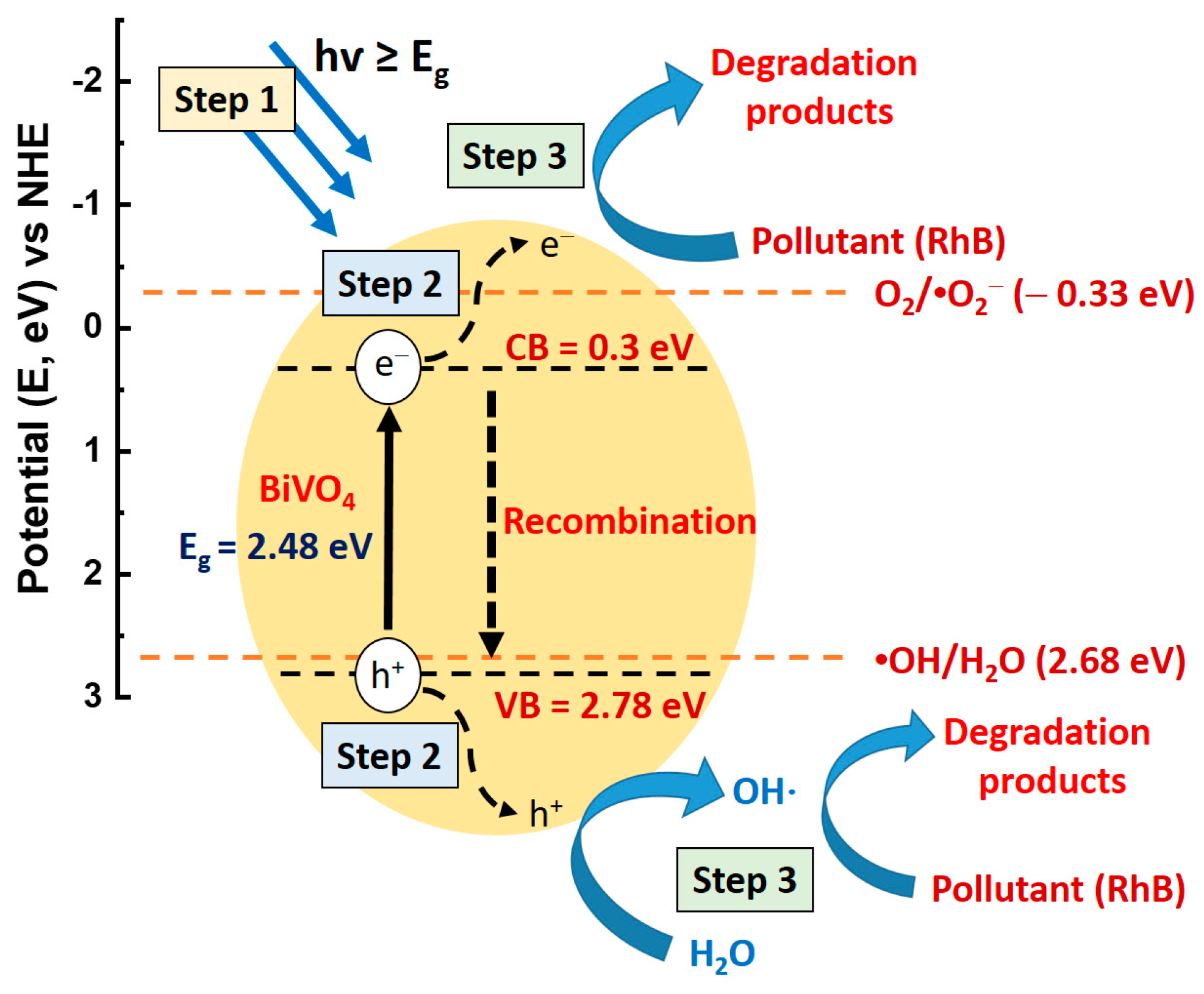
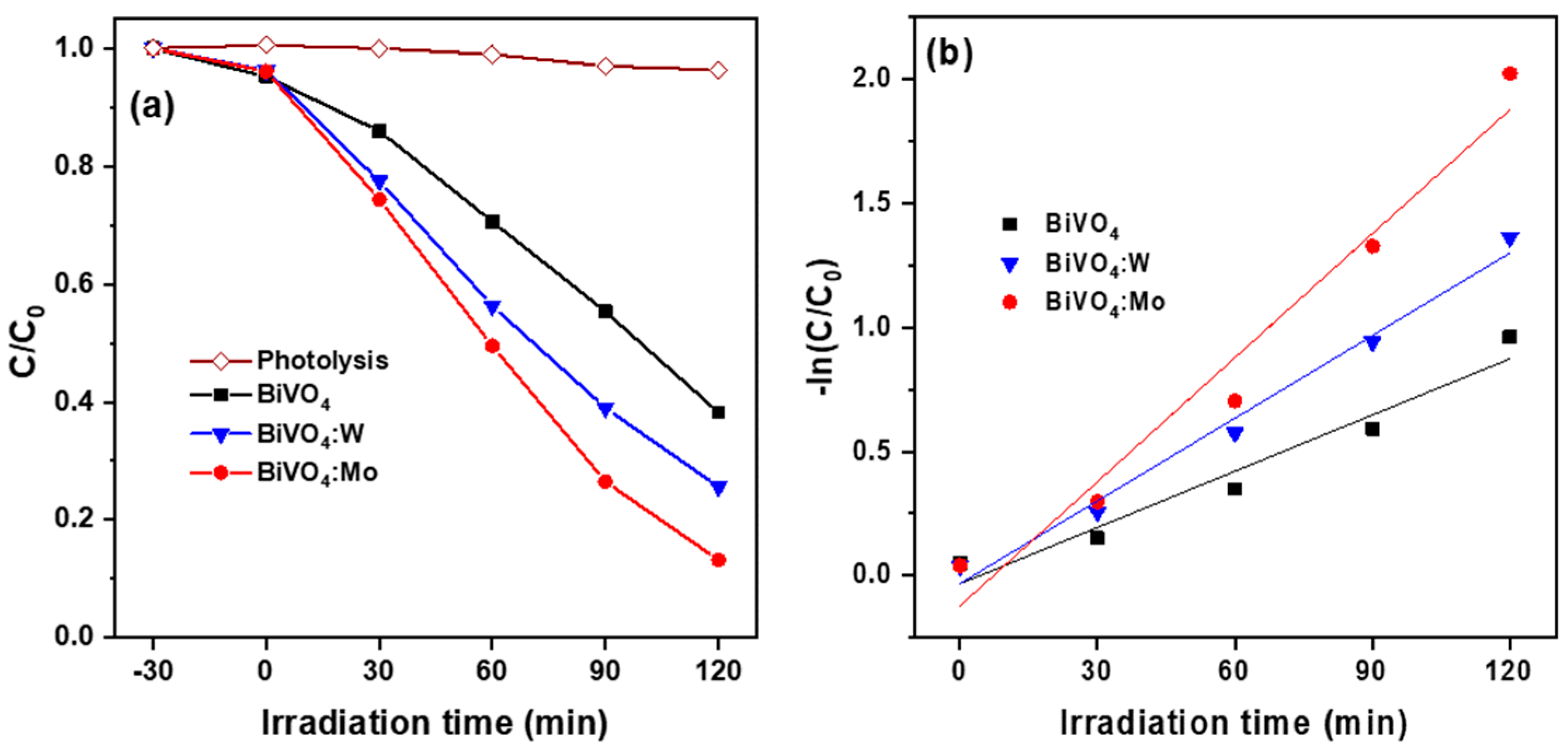
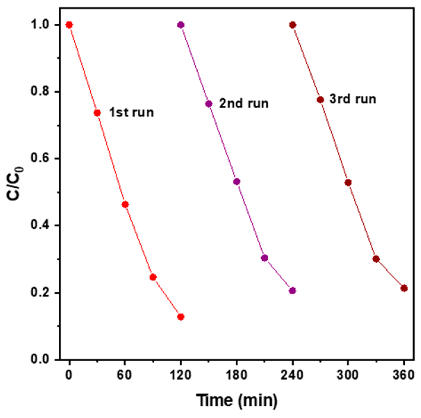
2.2. Photocatalytic Degradation Performance of BiVO4-Based Photocatalysts

The proposed photocatalytic reaction principle and mechanism [1] of a BiVO4 photocatalyst under visible light illumination are illustrated in Figure 8. Knowing the relative positions of the VB and CB potentials of the oxide semiconductor with respect to the potentials of •OH/H2O and O2/•O2 is helpful to understanding the photocatalytic reaction procedures and mechanisms. The positions of the conduction band minimum (CBM) and valence band maximum (VBM) are +0.3 eV and +2.78 eV, respectively [33,34]. In addition, the potential for the generation of •OH [•OH/H2O] radicals is +2.68 eV for a normal hydrogen electrode (NHE) and that for •O2 [O2/•O2] radicals is +0.13 eV for an NHE [35]. Under the excitation of visible light, the photogenerated electrons transition from the VB to the CB of the BiVO4 semiconductor, and photogenerated holes remain in the VB due to electron deletion. Photogenerated electrons and holes then migrate to the surface of the BiVO4 nano-sized semiconductor. Subsequently, photogenerated electrons reduce O2 to •O2 and photogenerated holes convert H2O into •OH. Both •O2 and •OH support the photodegradation of RhB.
The photocatalytic degradation reaction is mainly related to the absorption and desorption of molecules on the surface of the photocatalyst. It is generally accepted that adding surfactants into the synthesis process could reduce the surface tension of the solution and make the precursors of oxide nanopartcles highly dispersive, therefore greatly increasing the photocatalytic performance [36,37]. Sodium dodecylbenzene sulfonate (SDBS) is a neutral anionic surfactant and is widely used as a morphology-directing agent. Zeng et al. reported that while adding SDBS into the reaction solution of hydrothermal synthesis did not change the crystal structure of BiVO4-based nanoparticles, it could reduce the average particle size, increase the specific surface area, and rise hydroxyl content on the as-synthesized oxide nanoparticles to improve the photocatalytic activity properties [36].
The photocatalytic degradation rate of diluted RhB solution as a function of visible light illumination time for the three BiVO4-based nanoparticles is shown in Figure 9a. Notably, the degradation of RhB dye by visible light irradiation was negligible due to its relatively stable structure, and the dark adsorption ability of these BiVO4-based photocatalysts was approximately 4% (after standing in the dark for 30 min). For each mixed suspension, after visible light illumination for 120 min the photocatalytic activity efficiencies of different photocatalysts were found to be as follows: BiVO4:Mo (86.8%) > BiVO4:W (74.4%) > BiVO4 (61.8%). The RhB solution degradation efficiency of the undoped BiVO4 nanoparticles in this study is higher than that of hydrothermally synthesized pure BiVO4 microcrystals reported by Tian et al. (55.9%) [20] and comparable to the photocatalytic activity of mBiVO4 hollow microspheres with the pH of the reaction solution controlled at 4 (70%) [3]. Based on the above evaluation and analysis, it is found that a small amount of Mo doping is a simple and effective approach to significantly improve the photocatalytic activity.
When the initial concentration of organic dye is low, the pseudo-first-order approximation, expressed as Equation (1), can be utilized to quantitatively analyze the reaction rate of dye degradation [13,38].
ln (C0/Ct) = kt
where C0 and Ct represent the concentrations of dye in the mixed solution before and after light irradiation for t min, k is the degradation rate constant (min−1), and t is the irradiation time (min). Each RhB aqueous solution photodegradation curve was analyzed by the pseudo-first-order degradation model as shown in Figure 9b, and the calculated k values are listed in the last column of Table 1. The tendency of the degradation rate constant depends on the impurity dopant and corresponds well with the tendency of photodegradation efficiency. In addition, it is worth noting that the k value difference was more than double between the undoped and Mo-doped mBiVO4 powder samples.
Figure 10 presents recycled results of BiVO4:Mo nanoparticles under visible light irradiation. After three photodegradation reaction cycles, the removal efficiency of RhB dye was still at 80% compared to the performance of the fresh oxide nanoparticle photocatalyst (~87%), indicating stability. The monoclinic BiVO4 nanoparticles doped with W and Mo enhanced the photocatalytic activity because the dopants enhanced the density of photogenerated carriers, reduced charge-transfer resistance, and minimized the recombination ratio of photoexcited electron–hole pairs [11,13]. Moreover, the surface condition and surface area play critical roles in photocatalytic activity. It can be well summarized that the photocatalytic activity is not only related to the specific surface area associated with the morphology and diameter of the nano-sized BiVO4-based photocatalysts; it also determines the charge generation and separation efficiency of the electron–hole pairs and efficient suppression of electron–hole recombination ability, which are correlated with their compositions and crystallinity.

3. Materials and Methods

3.1. Chemicals and Synthesis Procedures of BiVO4-Based Nanoparticles

Three kinds of bismuth vanadate (BiVO4)-based powder samples, including undoped, W-doped, and Mo-doped BiVO4, were synthesized using a hydrothermal method. First, stoichiometric amounts of bismuth(III) nitrate pentahydrate (Bi(NO3)3·5H2O, Alfa Aesar, Ward Hill, MA, USA) and ammonium metavanadate (NH4VO3, Acros, Geel, Belgium) were individually dissolved in 2M nitric acid (HNO3, J.T. Baker, Phillipsburg, NJ, USA) solution (10 mL), and each mixture was stirred for 30 min at room temperature (RT). Following this, 2.5 mg sodium dodecylbenzene sulfonate (SDBS, Sigma-Aldrich, Burlington, MA, USA) was added to the former as the morphology-directing agent [39]. The stoichiometric amount of W or Mo sodium salt was added to the latter to prepare the impurity-doped BiVO4-x precursors. Sodium tungstate dihydrate (Na2WO4·2H2O, Alfa Aesar), and sodium molybdenum oxide dihydrate (Na2MoO4.2H2O, Alfa Aesar) were selected as the raw materials for the W and Mo ion sources, respectively. The impurity doping levels were fixed at 2 at% (M/(V + M), M = W or Mo) in each resultant solution for the preparation of the impurity-doped samples. Following this, the 2 mixtures (Bi and V ions precursors) were mixed together in molar ratio of 1:1 and again stirred for 30 min at RT to obtain a stable and homogenous yellow precursor solution. Each chemical used in this study was an analytical grade reagent. The pH value of the reaction solutions was adjusted to 4 by adding an appropriate amount of NaOH solution. The as-prepared precursor solution was transferred into a 45 mL capacity Teflon-lined stainless-steel autoclave (Parr Instrument Company, model 4744, Moline, IL, USA), and the hydrothermal reaction synthesis was performed at 200 °C for 8 h under autogenous pressure. After the autoclave naturally cooled to RT, the Bi-based oxide precipitates were collected and washed 3 times with distilled water and once with ethanol before finally being dried at 60 °C for 10 h in the atmosphere.

3.2. Physical Properties Characterization and Photocatalytic Activity Measurement

The crystal structure of the as-prepared products was examined on an X-ray diffractometer (Bruker, Billerica, MA, USA) in the region of 2θ = 10°–70° using Cu Kα radiation. The morphologies and particle sizes of the as-synthesized BiVO4-based powders were observed and investigated with a Hitachi S-4800 scanning electron microscope (SEM, Tokyo, Japan) and JEOL JEM2100F transmission electron microscope (TEM, Tokyo, Japan). The surface area of the oxide powders was measured using the BET technique using N2 adsorption/desorption isotherms with a Micromeritics ASAP2020 surface area and porosimetry system (Norcross, GA, USA). The elemental compositions and core-level binding energies were examined and recorded with a ULVAC-PHI PHI 5000 VersaProbe X-ray photoelectron spectroscope (XPS, Kanagawa, Japan). The electron spin resonance (ESR) spectra were collected with a Bruker EMX-10 electron paramagnetic resonance (EPR) spectrometer (Bremen, Germany) operating in the X-band frequency at approximately 9.83 GHz and 15 mW. The Raman spectra were examined and collected with a Nanobase XperRAM S Raman spectrometer (Seoul, South Korea) with an excitation wavelength of 532 nm. Fluorescence (FL) emission spectra were measured using a Shimadzu RF-5301PC spectrofluorophotometer (Koyoto, Japan) with a Xenon lamp as a 350 nm excitation light source. The near UV-visible absorption spectra were recorded on a JASCO V-770 UV-Vis/NIR spectrophotometer (Oklahoma City, OK, USA), which was used for a reflectance standard UV-Vis diffuse reflectance experiment in the wavelength range of 300–800 nm.
The photocatalytic activity of the BiVO4-based nanoparticles was evaluated by measuring and analyzing the photodegradation efficiency of dilute Rhodamine B (RhB) solution under visible light at different times. The Bi-based oxide photocatalyst (0.01 g) was added into the as-prepared aqueous solution (100 mL) containing RhB dye (10 mg/L). In the dark condition, the mixture was stirred for 30 min to achieve the adsorption saturation of each species of photocatalyst suspension. Subsequently, the mixture was subjected to 300 W visible light illumination using an Xe lamp (500 W, λ ≥ 400 nm). At specified time intervals, the suspension (0.8 mL) was collected by centrifugation at 500 rpm for 5 min for the next measurement. The concentration of RhB was determined by measuring the absorption characteristics on a Hitachi U-2900 double-beam spectrophotometer (Tokyo, Japan). Cycling experiments were carried out to evaluate recyclability and stability of photocatalytic performance for developing the BiVO4-based photocatalyst.

4. Conclusions

Undoped, W-doped, and Mo-doped BiVO4 nanocrystalline particles were successfully prepared by the green hydrothermal route. These hydrothermally synthesized products had a single-phase monoclinic structure and high crystallinity. Results showed that lightly doping mBiVO4 with W or Mo could modify the surface morphology, alter the particle shape, refine the particle size, and thereby increase the specific surface area. In addition, W or Mo ions partly replaced V5+ in mBiVO4 crystals, both increasing the density of oxygen vacancies and promoting their n-type character. Such features helped to enhance the photocatalytic activity. The W- and Mo-doped BiVO4 nanoparticles showed higher photocatalytic activity for photodegradation of diluted RhB solution than the undoped BiVO4 nanoparticles, and the BiVO4:Mo photocatalyst exhibited the best photodegradation efficiency of almost 87% under visible light illumination for 120 min. The enhanced photocatalytic degradation efficiency was related to neither the specific surface area value nor the generation/recombination rate of the photogenerated charge carriers of the as-prepared oxide semiconductor photocatalysts.

Supplementary Materials

The following supporting information can be downloaded at: https://www.mdpi.com/article/10.3390/catal13030475/s1, Figure S1: SEM micrographs of hydrothermally synthesized BiVO4-based nanoparticles. (a) undoped, (b) W-doped, and (c) Mo-doped BiVO4 samples, Figure S2: (a) W 4f and (b) Mo 3d core-level spectra of hydrothermally synthesized BiVO4:W and BiVO4:Mo powder samples, respectively.

Author Contributions

Conceptualization, C.-Y.T. and C.-Y.C. (Ching-Yu Chung); methodology, C.-Y.T., C.-Y.C. (Ching-Yu Chung), C.-Y.C. (Chin-Yi Chen), Y.-C.C., C.-J.C., and J.J.W.; validation, C.-Y.T.; investigation, C.-Y.T. and C.-Y.C. (Ching-Yu Chung); resources, C.-Y.T.; data curation, C.-Y.T. and C.-Y.C. (Ching-Yu Chung); writing—original draft preparation, C.-Y.T., C.-Y.C. (Ching-Yu Chung), C.-Y.C. (Chin-Yi Chen), Y.-C.C., C.-J.C., and J.J.W.; writing—review and editing, C.-Y.T., C.-Y.C. (Ching-Yu Chung), C.-Y.C. (Chin-Yi Chen), Y.-C.C., C.-J.C., and J.J.W.; project administration, C.-Y.T.; funding acquisition, C.-Y.T. All authors have read and agreed to the published version of the manuscript.

Funding

This study was founded by the Ministry of Science and Technology (MOST), Taiwan, under grant number MOST 109-2221-E-035-045.

Data Availability Statement

Not applicable.

Acknowledgments

The authors would like to thank the Instrument Center of National Chung Hsing University, Taichung, Taiwan, for assisting with the XPS examinations, and the Institute of Materials Science and Engineering of National Taipei University of Technology, Taipei, Taiwan, for assisting with the TEM observations.

Conflicts of Interest

The authors declare no conflict of interest.

References

  1. Malathi, A.; Madhavan, J.; Ashokkumar, M.; Arunachalam, P. A review on BiVO4 photocatalyst: Activity enhancement methods for solar photocatalytic applications. Appl. Catal. A Gen. 2018, 555, 47–74. [Google Scholar] [CrossRef]
  2. Tan, H.L.; Amai, R.; Ng, Y.H. Alternative strategies in improving the photocatalytic and photoelectrochemical activities of visible light-driven BiVO4: A review. J. Mater. Chem. A 2017, 5, 16498–16521. [Google Scholar] [CrossRef]
  3. Li, F.; Yang, C.; Li, Q.; Cao, W.; Li, T. The pH-controlled morphology transition of BiVO4 photocatalysts from microparticles to hollow microspheres. Mater. Lett. 2015, 145, 52–55. [Google Scholar] [CrossRef]
  4. Mukhtar, F.; Munawar, T.; Nadeem, M.S.; Khan, S.A.; Koc, M.; Batool, S.; Hasan, M.; Iqbal, F. Enhanced sunlight-absorption of Fe2O3 covered by PANI for the photodegradation of organic pollutants and antimicrobial inactivation. Adv. Powder Technol. 2022, 33, 103708. [Google Scholar] [CrossRef]
  5. Mukhtar, F.; Munawar, T.; Nadeem, M.S.; Ur Rehman, M.N.; Khan, S.A.; Koc, M.; Batool, S.; Hasan, M.; Iqbal, F. Dual Z-scheme core-shell PANI-CeO2-Fe2O3-NiO heterostructured nanocomposite for dyes remediation under sunlight and bacterial disinfection. Environ. Res. 2022, 215, 114140. [Google Scholar] [CrossRef]
  6. Liu, X.; Gu, S.; Zhao, Y.; Zhou, G.; Li, W. BiVO4, Bi2WO6 and Bi2MoO6 photocatalysis: A brief review. J. Mater. Sci. Technol. 2020, 56, 45–68. [Google Scholar] [CrossRef]
  7. Nguyen, T.D.; Nguyen, V.H.; Nanda, S.; Vo, D.V.N.; Nguyen, V.H.; Tran, T.V.; Nong, L.X.; Nguyen, T.T.; Bach, L.G.; Abdullah, B.; et al. BiVO4 photocatalysis design and applications to oxygen production and degradation of organic compounds: A review. Environ. Chem. Lett. 2020, 18, 1779–1801. [Google Scholar] [CrossRef]
  8. Zhang, A.; Zhang, J.; Cui, N.; Tie, X.; An, Y.; Li, L. Effects of pH on hydrothermal synthesis and characterization of visible-light-driven BiVO4 photocatlyst. J. Mol. Catal. A Chem. 2009, 304, 28–32. [Google Scholar] [CrossRef]
  9. Thalluri, S.M.; Hernandez, S.; Bensaid, S.; Saracco, G.; Russo, N. Green-synthesized W- and Mo-doped BiVO4 oriented along the {040} facet with enhanced activity for the sun-driven water oxidation. Appl. Catal. B Environ. 2018, 555, 47–74. [Google Scholar] [CrossRef]
  10. Dong, S.; Feng, J.; Li, Y.; Hu, L.; Liu, M.; Wang, Y.; Pi, Y.; Sun, J.; Sun, J. Shape-controlled synthesis of BiVO4 hierarchical structures with unique natural-sunlight-driven photocatalytic activity. Appl. Catal. B Environ. 2014, 152–153, 413–424. [Google Scholar] [CrossRef]
  11. Parmar, K.P.S.; Kang, H.J.; Bist, A.; Dua, P.; Jang, J.S.; Lee, J.S. Photocatalytic and photoelectrochemical water oxidation over metal-doped monoclinic BiVO4 photoanodes. ChemSusChem 2012, 5, 1926–1934. [Google Scholar] [CrossRef] [PubMed]
  12. Park, H.S.; Kweon, K.E.; Ye, H.; Paek, E.; Hwang, G.S.; Bard, A.J. Factors in the metal doping of BiVO4 for improved photoelectrocatalytic activity as studied by scanning electrochemical microscopy and first-principles density-functional calculation. J. Phys. Chem. C. 2011, 115, 17870–17879. [Google Scholar] [CrossRef]
  13. Zhao, G.; Ding, J.; Zhou, F.; Zhao, Q.; Wang, K.; Chen, X.; Gao, Q. Insight into a novel microwave-assisted W doped BiVO4 self-assembled sphere with rich oxygen vacancies oriented on rGO (W-BiVO4-x/rGO) photocatalyst for efficient contaminants removal. Sep. Purif. Technol. 2021, 277, 119610. [Google Scholar] [CrossRef]
  14. Tolod, K.R.; Hernández, S.; Russo, N. Recent advances in the BiVO4 photocatalyst for sun-driven water oxidation: Top-performing photoanodes and scale-up challenges. Catalytic 2017, 7, 13. [Google Scholar] [CrossRef] [Green Version]
  15. Berglund, S.P.; Rettie, A.J.E.; Hoang, S.; Mullins, C.B. Incorporation of Mo and W into nanostructured BiVO4 films for efficient photoelectrochemical water oxidation. Phys. Chem. Chem. Phys. 2012, 14, 7065–7075. [Google Scholar] [CrossRef] [PubMed]
  16. Iwase, A.; Nozawa, S.; Adachi, S.I.; Kudo, A. Preparation of Mo- and W-doped BiVO4 fine particles prepared by an aqueous route for photocatalytic and photoelectrochemical O2 evolution. J. Photochem. Photobiol. A 2018, 353, 284–291. [Google Scholar] [CrossRef]
  17. Talasil, G.; Sachdev, S.; Srivastva, U.; Saxena, D.; Ramakumar, S.S.V. Modified synthesis of BiVO4 and effect of doping (Mo or W) on its photoelectrochemical performance for water splitting. Energy Rep. 2020, 6, 1963–1972. [Google Scholar] [CrossRef]
  18. Tian, X.; Zhu, Y.; Zhang, W.; Zhang, Z.; Hua, R. Preparation and photocatalytic properties of Mo-doped BiVO4. J. Mater. Sci. Mater. Electron. 2019, 30, 19335–19342. [Google Scholar] [CrossRef]
  19. Samsudin, M.F.R.; Sufian, S.; Hameed, B.H. Epigrammatic progress and perspective on the photocatalytic properties of BiVO4-based photocatalyst in photocatalytic water treatment technology: A review. J. Mol. Liq. 2018, 268, 438–459. [Google Scholar] [CrossRef]
  20. Obregón, S.; Caballero, A.; Colón, G. Hydrothermal synthesis of BiVO4: Structural and morphological influence on the photocatalytic activity. Appl. Catal. B Environ. 2012, 117, 59–66. [Google Scholar] [CrossRef]
  21. Lei, B.X.; Zeng, L.L.; Zhang, P.; Sun, Z.F.; Sun, W.; Zhang, X.X. Hydrothermal synthesis and photocatalytic properties of visible-light induced BiVO4 with different morphologies. Adv. Powder Technol. 2014, 25, 946–951. [Google Scholar] [CrossRef]
  22. Ressnig, D.; Kontic, R.; Patzke, G.R. Morphology control of BiVO4 photocatalysts: pH optimization vs. self-organization. Mater. Chem. Phys. 2012, 135, 457–466. [Google Scholar] [CrossRef]
  23. Sharifi, T.; Crmaric, D.; Kovacic, M.; Popovic, M.; Rokovic, M.K.; Kusic, H.; Jozić, D.; Ambrožić, G.; Kralj, D.; Kontrec, J.; et al. Tailored BiVO4 for enhanced visible-light photocatalytic performance. J. Environ. Chem. Eng. 2021, 9, 106025. [Google Scholar] [CrossRef]
  24. Feng, X.; Lv, B.; Lu, L.; Feng, X.; Wang, H.; Xu, B.; Yang, Y.; Zhang, F. Role of surface oxygen vacancies in zinc oxide/graphitic carbon nitride composite for adjusting energy band structure to promote visible-light-driven photocatalytic activity. Appl. Surf. Sci. 2021, 562, 150106. [Google Scholar] [CrossRef]
  25. Gu, X.; Luo, Y.; Li, Q.; Wang, R.; Fu, S.; Lv, X.; He, Q.; Zhang, Y.; Yan, Q.; Xu, X.; et al. First-principle insight into the effects of oxygen vacancies on the electronic, photocatalytic, and optical properties of monoclinic BiVO4 (001). Front. Chem. 2020, 8, 601983. [Google Scholar] [CrossRef] [PubMed]
  26. Venkatesan, R.; Velumani, S.; Tabellout, M.; Errien, N.; Kassiba, A. Dielectric behavior, conduction and EPR active centres in BiVO4 nanoparticles. J. Phys. Chem. Solids 2013, 74, 1695–1702. [Google Scholar] [CrossRef]
  27. Frost, R.L.; Henry, D.A.; Weier, M.L.; Martens, W. Raman spectroscopy of three polymorphs of BiVO4: Clinobisvanite, dreyerite and pucherite, with comparisons to (VO4)3-bearing minerals: Namibite, pottsite and schumacherite. J. Raman Spectrosc. 2006, 37, 722–732. [Google Scholar] [CrossRef] [Green Version]
  28. Yu, J.; Kudo, A. Effects of structural variation on the photocatalytic performance of hydrothermally synthesized BiVO4. Adv. Funct. Mater. 2006, 16, 2163. [Google Scholar] [CrossRef]
  29. Liu, B.; Yan, X.; Yan, H.; Yao, Y.; Cai, Y.; Wei, J.; Chen, S.; Xu, X.; Li, L. Preparation and characterization of Mo doped in BiVO4 with enhanced photocatalytic properties. Materials 2017, 10, 976. [Google Scholar] [CrossRef] [PubMed] [Green Version]
  30. Xu, P.; Wang, P.; Wang, Q.; Wei, R.; Li, Y.; Xin, Y.; Zheng, T.; Hu, L.; Wang, X.; Zhang, G. Facile synthesis of Ag2O/ZnO/rGO heterojunction with enhanced photocatalytic activity under simulated solar light: Kinetics and mechanism. J. Hazard. Mater. 2021, 403, 124011. [Google Scholar] [CrossRef]
  31. Zhang, M.; Shao, C.; Li, X.; Zhang, P.; Sun, Y.; Su, C.; Zhang, X.; Ren, J.; Liu, Y. Carbon-modified BiVO4 microtubes embedded with Ag nanoparticles have high photocatalytic activity under visible light. Nanoscale 2012, 4, 7501–7508. [Google Scholar] [CrossRef]
  32. Oshikiri, M.; Boero, M.; Ye, J.; Zou, Z.; Kido, G. Electronic structures of promising photocatalysts (M = V, Nb, Ta) and for water decomposition in the visible wavelength region. J. Chem. Phys. 2002, 117, 7313. [Google Scholar] [CrossRef]
  33. Regmi, C.; Dhakal, D.; Lee, S.W. Visible-light-induced Ag/BiVO4 semiconductor with enhanced photocatalytic and antibacterial performance. Nanotechnology 2018, 29, 064001. [Google Scholar] [CrossRef] [PubMed]
  34. Shi, J.; Zhang, W.; Gu, Q. Ab Initio Calculation of Surface-Controlled Photocatalysis in Multiple-Phase BiVO4. J. Phys. Chem. C 2022, 126, 9541–9550. [Google Scholar] [CrossRef]
  35. Naresh, G.; Malik, J.; Meena, V.; Mandal, T.K. pH-mediated collective and selective solar photocatalysis by a series of layered Aurivillius perovskites. ACS Omega 2018, 3, 11104–11116. [Google Scholar] [CrossRef] [PubMed]
  36. Zeng, J.; Zhong, J.; Li, J.; Xiang, Z.; Liu, X.; Chen, J. Improvement of photocatalytic activity under solar light of BiVO4 microcrystals synthesized by surfactant-assisted hydrothermal method. Mater. Sci. Semicond. Process. 2014, 27, 41–46. [Google Scholar] [CrossRef]
  37. Zhang, A.; Zhang, J. Characterization of visible-light-driven BiVO4 photocatalysts synthesized via a surfactant-assisted hydrothermal method. Spectrochim. Acta A Mol. Biomol. Spectrosc. 2009, 73, 336–341. [Google Scholar] [CrossRef]
  38. Ding, J.; Bu, L.; Zhao, Q.; Kabutey, F.T.; Wei, L.; Dionysiou, D.D. Electrochemical activation of persulfate on BDD and DSA anodes: Electrolyte influence, kinetics and mechanisms in the degradation of bisphenol A. J. Hazard Mater. 2020, 388, 121789. [Google Scholar] [CrossRef]
  39. Zhang, L.; Chen, D.; Jiao, X. Monoclinic structured BiVO4 nanosheets: Hydrothermal preparation, formation, mechanism, and coloristic and photocatalytic properties. J. Phys. Chem. B 2006, 110, 2668–2673. [Google Scholar] [CrossRef]
Figure 1. XRD patterns of BiVO4, W-doped BiVO4 (BiVO4:W), and Mo-doped (BiVO4:Mo) nanoparticles synthesized using a hydrothermal method at pH 4.0.
Catalysts 13 00475 g001
Figure 2. TEM micrographs of hydrothermally synthesized BiVO4-based nanoparticles. (a) undoped, (b) W-doped, and (c) Mo-doped BiVO4 samples.
Catalysts 13 00475 g002
Figure 3. Core-level XPS spectra of (a) Bi 4f, (b) V 2p, and (c) O 1s regions of three BiVO4-based powder samples.
Catalysts 13 00475 g003
Figure 4. ESR spectra of undoped and impurity-doped BiVO4 powder samples.
Catalysts 13 00475 g004
Figure 5. Raman spectra of undoped and impurity-doped BiVO4 powder samples.
Catalysts 13 00475 g005
Figure 6. Room temperature FL spectra of undoped and impurity-doped BiVO4 powder samples.
Catalysts 13 00475 g006
Figure 7. (a) UV-visible absorption spectra and (b) Tauc plots of three BiVO4-based powder samples.
Catalysts 13 00475 g007
Figure 8. Schematic illustration of proposed photocatalytic reaction mechanism of BiVO4 photocatalyst under visible illumination.
Catalysts 13 00475 g008
Figure 9. (a) Photocatalytic activity and (b) the first-order kinetics for degradation of diluted RhB solution by BiVO4-based nanoparticles under visible light irradiation.
Catalysts 13 00475 g009
Figure 10. Stability of BiVO4:Mo nanoparticles in photocatalytic degradation of diluted RhB solution.
Catalysts 13 00475 g010
Table 1. Microstructural features, optical characteristics, and photocatalytic activities of hydrothermally synthesized undoped and impurity-doped BiVO4 nanoparticles.
Photo-CatalystAverage Particle Size
(nm)
SBET 1
(m2g−1)
Main Raman Peak Wavenumber
(cm−1)
Optical Bandgap
(eV)
Photo-Degradation Efficiency
(%)
Reaction Rate Constant
(min−1)
BiVO41644.709824.42.4861.80.0076
BiVO4:W1377.079816.42.4974.40.0111
BiVO4:Mo1358.214818.12.5086.80.0167
1 Specific surface area of powder sample.
Disclaimer/Publisher’s Note: The statements, opinions and data contained in all publications are solely those of the individual author(s) and contributor(s) and not of MDPI and/or the editor(s). MDPI and/or the editor(s) disclaim responsibility for any injury to people or property resulting from any ideas, methods, instructions or products referred to in the content.

