Article

Hydrothermal Synthesis of Bimetallic (Zn, Co) Co-Doped Tungstate Nanocomposite with Direct Z-Scheme for Enhanced Photodegradation of Xylenol Orange

by
Fahad A. Alharthi
*,
Wedyan Saud Al-Nafaei
,
Alanoud Abdullah Alshayiqi
,
Hamdah S. Alanazi
and
Imran Hasan
*
Department of Chemistry, College of Science, King Saud University, Riyadh 11451, Saudi Arabia
*
Authors to whom correspondence should be addressed.
Catalysts 2023, 13(2), 404; https://doi.org/10.3390/catal13020404
Submission received: 13 December 2022 / Revised: 15 January 2023 / Accepted: 17 January 2023 / Published: 14 February 2023
(This article belongs to the Special Issue Advanced Oxidation Catalysts)

Abstract

:
In the present study, pristine ZnWO4, CoWO4, and mixed metal Zn0.5Co0.5WO4 were synthesized through the hydrothermal process using a Teflon-lined autoclave at 180 ℃. The synthesized nanomaterials were characterized by various spectroscopic techniques, such as TEM, FTIR, UV–vis, XRD, and SEM-EDX-mapping to confirm the formation of nanocomposite material. The synthesized materials were explored as photocatalysts for the degradation of xylenol orange (XO) under a visible light source and a comparative study was explored to check the efficiency of the bimetallic co-doped nanocomposite to the pristine metal tungstate NPs. XRD analysis proved that reinforcement of Co2+ in ZnWO4 lattice results in a reduction in interplanar distance from 0.203 nm to 0.185 nm, which is reflected in its crystallite size, which reduced from 32 nm to 24 nm. Contraction in crystallite size reflects on the optical properties as the energy bandgap of ZnWO4 reduced from 3.49 eV to 3.33 eV in Zn0.5Co0.5WO4, which is due to the formation of a Z-scheme for charge transfer and enhancement in photocatalytic efficiency. The experimental results suggested that ZnWO4, CoWO4, and Zn0.5Co0.5WO4 NPs achieved a photocatalytic efficiency of 97.89%, 98.10%, and 98.77% towards XO in 120 min of visible solar light irradiation. The kinetics of photodegradation was best explained by pseudo-first-order kinetics and the values of apparent rate const (kapp) also supported the enhanced photocatalytic efficiency of mixed metal Zn0.5Co0.5WO4 NPs towards XO degradation.

1. Introduction

The development of societies and industries has led to the deterioration of freshwater qualities through the release of untreated effluents containing hazardous chemical contaminants, such as dyes and pharmaceutical products, etc. [1,2]. The toxic organic compounds, especially dyes and pharmaceuticals, contaminated wastewater in the environment, which can cause a variety of health issues to humans [3,4]. Among various organic pollutants, xylenol orange (XO; C31H32N2 O13S) belongs to the acidic dye family commonly used in the textile, paper, and printing industries, but beyond the permissible limit, it causes nervous system breakdown, liver–kidney damage, blood disorders, and skin irritation [5,6]. So, it has become a matter of serious concern to develop new technology-based methods and resolutions which can treat these hazardous chemicals in the water and regulate their limits in the aquatic environment. Among various chemical, physical, and biological wastewater treatment methods, photocatalysis enables the adsorbed organic molecules to be degraded at a low cost into H2O and CO2 using solar irradiation, which is abundant, renewable, and environmentally friendly [7,8]. Moreover, advanced oxidation processes (AOPs) were created to promote the removal or degradation of contaminant species through redox processes, focused on the remediation of contaminants in wastewater; they are a group of techniques that use reactive oxidant species (ROS) to mineralize a wide range of resistant organics [9,10].
Metal tungstate nanoparticles (NPs) have received a lot of interest among the different types of semiconductor photocatalysts [11]. For this reason, the study and synthesis of single photocatalysts that successfully use visible light to stimulate photocatalysis have recently become a promising idea. Until now, various semiconductors with sufficient energy band gaps to absorb visible light, such as Fe2O3, Ag2O, Bi2WO6, g-C3N4, MnWO4, CdWO4, CdS, NiMoO4, Cu2O, ln2S3, and Si3N4, have been effectively synthesized and utilized for photocatalysis, because of its high average refractive index, high chemical stability, excellent light absorbing property, and high catalytic activity [12,13,14]. In the tungstate family, zinc tungstate (ZnWO4) has been a widely known semiconductor photocatalyst owing to its high physical and chemical stability and difficulty to dissolve [15]. However, due to homogeneity, it is only UV light active and possesses a high rate of electron–hole pair recombination, which poses an obstacle in the practical applications of ZnWO4 [16]. Various methods have been applied previously to increase the photocatalytic efficiency of ZnWO4, such as semiconductor coupling [17], noble metal loading [18], and metal deposition [19]. The metal deposition of ZnWO4 with other metals having a narrow band gap could effectively inhibit the electron–hole pair recombination by expanding the light absorption range, and thus a robust enhancement in photocatalytic efficiency will appear [20,21]. So, the Co deposition method was applied in this study to synthesize the Co-doped nanocomposite Zn0.5Co0.5WO4 with improved energy bandgap, optical properties (UV–vis light active), and photocatalytic efficiency. Since the ionic radii of both Co2+ and Zn2+ are approximately the same, it can be easily deposited in the solid matrix of ZnWO4 and result in the reduction of the energy bandgap from 3.87 eV (ZnWO4) to 1.95 eV (Zn0.5Co0.5WO4). ZnWO4 was identified as a large band gap semiconductor (band gap = 3.87 eV) with the appropriate conduction band and valance band locations based on its electronic characteristics, while, on the other hand, cobalt tungstate (CoWO4) has been recognized as having excellent phase composition, strong chemical stability, and a low energy bandgap, which makes it adaptable to degrade the dyestuff in the visible light range [22].
In the present work, a hydrothermal method was used for the synthesis of pristine metal tungstate nanomaterials (ZnWO4, CoWO4 NPs) and mixed metal tungstate nanocomposite (Zn0.5Co0.5WO4 NPs) using a Teflon line autoclave. The synthesized NPs were explored for the photocatalytic degradation of XO dye and a comparative study was designed to explore the photocatalytic mechanism and efficiency of the photocatalyst.

2. Results and Discussion

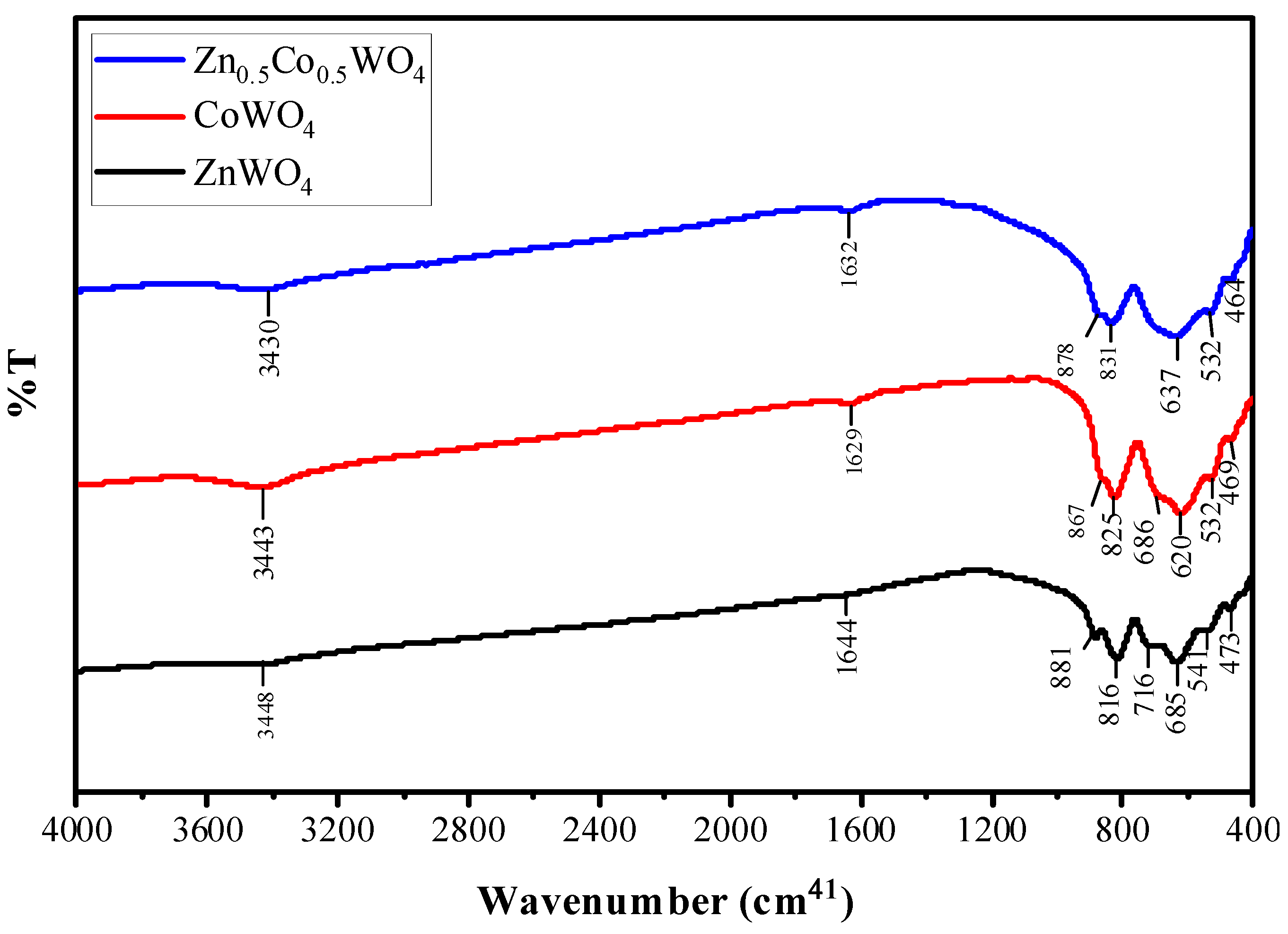
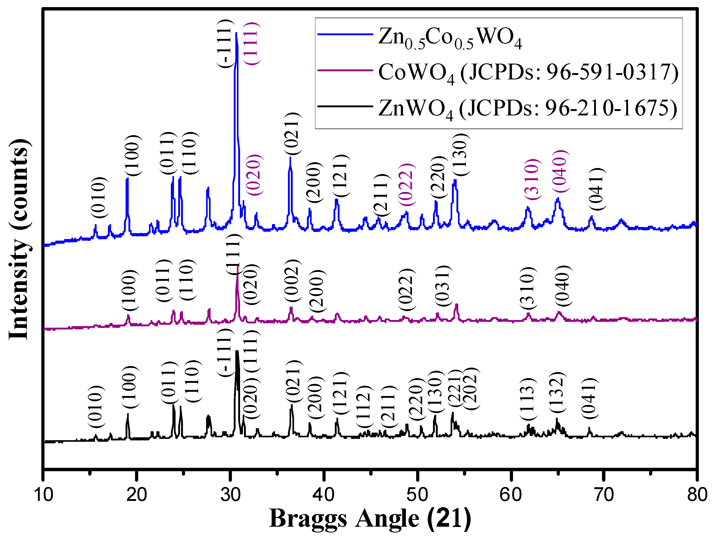
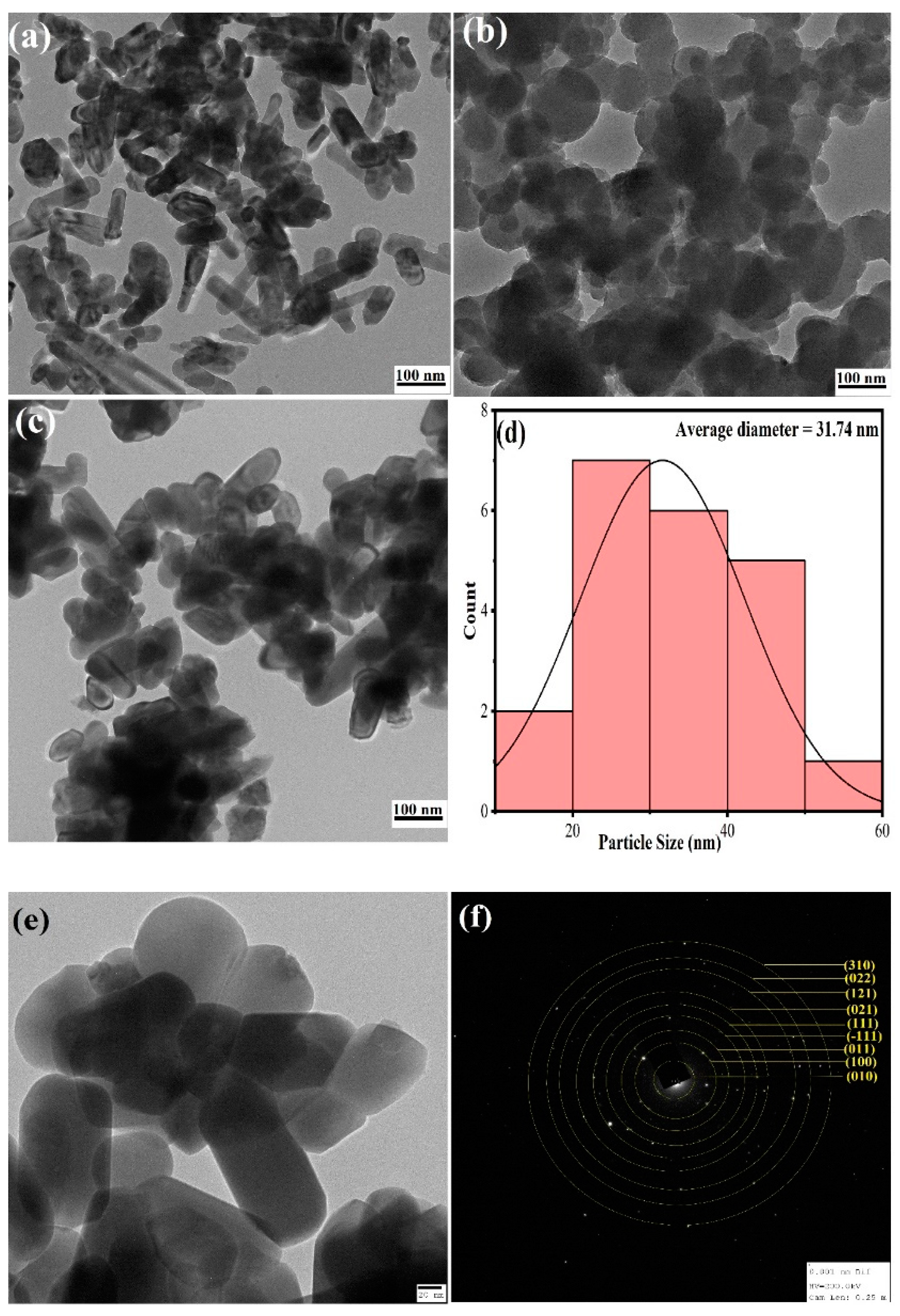
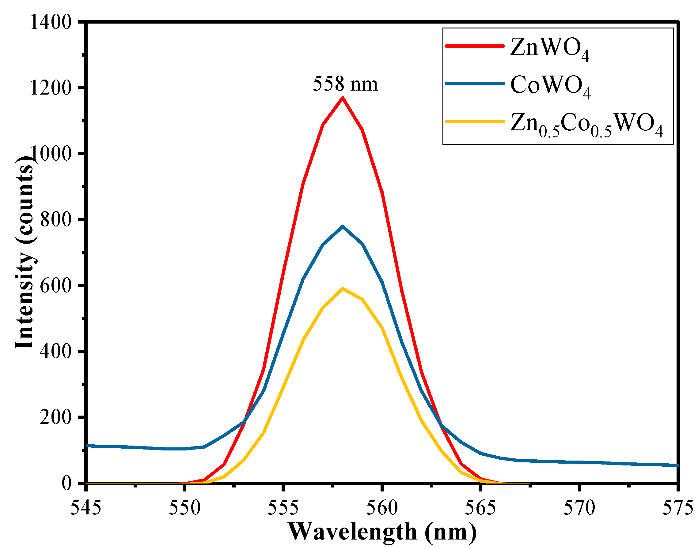
2.1. Material Characterization

Figure 1 represents the FTIR of ZnWO4, CoWO4, and Zn0.5Co0.5WO4, in which the characteristic peaks of ZnWO4 are 3448 and 1644 cm−1 (stretching and bending vibrations of –OH group) due to surface adsorbed water, 473 to 881 cm−1 (W2O8 bonds (WO6) groups shared at edges and WO2 groups), and 473 cm−1 (stretching vibrational of the Zn–O) [23]. Three peaks at 541, 685, and 716 cm−1 correspond to symmetric and asymmetric stretching vibrations of the bridging atom of the WO2 groups of distorted octahedral (WO6) clusters [24]. Broad absorption bands at 816 cm−1 and 881 cm−1 are caused by symmetrical vibrations of bridge oxygen atoms of Zn–O–W groups [24]. Similar types of stretching and vibrational bands are also shown by CoWO4 with different peak values and 469 cm−1 belongs to the Co–O bond. As compared to ZnWO4, the FTIR of mixed metal Zn0.5Co0.5WO4 showed the difference in peak shapes and intensities due to the partial replacement of Zn2+ ions by Co2+ ions [25].
The formation of ZnWO4, CoWO4, and Zn0.5Co0.5WO4 was confirmed by the XRD analysis. Figure 2 shows the XRD pattern of the ZnWO4, CoWO4, and Zn0.5Co0.5WO4 prepared by the hydrothermal method at 180℃ for 24 h. The XRD spectra of ZnWO4 shows characteristic peaks at 2θ value of 15.58°, 19.04°, 23.94°, 24.66°, 30.59°, 31.38°, 36.54°, 38.47°, 41.41°, 44.40°, 46.55°, 50.39 51.84°, 53.76°, 54.11°, 61.85°, 64.90°, and 68.34°, which belong to miller indices (010), (100), (011), (110), (111), (020), (021), (200), (121), (112), (211), (220) (130), (221), (202), (113), (132), and (041), respectively (JCPDs card no. 96-210-1675). Then, the XRD spectra of CoWO4 shows characteristic peaks at 2θ value of 19.02°, 23.88°, 27.72°, 31.50°, 36.44°, 38.69°, 48.52°, 52.17°, 61.89°, and 65.14°, which belong to miller indices (100), (011), (110), (020), (002), (200), (022), (031), (310), and (040), respectively (JCPDs card no. 96-591-0317). Finally, the XRD pattern of Zn0.5Co0.5WO4 NPs shows peaks ascribed to the ZnWO4 at 15.54° (010), 36.41° (021), 41.35° (211), 51.95° (220), 53.94° (130), and 68.56° (041), and peaks ascribed to the CoWO4 at 48.81° (022), 61.81° (310), and 64.96° (040), respectively, which suggests that Co has been successfully doped in the solid matrix of ZnWO4. Moreover, the peak intensities of Zn0.5Co0.5WO4 NPs are greater than pristine ZnWO4 suggesting the increase in crystallinity of the complex upon being mixed with Co2+, which has been proved by data given in Table 1. There are several extra peaks observed in the XRD spectra of Zn0.5Co0.5WO4 which do not belong to either ZnWO4 or CoWO4. These peaks are due to the formation of Na2W2O7 and Na2W4O13 phases with the Zn0.5Co0.5WO4 phase [26,27].
Further information about crystallite size and dislocation density; Scherrer’s equation was taken into consideration [28].
D = 0 . 9 λ β cos θ
Dislocation   Density ( δ ) = 1 D 2  
  Interlayer   Spacing ( d 111 ) = n λ 2 Sin θ  
% Crystallinity = Area   under   the   crystalline   peaks Total   area × 100
where D is the crystallite size, λ is the characteristic wavelength of the X-ray, β represents the angular width in radian at an intensity equal to half of its maximum (HWMI) of the peak, and θ is the diffraction angle. Using Equation (1), the average particle size of ZnWO4, CoWO4, and Zn0.5Co0.5WO4 was 31.98, 32.19, and 24.24 nm suggesting a contraction in particle size upon Co inclusion in the ZnWO4 solid lattice.
The surface morphology of the ZnWO4, CoWO4, and Zn0.5Co0.5WO4 were studied using SEM analysis given in (Figure 3). ZnWO4 reveals an aggregation of irregularly shaped crystals with plenty of nanorods (Figure 3a). CoWO4 exhibit uniform nanoparticles of various size (Figure 3b), while Zn0.5Co0.5WO4 revealed an assembly of fine nanoparticles with a porous surface (Figure 3c). The elemental composition of the synthesized Zn0.5Co0.5WO4 NPs was observed by EDX analysis and the results showed the presence of the element (weight%) as O (28.52%), Zn (4.55%), Co (4.22%), and W (62.81%), which confirms the successful deposition of Co2+ in the ZnWO4 lattice (Figure 3d). The high atomic percentage of W in the EDX spectra is due to the presence of impurity in the form of Na2W2O7 and Na2W4O13 phases with the Zn0.5Co0.5WO4, which resulted in an extra amount of W in the EDX spectra [27].
Furthermore, to figure out the size of the ZnWO4, CoWO4, and Zn0.5Co0.5WO4, TEM analysis was performed (Figure 4a–c). ZnWO4 (Figure 4a) is composed of particles with a rod-like shape; CoWO4 (Figure 4b) presented a morphologically spherical shaped particle. The synthesized material Zn0.5Co0.5WO4 (Figure 4c) exhibited particles mixed of nanorods with some spherical particles with size in a range from 5 to 60 nm, and the Gaussian distribution of the average size was found to be 31 nm given in Figure 4d. Figure 4e represents the TEM image of Zn0.5Co0.5WO4, which suggested the shape of particles to be distorted monoclinic due to the doping of Co into the ZnWO4 lattice. Selected area electron diffraction (SAED) analysis (Figure 4f) was further considered to simulate the TEM data with XRD to assess the purity and shape of the nanoparticles. It contains patterns originating from diffraction points in the TEM screen belonging to randomly oriented nanoparticles. The occurrence of diffraction patterns in Figure 4f suggested that the material is crystalline and pure. The hkl values belonging to yellow fringes simulate the XRD hkl planes of Zn0.5Co0.5WO4 suggesting the shape of the nanoparticles to be monoclinic.
The optical properties of the synthesized NPs and their energy bandgap were estimated by using UV–vis spectroscopy in the range of 200–700 nm and the results are given in Figure 5. The absorption spectra of all the synthesized NPs ZnWO4, CoWO4, and Zn0.5Co0.5WO4 exhibited two peaks, one around 260–312 nm and another broad peak around 530–650 nm, suggesting the UV–vis activeness of the NPs. The inset in Figure 5 shows Tauc’s plot, which is used to calculate band gap energy (Eg) of the prepared samples by using the Equation (5) [29];
( α h ν ) = A ( h ν E g ) n  
where α is the absorption coefficient, A is constant, h is plank constant, ν is the frequency of radiations, and n is a constant of transition variations, i.e., n = 1/2 for direct transitions and n = 2 for the indirect transitions. Tauc’s plot indicated that the synthesized ZnWO4, CoWO4, and Zn0.5Co0.5WO4 were found to have an energy bandgap value of 3.49 eV, 3.42 eV, and 3.33 eV, respectively. Results showed that the deposition of Co2+ in the ZnWO4 solid lattice results in the increase in absorption edge by decreasing the energy bandgap, which inhibits the rate of electron–hole pair recombination, and thus improves the photocatalytic efficiency of the synthesized material [30]. The doping of Co2+ into ZnWO4 leads to the induction of deformations in the pure monoclinic lattice of ZnWO4, which creates mobile oxygen vacancies in the cluster and makes an improvement in the catalytic [31,32].
Figure 6 consists of the photoluminescence emission spectra of ZnWO4, CoWO4, and Zn0.5Co0.5WO4 recorded at a 594 nm excitation wavelength. It can be seen that the PL spectra spans in the range of 545 to 575 nm with a prominent emission peak at 558 nm. The information obtained from the PL spectra suggested that the PL intensity of synthesized Zn0.5Co0.5WO4 was found to be lower than that of ZnWO4 and CoWO4. The inclusion of Co in the wolframite monoclinic structure of ZnWO4 results in emissions due to the radiative transition between W and O in the WO66− molecular complex, which effectively slows down the electron–hole [33,34,35].

2.2. Photocatalytic Experiments and Optimization of Reaction Parameters

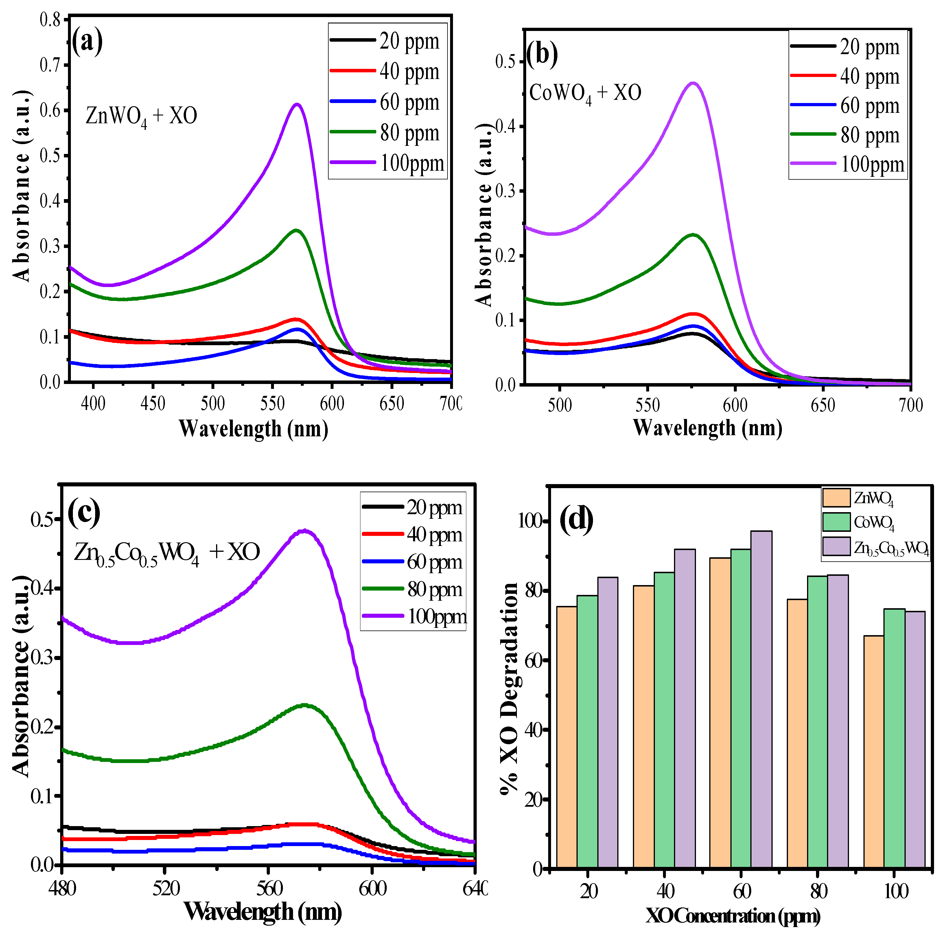
2.2.1. Effect of Variable XO Concentration

The concentration of the dye solution is an important parameter for elucidating the photocatalytic efficiency of the material. Photocatalytic experiments were conducted by taking 10 mg of NPs as a catalyst with variable (20–100 ppm) XO concentration for 120 min of visible light irradiation. The results obtained are given in Figure 7a–d, which suggest that Zn0.5Co0.5WO4 possesses better photocatalytic efficiency (97.17%) as compared to CoWO4 (91.81%), followed by ZnWO4 (89.51%) for 60 ppm of XO, but as the concentration of XO increases beyond 60 ppm, the percentage degradation decreases (Figure 7d). The causes could include: (1) XO may cover more ZnWO4, CoWO4, and Zn0.5Co0.5WO4 active sites, which inhibits the production of oxidants (OH or O2 radicals) and lowers the efficiency of degradation: and (2) higher XO concentration absorbs a greater number of photons, and as a result, there are not enough photons to activate the surfaces of ZnWO4, CoWO4, and Zn0.5Co0.5WO4 nanoparticles, which slows down XO’s degradation at higher concentrations [36]. So, 60 ppm XO concentration was chosen as the optimum concentration for further photocatalytic experiments.

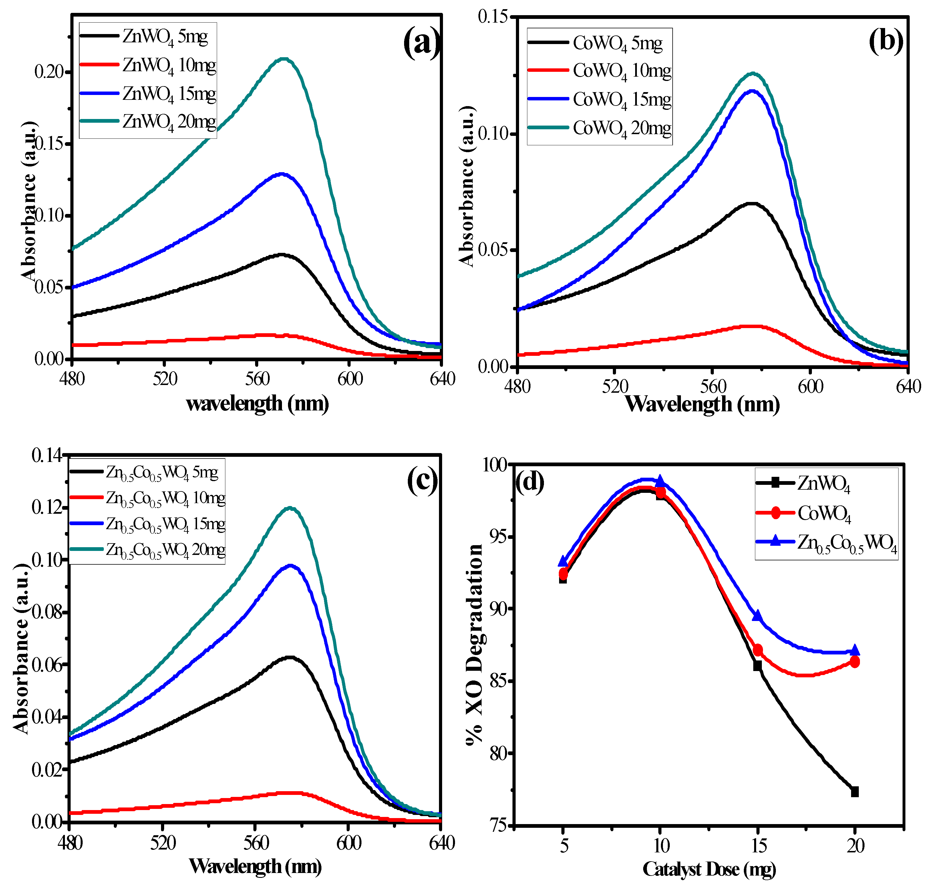
2.2.2. Effect of Variable Catalyst Dose
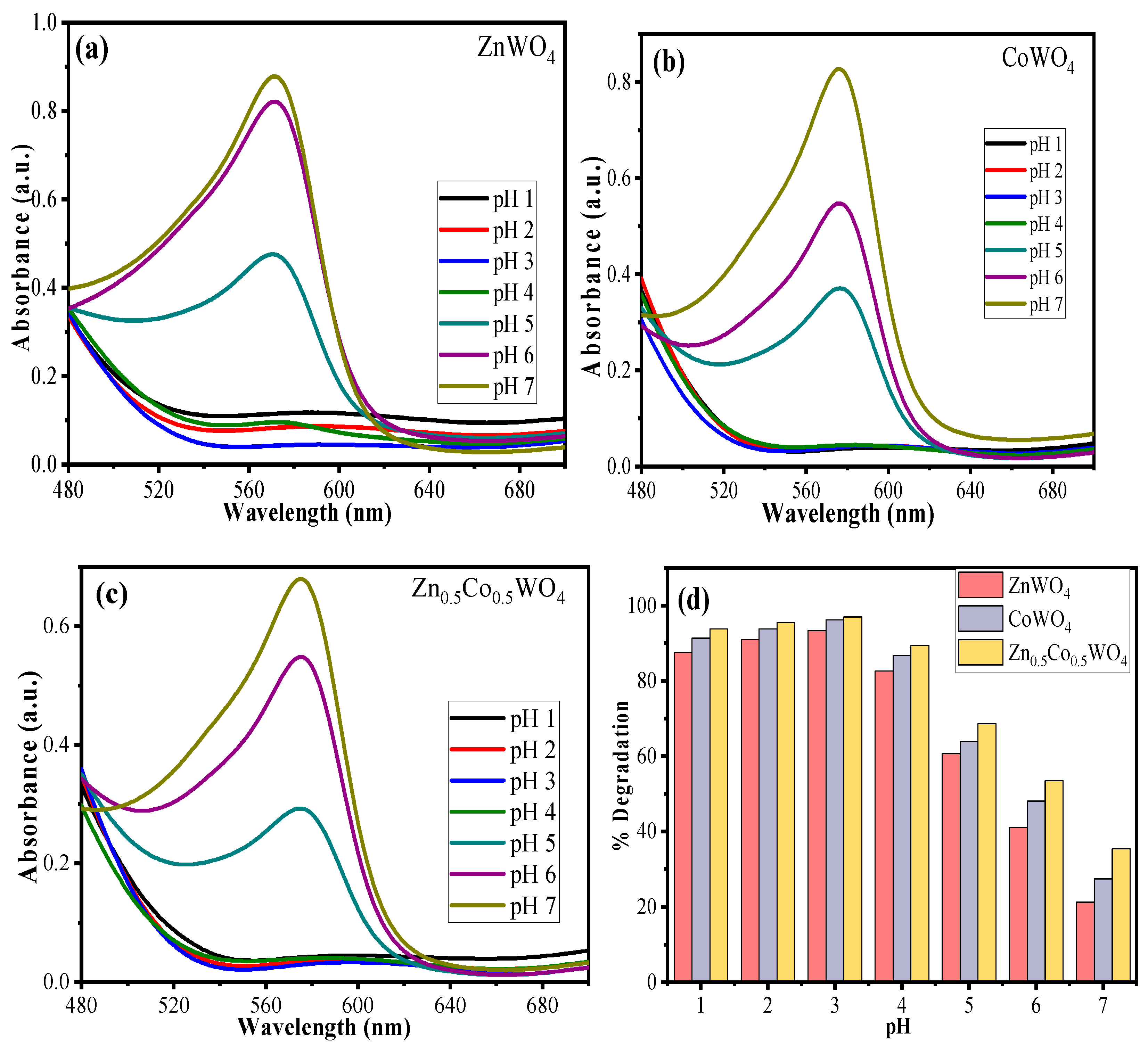
The physicochemical characteristics of nanomaterials, such as surface area, phase structure, particle size, and interface charge, make them able to complete adsorption action [27]. To efficiently degrade XO through catalysis using the catalyst, the photocatalytic experiment was performed by varying catalyst doses, such as 5, 10, 15, and 20 mg with 10 mL of 10 ppm XO. The results given in Figure 8a–d suggested that the photocatalytic efficiency increased from 5 mg catalyst dose to 10 mg and starts declining with further increase. All the synthesized NPs exhibit maximum photocatalytic efficiency at 10 mg catalyst dose as 97.89% for ZnWO4, 98.10% for CoWO4, and 98.77% for Zn0.5Co0.5WO4. More active sites are produced on the catalyst’s surface at lower concentrations, which promotes the creation of OH or O2 radicals. After that, increasing the catalytic dose causes the particles to agglomerate and sediment, which increases the turbidity and opacity of the slurry. Hence, the generation of OH or O2 radicals automatically decrease [24]. Therefore, the catalyst dose of 10 mg was chosen to be the optimum dose for the degradation.

2.2.3. Effect of pH

Experiments were performed to evaluate the impact of pH on the photocatalytic efficiencies of ZnWO4, CoWO4, and Zn0.5Co0.5WO4. A total of 10 mg of the catalyst was dispersed in 20 mL of 60 ppm XO solution with a variable pH range from 1–7, and irradiated for 120 min under the visible light source. The obtained results after the experiments are given in Figure 9, which suggests that the photocatalytic efficiency of all the catalysts increased until pH 3, giving a maximum value of 93.35% for ZnWO4, 96.21% for CoWO4, and 97.97% for Zn0.5Co0.5WO4. Further, an increase in pH value beyond 3 results in a decrease in photocatalytic efficiency with a continuous fall until pH 7. So, pH 3 was taken as the optimum pH value for the photocatalytic experiments. The trend can be explained by the fact that as XO is an anionic dye, it can easily bind with the positive surface of the catalyst at low pH through electrostatic interactions, and thereby, in the presence of light, it gets degraded by OH or O2 radicals [7]. As the pH values increase, the surface becomes negative, which will exert a repulsive force on the anionic XO molecule, and hence, the photocatalytic efficiency decreases.

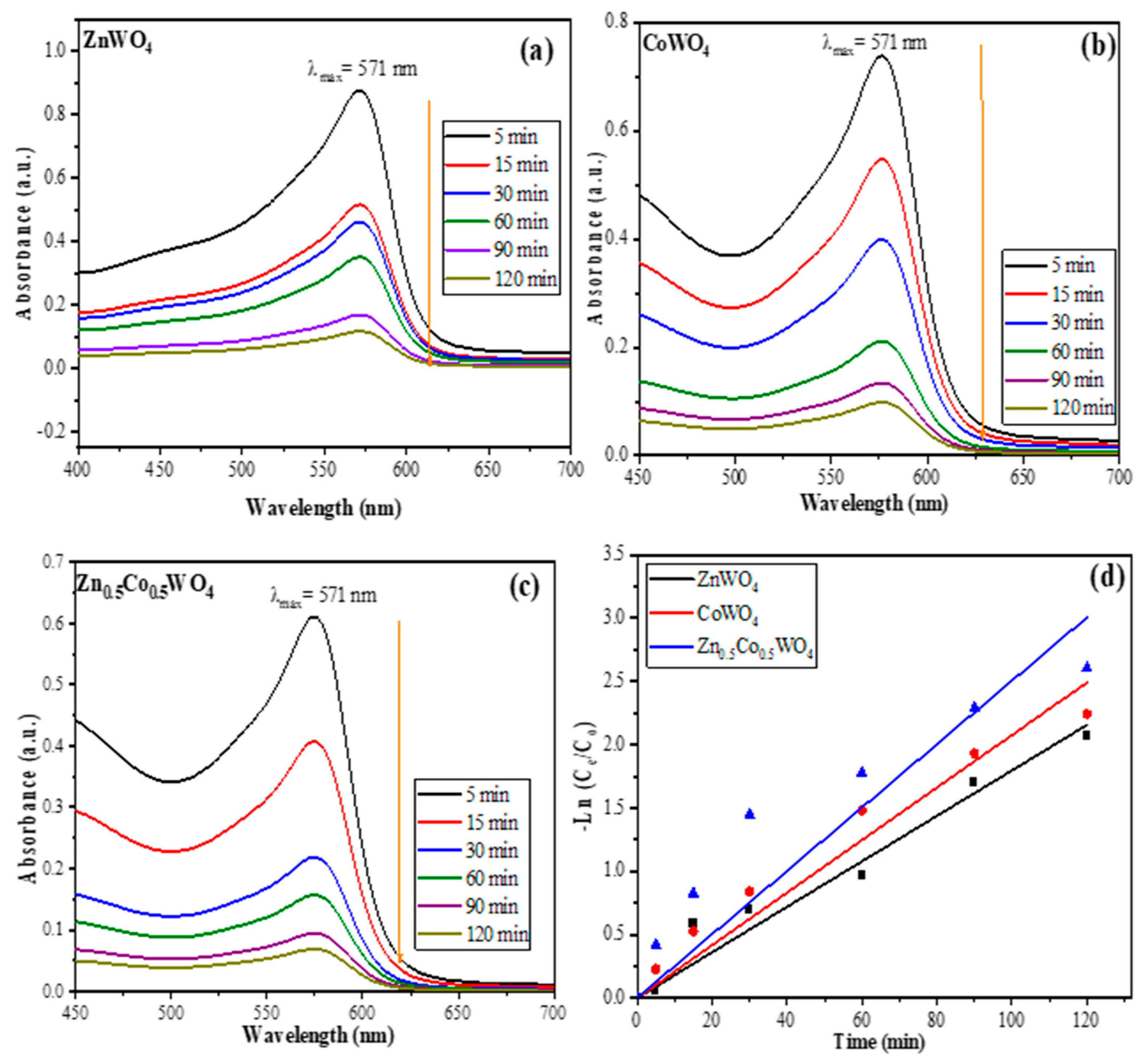
2.3. Kinetics of Photodegradation

A variety of experiments were carried out by taking 10 mL of 20 ppm XO with 10 mg of catalyst at varying irradiation time from 0 to 120 min under visible solar radiation, and the data obtained was adjusted to the Langmuir–Hinshelwood (L–H) pseudo-first-order kinetic model for the quantitative analysis of XO degradation [37,38].
ln ( C e C o ) = k app × t
where Co represents the initial concentration and Ct represents the final concentration of XO at time t. The results are given in Figure 10a–d, in which it was observed that with an increase in irradiation time, the photocatalytic efficiency increases. This may be due to an increase in irradiation time, the surface of the catalyst becomes more activated by absorbing the solar radiation and generates a greater number of ROS OH or O2 radicals which interact with dye molecules and degrade them [39]. The slope of the pseudo-first-order reaction (Figure 10d) can be utilized to determine the value of kapp from the plot of –ln (Ce/C0) vs. time (min), which shows a linear response. The obtained values of kapp given in Table 2 as 0.018 min−1 for ZnWO4, 0.021 min−1 for CoWO4, and 0.025 min−1 for Zn0.5Co0.5WO4 proved the high efficiency of Zn0.5Co0.5WO4 as compared to pristine ZnWO4 and CoWO4. The obtained value of R2 = 0.99 for all the nanoparticles suggested that the photocatalytic data can be realistically simulated by the L–H model. The outcomes demonstrate that the deposition of Co2+ in ZnWO4 of both catalysts considerably improves the degrading ability compared to each catalyst individually. Finally, Zn0.5Co0.5WO4was the novel composite having distinctive characteristics when compared to other composites.

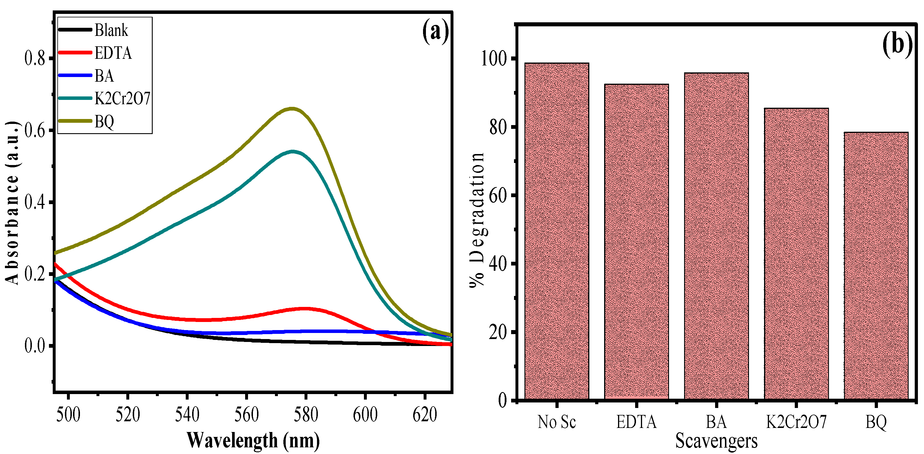
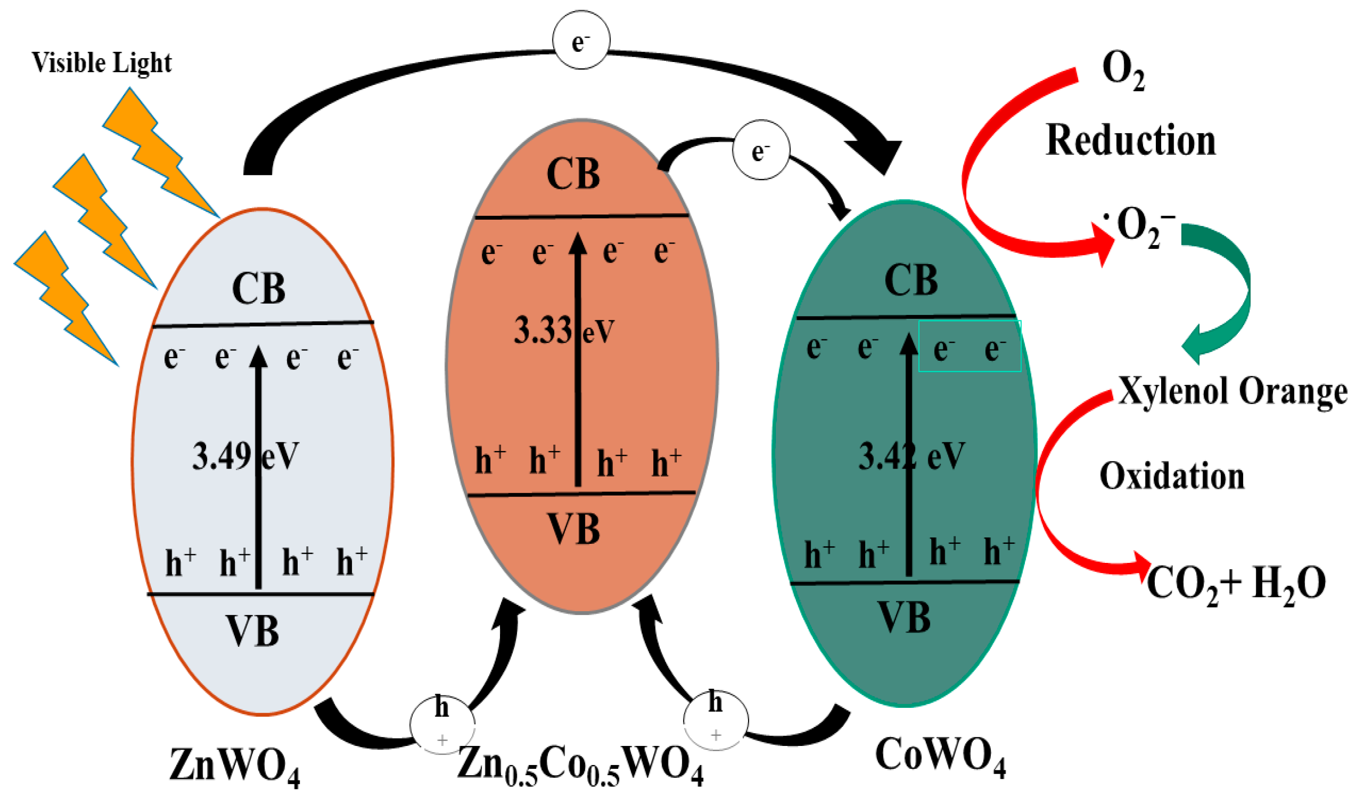
2.4. Scavenging Study and Mechanism of Photodegradation

It was suggested that a variety of reactive species, including the hydroxyl radical (OH), superoxide (O2), valence band hole (h+), and electron (e) play important roles in the photodegradation of organic pollutants, and their effectiveness depends upon the band structure and chemical nature of the photocatalysts [40]. So, scavenger studies were performed using a 1.3 mM solution of Benzoic acid (BA), Potassium dichromate (K2Cr2O7), (AA), ethylenediaminetetraacetic acid (EDTA), and benzoquinone (BQ) by taking 10 mL of 20ppm XO with 10 mg at pH 3 for an irradiation time of 120 min. We know that BA is a scavenger for OH, EDTA for h+, K2Cr2O7 for e, and BQ for O2 [41,42]. Figure 11 displays the results, which show that without the use of a scavenger, the degrading efficiency of Zn0.5Co0.5WO4 towards XO achieved 98.27%. After adding BA and EDTA, there appears no appreciable change in photocatalytic efficiency, suggesting no primary role of OH and h+ on the degradation of XO at the optimized conditions. On the contrary, when BQ and K2Cr2O7 are added, an appreciable decrease in photocatalytic efficiency happens, demonstrating that photogenerated electrons (e) and O2 radicals are predominant species in this photodegradation reaction scheme. The results showed that the degradation of XO was primarily controlled by e and O2 radicals (majorly).
The plausible mechanism of degradation of XO by O2 radicals is given in Figure 12 [43,44].

2.5. Comparison with the Literature

The outcomes of this study were compared with the information available in the literature, and a comparison in Table 3 was made to investigate the upgraded scientific information added by the present study. It was found that there is no research where tungstate base nanomaterial is used for the photocatalytic degradation of xylenol orange.

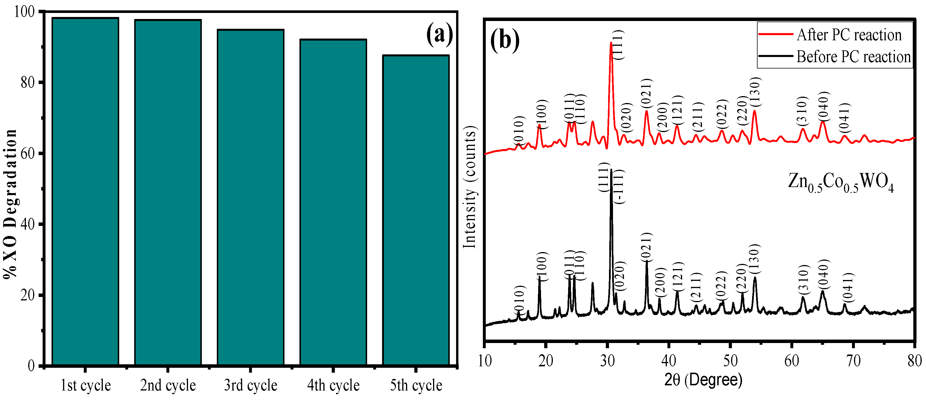
2.6. Reusability Test

The reusability test was performed for the synthesized Zn0.5Co0.5WO4 up to five cycles to assess the stability of the material towards XO degradation, and the results are given in Figure 13a. It can be seen that there is no appreciable change in photocatalytic efficiency up to the first two cycles with greater than 98% of XO degradation. Furthermore, from cycle 3 to cycle 5, there appears some appreciable decrease in photocatalytic efficiency attaining 85% of XO degradation in cycle 5. The overall results suggest that the synthesized Zn0.5Co0.5WO4 have good stability toward the XO degradation under optimized reaction conditions. To support the stability of the material, XRD analysis of the synthesized material before and after the photocatalytic reaction was taken into consideration, which is given in Figure 13b. The XRD spectra of Zn0.5Co0.5WO4 after five consecutive cycles of reuse represented almost similar hkl planes, but with reduced intensity as compared to pristine Zn0.5Co0.5WO4 before the photocatalytic reaction. Only one change was observed, that after the photocatalytic reaction the (-111) plane diminished, which may be due to the absorption of water.

3. Materials and Methods

3.1. Chemicals and Reagents

Sodium tungstate dihydrate (Na2WO4·2H2O, ACS grade, 99%) was purchased from Merck, Rahway, NJ, USA. Cobalt (II) nitrate hexahydrate (Co (NO3)2.6H2O ACS grade 98%), Zinc nitrate hexahydrate (Zn (NO3)2.6H2O, reagent grade 98%), were purchased from Sigma Aldrich, St. Louis, Missouri USA. Xylenol orange (C31H28N2Na4O13S, GR) and Ammonia solution (25%) were acquired from Otto Chemie, Mumbai, India. All the solutions were prepared in deionized (DI) water and used throughout all the photocatalytic experiments. Figure 14 represents the molecular structure of xylenol orange.

3.2. Synthesis of ZnWO4, CoWO4, and Zn0.5Co0.5WO4 Nanoparticles

The synthesis of pristine metal tungstate nanomaterials (ZnWO4, CoWO4 NPs) and mixed metal tungstate nanocomposite (Zn0.5Co0.5WO4) was done according to the hydrothermal method reported elsewhere [51]. Three Erlenmeyer flasks of 50 mL capacity were equipped with 25 mL of 3mmol of sodium tungstate dihydrate (Na2WO4·2H2O), Zinc nitrate hexahydrate (Zn(NO3)2·6H2O; Flask 1), Cobalt(II)nitrate hexahydrate (Co (NO3)2·6H2O; Flask 2), and 1:1 ratio, i.e., 1.5 mmol each, of Zn(NO3)2·6H2O and Co (NO3)2·6H2O (Flask 3). All the flasks were placed on a magnetic stirring plate for 30 min to completely dissolve the salts and attain homogeneity. Then, the mixtures (F1, F2, and F3) were transferred into a Teflon-lined steel autoclave and heated in a convection oven at 180 ℃ for 24 h. The resulting precipitates of ZnWO4, CoWO4, and Zn0.5Co0.5WO4 were centrifuged at 8000 rpm for 5 min and purified several times with DI water and finally with ethanol (2 times). After that, the materials were dried at 100 °C for 5 h and calcined at 600 °C for 4 h, then stored in a desiccator using a polystyrene Petri dish for further characterization and photocatalytic experiments.

3.3. Characterization Techniques

The ZnWO4, CoWO4, and Zn0.5Co0.5WO4 were characterized using various techniques. The change in crystallite size and interplanar distance on Co deposition in the crystal lattice of ZnWO4 was analyzed by an X-ray powder diffractometer (XRD, Rigaku Ultima IV, Tokyo, Japan) at Cu Kα wavelength of 1.5418 Å as a source of radiation. The change of WO66− octahedron vibrations, Zn–O, and formation of Co–O bonding was assessed through Fourier transform infrared (FTIR) spectroscopy using a Perkin Elmer Spectrum 2 ATR spectrometer (Perkin Elmer, Waltham, MA, USA). The optical properties, energy bandgap of the synthesized NPs, and remaining concentration of XO in the effluent after the photocatalytic experiment was calculated by using a Shimadzu UV–1900 double-beam spectrophotometer (Kyoto, Japan). The surface morphology of the synthesized NPs was observed by scanning electron microscopy (SEM; JEOL GSM 6510LV, Tokyo, Japan) associated with an X-ray energy dispersive detector (EDX) to investigate the elemental composition and mapping. The morphological change in shape and size of the nanocrystalline solid upon Co deposition in ZnWO4 was investigated using a transmission electron microscope (TEM; JEM 2100, Tokyo, Japan).

3.4. Photocatalysis Experiment

The photocatalytic efficiency of ZnWO4, CoWO4, and Zn0.5Co0.5WO4 was analyzed towards the degradation of xylenol orange under visible light irradiation in a photocatalytic reactor. An aliquot of 10 mL of 50 ppm XO was taken in a 20 mL 1.8 cm glass tube with a magnetic bar of 10 mg of synthesized NPs in a photocatalytic reactor using Xe lamp (420 W cm −1) for 5–120 min of irradiation. The aliquots are taken out of the photoreactor at a regular time interval, centrifuged to separate the nanocatalyst, and then placed in a UV–vis spectrophotometer at λmax = 580 nm to check the photocatalytic efficiency of the synthesized NPs, which is given by Equation (7);
XO   Degradation ( % ) = ( C 0 - C e C 0 ) × 100
where C0 and Ce represents the initial concentration of an organic pollutant at time t = 0 and at any time t, respectively. The experiments were repeated by changing the parameters, for instance, organic pollutant concentration (ppm), irradiation time (min), catalytic load (mg), and pH of the media to optimize the reaction condition and predict the mechanism of the degradation.

4. Conclusions

The present study reveals the hydrothermal synthesis of pristine ZnWO4, CoWO4, and mixed metal Zn0.5Co0.5WO4 nanoparticles (NPs) in a Teflon-lined autoclave at 180 °C for 24 h. The structural, morphological, elemental, and optical analyses supported the formation of Zn0.5Co0.5WO4. The XRD analysis revealed a contraction in crystallite size from 32 nm to 24 nm with a decrease in crystallinity from 45% to 42% in ZnWO4 to Zn0.5Co0.5WO4 suggesting the successful deposition of Co2+ in the ZnWO4 lattice. The TEM analysis suggested the nanorod-like shape of the Zn0.5Co0.5WO4 NPs with an average particle size of 31.74 nm. The optical studies revealed the UV–vis light activeness of materials with the energy band gap of ZnWO4 (3.49 eV), CoWO4 (3.42 eV), and mixed metal Zn0.5Co0.5WO4 (3.33 eV). The material was explored as a catalyst for the photodegradation of XO dye under visible solar radiation and the results suggested the order of photocatalytic efficiency as Zn0.5Co0.5WO4 (98.77%) > CoWO4 (98.10%) > ZnWO4 (97.89%) utilizing 10 mg of catalyst for 60 ppm XO for 120 min of solar irradiation. The photocatalytic data were well explained by L–H pseudo–first–order kinetics with R2 = 0.99, and apparent rate constant (kapp) as 0.018 min−1 for ZnWO4, 0.021 min−1 for CoWO4, and 0.025 min−1 for Zn0.5Co0.5WO4. The scavenger experiments disclosed that superoxide anion radicals (•O2) and photogenerated electrons (e) played important roles in the degradation of XO. The outcomes of this study revealed that the eco-friendly and economical hydrothermal synthesized material (Zn0.5Co0.5WO4) can be used more efficiently for the treatment of XO-contaminated water in industries without the generation of any secondary pollution.

Author Contributions

Conceptualization, F.A.A.; Methodology, F.A.A., H.S.A. and I.H.; Software, I.H.; Validation I.H.; Investigation, W.S.A.-N. and A.A.A.; Resources, W.S.A.-N. and A.A.A.; Data curation, I.H.; Writing—original draft, W.S.A.-N.; Writing—review & editing, H.S.A. and I.H.; Visualization, I.H.; Supervision, F.A.A. and H.S.A.; Project administration, F.A.A.; Funding acquisition, F.A.A. All authors have read and agreed to the published version of the manuscript.

Funding

The authors extend their appreciation to the Deputyship of Research and Innovation, Ministry of Education in Saudi Arabia for funding this research work through project number (IFKSURG-2-255).

Conflicts of Interest

The authors declare no conflict of interest.

References

  1. You, J.; Wang, L.; Zhao, Y.; Bao, W. A Review of Amino-Functionalized Magnetic Nanoparticles for Water Treatment: Features and Prospects. J. Clean. Prod. 2021, 281, 124668. [Google Scholar] [CrossRef]
  2. Surendra, B.S.; Shashi Shekhar, T.R.; Veerabhadraswamy, M.; Nagaswarupa, H.P.; Prashantha, S.C.; Geethanjali, G.C.; Likitha, C. Probe Sonication Synthesis of ZnFe2O4 NPs for the Photocatalytic Degradation of Dyes and Effect of Treated Wastewater on Growth of Plants. Chem. Phys. Lett. 2020, 745, 137286. [Google Scholar] [CrossRef]
  3. Akbari, A.; Sabouri, Z.; Hosseini, H.A.; Hashemzadeh, A.; Khatami, M.; Darroudi, M. Effect of Nickel Oxide Nanoparticles as a Photocatalyst in Dyes Degradation and Evaluation of Effective Parameters in Their Removal from Aqueous Environments. Inorg. Chem. Commun. 2020, 115, 107867. [Google Scholar] [CrossRef]
  4. Sarkar, S.; Ponce, N.T.; Banerjee, A.; Bandopadhyay, R.; Rajendran, S.; Lichtfouse, E. Green Polymeric Nanomaterials for the Photocatalytic Degradation of Dyes: A Review. Environ. Chem. Lett. 2020, 18, 1569–1580. [Google Scholar] [CrossRef] [PubMed]
  5. Xu, P.P.; Zhang, L.; Jia, X.; Wang, X.; Cao, Y.; Zhang, Y. Visible-Light-Enhanced Photocatalytic Activities for Degradation of Organics by Chromium Acetylacetone Supported on UiO-66-NH2. ChemistrySelect 2020, 5, 14877–14883. [Google Scholar] [CrossRef]
  6. Garg, N.; Bera, S.; Rastogi, L.; Ballal, A.; Balaramakrishna, M.V. Synthesis and Characterization of L-Asparagine Stabilised Gold Nanoparticles: Catalyst for Degradation of Organic Dyes. Spectrochim. Acta Part A Mol. Biomol. Spectrosc. 2020, 232, 118126. [Google Scholar] [CrossRef]
  7. Kumar, O.P.; Shahzad, K.; Nazir, M.A.; Farooq, N.; Malik, M.; Ahmad Shah, S.S.; ur Rehman, A. Photo-Fenton Activated C3N4x/AgOy@Co1-XBi0.1-YO7 Dual s-Scheme Heterojunction towards Degradation of Organic Pollutants. Opt. Mater. 2022, 126, 112199. [Google Scholar] [CrossRef]
  8. Jamshaid, M.; Nazir, M.A.; Najam, T.; Shah, S.S.A.; Khan, H.M.; ur Rehman, A. Facile Synthesis of Yb3+-Zn2+ Substituted M Type Hexaferrites: Structural, Electric and Photocatalytic Properties under Visible Light for Methylene Blue Removal. Chem. Phys. Lett. 2022, 805, 139939. [Google Scholar] [CrossRef]
  9. Shahzad, K.; Hussain, S.; Nazir, M.A.; Jamshaid, M.; ur Rehman, A.; Alkorbi, A.S.; Alsaiari, R.; Alhemiary, N.A. Versatile Ag2O and ZnO Nanomaterials Fabricated via Annealed Ag-PMOS and ZnO-PMOS: An Efficient Photocatalysis Tool for Azo Dyes. J. Mol. Liq. 2022, 356, 119036. [Google Scholar] [CrossRef]
  10. Xu, L.; Wang, X.; Xu, M.L.; Liu, B.; Wang, X.F.; Wang, S.H.; Sun, T. Preparation of Zinc Tungstate Nanomaterial and Its Sonocatalytic Degradation of Meloxicam as a Novel Sonocatalyst in Aqueous Solution. Ultrason. Sonochem. 2020, 61, 104815. [Google Scholar] [CrossRef]
  11. Taneja, P.; Sharma, S.; Umar, A.; Mehta, S.K.; Ibhadon, A.O.; Kansal, S.K. Visible-Light Driven Photocatalytic Degradation of Brilliant Green Dye Based on Cobalt Tungstate (CoWO4) Nanoparticles. Mater. Chem. Phys. 2018, 211, 335–342. [Google Scholar] [CrossRef]
  12. Karthikeyan, C.; Arunachalam, P.; Ramachandran, K.; Al-Mayouf, A.M.; Karuppuchamy, S. Recent Advances in Semiconductor Metal Oxides with Enhanced Methods for Solar Photocatalytic Applications. J. Alloys Compd. 2020, 828, 154281. [Google Scholar] [CrossRef]
  13. Chawla, H.; Chandra, A.; Ingole, P.P.; Garg, S. Recent Advancements in Enhancement of Photocatalytic Activity Using Bismuth-Based Metal Oxides Bi2MO6 (M = W, Mo, Cr) for Environmental Remediation and Clean Energy Production. J. Ind. Eng. Chem. 2021, 95, 1–15. [Google Scholar] [CrossRef]
  14. Ikram, M.; Rashid, M.; Haider, A.; Naz, S.; Haider, J.; Raza, A.; Ansar, M.T.; Uddin, M.K.; Ali, N.M.; Ahmed, S.S.; et al. A Review of Photocatalytic Characterization, and Environmental Cleaning, of Metal Oxide Nanostructured Materials. Sustain. Mater. Technol. 2021, 30, e00343. [Google Scholar] [CrossRef]
  15. Lin, J.; Lin, J.; Zhu, Y. Controlled Synthesis of the ZnWO4 Nanostructure and Effects on the Photocatalytic Performance. Inorg. Chem. 2007, 46, 8372–8378. [Google Scholar] [CrossRef] [PubMed]
  16. Bai, X.; Wang, L.; Zhu, Y. Visible Photocatalytic Activity Enhancement of ZnWO4 by Graphene Hybridization. ACS Catal. 2012, 2, 2769–2778. [Google Scholar] [CrossRef]
  17. Ke, J.; Niu, C.; Zhang, J.; Zeng, G. Significantly Enhanced Visible Light Photocatalytic Activity and Surface Plasmon Resonance Mechanism of Ag/AgCl/ZnWO4 Composite. J. Mol. Catal. A Chem. 2014, 395, 276–282. [Google Scholar] [CrossRef]
  18. Dong, T.; Li, Z.; Ding, Z.; Wu, L.; Wang, X.; Fu, X. Characterizations and Properties of Eu3+-Doped ZnWO4 Prepared via a Facile Self-Propagating Combustion Method. Mater. Res. Bull. 2008, 43, 1694–1701. [Google Scholar] [CrossRef]
  19. Zhang, X.; Wang, B.; Wang, X.; Xiao, X.; Dai, Z.; Wu, W.; Zheng, J.; Ren, F.; Jiang, C. Preparation of M@BiFeO3 Nanocomposites (M = Ag, Au) Bowl Arrays with Enhanced Visible Light Photocatalytic Activity. J. Am. Ceram. Soc. 2015, 98, 2255–2263. [Google Scholar] [CrossRef]
  20. Wang, F.; Li, W.; Gu, S.; Li, H.; Liu, X.; Wang, M. Fabrication of FeWO4@ZnWO4/ZnO Heterojunction Photocatalyst: Synergistic Effect of ZnWO4/ZnO and FeWO4@ZnWO4/ZnO Heterojunction Structure on the Enhancement of Visible-Light Photocatalytic Activity. ACS Sustain. Chem. Eng. 2016, 4, 6288–6298. [Google Scholar] [CrossRef]
  21. Li, P.; Zhao, X.; Jia, C.J.; Sun, H.; Sun, L.; Cheng, X.; Liu, L.; Fan, W. ZnWO4/BiOI Heterostructures with Highly Efficient Visible Light Photocatalytic Activity: The Case of Interface Lattice and Energy Level Match. J. Mater. Chem. A Mater. 2013, 1, 3421–3429. [Google Scholar] [CrossRef]
  22. Mgidlana, S.; Nyokong, T. Asymmetrical Zinc(II) Phthalocyanines Cobalt Tungstate Nanomaterial Conjugates for Photodegradation of Methylene Blue. J. Photochem. Photobiol. A Chem. 2021, 418, 113421. [Google Scholar] [CrossRef]
  23. Siriwong, P.; Thongtem, T.; Phuruangrat, A.; Thongtem, S. Hydrothermal Synthesis, Characterization, and Optical Properties of Wolframite ZnWO4 Nanorods. Crystengcomm 2011, 13, 1564–1569. [Google Scholar] [CrossRef]
  24. Pavithra, N.S.; Nagaraju, G.; Patil, S.B. Ionic Liquid-Assisted Hydrothermal Synthesis of ZnWO4 Nanoparticles Used for Photocatalytic Applications. Ionics 2021, 27, 3533–3541. [Google Scholar] [CrossRef]
  25. Han, S.; Xiao, K.; Liu, L.; Huang, H. Zn1−xCoxWO4 (0 ≤ x ≤ 1) Full Range Solid Solution: Structure, Optical Properties, and Magnetism. Mater. Res. Bull. 2016, 74, 436–440. [Google Scholar] [CrossRef]
  26. Rahmani, M.; Sedaghat, T. Nitrogen-Doped ZnWO4 Nanophotocatalyst: Synthesis, Characterization and Photodegradation of Methylene Blue under Visible Light. Res. Chem. Intermed. 2019, 45, 5111–5124. [Google Scholar] [CrossRef]
  27. Rahmani, M.; Sedaghat, T. A Facile Sol–Gel Process for Synthesis of ZnWO4 Nanopartices with Enhanced Band Gap and Study of Its Photocatalytic Activity for Degradation of Methylene Blue. J. Inorg. Organomet. Polym. Mater. 2019, 29, 220–228. [Google Scholar] [CrossRef]
  28. Scherrer, P. Estimation of the Size and Internal Structure of Colloidal Particles by Means of Rontgen Rays. Nachr. Ges. Wiss. Göttingen 1918, 26, 98–100. [Google Scholar]
  29. Tauc, J. Optical Properties of Solids; Abelès, F., Ed.; Elsevier: Amsterdam, The Netherlands, 1970. [Google Scholar]
  30. Sadeghfar, F.; Zalipour, Z.; Taghizadeh, M.; Taghizadeh, A.; Ghaedi, M. Photodegradation Processes. Interface Sci. Technol. 2021, 32, 55–124. [Google Scholar] [CrossRef]
  31. Li, X.; Yu, J.; Low, J.; Fang, Y.; Xiao, J.; Chen, X. Engineering Heterogeneous Semiconductors for Solar Water Splitting. J. Mater. Chem. A Mater. 2015, 3, 2485–2534. [Google Scholar] [CrossRef]
  32. Fujito, H.; Kunioku, H.; Kato, D.; Suzuki, H.; Higashi, M.; Kageyama, H.; Abe, R. Layered Perovskite Oxychloride Bi4NbO8Cl: A Stable Visible Light Responsive Photocatalyst for Water Splitting. J. Am. Chem. Soc. 2016, 138, 2082–2085. [Google Scholar] [CrossRef]
  33. Li, Y.; Hua, S.; Zhou, Y.; Dang, Y.; Cui, R.; Fu, Y. Activating ZnWO4 Nanorods for Efficient Electroanalysis of Bisphenol A via the Strategy of In Doping Induced Band Gap Change. J. Electroanal. Chem. 2020, 856, 113613. [Google Scholar] [CrossRef]
  34. Yu, X.; Williams, C.T. Recent Advances in the Applications of Mesoporous Silica in Heterogeneous Catalysis. Catal. Sci. Technol. 2022, 12, 5765–5794. [Google Scholar] [CrossRef]
  35. Malik, J.; Kumar, S.; Srivastava, P.; Bag, M.; Mandal, T.K. Cation Disorder and Octahedral Distortion Control of Internal Electric Field, Band Bending and Carrier Lifetime in Aurivillius Perovskite Solid Solutions for Enhanced Photocatalytic Activity. Mater. Adv. 2021, 2, 4832–4842. [Google Scholar] [CrossRef]
  36. Kirankumar, V.S.; Sumathi, S. Copper and Cerium Co-Doped Cobalt Ferrite Nanoparticles: Structural, Morphological, Optical, Magnetic, and Photocatalytic Properties. Environ. Sci. Pollut. Res. 2019, 26, 19189–19206. [Google Scholar] [CrossRef] [PubMed]
  37. Pan, Y.M.; Zhang, W.; Hu, Z.F.; Feng, Z.Y.; Ma, L.; Xiong, D.P.; Hu, P.J.; Wang, Y.H.; Wu, H.Y.; Luo, L. Synthesis of Ti4+-Doped ZnWO4 Phosphors for Enhancing Photocatalytic Activity. J. Lumin. 2019, 206, 267–272. [Google Scholar] [CrossRef]
  38. Otrokov, M.M.; Klimovskikh, I.I.; Calleja, F.; Shikin, A.M.; Vilkov, O.; Rybkin, A.G.; Estyunin, D.; Muff, S.; Dil, J.H.; Vázquez De Parga, A.L.; et al. Evidence of Large Spin-Orbit Coupling Effects in Quasi-Free-Standing Graphene on Pb/Ir(1 1 1). 2d Mater 2018, 5, 035029. [Google Scholar] [CrossRef]
  39. Magdalane, C.M.; Kaviyarasu, K.; Vijaya, J.J.; Siddhardha, B.; Jeyaraj, B.; Kennedy, J.; Maaza, M. Evaluation on the Heterostructured CeO2/Y2O3 Binary Metal Oxide Nanocomposites for UV/Vis Light Induced Photocatalytic Degradation of Rhodamine–B Dye for Textile Engineering Application. J. Alloys Compd. 2017, 727, 1324–1337. [Google Scholar] [CrossRef]
  40. Zheng, X.; Yuan, J.; Shen, J.; Liang, J.; Che, J.; Tang, B.; He, G.; Chen, H. A Carnation-like RGO/Bi2O2CO3/BiOCl Composite: Efficient Photocatalyst for the Degradation of Ciprofloxacin. J. Mater. Sci. Mater. Electron. 2019, 30, 5986–5994. [Google Scholar] [CrossRef]
  41. Balu, S.; Velmurugan, S.; Palanisamy, S.; Chen, S.W.; Velusamy, V.; Yang, T.C.K.; El-Shafey, E.S.I. Synthesis of α-Fe2O3 Decorated g-C3N4/ZnO Ternary Z-Scheme Photocatalyst for Degradation of Tartrazine Dye in Aqueous Media. J. Taiwan Inst. Chem. Eng. 2019, 99, 258–267. [Google Scholar] [CrossRef]
  42. Naresh, G.; Malik, J.; Meena, V.; Mandal, T.K. PH-Mediated Collective and Selective Solar Photocatalysis by a Series of Layered Aurivillius Perovskites. ACS Omega 2018, 3, 11104–11116. [Google Scholar] [CrossRef] [PubMed]
  43. Saher, R.; Hanif, M.A.; Mansha, A.; Javed, H.M.A.; Zahid, M.; Nadeem, N.; Mustafa, G.; Shaheen, A.; Riaz, O. Sunlight-Driven Photocatalytic Degradation of Rhodamine B Dye by Ag/FeWO4/g-C3N4 Composites. Int. J. Environ. Sci. Technol. 2021, 18, 927–938. [Google Scholar] [CrossRef]
  44. Zhang, P.; Liang, H.; Liu, H.; Bai, J.; Li, C. A Novel Z-Scheme BiOI/BiOCl Nanofibers Photocatalyst Prepared by One-Pot Solvothermal with Efficient Visible-Light-Driven Photocatalytic Activity. Mater. Chem. Phys. 2021, 272, 125031. [Google Scholar] [CrossRef]
  45. Santana, R.W.R.; Lima, A.E.B.; de Souza, L.K.C.; Santos, E.C.S.; Santos, C.C.; de Menezes, A.S.; Sharma, S.K.; Cavalcante, L.S.; Maia da Costa, M.E.H.; Sales, T.O.; et al. BiOBr/ZnWO4 Heterostructures: An Important Key Player for Enhanced Photocatalytic Degradation of Rhodamine B Dye and Antibiotic Ciprofloxacin. J. Phys. Chem. Solids 2023, 173, 111093. [Google Scholar] [CrossRef]
  46. Geetha, G.V.; Sivakumar, R.; Slimani, Y.; Sanjeeviraja, C.; Kannapiran, E. Rare Earth (RE: La and Ce) Elements Doped ZnWO4 Nanoparticles for Enhanced Photocatalytic Removal of Methylene Blue Dye from Aquatic Environment. Phys. B Condens. Matter 2022, 639, 414028. [Google Scholar] [CrossRef]
  47. Liu, X.; Shu, J.; Wang, H.; Jiang, Z.; Xu, L.; Liu, C. One-Pot Preparation of a Novel CoWO4/ZnWO4 p-n Heterojunction Photocatalyst for Enhanced Photocatalytic Activity under Visible Light Irradiation. J. Phys. Chem. Solids 2023, 172, 111061. [Google Scholar] [CrossRef]
  48. Kumar, G.M.; Lee, D.J.; Jeon, H.C.; Ilanchezhiyan, P.; Deuk Young, K.; Tae Won, K. One Dimensional ZnWO4 Nanorods Coupled with WO3 Nanoplates Heterojunction Composite for Efficient Photocatalytic and Photoelectrochemical Activity. Ceram. Int. 2022, 48, 4332–4340. [Google Scholar] [CrossRef]
  49. Jaramillo-Páez, C.; Navío, J.A.; Puga, F.; Hidalgo, M.C. Sol-Gel Synthesis of ZnWO4-(ZnO) Composite Materials. Characterization and Photocatalytic Properties. J. Photochem. Photobiol. A Chem. 2021, 404, 112962. [Google Scholar] [CrossRef]
  50. Jayamani, G.; Shanthi, M. An Efficient Nanocomposite CdS-ZnWO4 for the Degradation of Naphthol Green B Dye under UV-A Light Illumination. Nano-Struct. Nano-Objects 2020, 22, 100452. [Google Scholar] [CrossRef]
  51. Geetha, G.V.; Keerthana, S.P.; Madhuri, K.; Sivakumar, R. Effect of Solvent Volume on the Properties of ZnWO4 Nanoparticles and Their Photocatalytic Activity for the Degradation of Cationic Dye. Inorg. Chem. Commun. 2021, 132, 108810. [Google Scholar] [CrossRef]
Figure 1. FTIR spectra of ZnWO4 (black line), CoWO4 (purple line), and Zn0.5Co0.5WO4 (light blue line).
Catalysts 13 00404 g001
Figure 2. X-ray diffraction pattern of ZnWO4, CoWO4, and Zn0.5Co0.5WO4 calcined at 600 ℃.
Catalysts 13 00404 g002
Figure 3. SEM micrographs of (a) ZnWO4 NPs; (b) CoWO4 NPs; (c) Zn0.5Co0.5WO4 at 1 μm scale; (d) EDX spectra of Zn0.5Co0.5WO4 in 0–20 keV range.
Catalysts 13 00404 g003
Figure 4. TEM images of (a) ZnWO4; (b) CoWO4; (c) Zn0.5Co0.5WO4 at 100 nm magnification range; (d) particle size histogram; (e) TEM image of Zn0.5Co0.5WO4 at 20 nm magnification range; (f) selected area electron diffraction (SAED) image of Zn0.5Co0.5WO4.
Catalysts 13 00404 g004
Figure 5. UV–vis plot of ZnWO4 NPs, CoWO4, and Zn0.5Co0.5WO4 in the wavelength range of 200–700 nm (inset is the Tauc’s plot for calculating the band gap energy (Eg) of the material).
Catalysts 13 00404 g005
Figure 6. Photoluminescence (PL) spectra of ZnWO4, CoWO4 and Zn0.5Co0.5WO4 were recorded at a 594 nm excitation wavelength dispersed in methanol.
Catalysts 13 00404 g006
Figure 7. Effect of initial concentration of XO dye on photocatalytic degradation by (a) ZnWO4; (b) CoWO4; (c) Zn0.5Co0.5WO4; (d) XO degradation (%) vs. XO concentration bar graph.
Catalysts 13 00404 g007
Figure 8. Photocatalytic degradation of XO with a different catalytic dose of (a) ZnWO4; (b) CoWO4; and (c) Zn0.5Co0.5WO4; and (d) %degradation vs. catalyst dose graph.
Catalysts 13 00404 g008
Figure 9. Effect of solution pH on photocatalytic degradation by (a) ZnWO4; (b) CoWO4; (c) Zn0.5Co0.5WO4; (d) XO degradation (%) vs. pH bar graph.
Catalysts 13 00404 g009
Figure 10. UV–vis absorption spectra of degraded XO dye (60 ppm) using 10 mg of (a) ZnWO4; (b) CoWO4; (c) Zn0.5Co0.5WO4; and (d) L–H model pseudo-first-order kinetic graph with variable irradiation time (5–120 min).
Catalysts 13 00404 g010
Figure 11. (a) UV–vis spectra of XO degradation in the presence of various scavengers; (b) % XO degradation vs. bar graph associated with each scavenger.
Catalysts 13 00404 g011
Figure 12. A plausible mechanism of degradation of XO by synthesized Zn0.5Co0.5WO4.
Catalysts 13 00404 g012
Figure 13. (a) Reusability test for synthesized Zn0.5Co0.5WO4 NPs for XO degradation (b) XRD spectra of Zn0.5Co0.5WO4 before and after the photocatalytic (PC) reaction.
Catalysts 13 00404 g013
Figure 14. Structure of xylenol orange (XO).
Catalysts 13 00404 g014
Table 1. XRD parameter of ZnWO4, CoWO4, and Zn0.5Co0.5WO4.
Crystallinity
(%)
Dislocation Density (δ) Lines (1014 × m2)Crystallite
Size (nm) at 2θ value
Interlayer
Spacing (nm) at 2θ
FWHM
hkl)
Component
45.249.7831.980.2030.4530.72ZnWO4
48.469.6532.190.1950.8130.05CoWO4
42.1817.0224.240.1980.7630.33Zn0.5Co0.
5WO4
Table 2. The kinetic parameters for the degradation of XO dye (20ppm) using 10 mg ZnWO4, CoWO4, and Zn0.5Co0.5WO4 with variable irradiation time (5–120 min).
Materialk1 (min−1)R2t1/2 (min)
ZnWO40.0180.9938.50
CoWO40.0210.9933.00
Zn0.5Co0.5WO40.0250.9927.72
Table 3. Comparison of the literature information with the present study.
CatalystsIrradiation Time
(min)
Light SourceDye Used% DegradationReferences
BiOBr/ZnWO4170UV-A lightRhB99.40[45]
La: ZnWO490UV lightMB97.00[46]
CoWO4/ZnWO4 p-n heterojunction40Xe lampRhB93%[47]
ZnWO4/WO3 heterojunction120Visible lightMB83.60[48]
ZnWO4-(ZnO)120UV-illuminationMO99.00[49]
CDs-ZnWO4150UV-illuminationNGB93.00[50]
Zn0.5Co0.5WO4120Visible Solar LightXO98.77Present Study
Disclaimer/Publisher’s Note: The statements, opinions and data contained in all publications are solely those of the individual author(s) and contributor(s) and not of MDPI and/or the editor(s). MDPI and/or the editor(s) disclaim responsibility for any injury to people or property resulting from any ideas, methods, instructions or products referred to in the content.

