Article

7α and 7β Hydroxylation of Dehydroepiandrosterone by Gibberella sp. and Absidia Coerulea Biotransformation

1
School of Functional Food and Wine, Shenyang Pharmaceutical University, Shenyang 110016, China
2
School of Life Sciences and Biopharmaceuticals, Shenyang Pharmaceutical University, Shenyang 110016, China
3
School of Pharmaceutical Engineering, Shenyang Pharmaceutical University, Shenyang 110016, China
4
Beijing Global Biotechnologies, Co., Ltd., Beijing 100193, China
*
Authors to whom correspondence should be addressed.
Catalysts 2023, 13(2), 272; https://doi.org/10.3390/catal13020272
Submission received: 13 December 2022 / Revised: 19 January 2023 / Accepted: 20 January 2023 / Published: 25 January 2023
(This article belongs to the Special Issue Microbial Biocatalysis)

Abstract

:
The hydroxylation of dehydroepiandrosterone (DHEA) to 7α -hydroxy-5-androstene-17-one (7α-OH-DHEA) and 7β-hydroxy-5-androstene-17-one (7β-OH-DHEA) by Gibberella sp. CICC 2498 and Absidia coerulea CICC 41050 was investigated. The media ingredients were optimized. Single factors such as the DHEA concentration, culture time, medium volume, and inoculum rate were individually investigated to generate optimum biotransformation conditions. An orthogonal optimization process using a four-factor, three- level L9 (33) experiment was designed and performed. Finally, the maximum production of 7β-OH-DHEA from DHEA biotransformation by Absidia coerulea is 69.61%. This strategy would provide a possible way to enhance the 7β-OH-DHEA yield in the pharmaceutical industry.

1. Introduction

Dehydroepiandrosterone (DHEA) is a major C19 steroid hormone produced by the adrenal cortex. Meanwhile, it is also produced in small quantities in the gonads and brain [1]. Due to its long half-life in plasma, most DHEA would become dehydroepiandrosterone sulfate ester (DHEAS), reserved, and converted into specific hormones when needed [2]. As a kind of important pharmaceutical steroid, hydroxylation at different positions would exhibit diversified biological activities. For example, hydroxylation at position 9α/16α is crucial for the bioactivities of glucocorticoids (dexamethasone, triamcinolone, etc.) [3,4]. Hydroxylation at position 11α is essential for anti-inflammatory activities (hydrocortisone, prednisolone) [5,6]. Hydroxylation at position 14α is vital for the production of the 21-acetoxy analog of proligestone, which is a prodrug of Promegestone [7]. Hydroxylation at position 15α is a key intermediate for the production of progesterone [8].
Previous biotransformation investigation towards DHEA-analog steroids had demonstrated a varieties of metabolites spectrum. Huang et.al reported that 15α-hydroxy-17 a-oxa-D-homo-androst-4-ene-3,17-dione and androst-4-en-3,17-dione, were produced by Penicillium griseopurpureum [9]. Kołek et al. used androstenediol as a substrate and produced di- and trihydroxylation products such as 3β,17β-Dihydroxyandrost-5-en-7-one, 3β,7α,17β-trihydroxyandrost-5-ene, 3β,7β,17β-Trihydroxyandrost-5-ene [10,11].
When 7-oxo-DHEA was used as substrate, 3β,16β-dihydroxy-androst-5-en-7,17-dione, 3β-hydroxy-17α-oxa-D-homo-androst-5-en-7,17-dione, and 3β-acetoxy-androst-5-en-7,17-dione could be produced by Laetiporus sulphureus AM498, Fusicoccum amygdali AM258 and Spicaria divaricata AM423 [12]. Incubation of DHEA with Ulocladium chartarum MRC 72584 produced seven DHEA derivatives, such as 3β-hydroxyandrost-5-en-7,17-dione, 3β,7β-dihydroxyandrost-5-en-17-one, 3β,7α-dihydroxyandrost-5-en-17-one, etc. [13]. A 7α,15α-dihydroxyl-DHEA product was reported by Li et al. using Colletotrichum lini [14]. Microbial transformation by using Mortierella isabellina AM212 produced 7-Oxo-DHEA, 7α-Hydroxy-DHEA, 7β-Hydroxy-DHEA [10], and Backusella lamprospora VKM F- 944 could transform DHEA into 7α-hydroxy-DHEA [15]. These single, double, and triple hydroxylation reactions greatly enriched the DHEA metabolite ingredients.
In this research, two out of twelve filamentous strains of fungi demonstrated their metabolic abilities for DHEA (Table 1, Figure S1). After the culture, extraction, and isolation, it could be identified that 7α-Hydroxy-DHEA was produced by Gibberella sp. CICC 2498 and 7β-Hydroxy-DHEA was produced by Absidia coerulea CICC 41050 (Figure 1). Previous literature had reported several optimizations for the production of 7α-hydroxy-DHEA. As far as we know, it is the first time that the 7β-Hydroxy-DHEA is obtained by Absidia coerulea CICC 41050 biotransformation. Thus, we focused on the optimization of the 7β-Hydroxy-DHEA in this study, and the optimized transformation rate of 7β-Hydroxy-DHEA is 69.61%.

2. Results and Discussion

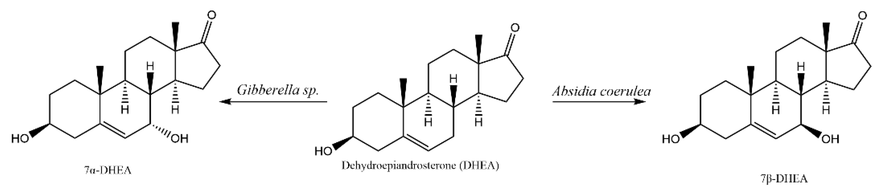
2.1. Whole-Cell Biotransformation Results of DHEA

Thin layer chromatography (TLC) was used to identify whether Gibberella sp. CICC 2498 and Absidia coerulea CICC 41050 could transform dehydroepiandrosterone (DHEA). Figure 2 showed that the substrate control group (Group1, DHEA) has an ocher band, and the transformation groups (Group 4 and 6) have blue bands of different shades with good separation between bands, and almost no ocher substrate bands can be seen. The blue bands (products) are below the red band (DHEA), indicating that the product polarity is greater than DHEA. In addition to the major metabolite, some other products were also generated but could not be further identified due to their lower concentration.

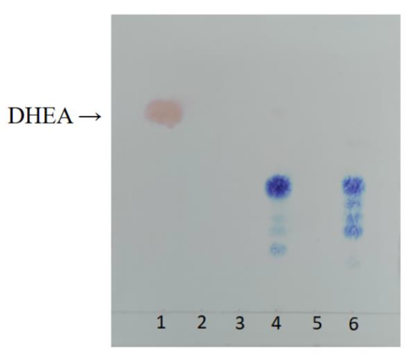
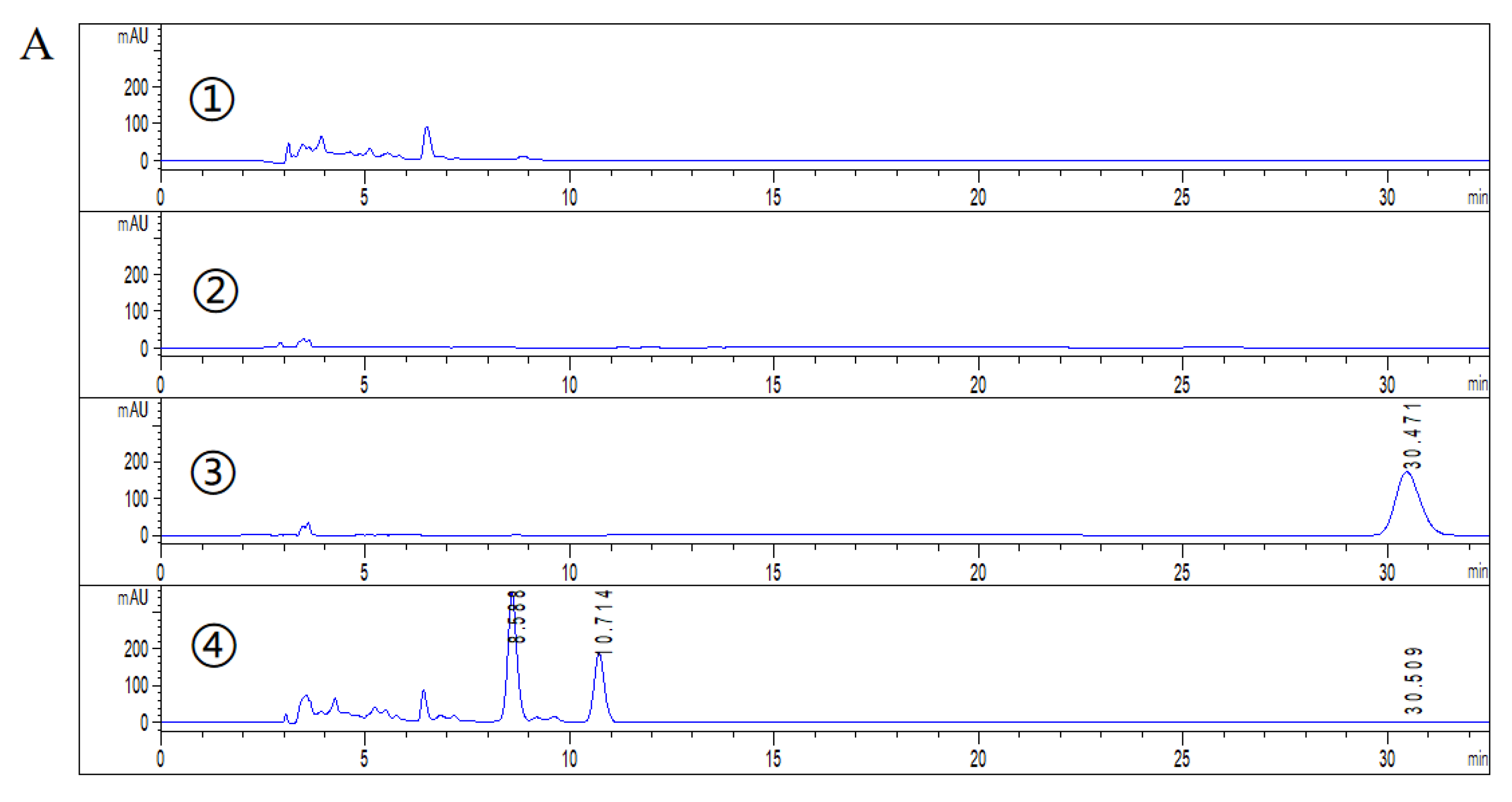
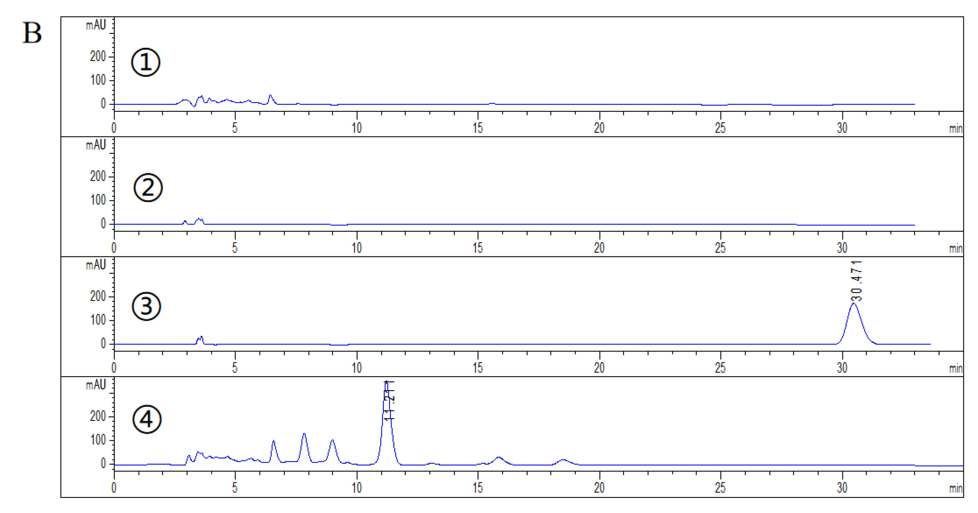
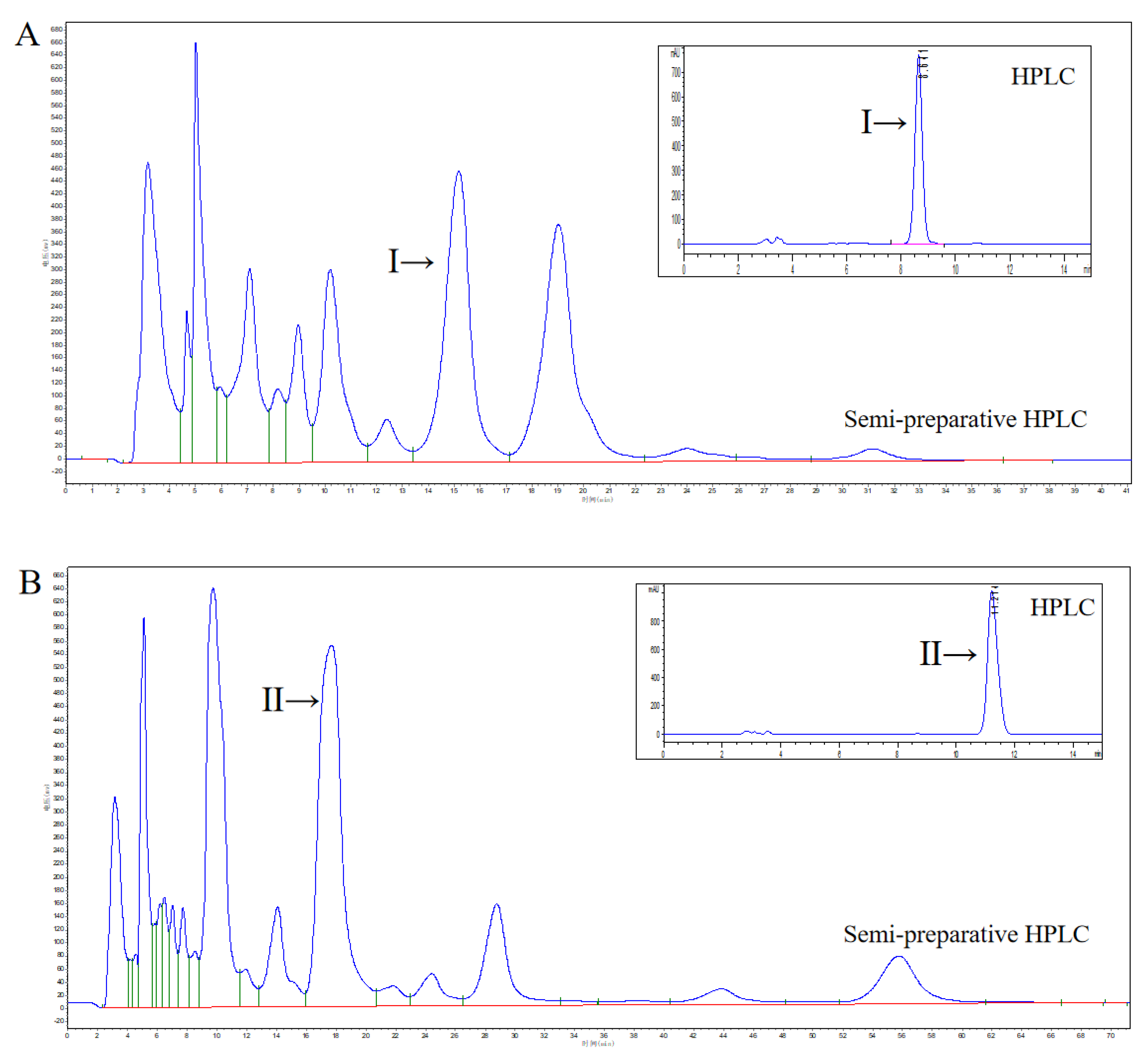
2.2. HPLC Analysis of DHEA Transformed by Gibberella sp. CICC 2498 and Absidia Coerulea CICC 41050

Figure 3A showed the transformation results of DHEA by Absidia coerulea CICC 41050. It can be seen from the comparison between the transformation group 4, and the substrate control group 3 that the substrate (DHEA) peak in the transformation group was significantly reduced. This indicated that DHEA was transformed by Absidia coerulea CICC 41050. By comparing the results of transformation group 4, strain control group 1, and cosolvent control group 3, the increased peak in transformation group 4 was most likely the metabolite of DHEA transformed by Absidia coerulea CICC 41050, rather than the substance produced by microbial growth and metabolism. The types of metabolites of DHEA transformed by Absidia coerulea CICC 41050 were few, and the content of metabolite I was significant, which was conducive to the later separation and purification. The retention time of metabolite I was 8.588 min (Figure 3A). In brief, incubation of Absidia coerulea CICC 41050 with DHEA (1 g/L) resulted in selective accumulation of the metabolite I.
Figure 3B showed that the transformation of DHEA by Gibberella sp. CICC 2498. The separation of the metabolites of DHEA transformed by Gibberella sp. CICC 2498 was good, which was conducive to the later separation and purification. The main metabolite is named metabolite II, and the retention time of metabolite II is 11.211 min (Figure 3B).

2.3. Isolation, Purification and Structural Identification of Metabolites

The fermentation broth was further purified by semi-preparative HPLC. Figure 4 showed that the retention times of metabolites I and II were 15.198 min and 17.698 min, respectively. Metabolite I and II obtained by semi-preparative HPLC were confirmed by HPLC (retention times were 8.588 min and 11.211 min, respectively). Finally, metabolites I and II can be isolated from fermentation broth with purities of 94.0% and 96.0%, respectively.
According to MS (ESI) m/z [M+H]+ 305.1 and [M−H]+ 303.2, the relative molecular weight of metabolite I is calculated to be 304. Compared with the relative molecular weight of 288 of the substrate DHEA (C19 H28 O2), an oxygen atom is added, and the molecular formula is C19 H28 O3 (Figures S2 and S3).
According to MS (ESI) m/z [M+H]+ 305.1 and [M−H]+ 303.2, the relative molecular weight of metabolite II is calculated to be 304. Compared with the relative molecular weight of 288 of the substrate DHEA (C19 H28 O2), an oxygen atom is added, and the molecular formula is C19 H28 O3 (Figures S4 and S5).
The position of the introduced hydroxyl group is analyzed according to the 13 C NMR and 1 H NMR spectral data (Figures S6–S9).
Metabolite I: 7β-Hydroxy-DHEA: 1 H-NMR (600 MHz, CDCl3) δH: 0.90 (3 H, s, 18-Me); 1.08 (3 H, s, 19-Me); 1.23–1.28 (1 H, m), 1.31 (1 H, d, J = 6.7 Hz, 6-H), 1.42–1.62 (7 H, m), 1.67–1.72 (1 H, m), 1.82–1.89 (4 H, m), 2.08–2.14 (1 H, m), 2.22–2.29 (2 H, m), 2.33–2.37 (1 H, m), 2.45–2.49 (1 H, m), 3.52–3.59 (1 H, m, 3α-H); 3.96 (1 H, dt, J1 = 5.3 Hz, J2 = 14.8 Hz, 7α-H); 5.31 (1 H, t, J = 1.8 Hz, 6-H). 13 C-NMR (151 MHz, CDCl3) δC: 13.5 (18-C), 19.1 (19-C), 20.3 (11-C), 24.1 (15-C), 31.2 (2-C), 31.4 (12-C), 35.9 (16-C), 36.6 (10-C), 36.8 (1-C), 40.4 (8-C), 41.6 (4-C), 47.7 (13-C), 48.2 (9-C), 51.1 (14-C), 71.2 (3-C), 72.8 (7-C), 125.4 (6-C), 143.7 (5-C), 221.1 (17-C).
Metabolite II: 7α-Hydroxy-DHEA: 1 H-NMR (600 MHz, CDCl3) δH: 0.89 (3 H, s, 18-Me), 1.02 (3 H, s, 19-Me), 1.10–1.15 (1 H, m), 1.26–1.31 (2 H, m), 1.50–1.61 (3 H, m), 1.66–1.73 (4 H, m), 1.78–1.91 (4 H, m), 2.09–2.19 (2 H, m), 2.28–2.33 (1 H, m), 2.35–2.39 (1 H, m), 2.45–2.50 (1 H, m), 3.55–3.61 (1 H, m, 3α-H), 3.98 (1 H, t, J = 4.7 Hz, 7β-H), 5.65 (1 H, d, J = 7.4 Hz, 6-H). 13 C-NMR (151 MHz, CDCl3) δC: 13.4 (18-C), 18.4 (19-C), 20.2 (11-C), 22.1 (15-C), 31.2 (2-C), 31.4 (12-C), 35.9 (16-C), 37.1 (10-C), 37.3 (1-C), 37.7 (8-C), 42.1 (4-C), 42.8 (13-C), 45.1 (9-C), 47.2 (14-C), 64.4 (7-C), 71.3 (3-C), 123.7 (6-C), 146.7 (5-C), 221.3 (17-C).The 1 H and 13 C NMR data of metabolites I and II are in agreement with those reported in the literature [10], which indicated that both 7 α/β hydroxylation DHEA were obtained (Figure 1) [10].

2.4. Optimization of 7β-OH-DHEA Production by Absidia Coerulea CICC 41050

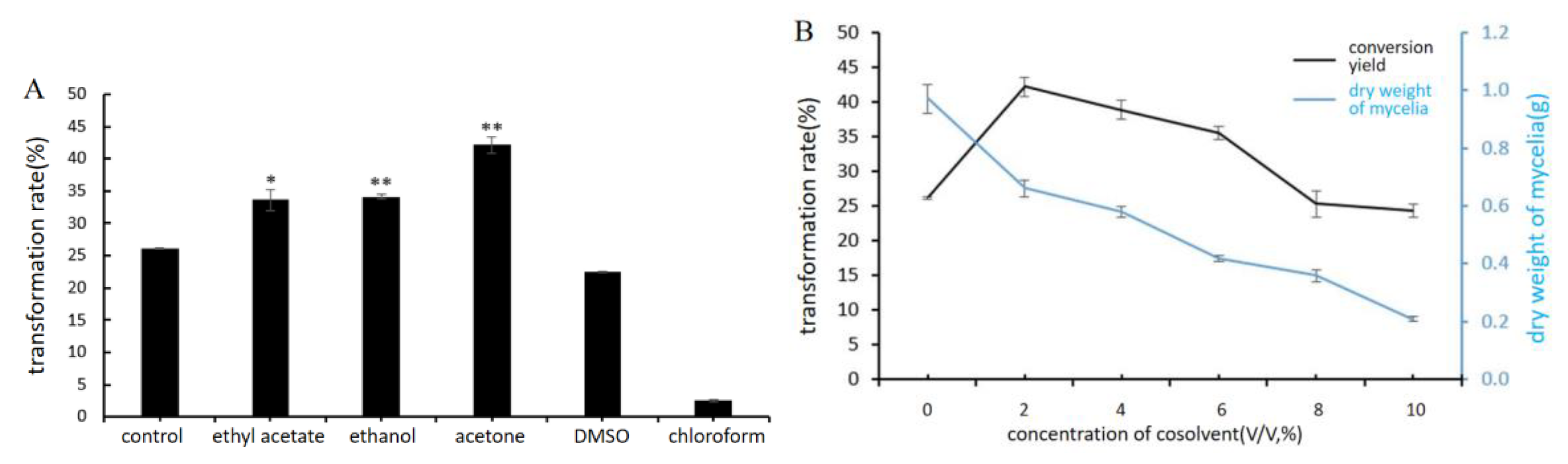
2.4.1. Influence of Different Cosolvents

Figure 5A showed that, compared with the control group, using ethyl acetate, acetone, and ethanol as cosolvents can increase the transformation rate; when DMSO and chloroform were used as cosolvents, the transformation rate decreased. Therefore, acetone was selected as the best cosolvent. It can be seen from the results in Figure 5B that when the acetone concentration was 2%, the transformation rate was the highest. If the concentration of acetone was too low, the substrate could not be completely dissolved. However, with the increase in acetone concentration, acetone will have a toxic effect on fungi, inhibiting their growth and the activity of hydroxylase, thus affecting the conversion rate of substrate. Therefore, 2% acetone was selected as the cosolvent for the subsequent experiment.

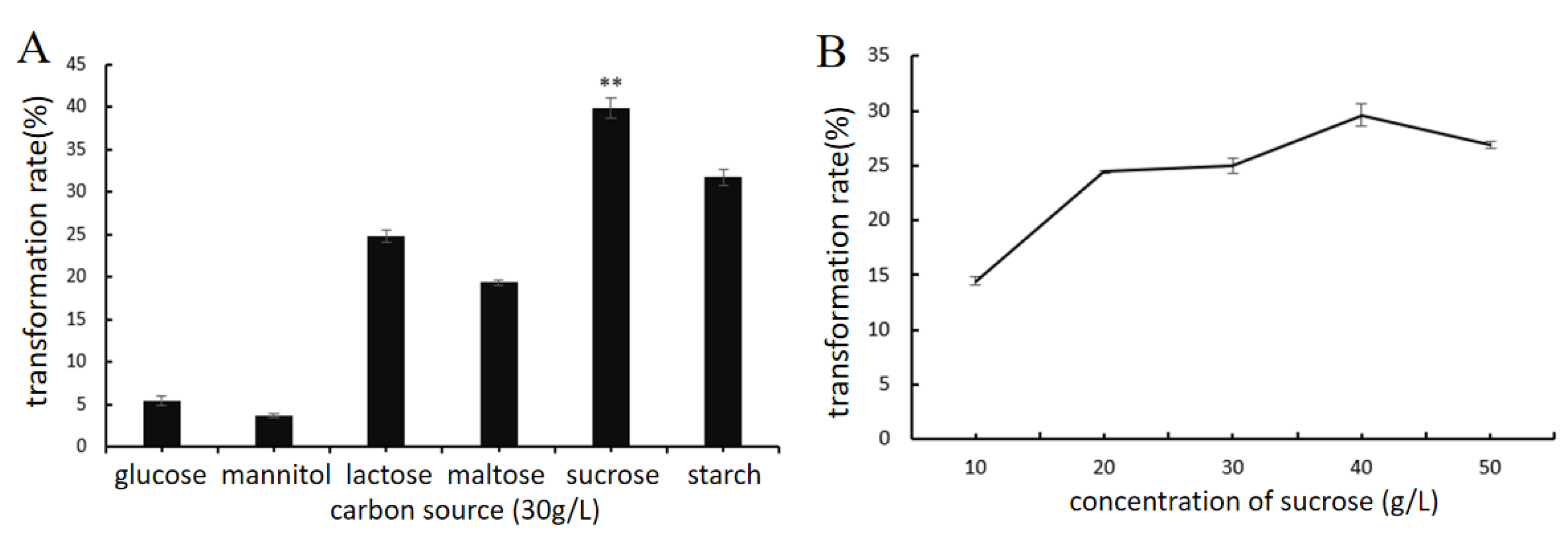
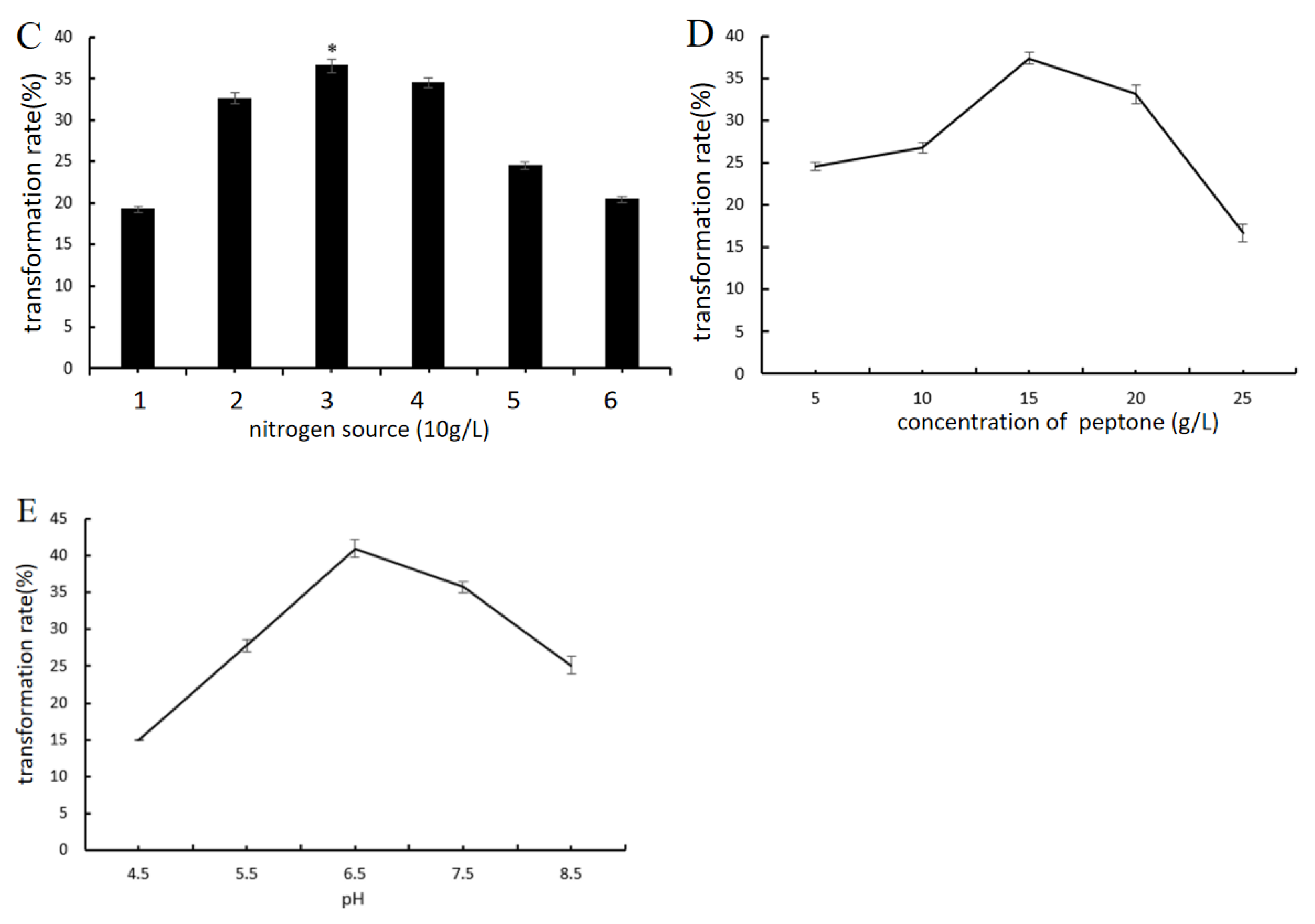
2.4.2. Effect of Key Nutrient Components and pH

It can be seen from the results in Figure 6A that the type of carbon source has a great impact on the transformation rate of 7β-OH-DHEA. When sucrose is the carbon source, the transformation rate is the highest, which is consistent with the type of carbon source in the transformation medium (Section 3.2). The production of 7β-OH-DHEA by Absidia coerulea CICC 41050 also depends on the concentration of sucrose. The concentration of sucrose (40 g/L) provides the highest yield of 7β-OH-DHEA, which is higher than that of 30 g/L in the original transformation medium (Section 3.2) (Figure 6B).
The Impact of the alternative to yeast extract, nitrogen sources (NH4 NO3, (NH4)2 SO4, peptone, yeast extract powder, and beef extract) on the improvement of 7β-hydroxylation catalyzed by Absidia coerulea CICC 41050 was investigated (Figure 6C). Replacement of yeast extract with peptone provided an 8–10% higher 7β-OH-DHEA yield (up to 37%). The results showed that peptone as a nitrogen source was superior to the original nitrogen source (yeast extract) of the transformation medium (Section 3.2). The effect of peptone at various concentrations on the transformation of DHEA by Absidia coerulea CICC 41050 was evaluated. The highest 7β-hydroxylase activity towards DHEA was reached at peptone content (15 g/L) (Figure 6D).
When studying the influence of pH of the transformation medium on DHEA conversion, it was shown that pH 6.5 provided the highest yield of 7β-OH-DHEA, while higher acidic or alkaline pH negatively affected the transformation rate.
According to the results of the single factor experiment, a three-factor and three-level orthogonal experiment were designed to explore the best medium composition (Table 2). The orthogonal experiment results are shown in Table 3.
Table 3 showed that the order of influence of the three factors on the transformation rate is C > A > B. Through range analysis, the optimal combination of the three factors is C2 A1 B3: initial pH 6.5, sucrose 30 g/L, and peptone 20 g/L. However, the composition-optimized medium is not in Table 3, and verification experiments are required. Three parallel experiments were carried out. It was defined that an initial pH 6.5, sucrose 30 g/L, and peptone 20 g/L provided the maximum production (50.48%) of 7β-OH-DHEA by Absidia coerulea CICC 41050.

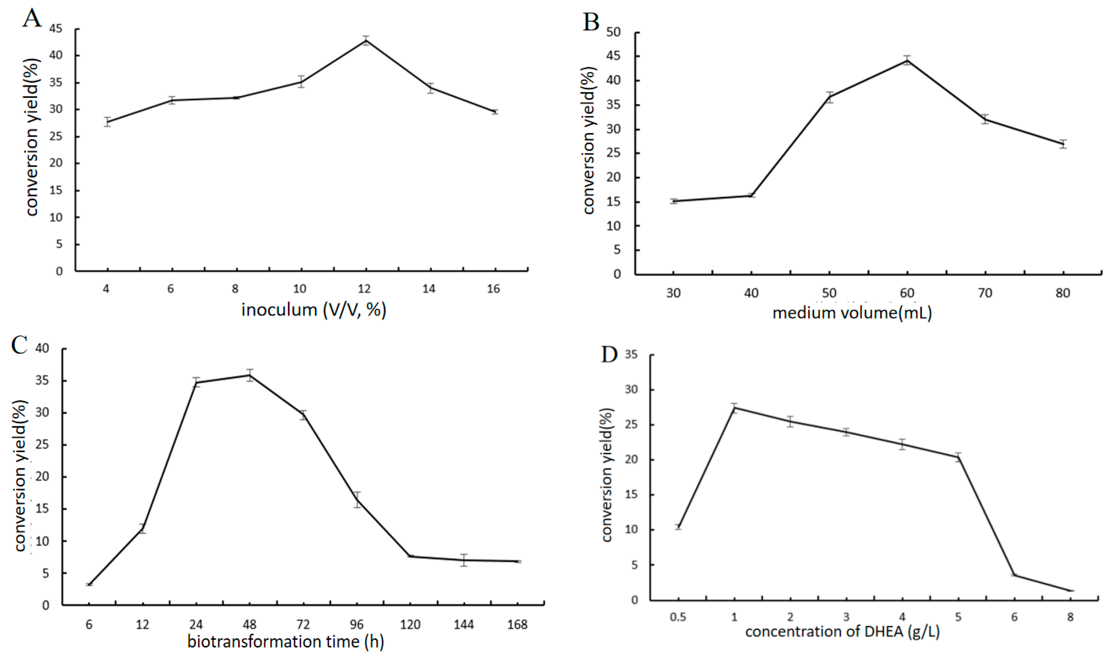
2.4.3. Effect of Biotransformation Conditions

Figure 7A shows that when the inoculum is less than 12%, the transformation rate increases with the increase of the inoculum, and when the inoculum is more than 12%, the transformation rate decreases. The production of 7β-OH-DHEA by Absidia coerulea CICC 41050 also depended on medium volume (Figure 7B). The transformation rate is highest when the medium volume is 60 mL in a 250 mL Erlenmeyer flask. When the volume of the medium is too large, the ventilation and dissolved oxygen in the medium are poor. The transformation rate reached its highest when the substrate was added for 48 h and became extremely low after 96 h (Figure 7C). It is speculated that the nutrients in the medium were consumed and the enzyme activity decreased. The effect of different concentrations of substrate (DHEA, 0.5–8.0 g/L) in the transformation medium was estimated. Figure 7D shows that 1 g/L DHEA can provide the highest transformation rate of 7β-OH-DHEA, while at more than 1 g/L, the transformation rate is declining; greater than 6 g/L, the transformation rate is very low, and the substrate is almost completely converted. The reason may be that the concentration of cosolvent increases with the increase in substrate concentration, and the toxicity of cosolvent inhibits the growth of fungi, thus negatively affecting DHEA conversion.
According to the results of the single-factor experiment, an orthogonal experiment with four factors and three levels was designed to explore the optimal biotransformation conditions (Table 4). The orthogonal experiment results are shown in Table 5.
It can be seen from the results in Table 5 that the order of influence of the four factors in the orthogonal experiment of biotransformation conditions on the transformation rate is A > C > B > D. Through range analysis, the optimal combination of the four factors is A1 C2 B2 D1: DHEA 1 g/L, medium volume 60 mL, biotransformation time 48 h, and inoculum 10%. As the optimal biotransformation conditions are not listed in Table 5, validation tests are required.
Three parallel experiments were carried out. It was defined that sucrose 30 g/L, peptone 20 g/L, corn steep liquor 10 g/L, K2 HPO4 2 g/L, KH2 PO4 1.6 g/L, MgSO4 0.5 g/L, FeSO4 0.05 g/L, pH 6.5, DHEA 1 g/L, medium volume 60 mL, biotransformation time 48 h, and inoculum 10% provided maximum production (69.61%) of 7β-OH-DHEA by Absidia coerulea CICC 41050. Compared with the highest transformation rate of 62.81% in the orthogonal test and the primary transformation rate of 27.23%, transformation rate was increased by 6.80% and 42.38% respectively.

3. Materials and Methods

3.1. Chemicals

Dehydroepiandrosterone (DHEA) was obtained from Hubei Gongtong Pharmaceutical Co., Ltd. (Xiangyang city, Hubei, China). Methanol and acetonitrile were purchased from Concord Technology Co., Ltd. (Tianjin, Tianjin, China). Yeast extract was purchased from HopeBio Co., Ltd. (Qingdao, Shandong, China). All other chemical reagents were purchased from Yuwang Chemical Co., Ltd. (Shenyang, Liaoning, China).

3.2. Microorganism and Cultivation

Absidia coerulea 41050 and Gibberella sp. 2498 were purchased from the China Center of Industrial Culture Collection (CICC).
Potato dextrose agar (PDA) is composed of potatoes (200 g), glucose (20 g), agar (20 g), and 1000 mL distilled water. Seed culture media (g/L) are composed of potato starch (45 g), yeast extract (3 g), corn steep liquor (10 g), CaCO3 (3 g), MgSO4 (0.5 g), and FeSO4 (0.05 g). Transformation media (g/L): sucrose (30 g), yeast extract (10 g), corn steep liquor (10 g), K2 HPO4 (2 g), KH2 PO4 (1.6 g), MgSO4 (0.5 g), FeSO4 (0.05 g), pH 6.5.
The fungi were routinely maintained on PDA slants. To obtain first-generation mycelium, the spore suspension from one agar slant (1 week old) was inoculated aerobically in 50 mL of seed culture media on a rotary shaker (200 rpm) at 28 °C, for 48 h in Erlenmeyer flasks (250 mL). Then 5 mL of seed culture were inoculated into the transformation medium (50 mL in 250 mL Erlenmeyer flask) and cultured at 28 °C, 200 rpm for 5 days. Substrate controls were set without inoculating the fungi into the media and strain controls were set without adding the substrate into the media, with all other conditions remaining the same.

3.3. Sample Preparation

The cultivation broth was centrifuged (3000 r/min, 10 min) to obtain mycelia and transformation solution after 5 days. The mycelium and the transformation solution, with an equal volume of ethyl acetate, were extracted. After three extractions, the extraction solution was combined, evaporated under reduced pressure on the rotary evaporator, and then redissolved with 5 mL of methanol. Added an appropriate amount of anhydrous magnesium sulfate to dry and obtained the transformation sample for detection.

3.4. Thin Layer Chromatography (TLC)

The concentrated extract was analyzed by TLC. TLC on silica gel 60 F254 (25 aluminum sheets 20 × 20 cm; Merck, New York, NY, USA) with a solvent mixture of CHCl3-CH3 OH (10:1, v/v) was applied to separate the metabolites and stained by spraying the plates with H2 SO4/CH3 CH2 OH mixture (1:9, v/v). A UV light at 254 nm was used to visualize them.

3.5. HPLC Detection

A 0.1 mL of conversion product containing dehydroepiandrosterone was diluted five times with methanol and then filtrated by 0.45 μM organic membrane to obtain the sample solution. HPLC analysis was performed on a WondaSil C18 Superb column (5 μm, 4.6 mm × 250 mm, Shimadzu, Kyoto, Japan) with a methanol/water mixture (62:38, v/v), as mobile phase was at 30 °C with UV absorbance detection of 206 nm. Flow rate: 0.8 mL/min; injection volume: 10 μL.

3.6. Isolation and Identification of Major Metabolite

Isolation of the target metabolite was performed by semi-preparative HPLC. HPLC analysis was performed on a SinoChrom ODS-BP column (5 μm, 10 mm × 250 mm, Elite, China) with an acetonitrile/water mixture (30:70, v/v) as mobile phase at 36 °C with UV absorbance detection of 206 nm. Flow rate: 3.7 mL/min; injection volume: 100 μL.
Purified metabolites were identified by ESI-MS and NMR analysis under standard conditions. 1 H, 13 C NMR spectra were taken using a Brüker AVANCE III 400 instrument (Bruker Biospin AG, Fallanden, Switzerland). 1 H NMR spectra were recorded in CDCl3 and DMSO-d6 using tetramethylsilane (TMS) as an internal standard. Mass spectra were taken in ESI mode on an Agilent 1200 LC-MS (Agilent, Santa Clara, CA, USA).

3.7. Establishment of Standard Curve and Calculation of Transformation Rate

The 7β-OH-DHEA samples were dissolved in methanol and prepared into solutions with different concentration gradients (0.05, 0.10, 0.15, 0.20, 0.25 mg/mL). After filtering, carry out HPLC detection. Draw a standard curve with the concentration of DHEA as the abscissa and the peak area as the ordinate. The transformation rate is calculated as follows:
T r a n s f o m a t i o n   r a t e = A × M b B × M a × 100 %
where A is the quantity of product (g), and B is the quantity of substrate (g). Ma and Mb are the relative molecular weight of the product (7β-OH-DHEA, 304.41) and the relative molecular weight of the substrate (DHEA, 288.43), respectively (Figure S10).

3.8. Optimization of DHEA Converted to 7β-OH-DHEA by Absidia Coerulea CICC 41050

3.8.1. Effect of the Type and Concentration of Cosolvent on Transformation Rate

Take 1 mL ethanol, acetone, ethyl acetate, dimethyl sulfoxide, and chloroform as cosolvent, respectively; dissolve 50 mg DHEA in the cosolvent, add the transformation medium, and use no cosolvent in the control group to calculate the transformation rate.

3.8.2. The Biotransformation Medium Was Studied by Single-Factor Experiment and Orthogonal Experiment

On the basis of the best cosolvent, the nitrogen source, carbon source, and initial pH were studied by a single-factor experiment. The 30 g/L sucrose in the transformation medium was replaced with glucose, mannitol, lactose, maltose, starch, etc. of the same concentration to explore the effect of carbon sources on the transformation yield. And then, the carbon source concentration in the transformation medium was set to 10 g/L, 20 g/L, 30 g/L, 40 g/L, 50 g/L to explore the effect of carbon source concentration on the transformation rate. The 10 g/L yeast extract in the transformation medium was replaced with sodium nitrate, ammonium sulfate, peptone, yeast extract powder, and beef extract of the same concentration to explore the effect of the nitrogen source on the transformation rate. The concentration of nitrogen source in the transformation medium was set to 5 g/L, 10 g/L, 15 g/L, 20 g/L, and 30 g/L to explore the effect of nitrogen source concentration on the transformation rate. Set the initial pH of the transformation medium to 4.5, 5.5, 6.5, 7.5, or 8.5 and explore the impact of different initial pH values on the transformation yield.
According to the results of the single-factor experiment, an orthogonal experiment with three factors and three levels L9 (33) was designed.

3.8.3. Biotransformation Conditions Were Studied by Single-Factor Experiment and Orthogonal Experiment

On the basis of the best cosolvent and the best culture medium, the single-factor experiment of transformation conditions was conducted. Set inoculum at 4%, 6%, 8%, 10%, 12%, 14%, and 16% (v/v, 50 mL). Fill a 250 mL Erlenmeyer flask with 30 mL, 40 mL, 50 mL, 60 mL, 70 mL, and 80 mL of transformation medium, respectively. The biotransformation times are 6 h, 12 h, 24 h, 48 h, 72 h, 96 h, 120 h, 144 h, and 168 h. Add 25 mg, 50 mg, 100 mg, 150 mg, 200 mg, 250 mg, 300 mg, and 400 mg of DHEA to 50 mL of transformation medium, respectively. Inoculate volume, medium volume, biotransformation time, and concentration of substrate (DHEA) were investigated to explore the effect on the transformation rate.
The orthogonal experiment with four factors and three levels L9 (34) was designed according to the inoculate volume, medium volume, biotransformation time, and concentration of substrate (DHEA) determined by the single-factor experiment.

3.9. Statistical Analysis

All the experiments were carried out in triplicate, and each presented value was the average of three independent experiments. Standard deviations (SD) were estimated using the following equation and shown as errors on the graphs:
SD = | x μ | 2 N
where ∑ means “sum of”, x is a value in the data set, μ is the mean of the data set, and N is the number of data points in the population. SPSS 20.0 software was used to conduct a t-test on the data to determine the statistical difference; p < 0.05 was significant (*), p < 0.01 was extremely significant (**).

4. Conclusions

The aim of this study was to evaluate 7α and 7β hydroxylation of dehydroepiandrosterone (DHEA) by Gibberella sp. CICC 2498 and Absidia coerulea CICC 41050 biotransformation. The biotransformation products were analyzed by HPLC. The retention time of the main product of DHEA transformation by Absidia coerulea CICC 41050 was 8.588 min. The retention time of the main product of DHEA transformation by Gibberella sp. CICC 2498 was 11.211 min. A semi-preparative HPLC method was successfully established to separate the biotransformation products of DHEA. The purity of the two metabolites was 94% and 96%, respectively. The isolated products were identified by NMR and MS, and the product of DHEA transformation by Absidia coerulea CICC 41050 was 7 β- OH-DHEA, and the product of DHEA transformation by Gibberella sp. CICC 2498 was 7α-OH-DHEA. Determined by single-factor experiment and subsequent orthogonal experiment, the optimized media composition to produce 7 β-OH-DHEA by Absidia coerulea CICC 41050 was consisted of 30 g/L sucrose, 20 g/L peptone, 10 g/L corn steep liquor, 2 g/L K2 HPO4, 1.6 g/L KH2 PO4, 0.5 g/L MgSO4, and 0.05 g/L FeSO4 with pH 6.5. The transformation rate of 7β-OH-DHEA reached 50.48%. The optimal biotransformation conditions (DHEA 1 g/L, medium volume 60 mL, biotransformation time 48 h, and inoculum 10%) provided maximum production (69.61%) of 7β-OH-DHEA by Absidia coerulea CICC 41050. The transformation of the DHEA substrate by Absidia coerulea CICC 41050 was described for the first time. Meanwhile, the conversion period was shortened to 48 h.
Previous literature had reported that Absidia griseolla var. igachii could provide C6β, C7α/β and C14α hydroxylation on androst-4-ene-3,17-dione (4-AD) [4], and Absidia coerulea AM93 could generate C7α/β hydroxylation on androstenediol [11]. This work extended our knowledge of DHEA hydroxylation on C7 position to the Absidia coerulea CICC 41050. It seems that the Absidia species possesses the C7 hydroxylation potential. Further genomic and proteomic data mining are worthy fo exploration for the rest of the story.
Steroids are lipophilic compounds with a gonane skeleton and play an important role in higher organisms. Due to different hydroxylations of steroid molecules, they vary greatly in their mode of action [16]. Hydroxylation of dehydroepiandrosterone (DHEA) to positions 3,7, and15 is an essential step in the synthesis of many steroidal drugs [17]. However, low hydroxylation of DHEA production is a difficult issue that must be solved urgently in industry. At present, DHEA and other steroid substrates could be hydroxylated by cytochromes P450 [18]. However, cytochrome P450 is a membrane bound protein, that is not very easy to get in a purified form for extensive research. To overcome at least some of these drawbacks, whole-cell systems are the method of choice to accomplish hydroxylation of the DHEA. Thus, this strategy would provide a possible way to enhance the 7β-OH-DHEA yield in the pharmaceutical industry.

Supplementary Materials

The following supporting information can be downloaded at: https://www.mdpi.com/article/10.3390/catal13020272/s1, Figure S1. Morphology of Absidia coerulea and Gibberella sp. Figure S2. (ESI) m/z [M+H]+ diagrams for the metabolite I. Figure S3. (ESI) m/z [M+H] diagrams for the metabolite I. Figure S4. (ESI) m/z [M+H]+ diagram for the metabolite II. Figure S5. (ESI) m/z [M+H] diagram for the metabolite II. Figure S6. 1 H NMR diagram for the 7α-OH-DHEA. Figure S7. 13 C NMR diagram for the 7α-OH-DHEA. Figure S8. 1 H NMR diagram for the 7β-OH-DHEA. Figure S9. 13 C NMR diagram for the 7β-OH-DHEA. Figure S10. Standard curve of 7β-OH-DHEA by HPLC.

Author Contributions

M.S.: Data Curation, Investigation; R.F.: Investigation, Methodology; S.C. and X.J.: Data Curation; F.W.: Resource; W.X. (Weizhuo Xu) and W.X. (Wei Xu): Resources, Supervision, Writing-Review and Editing. All authors have read and agreed to the published version of the manuscript.

Funding

This research received no external funding.

Data Availability Statement

Data are available upon reasonable request.

Conflicts of Interest

The authors declare no conflict of interest.

References

  1. EMBL-EBI. CHEBI:28689—Dehydroepiandrosterone. Available online: http://www.ebi.ac.uk/chebi/searchId.do?chebiId=CHEBI:28689 (accessed on 1 January 2020).
  2. Corrigan, B. DHEA and Sport. Clin. J. Sport Med. 2002, 12, 236–241. [Google Scholar] [CrossRef] [PubMed]
  3. Chuang, T.Y.; Cheng, A.J.; Chen, I.T.; Lan, T.Y.; Huang, I.H.; Shiau, C.W.; Hsu, C.L.; Liu, Y.W.; Chang, Z.F.; Tseng, P.H.; et al. Suppression of LPS-induced inflammatory responses by the hydroxyl groups of dexamethasone. Oncotarget 2017, 8, 49735–49748. [Google Scholar] [CrossRef] [PubMed]
  4. Heidary, M.; Habibi, Z. Microbial transformation of androst-4-ene-3,17-dione by three fungal species Absidia griseolla var. igachii, Circinella muscae and Trichoderma virens. J. Mol. Catal. B-Enzym. 2016, 126, 32–36. [Google Scholar] [CrossRef]
  5. Dragan, C.A.; Zearo, S.; Hannemann, F.; Bernhardt, R.; Bureik, M. Efficient conversion of 11-deoxycortisol to cortisol (hydrocortisone) by recombinant fission yeast Schizosaccharomyces pombe. FEMS Yeast Res. 2005, 5, 621–625. [Google Scholar] [CrossRef] [PubMed] [Green Version]
  6. Dumas, B.; Cauet, G.; Lacour, T.; Degryse, E.; Laruelle, L.; Ledoux, C.; Spagnoli, R.; Achstetter, T. 11 beta-hydroxylase activity in recombinant yeast mitochondria. In vivo conversion of 11-deoxycortisol to hydrocortisone. Eur. J. Biochem. 1996, 238, 495–504. [Google Scholar] [CrossRef] [PubMed]
  7. Andryushina, V.A.; Voishvillo, N.E.; Druzhinina, A.V.; Stytsenko, T.S.; Yaderets, V.V.; Petrosyan, M.A.; Zeinalov, O.A. 14 alpha-Hydroxylation of steroids by mycelium of the mold fungus Curvularia lunata (VKPM F-981) to produce precursors for synthesizing new steroidal drugs. Pharm. Chem. J. 2013, 47, 103–108. [Google Scholar] [CrossRef]
  8. Faramarzi, M.A.; Tabatabaei Yazdi, M.; Amini, M.; Zarrini, G.; Shafiee, A. Microbial hydroxylation of progesterone with Acremonium strictum. FEMS Microbiol. Lett. 2003, 222, 183–186. [Google Scholar] [CrossRef] [PubMed] [Green Version]
  9. Huang, L.-H.; Li, J.; Xu, G.; Zhang, X.H.; Wang, Y.G.; Yin, Y.L.; Liu, H.M. Biotransformation of dehydroepiandrosterone (DHEA) with Penicillium griseopurpureum Smith and Penicillium glabrum (Wehmer) Westling. Steroids 2010, 75, 1039–1046. [Google Scholar] [CrossRef] [PubMed]
  10. Kolek, T.; Milecka, N.; Świzdor, A.; Panek, A.; Białońska, A. Hydroxylation of DHEA, androstenediol and epiandrosterone by Mortierella isabellina AM212. Evidence indicating that both constitutive and inducible hydroxylases catalyze 7 alpha- as well as 7beta-hydroxylations of 5-ene substrates. Org. Biomol. Chem. 2011, 9, 5414–5422. [Google Scholar] [CrossRef] [PubMed]
  11. Milecka-Tronina, N.; Kołek, T.; Świzdor, A.; Panek, A. Hydroxylation of DHEA and its analogues by Absidia coerulea AM93. Can an inducible microbial hydroxylase catalyze 7alpha- and 7beta-hydroxylation of 5-ene and 5alpha-dihydro C19-steroids? Bioorganic Med. Chem. 2014, 22, 883–891. [Google Scholar] [CrossRef] [PubMed]
  12. Lyczko, P.; Panek, A.; Ceremuga, I.; Świzdor, A. The catalytic activity of mycelial fungi towards 7-oxo-DHEA—An endogenous derivative of steroidal hormone dehydroepiandrosterone. Microb. Biotechnol. 2021, 14, 2187–2198. [Google Scholar] [CrossRef] [PubMed]
  13. Yildirim, K.; Kuru, A.; Yılmazer Keskin, S.; Ergin, S. Microbial transformation of dehydroepiandrosterone (DHEA) by some fungi. Biocatal. Biotransformation 2021, 39, 465–474. [Google Scholar] [CrossRef]
  14. Li, H.; Sun, J.; Xu, Z. Biotransformation of DHEA into 7α,15α-diOH-DHEA. In Microbial Steroids: Methods and Protocols; Barredo, J.-L., Herráiz, I., Eds.; Springer: New York, NY, USA, 2017; pp. 289–295. [Google Scholar]
  15. Kollerov, V.; Shutov, A.; Kazantsev, A.; Donova, M. Hydroxylation of pregnenolone and dehydroepiandrosterone by zygomycete Backusella lamprospora VKM F-944: Selective production of 7alpha-OH-DHEA. Appl. Microbiol. Biotechnol. 2022, 106, 535–548. [Google Scholar] [CrossRef] [PubMed]
  16. Schmitz, D.; Zapp, J.; Bernhardt, R. Steroid conversion with CYP106A2—Production of pharmaceutically interesting DHEA metabolites. Microb. Cell Factories 2014, 13, 81. [Google Scholar] [CrossRef] [PubMed] [Green Version]
  17. Li, C.; Li, H.; Sun, J.; Zhang, X.; Shi, J.; Xu, Z. Production of 7alpha,15alpha-diOH-DHEA from dehydroepiandrosterone by Colletotrichum lini ST-1 through integrating glucose-feeding with multi-step substrate addition strategy. Bioprocess Biosyst. Eng. 2016, 39, 1259–1266. [Google Scholar] [CrossRef] [PubMed]
  18. Sakaki, T. Practical application of cytochrome P450. Biol. Pharm. Bull. 2012, 35, 844–849. [Google Scholar] [CrossRef] [PubMed]
Figure 1. 7α- and 7β-hydroxylated DHEA obtained by Gibberella sp. CICC 2498 and Absidia coerulea CICC 41050.
Catalysts 13 00272 g001
Figure 2. TLC analysis of microbial transformation of DHEA. Group 1, substrate (DHEA); Group 2, cosolvent (acetone); Group 3, Absidia coerulea CICC 41050; Group 4, Absidia coerulea CICC 41050 + DHEA; Group 5, Gibberella sp. CICC 2498; Group 6, Gibberella sp. CICC 2498 + DHEA. Colored with a 10% sulfuric acid-ethanol.
Catalysts 13 00272 g002
Figure 3. HPLC analysis of biotransformation of DHEA by Absidia coerulea CICC 41050 (A), and Gibberella sp. CICC 2498 (B). Group 1, strain; Group 2, cosolvent (acetone); Group 3, substrate (DHEA); Group 4, strain + substrate (DHEA).
Catalysts 13 00272 g003aCatalysts 13 00272 g003b
Figure 4. Semi-preparative HPLC separation diagram. (A), the semi-preparative separation of metabolite I from fermentation broth after incubation of Absidia coerulea CICC 41050 with DHEA (5 days, 1 g/L), metabolite I was identified by HPLC. (B), the semi-preparative separation of metabolite II from fermentation broth after incubation of Gibberella sp. CICC 2498 with DHEA (5 days, 1 g/L), metabolite II was identified by HPLC.
Catalysts 13 00272 g004
Figure 5. The effect of cosolvent type (A) and concentration (B) on transformation rate. (A), volume of cosolvent 1 mL (2%, V:V), concentration of DHEA 1 g/L, pH 6.5, 28 °C, 220 r/min. * p < 0.05, ** p < 0.01. (B), concentration of DHEA 1 g/L.
Catalysts 13 00272 g005
Figure 6. The effect of carbon source (A,B), nitrogen source (C,D) and initial pH (E) on transformation rate. (A,B), DHEA 1 g/L, acetone 2%, yeast extract 10 g/L, pH = 6.5, (A), 48 h (B), 24 h. ** p < 0.01. (C,D), 1: NH4 NO3, 2: (NH4)2 SO4, 3: peptone, 4: yeast extract, 5: yeast extract powder, 6: beef extract, DHEA 1 g/L, acetone 2%, sucrose 30 g/L, pH = 6.5, (C), 48 h (D), 24 h. * p < 0.05. (E), DHEA 1 g/L, acetone 2%, yeast extract 10 g/L, sucrose 30 g/L.
Catalysts 13 00272 g006aCatalysts 13 00272 g006b
Figure 7. The effect of biotransformation conditions. (AD), the composition-optimized medium: initial pH 6.5, sucrose 30 g/L, and peptone 20 g/L.
Catalysts 13 00272 g007
Table 1. Ability of DHEA biotransformation by the tested microorganisms.
MicroorganismAbility *MicroorganismAbility *
Absidia coerulea CICC 41050+Cunninghamella elegans 3.910
Aspergillus fumigatus
Aspergillus niger

Gibberella fujikuroi CICC 40272
Gibberella sp. CICC 2498

+
Caldariomyces fumago CGMCC 16373Paecilomyces lilacinus
Cunninghamella blakesleeana 3.970
Cunninghamella echinulata 3.967

Penicillium
Trichoderma virens CICC 2535

* Ability of DHEA biotransformation: (+) able, (−) not able.
Table 2. Orthogonal experiment factors and levels assignment for medium composition.
FactorA/Sucrose (g/L)B/Peptone (g/L)C/Initial pH
Level
130105.5
240156.5
350207.5
Table 3. Orthogonal experimental design and results for medium composition.
FactorABCTransformation Rate (%)
No.
111141.75
212240.48
313337.92
421239.80
522332.02
623128.75
731323.58
832139.29
933246.57
K140.0535.0436.60
K233.5237.2642.28
K336.4837.7531.17
R 6.532.7011.11
Table 4. Orthogonal experiment factors and levels assignment for biotransformation conditions.
FactorA/DHEA (g/L)B/Time (h)C/Medium Volume (mL)D/Inoculum Rate (V/V, %)
Level
11245010
22486012
33727014
Table 5. Orthogonal experimental design and results for biotransformation conditions.
FactorABCDTransformation Rate (%)
No.
1111153.62
2122261.28
3133362.81
4212338.94
5223147.61
6231220.07
7313230.03
8321328.10
9332142.73
K159.2440.8633.9347.99
K235.5445.9847.6537.13
K333.6244.1046.8245.56
R19.984.0116.623.31
Disclaimer/Publisher’s Note: The statements, opinions and data contained in all publications are solely those of the individual author(s) and contributor(s) and not of MDPI and/or the editor(s). MDPI and/or the editor(s) disclaim responsibility for any injury to people or property resulting from any ideas, methods, instructions or products referred to in the content.

