Article

Enhancing Photocatalysis of Ag Nanoparticles Decorated BaTiO3 Nanofibers through Plasmon-Induced Resonance Energy Transfer Turned by Piezoelectric Field

1
State Key Laboratory of Quantum Optics and Quantum Optics Devices, School of Physics and Electronic Engineering, Institute of Laser Spectroscopy, Shanxi University, Taiyuan 030006, China
2
Collaborative Innovation Center of Extreme Optics, Shanxi University, Taiyuan 030006, China
*
Authors to whom correspondence should be addressed.
Catalysts 2022, 12(9), 987; https://doi.org/10.3390/catal12090987
Submission received: 13 July 2022 / Revised: 18 August 2022 / Accepted: 30 August 2022 / Published: 1 September 2022
(This article belongs to the Special Issue Advances in Heterojunction Photocatalysts)

Abstract

:
Revealing the charge transfer path is very important for studying the photocatalytic mechanism and improving photocatalytic performance. In this work, the charge transfer path turned by the piezoelectricity in Ag-BaTiO3 nanofibers is discussed through degrading methyl orange. The piezo-photocatalytic degradation rate of Ag-BaTiO3 is much higher than the photocatalysis of Ag-BaTiO3 and piezo-photocatalysis of BaTiO3, implying the coupling effect between Ag nanoparticle-induced localized surface plasmon resonance (LSPR), photoexcited electron-hole pairs, and deformation-induced piezoelectric field. With the distribution density of Ag nanoparticles doubling, the LSPR field increases by one order of magnitude. Combined with charge separation driven by the piezoelectric field, more electrons in BaTiO3 nanofibers are excited by plasmon-induced resonance energy transfer to improve the photocatalytic property.

1. Introduction

The increasing worsening of global water pollution seriously threatens human health and social development. Solar-induced photocatalytic degradation of pollutants based on semiconducting photocatalysts is an effective method for wastewater treatment [1,2,3,4]. Under light irradiation with applicable energy, the semiconductor photocatalysts are excited to generate free electron-hole pairs, which migrate to the solid/liquid interface and generate reactive oxygen species (ROSs) for participating in the degradation of various pollutants [5,6,7,8]. However, the electron-hole pairs are easy to recombine due to the conduction of the Coulomb force; only a few carriers can successfully migrate to the surface of semiconductor particles to participate in the photocatalytic reaction [9,10,11,12,13]. It is a key problem to effectively improve the separation of electron-hole pairs for photocatalysis.
Recently, a novel method, named “piezo-photocatalysis” has been developed based on piezoelectric photocatalysts by coupling the semiconductor, photoexcitation, and piezoelectric effect to achieve simple and efficient separation of electron-hole pairs [14,15,16,17]. BaTiO3 (BTO) is one of the most promising piezoelectric materials that can generate a strong internal field through crystal deformation under mechanical strain [18,19,20], which is considered a potential piezoelectric photocatalyst [21,22,23,24]. However, pure BTO can only absorb UV light due to its wide band gap (~3.6 eV). On the other hand, piezo-photocatalysis can be further improved by coupling with other photocatalytic enhancement methods, including heterojunctions, plasmons, and defect projects. In particular, the plasmons raised from metal nanoparticles loading on semiconductors with an appropriate amount are efficient to enhance the catalytic performance, which has been widely studied in photocatalytic degradation, nitrogen fixation, CO2 reduction, and electrocatalysis [25,26,27,28].
In this work, BTO nanofibers are synthesized to study the photocatalytic and piezo-photocatalytic performances through degrading methyl orange (MO). Ag nanoparticles with different densities are coated on the BTO surface to further improve the catalytic performance. The BTO nanofibers with high Ag loading mass exhibit better photocatalytic performance, which is significantly improved by ultrasonication-induced piezoelectricity. To study the charge transfer paths, the BTO and Ag-BTO are excited by UV and visible light, respectively. Combining the simulation results, the energy transfer mechanism of piezo-photocatalysis of Ag-BTO is proposed.

2. Results and Discussion

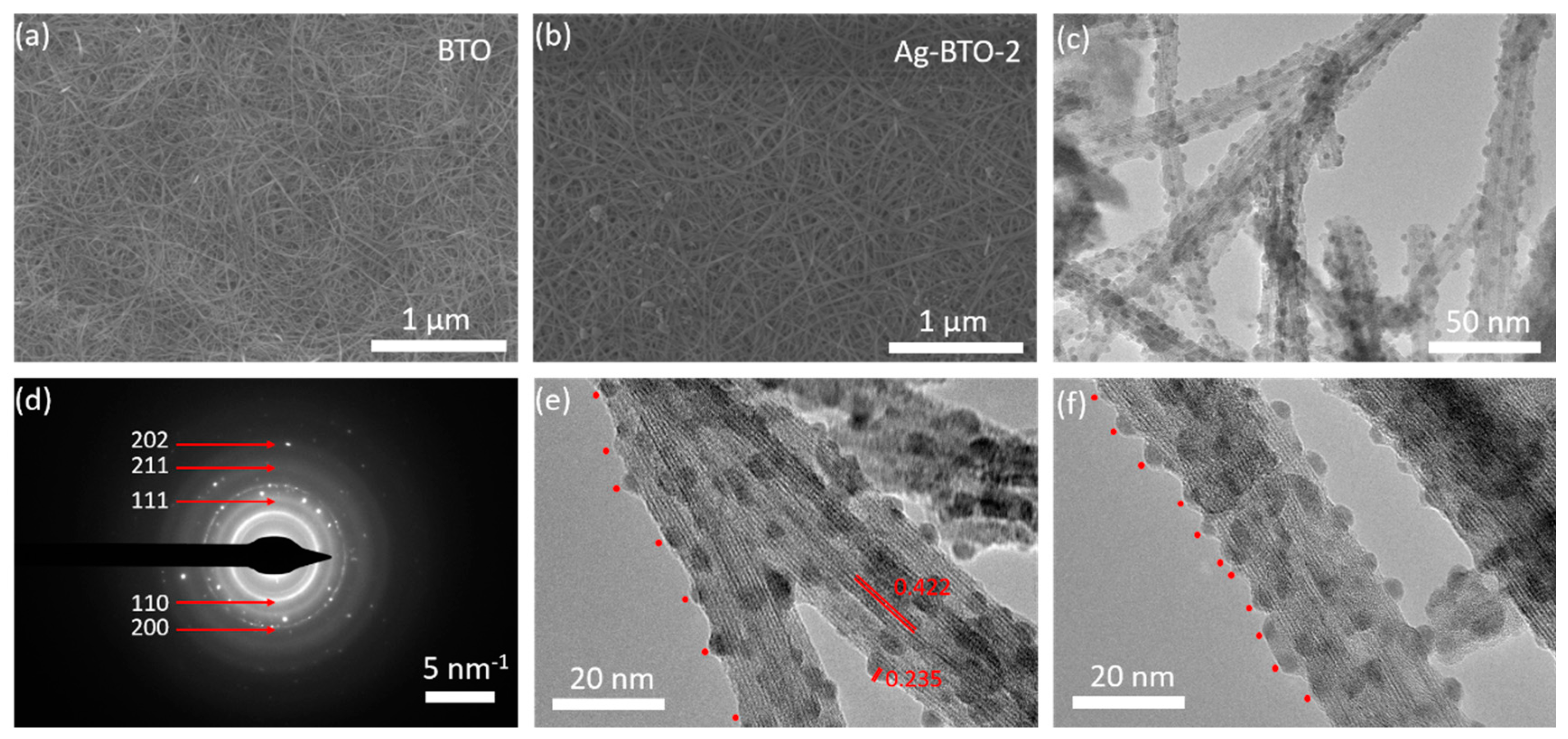
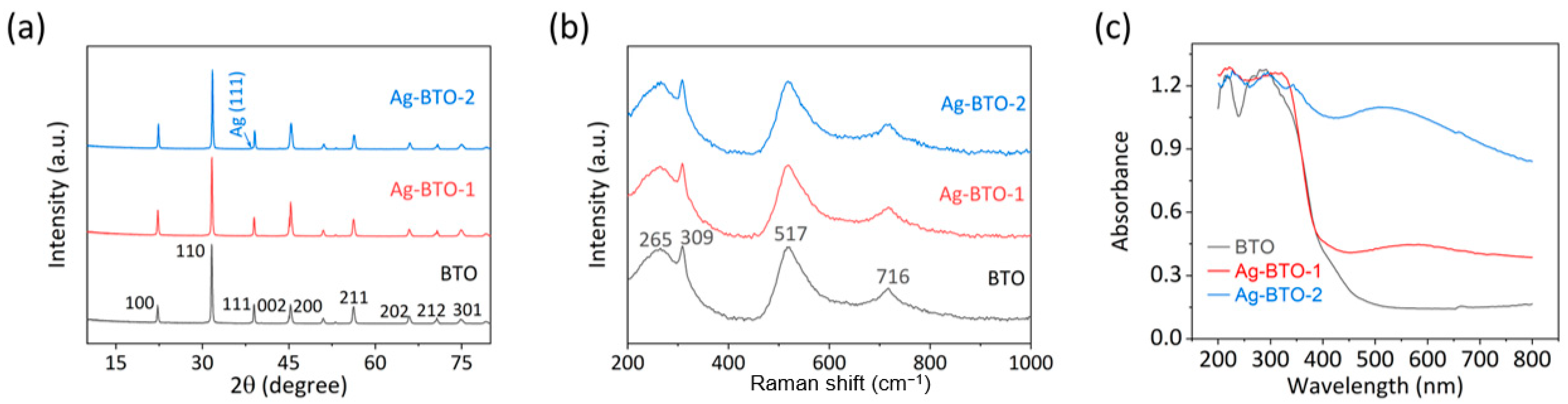
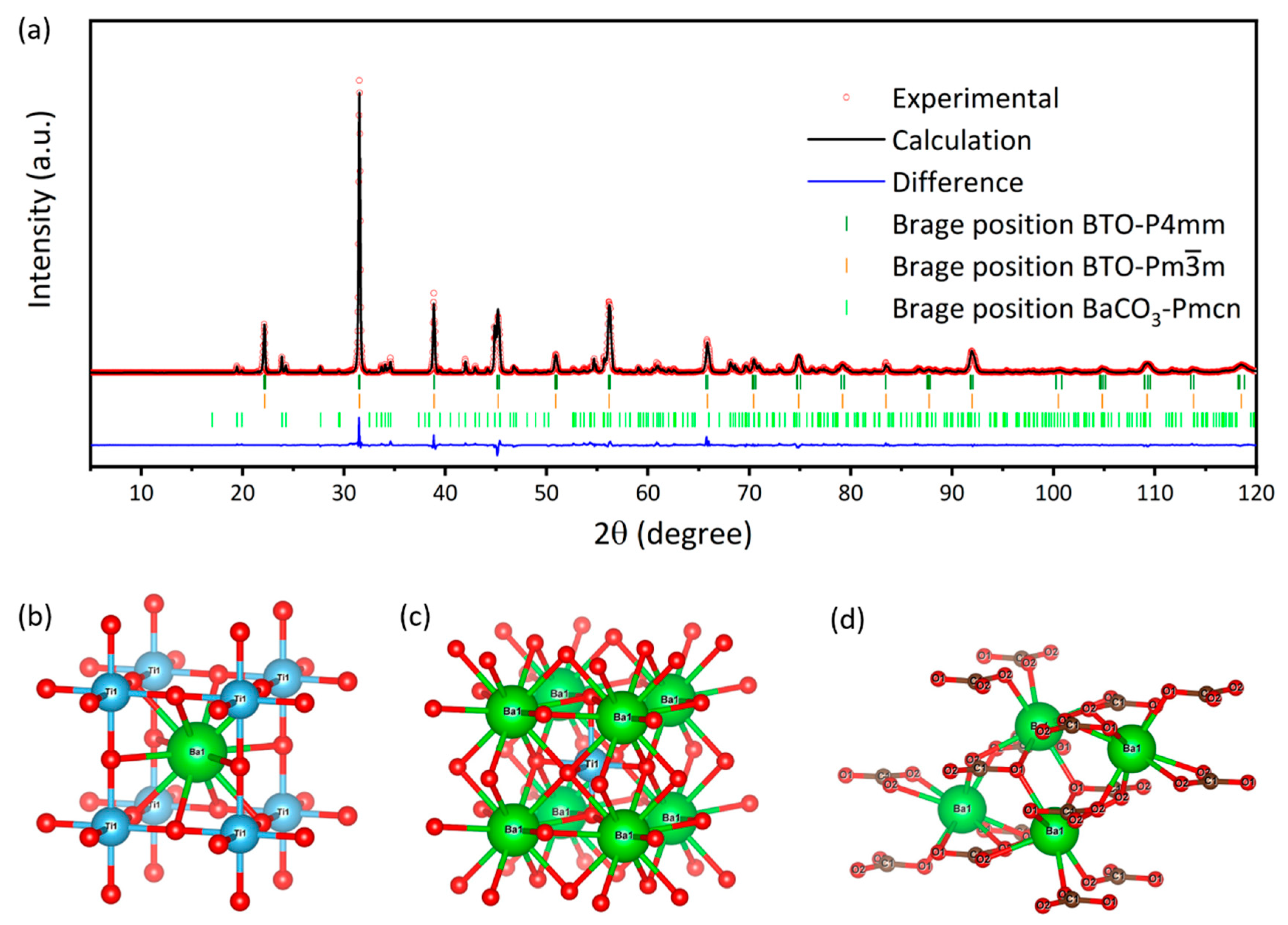
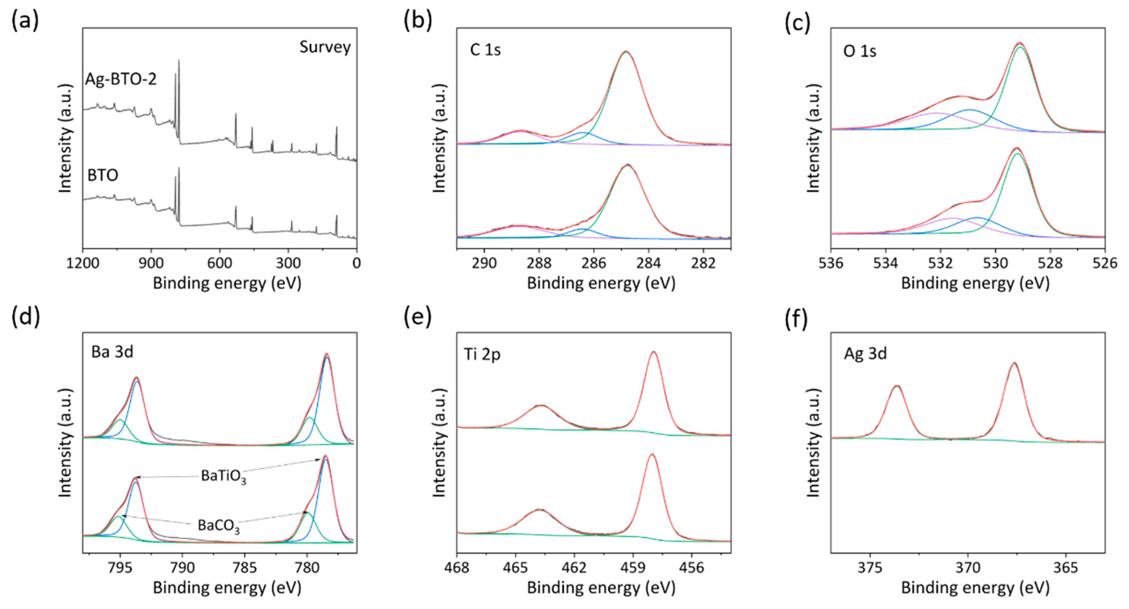
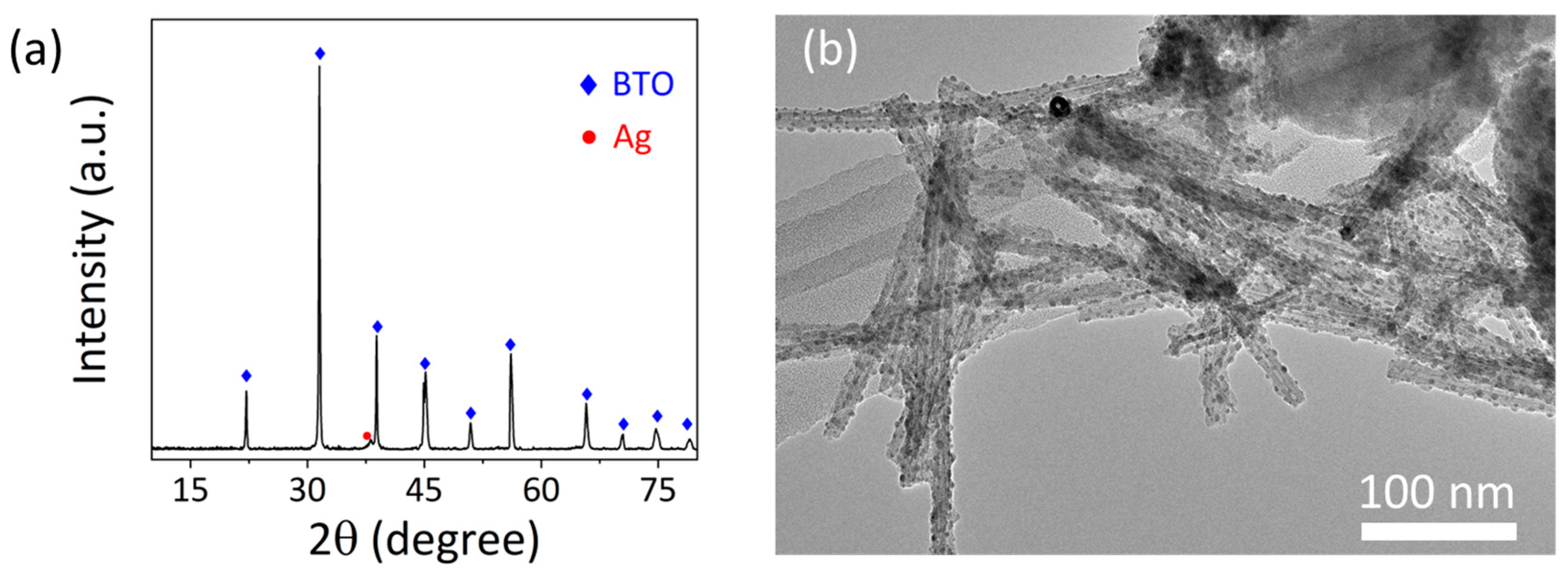
2.1. Characterization of Ag-BTO Nanofibers

Figure 1a shows the SEM image of pure BTO nanofibers, illustrating the ultralong un-directional nanofibers with few fractures. Figure 1b is the SEM image of Ag-BTO-2 nanofibers, displaying a similar morphology with pure BTO. The low-resolution TEM image of Ag-BTO-2 nanofibers is shown in Figure 1c, depicting the uniform distribution of Ag nanoparticles on the whole surface of BTO nanofibers. The corresponding SAED pattern is shown in Figure 1d, where the diffraction rings depending on (110), (111), (200), (211), and (202) crystal planes of BTO are observed [29]. The HRTEM images of Ag-BTO-1 and Ag-BTO-2 are shown in Figure 1e,f, respectively. The distances of 0.235 and 0.422 nm belong to the (111) and (001) crystal planes of Ag and BTO, respectively. For Ag-BTO-2, the size of the Ag nanoparticle is equal to Ag-BTO-1, while the distribution density is double.
The XRD and Raman curves of pure BTO, Ag-BTO-1, and Ag-BTO-2 are compared in Figure 2. As shown in Figure 2a, all the major diffraction peaks of the three samples are assigned to BTO (PDF No. 79-2265) [30]. Particularly, the peak splitting around 45° indicates the high purity of tetragonal BTO. The trace peak at 38.62° assigned to (111) plane of Ag crystal can only be observed in Ag-BTO-2, which is attributed to the low proportion of Ag nanoparticles. In Figure 2b, the three normalized Raman curves are basically the same, where the peaks at 265, 309, 517, and 716 cm−1 correspond to BTO [31]. A sharp peak appearing around 309 cm−1 further confirms the presence of tetragonal BTO. The DRS spectra of the three samples are shown in Figure 2c. Both the samples display two similar adsorption peaks in UV range around 200 and 300 nm. For Ag-BTO-1, a gentle peak appears around 500–700 nm due to the LSPR of Ag nanoparticles. For Ag-BTO-2, the intensity of the gentle peak triples, implying the LSPR of Ag nanoparticles drastically increases. To characterize the crystal structure of the synthesized BTO nanofibers in detail, the refined XRD pattern of pure BTO nanofibers is tested and analyzed by Rietveld refinement, as shown in Figure 3. The refined XRD pattern is indexed by tetragonal BaTiO3 (piezoelectric, space group P4mm) and cubic BaTiO3 (non-piezoelectric, space group Pm–3m) with a ratio of 2:1, as well as a small portion of BaCO3. The refinement results are shown in Table 1, where the Rwp = 6.06%, Rp = 3.98%, and GOF = 3.32. The lattice parameters for tetragonal and cubic BTO are calculated to be a = 4.0150, c = 3.9984, and a = 4.0081 Å, respectively, which are very close to the standard data. These results suggest that the piezoelectric BaTiO3 are successfully synthesized under a relatively low temperature.
The XPS spectra of pure BTO and Ag-BTO-2 are almost the same, as shown in Figure 4. The survey spectra depict the presence of C, O, Ti, and Ba in pure BTO and C, O, Ti, Ba, and Ag in Ag-BTO, respectively (Figure 4a). The C 1s spectra are divided into three peaks around 284.80, 286.48, and 288.78 eV corresponding to the environmental C–C (or C–H) groups, CO32− ions, and C–O groups, respectively (Figure 4b) [32]. The O 1s spectra are also divided into three peaks assigning BaTiO3, CO32− ions, and C–O groups, respectively (Figure 4c). The Ba 3d spectra shows two peaks around 793.65 and 778.48 eV, which are assigned to Ba 3d3/2 and Ba 3d5/2, respectively. Furthermore, the peaks are well divided into two couple of peaks assigned to BaTiO3 and BaCO3, respectively (Figure 4d). Ti 2p photoelectron peaks reveal the purity of BaTiO3 without TiO2 or sodium titanates (Figure 4e). For Ag-BTO-2, the energy difference between Ag 3d3/2 (373.68) and 3d5/2 (367.67) is 6.01 eV, indicating the zero-valent state of Ag element (Figure 4f). These results imply that Ag nanoparticles are successfully loaded on the surface of BTO nanofibers without affecting the morphology, microstructure, and chemical state of BTO nanofibers.

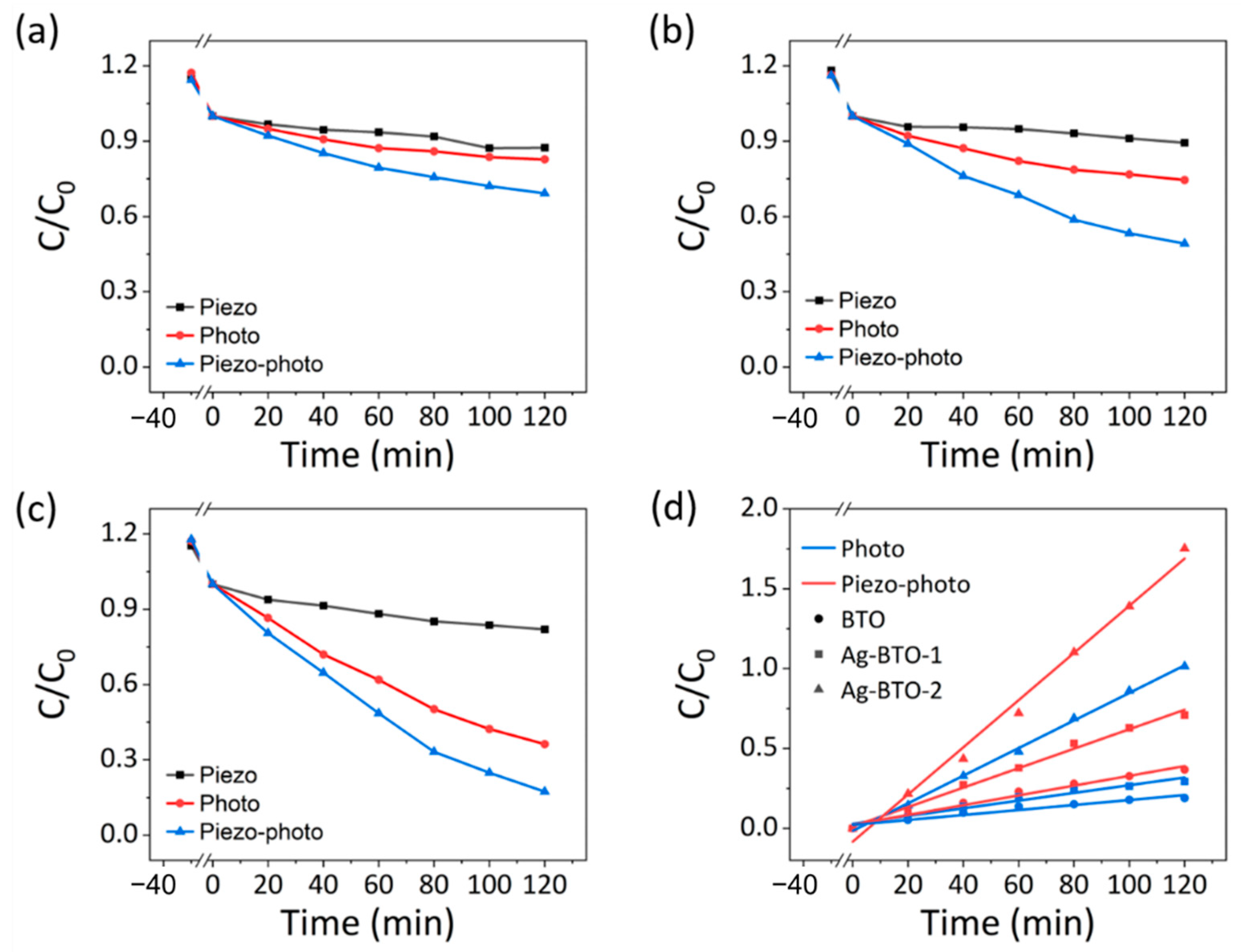
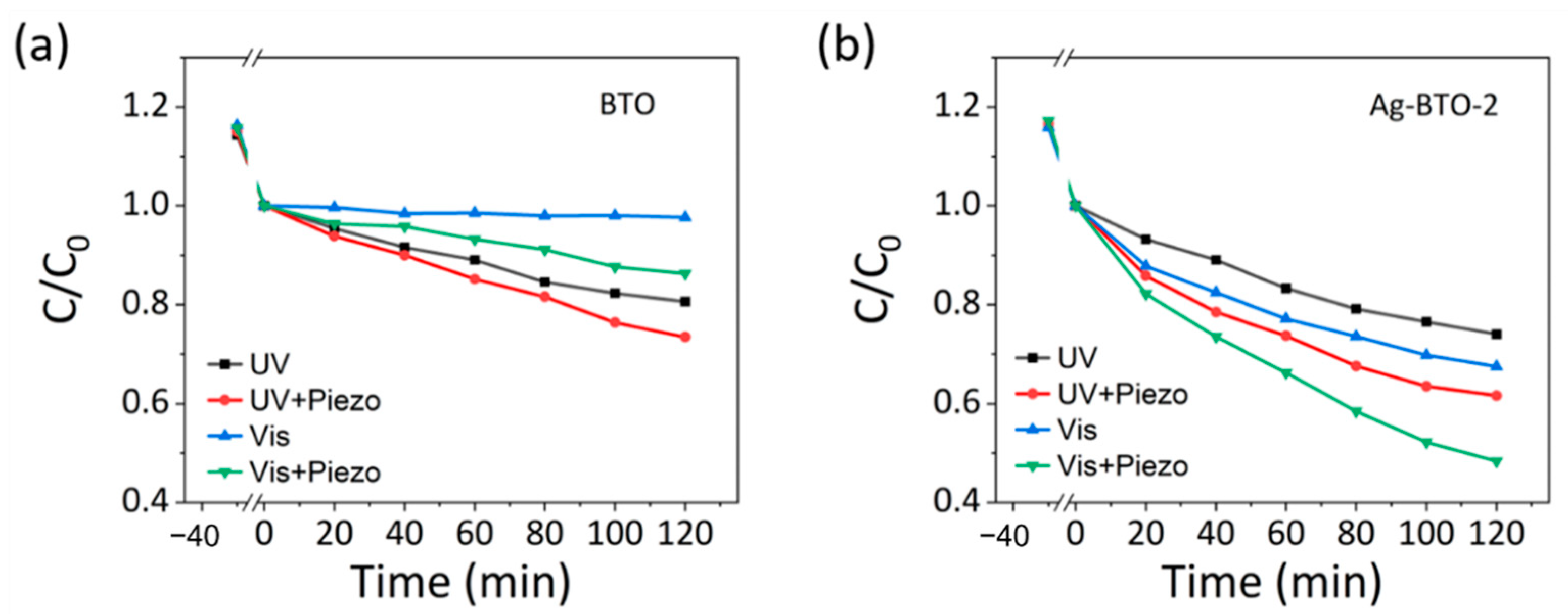
2.2. Piezo-Photocatalytic Property of Ag-BTO Nanofibers

The piezo-photocatalytic properties of the nanofibers are evaluated by degrading MO solution under ultrasonication and solar irradiation, as shown in Figure 5. Figure 5a–c show the MB degradation rates catalyzed by pure BTO, Ag-BTO-1, and Ag-BTO-2 under different conditions for 120 min, respectively. Under only ultrasonication, the degradation rates of the three samples are relatively low, indicating the ultrasound-induced piezoelectric field failed to efficiently degrade MO. Under light irradiation, the loading of Ag nanoparticles greatly improves the degradation rate, which further increases with the increasing Ag loading mass. The maximum degradation rate (82.7%) is achieved by Ag-BTO-2 under the cooperation of ultrasonication and light irradiation, suggesting the synergistic effect between piezoelectricity and photocatalysis. Figure 5d shows the degradation kinetic curves, indicating all the degradation processes follow the pseudo-first-order reaction kinetics.
To further study the function of the piezoelectricity on photocatalysis, the catalytic performances of BTO and Ag-BTO-2 samples are measured under UV and visible light, respectively, as shown in Figure 6. All the catalytic degradation rates of Ag-BTO-2 catalysts are higher than pure BTO. For pure BTO nanofibers (Figure 6a), the photocatalysis and piezo-catalysis under visible light are very weak (2.4% and 13.7%), and those under UV light are also limited (19.4% and 26.6%). As shown in Figure 6b, the UV-driven photocatalysis of the Ag-BTO-2 sample is moderately improved from 26.0% to 38.5% by piezoelectricity. In comparison, the visible-driven photocatalysis is developed from 32.4% to 51.7% by piezoelectricity, displaying a giant enhancement.
The stability of the catalysts is shown in Figure 7. Figure 7a is a TEM image of Ag-BTO-2 after piezo-photocatalytic process, keeping the original morphology and nanoparticle-nanofiber structure. The corresponding XRD pattern is shown in Figure 7b, also showing similar data with the sample before piezo-photocatalysis. Particularly, the minor peak related to Ag nanoparticles is still obtained, and no Ag2O peak is observed, indicating that the Ag nanoparticles maintain metallic state.

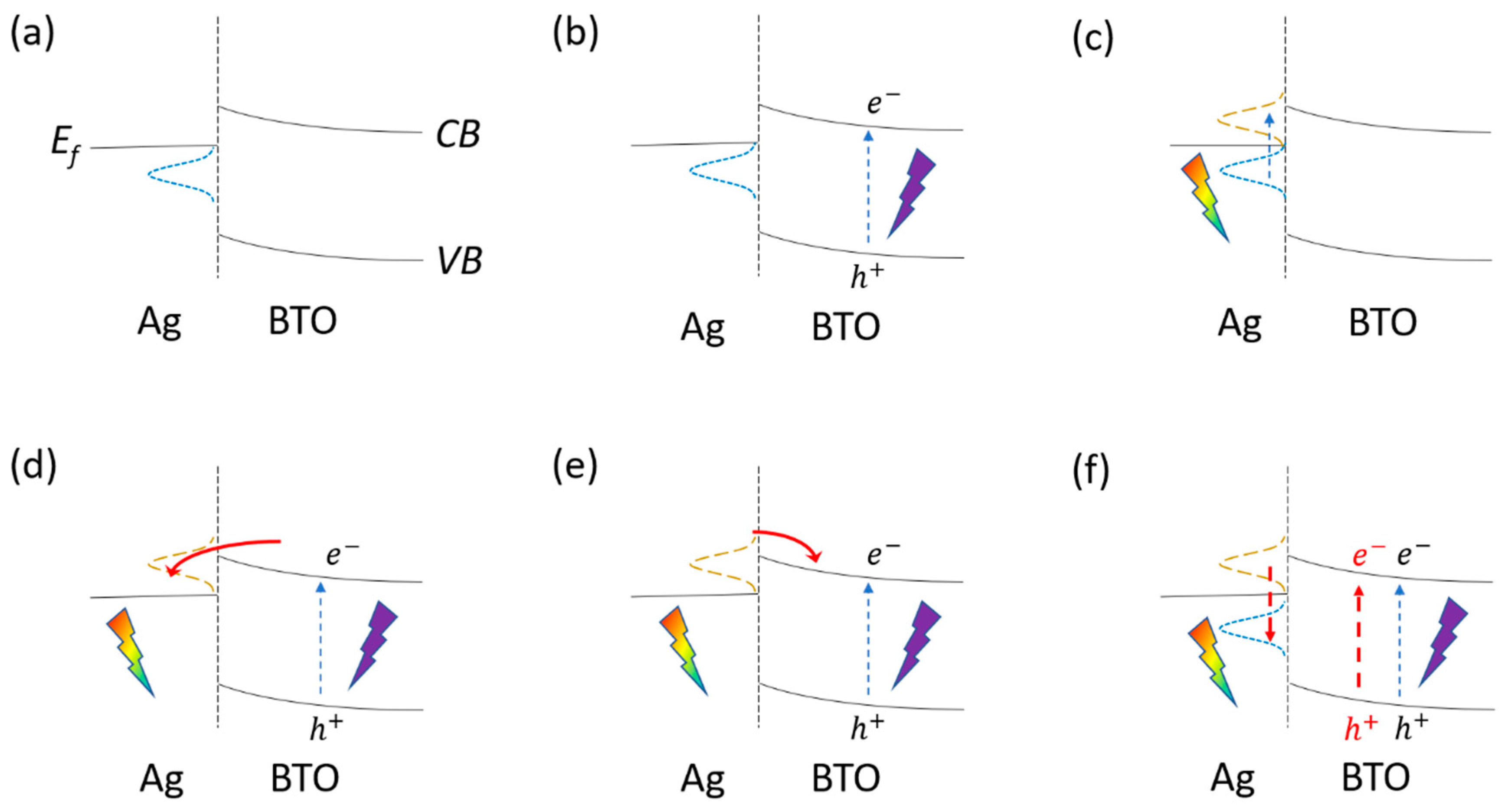
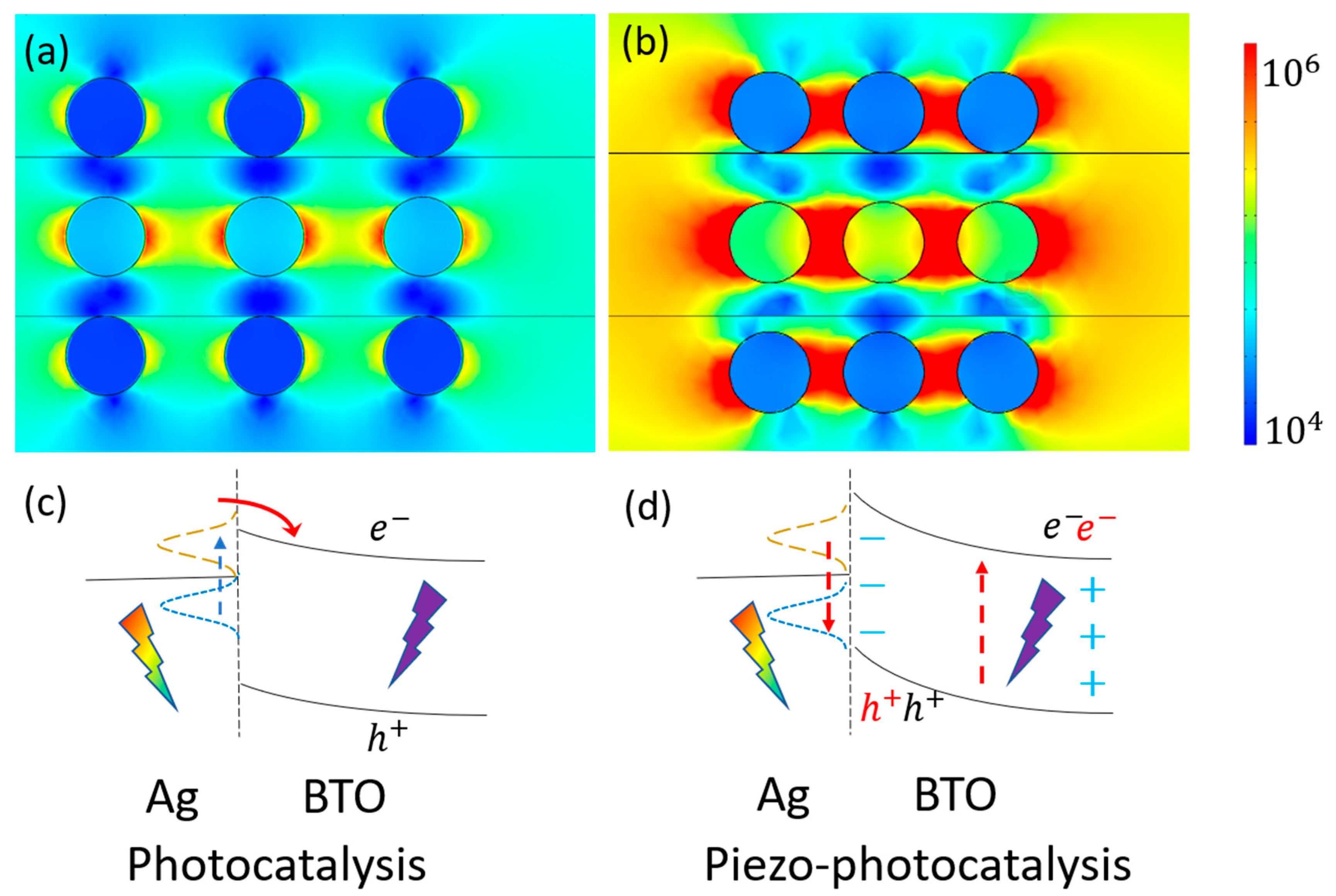
2.3. Mechanism Analysis

Based on the above experimental results, the mechanisms of photocatalysis and piezo-photocatalysis of Ag-BTO nanofibers are developed. The possible photocatalytic mechanism of charge transfer in Ag-BTO is simply discussed. The generation and separation of photoexcited electron-hole pairs are the most important for photocatalysis. BTO nanofibers and Ag nanoparticles form Schottky contact with band offset (Figure 8a) [33]. BTO can absorb UV light to generate electron-hole pairs but cannot absorb visible light due to the wide bandgap (Figure 8b). Ag nanoparticles can absorb visible light through located surface plasmon resonance (LSPR) [34,35,36], but the UV absorption is very low due to the large detuning from resonance frequency (Figure 8c). As the Ag nanoparticles are synthesized on the BTO surface, there are three possible paths to enhance the solar-driven photocatalysis: the first is that the photogenerated electrons in BTO migrate to Ag nanoparticles due to the lower work function, leading to the separation of electron-hole pairs (Figure 8d) [37,38,39]; the second is that the LSPR-induced hot electrons in Ag nanoparticles migrate to BTO, increasing the number of free electrons in BTO (Figure 8e) [40,41,42,43]; the last is that the plasmons in Ag can transfer energy to BTO to generate electron-hole pairs, named “plasmon-induced resonance energy transfer” (PIRET) through dipole-dipole resonance (Figure 8f) [44,45,46,47]. In this case, the UV photocatalytic property of Ag-BTO is a little higher than that of BTO, ruling out the existence of the first path. On the other hand, the Ag-BTO samples exhibit good visible photocatalytic properties, indicating that the visible light is absorbed by Ag nanoparticles through LSPR and transfers hot electrons or energy to BTO, corresponding to the second or third path.
The LSPR electromagnetic fields of Ag-BTO-1 and Ag-BTO-2 are simulated by FEM, as shown in Figure 9a,b, respectively. As the distance of Ag nanoparticle arrays decreases from 10 to 5 nm, the average strength of the LSPR electromagnetic field at 550 nm increases by one order of magnitude. Thus, the photocatalysis and piezo-photocatalysis of Ag-BTO-2 are much higher than those of Ag-BTO-1. Under ultrasonication, BTO is deformed by ultrasound-induced cavitation blasting and generates a built-in piezoelectric field, which promotes the electron-hole separation in BTO and increases the Ag-BTO interface barrier (Figure 9c). As reported, the hot-electron injection path is significantly affected by the interface barrier, while the PERET path is a non-contact route rising superior to the interface barrier [48]. Under solar irradiation, the electron density of state around Ag nanoparticles is much higher than that in BTO, and the migrating of free electrons from BTO to Ag is restrained. The piezoelectric-enhanced interface barrier prevents the hot electrons from injecting into BTO. With a large number of Ag nanoparticles loading on the BTO surface, the first path and second path are both excluded during the piezo-photocatalysis process. Thus, the piezo-photocatalytic mechanism of Ag-BTO nanofibers is described below: under solar irradiation, BTO is excited by UV light to generate electron-hole pairs. The piezoelectric field contributes to the separation of electron-hole pairs and increases the Ag-BTO interface barrier. On the other hand, visible light is captured by high-density Ag nanoparticles through LSPR, transferring energy to further excite more electron-hole pairs for catalysis.

3. Materials and Methods

3.1. Synthesis of BaTiO3 Nanofibers

All reagents were analytical reagents and purchased from Sinophram Chemical Reagent, Shanghai, China. A piece of Ti foil (Haiyuan aluminum, Xining, China) was ultrasound-cleaned in deionized water/acetone for 10 min, heated to 650 °C with a heating rate of 10 °C min−1, and maintained for 5 h in air. Then, the heat-treated Ti foil was immersed in 30 mL NaOH aqueous solution (12 M), sealed, and heated at 160 °C for 6 h, followed by immersion in Ba(OH)2 aqueous solution (0.02 M) at 210 °C for 7 h. After the reaction, the white film was carefully peeled from the surface of Ti foil and ground in an agate mortar for 30 min. Finally, the powders were ultrasound-dispersed in deionized water for 10 min and centrifuged at 4000 rpm for 3 min. To synthesize Ag-BTO nanofibers, the sample was dispersed in 50 mL AgNO3 ethanol solution (0.02 and 0.05 M) under 100 rpm stirring (JoanLab MS5s, Huzhou, China) and 300 W Hg lamp (PerfectLight CHF-XM 300, Beijing, China) irradiation for 2 h, labeled as Ag-BTO-1 and Ag-BTO-2, respectively.

3.2. Characterization and Measurements

The morphology and microstructure were obtained by scanning electron microscope (SEM, Hitachi S-8100, Tokyo. Japan), transmission electron microscope (TEM, FEI Tecnai F20, Portland, OR, USA), X-ray powder diffractometer (XRD, Rigaku SmartLab, Tokyo, Japan), Raman spectrometer (HORIBA LabRAM Nano, Montpellier, France), and X-ray photoelectron spectroscope (XPS, Thermo Scientific K-Alpha, Waltham, MA, USA). Conventional XRD patterns were measured with a sweeping rate of 10° min−1. The refined XRD pattern of pure BTO nanofibers was tested in a slow rate of 0.5° min−1 with a wide sweeping range from 5° to 120°, and Rietveld refinement is employed by TOPAS academic. The photocatalysis and piezo-photocatalysis were evaluated by degrading MO in a mechano-photo reaction apparatus (homemade). A 300 W Xe lamp (PerfectLight, Microsolar 300, Beijing, China) was used to directly provide the light source. UV and visible light were obtained by short-pass and long-pass filters, respectively. The ultrasound was generated by a 120 W ultrasonic vibrator (DongSen DS-120STS, Shenzhen, China) with a frequency of 24 kHz. During the catalytic process, 10 mg catalyst was used to degrade 50 mL MO solution (10 mg L−1). The MO concentration was determined by absorption spectroscopy at 464 nm.

4. Conclusions

In summary, the piezo-photocatalytic performance of BaTiO3 nanofibers is enhanced by loading with Ag nanoparticles. FEM simulation indicates that the visible-light absorbance of Ag nanoparticles exponentially increases with the distribution density. With the loading of Ag nanoparticles, the piezo-photocatalytic MO degradation rate increases from 30.8% to 50.8%, and further increases to 82.7% with doubling Ag density. The UV-driven and visible-driven catalytic performances were individually measured to study the contribution of Ag nanoparticles and BTO nanofibers, displaying that the visible-driven catalysis dominates the enhancement during the piezo-photocatalytic process. Finally, the piezo-photocatalytic mechanism of Ag-BTO was discussed. Under solar irradiation, electron-hole pairs are generated in BTO by UV light, separated by the piezoelectric field, and enhanced by the high-density Ag nanoparticles through PIRET. This work develops the investigation of plasmon-improved piezo-photocatalysis.

Author Contributions

Conceptualization, Y.F. and J.M.; methodology, Z.R. and P.L.; investigation, P.C. and X.L.; writing—original draft preparation, P.C. and Y.L.; writing—review and editing, Y.F. and J.W.; visualization, P.C. and W.L.; supervision, Y.F. and J.M.; project administration, Y.F. and J.M.; funding acquisition, J.M. All authors have read and agreed to the published version of the manuscript.

Funding

This research was funded by National Key R&D Program of China (2017YFA0304203), the National Natural Science Foundation of China (62020106014, 62175140, 61901249, 92165106, 12104276); PCSIRT (IRT—17R70), 111 project (D18001), the Program for the Outstanding Innovative Teams of Higher Learning Institutions of Shanxi (OIT), the Applied Basic Research Project of Shanxi Province, China (201901D211191, 201901D211188), the Shanxi 1331 KSC, and the collaborative grant by the Russian Foundation for Basic Research and NSF of China (62011530047, 20-53-53025 in the RFBR classification).

Data Availability Statement

The data presented in this study are available on request from the corresponding author.

Acknowledgments

The authors would like to thank Zhenyu Duan from Shiyanjia Lab (www.shiyanjia.com) for the helps on material characterizations.

Conflicts of Interest

The authors declare no conflict of interest.

References

  1. Chen, T.; Liu, L.Z.; Hu, C.; Huang, H.W. Recent advances on Bi2WO6-based photocatalysts for environmental and energy applications. Chin. J. Catal. 2021, 42, 1413–1438. [Google Scholar] [CrossRef]
  2. Goktas, S.; Goktas, A. A comparative study on recent progress in efficient ZnO based nanocomposite and heterojunction photocatalysts: A review. J. Alloys Compd. 2021, 863, 158734. [Google Scholar] [CrossRef]
  3. Kumar, A.; Raizada, P.; Hosseini-Bandegharaei, A.; Thakur, V.K.; Nguyen, V.; Singh, P. C-, N-Vacancy defect engineered polymeric carbon nitride towards photocatalysis: Viewpoints and challenges. J. Mater. Chem. A 2021, 9, 111–153. [Google Scholar] [CrossRef]
  4. Li, Y.; Wu, Y.; Jiang, H.; Wang, H. In situ stable growth of Bi2WO6 on natural hematite for efficient antibiotic wastewater purification by photocatalytic activation of peroxymonosulfate. Chem. Eng. J. 2022, 446, 136704. [Google Scholar] [CrossRef]
  5. Jin, P.; Wang, L.; Ma, X.; Lian, R.; Huang, J.; She, H.; Zhang, M.; Wang, Q. Construction of hierarchical ZnIn2S4@PCN-224 heterojunction for boosting photocatalytic performance in hydrogen production and degradation of tetracycline hydrochloride. Appl. Catal. B Environ. 2021, 284, 119762. [Google Scholar] [CrossRef]
  6. Moradi, M.; Hasanvandian, F.; Isari, A.A.; Hayati, F.; Kakavandi, B.; Setayesh, S.R. CuO and ZnO co-anchored on g-C3N4 nanosheets as an affordable double Z-scheme nanocomposite for photocatalytic decontamination of amoxicillin. Appl. Catal. B Environ. 2021, 285, 119838. [Google Scholar] [CrossRef]
  7. Wang, Z.; Jiang, L.; Wang, K.; Li, Y.; Zhang, G. Novel AgI/BiSbO4 heterojunction for efficient photocatalytic degradation of organic pollutants under visible light: Interfacial electron transfer pathway, DFT calculation and degradation mechanism study. J. Hazard. Mater. 2021, 410, 124948. [Google Scholar] [CrossRef]
  8. Bayan, E.M.; Lupeiko, T.G.; Pustovaya, L.E.; Volkova, M.G. Synthesis and photocatalytic properties of Sn-TiO2 nanomaterials. J. Adv. Dielectr. 2020, 10, 2060018. [Google Scholar] [CrossRef]
  9. Chen, J.; Li, G.; Lu, N.; Lin, H.; Zhou, S.; Liu, F. Anchoring cobalt single atoms on 2D covalent triazine framework with charge nanospatial separation for enhanced photocatalytic pollution degradation. Mater. Today Chem. 2022, 24, 100832. [Google Scholar] [CrossRef]
  10. Hu, J.; Chen, Y.; Zhou, Y.; Zeng, L.; Huang, Y.; Lan, S.; Zhu, M. Piezo-enhanced charge carrier separation over plasmonic Au-BiOBr for piezo-photocatalytic carbamazepine removal. Appl. Catal. B Environ. 2022, 311, 121369. [Google Scholar] [CrossRef]
  11. Idris, A.M.; Zheng, S.; Wu, L.; Zhou, S.; Lin, H.; Chen, Z.; Xu, L.; Wang, J.; Li, Z. A heterostructure of halide and oxide double perovskites Cs2AgBiBr6/Sr2FeNbO6 for boosting the charge separation toward high efficient photocatalytic CO2 reduction under visible-light irradiation. Chem. Eng. J. 2022, 446, 137197. [Google Scholar] [CrossRef]
  12. Liao, B.; Liao, X.; Xie, H.; Qin, Y.; Zhu, Y.; Yu, Y.; Hou, S.; Zhang, Y.; Fan, X. Built in electric field boosted photocatalytic performance in a ferroelectric layered material SrBi2Ta2O9 with oriented facets: Charge separation and mechanism insights. J. Mater. Sci. Technol. 2022, 123, 222–233. [Google Scholar] [CrossRef]
  13. Li, X.; Kang, B.; Dong, F.; Zhang, Z.; Luo, X.; Han, L.; Huang, J.; Feng, Z.; Chen, Z.; Xu, J.; et al. Enhanced photocatalytic degradation and H2/H2O2 production performance of S-pCN/WO2.72 S-scheme heterojunction with appropriate surface oxygen vacancies. Nano Energy 2021, 81, 105671. [Google Scholar] [CrossRef]
  14. Dai, B.Y.; Biesold, G.M.; Zhang, M.; Zou, H.Y.; Ding, Y.; Wang, Z.L.; Lin, Z.Q. Piezo-phototronic effect on photocatalysis, solar cells, photodetectors and light-emitting diodes. Chem. Soc. Rev. 2021, 50, 13646–13691. [Google Scholar] [CrossRef] [PubMed]
  15. Guo, L.X.; Chen, Y.D.; Ren, Z.Q.; Li, X.; Zhang, Q.W.; Wu, J.Z.; Li, Y.Q.; Liu, W.L.; Li, P.; Fu, Y.M.; et al. Morphology engineering of type-II heterojunction nanoarrays for improved sonophotocatalytic capability. Ultrason. SonoChem. 2021, 81, 105849. [Google Scholar] [CrossRef]
  16. Zhang, C.X.; Lei, D.; Xie, C.F.; Hang, X.S.; He, C.A.X.; Jiang, H.L. Piezo-Photocatalysis over Metal-Organic Frameworks: Promoting Photocatalytic Activity by Piezoelectric Effect. Adv. Mater. 2021, 33, 2106308. [Google Scholar] [CrossRef]
  17. Li, X.; Wang, W.; Dong, F.; Zhang, Z.; Han, L.; Luo, X.; Huang, J.; Feng, Z.; Chen, Z.; Jia, G.; et al. Recent Advances in Noncontact External-Field-Assisted Photocatalysis: From Fundamentals to Applications. ACS Catal. 2021, 11, 4739–4769. [Google Scholar] [CrossRef]
  18. Marshall, J.; Walker, D.; Thomas, P. Bismuth zinc niobate: BZN-BT, a new lead-free BaTiO3-based ferroelectric relaxor? J. Adv. Dielectr. 2020, 10, 2050033. [Google Scholar] [CrossRef]
  19. Sidorenko, E.; Ngoc, C.T.B.; Prikhodko, G.; Natkhin, I.; Shloma, A.; Kharchenko, D. The constant electric field effect on the radio absorption of crystals BaTiO3 and piezoceramics PCR-1. J. Adv. Dielectr. 2020, 10, 2060020. [Google Scholar] [CrossRef]
  20. Yao, M.; Li, L.; Wang, Y.; Yang, D.; Miao, L.; Wang, H.; Liu, M.; Ren, K.; Fan, H.; Hu, D. Mechanical Energy Harvesting and Specific Potential Distribution ofa Flexible Piezoelectric Nanogenerator Based on 2-D BaTiO3-Oriented Polycrystals. ACS Sustain. Chem. Eng. 2022, 10, 3276–3287. [Google Scholar] [CrossRef]
  21. Cheng, S.S.; Luo, Y.; Zhang, J.; Shi, R.; Wei, S.T.; Dong, K.J.; Liu, X.M.; Wu, S.L.; Wang, H.B. The highly effective therapy of ovarian cancer by Bismuth-doped oxygen-deficient BaTiO3 with enhanced sono-piezocatalytic effects. Chem. Eng. J. 2022, 442, 136380. [Google Scholar] [CrossRef]
  22. Fu, Y.M.; Ren, Z.Q.; Guo, L.X.; Li, X.; Li, Y.Q.; Liu, W.L.; Li, P.; Wu, J.Z.; Ma, J. Piezotronics boosted plasmonic localization and hot electron injection of coralline-like Ag/BaTiO3 nanoarrays for photocatalytic application. J. Mater. Chem. C 2021, 9, 12596–12604. [Google Scholar] [CrossRef]
  23. Liu, Q.; Li, Z.Y.; Li, J.; Zhan, F.Q.; Zhai, D.; Sun, Q.W.; Xiao, Z.D.; Luo, H.; Zhang, D. Three dimensional BaTiO3 piezoelectric ceramics coated with TiO2 nanoarray for high performance of piezo-photoelectric catalysis. Nano Energy 2022, 98, 107267. [Google Scholar] [CrossRef]
  24. Tang, Q.; Wu, J.; Kim, D.; Franco, C.; Terzopoulou, A.; Veciana, A.; Puigmarti-Luis, J.; Chen, X.Z.; Nelson, B.J.; Pane, S. Enhanced Piezocatalytic Performance of BaTiO3 Nanosheets with Highly Exposed {001} Facets. Adv. Funct. Mater. 2022, 32, 2202180. [Google Scholar] [CrossRef]
  25. Sanchis-Gual, R.; Otero, T.F.; Coronado-Puchau, M.; Coronado, E. Enhancing the electrocatalytic activity and stability of Prussian blue analogues by increasing their electroactive sites through the introduction of Au nanoparticles. Nanoscale 2021, 13, 12676–12686. [Google Scholar] [CrossRef]
  26. Li, X.; Jiang, H.P.; Ma, C.C.; Zhu, Z.; Song, X.H.; Wang, H.Q.; Huo, P.W.; Li, X.Y. Local surface plasma resonance effect enhanced Z-scheme ZnO/Au/g-C3N4 film photocatalyst for reduction of CO2 to CO. Appl. Catal. B-Environ. 2021, 283, 119638. [Google Scholar] [CrossRef]
  27. Li, S.J.; Wang, C.C.; Liu, Y.P.; Xue, B.; Jiang, W.; Liu, Y.; Mo, L.Y.; Chen, X.B. Photocatalytic degradation of antibiotics using a novel Ag/Ag2S/Bi2MoO6 plasmonic p-n heterojunction photocatalyst: Mineralization activity, degradation pathways and boosted charge separation mechanism. Chem. Eng. J. 2021, 415, 128991. [Google Scholar] [CrossRef]
  28. Chen, L.W.; Hao, Y.C.; Guo, Y.; Zhang, Q.H.; Li, J.N.; Gao, W.Y.; Ren, L.T.; Su, X.; Hu, L.Y.; Zhang, N.; et al. Metal-Organic Framework Membranes Encapsulating Gold Nanoparticles for Direct Plasmonic Photocatalytic Nitrogen Fixation. J. Am. Chem. Soc. 2021, 143, 5727–5736. [Google Scholar] [CrossRef]
  29. Chen, T.; Meng, J.; Wu, S.; Pei, J.; Lin, Q.; Wei, X.; Li, J.; Zhang, Z. Room temperature synthesized BaTiO3 for photocatalytic hydrogen evolution. J. Alloys Compd. 2018, 754, 184–189. [Google Scholar] [CrossRef]
  30. On, D.V.; Vuong, L.D.; Chuong, T.V.; Quang, D.A.; Tuyen, H.V.; Tung, V.T. Influence of sintering behavior on the microstructure and electrical properties of BaTiO3 lead-free ceramics from hydrothermal synthesized precursor nanoparticles. J. Adv. Dielectr. 2021, 11, 2150014. [Google Scholar] [CrossRef]
  31. Charoonsuk, T.; Sriphan, S.; Nawanil, C.; Chanlek, N.; Vittayakorn, W.; Vittayakorn, N. Tetragonal BaTiO3 nanowires: A template-free salt-flux-assisted synthesis and its piezoelectric response based on mechanical energy harvesting. J. Mater. Chem. C 2019, 7, 8277–8286. [Google Scholar] [CrossRef]
  32. Miot, C.; Husson, E.; Proust, C.; Erre, R.; Coutures, J.P. Residual carbon evolution in BaTiO3 ceramics studied by XPS after ion etching. J. Eur. Ceram. Soc. 1998, 18, 339–343. [Google Scholar] [CrossRef]
  33. Nithya, P.M.; Devi, L.G. Effect of surface Ag metallization on the photocatalytic properties of BaTiO3: Surface plasmon effect and variation in the Schottky barrier height. Surf. Interfaces 2019, 15, 205–215. [Google Scholar] [CrossRef]
  34. Chen, Z.; Chen, M.; Yan, H.; Zhou, P.; Chen, X. Enhanced solar thermal conversion performance of plasmonic gold dimer nanofluids. Appl. Therm. Eng. 2020, 178, 115561. [Google Scholar] [CrossRef]
  35. Liu, X.-D.; Chen, B.; Wang, G.-G.; Ma, S.; Cheng, L.; Liu, W.; Zhou, L.; Wang, Q.-Q. Controlled Growth of Hierarchical Bi2Se3/CdSe-Au Nanorods with Optimized Photothermal Conversion and Demonstrations in Photothermal Therapy. Adv. Funct. Mater. 2021, 31, 2104424. [Google Scholar] [CrossRef]
  36. Sun, Q.; Hou, P.; Wu, S.; Yu, L.; Dong, L. The enhanced photocatalytic activity of Ag-Fe2O3-TiO2 performed in Z-scheme route associated with localized surface plasmon resonance effect. Colloids Surf. A 2021, 628, 127304. [Google Scholar] [CrossRef]
  37. Humayun, M.; Ullah, H.; Cheng, Z.-E.; Tahir, A.A.; Luo, W.; Wang, C. Au surface plasmon resonance promoted charge transfer in Z-scheme system enables exceptional photocatalytic hydrogen evolution. Appl. Catal. B Environ. 2022, 310, 121322. [Google Scholar] [CrossRef]
  38. Xu, Q.; Knezevic, M.; Laachachi, A.; Franger, S.; Colbeau-Justin, C.; Ghazzal, M.N. Insight into Interfacial Charge Transfer during Photocatalytic H2 Evolution through Fe, Ni, Cu and Au Embedded in a Mesoporous TiO2@SiO2 Core-shell. ChemCatChem 2022, 14, e202200102. [Google Scholar] [CrossRef]
  39. Zhang, J.; Gu, H.; Wang, X.; Zhang, H.; Chang, S.; Li, Q.; Dai, W.-L. Robust S-scheme hierarchical Au-ZnIn2S4/NaTaO3: Facile synthesis, superior photocatalytic H2 production and its charge transfer mechanism. J. Colloid Interface Sci. 2022, 625, 785–799. [Google Scholar] [CrossRef]
  40. Berdakin, M.; Soldano, G.; Bonafe, F.P.; Liubov, V.; Aradi, B.; Frauenheim, T.; Sanchez, C.G. Dynamical evolution of the Schottky barrier as a determinant contribution to electron-hole pair stabilization and photocatalysis of plasmon-induced hot carriers. Nanoscale 2022, 14, 2816–2825. [Google Scholar] [CrossRef]
  41. Gai, Q.; Ren, S.; Zheng, X.; Liu, W.; Dong, Q. Enhanced photocatalytic performance of Ag/CdS by L-cysteine functionalization: Combination of introduced co-catalytic groups and optimized injection of hot electrons. Appl. Surf. Sci. 2022, 579, 151838. [Google Scholar] [CrossRef]
  42. Manuel, A.P.; Shankar, K. Hot Electrons in TiO2-Noble Metal Nano-Heterojunctions: Fundamental Science and Applications in Photocatalysis. NanoMater 2021, 11, 1249. [Google Scholar] [CrossRef] [PubMed]
  43. Yuan, X.; Zhen, W.; Yu, S.; Xue, C. Plasmon Coupling-Induced Hot Electrons for Photocatalytic Hydrogen Generation. Chem. Asian J. 2021, 16, 3683–3688. [Google Scholar] [CrossRef] [PubMed]
  44. Choi, Y.M.; Lee, B.W.; Jung, M.S.; Han, H.S.; Kim, S.H.; Chen, K.; Kim, D.H.; Heinz, T.F.; Fan, S.; Lee, J.; et al. Retarded Charge-Carrier Recombination in Photoelectrochemical Cells from Plasmon-Induced Resonance Energy Transfer. Adv. Energy Mater. 2020, 10, 2000570. [Google Scholar] [CrossRef]
  45. Jia, H.; Wong, Y.L.; Wang, B.; Xing, G.; Tsoi, C.C.; Wang, M.; Zhang, W.; Jian, A.; Sang, S.; Lei, D.; et al. Enhanced solar water splitting using plasmon-induced resonance energy transfer and unidirectional charge carrier transport. Opt. Express 2021, 29, 34810–34825. [Google Scholar] [CrossRef]
  46. Li, J.; Cushing, S.K.; Meng, F.; Senty, T.R.; Bristow, A.D.; Wu, N. Plasmon-induced resonance energy transfer for solar energy conversion. Nat. Photonics 2015, 9, 601–607. [Google Scholar] [CrossRef]
  47. Ma, J.; Liu, X.; Wang, R.; Zhang, F.; Tu, G. Plasmon-induced near-field and resonance energy transfer enhancement of photodegradation activity by Au wrapped CuS dual-chain. Nano Res. 2022, 15, 5671–5677. [Google Scholar] [CrossRef]
  48. Kohan, M.G.; You, S.; Camellini, A.; Concina, I.; Zavelani-Rossi, M.; Vomiero, A. Optical field coupling in ZnO nanorods decorated with silver plasmonic nanoparticles. J. Mater. Chem. C 2021, 9, 15452–15462. [Google Scholar] [CrossRef]
Figure 1. The characterizations of the samples. (a,b) SEM images of BTO and Ag-BTO nanofibers. (c) TEM image of Ag-BTO nanofibers. (d) SAED pattern of Ag-BTO nanofibers. (e,f) HRTEM images of Ag-BTO-1 and Ag-BTO-2.
Catalysts 12 00987 g001
Figure 2. (a) XRD patterns, (b) Raman spectra, and (c) DRS spectra of the three samples.
Catalysts 12 00987 g002
Figure 3. (a) Rietveld refined XRD pattern of the BTO nanofibers. (bd) The refined crystal lattice structures of (b) BaTiO3-Pm-3m, (c) BaTiO3-P4mm, and (d) BaCO3-Pmcn.
Catalysts 12 00987 g003
Figure 4. The XPS spectra of BTO and Ag-BTO. (a) Survey spectra. (b) C1s. (c) O 1s. (d) Ba 3d. (e) Ti 2p. (f) Ag 3d.
Catalysts 12 00987 g004
Figure 5. Catalytic performance comparison. (ac) The piezocatalysis, photocatalysis, and piezo-photocatalysis of (a) BTO, (b) Ag-BTO-1, and (c) Ag-BTO-2. (d) The reaction kinetics.
Catalysts 12 00987 g005
Figure 6. The photocatalysis and piezo-photocatalysis under UV and visible light of (a) BTO and (b) Ag-BTO-2.
Catalysts 12 00987 g006
Figure 7. (a) XRD pattern and (b) TEM image of Ag-BTO-2 after piezo-photocatalysis.
Catalysts 12 00987 g007
Figure 8. The schematic diagrams of charge transfer paths of Ag–BTO. (a) Schottky contact of Ag–BTO. (b) UV excitation. (c) Visible light excitation. (df) The transfer paths of (d) charge separation, (e) hot-electron injection, and (f) PIRET.
Catalysts 12 00987 g008
Figure 9. The proposed mechanism of piezo-photocatalysis. (a,b) The FEM simulation of the LSPR electromagnetic field of (a) Ag-BTO-1 and (b) Ag-BTO-2. (c) The hot-electron injection process in photocatalysis. (d) The PIRET process in piezo-photocatalysis.
Catalysts 12 00987 g009
Table 1. Rietveld refined cell parameters of the synthesized BTO nanofibers.
Cell ParametersTetragonal BaTiO3Cubic BaTiO3BaCO3
Proportion64.32%32.20%3.48%
Space GroupP4mmPm-3mPmcn
a (Å)4.01504.00815.3130
b (Å)4.01504.00818.9038
c (Å)3.99844.00816.4361
α (°)909090
β (°)909090
γ (°)909090
Volume (Å3)64.4564.392304.47
Publisher’s Note: MDPI stays neutral with regard to jurisdictional claims in published maps and institutional affiliations.

