
Influence of Wurtzite ZnO Morphology on Piezophototronic Effect in Photocatalysis

 and
and 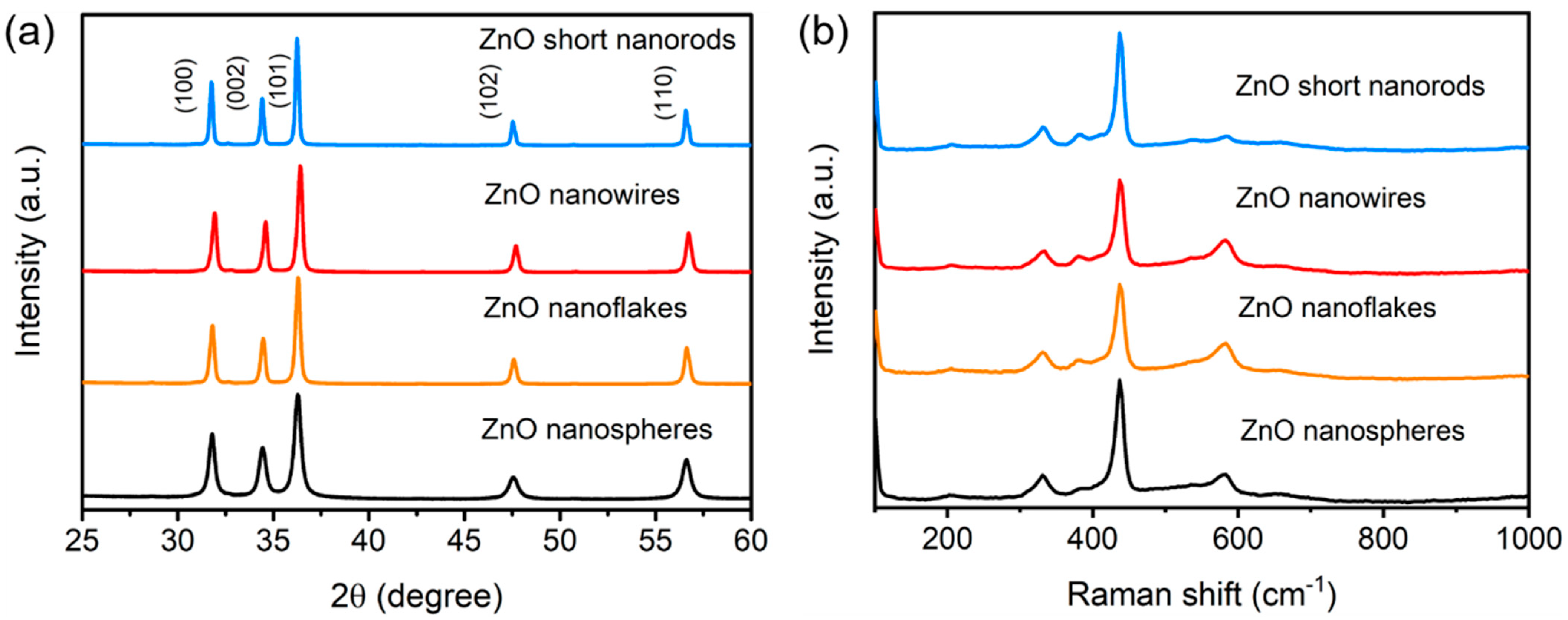
Abstract
:1. Introduction
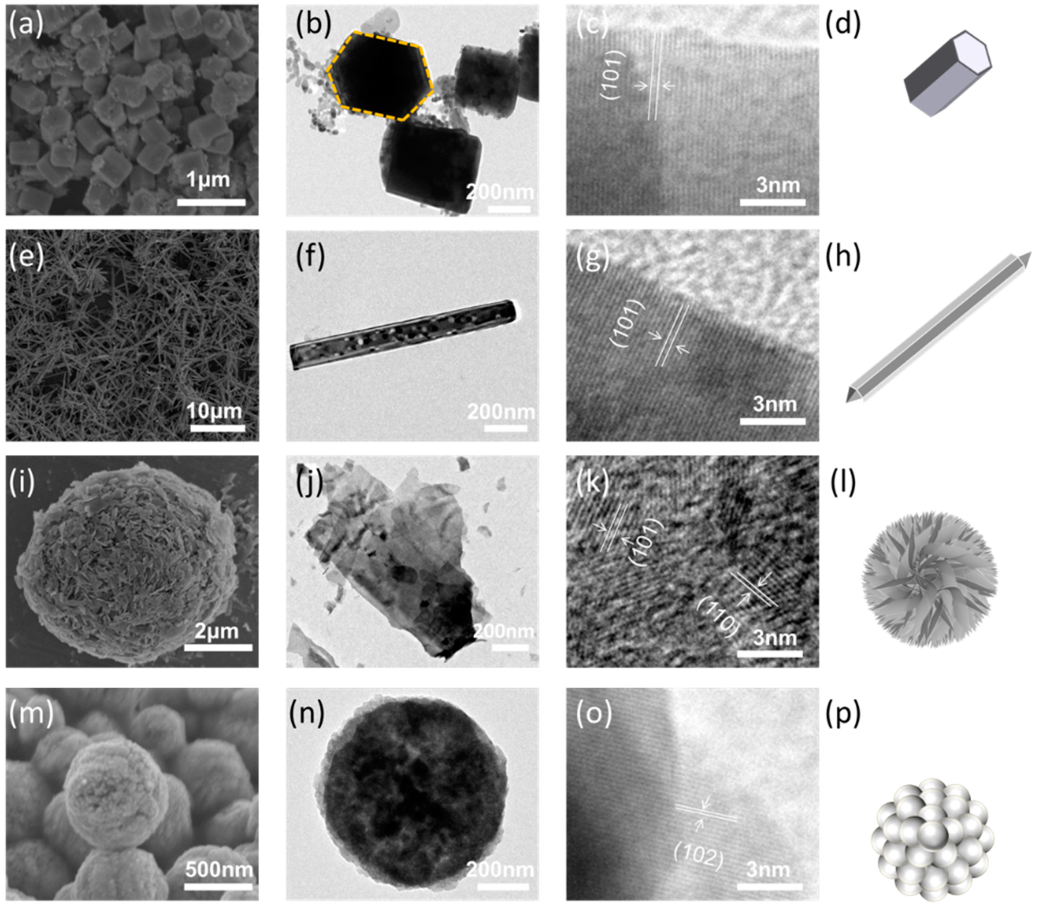
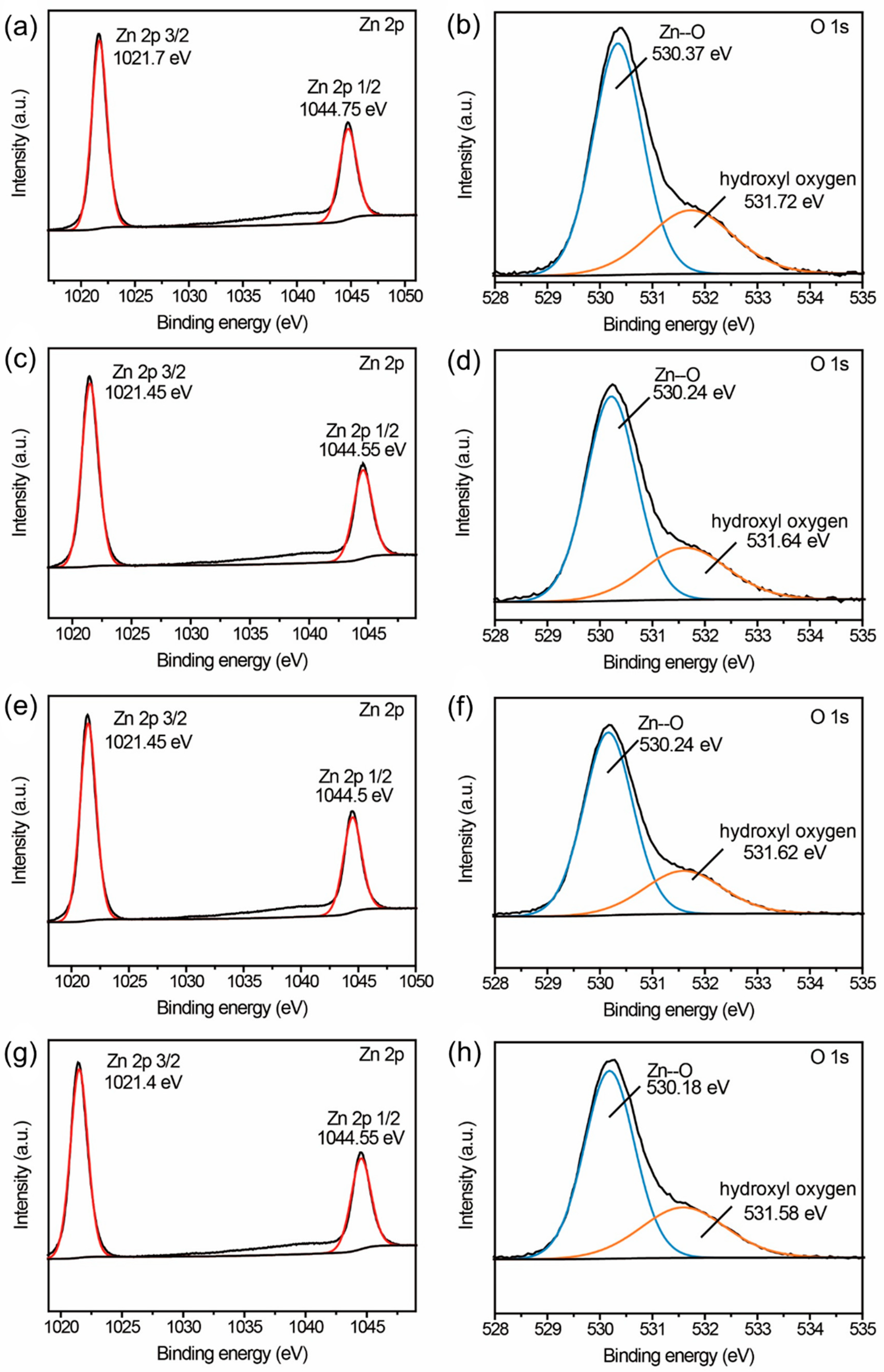
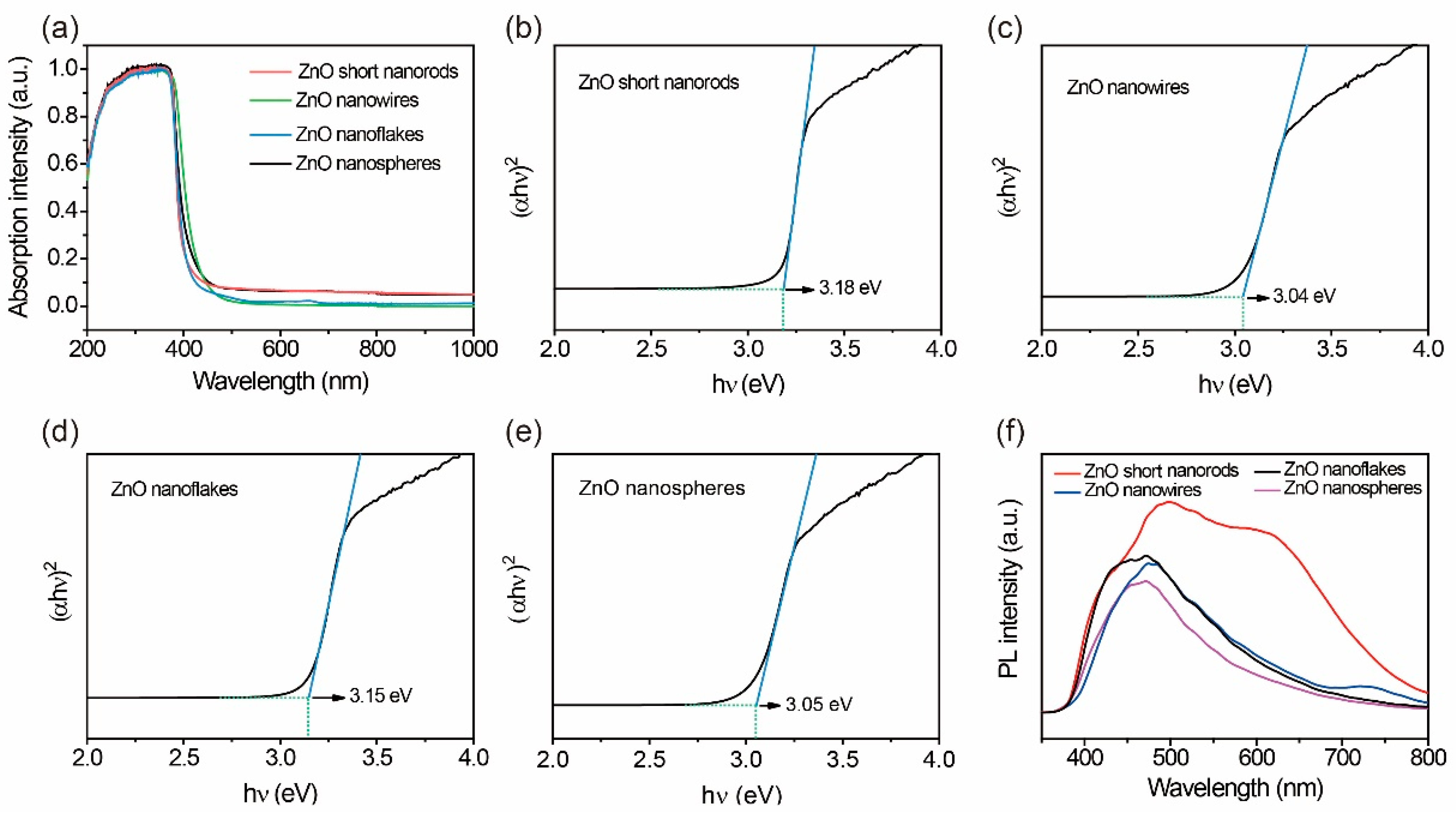
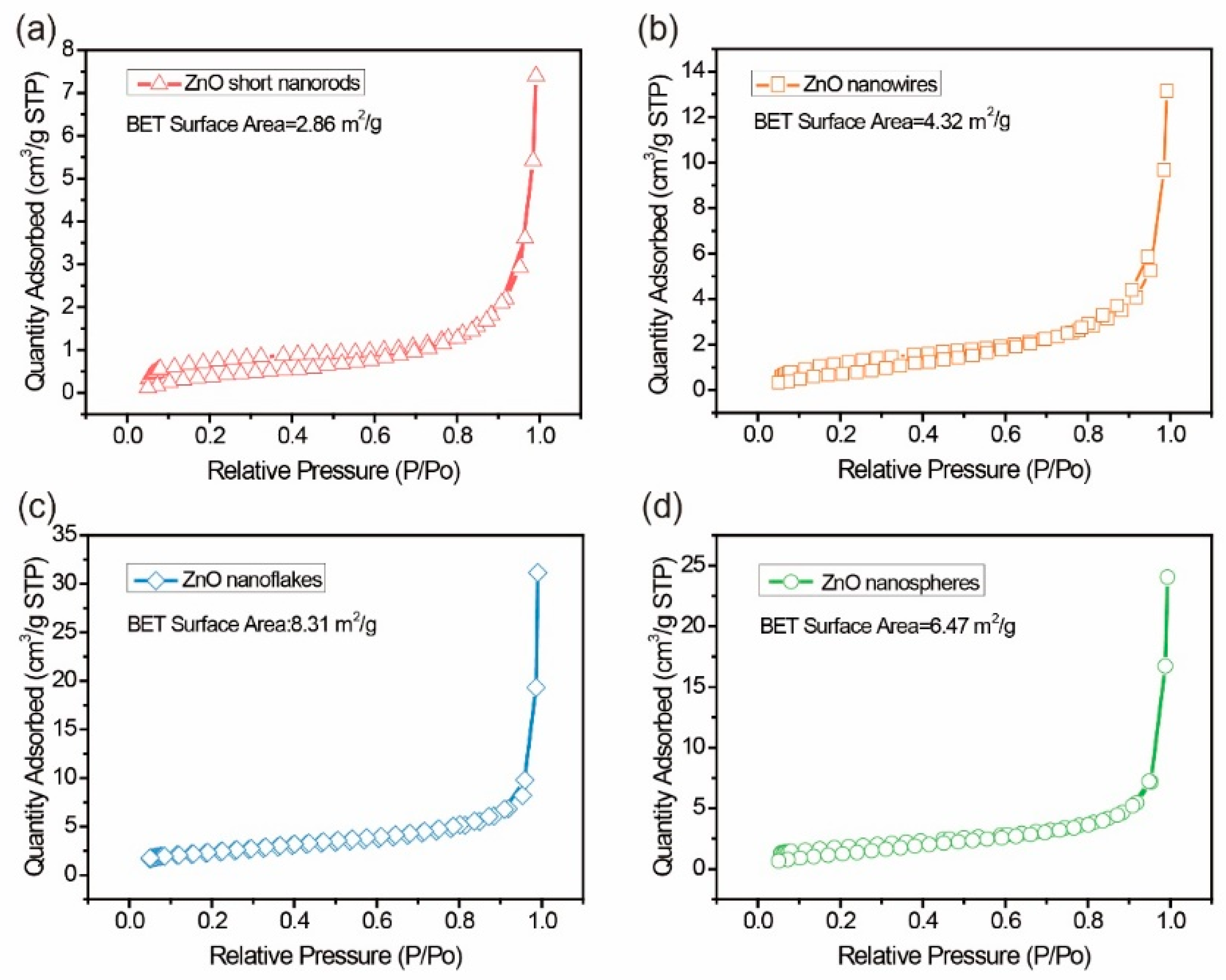
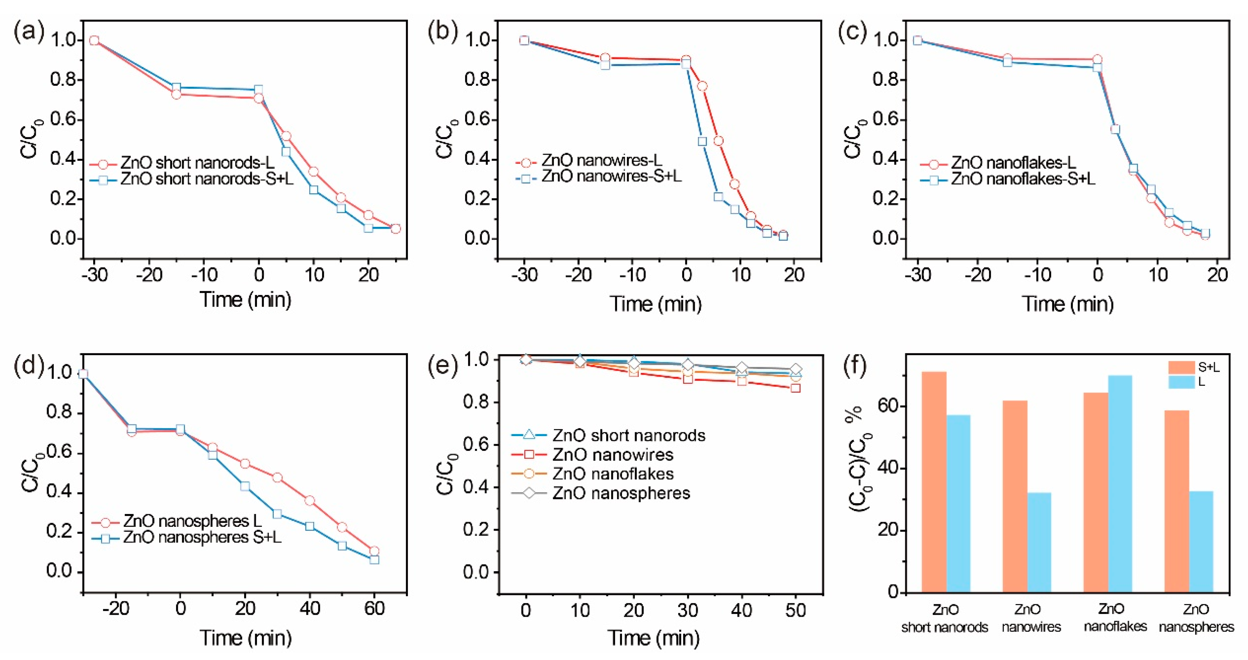
2. Results and Discussion
3. Experimental procedures
3.1. Materials
3.2. ZnO Synthesis
3.3. Characterization
3.4. Photocatalytic Property Evaluation
4. Conclusions
Author Contributions
Funding
Conflicts of Interest
References
- Bartolomeu, M.; Neves, M.G.P.M.S.; Faustino, M.A.F.; Almeida, A. Wastewater chemical contaminants: Remediation by advanced oxidation processes. Photochem. Photobiol. Sci. 2018, 17, 1573–1598. [Google Scholar] [CrossRef]
- Miletto, M. Water and Energy Nexus: Findings of the World Water Development Report 2014. In Proceedings of the 11th Kovacs Colloquium on Hydrological Sciences and Water Security: Past, Present and Future, UNESCO Headquarters, Paris, France, 16–17 June 2014. [Google Scholar]
- Chiu, Y.-H.; Chang, T.-F.; Chen, C.-Y.; Sone, M.; Hsu, Y.-J. Mechanistic Insights into Photodegradation of Organic Dyes Using Heterostructure Photocatalysts. Catalysts 2019, 9, 430. [Google Scholar] [CrossRef]
- Chen, S.; Zhang, J.; Zhang, C.; Yue, Q.; Li, Y.; Li, C. Equilibrium and kinetic studies of methyl orange and methyl violet adsorption on activated carbon derived from Phragmites australis. Desalination 2010, 252, 149–156. [Google Scholar] [CrossRef]
- Gurreri, L.; Tamburini, A.; Cipollina, A.; Micale, G. Electrodialysis Applications in Wastewater Treatment for Environmental Protection and Resources Recovery: A Systematic Review on Progress and Perspectives. Membranes 2020, 10, 146. [Google Scholar] [CrossRef] [PubMed]
- Mustafa, R.; Asmatulu, E. Preparation of activated carbon using fruit, paper and clothing wastes for wastewater treatment. J. Water Process Eng. 2020, 35, 101239. [Google Scholar] [CrossRef]
- Yang, X.; Wang, Z.; Shao, L. Construction of oil-unidirectional membrane for integrated oil collection with lossless transportation and oil-in-water emulsion purification. J. Membr. Sci. 2018, 549, 67–74. [Google Scholar] [CrossRef]
- Lin, L.; Li, R.-H.; Li, Y.; Xu, J.; Li, X.-Y. Recovery of organic carbon and phosphorus from wastewater by Fe-enhanced primary sedimentation and sludge fermentation. Process Biochem. 2016, 54, 135–139. [Google Scholar] [CrossRef]
- Routoula, E.; Patwardhan, S.V. Degradation of Anthraquinone Dyes from Effluents: A Review Focusing on Enzymatic Dye Degradation with Industrial Potential. Environ. Sci. Technol. 2020, 54, 647–664. [Google Scholar] [CrossRef]
- Ma, D.; Yi, H.; Lai, C.; Liu, X.; Huo, X.; An, Z.; Li, L.; Fu, Y.; Li, B.; Zhang, M.; et al. Critical review of advanced oxidation processes in organic wastewater treatment. Chemosphere 2021, 275, 130104. [Google Scholar] [CrossRef] [PubMed]
- Serrà, A.; Philippe, L.; Perreault, F.; Garcia-Segura, S. Photocatalytic treatment of natural waters. Reality or hype? The case of cyanotoxins remediation. Water Res. 2020, 188, 116543. [Google Scholar] [CrossRef]
- Yousefi, S.R.; Alshamsi, H.A.; Amiri, O.; Salavati-Niasari, M. Synthesis, characterization and application of Co/Co3O4 nanocomposites as an effective photocatalyst for discoloration of organic dye contaminants in wastewater and antibacterial properties. J. Mol. Liq. 2021, 337, 116405. [Google Scholar] [CrossRef]
- Orooji, Y.; Tanhaei, B.; Ayati, A.; Tabrizi, S.H.; Alizadeh, M.; Bamoharram, F.F.; Karimi, F.; Salmanpour, S.; Rouhi, J.; Afshar, S.; et al. Heterogeneous UV-Switchable Au nanoparticles decorated tungstophosphoric acid/TiO2 for efficient photocatalytic degradation process. Chemosphere 2021, 281, 130795. [Google Scholar] [CrossRef] [PubMed]
- Cheng, C.; Mao, L.H.; Shi, J.W.; Xue, F.; Zong, S.C.; Zheng, B.T.; Guo, L.J. NiCo2O4 Nanosheets as a Novel Oxygen-Evolution-Reaction Cocatalyst in Situ Bonded on the G-C3N4 Photocatalyst for Excellent Overall Water Splitting. J. Mater. Chem. A 2021, 9, 12299–12306. [Google Scholar] [CrossRef]
- Jiang, H.X.; Zhang, L.S.; Liu, H.Y.; Wu, D.S.; Wu, F.Y.; Tian, L.; Liu, L.L.; Zou, J.P.; Luo, S.L.; Chen, B.B. Silver Single Atom in Carbon Nitride Catalyst for Highly Efficient Photocatalytic Hydrogen Evolution. Angew. Chem. 2020, 59, 23112–23116. [Google Scholar] [CrossRef] [PubMed]
- Li, D.; Kassymova, M.; Cai, X.; Zang, S.-Q.; Jiang, H.-L. Photocatalytic CO2 reduction over metal-organic framework-based materials. Co-ord. Chem. Rev. 2020, 412, 213262. [Google Scholar] [CrossRef]
- Huang, H.Y.; Wang, K.; Guo, T.; Li, J.; Wu, X.Y.; Zhang, G.K. Construction of 2d/2d Bi2Se3/G-C3N4 Nanocomposite with High Interfacial Charge Separation and Photo-Heat Conversion Efficiency for Selective Photocatalytic CO2 Reduction. Appl. Catal. B Environ. 2020, 277, 119232. [Google Scholar] [CrossRef]
- Irshad, M.A.; Nawaz, R.; Rehman, M.Z.U.; Adrees, M.; Rizwan, M.; Ali, S.; Ahmad, S.; Tasleem, S. Synthesis, characterization and advanced sustainable applications of titanium dioxide nanoparticles: A review. Ecotoxicol. Environ. Saf. 2021, 212, 111978. [Google Scholar] [CrossRef]
- Humayun, M.; Raziq, F.; Khan, A.; Luo, W. Modification Strategies of TiO2 for Potential Applications in Photocatalysis: A critical Review. Green Chem. Lett. Rev. 2018, 11, 86–102. [Google Scholar] [CrossRef]
- Guo, C.; Li, L.; Chen, F.; Ning, J.; Zhong, Y.; Hu, Y. One-step phosphorization preparation of gradient-P-doped CdS/CoP hybrid nanorods having multiple channel charge separation for photocatalytic reduction of water. J. Colloid Interface Sci. 2021, 596, 431–441. [Google Scholar] [CrossRef]
- Huang, Y.X.; Lei, R.; Yuan, J.; Gao, F.; Jiang, C.K.; Feng, W.H.; Zhuang, J.D.; Liu, P. Insight into the Piezo-Photo Coupling Effect of PbTiO3/CdS Composites for Piezo-Photocatalytic Hydrogen Production. Appl. Catal. B Environ. 2021, 282, 119586. [Google Scholar] [CrossRef]
- Wang, K.; Xing, Z.P.; Du, M.; Zhang, S.Y.; Li, Z.Z.; Pan, K.; Zhou, W. Hollow MoSe2@Bi2S3/CdS Core-Shell Nanostructure as Dual Z-Scheme Heterojunctions with Enhanced Full Spectrum Photocatalytic-Photothermal Performance. Appl. Catal. B Environ. 2021, 281, 119482. [Google Scholar] [CrossRef]
- Tan, H.L.; Amal, R.; Ng, Y.H. Alternative strategies in improving the photocatalytic and photoelectrochemical activities of visible light-driven BiVO4: A review. J. Mater. Chem. A 2017, 5, 16498–16521. [Google Scholar] [CrossRef]
- Wang, L.Y.; Ding, K.; Xu, R.; Yu, D.; Wang, W.; Gao, P.; Liu, B.J. Fabrication of BiVO4/BiPO4/GO Composite Photocatalytic Material for the Visible Light-Driven Degradation. J. Clean. Prod. 2020, 247, 119108. [Google Scholar] [CrossRef]
- Yu, C.; Li, G.; Kumar, S.; Yang, K.; Jin, R. ChemInform Abstract: Phase Transformation Synthesis of Novel Ag2O/Ag2CO3 Heterostructures with High Visible Light Efficiency in Photocatalytic Degradation of Pollutants. ChemInform 2014, 45, 892–898. [Google Scholar] [CrossRef]
- Chen, Y.; Zhu, G.; Hojamberdiev, M.; Gao, J.; Zhu, R.; Wang, C.; Wei, X.; Liu, P. Three-dimensional Ag2O/Bi5O7I p–n heterojunction photocatalyst harnessing UV–vis–NIR broad spectrum for photodegradation of organic pollutants. J. Hazard. Mater. 2018, 344, 42–54. [Google Scholar] [CrossRef]
- Hieu, C.N.; Lien, T.M.; Van, T.T.T.; Juang, R.S. Enhanced Removal of Various Dyes from Aqueous Solutions by UV and Simulated Solar Photocatalysis over TiO2/ZnO/rGO Composites. Sep. Purif. Technol. 2020, 232, 115962. [Google Scholar] [CrossRef]
- Kumar, S.G.; Rao, K.S.R.K. Zinc oxide based photocatalysis: Tailoring surface-bulk structure and related interfacial charge carrier dynamics for better environmental applications. RSC Adv. 2014, 5, 3306–3351. [Google Scholar] [CrossRef]
- Goktas, S.; Goktas, A. A comparative study on recent progress in efficient ZnO based nanocomposite and heterojunction photocatalysts: A review. J. Alloys Compd. 2021, 863, 158734. [Google Scholar] [CrossRef]
- Pan, L.; Muhammad, T.; Ma, L.; Huang, Z.-F.; Wang, S.; Wang, L.; Zou, J.-J.; Zhang, X. MOF-derived C-doped ZnO prepared via a two-step calcination for efficient photocatalysis. Appl. Catal. B Environ. 2016, 189, 181–191. [Google Scholar] [CrossRef]
- Trandafilovic, V.L.; Jovanovic, D.J.; Zhang, X.; Ptasinska, S.; Dramicanin, M.D. Enhanced Photocatalytic Degradation of Methylene Blue and Methyl Orange by ZnO:Eu Nanoparticles. Appl. Catal. B Environ. 2017, 203, 740–752. [Google Scholar] [CrossRef] [Green Version]
- Ardekani, R.S.; Aghdam, A.S.R.; Nazari, M.; Bayat, A.; Saievar-Iranizad, E.; Liavali, M.N. Synthesis and Characterization of Photocatalytically Active Crumpled-Shape Nanocomposites of Nitrogen and Sulfur Co-Doped ZnO-CeO2. Sol. Energy Mater. Sol. Cells 2019, 203, 110195. [Google Scholar] [CrossRef]
- Bharathi, P.; Harish, S.; Archana, J.; Navaneethan, M.; Ponnusamy, S.; Muthamizhchelvan, C.; Shimomura, M.; Hayakawa, Y. Enhanced charge transfer and separation of hierarchical CuO/ZnO composites: The synergistic effect of photocatalysis for the mineralization of organic pollutant in water. Appl. Surf. Sci. 2019, 484, 884–891. [Google Scholar] [CrossRef]
- Aliaga, J.; Cifuentes, N.; González, G.; Sotomayor-Torres, C.; Benavente, E. Enhancement Photocatalytic Activity of the Heterojunction of Two-Dimensional Hybrid Semiconductors ZnO/V2O5. Catalysts 2018, 8, 374. [Google Scholar] [CrossRef]
- Wang, Z.L.; Wu, W.; Falconi, C. Piezotronics and piezo-phototronics with third-generation semiconductors. MRS Bull. 2018, 43, 922–927. [Google Scholar] [CrossRef]
- Yang, Q.; Wang, W.; Xu, S.; Wang, Z.L. Enhancing Light Emission of ZnO Microwire-Based Diodes by Piezo-Phototronic Effect. Nano Lett. 2011, 11, 4012–4017. [Google Scholar] [CrossRef]
- Xue, X.; Zang, W.; Deng, P.; Wang, Q.; Xing, L.; Zhang, Y.; Wang, Z.L. Piezo-potential enhanced photocatalytic degradation of organic dye using ZnO nanowires. Nano Energy 2015, 13, 414–422. [Google Scholar] [CrossRef]
- Bai, Y.; Zhao, J.; Lv, Z.; Lu, K. Enhanced piezo-phototronic effect of ZnO nanorod arrays for harvesting low mechanical energy. Ceram. Int. 2019, 45, 15065–15072. [Google Scholar] [CrossRef]
- Wu, W.; Yin, X.; Dai, B.; Kou, J.; Ni, Y.; Lu, C. Water flow drived piezo-photocatalytic flexible films: Bi-piezoelectric integration of ZnO nanorods and PVDF. Appl. Surf. Sci. 2020, 517, 146119. [Google Scholar] [CrossRef]
- Hong, K.-S.; Xu, H.; Konishi, H.; Li, X. Direct Water Splitting Through Vibrating Piezoelectric Microfibers in Water. J. Phys. Chem. Lett. 2010, 1, 997–1002. [Google Scholar] [CrossRef]
- Wu, J.M.; Sun, Y.-G.; Chang, W.-E.; Lee, J.-T. Piezoelectricity induced water splitting and formation of hydroxyl radical from active edge sites of MoS2 nanoflowers. Nano Energy 2018, 46, 372–382. [Google Scholar] [CrossRef]
- Raoufi, D. Synthesis and microstructural properties of ZnO nanoparticles prepared by precipitation method. Renew. Energy 2013, 50, 932–937. [Google Scholar] [CrossRef]
- Vorokh, A. Scherrer formula: Estimation of error in determining small nanoparticle size. Nanosyst. Phys. Chem. Math. 2018. [Google Scholar] [CrossRef]
- Montenegro, D.N.; Hortelano, V.; Martínez, O.; Martinez-Tomas, M.C.; Sallet, V.; Muñoz-Sanjose, V.; Jiménez, J. Non-radiative recombination centres in catalyst-free ZnO nanorods grown by atmospheric-metal organic chemical vapour deposition. J. Phys. D Appl. Phys. 2013, 46. [Google Scholar] [CrossRef]
- Walter, C.; Barg, S.; Ni, N.; Maher, R.C.; Garcίa-Tuñón, E.; Ismail, M.M.Z.; Babot, F.; Saiz, E. A novel approach for the fabrication of carbon nanofibre/ceramic porous structures. J. Eur. Ceram. Soc. 2013, 33, 2365–2374. [Google Scholar] [CrossRef]
- Su, X.; Wang, Z.; Huang, Y.; Miao, Z.; Wang, S.; Wang, J.; Zhang, X.L.; Sun, X.; Liu, H.; Sang, Y. Triethanolamine interface modification of crystallized ZnO nanospheres enabling fast photocatalytic hazard-free treatment of Cr(vi) ions. Nanotechnol. Rev. 2021, 10, 847–856. [Google Scholar] [CrossRef]
- Rambabu, K.; Bharath, G.; Banat, F.; Show, P.L. Green synthesis of zinc oxide nanoparticles using Phoenix dactylifera waste as bioreductant for effective dye degradation and antibacterial performance in wastewater treatment. J. Hazard. Mater. 2020, 402, 123560. [Google Scholar] [CrossRef]
- Cao, C.; Xie, X.; Zeng, Y.; Shi, S.; Wang, G.; Yang, L.; Wang, C.-Z.; Lin, S. Highly efficient and stable p-type ZnO nanowires with piezotronic effect for photoelectrochemical water splitting. Nano Energy 2019, 61, 550–558. [Google Scholar] [CrossRef]
- Yu, S.; Zhang, H.; Li, Z. Effects of pH on High-Performance ZnO Resistive Humidity Sensors Using One-Step Synthesis. Sensors 2019, 19, 5267. [Google Scholar] [CrossRef]
- Zhang, Q.; Gu, H.; Wang, X.; Li, L.; Zhang, J.; Zhang, H.; Li, Y.-F.; Dai, W.-L. Robust hollow tubular ZnIn2S4 modified with embedded metal-organic-framework-layers: Extraordinarily high photocatalytic hydrogen evolution activity under simulated and real sunlight irradiation. Appl. Catal. B Environ. 2021, 298, 120632. [Google Scholar] [CrossRef]
- Li, W.; Wang, G.; Chen, C.; Liao, J.; Li, Z. Enhanced Visible Light Photocatalytic Activity of ZnO Nanowires Doped with Mn2+ and Co2+ Ions. Nanomaterials 2017, 7, 20. [Google Scholar] [CrossRef] [Green Version]
- Li, Q.; Fu, S.; Song, C.; Wang, G.; Zeng, F.; Pan, F. Sputtering power dependence of structure and photoluminescence of ZnO on 6H–SiC. J. Mater. Sci. Mater. Electron. 2017, 28, 17881–17888. [Google Scholar] [CrossRef]
- Li, H.; Sang, Y.; Chang, S.; Huang, X.; Zhang, Y.; Yang, R.; Jiang, H.; Liu, H.; Wang, Z.L. Enhanced Ferroelectric-Nanocrystal-Based Hybrid Photocatalysis by Ultrasonic-Wave-Generated Piezophototronic Effect. Nano Lett. 2015, 15, 2372–2379. [Google Scholar] [CrossRef] [PubMed]
- Zhao, C.; Liang, Y.; Li, W.; Tian, Y.; Chen, X.; Yin, D.; Zhang, Q. BiOBr/BiOCl/carbon quantum dot microspheres with superior visible light-driven photocatalysis. RSC Adv. 2017, 7, 52614–52620. [Google Scholar] [CrossRef] [Green Version]


Publisher’s Note: MDPI stays neutral with regard to jurisdictional claims in published maps and institutional affiliations. |
© 2022 by the authors. Licensee MDPI, Basel, Switzerland. This article is an open access article distributed under the terms and conditions of the Creative Commons Attribution (CC BY) license (https://creativecommons.org/licenses/by/4.0/).
Share and Cite
Su, X.; Zhao, X.; Cui, C.; Xi, N.; Zhang, X.L.; Liu, H.; Yu, X.; Sang, Y. Influence of Wurtzite ZnO Morphology on Piezophototronic Effect in Photocatalysis. Catalysts 2022, 12, 946. https://doi.org/10.3390/catal12090946
Su X, Zhao X, Cui C, Xi N, Zhang XL, Liu H, Yu X, Sang Y. Influence of Wurtzite ZnO Morphology on Piezophototronic Effect in Photocatalysis. Catalysts. 2022; 12(9):946. https://doi.org/10.3390/catal12090946
Chicago/Turabian StyleSu, Xiaowen, Xiaolei Zhao, Chao Cui, Ning Xi, Xiao Li Zhang, Hong Liu, Xiaowen Yu, and Yuanhua Sang. 2022. "Influence of Wurtzite ZnO Morphology on Piezophototronic Effect in Photocatalysis" Catalysts 12, no. 9: 946. https://doi.org/10.3390/catal12090946