Article

C058 and Other Functional Microorganisms Promote the Synthesis of Extracellular Polymer Substances in Mycelium Biofloc

1
College of Resources and Environment, Zhongkai University of Agriculture and Engineering, Guangzhou 510225, China
2
Guangdong Provincial Engineering and Technology Research Center for Agricultural Land Pollution Prevention and Control, Guangzhou 510225, China
*
Author to whom correspondence should be addressed.
Catalysts 2022, 12(7), 693; https://doi.org/10.3390/catal12070693
Submission received: 21 April 2022 / Revised: 16 June 2022 / Accepted: 20 June 2022 / Published: 24 June 2022
(This article belongs to the Special Issue Microbial Biocatalysis)

Abstract

:
The mycelium biofloc bioaugmented by Cordyceps strain C058 effectively purifies water, which may be related to the synthesis of extracellular polymer substances. To verify this conjecture, we analyzed the changes in extracellular polymer substances content in the mycelium biofloc under various hydraulic retention times (36 h, 18 h, and 11 h). The microstructure and microflora composition were analyzed using a scanning electron microscope and high-throughput sequencing. The ordinary biofloc without bioaugmentation was taken as a control. The results showed that under the above hydraulic retention time, the extracellular polymer substances contents of the mycelium biofloc were 51.20, 55.89, and 33.84 mg/g, respectively, higher than that of the ordinary biofloc (14.58, 15.72, and 18.19 mg/g). The protein content or the polysaccharide content also followed the same trend. Meanwhile, the sedimentation performance of the mycelium biofloc was better than that of the ordinary biofloc, attributed to the content of the extracellular polymer substances. It is worth noting that C058 is the main biofloc content, which promotes the synthesis of extracellular polymer substances in the mycelium biofloc. Other functional microorganisms in the mycelium biofloc were Janthinobacterium, Phormidium, Leptolyngbya, Hymenobacter, and Spirotrichea, which also promote the synthesis of extracellular polymer substances.

1. Introduction

Extracellular polymeric substances (EPS) are sticky polymers secreted by microorganisms through metabolism and cell autolysis [1]. They contain proteins (PN), polysaccharides (PS), humic acid, and nucleic acids [2,3]. PN and PS are the main EPS constituents, accounting for about 70–80% of the total EPS [4]. Because EPS promote adhesion and aggregation between microbial cells [5], and their content can affect the properties of flocs, such as flocculation capacity, settling performance, surface charge, and microbial community structure [6,7,8], EPS are key substances that determine the physical, chemical, and biological properties of flocs, and thus the water purification performance. Biofloc technology (BFT) is widely used in aquacultural tailwater purification. Zoogloea and filamentous bacteria are the core of ordinary biofloc (OBF). They adhere to bacteria, fungi, algae, protozoa, and organic polymers, forming structurally diverse flocs using secreted extracellular polymer substances [9]. OBF removes ammonium nitrogen, total nitrogen, and total phosphorus from water through microbial assimilation and nitrification [10] and removes organic matter from water through biological adsorption [11]. Recent studies have shown that the bioaugmented biofloc efficiently purifies water, but the mechanism is not clearly described, in particular, the EPS synthesis [12,13,14].
Cordyceps sp. belongs to phylum Ascomycota, class Sordariomycetes, order Hypocreales, and family Clavicipitaceae. Its ascospores germinate into and belong to large filamentous mycelium. Numerous studies have shown that fungi can efficiently remove contaminants in wastewater [15,16,17,18]. Our previous study showed that C058 and its bioaugmented biofloc named mycelium biofloc (MBF) effectively purified water [19,20]; however, the mechanism underlying this process is unclear. Zhang [21] used a mycelium ball as a biomass carrier to load Pseudomonas stutzeri T13, and found that the total nitrogen removal rate in the SBR reactor was about 10% higher than in the traditional activated sludge reactor, and the protein content in EPS secreted by mycelium ball is high, which enhances the solid–liquid separation performance of the system by improving the hydrophobicity of activated sludge. Cordyceps secretes large amounts of PS, PN, and other macromolecules [22,23]. Hence, we speculate that the water purification effect of MBF may be related to its EPS synthesis. To validate this hypothesis, we analyzed the changes in EPS content, the microstructure, and the microflora composition in MBF under various hydraulic retention times (HRT) using a scanning electron microscope (SEM) and high-throughput sequencing. The OBF without bioaugmentation was taken as a control. The findings of this study provide a theoretical basis for the water purification effect of MBF.

2. Results and Discussion

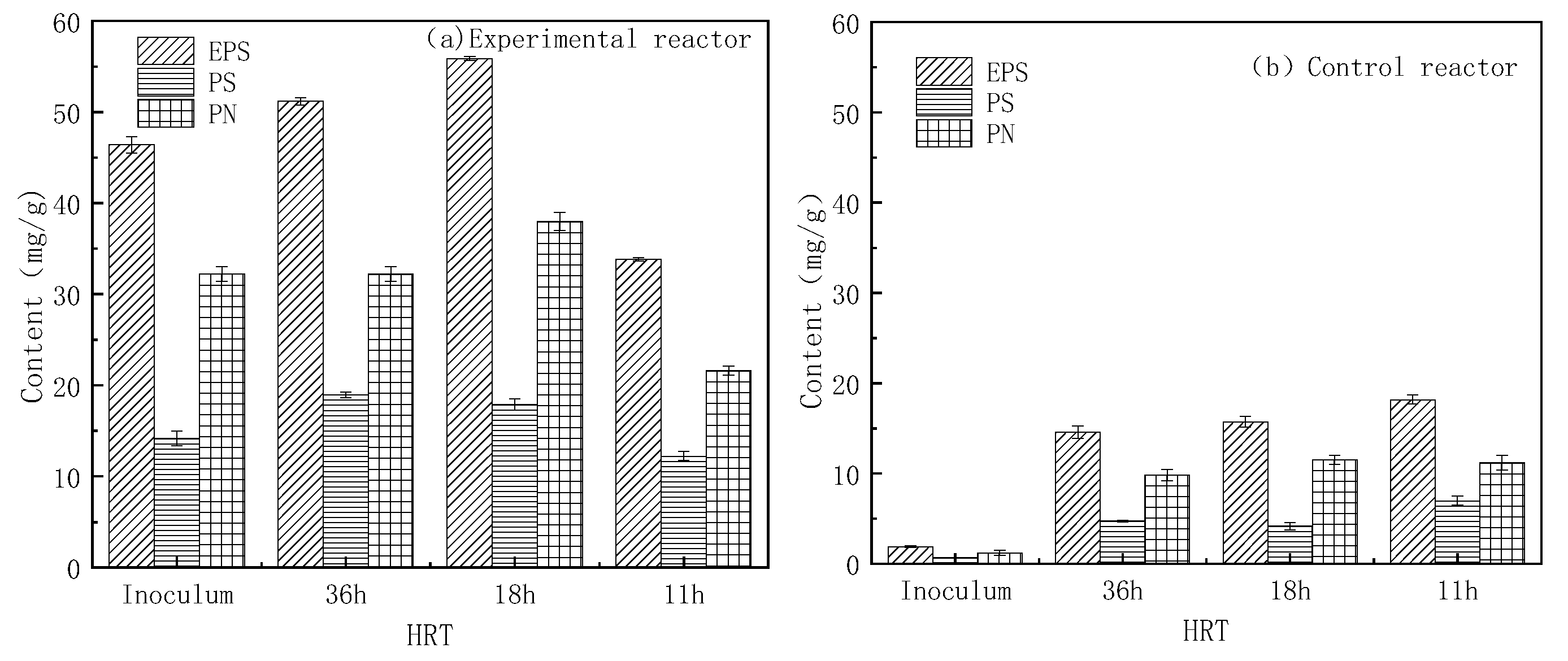
2.1. Changes in EPS Content in the Biofloc

Figure 1 shows that the EPS content was higher in the experimental reactor than that of the control reactor under various HRTs, which may be attributed to the C058 in the experimental reactor. C058 secretes a large amount of PS and PN [19], generating higher EPS content in MBF of 51.20, 55.89, and 33.84 mg/g under the above HRTs. Morgan et al. [24] proposed that the activity of functional microorganisms affects the EPS content. Accordingly, the EPS contents were different in the two reactors [25]. There was lower EPS content in OBF (14.58, 15.72, and 18.19 mg/g under the above HRTs). Meanwhile, It can be seen from Figure 1 that the PN content or PS content also followed the same trend. In addition, the flow rate influenced the HRT and thus the shear forces. Appropriated shear force is essential for optimal EPS secretion in aerobic granular sludge, but excessive shear force destabilizes the sludge [26]. This may explain the increase in EPS content in both reactors during the first and second operation stages (Figure 1). Filamentous bacteria are intolerant to shear forces, and HRT 11 h may cause autolysis of filamentous bacteria with a decrease in EPS content in MBF.

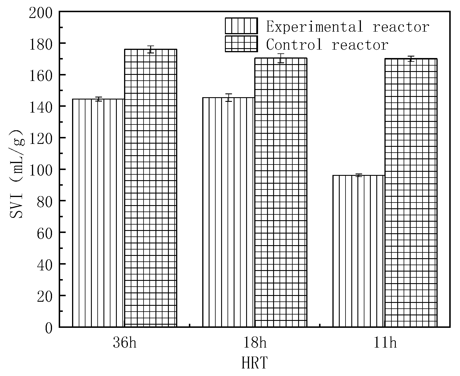
2.2. Sludge Volume Index (SVI) Changes of Biofloc

The settling performance of flocs can indirectly reflect the content and composition of EPS. It can be seen from Figure 2 that the SVI was lower in the experimental reactor than that in the control reactor under various HRTs, and all the SVI in the experimental reactor were lower than 150 mL/g, implying that the settling performance of MBF occurred within a given favorable range. That of the control group was above 150 mL/g under various HRTs. The OBF was loose and was detrimental to sedimentation [27]; thus, the settling performance of MBF was better than that of OBF. Meanwhile, there was a positive correlation between EPS and SVI in the experimental reactor, consistent with a previous study [28]. Li et al. [29] found that microorganisms can secrete EPS to enhance flocculation and sedimentation. In the present study, the sedimentation performance of MBF was better than that of OBF, which indirectly proved that the EPS content of MBF was higher than that of OBF. In addition, the settling performance of flocs reflects the composition of EPS. Previous studies have shown that PN possesses hydrophobic groups, which improve the hydrophobicity of the cells and enhance the mutual attraction between bacteria, promoting their sedimentation [30,31]. In this study, the PN content of the experimental reactor was higher than that of the control reactor under various HRTs (Figure 1).

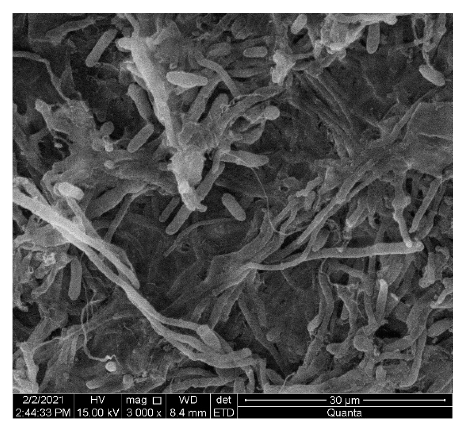
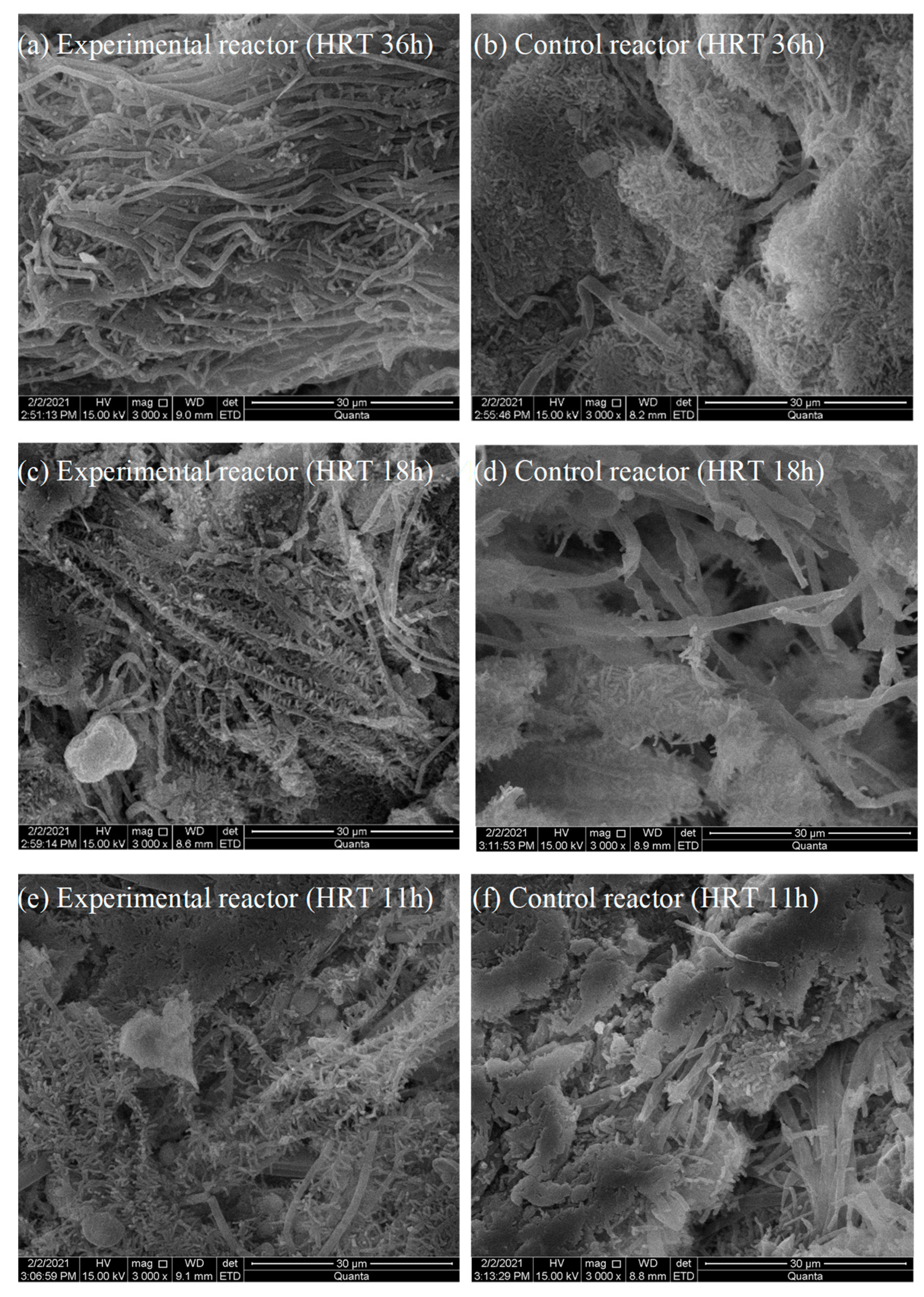
2.3. Changes in the Appearance of Biofloc

The appearance of MBF and OBF were analyzed through a visual examination using an SEM. The morphological structure of C058 is shown in Figure 3. C058 is filamentous and supercoiled. The bioflocs in both reactors under various HRTs are shown in Figure 4. As shown in Figure 4a,c,e, bacteria aggregates on the MBF surface increased over time, forming a symbiotic structure with C058. Although MBF was constructed by fungi and bacteria together, fungi were still the dominant microorganism. As shown in Figure 4b,d,f, the OBF mainly comprised zoogloea, whose size decreased with time. It revealed that the shortened HRT strengthened the shear force and minimized the zoogloea size.

2.4. Changes in the Microbial Community Structure in Biofloc

During the operation of both reactors, the biofloc was sampled when it had achieved stable operation under various HRTs. A total of eight samples were collected and included CKCB (inoculum of the experimental reactor), CB-1 (sample of the experimental reactor at HRT 36 h), CB-2 (sample of the experimental reactor at HRT 18 h), CB-3 (sample of the experimental reactor at HRT 11 h), CKWB (inoculum of the control reactor), BF-1 (sample of the control reactor at HRT 36 h), BF-2 (sample of the control reactor at HRT 18 h), BF-3 (sample of the control reactor at HRT 11 h).
The microbial community structure in biofloc was determined using 16S rRNA and 18S rRNA gene sequencing, and the sequencing process was completed in Guangdong Meilikang Biotechnology Co., Ltd., Guangzhou, China.

2.4.1. Changes in Prokaryote in Biofloc

(1)
The diversity index of prokaryote
The diversity of the microbial community, including the species diversity and relative abundance of each group, was described based on the α-diversity index. We found that the Chao1 index in the experimental reactor negatively correlated with the HRT, and was at the lowest level (1906.67) at HRT 11 h. The change of the Chao1 index in the control reactor was the same as in the experimental reactor, and the lowest Chao1 index was 1489.73 at HRT 11 h (Table 1). The change of HRT had less of an effect on the bacteria abundance in the experimental reactor than in the control reactor. Meanwhile, the Shannon index and Simpson index also followed the same trend. Hence, the species diversity of microorganisms was higher in the experimental reactor than in the control reactor, implying that the microbial community in the experimental reactor was more stable.
(2)
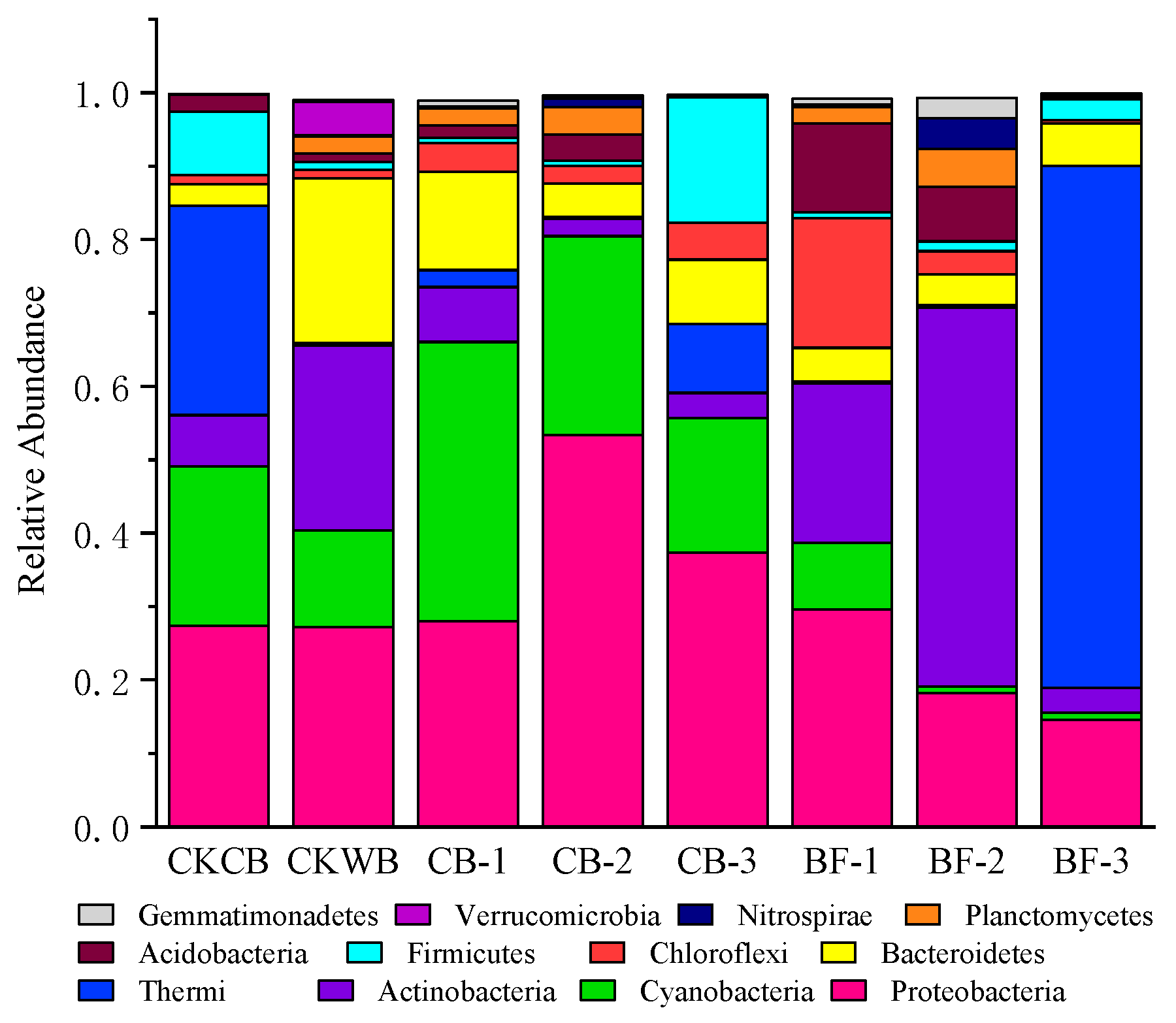
The relative abundance of prokaryotes at the phylum level
It can be seen from Figure 5 that the most dominant phylum in the experimental reactor at the first operation stage was Thermi, followed by Proteobacteria and Cyanobacteria. At HRT 36 h, Cyanobacteria became the most dominant phylum, with an abundance of 38.05%, followed by Proteobacteria (27.99%) and Bacteroidetes (13.34%). At HRT 18 h, the abundance of Proteobacteria increased to 53.34%, making it the most dominant phylum, followed by Cyanobacteria and Bacteroidetes. At HRT 11 h, the abundance of Proteobacteria was 37.35%, and it was still the most dominant phylum, followed by Cyanobacteria and Firmicutes. In the control reactor, the most dominant phylum at the first operation stage was Proteobacteria, followed by Actinobacteria and Bacteroidetes. The abundance of Proteobacteria decreased gradually from 27.21% (HRT 18 h) to 14.59% (HRT 11 h) with the shorting of HRT, and it was no longer the most dominant bacteria. The abundance of Actinobacteria also displayed the same trend. Instead, Thermi became the most dominant phylum at HRT 11 h; therefore, MBF was dominant by Proteobacteria. Proteobacteria plays an indispensable role in the material cycle, including nitrogen transformation in the water [32,33], enabling nitrogen removal in aquacultural wastewater [34]. Meanwhile, the abundance of Bacteroidetes and Cyanobacteria was also higher in the experimental reactor than in the control reactor under various HRTs. Bacteroidetes had the particularity of using nitrogen compounds for growth and Cyanobacteria had nitrogen fixation capacity [32,35]. These bacteria can promote the synthesis of EPS in MBF.
(3)
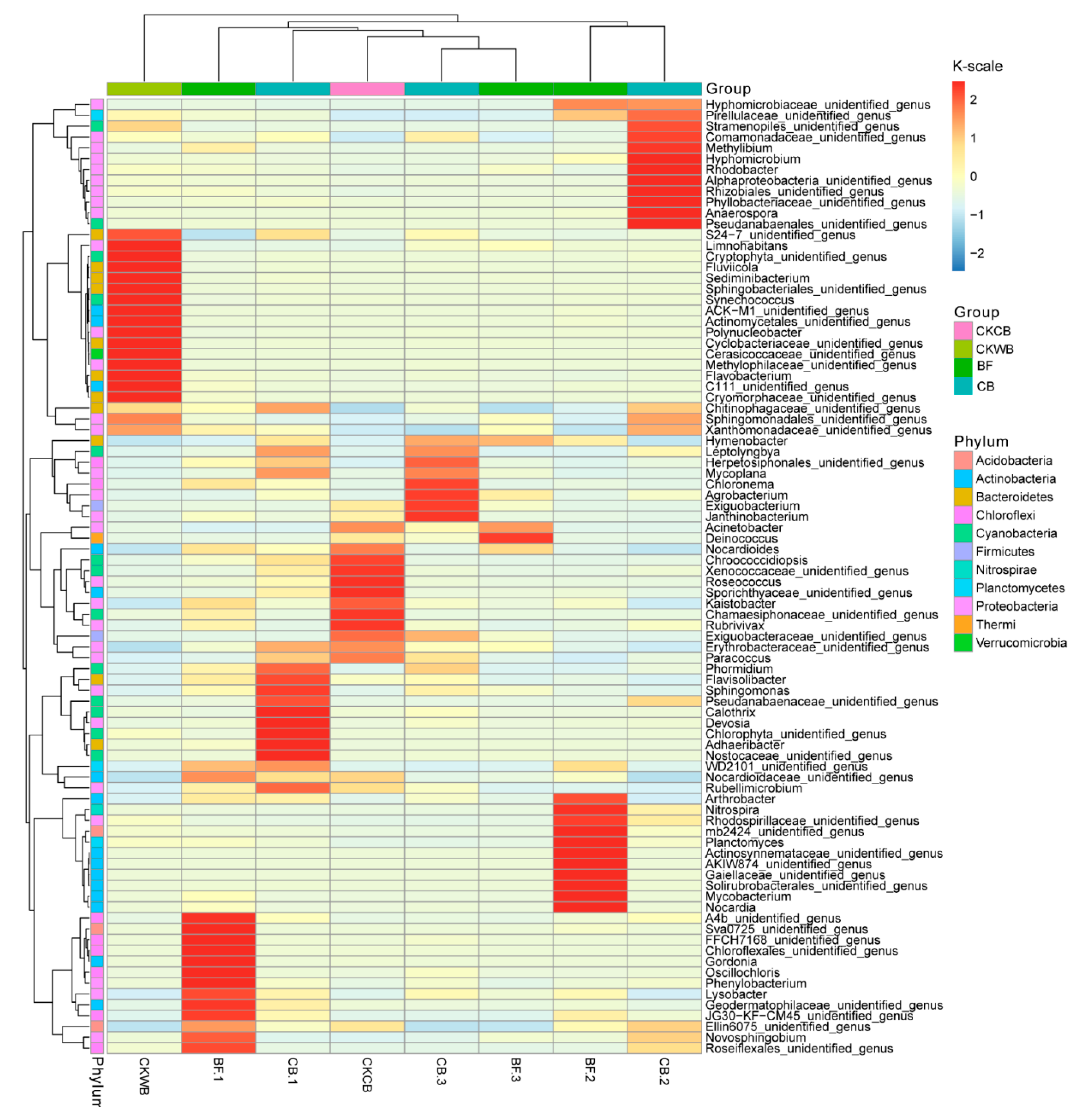
The relative abundance of prokaryotes at the genus level
It can be seen from Figure 6 that the abundance of Janthinobacterium belonging to Proteobacteria in the experimental reactor reached 20.74% at HRT 11 h, significantly higher than that in the control reactor (0.68%). This may explain the effective water purification capability of MBF at short HRT, as reported in our previous study [18]. Other studies have shown that Janthinobacterium has nitrogen removal capacity in the water. Yang et al. [36] found that Janthinobacterium sp. M-11 still exhibited nitrite removal capacity under low temperature, and the removal rate of nitrite and nitrate even reached 93% and 98%, respectively, under anaerobic conditions, which was beneficial for the treatment of eutrophic water. Meanwhile, as dominant genera, the abundance of Phormidium and Leptolyngbya belonging to Cyanobacteria was higher in the experimental reactor than in the control reactor under various HRTs. Other studies have shown that Phormidium and Leptolyngbya had excellent nitrogen fixation activity and PN and PS production capacity. Through the 15N2 gas tracer method combined with the stable isotope nucleic acid probe technique (DNA-stable isotope probing), it was confirmed that Leptolyngbya was one of the most important nitrogen fixation microorganisms in paddy soil [37]. Bar et al. [38] found that Phormidium secretes PS for flocculation. Kim et al. [39,40] found that Leptolyngbya sp. KIOST-1 produces a large amount of PN. In addition, the abundance of Hymenobacter belonging to Bacteroides was higher in the experimental reactor than in the control reactor under various HRTs. Hymenobacter possesses nitrogen fixation capacity [41]; therefore, in addition to C058, many bacteria that can synthesize EPS were present in the experimental reactor, which promoted the synthesis of EPS in MBF, enhancing the water purification efficiency of the system.

2.4.2. Changes in Eukaryotes in Biofloc

(1)
The diversity index of eukaryotes
The microflora abundance in the experimental reactor was on the rise on the whole with the shortening of HRT, but the control reactor displayed an opposite trend (Table 2). Compared with the control reactor, the microflora diversity was lower in the experimental reactor, probably because C058 filamentous fungi dominated the experimental reactor, and competition limited the growth of the other eukaryotes.
(2)
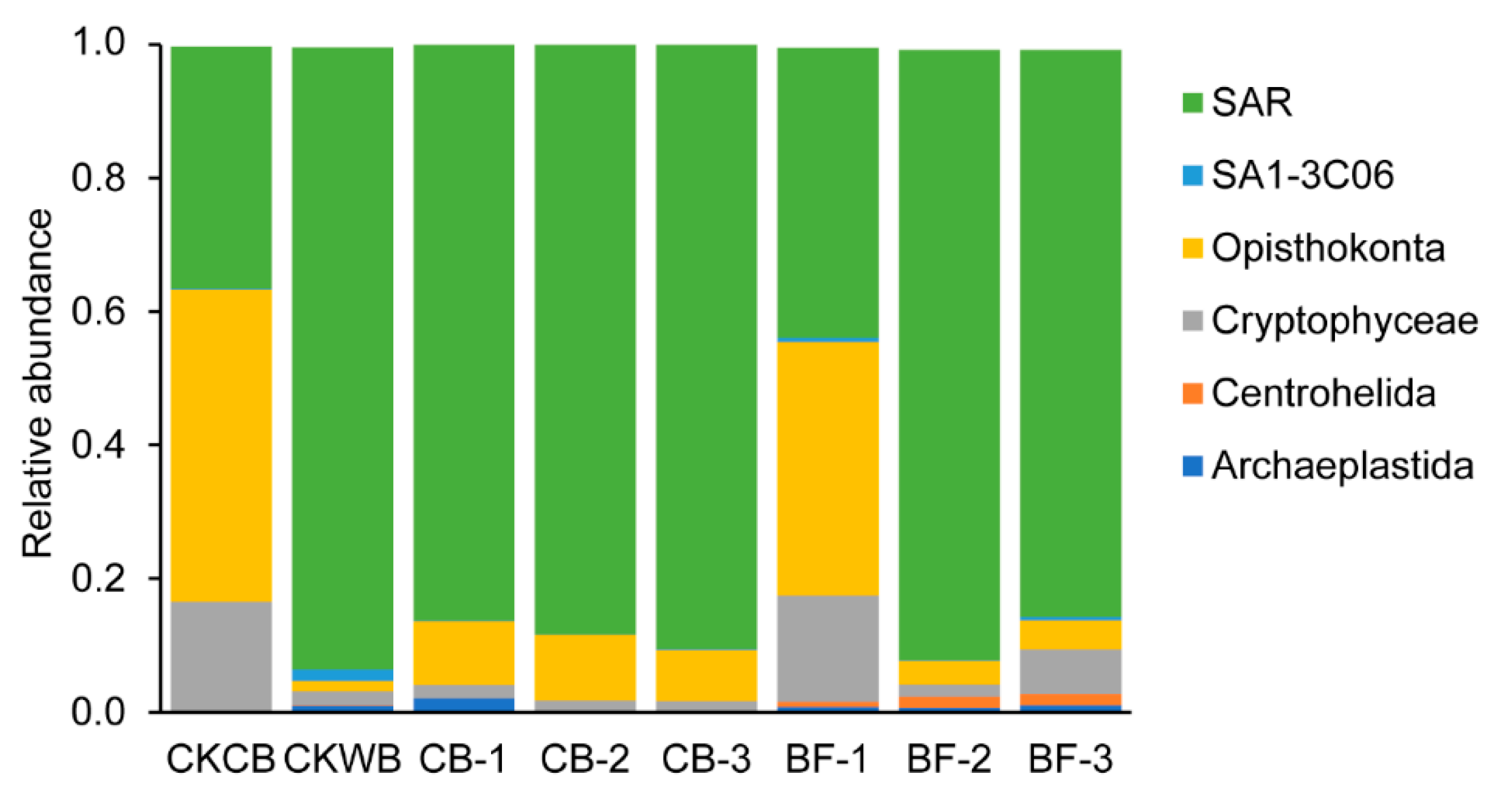
The relative abundance of eukaryotes at the phylum level
It can be seen from Figure 7 that the most abundant eukaryote was Opisthokonta at the first operation stage of the experimental reactor, achieving an abundance of 46.72%. In the latter stages, SAR became the most abundant eukaryote, at 88.36% (HRT 36 h), 86.19% (HRT 18 h), and 90.49% (HRT 11 h), respectively; however, the proportion of SAR in the control reactor declined. Studies have shown that protozoa caused flocs formation by excreting gelatinous mucus before ingesting the bacteria [42]. After the formation of the floc, ciliated protozoa become the main protozoan group, and the secretions during its predation further promote the accumulation of microorganisms in the sludge [43]. Hence, the abundance of SAR remained higher in the experimental reactor, which promoted the flocs’ formation and EPS synthesis.
(3)
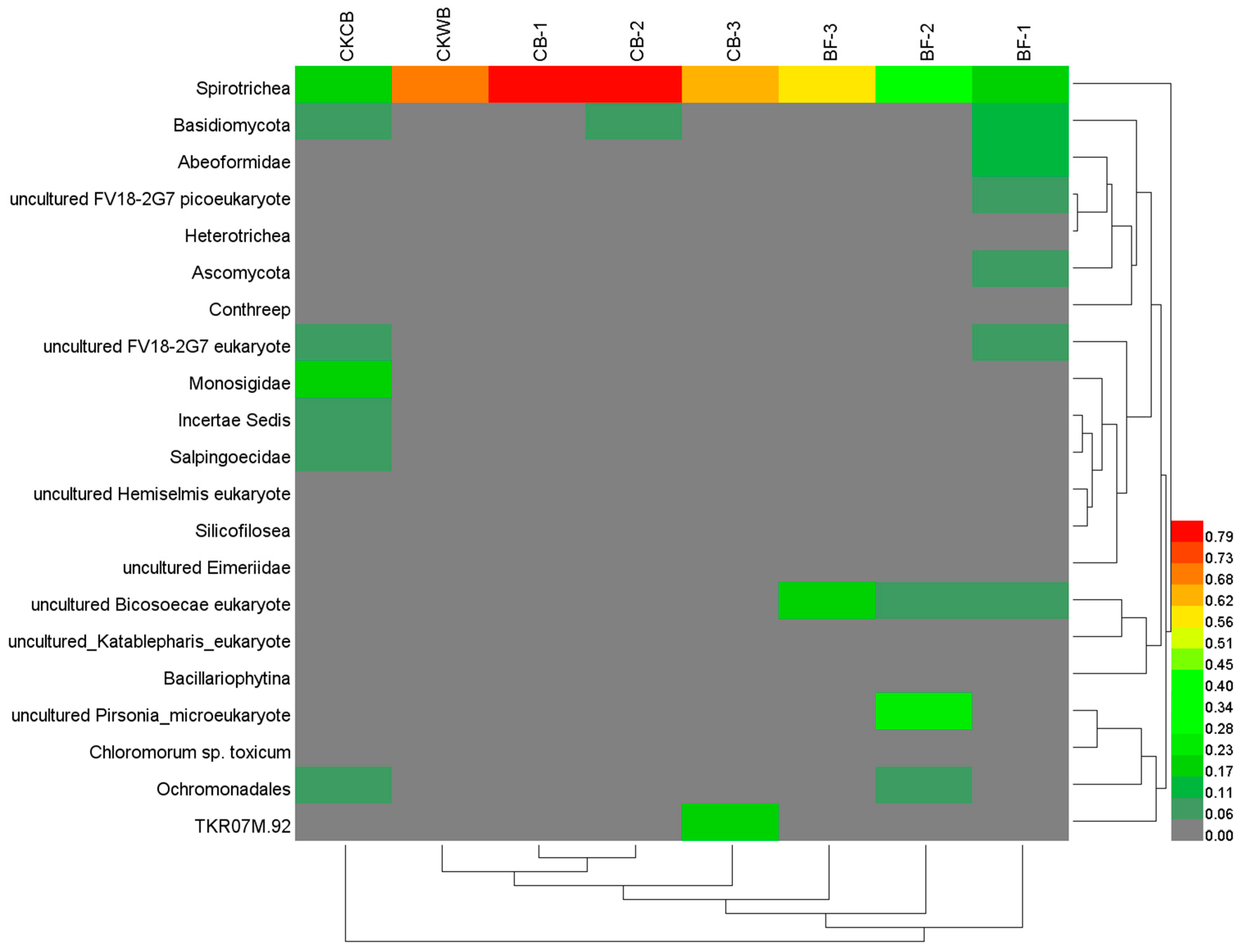
The relative abundance of eukaryotes at the genus level
It can be seen from Figure 8 that Spirotrichea was the most dominant group in both reactors, but their abundance changed with time. The abundance of Spirotrichea in the experimental reactor increased from 18.67% (inoculum) to 79.02% (HRT 36 h), 84.67% (HRT 18 h), and 62.26% (HRT 11 h), but decreased in the control reactor from 70.64% (inoculum) to 21.24% (HRT 36 h), 40.82% (HRT 18 h), and 56.42% (HRT 11 h). Spirotrichea belonging to Ciliophora is a complex and diverse group of ciliates. It is an indispensable part of the microfood ring and is the main contributor to the energy cycle. Its abundance in marine water was negatively correlated with the N and P contents [44,45,46]. Ciliates stimulate the formation and adhesion of bacteria colonies, which are efficient for nutrient uptake [47]; Ciliates appeared on the surface of aerobic granular sludge and secreted the viscous substances that can absorb suspended particles and bacteria, and the dead remains were used as the skeleton to form granular sludge [48]; therefore, the increase in the abundance of Spirotrichea in the experimental reactor promoted flocs’ formation, and thus improved the efficiency of water purification.

3. Materials and Methods

3.1. Experimental Materials

The Cordyceps strain C058 was donated by the Medicinal Fungi Research Group of the Institute of Microbiology, Guangdong Academy of Sciences. The formula of the C8 medium was referred to by Li et al. [22]. The formula of the simulated wastewater was referred to by Schryver and Verstraete [49]. The size and structure of the baffled reactor were described by Yang [20].

3.2. Experimental Methods

3.2.1. Culture and Collection of C058

The mycelium was taken from the C058 slope and inoculated into a C8 culture medium. After a large number of new mycelium balls were grown out at 28 °C, 150 rpm in a rotating incubator, they were collected by centrifugation under the condition of 2000 rpm, 10 min, and were washed three times with sterilized water. The washed mycelium balls were used for inoculation.

3.2.2. Operation of the Reactors

The experiment was carried out in two sets of identical baffled reactors. After filling with the simulated wastewater, one reactor, as an experimental reactor, was inoculated with 1% of C058 and 1% of Pearl River water (23°06′37.9″ N, 113°16′51.6″ E) to form mycelium biofloc (MBF), and the other reactor as a control reactor was only inoculated with 1% of Pearl River water to form OBF. Here, Pearl River is chosen as a typical natural water body that is rich in natural microflora. The reactors were operated at room temperature and contained above 5 mg/L of dissolved oxygen by continuous aeration.
After a consideration of the effect of HRT on the EPS content [26], the reactor was operated under three stages: HRT 36 h (v = 4.7 L/h), 18 h (v = 9.2 L/h) and 11 h (v = 15.3 L/h). The first operation stage was HRT 36 h. When the reactor became stable, sampling for latter index determination was carried out. Then, the influent velocity was changed to control the HRT of 18 h for a second operation stage; so did the third operation stage at HRT 11 h.

3.3. Analysis Method

3.3.1. Extraction and Determination of EPS

The EPS was extracted using the centrifugal heating method [50]. In this study, the main components of EPS, PN, and PS, were used to characterize EPS. The PN content was determined using Coomassie brilliant blue spectrophotometry, and the PS content was determined using phenol-sulfuric acid spectrophotometry [51].

3.3.2. Determination of SVI

Sludge settling velocity in 30 min (SV30) and mixed liquid suspended solids (MLSS) content were determined according to “Monitoring of Water and waste Water” [52], and SVI = SV30/MLSS.

3.3.3. SEM Analysis

Samples were dried (Tousimis Autosamdri-815, American) and sprayed with gold (EMS 150T, American) before the electron microscope scanning (Tungsten Filament SEM Q25, American). The specific operation was referred to by Shen et al. [53].

3.3.4. Analysis of Microbial Community Structure

(1)
The 16S rRNA sequence analysis
Total DNA was extracted using the soil strong DNA extraction kit (DNeasy PowerSoil Kit, QIAGEN, Hilden, Germany). The DNA was diluted to 10 ng/μL for PCR amplification. According to Tamaki [54], the V4-V5 hypervariable region of the 16S rRNA gene of the experimental sample was amplified by primers 515F(5′-GTGYCAGCMGCCGCGGTA-3′) and 909R (5′-CCCCGYCAATTCMTTTRAGT-3′). Reaction system: every 25 μL PCR reaction solution contains 1× PCR buffer, 1.5 mM MgCl2, 0.2 mM dNTP (Transgen, Beijing, China), 1.0 μM primers, 0.25U Ex Taq (TaKaRa, Beijing, China), and 10 ng DNA template. The PCR reaction procedure was pre-denatured at 94 °C for 3 min, then extended for 10 min at 72 °C after 30 cycles of conventional amplification (denatured at 94 °C for 40 s, annealing at 56 °C for 60 s, extension at 72 °C for 60 s). The same sample was amplified twice, and the two PCR products obtained from the same sample were mixed. After 1.2% agarose gel electrophoresis, the gel was cut and purified by sanPrep DNA gel recovery kit (Raw engineering, China). After all the purified DNA was mixed in the same amount, PE250 sequencing was carried out using the Illumina Miseq platform [55].
The original sequencing data were spliced using the FLASH1.2.8 software and screened using the QIIME1.9.0 software after splicing [29,56,57], using the Uchime program [58] to detect and remove chimera sequences, then using the QIIME1.9.0 software to divide OTUs (Operational Taxonomic Units) according to 97% similarity of sequences, and using the RDP classifier [46] to annotate each species.
(2)
The 18S rRNA sequence analysis
The 18S rRNA was extracted as 16s rRNA. According to Lejzerowicz et al. [59], the V4 hypervariable region of 18s rRNA gene of the experimental sample was amplified by primer TAReuk454WD1 (5′-CCAGCASCYGCGGTAATTCC-3′) and primer TAReukREV3 (5′-ACTTTCGTTCTTGATYRA-3′). PCR reaction system: every 25 μL PCR reaction solution contains 1× PCR buffer, 1.5 mM MgCl2, 0.2 mM dNTP (Transgen, China), 1.0 μM primers, 0.25 U TransFast Taq DNA polymerase (Transgen, China), and 10 ng DNA template. The PCR reaction procedure was the same as that of 16S rRNA. After the completion of the PCR reaction, two PCR products amplified from the same sample were mixed, and the gel was cut using 1.2% agarose gel electrophoresis and purified by the AxyPrep DNA gel recovery kit (Axygen, Hangzhou, China). After all the purified DNA was mixed in the same amount, PE250 sequencing was carried out using the Illumina Miseq platform [29].
The original sequencing data were analyzed as 16S rRNA.

3.4. Data Processing

Microsoft Excel 2019 was used for data processing and analysis, and Origin 2018 was used for drawing.

4. Conclusions

(1)
The EPS contents in MBF were 51.20 mg/g (HRT 36 h), 55.89 mg/g (HRT 18 h), and 33.84 mg/g (HRT 11 h), respectively, higher than the EPS content of OBF under the corresponding HRTs. PN content or PS content also followed the same trend.
(2)
The sedimentation performance of MBF was better than that of OBF, attributed to higher EPS PN contents.
(3)
MBF was constructed by fungi and bacteria together, and C058 was the main component, promoting the synthesis of EPS.
(4)
Compared with OBF, MBF bioaugmented by Cordyceps strain C058 had higher diversity and abundance of microorganisms, realizing a more stable operation of the experimental reactor. More importantly, C058 promoted the growth of some functional bacteria, including Janthinobacterium belonging to Proteobacteria, Phormidium and Leptolyngbya belonging to Cyanobacteria, and Hymenobacter belonging to Bacteroides, which participate in nitrogen fixation and PN and PS production, promoting the EPS synthesis. In addition, C058 also promoted the growth of Spirotrichea belonging to Ciliophora, which benefited floc formation and enhanced the water purification.

5. Patents

Chinese patent: A MPBR reactor suitable for sewage purification, CN212833060U. Applicant: Zhongkai University of Agriculture and Engineering. Inventor: Yiyong Li, Yongcong Yang, Kangchun Peng, Baoe Wang, Xueqin Tao, Chong Lin, Zexiang Lei, Jianjun Du.

Author Contributions

Conceptualization, Y.L. and B.W.; methodology, Y.L. and Y.Y.; software, W.L. (Wanyi Luo); validation, W.L. (Wen Liu), Z.L., X.T. and B.W.; formal analysis, W.L. (Wanyi Luo); data curation, B.W.; writing—original draft preparation, W.L. (Wanyi Luo); writing—review and editing, Y.L.; supervision, B.W.; funding acquisition, Y.L. All authors have read and agreed to the published version of the manuscript.

Funding

This research was funded by the Science and Technology Planning Project of Guangzhou, China, grant numbers 201803030039, 201704020187, and B318221315.

Institutional Review Board Statement

Not applicable.

Informed Consent Statement

Not applicable.

Data Availability Statement

The data presented in this study are available on request from the corresponding author. The data are not publicly available due to confidentiality issues.

Acknowledgments

The authors would like to thank the Medicinal Fungi Research Group, Institute of Microbiology, Guangdong Academy of Sciences for the donation of Cordyceps sp. C058 strain. The authors would also like to thank Guangdong Meilikang Biotechnology Co., Ltd. for the sequencing process and all the staff who contributed to this experiment.

Conflicts of Interest

The authors declare no conflict of interest.

References

  1. Wang, Z.C.; Gao, M.C.; Wei, J.F.; Yang, Y.S.; Zhang, J.; Yu, S.P.; Wang, Y.J. Effect of salinity change on extracellular polymer of anaerobic sludge. Acta Sci. Circumst. 2016, 36, 3273–3281. [Google Scholar]
  2. Sheng, G.P.; Yu, H.Q.; Li, X.Y. Extracellular polymeric substances (EPS) of microbial aggregates in biological wastewater treatment systems: A review. Biotechnol. Adv. 2010, 28, 882–894. [Google Scholar] [CrossRef]
  3. More, T.; Yadav, J.; Yan, S.; Tyagi, R.; Surampalli, R. Extracellular polymeric substances of bacteria and their potential environmental applications. J. Environ. Manag. 2014, 144, 1–25. [Google Scholar] [CrossRef]
  4. Dignac, M.; Urbain, V.; Rybacki, D.; Bruchet, A.; Snidaro, D.; Scribe, P. Chemical description of extracellular polymers: Implication on activated sludge floc structure. Water Sci. Technol. 1998, 38, 45–53. [Google Scholar] [CrossRef]
  5. Jagaba, A.H.; Kutty, S.; Noor, A.; Haruna Birniwa, A.; Affam, A.; Lawal, I.M.; Usman Kankia, M.; Usman Kilaco, A. A systematic literature review of biocarriers: Central elements for biofilm formation, organic and nutrients removal in sequencing batch biofilm reactor. J. Water Process Eng. 2021, 42, 102178. [Google Scholar] [CrossRef]
  6. Wang, Z.C.; Gao, M.; Wang, S.; Xin, Y.J.; Ma, D.; She, Z.L.; Wang, Z.; Chang, Q.B.; Ren, Y. Effect of hexavalent chromium on extracellular polymeric substances of granular sludge from an aerobic granular sequencing batch reactor. Chem. Eng. J. 2014, 251, 165–174. [Google Scholar] [CrossRef]
  7. Basuvaraj, M.; Fein, J.; Liss, S.N. Protein and polysaccharide content of tightly and loosely bound extracellular polymeric substances and the development of a granular activated sludge floc. Water Res. 2015, 82, 104–117. [Google Scholar] [CrossRef]
  8. Zhu, N.; Liu, L.; Xu, Q.; Chen, G.W.; Wang, G. Resources availability mediated EPS production regulate microbial cluster formation in activated sludge system. Chem. Eng. J. 2015, 279, 129–135. [Google Scholar] [CrossRef]
  9. Ray, A.J.; Seaborn, G.; Leffler, J.W.; Wilde, S.B.; Lawson, A.; Browdy, C.L. Characterization of microbial communities in minimal-exchange, intensive aquaculture systems and the effects of suspended solids management. Aquaculture 2010, 310, 130–138. [Google Scholar] [CrossRef]
  10. Zhao, D.H.; Pan, L.Q.; Wang, C. Cleaning effect of biofloc on culture environment and effects of physiological indexes of Penaeus vannamei. Trans. Oceanol. Limnol. 2014, 67–73. [Google Scholar]
  11. Jagaba, A.H.; Kutty, S.R.M.; Noor, A.; Affam, A.C.; Ghfar, A.A.; Usman, A.K.; Lawal, I.M.; Birniwa, A.H.; Kankia, M.U.; Afolabi, H.K.; et al. Parametric optimization and kinetic modelling for organic matter removal from agro-waste derived paper packaging biorefinery wastewater. Biomass Convers. Biorefin. 2022, 1–18. [Google Scholar] [CrossRef]
  12. Zhang, H.; Wang, X.; Li, C. Isolation and identification of a bacillus sp. strain and its role in bioflocs for the shrimp culture system. Prog. Fish. Sci. 2016, 37, 111–118. [Google Scholar]
  13. Gao, G.; Zhu, K.L.; Zhang, Q.Q.; Wang, Z.J.; Huang, J. Simplified fermentation of a functional probiotics and the application in prawn (Litopenaeus vannamei) bio-floc breeding. Prog. Fish. Sci. 2017, 38, 140–147. [Google Scholar]
  14. Wang, S.H.; Tan, H.X.; Luo, G.Z.; Sun, D.C. Effects of temperature, pH and dissolved oxygen on the construction of biofloc system by Lactobacillus plantarum. Genom. Appl. Biol. 2020, 39, 2628–2637. [Google Scholar]
  15. Baldrian, P. Interactions of heavy metals with white-rot fungi. Enzyme Microb. Technol. 2003, 32, 78–91. [Google Scholar] [CrossRef]
  16. Crini, G. Non-conventional low-cost adsorbents for dye removal: A review. Bioresour. Technol. 2006, 97, 1061–1085. [Google Scholar] [CrossRef]
  17. Zhao, L.H.; Jin, R.F.; Sun, H.J.; Gao, Y.J. Study on treatment of cotton pulp black liquor by white rot fungi and activated sludge. Ecol. Environ. 2009, 18, 28–31. [Google Scholar]
  18. Wang, L.; Chen, G.Q.; Zeng, G.M.; Zhang, W.Q.; Chen, Y. Research progress on extracellular polymers of fungi and their interaction with heavy Metals. Environ. Pollut. Control 2010, 32, 74–80. [Google Scholar]
  19. Li, Y.; Yang, Y.C.; Wang, B.E.; Lin, C.; Tao, X.Q.; Du, J.J. Optimization and effect of purification of aquaculture wastewater by Cordyceps sinensis. Adv. Environ. Prot. 2020, 10, 10. [Google Scholar]
  20. Yang, Y.C. Purification Effect and Mechanism of Cordyceps Sinensis on Aquaculture Wastewater; Zhongkai University of Agriculture and Engineering: Guangzhou, China, 2020. [Google Scholar]
  21. Zhang, R.Y. Occurrence Regularity of Mycelium Pellets in Activated Sludge System and Its Bioaugmentation Application; Hebei University of Engineering: Handan, China, 2020. [Google Scholar]
  22. Li, Y.; Chen, Y.C.; Li, D.L.; Zhang, Y.X.; Zhang, W.M. Study on antibacterial and antitumor activity of several fermented extracts of Cordyceps sinensis. Sci. Technol. Food Ind. 2010, 31, 88–90. [Google Scholar]
  23. Zhou, X.; Huang, M.Q.; Qiu, R.; Li, Z.Q.; Guo, S.H.; Cui, Y.H. Research progress on composition, production and application of Cordyceps sinensis. Priv. Sci. Technol. 2018, 98–99. [Google Scholar]
  24. Morgan, J.W.; Forster, C.F.; Evison, L. A comparative study of the nature of biopolymers extracted from anaerobic and activated sludges. Water Res. 1990, 24, 743–750. [Google Scholar] [CrossRef]
  25. Ge, L.Y.; Wang, H.W.; Ma, L.M.; Deng, H.H.; Liu, Y. Study on influencing factors of extracellular polymers in aerobic activated sludge. Environ. Sci. Technol. 2007, 02, 8–9. [Google Scholar]
  26. Gu, Y.N. Study on the Efficiency and Stability of Aerobic Granular Sludge in the Treatment of Domestic Wastewater; Harbin Institute of Technology: Harbin, China, 2020. [Google Scholar]
  27. Zhang, X.; Zhang, Q.S.; Xu, Y.; Liu, X. Study on sedimentation and dewatering performance of activated sludge from a sewage treatment plant. Environ. Prot. Sci. 2011, 37, 20–22. [Google Scholar]
  28. Zhang, L.H.; Li, J.; Guo, J.B.; Jia, Y.P.; Zhang, H.F. Effect of EPS on flocculation settling performance and surface properties of activated sludge. J. Chem. Ind. Eng. 2012, 63, 1865–1871. [Google Scholar]
  29. Li, H.; Qu, J.; Li, T.; Li, j.; Lin, Q.; Li, X. Pika population density is associated with the composition and diversity of gut microbiota. Front. Microbiol. 2016, 7, 758. [Google Scholar] [CrossRef]
  30. Tsuneda, S.; Jung, J.; Hayashi, H.; Aikawa, H.; Hirata, A.; Sasaki, H. Influence of extracellular polymers on electrokinetic properties of heterotrophic bacterial cells examined by soft particle electrophoresis theory. Colloids Surf. B Biointerfaces 2003, 29, 181–188. [Google Scholar] [CrossRef]
  31. Satoshi, T.; Hirotoshi, A.; Hiroshi, H.; Atsushi, Y.; Akira, H. Extracellular polymeric substances responsible for bacterial adhesion onto solid surface. FEMS Microbiol. Lett. 2010, 223, 287–292. [Google Scholar]
  32. Cardona, E.; Gueguen, Y.; Magré, K.; Lorgeoux, B.; Piquemal, D.; Pierrat, F.; Noguier, F.; Saulnier, D. Bacterial community characterization of water and intestine of the shrimp Litopenaeus stylirostris in a biofloc system. BMC Microbiol. 2016, 16, 157. [Google Scholar] [CrossRef]
  33. Addo, F.G.; Zhang, S.; Manirakiza, B.; Ohore, E.O.; Yuan, S.D. The impacts of straw substrate on biofloc formation, bacterial community and nutrient removal in shrimp ponds. Bioresour. Technol. 2021, 326, 124727. [Google Scholar] [CrossRef]
  34. Yang, C.; Wei, Z.; Liu, R.; LI, Q.; Li, B.; Wang, S.; Song, C.; Qiao, C.; Aulchandani, A. Phylogenetic diversity and metabolic potential of activated sludge microbial communities in full-scale wastewater treatment plants. Environ. Sci. Technol. 2011, 45, 7408–7415. [Google Scholar] [CrossRef] [PubMed]
  35. Lee, I.S.; Parameswaran, P.; Rittmann, B.E. Effects of solids retention time on methanogenesis in anaerobic digestion of thickened mixed sludge. Bioresour. Technol. 2011, 102, 10266–10272. [Google Scholar] [CrossRef]
  36. Yang, M.; Lu, D.W.; Qin, B.D.; Liu, Q.L.; Zhao, Y.M.; Liu, H.L.; Ma, J. Highly efficient nitrogen removal of a coldness-resistant and low nutrient needed bacterium, Janthinobacterium sp M-11. Bioresour. Technol. 2018, 256, 366–373. [Google Scholar] [CrossRef] [PubMed]
  37. Ma, J.; Bei, Q.C.; Wang, X.J.; Lan, P.; Liu, G.; Lin, X.W.; Liu, Q.; Lin, Z.B.; Liu, B.J.; Zhang, Y.H.; et al. Impacts of Mo application on biological nitrogen fixation and diazotrophic communities in a flooded rice-soil system. Sci. Total Environ. 2019, 649, 686–694. [Google Scholar] [CrossRef] [PubMed]
  38. Bar-Or, Y.; Shilo, M. Characterization of macromolecular flocculants produced by Phormidium sp. strain J-1 and by Anabaenopsis circularis PCC 6720. Appl. Environ. Microbiol. 1987, 53, 2226–2230. [Google Scholar] [CrossRef] [Green Version]
  39. Kim, J.H.; Choi, W.; Jeon, S.M.; Kim, T.; Park, A.; Kim, J.; Heo, S.J.; Oh, C.; Shim, W.B.; Kang, D.H. Isolation and characterization of Leptolyngbya sp. KIOST-1, a basophilic and euryhaline filamentous cyanobacterium from an open paddle-wheel raceway Arthrospira culture pond in Korea. J. Appl. Microbiol. 2015, 119, 1597–1612. [Google Scholar] [CrossRef] [Green Version]
  40. Kim, J.H.; Kang, D.H. Draft genome sequence of Leptolyngbya sp. KIOST-1, a filamentous cyanobacterium with biotechnological potential for alimentary purposes. Genome Announc. 2016, 4, e00984-16. [Google Scholar] [CrossRef] [Green Version]
  41. Lu, X.P.; Tang, K.; Li, H.; Cheng, Y.L.; Yang, S.S.; Guo, H.L.; Guo, H.Q.; Yun, X.Y.; Feng, F.Y. Isolation and Identification of thin layer strain L28 and its promoting effect on the growth of Potato seedlings. Microbiol. China 2020, 47, 4050–4058. [Google Scholar]
  42. Tenney, M.W.; Stumm, W. Chemical flocculation of microorganisms in biological waste treatment. J. Water Pollut. Control Fed. 1965, 37, 1370–1388. [Google Scholar]
  43. Chen, Y.H. Composition and Function Analysis of Extracellular Polymers during Activated Sludge Aggregation; Chongqing University: Chongqing, China, 2019. [Google Scholar]
  44. Andersen, P. The quantitative importance of the microbial loop in the marine pelagic: A case study from the North Bering/Chukchi seas. Arch. Hydrobiol. Beih. 1988, 31, 243–251. [Google Scholar]
  45. Nielsen, T.G.; Hansen, B. Plankton community structure and carbon cycling on the western coast of Greenland during and after the sedimentation of a diatom bloom. Mar. Ecol. Prog. Ser. 1995, 125, 239–257. [Google Scholar] [CrossRef]
  46. Wang, Q.; Garrity, G.M.; Tiedje, J.M.; Cole, J.R. Naïve Bayesian classifier for rapid assignment of rRNA sequences into the new bacterial taxonomy. Appl. Environ. Microbiol. 2007, 73, 5261–5267. [Google Scholar] [CrossRef] [PubMed] [Green Version]
  47. Weerman, E.J.; Van Der Geest, H.G.; Van Der Meulen, M.D.; Manders, E.M.M.; Koppel, J.V.D.; Herman, P.M.J.; Admiraal, A.W. Ciliates as engineers of phototrophic biofilms. Freshw. Biol. 2011, 56, 1358–1369. [Google Scholar] [CrossRef]
  48. Weber, S.D.; Ludwig, W.; Schleifer, K.H.; Fried, J. Microbial composition and structure of aerobic granular sewage biofilms. Appl. Environ. Microbiol. 2007, 73, 6233–6240. [Google Scholar] [CrossRef] [Green Version]
  49. Schryver, P.D.; Verstraete, W. Nitrogen removal from aquaculture pond water by heterotrophic nitrogen assimilation in lab-scale sequencing batch reactors. Bioresour. Technol. 2009, 99, 1162–1167. [Google Scholar] [CrossRef]
  50. Zhang, X.; Bishop, P.L.; Kinkle, B.K. Comparison of extraction methods for quantifying extracellular polymers in biofilms. Water Sci. Technol. 1999, 39, 211–218. [Google Scholar] [CrossRef]
  51. Bo, F.; Palmgren, R.; Keiding, K.; Nielsen, P.H. Extraction of extracellular polymers from activated sludge using a cation exchange resin. Water Res. 1996, 30, 1749–1758. [Google Scholar]
  52. Eaton, A.D.; Clesceri, L.S.; Greenberg, A.E.; Franson, M.A.H. Standard methods for the examination of water and wastewater. Am. J. Public Health Nations Health 1966, 56, 387–388. [Google Scholar]
  53. Shen, P.; Chen, X.D. Microbiology Experiment; Higher Education Press: Beijing, China, 2007. [Google Scholar]
  54. Tamaki, H.; Wright, C.L.; Li, X.; Lin, Q.; Hwang, C.; Wang, S.; Thimmapuram, J.; Kamagata, Y.; Liu, W.T. Analysis of 16S rRNA amplicon sequencing options on the Roche/454 Next-Generation Titanium Sequencing Platform. PLoS ONE 2011, 6, e25263. [Google Scholar] [CrossRef]
  55. Caporaso, J.G.; Kuczynski, J.; Stombaugh, J.; Bittinger, K.; Bushman, F.D.; Costello, E.K.; Fierer, N.; Peña, A.G.; Goodrich, J.K.; Gordon, J.I.; et al. QIIME allows analysis of high-throughput community sequencing data. Nat. Methods 2010, 7, 335–336. [Google Scholar] [CrossRef] [Green Version]
  56. Li, S.; Peng, Y.Z.; Wang, R.D. The role of extracellular polymers substance in biological treatment of wastewater. J. Nat. Sci. Heilongjiang Univ. 2016, 33, 515–520. [Google Scholar]
  57. Ni, J.J.; Li, X.J.; He, Z.L.; Xu, M.Y. A novel method to determine the minimum number of sequences required for reliable microbial community analysis. J. Microbiol. Methods 2017, 139, 196–201. [Google Scholar] [CrossRef] [PubMed]
  58. Edgar, R.C.; Haas, B.J.; Clemente, J.C.; Quince, C.; Knight, R. UCHIME improves sensitivity and speed of chimera detection. Bioinformatics 2011, 27, 2194–2200. [Google Scholar] [CrossRef] [Green Version]
  59. Lejzerowicz, F.; Esling, P.; Pillet, L.; Wilding, T.A.; Black, K.D.; Pawlowski, J. High-throughput sequencing and morphology perform equally well for benthic monitoring of marine ecosystems. Sci. Rep. 2015, 5, 13932. [Google Scholar] [CrossRef] [PubMed] [Green Version]
Figure 1. Contents of EPS and its components in biofloc from the two reactors under various HRTs.
Catalysts 12 00693 g001
Figure 2. SVI changes of biofloc in both reactors under various HRTs.
Catalysts 12 00693 g002
Figure 3. The morphological structure of Cordyceps strain C058.
Catalysts 12 00693 g003
Figure 4. SEM of biofloc from both reactors under various HRTs.
Catalysts 12 00693 g004
Figure 5. Composition of dominant prokaryote phyla.
Catalysts 12 00693 g005
Figure 6. Heatmap diagram for the dominant prokaryote genera.
Catalysts 12 00693 g006
Figure 7. Composition of dominant eukaryote phyla.
Catalysts 12 00693 g007
Figure 8. Heatmap diagram for the dominant eukaryote genera.
Catalysts 12 00693 g008
Table 1. The alpha diversity index of biofloc from both reactors based on the 16S rRNA sequences.
Chao1ShannonSimpson
CKCB1701.596.160.969
CB-12868.928.420.988
CB-22720.188.270.987
CB-31906.676.160.942
CKWB2477.067.800.988
BF-12242.127.820.981
BF-22059.896.670.915
BF-31489.733.970.745
Table 2. The alpha diversity index of biofloc from both reactors based on the 18S rRNA sequences.
Chao1ShannonSimpson
CKCB290.254.430.92
CB-1387.692.940.63
CB-2259.122.210.52
CB-3393.453.170.75
CKWB492.114.060.87
BF-1481.274.930.93
BF-2372.074.160.88
BF-3353.573.750.86
Publisher’s Note: MDPI stays neutral with regard to jurisdictional claims in published maps and institutional affiliations.

