Review

The Recent Advances in Bulk and Microfluidic-Based pH Sensing and Its Applications

Department of Chemistry, Xi’an Jiaotong-Liverpool University, No. 111 Ren Ai Road, Suzhou Industrial Park, Suzhou 215123, China
*
Author to whom correspondence should be addressed.
Catalysts 2022, 12(10), 1124; https://doi.org/10.3390/catal12101124
Submission received: 18 August 2022 / Revised: 13 September 2022 / Accepted: 16 September 2022 / Published: 27 September 2022

Abstract

:
The determination of pH is of paramount importance in environmental, pharmaceutical, and medicinal sciences, etc., for accurate controlling, monitoring, and adjusting whole processes on microscale and macroscale. Therefore, the pH measurements have drawn continuous efforts from a great deal of research. The bulk pH determination can tackle part of the demands from laboratories and industrial applications. However, more and more studies have started to pay more attention to microfluidic-based pH sensing by integrating with metal oxides and solid-state-based pH sensing applications. This review paper focuses on the recent development of pH sensing, the mechanisms of pH sensing, a few common pH sensors, and microfluidic-based pH determinations from the aspects of fabrication techniques to the various applications in biology, environmental study, and food safety. The future trends of pH sensing, as well as microfluidic-based pH sensing, were discussed as well at the end of this review.

1. Introduction

1.1. Background

The determination of pH is of great significance in various types of inspections such as chemistry, biology, agriculture, architecture, and so on [1]. Since the first research on pH was announced in 1914 and the main role of the hydrogen ion concentration and its determination of H+ in biology had been established [2], there has been a great historical development in pH measurement and different specific methods of determination have been used depending on diverse needs (different equipment, samples, analytically time or locations [3]) in the past few decade’s years. In this case, pH measurements, the universal quality analysis, have been widely used in both laboratory level and industry because of reasonable prices, easy operations, efficiency, and so on. In the following parts, the definition, history, specific methods, and applications of pH are included.

1.2. Definition and History of pH

In terms of Petra Spitzer and Kenneth W. Pratt’s research [4], lots of scientists [5,6] had studied and developed various ionic theories to define what the pH is. In the 19th century, Arrhenius et al. [7] revealed the ionic theory, which offered the basic correlation between hydroxyl and hydrogen ions and leads to further study of the dissociation constant of water in the following years. After a few years, Friedenthal et al. [8] was the man who firstly published a range of acidity by examining the color changes of indicator dyes in suitable hydrogen ion concentrations from 14 prepared known hydrogen concentration solutions. With the help of the prior theories and published data, Sørensen et al. [4] published the concept of pH on a scale from 0 to 14 in the environment at 25 °C according to the ionic product of water, which simplified the measurements of hydrogen ion concentration. In the next few years, with the concept of activity (see Equation (1) below), the theory of interionic interaction published by Debye and Hückel [9], Sørensen and K. Linderstrøm-Lang [10] announced the concept of pH which is widely acknowledged today:
pH = −log aH = −log(mHγH/m0).
In Equation (1), aH is the molality-based activity; γH is the molal activity coefficient of the H+ at the molality (mH); m0 means the standard state, which is selected to the 1 mol kg−1. Although this equation can be used to measure the activity of hydrogen ions in solution, it is stated by a quantity that is unable to be determined based on thermodynamical methods. Therefore, this concept and equation of pH cannot ensure the accuracy of sample determinations and new equations needs to be introduced and relates to improve the results of measurement and get closer to the definition of pH.
Based on the concept of pH by Sørensen, more and more studies and research have been developed. By characterizing various standard buffer solutions with selected pH values several pH scales have been established [11]. Additionally, Bates et al. [12] had specially provided great progress since 1948, he mentioned that “several scales, all dissemblance under the name pH, are in mutual use. A number of scientists studied based on one concept and determine a different amount.” After decades, Bates and his workmates at the National Bureau of Standards (NBS) published relevant research on the basics of buffer solutions and appropriate electrochemical cells and established the concept of multipoint pH protocol [13]. As for the multipoint pH protocol, every pH standard is a dilute liquid solution with a specific ionic strength. This new definition was recommended and included in 1979 by the International Union of Pure and Applied Chemistry(IUPAC) [14] and IUPAC Recommendation also published a concept of pH that depended on an unknown solution—pH(X), in a cell including hydrogen and a suitable electrode with a liquid junction which was relevant to one standard solution—pH(S) in Equation (2):
pH ( X ) = pH ( S ) + E S E X ] F R T   l n 10
According to Equation (2), the quantities E(S) and E(X) mean the standard solution and the unknown sample constitute the potentials of this cell, and pH(X) and pH(S) indicates the pH of the unknown sample liquid and the standard solution, respectively. The quantities R, T, and F are the gas constant, thermodynamic temperature, and the Faraday constant, respectively. The quantity RT ln10/F is often considered as the thermodynamic or Nernstian slope, k. This equation had already addressed the problems of low accuracy but there is one major defect—the contribution from the liquid junction potential (LJP) is excluded, which may lead to liquid contact between solution S or X and the reference electrode and is a complicated function of solution composition and other factors. A basic source of deviation in operational measurements of pH is the difference in residual LJP between solution S and solution X. So, the pH value is a number and in a good approximation, this kind of bias is permissible to displace the hydrogen electrodes in both cells with other hydrogen ion responsive electrodes, such as glass or quinhydrone [14].
The potentials of standard solutions and samples can be studied by the potentiometric analysis including direct potential titration and potentiometric titration. The study of potentiometric analysis, especially for the measurement of hydrogen ions and pH values, is firstly announced in the early 1900s in the first half of the 20th century, which can be traced back to the work of Michaelis [8], and Kolthoff and Furman [15]. Compared to direct potential titration, potentiometric titration is more useful because of the reasonable device design and high precision result in many fields (such as ocean and sediment pore water) [16].
In the following time, the new concept and theory of pH continued to bloom. In the 1980s, the British Standard Organization (BSI) presented the single-standard scale [17] and the result of pH measurement meant a stable reference point. The pH values of other solutions in the BSI protocol are defined by operational definitions and are implemented in cells with liquid contacts of specified geometry. However, these two concepts of pH only were acknowledged in a short time and other research and theory of pH scale recommendations [18] were published in a failure.
In 1985, IUPAC presented a recommendation, “Concepts of pH, Normal Reference Values, Determination of pH and Relevant terminology [19], which was modified as a compromise proposal that agreed with the effectiveness of the NBS and BSI methods but this situation is very ungratified. Two possible pH values for each unknown solution can be obtained by applying two pH scales in parallel. As more and more studies of pH included various types of recommendations and definitions had been developed and uncovered by F. G. K. Baucke’s critical analysis [20], the shortcomings of the IUPAC in 1985 have been serious. Therefore, the working party—the DIN (Deutsches Institut für Normung e.V.) Committee Technical pH measurement had extensively deliberated with IUPAC and the new document was published and replaced the old one in 2002. The “dual pH scale” in the 1985 file was canceled and the basis for the main measurement of pH was formed depending on different needs.

1.3. Applications

According to the above introduction, it is noticeable that pH determinations are critical in quality analyses in the laboratory, especially the pH determination in aqueous solution. There are a few major aspects introduced in the following sections.

1.3.1. Environmental Determination

As the soil pH is one of the typical indexes in environmental analysis, there are lots of various determinations to measure pH to ensure whether the soil is polluted. The pH of the soil, the strength index of soil pH, is the basic chemical properties of soil. With the benefits of easy measurement, the determination of pH is always used as an important reference for multiple parameters of water quality. Xu and Dong et al. [21] used microelectrode array (MEA) based on inkjet printing technology (IPT) to fabricate real-time sensors in soil and water quality detection along with the pH sensing. The design of this in situ sensing is shown in Figure 1. Meanwhile, pH measurement of soil [22], one of the series of physical and chemical analysis, not only relates to many other projects of analysis methods and analysis but also its results are considered as a basis for inspection of other quality analysis.

1.3.2. Pharmacy

The pH is an important index in the quality standard of drugs to man’s plasma proteins [23], which affects the stability and safety of products. Many factors affect pH, including water source, container, buffer preparation, temperature, and testing methods, etc. [24], involving biological products from feeding production to transportation and other links. In this case, the control of pH is beneficial to improve the safety of drug preparation and standardize drug production [25]. Wang and Zhang [26] studied that the pH-sensitive nanoparticles could be a protection of oral protein drugs to improve these drugs’ bioavailability.

1.3.3. Medical Science

In recent years, with the rapid development of science and technology, there has been great progress in the medical field. Many remarkable curatives of safe medicine have gradually been invented. In drug production, the solid preparation of drugs is easier to store and deliver. Therefore, most drugs are made from solid preparation, micro ring, and solid preparation environments, which have a great influence on all aspects of the performance of solid preparations. To ensure the quality of drugs, solid preparations can be applied for storage in the most suitable microenvironment. People often use the pH detection method to determine the microenvironment of solid preparations so that the solid preparations can have a better preservation [27].

1.3.4. Biological Analysis

As for organisms, the specific pH value is an essential assessment for the metabolic process of many cells, and cell dysfunction is always related to abnormal pH values in organelles [28,29]. The slight drop or raise of pH in cells may lead to improper growth and cleavage of normal cells. Therefore, testing the pH value of living cells in living organisms is of great significance in the diagnosis and treatment of diseases [30,31,32,33]. In this case, there are lots of studies related to the pH measurements for different types of cells. Nie et al. [34] used the carbon dots as a pH sensor to measure the cancer diagnosis and intracellular pH values (shown in Figure 2). In addition, pH changes in the skin surface affected by using two different pH values of synthetic detergent were studied to figure out suitable skin care [35].

1.3.5. Chemical Analysis

Determining pH values seems to be an extensive use of chemical analysis as the introduction of pH sensing in other fields (medical, pharmacy, biology, etc.), can be considered as specific employment in chemistry. There are still some specific studies related to some other chemical issues. Constant pH molecular dynamics (pHMD), an emerging approach over the last decade, was developed for more precise pH influence on chemical reactions [36]. These references became a key to studying the mechanism of pH-dependent conformational processes in detail. A representative application is to identify the mechanism of acid or base catalysis based on the specific protonation states in different pH values [37].
In conclusion, pH determination is of great importance in different aspects such as environmental determination, pharmacy, medical science, biological analysis, and chemical analysis, etc. Meanwhile, the specific methods of pH measurement not only depend on different types of science but also may change in microscale and bulk scale. In this case, the most correct pH measurements are selected by diverse kinds of applications and the precision of request of experiment results.

2. The Overview of Mechanism of pH Sensing

As pH is considered a single ion quantity, it cannot be determined by a basic unit of any measurement system, it is difficult to achieve an accurate basis for traceability by pH determining. In this case, the definition of “primary method of measurement” is established and considered as the standard to ensure whether the determination is fulfilled to be satisfied, which leads to one of the parts of SI [38] (International System of Units).

2.1. Specific pH Measurement

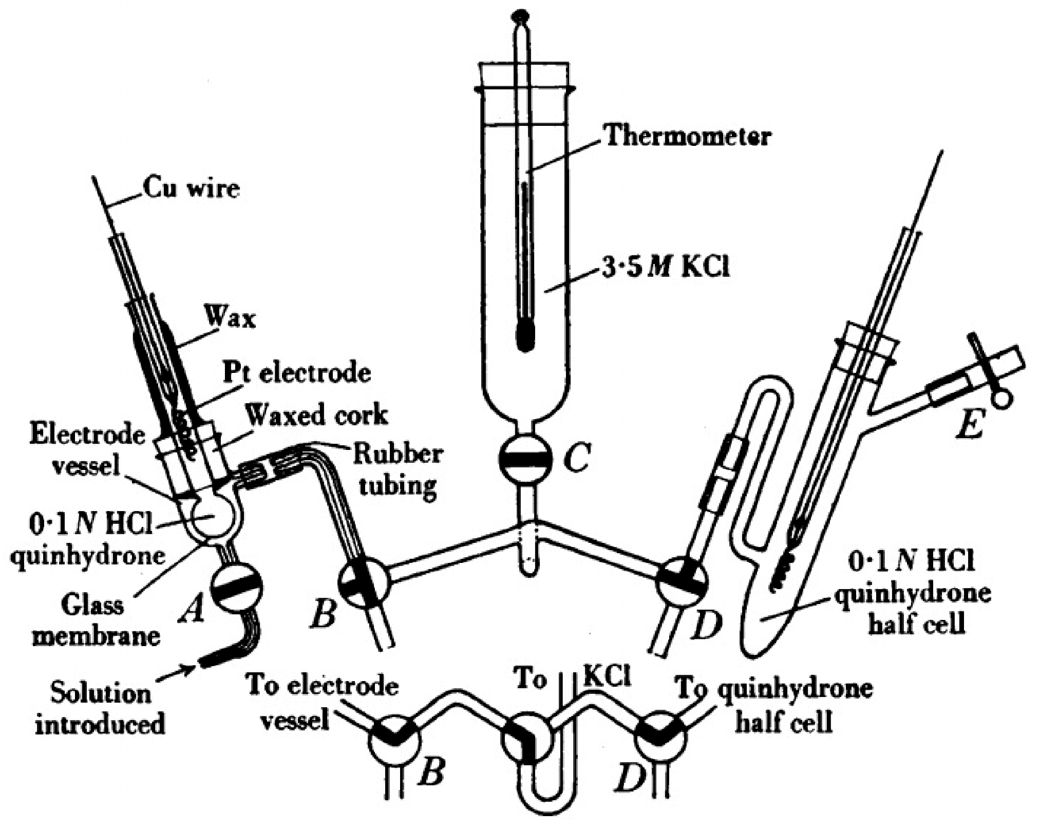
2.1.1. Cell for a Primary Method Measuring pH

Based on cell I (consisting of a platinum hydrogen gas electrode, a chosen buffer solution, and a silver/silver chloride reference electrode [39]), a new cell called the Harned cell is developed by Harned et al. [40] to measure the acidity of the sample in the 1930s. This cell was developed at the NBS (National Bureau of Standards, USA, now NIST) by Bates [41] and Hamer [42] to ensure the accuracy of pH determination in the next decades.
Adding the chloride ions to the chloride-free buffer solution at some different Cl molarities to keep the balance into the potential of the Ag/AgCl electrode. With the help of the Harned cell, it can solve the problem of the LJP and connection so that the cell potential only includes the disparity in the two electrode potentials.
To develop a battery without a liquid junction, it is certain to follow a convention that the single ion activity consisted of Equation (1) above. In this case, the leading method for pH based on this aim, the Bates–Guggenheim convention [43] is used and can be related to the Debye–Hückel theory of strong electrolytes but this convention only suits the solutions of low ionic strength, I ≤ 0.1 mol·kg−1.
If the expanded uncertainty relates to the other thermodynamic assumption, the traceability of the determined pH value to the SI can be described in terms of pH(S) values so that the Bates–Guggenheim convention can be considered. However, compared with the experimental expanded measurement uncertainty (k = 2) for a pH value of a selected primary buffer solution (0.003–0.004), the measurement uncertainty contribution growing from the used convention is 0.01. Fortunately, this convention is still common use for most of the applications [44].

2.1.2. The Differential pH Cell (Baucke Cell)

There is one specific drawback of Harned cell—long-time production to ensure accuracy, which leads to a less suitable economy for most of the conventional laboratories and determinations. Therefore, to meet the basic need of pH measurement, lots of cells with liquid junctions may be applied to measure the pH values of a secondary reference buffer liquid by comparing that of a major buffer solution [45], which leads to a more popular application in the public and provides operators with critical reasonable methods.
Similar to the content of the primary standard, the secondary reference buffer solutions are developed, and the differential potentiometric cell is produced by Baucke first [46,47,48,49]. It is called a differential cell or Baucke cell, cell II, with the aim of standardizing pH buffers solution, is composited by two identical metal electrodes, Pt (1) and Pt (2), and two quasi-identical buffers solutions, S1 and S2, with pH values, pH(S1) and pH(S2).
Compared to cell I, cell II is employed to distribute pH(S2) for a certified reference material (CRM) below so S2 is a CRM buffer for the assignment of pH (S2) and S1 is considered a primary pH buffer [38]. S1 is merely a small batch as a primary reference while S2 is a larger batch for the same nominal construction of S1. As the traceability of pH (S2) for the CRM to the SI is measured from the Harned cell (from pH(S1) to the SI) and from the differential measurement (from pH (S2) to pH(S1)), some uncertainty can be ignored and do not lower the accuracy of measurement.
As Baucke developed cell II, this cell is also called the “Baucke cell”. Contradistinguish the main determination of pH, the differential measurement of pH was easily operated and improves the efficiency of determination, which led to the wide usage in projects for the dissemination of pH measurement capability of a number of countries [50].

2.1.3. Ordinary pH Measurements: The Glass Electrode

The primary and differential measurements of pH are applied at NMIs (National Metrology Institutes) and authoritative laboratories to realize and publish the accurate quantity of pH because of their characteristics—complexity and restriction to specific buffers. So, there are some other routine pH measurements for most of the labs. The glass electrode might be one of the typical examples [4].
According to the 2002 IUPAC, the definition of the glass electrode is that a bulb or other suitable form of glass is always constructed into the hydrogen ion-responsive electrode contacted to a stem of high-resistance glass complete with an internal reference electrode and internal filling solution system. Choosing different geometrical forms of glass depends on different types of applications. For instance, for medical science, it is suitable for some special applications such as a capillary electrode for blood measurement [49].
The routine pH determinations consist of a pH test step including a pH meter and a pH electrode. As for the pH electrode, it is usually called a “glass electrode” while it is composed of two independent electrodes—the internal glass electrode and the external reference electrode having a liquid junction. The electrode paid is always considered to be designed as a combination or single rob. The representative electrode of this method is based on cell III:
Ag|AgCl|KCl|sample, buffer|glass|internal buffer (pH 7) + KCl|AgCl|Ag
The Ag|AgCl reference electrode and the KCl buffer solution on the left side of cell III include the external reference electrode. The diaphragm between the sample buffer liquid and KCl liquid offers electrical contact while minimizing the flow of the KCl solution into the sample buffer. An LJP rises at this boundary of solution phases.
Because of many uncertain and systematic influences, notably including the LJP, it is necessary to calibrate cell III concerning reference buffer solutions traceable to primary pH standards. The calibration processes can be divided into single-point, two-point, and multipoint calibrations [38].
During these three calibration procedures, it is important for the indeterminacies of the residual LJP and the buffer pH values to be contained in the calculation of the no determinacy of the pH of the sample.
As for single-point calibration, it uses one criterion. According to the measured potential of the standard and the thermodynamic (Nernstian) slope, k, there is an assumption of calibration function—a straight line defined according to the test potential of the standard and the thermodynamic (Nernstian) slope, k. Single-point calibration is of great benefit to acquiring an approximate pH value prior to achieving more accurate results due to the application of two-point or multipoint calibration. As for the two-point or bracketing process, it is adopted in most routine pH measurements as this calibration uses two standards with values that “bracket” the range where the uncharted lies.
For multipoint calibration, it is suggested that multipoint calibration is adopted because the large slope of the pH(X) value requires minimum uncertainty and maximum consistency. The pH of the reference buffer solution, which is different from the main standard buffer in composition and buffer capacity, is also tested by multipoint calibration. These pH reference buffers often play the role of reference to technical buffers or ready-to-use buffers. Electrode calibration can be calculated in terms of linear regression of the cell potential difference of the least square line at the pH of each calibrator [4].
As the last two calibration procedures, the two-point and multipoint calibration protocols, the glass electrode typically produces a practical slope, k′ which is slightly smaller than k. The main cause in this deviation is the variation in the LJP with pH [38]. However, Baucke et al. [51] stated that this deviation is due in part to shifts in the surface activity of the silanol groups on the surface of the glass film, which is therefore an inherent property of the glass electrode itself by comparing the response of glass and Pt|H2 electrodes.
In conclusion, the electrochemical measurement includes a working electrode and the reference electrode of two electrodes to form a complete circuit. During the process of measurement, the reference electrode provides a constant potential effect, which leads to maintaining its potential under the concentration of the solution of the test. Therefore, the reference electrode must be requested with good reversibility, reproducibility, and stability. There are three main types of pH measurement (Harned Cell, Baucke Cell and the glass electrode) developed and provided for researchers to use for different requirements.

3. The Overview of pH Sensors

On the basis of the study of pH determination, it is noticeable that pH sensors play a universal role in pH measurement techniques. The pH sensor is the earliest and with the help of these kinds of electrodes, the cells can be used as the pH sensors consisted of various types of electrodes. pH sensors, usually composed of a chemical part and a signal transmission part of electrodes, are commonly used for industrial measurement of solutions, water, and other substances. In most of the developed chemical sensors, the earliest application of pH electrodes is hydrogen electrodes. With the development of scientific research level, glass electrodes, hydroquinone electrodes, and metal/metal oxide electrodes have especially been developed successfully and the most common usage of pH sensors is glass electrodes. These electrodes’ detection of their principles is similar, but the results of the determination are extraordinary and different. In the next section, detailed information on pH sensors and some main typical applications of this technique are included.

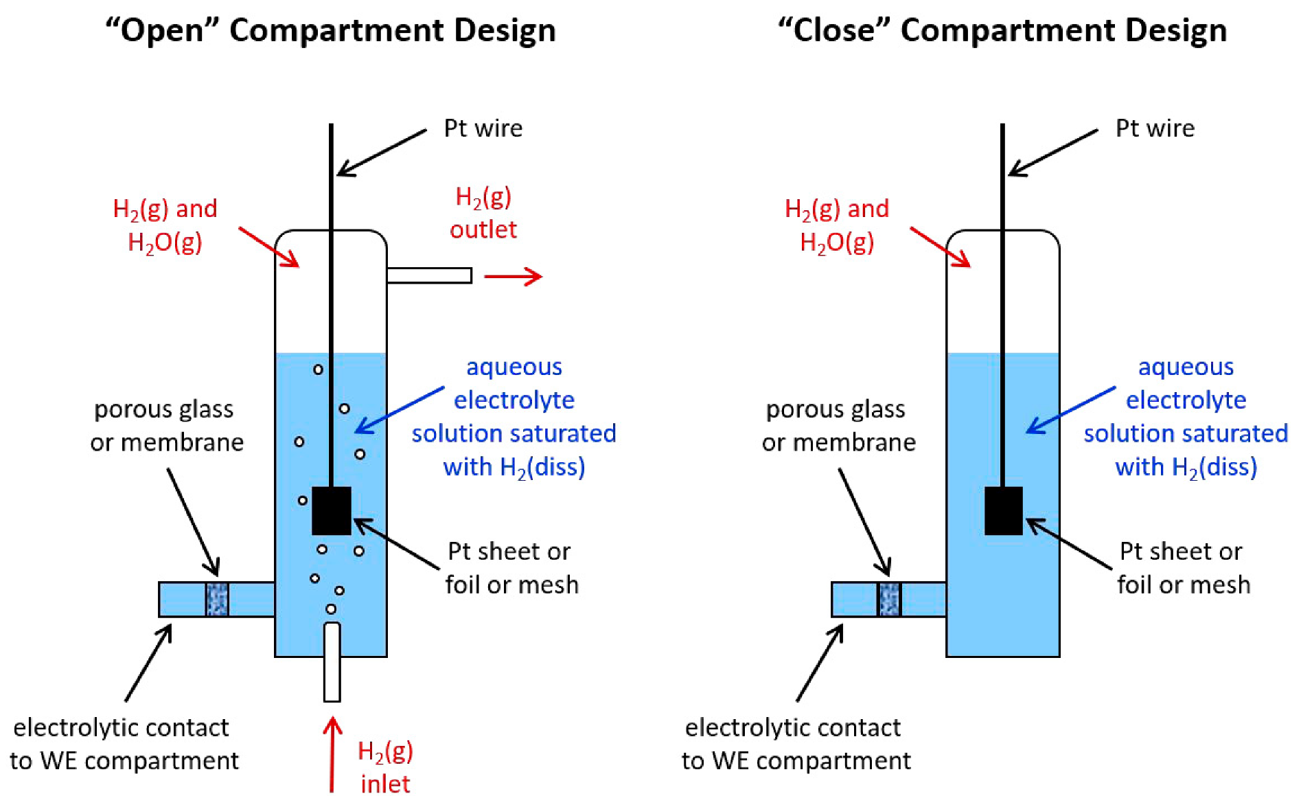
3.1. Hydrogen Electrode

Prior to glass electrodes, the pH sensor is mainly a hydrogen electrode, which can be used in both aqueous solutions and non-aqueous solutions. The characteristic of the hydrogen electrode is that it is plated with spongy platinum black as the sensitive layer of H+ [52]. There are two types of hydrogen electrodes—standard and reversible hydrogen electrodes. In general, these two kinds of techniques have the same design and only two main systems based on the same principle (potential difference arises from different values of two variables—an (H+) and f(H2) [53,54] (“open” and “close” designs shown in Figure 3) are commonly used in these years. The main difference between these two-compartment designs is the supply of hydrogen. As for the “open” system, the hydrogen can be achieved by the bottom a small opening and the “close” device can achieve hydrogen not only by the bottom small opening but also use negative potential as a cathode in water electrolysis to generate hydrogen. Devynck et al. [55] investigated the hydrogen electrode that served as a pH indicator in the hydrogen fluoride and superacid media by the means of voltammetric and potentiometric measurements in the acid ranges.
However, there are many disadvantages of hydrogen electrodes. Firstly, platinum black is prone to inactivation poisoning affected by mercury, and oxygen cyanide, which leads to loss of original activity of platinum black. In addition, the structure of the hydrogen electrode (shown in Figure 3) is complex and the use of high purity hydrogen (99.9999%), and the test environment is demanding, which brings a lot of inconvenience to the actual use and has little practical value. These kinds of hydrogen electrodes have good reversibility, stability, and reproducibility, so they are often used as reference electrodes to measure the standard electrode potential of other electrodes.

3.2. The Glass Electrode

According to the above introduction in Section 2.1.3 of ordinary pH measurements, the glass electrode is routinely used for pH measurements in a common lab setting. The glass electrode, the early use of pH sensors, is currently the most popular use of a hydrogen ion-selective electrode (shown in Figure 4). Its sensitive structure is a hyaline membrane which is the common chemical, silica. This kind of chemical consists of a silica bond to form a mesh skeleton. In this case, alkali metal ions such as sodium–potassium, lithium, etc., can be saved in the mesh structure (examples shown in Table 1). When the glass electrode is immersed and activated, the metal ions existing in the mesh construction can generate hydration ions with water, and the reaction between metal ions and hydration ions occurs, resulting in a pH response. The silver/silver chloride or calomel electrodes are usually used as internal reference electrodes, and the saturated calomel electrode (SCE) is used as an external reference electrode during the measurement.
Although it has the advantages of sensitivity, accuracy (high measurement accuracy of 0.02 pH [58]), short reaction time and high stability, there are still many defects, such as fragility, high internal resistance, high film impedance, the difficulty of miniaturization and inconvenience for connection with other instruments [59], which leads to the limitation of their determination scope. Therefore, various pH sensors have been developed to address these problems.
Table 1. The development of glass electrodes.
FormRepresentative CompositionEvaluationShortcomingReference
Sodium silicate pH glass electrode22Na2O–6CaO–72SiO2Combing with the vacuum tube amplifiers successfully in the industry since the 1930sDeviations were observed in more acid or base solutions[60,61]
Lithium silicate pH glass electrode18.1Li2O–9.6CaO–72.3SiO2Higher pH limit in Li+ or Na+ containing solutionsDue to the high electrical resistance, this technique is failed to confirm its characteristics by other scientists.[62,63]
26Li2O–3.6BaO–70.4SiO2 and 26.5Li2O–12.3MgO–61.2SiO2Only using low electrical resistance to fabricate the glass electrode/[63]
Li2O-Cs2O-La2O3-SiO2The most successful improvement of lithium silicate glasses for glass electrode and have been widely employed since the 1950s/[64]

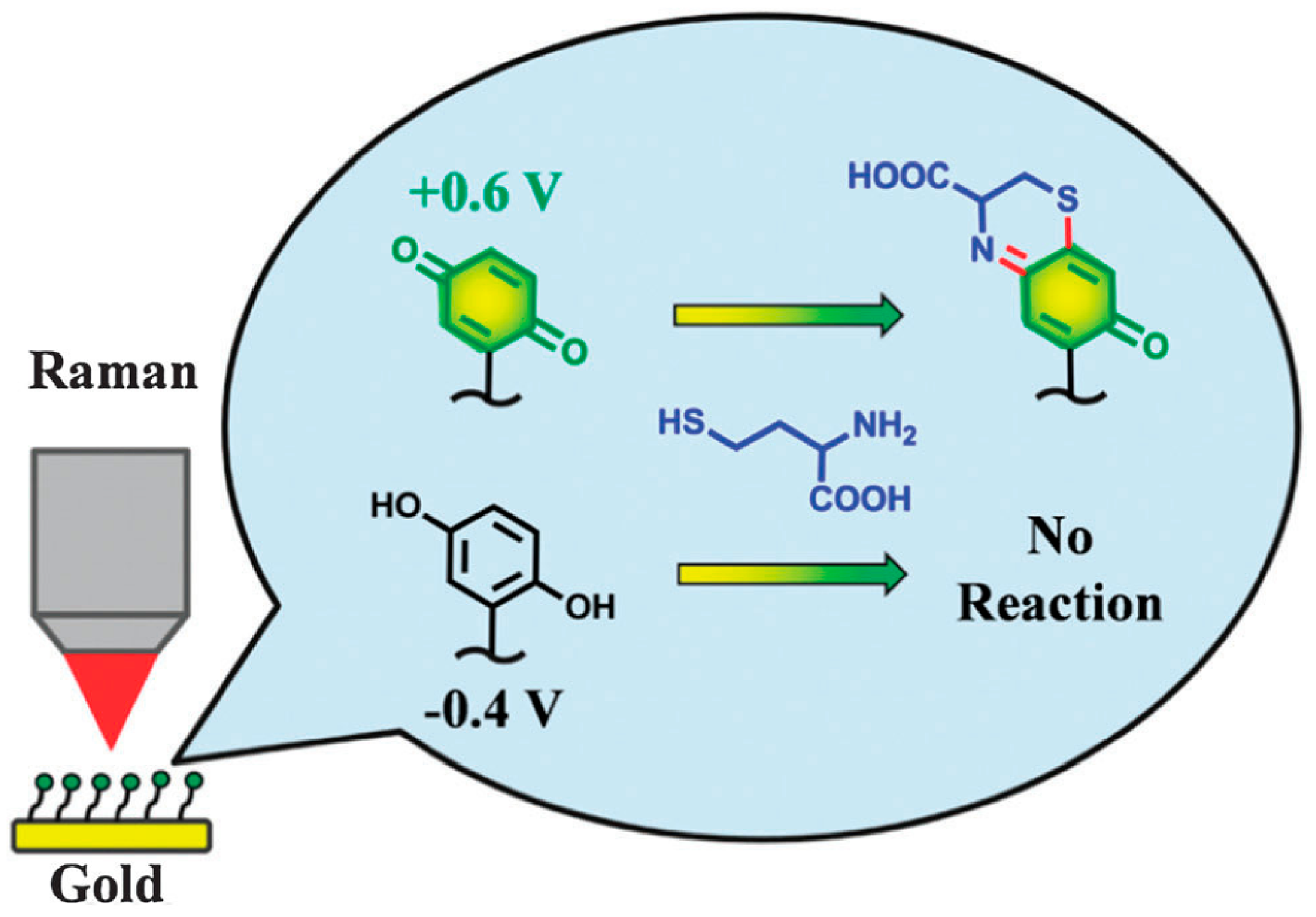
3.3. Quinone Hydroquinone pH Electrode

Bulmnan et al. [65] firstly proposed the Redox reaction between hydroquinone and quinone, which can be used to measure the concentration of hydrogen ions. Quinone hydroquinone electrodes are composed of smooth platinum or gold electrodes immersed in a quinone hydroquinone saturated solution. Its electrode reaction:
Q + 2H+ = H2Q (Q: Quinone, H2Q: hydroquinone)
The quinone hydroquinone electrode is dependent on the same Nernst equation for computing basis, hydrogen ion concentration of the solution is achieved by the potential difference. Additionally, the quinone hydroquinone electrode is not only simple to prepare but also has fast potential stability, low internal resistance, prevention from interference antioxidants and other substances. This technique can be used in a solution containing soluble gases or acetone formic acid with a carbonyl structure and other organic acids. However, the main disadvantage of the quinone hydroquinone electrode is the lack of understanding the complex interactions of structural types (various kinds of natural membranes or interfaces and the network of extra- and intracellular) [66]. In this case, with the help of basic and applied science, it is necessary to enrich different types of biomimetic interfaces on the surface of the nanomaterial [67].
The most common method to fabricate quinone hydroquinone sensors is self-assembly (SAM) because of simple operation, chemical availability, etc. [67]. There are different kinds of SAM surface reactions. For instance, Zhang et al. [68] demonstrated that a representative reaction, which is a modulated quinone hydroquinone transition and heterocyclization produced by benzoquinone and L-cysteine (shown in Figure 5). Quinone hydroquinone can be used as the composition of nanomaterial to achieve biosensors. As the transform of dopamine and dopaquinone can be a symbol of pH changes, Medintz et al. [69,70] studied that the biosensor consisted of dopamine and quantum dots conjugates to detect whether cells undergo drug-induced alkalosis by measuring cytoplasmic pH.

3.4. Optical Fiber pH Sensor

The optical fiber pH sensor is the sensor with a very high sensitivity that appeared in the 1980s, and it can be continuously used to measure automatically. Different from other electrochemical measurement methods of pH electrodes, an optical fiber pH sensor using optical properties for pH measurement has been developed as the light electrode within the scope of different sections in the range of pH (from 1.0 to 8.0) for various needs in pH measurement. For instance, their accuracy reaches 0.1–0.05 pH unit when sample pH changes. As for the fabrication of optical sensors, the choice of platform is the key to designing relevant devices successfully. In these years, lots of absorption- and luminescence-based optical sensing techniques have been studied in these years [71,72,73,74,75,76]. The optical fiber sensor platforms are considered the traditional platforms and there have been lots of studies on their developments (shown in Table 2).
The mechanism of optical fiber pH sensor is to use chemicals, which is sensitive to hydrogen ions as sensitive element material, to produce different spectral characteristics by these chemicals to obtain the pH values. The sensor is composed of a light wave as energy transfer medium and optical fiber as a light transmission medium. The main drawback of the optical fiber sensor is its narrow linear range and long response time. The application is limited of this instrument because different pH testing scopes need to change different probes and cannot be used for suspension. To deal with the problems of optical fiber sensors, another platform, a planar waveguide-based sensor has been developed as they are not only more stable but also more attractive with the help of advanced manufacturing techniques [77].
According to the shifts of test optical signal characteristics generated by indicators and the interactions between the materials, fiber optic pH sensors can be divided into fluorescent light, absorption type, and reflection type. Peterson et al. [78] developed the first optical fiber pH sensor based on the principle of light absorption in the 1990s, and the optical fiber pH sensor based on light absorption has been continuously developing: Tzonkov et al. [79] improved the response time of the electrode to 30 s through the study of the dynamic model. As for technology, the invention of optical waveguide technology realized long-distance telemetry, and the invention of disappeared wave technology expanded the detection range, which leads to the development of optical absorption fiber pH sensors. Since the 1990s, lots of polymer films have been used on the surface of electrodes to enhance the reflected light, which can lead to improving the sensitivity of reflective fiber pH sensors. For instance, the PAN membranes were developed by radical polymerization: poly(acrylonitrile acrylic acid-2-vinylnaphthalene) and poly(acrylonitrile-methacrylic acid-2-vinylnaphthalene) [80]. Gu et al. [81] used polyethylenimine (PEI) and sodium alginate (SA) to deposit on the side surface of the thin-core fiber modal interferometer (TCFMI) by self-assembly technique for the determination of pH values in biology. Alabbas et al. [82] describe the design of a reflection-type fiber-optic pH sensor for improved reproducibility, which can be used to measure the pH values of acidic and alkaline vapors. As fluorescence is a highly sensitive analysis method with a short response time, most of these fiber-optic pH fluorescence sensors use fluorescent reagents as stationary phases. Bastien et al. [83] used polymethylmethacrylate (PMMA) as optical fibers for on-body monitoring in biological solutions (shown in Figure 6).
Table 2. The introduction of optical fiber-based platform.
Types of Optical Fiber pH SensorConfigurationSpecific Indicator DyespH RangeResponse TimeAccuracyReference
Fluorescent typeprobeFluorescein 9–32.5 minNot specified[84]
3–96 min
Absorption typePhenol red.7.0–7.40.5 min0.017[85]
Reflection typeBromothymol blue7–122.5 minNot specified[86]

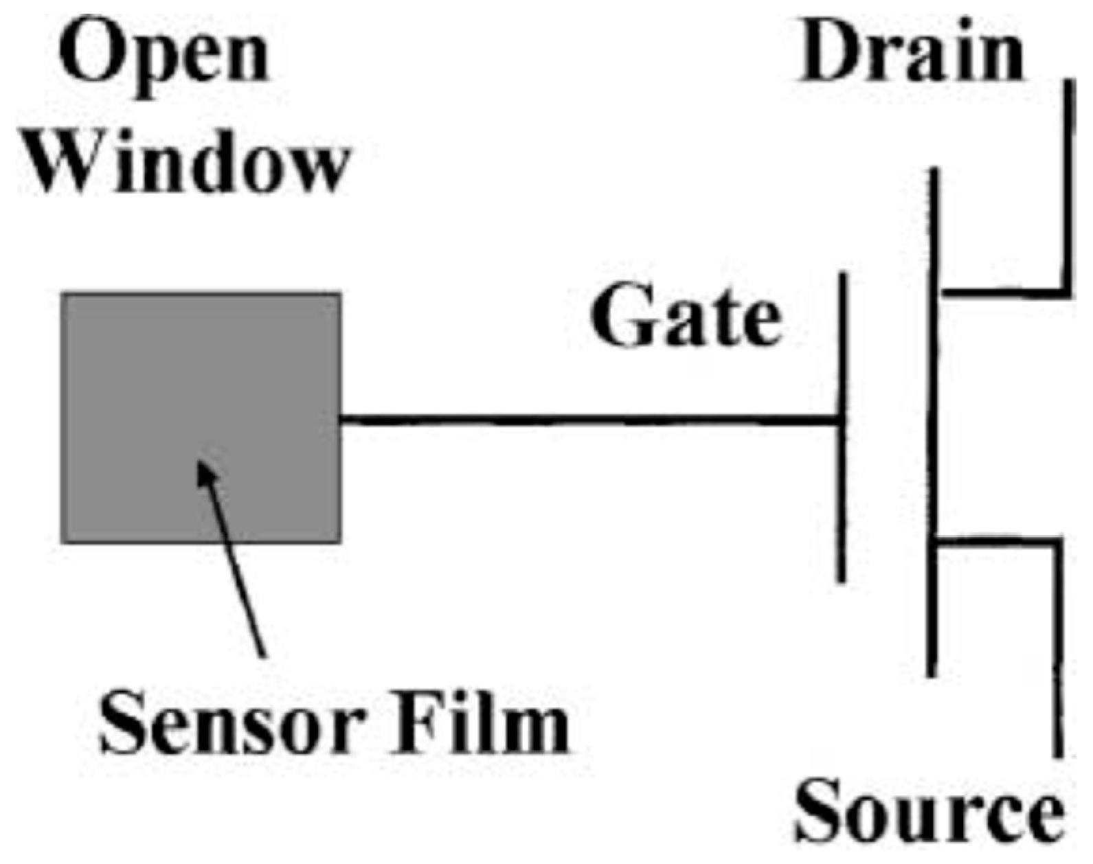
3.5. Ion Sensitive Field Effect Transistor (ISFET) pH Sensor

ISFET, a new type of pH-sensitive device invented by Bergveld in 1981 [87], the main material is a silicon chip. The structure of ISFET is similar to that of MOSFET (metal-oxide-semiconductor field effect transistor) but the ion-sensitive membrane is used to replace the metal gate of the MOSFET [88]. After the contact between the sensitive membrane and solution, the Nernst response to hydrogen ions occurs. The specific structure and design of the device are shown in Figure 7. Compared to other pH sensors, the ISFET pH electrode has good precision, repeatability, miniaturization, and short response time while the limitation of the large size of the ISFET sensor for further employment becomes the main disadvantage [89,90].

3.6. Metal/Metal Oxide pH Sensor

Metal/metal oxide pH sensor, as an important substitute system for pH glass electrodes, is also a major research direction of pH electrodes up to now. There are some characteristics of this technique—simple preparation, small internal resistance, quick response, high mechanical strength, and miniaturization. In addition, this type of sensor can not only adapt to the test environment of high temperature or strong agitation system but also can be used in the system containing F- solution. Morishita et al. [91] used the sensor consisting of a lead electrode and an oxidized stainless steel electrode for oil deterioration based on the principle of pH value measurement. Researchers [91] used the sensor consisting of a lead electrode and an oxidized stainless steel electrode for the pH value measurement. With a good linear relationship of the sensor signal to the quasi-pH value of oil, the region where oil deterioration proceeded can be identified. In addition, the residual basic additives in the oil can be easily estimated from the sensor signal. Some achievements have also been made in the miniaturization development of metal/metal oxide pH sensors [92]. For example, with the help of the connection of carbon substrates and immobilized noble metal nanostructures [93], sensitive biosensors can be developed in medical products, instruments, and techniques [94,95,96] because of their positive effect on redox reactions.
The principle of metal/metal oxide electrode is based on redox reaction, the substance of reaction is the reduction reaction of metal oxide with the participation of hydrogen ions. At present, the study of this sensor mainly focuses on a few elements in the fourth, fifth, and sixth periods, and the most are the research on the platinum group elements in the sixth group. Metal/metal oxide electrode preparation methods include sputtering, electrochemical cycle voltammetry and thermal oxidation method, etc.
The metal/metal oxide electrodes are considered suitable electrodes for embedding in concrete due to their excellent stability and strength, as well as their physical activity in the size of the electrode. Different metal oxides currently used to make pH electrodes include IrOX [97,98,99,100] PbO2 [101], OsO2 [102], TiO2, WO3 [103], PtO2 [102] depending on different subjects (details are demonstrated in Table 1). The WO3 and IrOx (or IrO2) have shown a wider pH response and a close-to-theoretical Nernst constant in fairly quick response time (a few minutes) and stable up to a week (shown in Table 3).

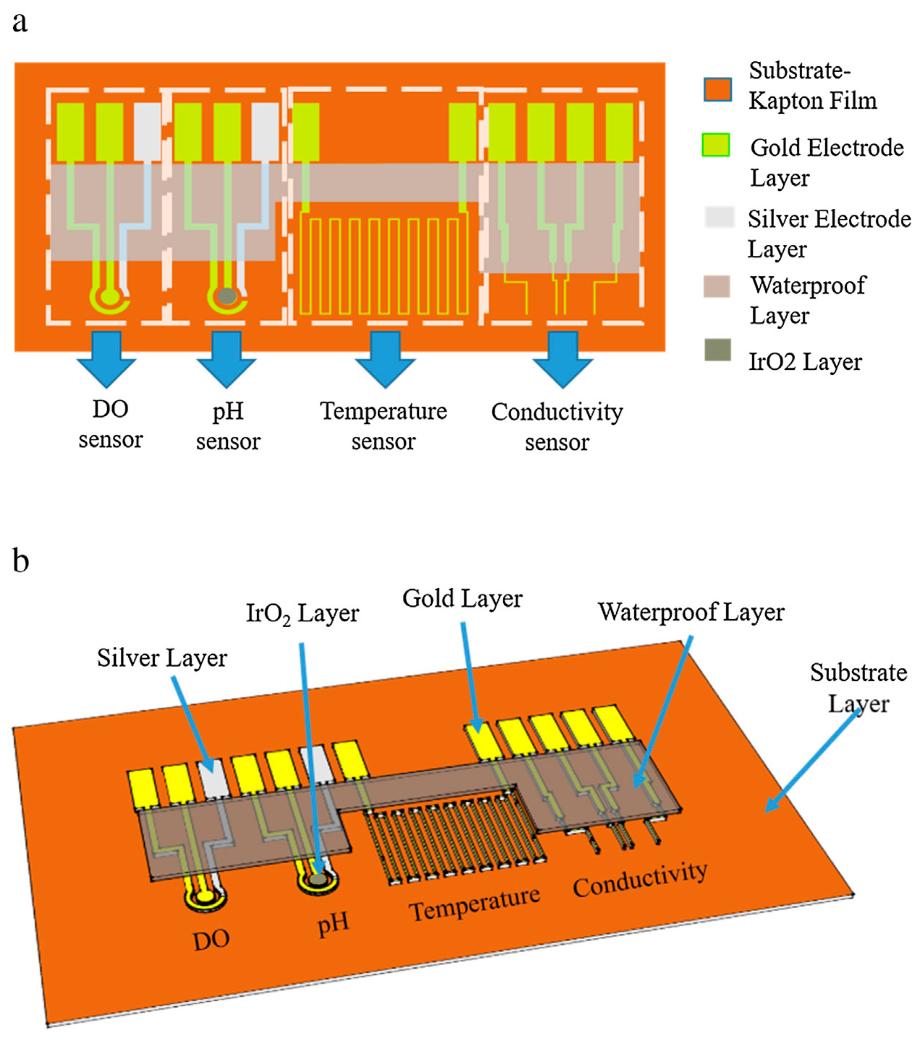
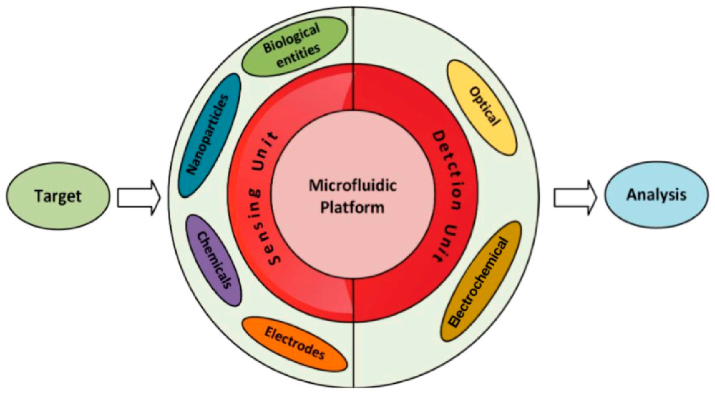
4. The Microfluidic-Based pH Sensing

Microfluidics is the skill that accurately controls a small volume of fluids by using channels of different sizes with a range from 10 s to 100 s of micrometers [106,107,108,109]. There are several merits such as shorter reaction times, better process control, lower waste generation, compacted system and parallelization, reasonable prices, and disposability [106,110,111,112,113] while most of the microfluidic devices are disposable and only used for one-time measurement. In the earlier study of microfluidics, the integration of microsensors with fluidic constructions (actuators, pumps, valves, etc.), and miniaturization of analytical assays was the main direction. With the development of research on microfluidics, micro total analysis systems (µTAS) were established in terms of micro-fabricated structures. Owing to the miniaturization with microfluidics, there had been great progress in many biological aspects such as genetic analysis, cell biology, and protein analysis [114]. Up to now, these instruments are widely adhibited in all fields of science mentioned above such as engineering, chemistry, biology, biomedical sciences, etc.

4.1. The Fabrication of Microfluidic-Based Sensing

Silicon was the first use for fabrication of the microfluidic instruments [115,116,117]. Then, glass and polydimethylsiloxane (PDMS) were the major material to fabricate the purposed devices. Nowadays, more and more materials including thermoplastic, and paper are accepted as fabrication materials [118,119,120]. The common manufacturing approaches for microfluidic sensors are injection molding, soft lithography, and mass-production technologies such as etching. The soft lithography technique using PDMS is considered as the most popular method compared with other techniques [121,122]. However, the special devices for fabrication and clean room requirements may lead to the high demand during the process [123], which hinders the cost-effectiveness of the fabricated sensors. In addition, 3D printer techniques (inkjet printing, stereolithography, extrusion printing, and so on [124,125]), the commercial and popular technology, is also the object of fabrication for microfluidic sensors because of the possibility of fabricating the microstructures designed by computers.

4.2. Microfluidic-Based Sensing Applications

Microfluidic sensors can be divided into two types—in which the microfluidic system measures the parameters inside it and in which the measurement of parameters takes place with the external integrated equipment, respectively [126,127,128]. According to Figure 8 [129], it can be known that the microfluidic system consists of two main parts—the sensing unit and the detection unit. As for the sensing unit, it involves elements such as biological entities, functionalized nanoparticles, and metal electrodes while the most universally applied detection systems with microfluidics are optical- and electrochemical-based systems [127,130].
As micromixers have the advantage of the high sensitivity of the microfluidic-based sensors, it is simple to modify their microchannel in order to determine various types of analyses [123,131,132]. Additionally, there is no other preparation of analysis before the measurement such as the detection of pollutants using sensors in microscale [129]. Therefore, microfluidic sensors have enriched the detection of different fields of science biology, analytical chemistry, and assessment of food products, which is considered as a substitute in labs.

4.2.1. Biology

The extracellular or intracellular pH values may change because of the reactions in cells such as growth, metabolism, signal transduction, etc. [133,134]. For instance, when the growth of cancer cells increases rapidly, the lactic acid will be produced, which leads to the low extracellular pH value in the microenvironment [135,136]. Therefore, pH was known as the key to dynamic determination of extracellular pH in cell culture experiments in both discovery and applied biomedical research [137,138]. Gang Xu et al. [139] developed a biosensor for pH measurement in air–liquid interface cell culture for wider application of microfluidic based sensing in biology. They used the tungsten microwires and silver microwires coated with silver/silver chloride as working and pseudo reference electrodes, respectively, which can measure cell pH values sensitively and ensure the repeatability of experiment results. In addition, Ges and Baudenbacher [140] integrate pH sensors consisted of IrOx films with miniature metal microelectrodes with microfluidic network to detect the acidification rates of single cardiomyocytes in a trapped extracellular room (shown in Figure 9). As for the monitoring different pH values in nL cell culture volumes, two IrOx thin film electrodes can be applied for the acidification rate of nL cell [141].

4.2.2. Environmental Detection

The detection of pH values of wastewater is the most typical environmental monitoring in the world. Although there are lots of methods developed for polluted water pH measurement, such as the colorimetric method [142,143,144], H+ reagent analysis [145,146,147], potential method [148,149], etc., these tools measure the pH values of solution by sampling method, which leads to the limitation of update response of samples and frustration of specific sewage treatment. In this case, it is necessary to develop new tools that can monitor pH micro-differences in short response time.
In the 1990s, the microfluidic chip, first invented by Manz and Widmer [150] during microchip capillary electrophoresis research, is characterized by the network of micropipes and controlled fluid flows through the whole system. This emerging technology is considered as the core of µTAS based on analytical chemistry and the micro-electro-machining technology (MEMS) [151]. McCormick et al. [152] demonstrated that the use of microchannels can offer high-resolution separations of DNA fragments in less than 3 min (shown in Figure 10). As the detection and fabrication of fiber optical pH sensors have already been developed for years, the combination of the microfluidic chip and fiber optical pH sensors can bring the benefits of both these two devices into one emerging instrument for pH measurements, especially for wastewater in our life. The integration of microfluidic devices and optical pH sensors was developed for quick pesticide detection by determining pH values of tap water [153].
In addition, Liu and Yu [154] reported different methods for pH measurements based on the in-channel photopolymerization technique such as a 3D hybrid valve, and the microchannel is covered with PDMS to separate the pH solution. The hydrogel swells or shrinks depending on solutions with different pH values. The results show the changes in hydrogel sizes (from 280 µm to 460 µm, respectively), in a pH region (2.0–11.0), which ensured the reversibility and reproducibility of hydrogel swelling and shrinkage. The effective optical thickness of the pH sensor layer will become stronger. The change in pH value in the microchannel can be monitored in real-time by Fourier transform integral sphere reflectance interferometry (FT-FS).

4.2.3. Food Safety

As there are still thousands of people who suffer from illnesses brought by spoiled food (caused by the microorganism and bacteria), food safety issue is the main concern in public [155]. Based on this situation, many techniques for the food industry have already been developed——the most traditional methods are microbiological culture and colony counting [156] while the progress in biosensors has emerged and been used in food safety due to its quick detection, limit of detection (LOD), feasible operation, and reasonable price. For example, Liu et al. [157] detected salmonella in food products by the microfluidic sensors. As for the pH measurement in food, the wireless device fabricated by miniature iridium oxide and silver chloride can sense the pH values of fish meat in long term to tell the microbiological spoilage of food remotely [158].
Overall, according to the principles of microfluidics, it is obvious that the sensors based on the microfluidic channel can bring great progress in the lab detection of liquid. In this case, solid-state pH sensors, especially IrOx, can be considered to combine with microfluidic technology to determine liquid at the micro or nanoscale.

4.2.4. Medical Engineering

Based on the research of biology from Section 4.2.1, the pH measurement of different types of cells or organs can monitor the animal’s health condition. In this case, the related pH biosensors can be developed and applied as the main technique of medical engineering. In past decades, lots of researchers have used the non-invasive fluorescent probes as pH biosensors to prevent humans from having diseases [159,160,161,162,163,164], especially cancer [165].
The pH fluorescent probes are developed sustainably in these years and the breakthrough is that the resolution of fluorescents proves to be higher. In this case, this kind of sensor was used for a more accurate analysis of pH-related physiological processes. Figure 11 shows the representative design of fluorescent probes. Table 4 shows each reflected structure’s pka and the excitation and emission properties accordingly. Probe 1-a is one of the strongest acids among probes 1-a, 1-b, and 1-c. As for microfluidic biosensors for medical pH detection [166,167], the paper-based microfluidic bioassays have been studied for a wide range of detected targets, especially pH, because of the reasonable price and easy operation [168].

5. Future Trend of pH Sensing and Microfluidic-Based pH Sensing

Due to the continuous updating of industry, innovation and reform of technology, and sustainable exploration in unknown fields, it will put higher requirements for the sensors. As one of the core technologies of sensing, the request for pH sensor techniques also keeps improving, which is mainly reflected in these three general trends or directions: miniaturization, intellectualization, and sustainability.

5.1. Miniaturization

According to the overview of pH sensors, it is obvious that the pH sensor has already developed in these decades, which can be applied in various fields at the bulk scale. Based on these existing pH sensors such as optical fiber sensors, metal/metal oxide sensors, etc., pH sensing was studied for more precise results at the microenvironment or nanoscale. In this case, it is undoubtful that the main trend of bulk pH sensing is miniaturization.
With the integration of the continuous development of nanotechnology, pH sensors will have a breakthrough in many fields of sensors. Based on the example of the reduced size of metal oxide pH sensors for detecting bacteria designed by Naora et al. [170], in the future nanorobots can be developed for human healthy detection. The device can diagnose cellular behavior or metabolism by determining pH in the microenvironment in the human body [171]. Additionally, the updating of applied materials of pH sensing is also the method to miniaturize this device. It is well known that nanotechnology is an emerging research field with huge potential for advanced applications. To combine nanotechnology with sensors, electrospinning is designed for the preparation of polymer fiber materials at the micro or nanoscale [172,173]. In 2010, Schueren et al. [174]. reported that pH indicators did not influence the halochromic behavior of dyes and the fiber structure, but the application of each dye is restricted by the limited range of pH values. Therefore, it is necessary to select a suitable association of dyes to measure the change in pH values for optical pH sensors.

5.2. Intellectualization

Due to the advanced computer technology, there will be a breakthrough in many fields of sensors integrated with computers. In this case, future sensors will combine with the Internet of Things (IoT) tools to provide more artificial intelligence (AI) such as detection, judgment, and self-solution [175,176]. Specifically, the wireless sensor networks (WSNs) including sensing devices and IoT can be implemented in environment monitoring (EM) especially in the weather forecasting [177], pollution control (air [178,179] and water [180,181,182]), temperature control [100] and food safety assessment [182,183]. These emerging methods of environmental monitoring are identified as smart environment monitoring (SEM) systems based on the use of wireless sensing, IoT, and AI.
As for pH sensors, there is no doubt that AI pH sensing will be employed not only in environmental detection but also in more areas such as the health assessment [184] with the Internet instead of the traditional pH sensors in the future. Although there have already been some microfluidic-based sensors applied in biology and medicine (e.g., sweat [185], tears [186], wounds [187], etc.), the collision from a large number of irrelevant compounds in bodily solutions is still the main challenge to achieve accurate and effective diagnosis results. AI can be a new solution to complement pH sensing, which leads to selective detecting capabilities, wider scope of detection, and reliable diagnosis [188,189,190,191]. For instance, the link of pH and temperature sensors can dynamically adjust the glucose data based on calibration parameters as the variable separation of software depends on post-measurement algorithms or calibration of sensors to calculate and analyze databases [192].

5.3. Sustainability

If the pH sensors can achieve these two characteristics above, it is admitted that this technique might bring lots of convenience to people’s life. However, the cost and service life of tools are also the primary elements to explore science nowadays, which is called sustainable development. Therefore, the low power requirement electrodes and long duration are critical factors to develop miniaturized and smart sensors for further study.
Currently, ISFET-based sensors require an application possibility and the electronic structure for online data analysis and warehouse also requires suitable power sources but there are many disadvantages in such application, including energy loss, poisonousness, frequent charging, etc. To address these problems, energy-independent sensors integrated with energy generators and storage will be tremendously employed in the online monitoring [193]. Manjakkal et al. [194,195,196] reported that supercapacitors and flexible solar cells can be utilized as power packs of voltage sources in wearable applications and water quality detection connected with robots [197,198].
Meanwhile, using reversable material, swelling hydrogels can produce reusable pH sensors for soil pH measurement [199]. According to these existing techniques, new targets can be set for further development such as the flexibility of sensors and the stability in extreme environmental condition [193].

6. Conclusions

Based on the historical theory of pH, pH plays a crucial role in current scientific studies and various types of applications, especially environmental detection, medicine, and pharmacy. Many specific traditional pH measurements are reported according to the mechanism of pH sensing and the representative method is pH sensors including hydrogen electrodes, glass electrodes, optical fiber electrodes, metal/metal oxide pH sensors, etc. As the fabrication of microfluidic-based sensing is operated universally, this equipment attracts more and more scientists to have research to achieve accurate and on-time experiment results, which leads to its wide application in daily life. However, there is still a large space for this technique as the growing demand for miniaturized, sustainable, and intelligent pH sensing and microfluidic-based pH sensing occurs with the background of “Green Science”. In this case, it is necessary to innovate pH sensing integrated with emerging strategies such as nanotechnology, the Internet, AI, etc.

Author Contributions

Conceptualization, Q.D. and W.X.; writing—original draft preparation, W.X.; writing—review and editing, Q.D.; visualization, W.X.; supervision, Q.D.; project administration, Q.D.; funding acquisition, Q.D. All authors have read and agreed to the published version of the manuscript.

Funding

This work was partially supported by Research Development Funding (RDF-21-1-005) under Xi’an Jiaotong-Liverpool University.

Conflicts of Interest

The authors declare no conflict of interest. The funders had no role in the design of the study; in the collection, analyses, or interpretation of data; in the writing of the manuscript, or in the decision to publish the results.

References

  1. Meinrath, G.; Spitzer, P. Uncertainties in Determination of pH. Microchim. Acta 2000, 135, 155–168. [Google Scholar] [CrossRef]
  2. Michaelis, L. Die Wasserstoffionenkonzentration. Naturwissenschaften 1914, 2, 829–834. [Google Scholar] [CrossRef]
  3. Brown, R.J.C.; Milton, M.J.T. Developments in accurate and traceable chemical measurements. Chem. Soc. Rev. 2007, 36, 904–913. [Google Scholar] [CrossRef] [PubMed]
  4. Spitzer, P.; Pratt, K.W. The history and development of a rigorous metrological basis for pH measurements. J. Solid State Electrochem. Curr. Res. Dev. Sci. Technol. 2011, 15, 69–76. [Google Scholar] [CrossRef]
  5. Hagerup. Études Enzymatiques: 2: Sur la Mesure et L’importance de la Concentration des Ions Hydrogène Dans les Réactions Enzymatiques, 2nd ed.; H.Hagerup: Kjøbenhavn, Denmark, 1925. [Google Scholar]
  6. Sfrensen, S.P.L. Enzymstudien. II. Mitteilung. Über die Messung und Die Bedeutung der Wasserstoffionen-Koncentration Bei Enzymatischen Prozessen; Biochem. Zeitschr. 21 (1909) 131-304, and 22 (1909) 352-356; Springer: Berlin/Heidelberg, Germany, 1909. [Google Scholar]
  7. Arrhenius, S. On the dissociation of substances dissolved in water. Z. Phys. Chem 1887, 1, 631. [Google Scholar] [CrossRef]
  8. Friedenthal, H. Die Bestimmung der Reaktion einer Flüssigkeit mit Hilfe von Indikatoren. Z. Für Elektrochem. Und Angew. Phys. Chem. 1904, 10, 113–119. [Google Scholar] [CrossRef]
  9. Debye, P.; Hückel, E. On the theory of electrolytes. I. Freezing point depression and related phenomena. Phys. Z 1923, 24, 185–206. [Google Scholar]
  10. Burton, R.F. Defining and teaching pH. J. Chem. Educ. 2007, 84, 1129. [Google Scholar] [CrossRef]
  11. Galster, H. pH Measurement; VCH: New York, NY, USA, 1991. [Google Scholar]
  12. Bates, R.G. Definitions of pH scales. Chem. Rev. 1948, 42, 1–61. [Google Scholar] [CrossRef]
  13. Bates, R.G.; Vijh, A.K. Determination of pH: Theory and practice. J. Electrochem. Soc. 1973, 120, 263C. [Google Scholar] [CrossRef]
  14. Whiffen, D.H. Manual of Symbols and Terminology for Physicochemical Quantities and Units; Elsevier: Amsterdam, The Netherlands, 2013; ISBN 1-4832-7887-5. [Google Scholar]
  15. Haring, M. Potentiometric Titrations. ACS Publ. 1926, 3, 846. [Google Scholar] [CrossRef] [Green Version]
  16. Haraldsson, C.; Anderson, L.G.; Hassellöv, M.; Hulth, S.; Olsson, K. Rapid, high-precision potentiometric titration of alkalinity in ocean and sediment pore waters. Deep Sea Res. Part I Oceanogr. Res. Pap. 1997, 44, 2031–2044. [Google Scholar] [CrossRef]
  17. Covington, A.K. Recent developments in pH standardisation and measurement for dilute aqueous solutions. Anal. Chim. Acta 1981, 127, 1–21. [Google Scholar] [CrossRef]
  18. Bates, R.G.; Popovych, O. The modern meaning of pH. C R C Crit. Rev. Anal. Chem. 1981, 10, 247–278. [Google Scholar] [CrossRef]
  19. Covington, A.K.; Bates, R.; Durst, R. Definition of pH scales, standard reference values, measurement of pH and related terminology (Recommendations 1984). Pure Appl. Chem. 1985, 57, 531–542. [Google Scholar] [CrossRef]
  20. Duffy, J.; Baucke, F. Effect of glass composition and basicity on reduction of metal ions to the metallic state in melts. Phys. Chem. Glasses 1997, 38, 25–26. [Google Scholar]
  21. Xu, Z.; Dong, Q.; Otieno, B.; Liu, Y.; Williams, I.; Cai, D.; Li, Y.; Lei, Y.; Li, B. Real-time in situ sensing of multiple water quality related parameters using micro-electrode array (MEA) fabricated by inkjet-printing technology (IPT). Sens. Actuators B Chem. 2016, 237, 1108–1119. [Google Scholar] [CrossRef]
  22. Pansu, M.; Gautheyrou, J. pH Measurement; Springer: Berlin/Heidelberg, Germany, 2006. [Google Scholar]
  23. Overton, T.W. Recombinant protein production in bacterial hosts. Drug Discov. Today 2014, 19, 590–601. [Google Scholar] [CrossRef]
  24. Han, U.; Seo, Y.; Hong, J. Effect of pH on the structure and drug release profiles of layer-by-layer assembled films containing polyelectrolyte, micelles, and graphene oxide. Sci. Rep. 2016, 6, 24158. [Google Scholar] [CrossRef]
  25. Pudipeddi, M.; Zannou, E.A.; Vasanthavada, M.; Dontabhaktuni, A.; Royce, A.E.; Joshi, Y.M.; Serajuddin, A.T.M. Measurement of Surface pH of Pharmaceutical Solids: A Critical Evaluation of Indicator Dye-Sorption Method and its Comparison with Slurry pH Method. J. Pharm. Sci. 2008, 97, 1831–1842. [Google Scholar] [CrossRef]
  26. Wang, X.-Q.; Zhang, Q. pH-sensitive polymeric nanoparticles to improve oral bioavailability of peptide/protein drugs and poorly water-soluble drugs. Eur. J. Pharm. Biopharm. 2012, 82, 219–229. [Google Scholar] [CrossRef] [PubMed]
  27. Taniguchi, C.; Kawabata, Y.; Wada, K.; Yamada, S.; Onoue, S. Microenvironmental pH-modification to improve dissolution behavior and oral absorption for drugs with pH-dependent solubility. Expert Opin. Drug Deliv. 2014, 11, 505–516. [Google Scholar] [CrossRef] [PubMed]
  28. Casey, J.R.; Grinstein, S.; Orlowski, J. Sensors and regulators of intracellular pH. Nat. Rev. Mol. Cell Biol. 2010, 11, 50–61. [Google Scholar] [CrossRef]
  29. Wu, Z.L.; Gao, M.X.; Wang, T.T.; Wan, X.Y.; Zheng, L.L.; Huang, C.Z. A general quantitative pH sensor developed with dicyandiamide N-doped high quantum yield graphene quantum dots. Nanoscale 2014, 6, 3868–3874. [Google Scholar] [CrossRef]
  30. Webb, B.A.; Chimenti, M.; Jacobson, M.P.; Barber, D.L. Dysregulated pH: A perfect storm for cancer progression. Nat. Rev. Cancer 2011, 11, 671–677. [Google Scholar] [CrossRef]
  31. Gethin, G. The significance of surface pH in chronic wounds. Wounds UK 2007, 3, 52. [Google Scholar]
  32. Schneider, L.A.; Korber, A.; Grabbe, S.; Dissemond, J. Influence of pH on wound-healing: A new perspective for wound-therapy? Arch. Dermatol. Res. 2007, 298, 413–420. [Google Scholar] [CrossRef]
  33. Stubbs, M.; McSheehy, P.M.; Griffiths, J.R.; Bashford, C.L. Causes and consequences of tumour acidity and implications for treatment. Mol. Med. Today 2000, 6, 15–19. [Google Scholar] [CrossRef]
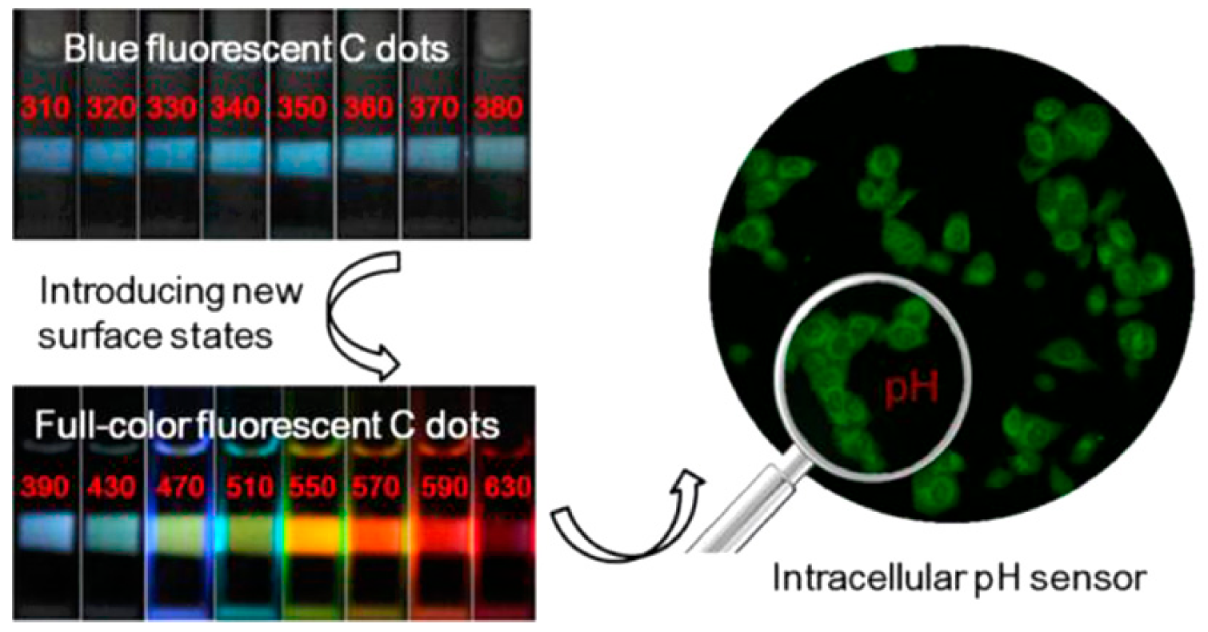
  34. Nie, H.; Li, M.; Li, Q.; Liang, S.; Tan, Y.; Sheng, L.; Shi, W.; Zhang, S.X.-A. Carbon dots with continuously tunable full-color emission and their application in ratiometric pH sensing. Chem. Mater. 2014, 26, 3104–3112. [Google Scholar] [CrossRef]
  35. Korting, H.; Hübner, K.; Greiner, K.; Hamm, G.; Braun-Falco, O. Differences in the skin surface pH and bacterial microflora due to the long-term application of synthetic detergent preparations of pH 5.5 and pH 7.0. Results of a crossover trial in healthy volunteers. Acta Derm Venereol 1990, 70, 429–431. [Google Scholar]
  36. Chen, W.; Morrow, B.H.; Shi, C.; Shen, J.K. Recent development and application of constant pH molecular dynamics. Mol. Simul. 2014, 40, 830–838. [Google Scholar] [CrossRef]
  37. Qian, J.; Khandogin, J.; West, A.H.; Cook, P.F. Evidence for a catalytic dyad in the active site of homocitrate synthase from Saccharomyces cerevisiae. Biochemistry 2008, 47, 6851–6858. [Google Scholar] [CrossRef]
  38. Milton, M.; Quinn, T. Primary methods for the measurement of amount of substance. Metrologia 2001, 38, 289. [Google Scholar] [CrossRef]
  39. Mariassy, M.; Pratt, K.W.; Spitzer, P. Major applications of electrochemical techniques at national metrology institutes. Metrologia 2009, 46, 199. [Google Scholar] [CrossRef]
  40. Harned, H.S.; Robinson, R.A. The ionic concentrations and activity coefficients of weak electrolytes in certain salt solutions. J. Am. Chem. Soc. 1928, 50, 3157–3178. [Google Scholar] [CrossRef]
  41. Bates, R.G.; Acree, S. pH values of certain phosphate-chloride mixtures and the second dissociation constant of phosphoric acid from 0° to 60 °C. J. Res. Natl. Bur. Stand. 1943, 30, 129–155. [Google Scholar] [CrossRef]
  42. Hamer, W.; Acree, S. A method for the determination of the pH of 0.05-molal solutions of acid potassium phthalate with or without potassium chloride. J. Res. Natl. Bur. Stand. 1944, 32, 215–227. [Google Scholar] [CrossRef]
  43. Bates, R.; Guggenheim, E. Report on the standardization of pH and related terminology. Pure Appl. Chem. 1960, 1, 163–168. [Google Scholar] [CrossRef]
  44. Baucke, F.G. New IUPAC recommendations on the measurement of pH–background and essentials. Anal. Bioanal. Chem. 2002, 374, 772–777. [Google Scholar] [CrossRef] [PubMed]
  45. Spitzer, P.; Werner, B. Improved reliability of pH measurements. Anal. Bioanal. Chem. 2002, 374, 787–795. [Google Scholar] [CrossRef]
  46. Baucke, F.G. Heiß-sterilisierte lagerfähige NBS-(DIN)-pH-Standardpufferlösungen–Untersuchung ihrer thermischen Stabilität. Chem. Ing. Tech. 1977, 49, 739–740. [Google Scholar] [CrossRef]
  47. Baucke, F. Lower temperature limit of NBS (DIN) pH standard buffer solution potassium tetroxalate. Electrochim. Acta 1979, 24, 95–97. [Google Scholar] [CrossRef]
  48. Baucke, F. Differential-potentiometric cell for the restandardization of pH reference materials. J. Electroanal. Chem. 1994, 368, 67–75. [Google Scholar] [CrossRef]
  49. Wilson, G.S.; Buck, R.; Rondinini, S.; Covington, A.; Baucke, F.; Brett, C.; Camões, M.; Milton, M.; Mussini, T.; Naumann, R. Measurement of pH. Definition, standards, and procedures. Pure Appl. Chem. 2002, 74, 2169–2200. [Google Scholar]
  50. Moritz, H. Geodetic reference system 1980. Bull. Géodésique 1980, 54, 395–405. [Google Scholar] [CrossRef]
  51. Baucke, F.G. Thermodynamic origin of the sub-Nernstian response of glass electrodes. Anal. Chem. 1994, 66, 4519–4524. [Google Scholar] [CrossRef]
  52. Mortimer, R.G. Physical Chemistry; Academic Press: Cambridge, MA, USA, 2000. [Google Scholar]
  53. Koryta, J.; Dvorak, J.; Kavan, L. Chapter 6. Principles of Electrochemistry, 2nd ed.; Wiley: New York, NY, USA, 1993; Volume 410. [Google Scholar]
  54. Bard, A.J.; Faulkner, L.R.; White, H.S. Electrochemical Methods: Fundamentals and Applications; John Wiley & Sons: Hoboken, NJ, USA, 2022. [Google Scholar]
  55. Devynck, J.; Hadid, A.B.; Fabre, P.-L. The hydrogen electrode as pH indicator in hydrogen fluoride and superacid media. J. Inorg. Nucl. Chem. 1979, 41, 1159–1161. [Google Scholar] [CrossRef]
  56. Jerkiewicz, G. Standard and reversible hydrogen electrodes: Theory, design, operation, and applications. ACS Catal. 2020, 10, 8409–8417. [Google Scholar] [CrossRef]
  57. Platt, B.S.; Dickinson, S. The technique of glass electrode measurements. Biochem. J. 1933, 27, 1069. [Google Scholar] [CrossRef] [PubMed]
  58. Khan, M.I.; Mukherjee, K.; Shoukat, R.; Dong, H. A review on pH sensitive materials for sensors and detection methods. Microsyst. Technol. 2017, 23, 4391–4404. [Google Scholar] [CrossRef]
  59. Yuqing, M.; Jianrong, C.; Keming, F. New technology for the detection of pH. J. Biochem. Biophys. Methods 2005, 63, 1–9. [Google Scholar] [CrossRef] [PubMed]
  60. MacInnes, D.A.; Dole, M. Tests of a new type glass electrode. Ind. Eng. Chem. Anal. Ed. 1929, 1, 57–59. [Google Scholar] [CrossRef]
  61. MacInnes, D.A.; Dole, M. The behavior of glass electrodes of different compositions. J. Am. Chem. Soc. 1930, 52, 29–36. [Google Scholar] [CrossRef]
  62. Dole, M. The Glass Electrode: Methods, Applications, and Theory; J. Wiley & Sons, Incorporated: Hoboken, NJ, USA, 1941. [Google Scholar]
  63. Belyustin, A.A. The centenary of glass electrode: From Max Cremer to FGK Baucke. J. Solid State Electrochem. 2011, 15, 47–65. [Google Scholar] [CrossRef]
  64. Perley, G.A. Glasses for measurement of pH. Anal. Chem. 1949, 21, 394–401. [Google Scholar] [CrossRef]
  65. Skoog, D.A.; Holler, F.J.; Crouch, S.R. Instrumental Analysis; Brooks/Cole, Cengage Learning Belmont: Boston, MA, USA, 2007. [Google Scholar]
  66. Tabaković, I.; Davidović, A.; Müller, W.E.; Zahn, R.K.; Sladić, D.; Dogović, N.; Gašić, M.J. Electrochemical reactivity of biologically active quinone/hydroquinone sesquiterpenoids on glassy carbon electrodes. Bioelectrochem. Bioenerg. 1987, 17, 567–577. [Google Scholar] [CrossRef]
  67. Ma, W.; Long, Y.-T. Quinone/hydroquinone-functionalized biointerfaces for biological applications from the macro-to nano-scale. Chem. Soc. Rev. 2014, 43, 30–41. [Google Scholar] [CrossRef]
  68. Li, J.; Sun, C.-L.; Tan, L.; Xie, Y.-L.; Zhang, H.-L. Investigation of an electrochemically switched heterocyclization reaction on gold surface. Langmuir 2013, 29, 5199–5206. [Google Scholar] [CrossRef]
  69. Ji, X.; Palui, G.; Avellini, T.; Na, H.B.; Yi, C.; Knappenberger, K.L., Jr.; Mattoussi, H. On the pH-dependent quenching of quantum dot photoluminescence by redox active dopamine. J. Am. Chem. Soc. 2012, 134, 6006–6017. [Google Scholar] [CrossRef]
  70. Medintz, I.; Stewart, M.; Trammell, S.; Susumu, K.; Delehanty, J.; Mei, B.; Melinger, J.; Blanco, J. Canosa, PE Dawson and H. Mattoussi. Nat. Mater 2010, 9, 676–684. [Google Scholar] [CrossRef]
  71. O’Driscoll, S.; McEvoy, H.M.; McDonagh, C.; MacCraith, B.D. Enhanced fluorescence-based optical sensor performance using a simple optical collection strategy. IEEE Photonics Technol. Lett. 2011, 24, 425–427. [Google Scholar] [CrossRef]
  72. Blue, R.; Kent, N.; Polerecky, L.; McEvoy, H.; Gray, D.; MacCraith, B. Platform for enhanced detection efficiency in luminescence-based sensors. Electron. Lett. 2005, 41, 682–684. [Google Scholar] [CrossRef]
  73. Burke, C.S.; McGaughey, O.; Sabattié, J.-M.; Barry, H.; McEvoy, A.K.; McDonagh, C.; MacCraith, B.D. Development of an integrated optic oxygen sensor using a novel, generic platform. Analyst 2005, 130, 41–45. [Google Scholar] [CrossRef] [PubMed]
  74. Duveneck, G.L.; Abel, A.P.; Bopp, M.A.; Kresbach, G.M.; Ehrat, M. Planar waveguides for ultra-high sensitivity of the analysis of nucleic acids. Anal. Chim. Acta 2002, 469, 49–61. [Google Scholar] [CrossRef]
  75. Puyol, M.; Salinas, Í.; Garcés, I.; Villuendas, F.; Llobera, A.; Domínguez, C.; Alonso, J. Improved integrated waveguide absorbance optodes for ion-selective sensing. Anal. Chem. 2002, 74, 3354–3361. [Google Scholar] [CrossRef] [PubMed]
  76. Burke, C.; Polerecky, L.; MacCraith, B. Design and fabrication of enhanced polymer waveguide platforms for absorption-based optical chemical sensors. Meas. Sci. Technol. 2004, 15, 1140. [Google Scholar] [CrossRef]
  77. Wencel, D.; Abel, T.; McDonagh, C. Optical chemical pH sensors. Anal. Chem. 2014, 86, 15–29. [Google Scholar] [CrossRef] [PubMed]
  78. Peterson, J.I.; Goldstein, S.R.; Fitzgerald, R.V.; Buckhold, D.K. Fiber optic pH probe for physiological use. Anal. Chem. 1980, 52, 864–869. [Google Scholar] [CrossRef]
  79. Kostov, Y.; Tzonkov, S.; Yotova, L. Dynamic model of an optical absorption-based pH sensor. Analyst 1993, 118, 987–990. [Google Scholar] [CrossRef]
  80. Gabor, G.; Chadha, S.; Walt, D.R. Sensitivity enhancement of fluorescent pH indicators using pH-dependent energy transfer. Anal. Chim. Acta 1995, 313, 131–137. [Google Scholar] [CrossRef]
  81. Gu, B. Biocompatible Fiber-Optic pH Sensor Based on Optical Fiber Modal Interferometer Self-Assembled With Sodium Alginate/Polyethylenimine Coating. IEEE Sens. J. 2012, 12, 1477–1482. [Google Scholar] [CrossRef]
  82. Alabbas, S.; Ashworth, D.; Narayanaswamy, R. Design and Performance Features of an Optical-Fibre Reflectance pH Sensor; IEEE Xplore: New York, NY, USA, 1989; Volume 26, pp. 373–380. [Google Scholar]
  83. Schyrr, B.; Pasche, S.; Scolan, E.; Ischer, R.; Ferrario, D.; Porchet, J.-A.; Voirin, G. Development of a polymer optical fiber pH sensor for on-body monitoring application. Sens. Actuators B Chem. 2014, 194, 238–248. [Google Scholar] [CrossRef]
  84. Cajlakovic, M.; Lobnik, A.; Werner, T. Stability of new optical pH sensing material based on cross-linked poly (vinyl alcohol) copolymer. Anal. Chim. Acta 2002, 455, 207–213. [Google Scholar] [CrossRef]
  85. Goldstein, S.; Peterson, J.; Fitzgerald, R. A miniature fiber optic pH sensor for physiological use. J. Biomech. Eng. 1980, 102, 141–146. [Google Scholar] [CrossRef]
  86. Kirkbright, G.F.; Narayanaswamy, R.; Welti, N.A. Fibre-optic pH probe based on the use of an immobilised colorimetric indicator. Analyst 1984, 109, 1025–1028. [Google Scholar] [CrossRef]
  87. Bergveld, P. Development of an Ion-Sensitive Solid-State Device for Neurophysiological Measurements. IEEE Trans. Biomed. Eng. 1970, 17, 70–71. [Google Scholar] [CrossRef]
  88. Swaminathan, S.; Krishnan, S.M.; Khiang, L.W.; Ahamed, Z.; Chiang, G. Microsensor characterization in an integrated blood gas measurement system. In Proceedings of the Asia-Pacific Conference on Circuits and Systems, Singapore, 16–18 December 2002; IEEE: Bali, Indonesia, 2002; pp. 15–20. [Google Scholar]
  89. Rani, R.A.; Sidek, O. ISFET pH Sensor Characterization: Towards Biosensor Microchip Application; IEEE: Bali, Indonesia, 2004; pp. 660–663. [Google Scholar]
  90. Chin, Y.-L.; Chou, J.-C.; Sun, T.-P.; Liao, H.-K.; Chung, W.-Y.; Hsiung, S.-K. A novel SnO2/Al discrete gate ISFET pH sensor with CMOS standard process. Sens. Actuators B Chem. 2001, 75, 36–42. [Google Scholar] [CrossRef]
  91. Morishita, S.; Suzuki, K.; Ashida, T.; Tasaka, K.; Nakada, M. Development of an On-Board Type Oil Deterioration Sensor; SAE Technical Paper: Chiang Mai, Thailand, 1993. [Google Scholar]
  92. Xiao, F.; Li, Y.; Zan, X.; Liao, K.; Xu, R.; Duan, H. Growth of metal–metal oxide nanostructures on freestanding graphene paper for flexible biosensors. Adv. Funct. Mater. 2012, 22, 2487–2494. [Google Scholar] [CrossRef]
  93. Campuzano, S.; Wang, J. Nanobioelectroanalysis based on carbon/inorganic hybrid nanoarchitectures. Electroanalysis 2011, 23, 1289–1300. [Google Scholar] [CrossRef]
  94. Wu, W.-Y.; Zhong, X.; Wang, W.; Miao, Q.; Zhu, J.-J. Flexible PDMS-based three-electrode sensor. Electrochem. Commun. 2010, 12, 1600–1604. [Google Scholar] [CrossRef]
  95. Li, C.; Han, J.; Ahn, C.H. Flexible biosensors on spirally rolled micro tube for cardiovascular in vivo monitoring. Biosens. Bioelectron. 2007, 22, 1988–1993. [Google Scholar] [CrossRef]
  96. Pradhan, D.; Niroui, F.; Leung, K. High-performance, flexible enzymatic glucose biosensor based on ZnO nanowires supported on a gold-coated polyester substrate. ACS Appl. Mater. Interfaces 2010, 2, 2409–2412. [Google Scholar] [CrossRef]
  97. Yao, S.; Wang, M.; Madou, M. A pH electrode based on melt-oxidized iridium oxide. J. Electrochem. Soc. 2001, 148, H29. [Google Scholar] [CrossRef]
  98. Elsen, H.A.; Monson, C.F.; Majda, M. Effects of electrodeposition conditions and protocol on the properties of iridium oxide pH sensor electrodes. J. Electrochem. Soc. 2008, 156, F1. [Google Scholar] [CrossRef]
  99. Lu, Y.; Wang, T.; Cai, Z.; Cao, Y.; Yang, H.; Duan, Y.Y. Anodically electrodeposited iridium oxide films microelectrodes for neural microstimulation and recording. Sens. Actuators B Chem. 2009, 137, 334–339. [Google Scholar] [CrossRef]
  100. Wang, M.; Yao, S.; Madou, M. A long-term stable iridium oxide pH electrode. Sens. Actuators B Chem. 2002, 81, 313–315. [Google Scholar] [CrossRef]
  101. Lima, A.C.; Jesus, A.A.; Tenan, M.A.; de Souza Silva, A.F.; Oliveira, A.F. Evaluation of a high sensitivity PbO2 pH-sensor. Talanta 2005, 66, 225–228. [Google Scholar] [CrossRef] [PubMed]
  102. Fog, A.; Buck, R.P. Electronic semiconducting oxides as pH sensors. Sens. Actuators 1984, 5, 137–146. [Google Scholar] [CrossRef]
  103. Zhang, W.-D.; Xu, B. A solid-state pH sensor based on WO3-modified vertically aligned multiwalled carbon nanotubes. Electrochem. Commun. 2009, 11, 1038–1041. [Google Scholar] [CrossRef]
  104. Baur, J.E.; Spaine, T.W. Electrochemical deposition of iridium (IV) oxide from alkaline solutions of iridium (III) oxide. J. Electroanal. Chem. 1998, 443, 208–216. [Google Scholar] [CrossRef]
  105. Dong, Q.; Song, D.; Huang, Y.; Xu, Z.; Chapman, J.H.; Willis, W.S.; Li, B.; Lei, Y. High-temperature annealing enabled iridium oxide nanofibers for both non-enzymatic glucose and solid-state pH sensing. Electrochim. Acta 2018, 281, 117–126. [Google Scholar] [CrossRef]
  106. Whitesides, G.M. The origins and the future of microfluidics. Nature 2006, 442, 368–373. [Google Scholar] [CrossRef]
  107. Lafleur, J.P.; Jönsson, A.; Senkbeil, S.; Kutter, J.P. Recent advances in lab-on-a-chip for biosensing applications. Biosens. Bioelectron. 2016, 76, 213–233. [Google Scholar] [CrossRef]
  108. Kou, S.; Cheng, D.; Sun, F.; Hsing, I.-M. Microfluidics and microbial engineering. Lab A Chip 2016, 16, 432–446. [Google Scholar] [CrossRef] [PubMed]
  109. Liao, Z.; Wang, J.; Zhang, P.; Zhang, Y.; Miao, Y.; Gao, S.; Deng, Y.; Geng, L. Recent advances in microfluidic chip integrated electronic biosensors for multiplexed detection. Biosens. Bioelectron. 2018, 121, 272–280. [Google Scholar] [CrossRef] [PubMed]
  110. Dutse, S.W.; Yusof, N.A. Microfluidics-based lab-on-chip systems in DNA-based biosensing: An overview. Sensors 2011, 11, 5754–5768. [Google Scholar] [CrossRef] [PubMed]
  111. Zhang, J.; Yan, S.; Yuan, D.; Alici, G.; Nguyen, N.-T.; Warkiani, M.E.; Li, W. Fundamentals and applications of inertial microfluidics: A review. Lab A Chip 2016, 16, 10–34. [Google Scholar] [CrossRef] [PubMed]
  112. Yi-Qiang, F.; Hong-Liang, W.; Ke-Xin, G.; Jing-Ji, L.; Dong-Ping, C.; ZHANG, Y.-J. Applications of modular microfluidics technology. Chin. J. Anal. Chem. 2018, 46, 1863–1871. [Google Scholar]
  113. Samiei, E.; Tabrizian, M.; Hoorfar, M. A review of digital microfluidics as portable platforms for lab-on a-chip applications. Lab A Chip 2016, 16, 2376–2396. [Google Scholar] [CrossRef] [PubMed]
  114. Andersson, H.; Van den Berg, A. Microfluidic devices for cellomics: A review. Sens. Actuators B Chem. 2003, 92, 315–325. [Google Scholar] [CrossRef]
  115. Du, G.; Fang, Q.; den Toonder, J.M. Microfluidics for cell-based high throughput screening platforms—A review. Anal. Chim. Acta 2016, 903, 36–50. [Google Scholar] [CrossRef]
  116. Mashaghi, S.; Abbaspourrad, A.; Weitz, D.A.; van Oijen, A.M. Droplet microfluidics: A tool for biology, chemistry and nanotechnology. TrAC Trends Anal. Chem. 2016, 82, 118–125. [Google Scholar] [CrossRef]
  117. Reverté, L.; Prieto-Simón, B.; Campàs, M. New advances in electrochemical biosensors for the detection of toxins: Nanomaterials, magnetic beads and microfluidics systems. A review. Anal. Chim. Acta 2016, 908, 8–21. [Google Scholar] [CrossRef] [PubMed]
  118. Ren, K.; Zhou, J.; Wu, H. Materials for microfluidic chip fabrication. Acc. Chem. Res. 2013, 46, 2396–2406. [Google Scholar] [CrossRef]
  119. Fiorini, G.S.; Chiu, D.T. Disposable microfluidic devices: Fabrication, function, and application. BioTechniques 2005, 38, 429–446. [Google Scholar] [CrossRef]
  120. Yang, K.; Peretz-Soroka, H.; Liu, Y.; Lin, F. Novel developments in mobile sensing based on the integration of microfluidic devices and smartphones. Lab A Chip 2016, 16, 943–958. [Google Scholar] [CrossRef] [PubMed] [Green Version]
  121. Bhattacharjee, N.; Urrios, A.; Kang, S.; Folch, A. The upcoming 3D-printing revolution in microfluidics. Lab A Chip 2016, 16, 1720–1742. [Google Scholar] [CrossRef]
  122. Lee, K.G.; Park, K.J.; Seok, S.; Shin, S.; Park, J.Y.; Heo, Y.S.; Lee, S.J.; Lee, T.J. 3D printed modules for integrated microfluidic devices. Rsc Adv. 2014, 4, 32876–32880. [Google Scholar] [CrossRef]
  123. Martínez-López, J.I.; Mojica, M.; Rodríguez, C.A.; Siller, H.R. Xurography as a rapid fabrication alternative for point-of-care devices: Assessment of passive micromixers. Sensors 2016, 16, 705. [Google Scholar] [CrossRef] [PubMed]
  124. Waheed, S.; Cabot, J.M.; Macdonald, N.P.; Lewis, T.; Guijt, R.M.; Paull, B.; Breadmore, M.C. 3D printed microfluidic devices: Enablers and barriers. Lab A Chip 2016, 16, 1993–2013. [Google Scholar] [CrossRef] [PubMed]
  125. Yazdi, A.A.; Popma, A.; Wong, W.; Nguyen, T.; Pan, Y.; Xu, J. 3D printing: An emerging tool for novel microfluidics and lab-on-a-chip applications. Microfluid. Nanofluidics 2016, 20, 50. [Google Scholar] [CrossRef]
  126. Antony, R.; Nandagopal, G.; Sreekumar, N.; Selvaraju, N. Detection principles and development of microfluidic sensors in the last decade. Microsyst. Technol. 2014, 20, 1051–1061. [Google Scholar] [CrossRef]
  127. Jokerst, J.C.; Emory, J.M.; Henry, C.S. Advances in microfluidics for environmental analysis. Analyst 2012, 137, 24–34. [Google Scholar] [CrossRef] [PubMed]
  128. Li, H.-F.; Lin, J.-M. Applications of microfluidic systems in environmental analysis. Anal. Bioanal. Chem. 2009, 393, 555–567. [Google Scholar] [CrossRef] [PubMed]
  129. Jaywant, S.A.; Arif, K.M. A comprehensive review of microfluidic water quality monitoring sensors. Sensors 2019, 19, 4781. [Google Scholar] [CrossRef]
  130. Pol, R.; Céspedes, F.; Gabriel, D.; Baeza, M. Microfluidic lab-on-a-chip platforms for environmental monitoring. TrAC Trends Anal. Chem. 2017, 95, 62–68. [Google Scholar] [CrossRef]
  131. Lynn Jr, N.S.; Martínez-López, J.-I.; Bocková, M.; Adam, P.; Coello, V.; Siller, H.R.; Homola, J. Biosensing enhancement using passive mixing structures for microarray-based sensors. Biosens. Bioelectron. 2014, 54, 506–514. [Google Scholar] [CrossRef] [PubMed]
  132. Ward, K.; Fan, Z.H. Mixing in microfluidic devices and enhancement methods. J. Micromechanics Microengineering 2015, 25, 094001. [Google Scholar] [CrossRef]
  133. Ghosh, S.; Chang, Y.-F.; Yang, D.-M.; Chattopadhyay, S. Upconversion nanoparticle-mOrange protein FRET nanoprobes for self-ratiometric/ratiometric determination of intracellular pH, and single cell pH imaging. Biosens. Bioelectron. 2020, 155, 112115. [Google Scholar] [CrossRef]
  134. Sun, F.; Zhang, P.; Bai, T.; Galvan, D.D.; Hung, H.-C.; Zhou, N.; Jiang, S.; Yu, Q. Functionalized plasmonic nanostructure arrays for direct and accurate mapping extracellular pH of living cells in complex media using SERS. Biosens. Bioelectron. 2015, 73, 202–207. [Google Scholar] [CrossRef]
  135. Sakata, T.; Sugimoto, H.; Saito, A. Live monitoring of microenvironmental pH based on extracellular acidosis around cancer cells with cell-coupled gate ion-sensitive field-effect transistor. Anal. Chem. 2018, 90, 12731–12736. [Google Scholar] [CrossRef] [PubMed]
  136. McBeth, C.; Al Dughaishi, R.; Paterson, A.; Sharp, D. Ubiquinone modified printed carbon electrodes for cell culture pH monitoring. Biosens. Bioelectron. 2018, 113, 46–51. [Google Scholar] [CrossRef] [PubMed]
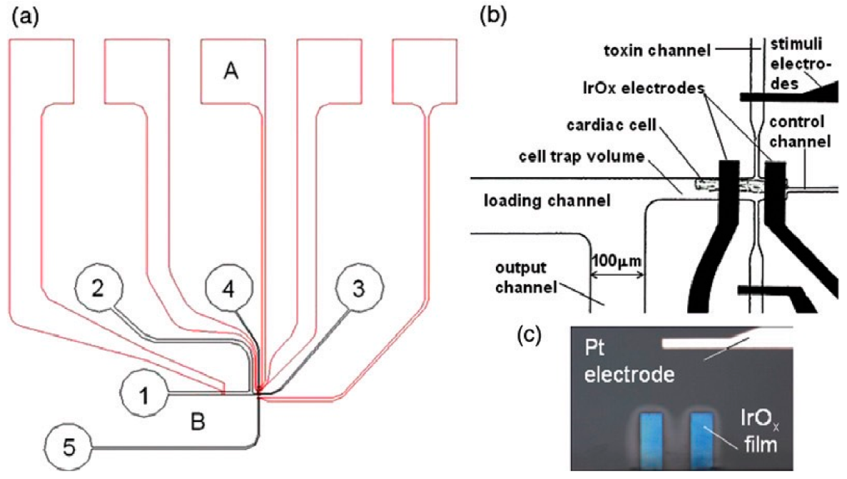
  137. Ges, I.A.; Ivanov, B.L.; Schaffer, D.K.; Lima, E.A.; Werdich, A.A.; Baudenbacher, F.J. Thin-film IrOx pH microelectrode for microfluidic-based microsystems. Biosens. Bioelectron. 2005, 21, 248–256. [Google Scholar] [CrossRef]
  138. Weltin, A.; Slotwinski, K.; Kieninger, J.; Moser, I.; Jobst, G.; Wego, M.; Ehret, R.; Urban, G.A. Cell culture monitoring for drug screening and cancer research: A transparent, microfluidic, multi-sensor microsystem. Lab A Chip 2014, 14, 138–146. [Google Scholar] [CrossRef] [PubMed]
  139. Dabaghi, M.; Saraei, N.; Xu, G.; Chandiramohan, A.; Yeung, J.; Nguyen, J.P.; Vukmirovic, M.; Selvaganapathy, P.R.; Hirota, J.A. PHAIR: A biosensor for pH measurement in air–liquid interface cell culture. Sci. Rep. 2021, 11, 3477. [Google Scholar] [CrossRef]
  140. Ges, I.; Baudenbacher, F. Microfluidic device to confine single cardiac myocytes in sub-nanoliter volumes for extracellular pH measurements. J. Exp. Nanosci. 2008, 3, 63–75. [Google Scholar] [CrossRef]
  141. Ges, I.A.; Ivanov, B.L.; Werdich, A.A.; Baudenbacher, F.J. Differential pH measurements of metabolic cellular activity in nl culture volumes using microfabricated iridium oxide electrodes. Biosens. Bioelectron. 2007, 22, 1303–1310. [Google Scholar] [CrossRef]
  142. Ge, Z.; Brown, C.W.; Sun, L.; Yang, S.C. Fiber-optic pH sensor based on evanescent wave absorption spectroscopy. Anal. Chem. 1993, 65, 2335–2338. [Google Scholar] [CrossRef]
  143. Motellier, S.; Noire, M.; Pitsch, H.; Dureault, B. pH determination of clay interstitial water using a fiber-optic sensor. Sens. Actuators B Chem. 1995, 29, 345–352. [Google Scholar] [CrossRef]
  144. Vishnoi, G.; Goel, T.C.; Pillai, P. pH Optrode for the Complete Working Range; SPIE: Boston, MA, USA, 1999; pp. 319–325. [Google Scholar]
  145. Lemos, S.G.; Nogueira, A.R.A.; Torre-Neto, A.; Parra, A.; Alonso, J. Soil calcium and pH monitoring sensor system. J. Agric. Food Chem. 2007, 55, 4658–4663. [Google Scholar] [CrossRef]
  146. Robinson, K.L.; Lawrence, N.S. Redox-sensitive copolymer: A single-component pH sensor. Anal. Chem. 2006, 78, 2450–2455. [Google Scholar] [CrossRef] [PubMed]
  147. Xu, X.; Goponenko, A.V.; Asher, S.A. Polymerized polyHEMA photonic crystals: pH and ethanol sensor materials. J. Am. Chem. Soc. 2008, 130, 3113–3119. [Google Scholar] [CrossRef] [PubMed]
  148. Johansson, G.; Karlberg, B.; Wikby, A. The hydrogen-ion selective glass electrode. Talanta 1975, 22, 953–966. [Google Scholar] [CrossRef]
  149. Wang, M.; Ha, Y. An electrochemical approach to monitor pH change in agar media during plant tissue culture. Biosens. Bioelectron. 2007, 22, 2718–2723. [Google Scholar] [CrossRef]
  150. Manz, A.; Graber, N.; Widmer, H. áM Miniaturized total chemical analysis systems: A novel concept for chemical sensing. Sens. Actuators B Chem. 1990, 1, 244–248. [Google Scholar] [CrossRef]
  151. Ahmet Burak, U. Development & Characterization of Multifunctional Microfluidic Materials. Ph.D. Thesis, NC State University, Raleigh, CA, USA, 2013. [Google Scholar]
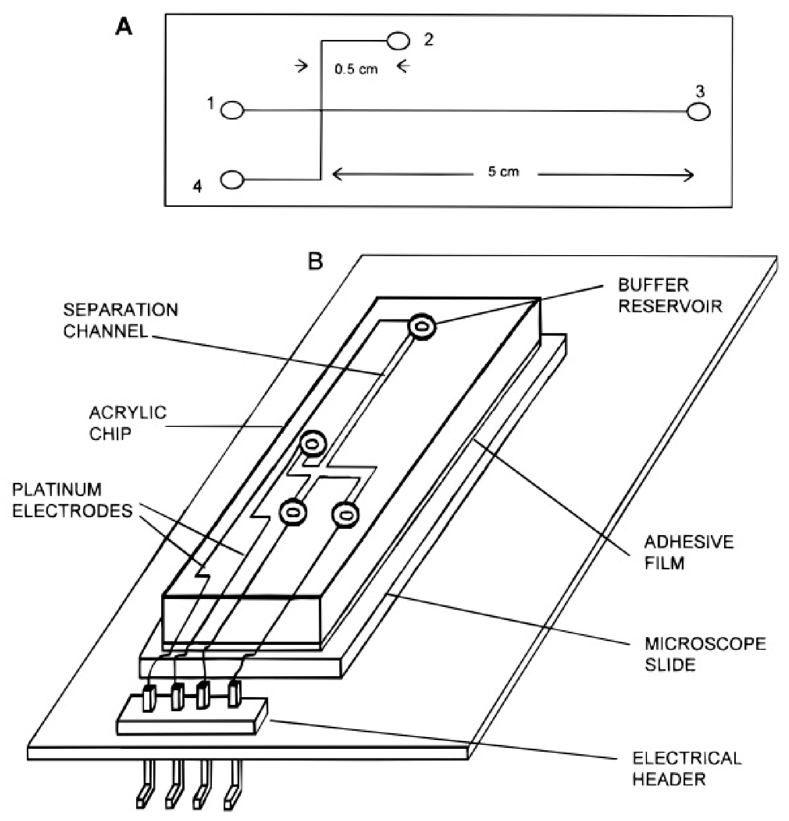
  152. McCormick, R.M.; Nelson, R.J.; Alonso-Amigo, M.G.; Benvegnu, D.J.; Hooper, H.H. Microchannel electrophoretic separations of DNA in injection-molded plastic substrates. Anal. Chem. 1997, 69, 2626–2630. [Google Scholar] [CrossRef]
  153. Tahirbegi, I.B.; Ehgartner, J.; Sulzer, P.; Zieger, S.; Kasjanow, A.; Paradiso, M.; Strobl, M.; Bouwes, D.; Mayr, T. Fast pesticide detection inside microfluidic device with integrated optical pH, oxygen sensors and algal fluorescence. Biosens. Bioelectron. 2017, 88, 188–195. [Google Scholar] [CrossRef]
  154. Liu, R.H.; Yu, Q.; Beebe, D.J. Fabrication and characterization of hydrogel-based microvalves. J. Microelectromech. Syst. 2002, 11, 45–53. [Google Scholar] [CrossRef]
  155. Buzby, J.C.; Roberts, T. Economic costs and trade impacts of microbial foodborne ilness. World Health Stat. Q. 1997, 50, 57–66. [Google Scholar]
  156. Bitton, G. Microbiology of Drinking Water Production and Distribution; Wiley: Hoboken, NJ, USA, 2014. [Google Scholar]
  157. Liu, J.; Jasim, I.; Shen, Z.; Zhao, L.; Dweik, M.; Zhang, S.; Almasri, M. A microfluidic based biosensor for rapid detection of Salmonella in food products. PLoS ONE 2019, 14, e0216873. [Google Scholar] [CrossRef]
  158. Huang, W.-D.; Deb, S.; Seo, Y.-S.; Rao, S.; Chiao, M.; Chiao, J. A passive radio-frequency pH-sensing tag for wireless food-quality monitoring. IEEE Sens. J. 2011, 12, 487–495. [Google Scholar] [CrossRef]
  159. Yue, Y.; Huo, F.; Lee, S.; Yin, C.; Yoon, J.; Chao, J.; Zhang, Y.; Cheng, F. A Dual Colorimetric/Fluorescence System for Determining pH Based on the Nucleophilic Addition Reaction of an o-Hydroxymerocyanine Dye. Chem. Eur. J. 2016, 22, 1239–1243. [Google Scholar] [CrossRef]
  160. Chen, S.; Hong, Y.; Liu, Y.; Liu, J.; Leung, C.W.; Li, M.; Kwok, R.T.; Zhao, E.; Lam, J.W.; Yu, Y. Full-range intracellular pH sensing by an aggregation-induced emission-active two-channel ratiometric fluorogen. J. Am. Chem. Soc. 2013, 135, 4926–4929. [Google Scholar] [CrossRef] [PubMed]
  161. Asanuma, D.; Takaoka, Y.; Namiki, S.; Takikawa, K.; Kamiya, M.; Nagano, T.; Urano, Y.; Hirose, K. Acidic-pH-activatable fluorescence probes for visualizing exocytosis dynamics. Angew. Chem. Int. Ed. 2014, 53, 6085–6089. [Google Scholar] [CrossRef] [PubMed]
  162. Best, Q.A.; Liu, C.; van Hoveln, P.D.; McCarroll, M.E.; Scott, C.N. Anilinomethylrhodamines: pH sensitive probes with tunable photophysical properties by substituent effect. J. Org. Chem. 2013, 78, 10134–10143. [Google Scholar] [CrossRef] [PubMed]
  163. Richter, C.; Schneider, C.; Quick, M.; Volz, P.; Mahrwald, R.; Hughes, J.; Dick, B.; Alexiev, U.; Ernsting, N. Dual-fluorescence pH probe for bio-labelling. Phys. Chem. Chem. Phys. 2015, 17, 30590–30597. [Google Scholar] [CrossRef]
  164. Park, Y.; Postupna, O.; Zhugayevych, A.; Shin, H.; Park, Y.-S.; Kim, B.; Yen, H.-J.; Cheruku, P.; Martinez, J.; Park, J. A new pH sensitive fluorescent and white light emissive material through controlled intermolecular charge transfer. Chem. Sci. 2015, 6, 789–797. [Google Scholar] [CrossRef] [PubMed] [Green Version]
  165. Trombetta, E.S.; Ebersold, M.; Garrett, W.; Pypaert, M.; Mellman, I. Activation of lysosomal function during dendritic cell maturation. Science 2003, 299, 1400–1403. [Google Scholar] [CrossRef]
  166. Punjiya, M.; Moon, C.H.; Matharu, Z.; Nejad, H.R.; Sonkusale, S. A three-dimensional electrochemical paper-based analytical device for low-cost diagnostics. Analyst 2018, 143, 1059–1064. [Google Scholar] [CrossRef]
  167. Lopez-Ruiz, N.; Curto, V.F.; Erenas, M.M.; Benito-Lopez, F.; Diamond, D.; Palma, A.J.; Capitan-Vallvey, L.F. Smartphone-based simultaneous pH and nitrite colorimetric determination for paper microfluidic devices. Anal. Chem. 2014, 86, 9554–9562. [Google Scholar] [CrossRef]
  168. Zhong, Q.; Ding, H.; Gao, B.; He, Z.; Gu, Z. Advances of microfluidics in biomedical engineering. Adv. Mater. Technol. 2019, 4, 1800663. [Google Scholar] [CrossRef]
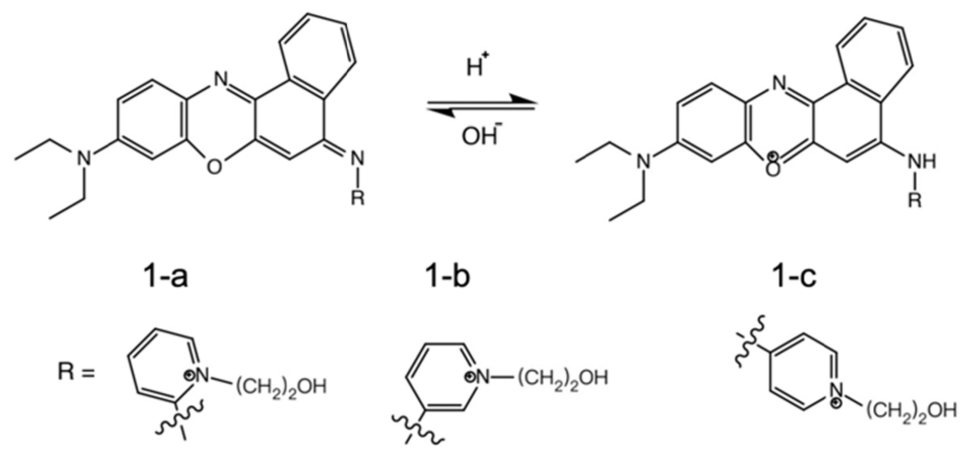
  169. Liu, W.; Sun, R.; Ge, J.-F.; Xu, Y.-J.; Xu, Y.; Lu, J.-M.; Itoh, I.; Ihara, M. Reversible Near-Infrared pH Probes Based on Benzo[a]phenoxazine. Anal. Chem. 2013, 85, 7419–7425. [Google Scholar] [CrossRef] [PubMed]
  170. Uria, N.; Abramova, N.; Bratov, A.; Muñoz-Pascual, F.-X.; Baldrich, E. Miniaturized metal oxide pH sensors for bacteria detection. Talanta 2016, 147, 364–369. [Google Scholar] [CrossRef]
  171. Hannah, S.; Blair, E.; Corrigan, D.K. Developments in microscale and nanoscale sensors for biomedical sensing. Curr. Opin. Electrochem. 2020, 23, 7–15. [Google Scholar] [CrossRef]
  172. Wang, H.-S.; Fu, G.-D.; Li, X.-S. Functional polymeric nanofibers from electrospinning. Recent Pat. Nanotechnol. 2009, 3, 21–31. [Google Scholar] [CrossRef]
  173. Li, D.; Xia, Y. Electrospinning of nanofibers: Reinventing the wheel? Adv. Mater. 2004, 16, 1151–1170. [Google Scholar] [CrossRef]
  174. Van der Schueren, L.; Mollet, T.; Ceylan, Ö.; De Clerck, K. The development of polyamide 6.6 nanofibres with a pH-sensitive function by electrospinning. Eur. Polym. J. 2010, 46, 2229–2239. [Google Scholar] [CrossRef]
  175. Jamil, M.S.; Jamil, M.A.; Mazhar, A.; Ikram, A.; Ahmed, A.; Munawar, U. Smart environment monitoring system by employing wireless sensor networks on vehicles for pollution free smart cities. Procedia Eng. 2015, 107, 480–484. [Google Scholar] [CrossRef]
  176. Ullo, S.L.; Sinha, G. Advances in smart environment monitoring systems using IoT and sensors. Sensors 2020, 20, 3113. [Google Scholar] [CrossRef]
  177. Kulkarni, P.; Kute, P. Internet of things based system for remote monitoring of weather parameters and applications. Int. J. Adv. Electron. Comput. Sci 2016, 3, 68–73. [Google Scholar]
  178. Jovanovska, E.M.; Davcev, D. No Pollution Smart City Sightseeing Based on WSN Monitoring System; IEEE: Bali, Indonesia, 2020; pp. 1–6. [Google Scholar]
  179. Arco, E.; Boccardo, P.; Gandino, F.; Lingua, A.; Noardo, F.; Rebaudengo, M. An integrated approach for pollution monitoring: Smart acquirement and smart information. ISPRS Ann. Photogramm. Remote Sens. Spat. Inf. Sci. 2016, 3, 67–74. [Google Scholar] [CrossRef]
  180. Pavithra, G. Intelligent monitoring device for agricultural greenhouse using IOT. J. Agric. Sci. Food Res. 2018, 9, 2–5. [Google Scholar]
  181. Elmustafa, S.A.A.; Mujtaba, E.Y. Internet of things in smart environment: Concept, applications, challenges, and future directions. World Sci. News 2019, 134, 1–51. [Google Scholar]
  182. Pathak, A.; AmazUddin, M.; Abedin, M.J.; Andersson, K.; Mustafa, R.; Hossain, M.S. IoT based smart system to support agricultural parameters: A case study. Procedia Comput. Sci. 2019, 155, 648–653. [Google Scholar] [CrossRef]
  183. Sivakannu, G.; Balaji, S. Implementation of smart farm monitoring using IoT. Int. J. Curr. Eng. Sci. Res 2017, 4, 21–27. [Google Scholar]
  184. Haick, H.; Tang, N. Artificial intelligence in medical sensors for clinical decisions. ACS Nano 2021, 15, 3557–3567. [Google Scholar] [CrossRef]
  185. Bandodkar, A.J.; Jeang, W.J.; Ghaffari, R.; Rogers, J.A. Wearable sensors for biochemical sweat analysis. Annu. Rev. Anal. Chem. 2019, 12, 1–22. [Google Scholar] [CrossRef]
  186. Wei, Y.; Gadaria-Rathod, N.; Epstein, S.; Asbell, P. Tear cytokine profile as a noninvasive biomarker of inflammation for ocular surface diseases: Standard operating procedures. Investig. Ophthalmol. Vis. Sci. 2013, 54, 8327–8336. [Google Scholar] [CrossRef]
  187. Qin, M.; Guo, H.; Dai, Z.; Yan, X.; Ning, X. Advances in flexible and wearable pH sensors for wound healing monitoring. J. Semicond. 2019, 40, 111607. [Google Scholar] [CrossRef]
  188. Vishinkin, R.; Haick, H. Nanoscale sensor technologies for disease detection via volatolomics. Small 2015, 11, 6142–6164. [Google Scholar] [CrossRef]
  189. He, J.; Baxter, S.L.; Xu, J.; Xu, J.; Zhou, X.; Zhang, K. The practical implementation of artificial intelligence technologies in medicine. Nat. Med. 2019, 25, 30–36. [Google Scholar] [CrossRef]
  190. Ahamed, F.; Farid, F. Applying Internet of Things and Machine-Learning for Personalized Healthcare: Issues and Challenges; IEEE: Bali, Indonesia, 2018; pp. 19–21. [Google Scholar]
  191. Jin, X.; Liu, C.; Xu, T.; Su, L.; Zhang, X. Artificial intelligence biosensors: Challenges and prospects. Biosens. Bioelectron. 2020, 165, 112412. [Google Scholar] [CrossRef] [PubMed]
  192. Shan, S.; Zhao, W.; Luo, J.; Yin, J.; Switzer, J.C.; Joseph, P.; Lu, S.; Poliks, M.; Zhong, C.-J. Flexibility characteristics of a polyethylene terephthalate chemiresistor coated with a nanoparticle thin film assembly. J. Mater. Chem. C 2014, 2, 1893–1903. [Google Scholar] [CrossRef]
  193. Manjakkal, L.; Szwagierczak, D.; Dahiya, R. Metal oxides based electrochemical pH sensors: Current progress and future perspectives. Prog. Mater. Sci. 2020, 109, 100635. [Google Scholar] [CrossRef]
  194. Manjakkal, L.; Navaraj, W.T.; Núñez, C.G.; Dahiya, R. Graphene–graphite polyurethane composite based high-energy density flexible supercapacitors. Adv. Sci. 2019, 6, 1802251. [Google Scholar] [CrossRef] [PubMed]
  195. Manjakkal, L.; Nikbakhtnasrabadi, F.; Dahiya, R. Energy Autonomous Sensors for Water Quality Monitoring; IEEE: Bali, Indonesia, 2018; pp. 1–4. [Google Scholar]
  196. Manjakkal, L.; Núñez, C.G.; Dang, W.; Dahiya, R. Flexible self-charging supercapacitor based on graphene-Ag-3D graphene foam electrodes. Nano Energy 2018, 51, 604–612. [Google Scholar] [CrossRef]
  197. Ryuh, Y.-S.; Yang, G.-H.; Liu, J.; Hu, H. A school of robotic fish for mariculture monitoring in the sea coast. J. Bionic Eng. 2015, 12, 37–46. [Google Scholar] [CrossRef]
  198. Valada, A.; Velagapudi, P.; Kannan, B.; Tomaszewski, C.; Kantor, G.; Scerri, P. Development of a Low Cost Multi-Robot Autonomous Marine Surface Platform; Springer: Berlin/Heidelberg, Germany, 2014; pp. 643–658. [Google Scholar]
  199. Sheppard Jr, N.F.; Tucker, R.C.; Salehi-Had, S. Design of a conductimetric pH microsensor based on reversibly swelling hydrogels. Sens. Actuators B Chem. 1993, 10, 73–77. [Google Scholar] [CrossRef]
Figure 1. Image of dissolved oxygen (DO), pH, temperature, and conductivity MEA sensors (a) and layers on each MEA sensor (b). Reprinted with permission from Ref. [21]. (Copyright, 2016, Elsevier).
Catalysts 12 01124 g001
Figure 2. The integration of carbon dots with different adjusted full-color emission and pH sensors. Reprinted with permission from Ref. [34]. (Copyright, 2014 ACS).
Catalysts 12 01124 g002
Figure 3. “Open” and “close” designs of the reference electrode compartment of the hydrogen reference electrode, reprinted with permission from reference [56]. (Copyright, ACS, 2020).
Catalysts 12 01124 g003
Figure 4. The design of glass electrode measurement, reprinted with permission from reference [57]. (Copyright, Portland Press, 1933).
Catalysts 12 01124 g004
Figure 5. The mechanism of quinone hydroquinone-SAM on a gold electrode surface. Reprinted with permission from Ref. [68]. (Copyright, ACS, 2013).
Catalysts 12 01124 g005
Figure 6. The specific structure of optical fiber pH sensor and its experimental design. (1) Signal light source (2) referred light source (3) photodiode and (4) dichroic mirror, reprinted with permission from reference [83]. (Copyright, Elsevier, 2014).
Catalysts 12 01124 g006
Figure 7. Integral diagram of the discrete ISFET. Reprinted with permission from Ref. [90]. (Copyright, 2001, Elsevier).
Catalysts 12 01124 g007
Figure 8. The diagram of a microfluidic sensing system. Reprinted with permission from Ref. [127]. (Copyright, 2019, MDPI).
Catalysts 12 01124 g008
Figure 9. (a) Diagram of a microfluidic sensing system including microfluidic instrument and pH electrodes to trap single cardiac myocytes and to test the metabolic activity: A (red)—microelectrode array system, B (black)—microfluidic network; for 1 to 5: they are input port, output port, control port, toxin, or stimuli port, respectively. (b) Specific structure of pH IrOx microelectrodes, stimulation electrodes, and the cell trap with loading and waste channels. (c) Optical picture of the IrOx pH-sensitive microelectrodes and the Pt stimulation electrode. Reprinted with permission from Ref. [138]. (Copyright, 2019, MDPI).
Catalysts 12 01124 g009
Figure 10. Design of the plastic microchannel chip (A) and ichnography (B) the details of the completed chip. Reprinted with permission from reference [152]. (Copyright, 1997, American Chemical Society).
Catalysts 12 01124 g010
Figure 11. The night-time ozone profile (NIR) fluorescent pH probes based on protonation of amine, three benzo[a]phenoxazine derivates [169].
Catalysts 12 01124 g011
Table 3. The pH measuring characteristics of metal oxide-based pH electrodes.
MaterialsMethodpH RangepH SensitivityStabilityReference
PbO2Thermal method1.2–7.5112 mV/decadeOnly stable in the acid region,Lima et al. [101] 2005
Over 7.588 mV/decadeNon-linear behavior of the pH response
OsO22–1151.2 mV/pHwere sensitive to oxidizing and reducing agentsWilliam et al. [102] 1984
TiO22–1255 mV/pH
PtO25–1046.7 mV/pH
WO3Magnetron sputtering2–1241 mV/pHHigh stability (over a month)Zhang et al. [103] 2009
IrOxElectrochemical63–82 mV/pHThe potential always stabilized in a few minutes.Baur et al. [104] 1998
IrO2Electrospinning3–1267.1–70.15 mV/pHstable in one weekDong et al. [105]
Table 4. The related properties of these three probes [95].
TypeProbe 1-a (nm)Probe 1-b (nm)Probe 1-c (nm)
λex597663599
λem688697694
pKa2.75.87.1
Publisher’s Note: MDPI stays neutral with regard to jurisdictional claims in published maps and institutional affiliations.

