Article

In Situ Electrochemical Characterization of a Microbial Fuel Cell Biocathode Running on Wastewater

by
Sudarsu V. Ramanaiah
1,2,*,
Cristina M. Cordas
3,*,
Sara Matias
1 and
Luís P. Fonseca
1
1
iBB—Institute for Bioengineering and Biosciences, Instituto Superior Técnico, Universidade de Lisboa, Av. Rovisco Pais, 1049-001 Lisboa, Portugal
2
Food and Biotechnology Research Lab, South Ural State University (National Research University), 454080 Chelyabinsk, Russia
3
LAQV, REQUIMTE, NOVA School of Science and Technology, FCT NOVA, Universidade NOVA de Lisboa, Campus de Caparica, 2829-516 Caparica, Portugal
*
Authors to whom correspondence should be addressed.
Catalysts 2021, 11(7), 839; https://doi.org/10.3390/catal11070839
Submission received: 16 May 2021 / Revised: 24 June 2021 / Accepted: 9 July 2021 / Published: 11 July 2021
(This article belongs to the Special Issue Bifunctional Oxygen Electrocatalysts)

Abstract

:
The electrochemical features of microbial fuel cells’ biocathodes, running on wastewater, were evaluated by cyclic voltammetry. Ex situ and in situ electrochemical assays were performed and the redox processes associated with the presence of microorganisms and/or biofilms were attained. Different controls using sterile media (abiotic cathode microbial fuel cell) and membranes covering the electrodes were performed to evaluate the source of the electrochemistry response (surface biofilms vs. biotic electrolyte). The bacteria presence, in particular when biofilms are allowed to develop, was related with the enhanced active redox processes associated with an improved catalytic activity, namely for oxygen reduction, when compared with the results attained for an abiotic microbial fuel cell cathode. The microbial main composition was also attained and is in agreement with other reported studies. The current study aims contributing to the establishment of the advantages of using biocathodes rather than abiotic, whose conditions are frequently harder to control and to contribute to a better understanding of the bioelectrochemical processes occurring on the biotic chambers and the electrode surfaces.

1. Introduction

Bacteria and yeasts are frequently used to catalyze the anodic and cathodic half-reactions in different bioelectrochemical systems and, in particular, in fuel cells, denominated as microbial fuel cells (MFC). On MFC organic compounds are used by these organisms as fuel using the anode and/or the cathode as electronic partners, taking advantage of the microorganisms’ ability to exchange electrons with electrodes, either directly or through extracellular small electron carriers, e.g., cytochromes, for which the surfaces mimic the physiological redox partners [1,2,3]. One of the advantages of MFC is their ability to use a wide sort of biomass-derived fuels with long term durability of microbial consortia [4]. Many different microorganisms have been utilized in MFCs, both as mixed and single strain cultures, such as Geobacteracea, Desulfobulbus or Desulfovibrio families, among others [5,6,7,8].
Electrochemistry techniques, namely cyclic voltammetry (CV), among others, allowed several authors to observe biofilms attached to anode and/or cathode electrodes’ effect. In studies electrochemical properties consistent with the hypothesis that biofilms work as catalysts, by producing electroactive biofilms or extracellular carriers, that enables the direct electron exchange with the electrode surfaces and, thus, enhancing the electrocatalytic response were reported [9,10,11,12,13,14]. In some systems, the electrochemical features of the anode and the cathode are different, even if the base materials are the same, reflecting possible differences in their extracellular electron transfer properties induced by the electrodes’ potential. In addition, the media also influences the electrochemical behavior of the electrodes covered with biofilms, for instance sulphate reducing bacteria possess different mechanism in the presence of nitrate or sulphate [15]. The potential, at which the electrodes are set, or the potential difference between them, has also been discussed as an important factor influencing the anode and cathode electrochemical response [10,12,14,15,16]. Monitoring the current as a function of potential and scan rate yields dependencies that can provide detailed mechanistic information about the electron transfer process from substrates (such as glucose, acetate, etc.) to the electrode surface. Changing the electrode potential will vary the driving force and may change the heterogeneous electron transfer step (across the catalyst/electrode interface). The present study focuses on the electrocatalytic activity in the anodic and cathodic chambers of a MFC running with a biocathode and harvesting electricity from bacteria cells present in wastewater. In situ electrochemical experiments were performed, including controls avoiding biofilms formation, to evaluate the biocathode MFC electrochemical behavior, together with experiments using sterile media in the cathodic compartment, enabling the comparison with an abiotic cathode running at equivalent conditions.

2. Results and Discussion

2.1. Ex Situ Electrochemical Characterization of Samples from MFC Using a Biocathode

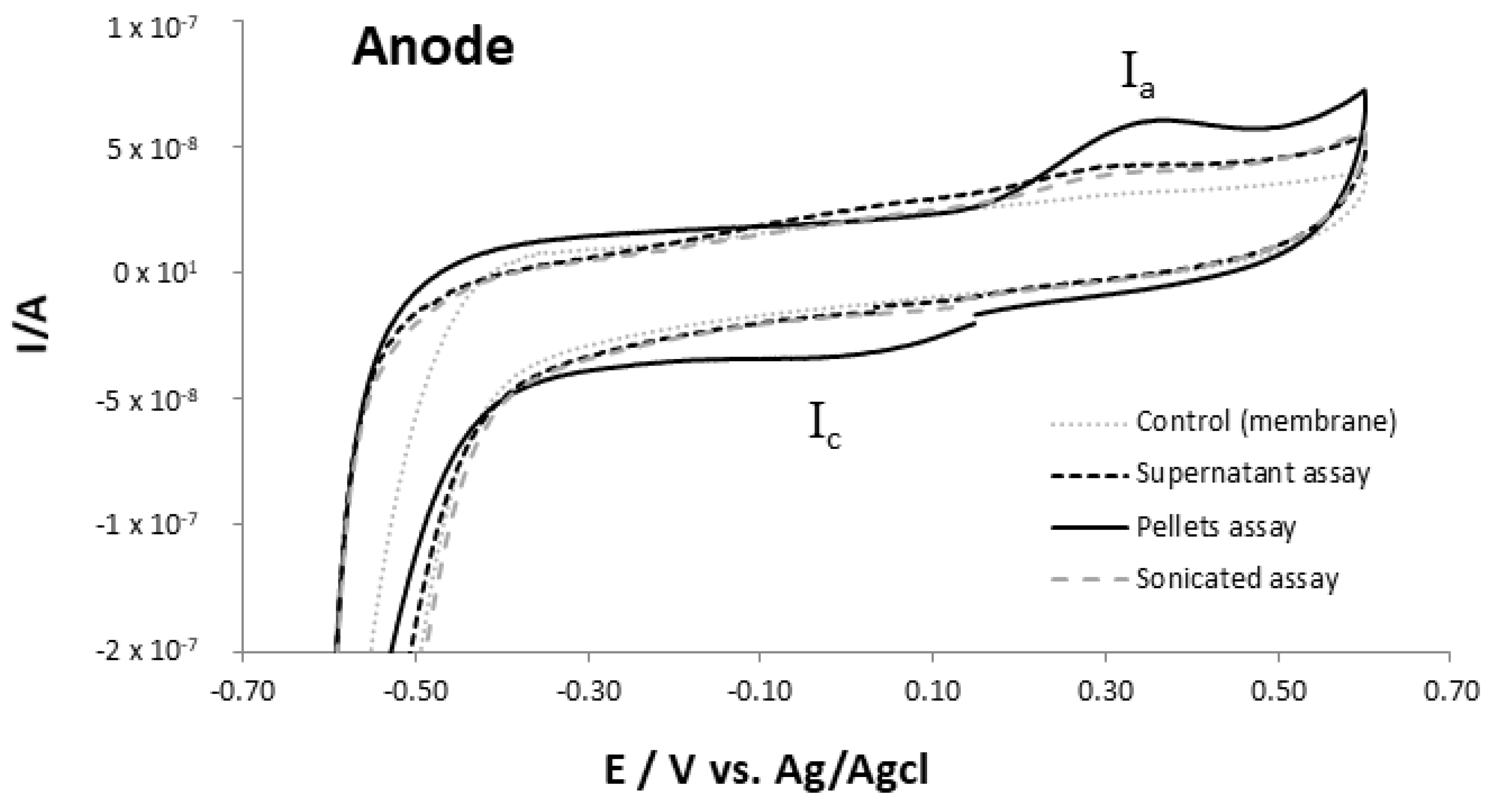
2.1.1. Anodic Chamber

The samples were taken and prepared as described on Section 3.4 and characterized by cyclic voltammetry to determine its electrochemical activity. From the results shown in Figure 1, it is possible to identify two redox processes associated with the presence of bacterial cells, namely an anodic wave around +0.4 V and a cathodic one at 0 V (respectively Ia and Ic), observed in all sample assays, more pronounced for the pellets’ samples.
These processes seem to be related with the presence of electron transfer proteins, such as cytochromes and small iron-sulfur proteins that possess metal centers [11,17,18,19], as pointed by other authors. The observed process I has a midpoint potential of approximately +0.2 V vs. Ag/AgCl (+0.39 V vs. NHE), which is within the range of several cytochromes reduction potential associated with biofilms and extracellular electron transfer pathways towards electrodes [20,21]. A high cathodic current starts to develop under −0.5 V, also visible in the controls, which agrees with the high electrocatalytic activity of the gold electrode surface towards the oxygen reduction (even if residual at the cell). The oxygen reduction, however, is slightly shifted to more negative potentials in the assays with the pellets sample, indicating the presence of biological material in the gold surface, lowering the oxygen diffusion and its electrocatalytic reaction on gold.

2.1.2. Cathodic Chamber

The ex situ electrochemical characterization of the bacterial cells collected from the cathodic chamber of the MFC reactor is summarized in Figure 2. The results of the biocathodic chamber do not show significant differences from the anodic. Again, it is possible to identify two redox processes associated with the presence of bacterial cells, namely an anodic wave around +0.3 V (I’a), slightly shifted towards more negative values when compared with the anodic chamber results and that seems due to more than one process (overlaid); and a cathodic one at +0.1 V (I’c), again, better observed for the pellets’ samples, corresponding to an approximately midpoint potential of +0.2 V vs. Ag/AgCl as observed for the anodic chamber samples. From the comparison with the control assays, these processes seem to be the result of the presence of proteins, possessing metal centers. The potentials at which the redox processes are visible are consistent with the potential values at which cytochromes and/or small iron sulfur proteins present redox activity [17,18,19,21,22]. In addition, other catalytic processes have been related with processes occurring on MFC electrodes, observed approximately at some potentials [11], also pointing to the effect of the microorganisms. A high cathodic current starts to develop around −0.5 V, also visible in the controls, that is slightly shifted to more negative potentials in the assays with the biological samples. As discussed before, this shift should be related with the presence of adsorbed material in the gold surface that hinders the electrode direct oxygen reduction process, as observed in the anodic samples. A small anodic wave develops around −0.6 V, more visible for the supernatant samples that seems associated to the reverse reaction, namely, the oxygen regeneration from intermediate molecules adsorbed or retain at the surface.
Comparing the results obtained between all the cathodic chamber assays samples (supernatant, pellets and sonicated) and corresponding controls, it is clear that the anodic and cathodic processes, showing midpoint potential close to +0.3 V, are visible in all samples, but not at the controls. The processes are more clearly observed in the pellet’s assays and are consequence from the larger biological material presence. The results attained in this section will be compared with the in situ assays (next section), allowing us to evaluate the role of the immobilized biological material for the MFC electroactivity, by comparing the electrochemical features attained from the sessile and the immobilized microorganisms on the electrode surfaces

2.2. In Situ Electrochemical Characterization of Samples from MFC Using a Biocathode

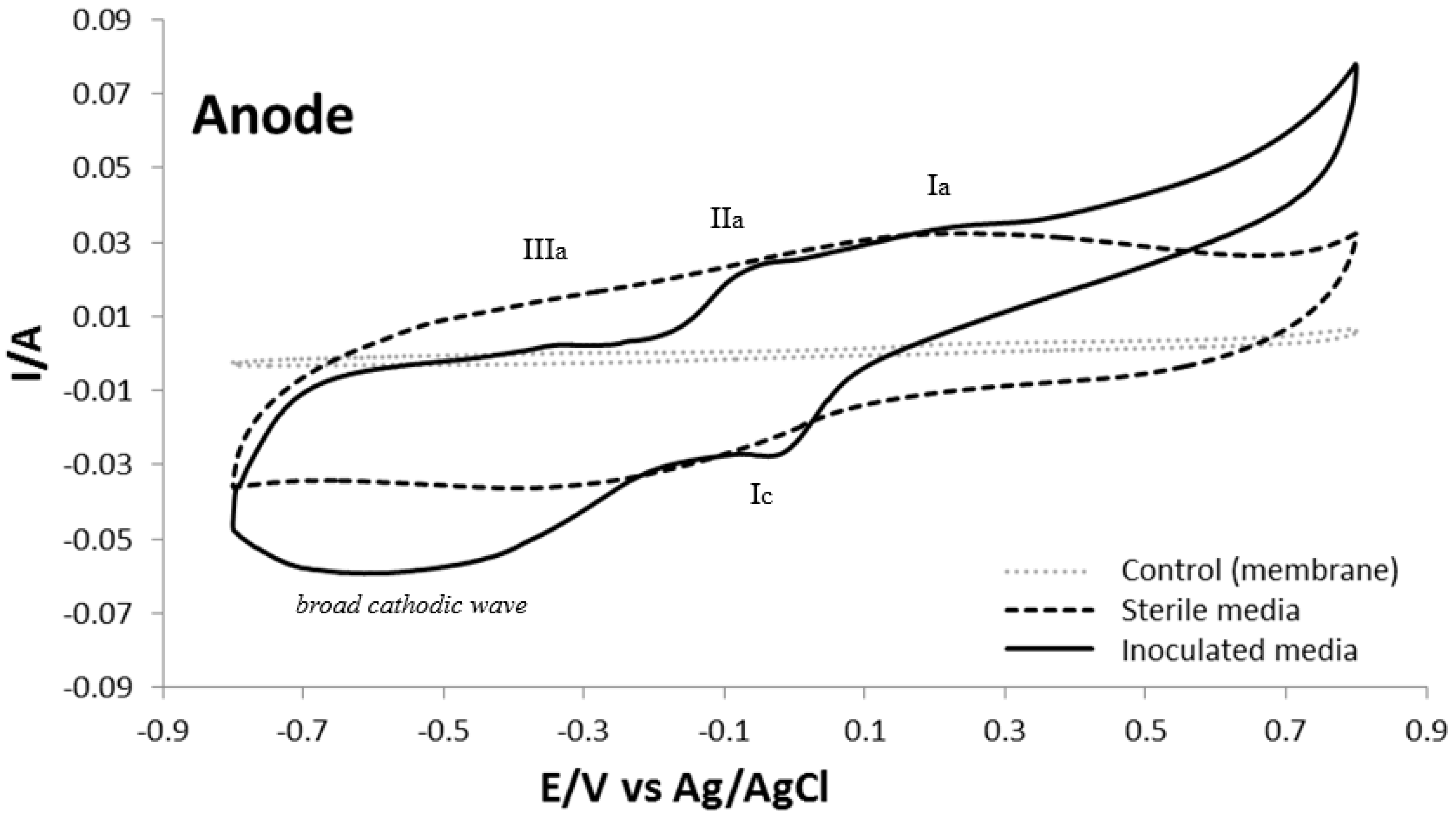
2.2.1. Anodic Chamber

Cyclic voltammetry assays were carried out in situ in the anodic chamber using regular inoculated media (1:3, wastewater accordingly with the procedure described in Section 3.2) and sterile media for comparison. Electrochemical features of assays with cellulose membranes covering both the anode and cathode electrodes, were also used as control to evaluate if the observed electrochemical activity is due to biofilms or redox compounds released into the solution resulting from the bacteria metabolism. The resulting voltammograms (Figure 3), from the anode containing biofilm, used as working electrode directly, present considerable differences from the controls (sterile and membranes’ electrodes). Three anodic processes are detected, approximately at +0.2, −0.1 and −0.35 V, denoted respectively as Ia, IIa and IIIa), whereas one cathodic peak, around 0 V (Ic) and a broad cathodic wave, between approximately −0.7 and −0.3 V, are observed.
Process I is similar to the one observed in the ex situ assays (see Figure 1), when using samples of the non-sterile media, being possible to estimate a midpoint potential approximately of +0.05 V vs. Ag/AgCl (+0.24 V vs. NHE). Although slightly more negative, this value is in agreement with the redox potentials found for small extracellular proteins, as mentioned before [21,22,23].
The broad cathodic wave was associated to the reduction of oxygen and oxygenated species in solution; it starts to develop around −0.3 V, at more positive values than the ones found when gold electrodes were used (in the ex situ assays), implying that biological material on the graphite felts present enhanced catalytic activity towards oxygen reduction, even when O2 presence is residual. The processes IIa and IIIa seem to be directly related with the reverse reaction of the cathodic processes observed in this broad cathodic wave. Using the sterile media, a different pattern is observed and two broad processes can be observed, an anodic wave presenting the maximum intensity around +0.15 V and a cathodic wave with maximum current intensity around −0.30 V. These are observed for many carbon materials and were associated to the carbon surface inherent redox processes in aqueous solution [24]. The membranes’ control has shown much less intense currents, also due to a less diffusion towards the electrode and small broad anodic and cathodic processes around +0.2 V and −0.35 V, respectively, are visible in a similar pattern to the sterile medium (Table 1; Figure 2). The results suggest that the anodic and cathodic media with biofilm formation present redox compounds (electron shuttles) resulting from the biological material presence that contribute for the enhanced electron transfer activity to the anode and catalytic activity and, consistently, producing an operative MFC.

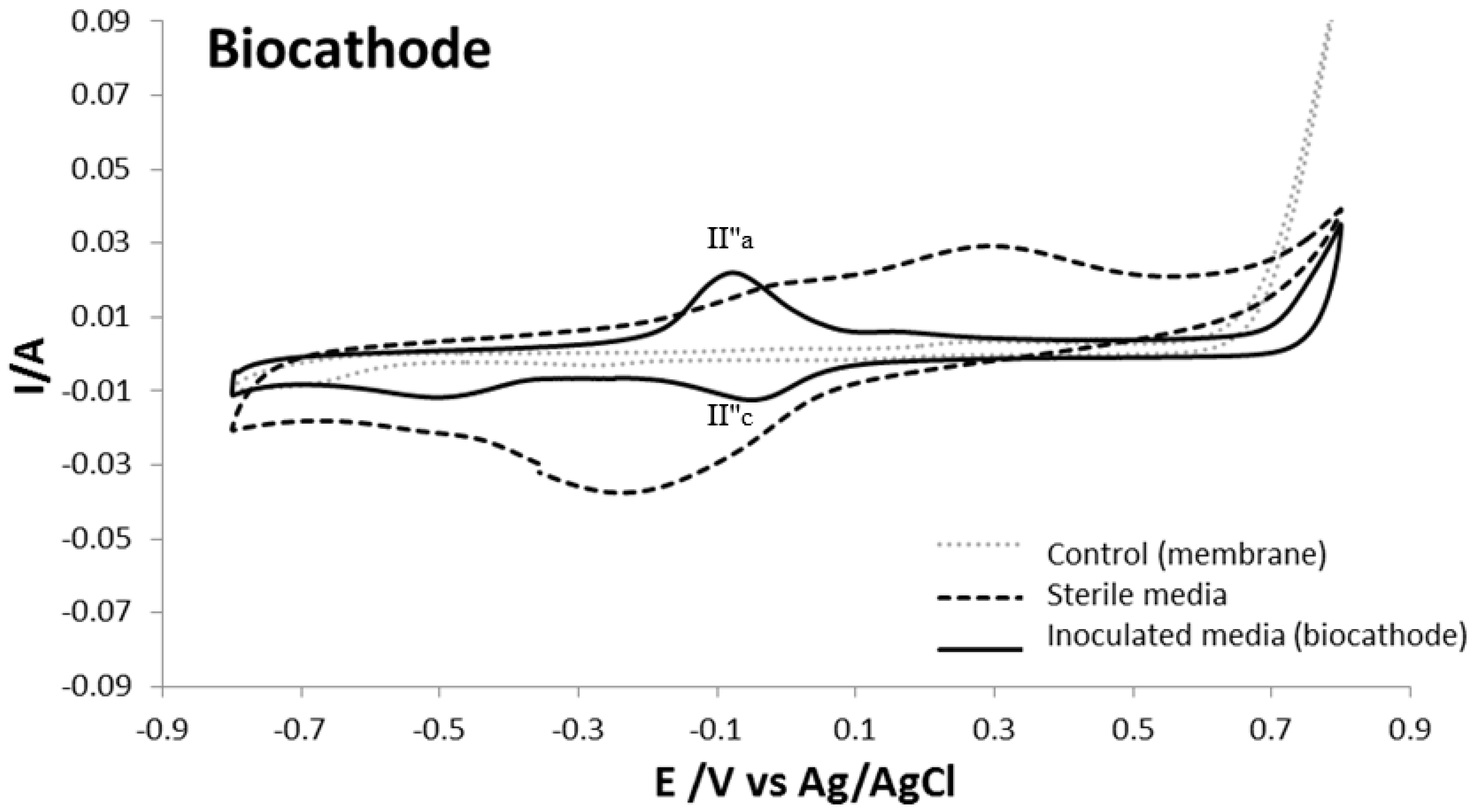
2.2.2. Biocathodic Chamber

The cyclic voltammetry in situ features of the aerobic biocathode chamber was performed as described for the anodic chamber in the previous Section 2.2.1, using the MFC cathode as working electrode. Again, the controls, performed using sterile media (corresponding to an abiotic cathode) and the MFC cathode covered by cellulose membranes, are shown (Figure 4).
There is a significant difference from the biocathode response functioning as working electrode in the CV assays (Figure 4) from the results attained ex situ (Figure 2) with the non-sterile (inoculated media) attained from the cathodic chamber. In the in situ study, a redox process (that seems, however, partially influenced by another overlaid process) is visible at potential values approximately to −0.1 V vs. Ag/Ag/Cl (≈+0.097 V vs. NHE). This process appears at more negative potentials than process I’ observed in the ex situ assays. In addition, the processes do not seem related with process II′a. As so, the anodic and cathodic processes were denoted as II″a and II″c. The lower potential difference between the anodic and cathodic processes seems related with surface confined redox processes, probably associated with the biofilm coating the electrode. Again, the potentials are close to others already reported for proteins and other electron carriers associated with the electron transfer between bacteria and the electrodes’ surfaces [25]. In addition, process II″, a small anodic wave at +0.15 V and a cathodic process at −0.5 V are observed. These seem to have some correspondence with the waves registered with the electrode in sterile media (abiotic cathode) and should be related with the presence of oxygen (and the carbon felt surface). The current intensity differences are most probably due to the presence of the biofilms (Figure 4) that hinders the oxygen diffusion to the carbon felt. No obvious redox processes associated with the microorganisms were observed from the control biocathode chamber, covered with the cellulose membrane. Using sterile media (corresponding to an abiotic cathode, as mentioned) two anodic waves develop around +0.3 and +0.05 V and two cathodic processes are also visible, a broad wave around −0.2 V and a smaller wave at −0.53 V all associated with the oxygen and oxygenated species reaction on the cathode carbon felt surface. The membranes’ control shows less intense currents, due to the diffusion of electrolyte that is reduced by the membrane presence and small anodic and cathodic processes around +0.26 and, approximately, at −0.25 V (Table 1; Figure 4) can be observed.
Based on all the results, the biocathode containing attached biofilms reveals more electron transfer and electrocatalytic activity towards oxygen reduction when compared to both the sterile media (abiotic cathode) and the membranes’ control. The results point to a significant role of the biofilms and the metabolic released redox biological compounds (electron shuttles) in the power density production in the biocathode MFC.

2.3. Composition of Wastewaters Analysis

The anode and the biocathode MFC chambers composition on microorganisms were attained, after 18 cycles showing some differences, in spite of the initial inoculum being the same (Table 2). This fact is in agreement with reported data on the adaptation of strains to the conditions when in the presence of electrified surfaces, namely the MFC electrodes [2,26].
It should be noted that the number of electroactive microorganisms is in constant expansion, such as the case of Methanobacteriales or Methanosarcinales (also found in abundance in the biocathode chamber), as recently described [27,28,29]. In addition, the results are in agreement with the observations that some microorganisms, in particular Methanosarcina, were enriched on carbon-based materials such as carbon felt [30] and recent reports point to the ability of this strain to direct exchange electrons with electrified surfaces [28]. Proteobacteria seems more abundant in the cathodic chamber as also already reported [25]. The presence of mixed consortium electroactive microorganisms in wastewater has been show as an advantage [31]. The direct electron transfer to electrodes and the ability to interspecies electron transfer contributes to enhance MFC properties in O2 and oxygenated species reduction, leading to an enhanced energy production, besides organic matter degradation. The understanding and exploration of microbial presence/biofilms formation in MFC is, thus, an increasing field of interest [32], including the best operational conditions allowing acclimation and selected enrichment of the most promising species considering the overall goals [2].

3. Materials and Methods

3.1. MFC Construction

Double chambered MFC was in-house constructed using acrylic glass material with equal volumes (working volume, 0.36 L) of anode and cathode compartments, separated by a cationic exchange membrane (Nafion, Alfa Aesar, Karlsruhe, Germany). Graphite felts (GF, Alfa Aesar, Karlsruhe, Germany, 6 × 6 cm; 5 mm thick; surface area 36 cm2) were used as electrodes. Copper wires were used for contact with electrodes, isolated with epoxy resin. In addition, the contacts with the chambers were isolated/separated by rubber gaskets. Appropriate sampling ports were designed. For the MFCs cathodes, a continuous air flow (passing through 0.2 μm filters) was provided through an air-pump to maintain constant the amount of dissolved oxygen.

3.2. Biocatalyst-Consortium from Wastewater

Aerobic mixed consortium from activated sludge was collected at the wastewater treatment plant of Chelas (Lisbon, Portugal) and was used to inoculate the MFC anode and cathode compartments with wastewater (1:3) and electrolyte (described in Section 3.3). For the MFC using abiotic cathode the inoculation was performed only in the anode compartment. MFC biocathode identification of microorganisms was done according to the protocol from Ramos et al. [33]. The 16S rRNA gene sequence analysis was conducted to identify the taxonomic affinities of a broad range of microorganisms [34]. Universal primers for archaea and bacteria were chosen (based on previous literature) to amplify the partial sequence of 16S rRNA to comprise the largest number of microorganisms. Nucleotide sequences of 16S rRNA encoding genes were retrieved from the Ribosomal Database Project [35] and GenBank (http://www.ncbi.nlm.nih.gov/Genbank/, accessed on Nucleic Acids Research, accessed on 1 January 2013; 41(D1):D36-42).

3.3. MFC Operation

MFC were operated in aerated abiotic cathode or biocathode conditions. MFC anodic (and cathodic for the biocathode MFC) chambers were inoculated with aerobic mixed consortia and operated under respective microenvironment. Anode and cathode chambers were fed with electrolyte with composition, in g/L, comprising of sodium acetate (0.82) and sodium carbonate (0.31) as the sole carbon sources in both chambers. For both chambers, the remaining media composition was 50 × 10−3 M phosphate buffer and nutrient solution (g/L): KH2PO4 (2.88), K2HPO4 (5.02), NH4Cl (0.53), C10H16N2O8 (0.50), MgSO4·7H2O (0.37), MnCl2·4H2O (0.59), COCl2·6H2O (0.08), CaCl2·2H2O (0.11), ZnCl2 (0.05), CuSO4·5H2O (0.01), AlK(SO4)2 (0.01), H3BO3 (0.01), Na2MoO4·2H20 (0.02), Na2SeO3 (0.001), Na2WO4·2H2O (0.01), NiCl2·6H2O (0.02) and FeCl3·6H2O (0.27) [36]. Prior to feeding, the pH of the electrolyte was maintained at 7.0 in both chambers. Anodic chamber was purged with N2 gas for at least 20 min to maintain the anaerobic microenvironment after the fed change and sampling; in the cathodic chamber a continuous supply of air was maintained through an air-pump (ELITE-801, Rolf C. Nagen, UK Ltd., Castleford, UK) to keep constant the amount of oxygen (the electron acceptor). The media solutions were changed when the voltage decreased to 50 × 10−3 V and the suspended biomass was reserved, forming a complete fed-batch cycle. MFC was operated at room temperature (app. 25 °C) and electrodes were connected through a copper wire to a fixed load of external resistance of 1000 Ω. This resistance value corresponds to the stabilized MFC operation established over a study with R between 15 to 15,000 Ω (see SI, Figure S1). MFC was operating in a total of 28 fed-batch cycles (corresponding to 220 days).

3.4. MFC Analysis

MFC was operating in a total of 28 fed-batch cycles, stabilizing at the 18 fed-batch cycle with high removal efficiency. The stabilized MFC biocathode was shown a maximum open circuit voltage (OCV) of +439 mV with a corresponding external resistor of 1000 Ω. The operational parameters are shown in Table 3 (the main operational parameters for the corresponding abiotic cathode MFC are presented in SI, Table S1, for comparison).
At this stage, wastewater samples from MFC anodic and cathodic chambers were collected and its features were analyzed by cyclic voltammetry (CV) to characterize the oxidation-reduction reactions of the suspension bacteria cells (the ex situ characterization, see Section 2.1). The ex situ cyclic voltammetry analysis used a CHI 440B potentiostat from CHI Instruments, USA. For the CV characterization of each chamber media, the used electrodes were a gold working electrode disk with ϕ = 2 mm (Bioanalytical systems, West Lafayette, IN, USA, model: 2014), an Ag/AgCl reference (RE-1B, BAS, Tokyo, Japan) and a platinum counter electrode (Bioanalytical systems, West Lafayette, IN, USA, model: 4230). One compartment electrochemical cell was used.
From each sample (control, supernatant, pellets and after sonication samples) 200 μL volume was taken and placed on a cellulose membrane (3.5 kDa cut-off), that covered the working electrode, in a thin-layer configuration. The control was performed with sterile media using the same procedures as for the samples. Nitrogen gas was used for oxygen removal, by bubbling at least for 20 min before the assays and continuously flushed into the electrochemical cell headspace during the measurements. CV was performed at 20 mV s−1 scan rate, after 5 min of equilibrium at open circuit potential. The potential window between +0.6 and −0.6 V was used to attain the oxidation/reduction processes characteristic of each sample. The working samples from the MFC anodic and cathodic chambers’ wastewaters (running with the biocathode), were separated into supernatant, pellets and sonicated (the pellets samples after sonication) and prepared for the CV analysis by the following procedures: each 1.5 mL of wastewater samples from the chambers were centrifuged at 12,000 RPM for 5 min and the upper layers (supernatant) were collected; the remaining pellets’ samples were washed with 50 mM phosphate buffer (pH 7.0), centrifuged at 12000 RPM for 5 min, to the final remaining samples (pellets); the preparation of sonicated samples were as the previous pellets, but with the additional step of sonication in a water bath (NAHITA, Ultra Sonic 220–240 V) for 5 min.
The in situ bioelectrochemical assays (see Section 2.2) were performed considering MFC reactor’s anode and cathode graphite felts as working and counter electrodes (and vice versa), using an Ag/AgCl reference electrode introduced in each chamber, interrupting momentarily the MFC operation. In these assays, scan rate was 20 × 10−3 V s−1 over the potential range +0.8 to −0.8 V. Additional controls were measured in parallel MFC reactors running the same time and using the same methodology, but with cellulose membranes (3.5 kDa cut-off) covering the electrodes, as described elsewhere [37], to avoid direct contact of bacteria with the electrodes, hindering biofilms formation.

4. Conclusions

The electrochemical assays of the wastewater samples retrieved from the MFC running with a biocathode clearly show the presence of anodic and cathodic redox processes associated with the microorganisms and biofilms’ presence. The possible occurrence of extracellular bacterial proteins with electron transfer properties must be taken into consideration and although its identification is not under the scope of this work, future studies should invest on this topic. The in situ MFC assays using the chambers’ electrodes, covered by biofilms, have shown interesting redox features evidencing the biofilms role in the production and conduction of electrons and in the catalytic properties towards oxygen reduction. Under the tested experimental conditions, the biocathode operating conditions seem the most favorable for cathodic electrochemical reduction of oxygen. The attained results confirm that biocathodes are viable and easier alternatives to the use of conventional catalysts in MFC devices. Studies to select the best operational MFC conditions using biocathodes to promote the enrichment of the most promising species, considering the goals, are a route that should be pursued.

Supplementary Materials

The following are available online at https://www.mdpi.com/article/10.3390/catal11070839/s1, Figure S1: Variation of the MFC (running with a biocathode) output voltage versus the applied external resistance (between 15 to 15,000 Ω); the remaining experimental conditions are described in the manuscript main text; Figure S2: Ex-situ cyclic voltammograms attained with anodic chamber wastewater sample with bacteria cells in suspension (on gold electrode); full potential window; scan rate 20 × 10−3 V s−1; Figure S3: Ex-situ cyclic voltammograms attained in the biocathode chamber wastewater sample with bacteria cells in suspension (on gold electrode); full potential window; scan rate 20 × 10–3 V s−1; Table S1: Abiotic cathode MFC operating parameters.

Author Contributions

S.V.R., C.M.C. and S.M.: experimental procedures, results discussion, data treatment, draft revision; L.P.F.: results discussion, draft revision. All authors have read and agreed to the published version of the manuscript.

Funding

FCT—Fundação para a Ciência e a Tecnologia, Portugal (Grant number grant No. FRH/BPD/33864/2009). This work was supported by the Associate Laboratory for Green Chemistry—LAQV which is financed by national funds from FCT/MCTES (UIDB/50006/2020 and UIDP/50006/2020).

Data Availability Statement

The datasets generated for this study are available on request to the corresponding.

Acknowledgments

S.V.R. acknowledges the financial support from FCT (Portuguese Foundation for Science and Technology) for Postdoctoral Research grant No. FRH/BPD/33864/2009. This work was supported by the Associate Laboratory for Green Chemistry—LAQV which is financed by national funds from FCT/MCTES (UIDB/50006/2020 and UIDP/50006/2020).

Conflicts of Interest

The authors declare no conflict of interest.

References

  1. Biffinger, J.C.; Pietron, J.; Ray, R.; Little, B.; Ringeisen, B.R. A Biofilm Enhanced Miniature Microbial Fuel Cell Using Shewanella Oneidensis DSP10 and Oxygen Reduction Cathodes. Biosens. Bioelectron. 2007, 22, 1672–1679. [Google Scholar] [CrossRef] [PubMed] [Green Version]
  2. Logan, B.E.; Rossi, R.; Ragab, A.; Saikaly, P.E. Electroactive Microorganisms in Bioelectrochemical Systems. Nat. Rev. Microbiol. 2019, 17, 307–319. [Google Scholar] [CrossRef] [PubMed]
  3. Schaetzle, O.; Barrière, F.; Baronian, K. Bacteria and Yeasts as Catalysts in Microbial Fuel Cells: Electron Transfer from Micro-Organisms to Electrodes for Green Electricity. Energy Environ. Sci. 2008, 1, 607–620. [Google Scholar] [CrossRef]
  4. Mohanakrishna, G.; Venkata Mohan, S.; Sarma, P.N. Bio-Electrochemical Treatment of Distillery Wastewater in Microbial Fuel Cell Facilitating Decolorization and Desalination along with Power Generation. J. Hazard. Mater. 2010, 177, 487–494. [Google Scholar] [CrossRef]
  5. Tender, L.M.; Gray, S.A.; Groveman, E.; Lowy, D.A.; Kauffman, P.; Melhado, J.; Tyce, R.C.; Flynn, D.; Petrecca, R.; Dobarro, J. The First Demonstration of a Microbial Fuel Cell as a Viable Power Supply: Powering a Meteorological Buoy. J. Power Sources 2008, 179. [Google Scholar] [CrossRef]
  6. Shukla, A.K.; Suresh, P.; Berchmans, S.; Rajendran, A. Biological Fuel Cells and Their Applications. Curr. Sci. 2004, 87, 455–468. [Google Scholar]
  7. Lovley, D.R. Bug Juice: Harvesting Electricity with Microorganisms. Nat. Rev. Microbiol. 2006, 4, 497–508. [Google Scholar] [CrossRef]
  8. Du, Z.; Li, H.; Gu, T. A State of the Art Review on Microbial Fuel Cells: A Promising Technology for Wastewater Treatment and Bioenergy. Biotechnol. Adv. 2007, 25, 464–482. [Google Scholar] [CrossRef]
  9. Bond, D.R.; Lovley, D.R. Electricity Production by Geobacter Sulfurreducens Attached to Electrodes. Appl. Environ. Microbiol. 2003, 69, 1548–1555. [Google Scholar] [CrossRef] [PubMed] [Green Version]
  10. Cordas, C.M.; Guerra, L.T.; Xavier, C.; Moura, J.J.G. Electroactive Biofilms of Sulphate Reducing Bacteria. Electrochim. Acta 2008, 54. [Google Scholar] [CrossRef]
  11. Massaglia, G.; Fiorello, I.; Sacco, A.; Margaria, V.; Pirri, C.F.; Quaglio, M. Biohybrid Cathode in Single Chamber Microbial Fuel Cell. Nanomaterials 2019, 9, 36. [Google Scholar] [CrossRef] [Green Version]
  12. Srikanth, S.; Marsili, E.; Flickinger, M.C.; Bond, D.R. Electrochemical Characterization of Geobacter Sulfurreducens Cells Immobilized on Graphite Paper Electrodes. Biotechnol. Bioeng. 2008, 99, 1065–1073. [Google Scholar] [CrossRef]
  13. Velvizhi, G.; Babu, P.S.; Mohanakrishna, G.; Srikanth, S.; Mohan, S.V. Evaluation of Voltage Sag-Regain Phases to Understand the Stability of Bioelectrochemical System: Electro-Kinetic Analysis. RSC Adv. 2012, 2, 1379–1386. [Google Scholar] [CrossRef]
  14. Venkata Mohan, S.; Srikanth, S.; Lenin Babu, M.; Sarma, P.N. Insight into the Dehydrogenase Catalyzed Redox Reactions and Electron Discharge Pattern during Fermentative Hydrogen Production. Bioresour. Technol. 2010, 101, 1826–1833. [Google Scholar] [CrossRef]
  15. Dall’Agnol, L.T.; Cordas, C.M.; Moura, J.J.G. Influence of Respiratory Substrate in Carbon Steel Corrosion by a Sulphate Reducing Prokaryote Model Organism. Bioelectrochemistry 2014, 97, 43–51. [Google Scholar] [CrossRef] [PubMed]
  16. Marsili, E.; Baron, D.B.; Shikhare, I.D.; Coursolle, D.; Gralnick, J.A.; Bond, D.R. Shewanella Secretes Flavins That Mediate Extracellular Electron Transfer. Proc. Natl. Acad. Sci. USA 2008, 105. [Google Scholar] [CrossRef] [PubMed] [Green Version]
  17. Nevin, K.P.; Lovley, D.R. Mechanisms for Accessing Insoluble Fe(III) Oxide during Dissimilatory Fe(III) Reduction by Geothrix Fermentans. Appl. Environ. Microbiol. 2002, 68, 2294. [Google Scholar] [CrossRef] [PubMed] [Green Version]
  18. Lovley, D.R. Extracellular Electron Transfer: Wires, Capacitors, Iron Lungs, and More. Geobiology 2008, 6, 225–231. [Google Scholar] [CrossRef] [PubMed]
  19. Wrighton, K.C.; Thrash, J.C.; Melnyk, R.A.; Bigi, J.P.; Byrne-Bailey, K.G.; Remis, J.P.; Schichnes, D.; Auer, M.; Chang, C.J.; Coates, J.D. Evidence for Direct Electron Transfer by a Gram-Positive Bacterium Isolated from a Microbial Fuel Cell. Appl. Environ. Microbiol 2011, 77, 7633. [Google Scholar] [CrossRef] [Green Version]
  20. Teixeira, L.R.; Dantas, J.M.; Salgueiro, C.A.; Cordas, C.M. Thermodynamic and Kinetic Properties of the Outer Membrane Cytochrome OmcF, a Key Protein for Extracellular Electron Transfer in Geobacter Sulfurreducens. Biochim. Biophys. Acta Bioenerg. 2018, 1859. [Google Scholar] [CrossRef]
  21. Teixeira, L.R.; Cordas, C.M.; Fonseca, M.P.; Duke, N.E.C.; Pokkuluri, P.R.; Salgueiro, C.A. Modulation of the Redox Potential and Electron/Proton Transfer Mechanisms in the Outer Membrane Cytochrome OmcF from Geobacter Sulfurreducens. Front. Microbiol. 2020, 10. [Google Scholar] [CrossRef] [PubMed]
  22. Mao, L.; Verwoerd, W.S. Model-Driven Elucidation of the Inherent Capacity of Geobacter Sulfurreducens for Electricity Generation. J. Biol. Eng. 2013, 7, 14. [Google Scholar] [CrossRef]
  23. Logan, B.E.; Hamelers, B.; Rozendal, R.; Schröder, U.; Keller, J.; Freguia, S.; Aelterman, P.; Verstraete, W.; Rabaey, K. Microbial Fuel Cells: Methodology and Technology. Environ. Sci. Technol. 2006, 40, 5181–5192. [Google Scholar] [CrossRef]
  24. Eifert, L.; Banerjee, R.; Jusys, Z.; Zeis, R. Characterization of Carbon Felt Electrodes for Vanadium Redox Flow Batteries: Impact of Treatment Methods. J. Electrochem. Soc. 2018, 165, A2577–A2586. [Google Scholar] [CrossRef] [Green Version]
  25. Santos, T.C.; de Oliveira, A.R.; Dantas, J.M.; Salgueiro, C.A.; Cordas, C.M. Thermodynamic and Kinetic Characterization of PccH, a Key Protein in Microbial Electrosynthesis Processes in Geobacter Sulfurreducens. Biochim. Biophys. Acta Bioenerg. 2015, 1847. [Google Scholar] [CrossRef] [Green Version]
  26. Jiang, Q.; Xing, D.; Zhang, L.; Sun, R.; Zhang, J.; Zhong, Y.; Feng, Y.; Ren, N. Interaction of Bacteria and Archaea in a Microbial Fuel Cell with ITO Anode. RSC Adv. 2018, 8, 28487–28495. [Google Scholar] [CrossRef] [Green Version]
  27. Valero, D.; Rico, C.; Canto-Canché, B.; Domínguez-Maldonado, J.A.; Tapia-Tussell, R.; Cortes-Velazquez, A.; Alzate-Gaviria, L. Enhancing Biochemical Methane Potential and Enrichment of Specific Electroactive Communities from Nixtamalization Wastewater Using Granular Activated Carbon as a Conductive Material. Energies 2018, 11, 2101. [Google Scholar] [CrossRef] [Green Version]
  28. Yee, M.O.; Rotaru, A.-E. Extracellular Electron Uptake in Methanosarcinales Is Independent of Multiheme C-Type Cytochromes. Sci. Rep. 2020, 10, 372. [Google Scholar] [CrossRef] [Green Version]
  29. Yee, M.O.; Deutzmann, J.; Spormann, A.; Rotaru, A.-E. Cultivating Electroactive Microbes—From Field to Bench. Nanotechnology 2020, 31, 174003. [Google Scholar] [CrossRef] [Green Version]
  30. Dang, Y.; Holmes, D.E.; Zhao, Z.; Woodard, T.L.; Zhang, Y.; Sun, D.; Wang, L.-Y.; Nevin, K.P.; Lovley, D.R. Enhancing Anaerobic Digestion of Complex Organic Waste with Carbon-Based Conductive Materials. Bioresour. Technol. 2016, 220, 516–522. [Google Scholar] [CrossRef] [PubMed]
  31. Cao, Y.; Mu, H.; Liu, W.; Zhang, R.; Guo, J.; Xian, M.; Liu, H. Electricigens in the Anode of Microbial Fuel Cells: Pure Cultures versus Mixed Communities. Microb. Cell Factories 2019, 18, 39. [Google Scholar] [CrossRef] [Green Version]
  32. Zhao, J.; Li, F.; Cao, Y.; Zhang, X.; Chen, T.; Song, H.; Wang, Z. Microbial Extracellular Electron Transfer and Strategies for Engineering Electroactive Microorganisms. Biotechnol. Adv. 2020, 107682. [Google Scholar] [CrossRef]
  33. Ramos, C.G.; Grilo, A.M.; Sousa, S.A.; Barbosa, M.L.; Nadais, H.; Jorge, H.L. A new methodology combining PCR, cloning, and sequencing of clones discriminated by RFLP for the study of microbial populations: Application to an UASB reactor sample. Appl. Microbiol. Biotechnol. 2010, 85, 801–806. [Google Scholar] [CrossRef]
  34. Baker, G.C.; Smith, J.J.; Cowan, D.A. Review and Re-Analysis of Domain-Specific 16S Primers. J. Microbiol. Methods 2003, 55, 541–555. [Google Scholar] [CrossRef] [PubMed] [Green Version]
  35. Larsen, N.; Olsen, G.J.; Maidak, B.L.; McCaughey, M.J.; Overbeek, R.; Macke, T.J.; Marsh, T.L.; Woese, C.R. The ribosomal database project. Nucleic Acids Res. 1993, 21, 3021–3023. [Google Scholar] [CrossRef] [PubMed]
  36. Chen, G.-W.; Choi, S.-J.; Lee, T.-H.; Lee, G.-Y.; Cha, J.-H.; Kim, C.-W. Application of Biocathode in Microbial Fuel Cells: Cell Performance and Microbial Community. Appl. Microbiol. Biotechnol. 2008, 79, 379–388. [Google Scholar] [CrossRef] [PubMed]
  37. Cordas, C.M.; Moura, J.J.G. Sulphate Reducing Bacteria—Electroactive Biofilm Formation; Nova Science Publishers: New York, NY, USA, 2012; ISBN 9781613244975. [Google Scholar]

Short Biography of Authors

Dr. Sudarsu V. Ramanaiah, Ph.D from CSIR—Indian Institute of Chemical Technology (CSIR-IICT). Working as a Senior Researcher in 5–100 (Russian Academic Excellence; Ministry of Science and Higher Education of the Russian Federation) at the SUSU/National Research University, Russian Federation. Postdoctoral research experience in the IRENA, ULe, Spain; FCT Fellow—IBB, IST, Portugal; PNU, South Korea and CAS Fellow—HIT, China. Major research includes Water and Wastewater Treatment, Bioelectrochemical routes of Energy/Fuel/Value added Products from Waste/Wastewater, Biosorption, Bioremediation and Environmental Sustainability.
Figure 1. Ex situ cyclic voltametric features of the anodic chamber wastewater sample with bacteria cells in suspension; potential window between −0.6 and +0.6 V; scan rate 20 × 10−3 V s−1; full scale at SI (Figure S2).
Catalysts 11 00839 g001
Figure 2. Ex situ cyclic voltametric features of the cathodic chamber wastewater sample with bacteria cells in suspension; potential window between −0.6 and +0.6 V; scan rate 20 × 10−3 V s−1; full scale at SI (Figure S3).
Catalysts 11 00839 g002
Figure 3. In situ cyclic voltammograms of the MFC anodic chamber (using the MFC anode as working electrode); non-sterile media (inoculated media) comparison with the controls, namely sterile media and the control by using membranes to hinder biofilms presence; potential window: −0.8 to +0.8 V; scan rate 20 × 10−3 mV s−1.
Catalysts 11 00839 g003
Figure 4. In situ cyclic voltammograms of the MFC cathodic chamber (using the MFC cathode as working electrode); non-sterile media (inoculated media) comparison with the controls, namely, sterile media (abiotic cathode) and the use of membranes to hinder biofilms presence; potential window: −0.8 to +0.8 V; scan rate 20 × 10−3 V s−1.
Catalysts 11 00839 g004
Table 1. Redox processes potential values attained for sterile (abiotic cathode), non-sterile (biocathode) and control (w/membrane) conditions in the in situ electrochemical assays.
Media/ConditionsAnodic ChamberCathodic Chamber
Epa/VEpc/VEpa/VEpc/V
Non-sterile;
control (membrane)
+0.20−0.35+0.26−0.25
Sterile media; control+0.15−0.30+0.05; +0.3−0.2; −0.53
Non-sterile
(biocathode)
+0.2; −0.1; −0.350; −0.3 to −0.75−0.1; +0.15−0.05; −0.5
Epa, anodic peak potential; Epc, cathodic peak potential (V vs. Ag/AgCl).
Table 2. Microbial Analysis of the Biocathode MFC chambers.
PhylumMFC Anode ChamberMFC Cathode Chamber
BacteriaArchaeaBacteriaArchaea
ProteobacteriaBurkholderiales Burkholderiales
Rhodocyclales Rhodocyclales
Xanthomonadales
Rhizobiales
Nitrosomonadales
Enterobacteriales
Desulfuromonadales
Pseudomonadales Pseudomonadales
Caulobacterales
Firmicutes Bacillales
Selenomonadales
BacteroidetesFlavobacteriales Flavobacteriales
Sphingobacteriales Sphingobacteriales
Chloroflexi unclassified Chloroflexi
Acidobacteria unclassified Acidobacteria
Euryarchaeota Methanosarcinales Methanosarcinales
Methanobacteriales
Methanomicrobiales Methanomicrobiales
Methanomassiliicoccus
Thaumarchaeota Nitrososphaerales
Table 3. Biocathode MFC operating parameters.
Operation ParametersBiocathode MFC
Batch mode operation time (days)150
OCV (mV)439
Power density (mW/m2)54
COD removal efficiency (%)94
Coulombic efficiency (%)33
Current density (mA /m2)122
Publisher’s Note: MDPI stays neutral with regard to jurisdictional claims in published maps and institutional affiliations.

