Article

Visible Light-Assisted Photocatalysis Using Spherical-Shaped BiVO4 Photocatalyst

1
Photocatalysis International Research Center, Research Institute for Science and Technology, Tokyo University of Science, 2641 Yamazaki, Noda, Chiba 278-8510, Japan
2
Department of Pure and Applied Chemistry, Faculty of Science and Technology, Tokyo University of Science, 2641 Yamazaki, Noda, Chiba 278-8510, Japan
3
Department of Automotive Engineering, Yeungnam University, 280 Daehak-ro, Gyeongsan, Gyeongbuk 38541, Korea
4
Center for Advance Studies in Material Science and Solid State Physics, Department of Physics, Savitribai Phule Pune University, (Formerly University of Pune), Pune 411 007, India
*
Author to whom correspondence should be addressed.
Catalysts 2021, 11(4), 460; https://doi.org/10.3390/catal11040460
Submission received: 11 March 2021 / Revised: 29 March 2021 / Accepted: 30 March 2021 / Published: 1 April 2021
(This article belongs to the Special Issue Commemorative Issue in Honor of Professor Akira Fujishima)

Abstract

:
In this research work, we reported the synthesis of a spherical-shaped bismuth vanadate (BiVO4) photocatalyst using a cost-effective, simple, chemical hydrothermal method and studied the effect of deposition temperatures on the structural, morphological, optical properties, etc. The XRD result confirmed the monoclinic scheelite phase of BiVO4. An XPS study confirmed the occurrence of Bi, V, and O elements and also found that Bi and V exist in +3 and +5 oxidation states, respectively. SEM micrographs revealed the spherical-shaped morphology of the BiVO4 photocatalyst. Optical investigation showed that the bandgap of the BiVO4 photocatalyst varied between 2.25 and 2.32 eV. The as-synthesized BiVO4 photocatalyst was used to study the photocatalytic degradation of crystal violet (CV) dye under visible light illumination. The photocatalytic degradation experiment showed that the degradation percentage of crystal violet dye using BiVO4 reached 98.21% after 120 min. Mineralization of crystal violet dye was studied using a chemical oxygen demand analysis.

1. Introduction

In recent decades, the semiconductor-assisted photocatalysis process has proven to be effective for the decomposition of organic pollutants, such as acids, dyes, and aromatic and phenolic compounds, etc., which are present in wastewater, due to its strong redox potential, environmental friendliness, moderate operation temperature, easy operation, and useful final products [1,2]. In recent years, researchers have tried to develop novel photocatalyst materials that are visible light-responsive due to their stability, modifiable energy band structure, low cost, non-toxicity, and environmental friendliness. Presently, several visible light-responsive photocatalysts, such as BiVO4, g-C3N4, and Ag3PO4, etc., have been developed and used in the photocatalytic destruction of organic pollutants. Among the different visible light-responsive photocatalysts, BiVO4 is one of the better, n-type visible light-driven photocatalysts, which has received much attention due to its narrow bandgap, non-toxicity, favorable valence band position, and high stability during the photocatalytic reaction [3,4,5]. It has been used in different applications such as supercapacitors, gas sensors, batteries, hydrogen production, photocatalysis, etc. Furthermore, it is used as a pigment and a ferroelectric material. It occurs in three major crystal phases, namely monoclinic scheelite, tetragonal zircon, and tetragonal scheelite [6,7]. Among these, the monoclinic BiVO4 phase has a bandgap energy of 2.4 eV, making it the most appropriate BiVO4 phase for visible light-induced photocatalysis applications [8].
Different methods have been reported for the synthesis of BiVO4 photocatalyst such as solid-state reaction, sonochemical, organic decomposition, precipitation, hydrothermal, sol–gel, etc. [9]. Among these, the hydrothermal method is considered to be a good technological process for preparing photocatalysts. There are several advantages of the hydrothermal method such as lower deposition temperatures, high purity of materials, good control of the size and shape of the particles, and the opportunity to obtain good crystalline material in a single step without post-annealing treatment, etc. [10]. Furthermore, this method is useful to produce different morphologies of the BiVO4 photocatalyst such as flower-like, flake-like, cuboid-like, and plate-like structures, etc., in order to improve the specific surface area and the photodegradation efficiency [11].
Kudo et al. [12] prepared highly crystalline monoclinic and tetragonal BiVO4 using an aqueous process via reaction of the layered potassium vanadates KV3O8 and K3V5O14 with Bi(NO3)3 at 20 °C for three days. Guo et al. [13] made a BiVO4 catalyst using a facile surfactant-free method and studied the photocatalytic degradation of methylene blue. Sarkar et al. [14] prepared a spherical-shaped BiVO4 photocatalyst using a precursor-mediated growth method and investigated the photocatalytic degradation of Rhodamine B under visible light illumination. Dong et al. [15] synthesized sponge-like BiVO4 films prepared with polystyrene (PS) as a pore-forming material and F-doped SnO2 (FTO) glass as a substrate and reported photoelectrocatalytic degradation of phenol under visible light irradiation. Deebasree et al. [16] synthesized a BiVO4 photocatalyst using a sol–gel-assisted ultrasonication method by varying the output power and studied the decolorization of methylene blue under visible light irradiation. Chen et al. [17] fabricated a BiVO4 photocatalyst using a microwave hydrothermal process and studied the overall water-splitting reaction.
Herein, we reported the preparation of a spherical-shaped BiVO4 photocatalyst using a simple, chemical, cost-effective hydrothermal method. Furthermore, we studied the effect of different deposition temperatures on the structural, morphological, optical, and photocatalytic properties of the BiVO4 photocatalyst. The prepared catalyst materials were characterized using different characterization tools such as X-ray diffraction (XRD), Raman spectroscopy, X-ray photoelectron spectroscopy (XPS), scanning electron microscopy (SEM), and UV–Vis spectroscopy, etc. Crystal violet (CV) dye is used in many industries such as the textile, paper, agriculture, and leather industries. Therefore, it was chosen as the model organic impurity in order to evaluate the photocatalytic performance of the spherical-shaped BiVO4 photocatalyst under visible light illumination. Crystal violet was successfully degraded using the BiVO4 photocatalyst without the use of external catalysts, such as H2O2, HClO4, and NaH2PO4, etc., which influence the degradation efficiency. Furthermore, we obtained good degradation efficiency compared to the literature (Table 1).

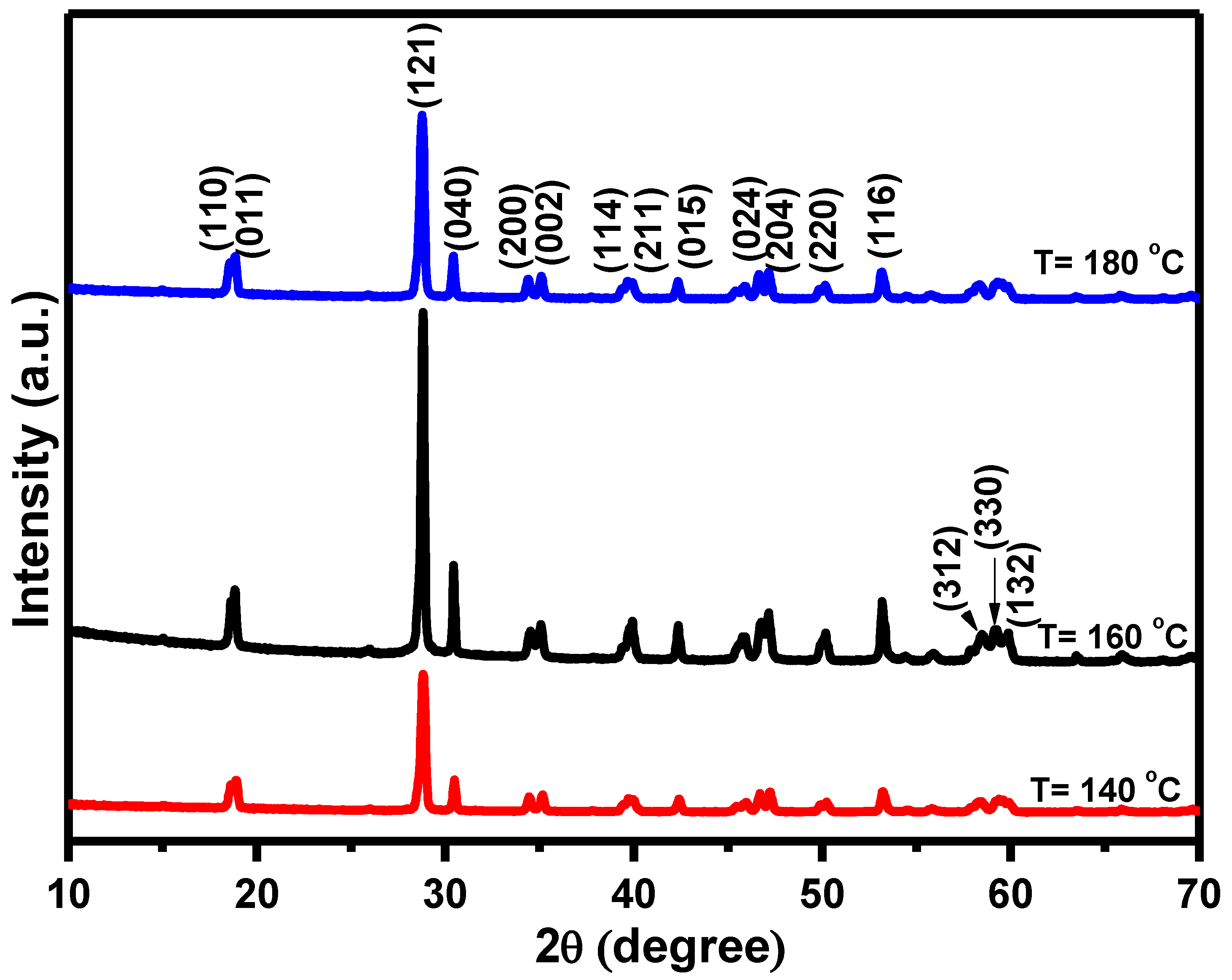
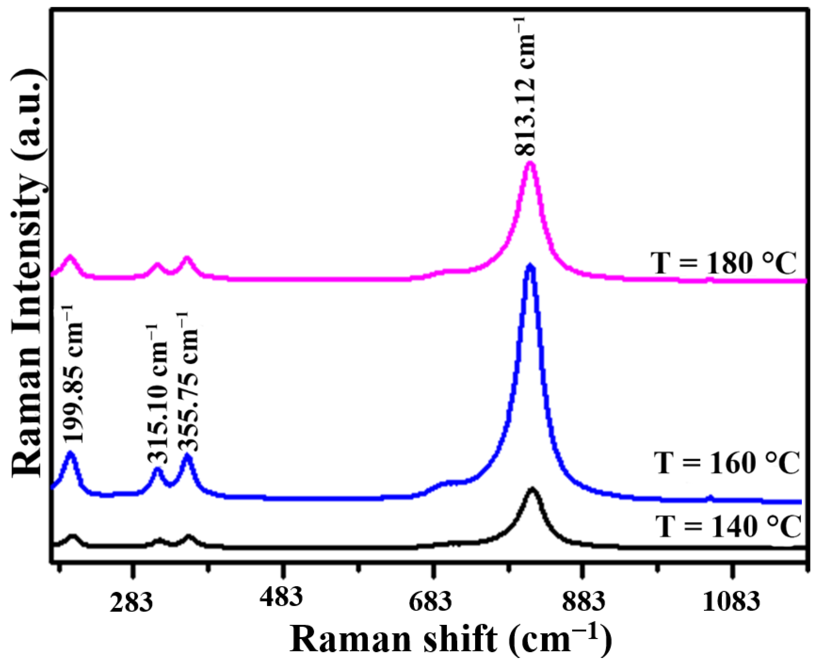
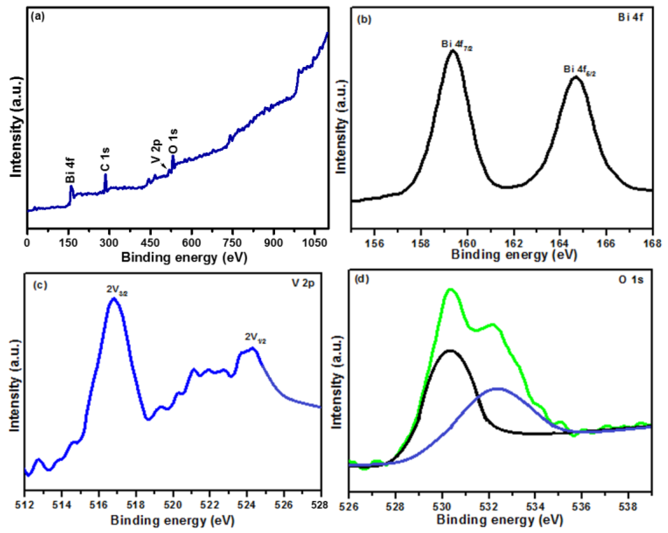
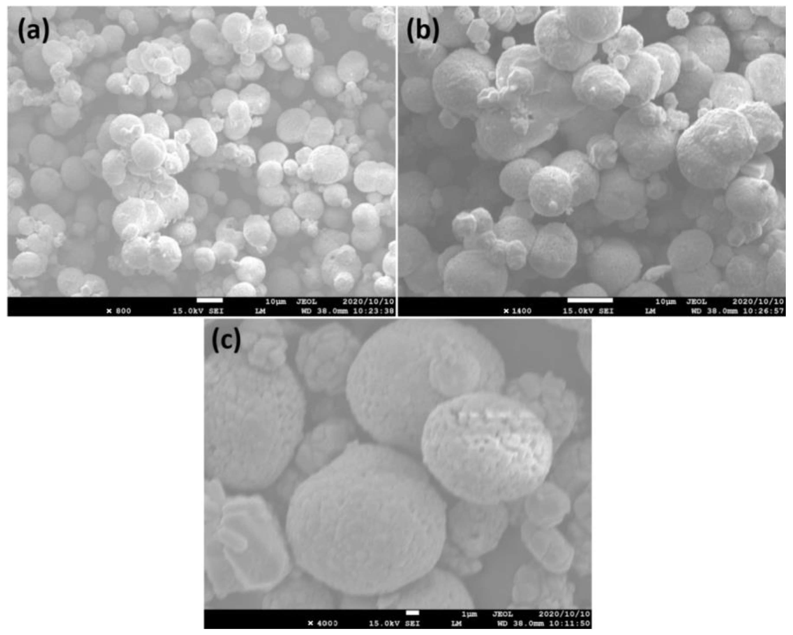
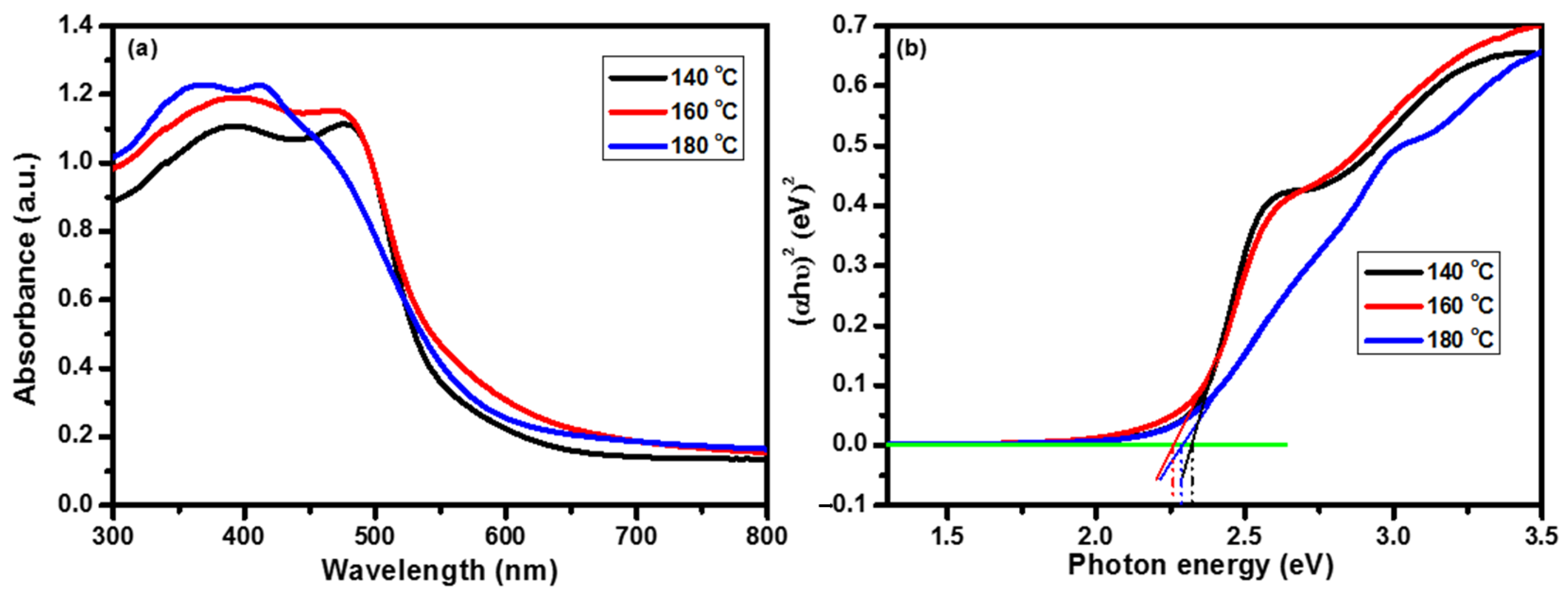
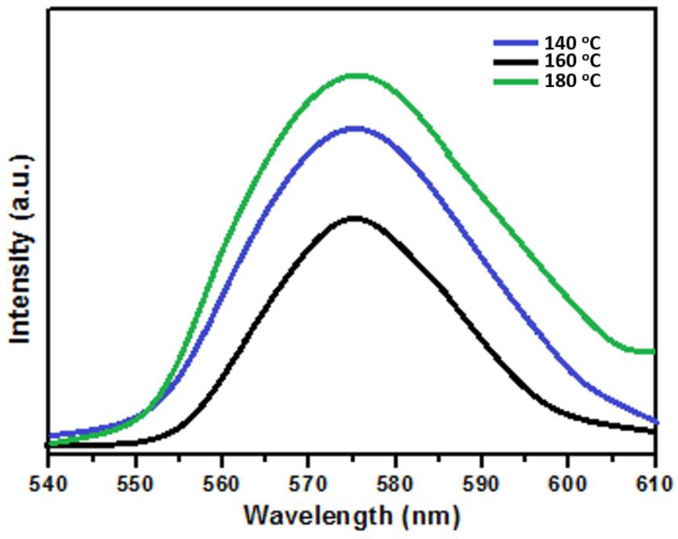
2. Results and Discussion

The crystal structures of the as-synthesized BiVO4 samples were studied using XRD. Figure 1 presents the XRD patterns of the BiVO4 photocatalysts synthesized at different deposition temperatures. The XRD patterns were measured by varying the diffraction angle (2θ) from 10° to 70°. All of the prepared samples were polycrystalline. For all of the synthesized photocatalysts, the diffraction peaks were well indexed to the monoclinic scheelite phase of BiVO4 (JCPDS Card number 01-083-1699) [18]. As the deposition temperature increased from 140 to 160 °C, the intensity of the (1 2 1) plane increased and then decreased with a further increase in deposition temperature. The BiVO4 sample deposited at 160 °C was highly crystalline and the preferred orientation was observed along the (1 2 1) plane for all samples. A higher intensity indicated a higher extent of crystallization along that plane [19]. The average crystallite size was calculated using Debye-Scherrer’s formula and was found to be 28.81, 26.90, and 27.68 nm for samples prepared at temperatures of 140, 160, and 180 °C, respectively. No impurity peaks or mixed phases were detected. Furthermore, confirmation of the formation of the BiVO4 photocatalysts was achieved using Raman spectroscopy.
Raman spectroscopy is a useful characterization tool to understand the local structure and symmetry of a material. Figure 2 shows the Raman spectra of the BiVO4 photocatalysts. From the Raman spectra, it is evident that there are four well-resolved characteristic peaks (199.85, 315.10, 355.75, and 813.12 cm−1) with different intensities. A similar trend was observed in the Raman study to that seen in the XRD result. The maximum peak intensity was observed for the sample prepared at 160 °C. The occurrence of a peak at 813.12 cm−1 corresponds to the to the symmetric V-O stretching mode (Ag symmetry) [20], while the other peaks at 315.10 and 355.75 cm−1 are attributed to the bending modes of the VO4 tetrahedra [21], and the peak at 199.85 cm−1 corresponds to the vibration of the crystal lattice [22]. The Raman and XRD results confirm the formation of the monoclinic scheelite phase of the BiVO4 photocatalysts.
An XPS study was used to investigate the chemical composition and oxidation states that the BiVO4 photocatalyst deposited at 160 °C and they are presented in Figure 3. Figure 3a presents the survey scan spectrum of the BiVO4 photocatalyst, which shows that the sample was composed of Bi, V, and O elements and no other impurity elements were found. The Bi 4f spectrum is shown in Figure 3b. The peaks located at binding energies of 159.28 and 164.60 eV correspond to Bi 4f 7/2 and Bi 4f 1/2, respectively [23]. This result proves that Bi exists in the +3 oxidation state. Figure 3c displays the V 2p spectrum, which splits into two major peaks at binding energy values of 516.75 (V 2p3/2) and 524.60 eV (V 2p1/2) that were assigned to V5+ [24]. Figure 3d presents the high-resolution O 1s spectra of the BiVO4 photocatalyst, which were fitted into two components at binding energy values of 530.40 and 532.15 eV, which are signals from the hydroxyl group and adsorbed water on the surface of the catalyst [25].
Surface morphology plays a crucial role in photocatalysis application. The morphology of the prepared BiVO4 photocatalysts was characterized using scanning electron microscopy. Figure 4a–c display SEM micrographs of the BiVO4 photocatalyst prepared at a 160 °C deposition temperature. From the first SEM image (Figure 4a), spherical-shaped BiVO4 particles with agglomeration in some places are evident [26]. Figure 4b,c present SEM micrographs at a higher magnification. Such spherical-shaped morphology is useful for photocatalysis application [27].
Optical study is an important characterization technique for studying photocatalysis application. Therefore, we employed UV–Vis spectroscopy to determine the absorption edges and bandgap energy of the BiVO4 photocatalysts. Figure 5a,b present the UV–Vis absorbance spectra and bandgap plot, respectively, of the BiVO4 photocatalysts at different deposition temperatures. Absorbance spectra were measured in the wavelength range of 300–800 nm. The bandgap edges of the BiVO4 photocatalysts were estimated at 645 (140 °C), 660 (160 °C), and 636 nm (180 °C). It was observed that all BiVO4 samples had stronger and wider absorption in the visible region. Figure 5b displays the bandgap plot of the BiVO4 samples. By plotting the graph of (αhν)n versus hν for the BiVO4 photocatalysts, the bandgap was determined. The bandgap energy of the BiVO4 photocatalysts was determined by plotting a tangent line across the vertical part of the curve and intersecting it with a horizontal reference line [28]. The bandgap energy for the BiVO4 photocatalysts was found to be 2.32 (140 °C), 2.25 (160 °C), and 2.28 eV (180 °C).
Figure 6 shows the photoluminescence (PL) spectra of the BiVO4 photocatalysts. PL spectra provide useful information about the migration, transfer, and recombination processes of the photogenerated charge carriers (electron–hole pairs) in semiconductor materials. The intensity of PL emission peaks is associated with the recombination of photogenerated electrons and holes. Therefore, a low-intensity PL emission peak implies a lower recombination of charge carriers upon light irradiation, and vice versa [29,30]. A broad emission peak was observed around 577 nm. The sample deposited at 160 °C exhibited the lowest PL intensity in the range of 540–610 nm compared to the BiVO4 samples deposited at 140 and 180 °C. These results shows that the sample deposited at 160 °C had the lowest recombination of photogenerated charge carriers. The XRD, Raman, UV–Vis, and PL results proved that the sample deposited at 160 °C was better for studying the photocatalytic activity because the sample deposited at 160 °C had good crystallinity, lower recombination of photogenerated charge carriers, etc.

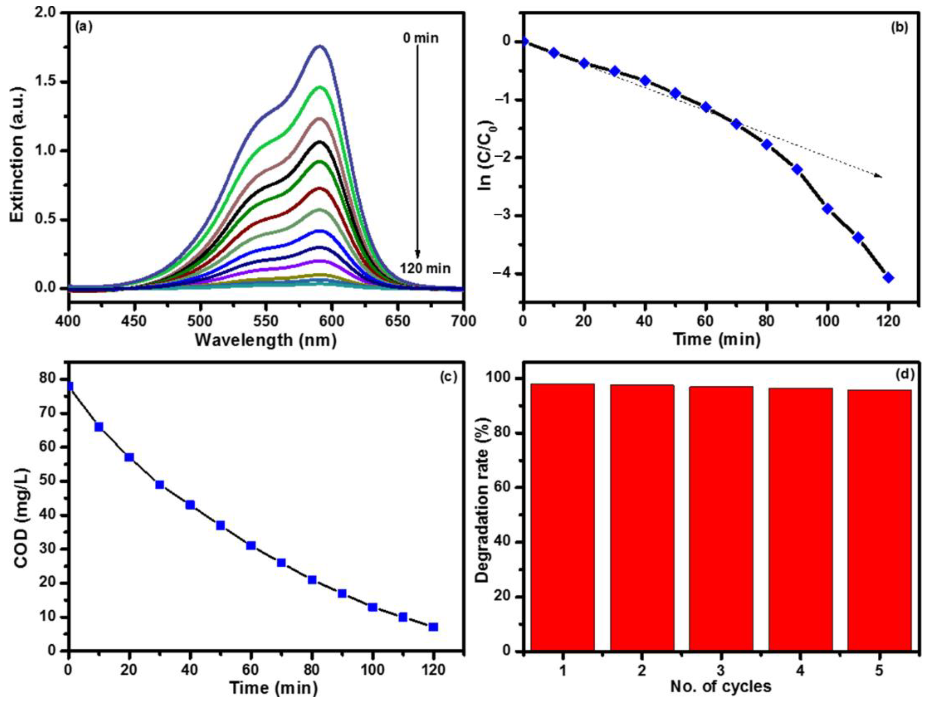
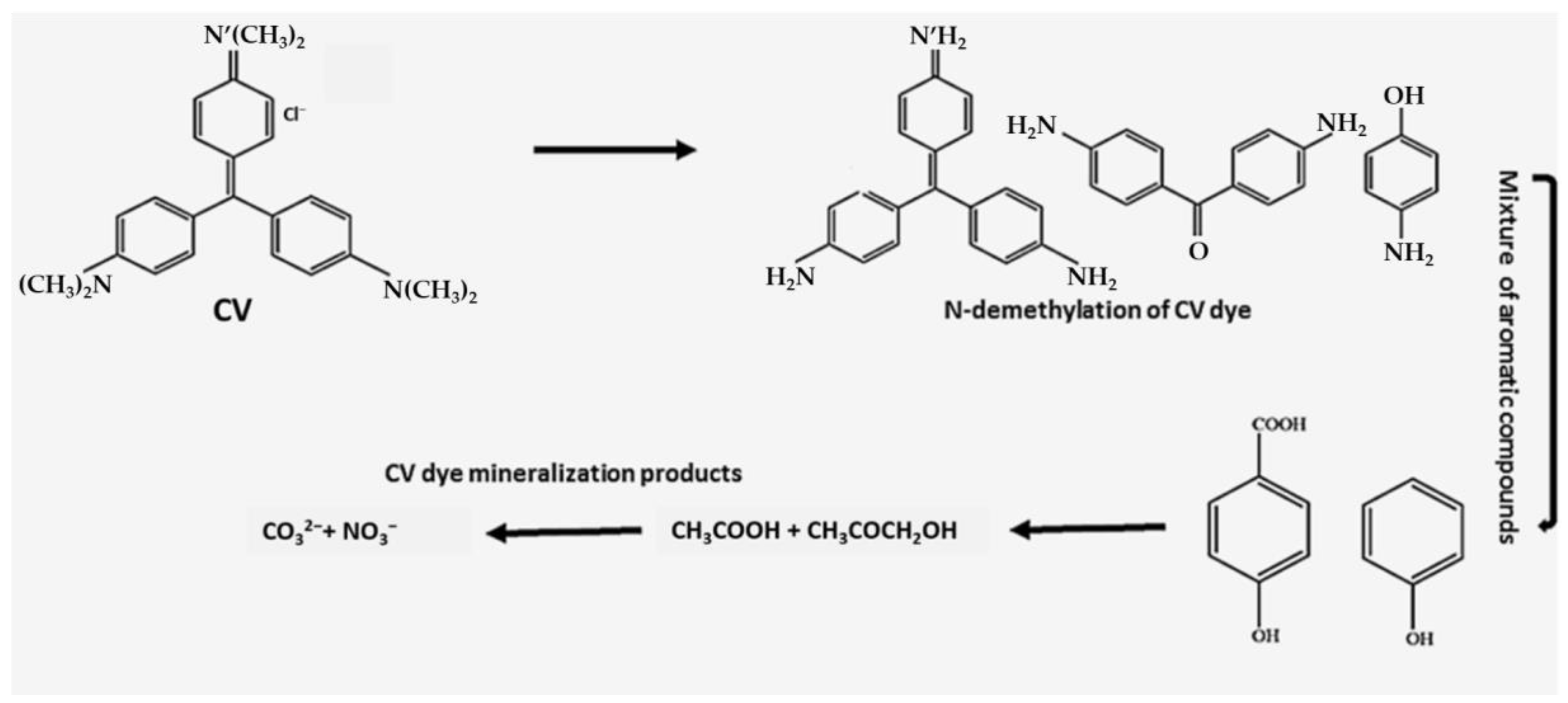
3. Photocatalytic Degradation Activity and Stability Performance

The photocatalytic performance of the BiVO4 photocatalyst prepared at 160 °C was used to study the degradation of crystal violet dye under visible light irradiation and the results are presented in Figure 7. Figure 7a displays the extinction spectra of crystal violet dye collected at different time intervals. The extinction spectra were recorded in the wavelength range 400–700 nm. A strong absorption peak was observed at 590 nm for the crystal violet dye [31]. From the extinction spectra, it was observed that the maximum absorbance at 590 nm decreased gradually and eventually disappeared after 120 min, indicating oxidative degradation of the crystal violet dye. During the progress of the degradation experiments, the frequent decrease in the extinction indicated a decrease in the crystal violet dye concentration with respect to time, and it was also visually confirmed by the reaction solution decoloration. This observation confirms that a redox reaction taking place on the surface of BiVO4 photocatalyst [32]. From this plot, degradation percentage was calculated. The following equation was used to calculate the degradation percentage [33].
Degradation   efficiency = C 0 C C 0 × 100 ( % )
where C and C0 are the final and initial concentrations of crystal violet dye, respectively. It was found that 98.21% degradation of CV dye occurs under visible light illumination. Using the extinction plot, the graph of In (C/C0) vs. reaction time was plotted and is shown in Figure 7b. From the slope, the plot’s reaction rate constant was calculated. Equation (2) was used to calculate the rate constant of the reaction [34].
ln ( C C 0 ) = k t
where t is the time and k can be taken as the apparent first-order rate constant of the degradation reaction. The value of the rate constant k was found to be 5.88 × 10⁻⁶ s−1. The variation in chemical oxygen demand (COD) values with respect to reaction time when using the BiVO4 photocatalyst is presented in Figure 7c. Unlike extinction, studying COD as a function of illumination time gives information about the concentration of oxidizable matter left in the electrolyte solution, not the concentration of the parent molecule. From the graph, it can be observed that the values of COD decrease with the increase in reaction time. COD values decreased from 78.1 to 7.4 mg/L. The variation in COD values indicates that the rate of degradation was high in the initial period, followed by a slow rate. This slow rate indicates the formation of a few long-lived intermediate by-products, which have a low reaction rate with hydroxyl radicals, which revealed the formation of intermediates such as aldehydes and aliphatic acids, etc. [35]. These intermediate products proceeded at a much slower reaction rate; therefore, the reduction in COD values resulted from the degradation of the CV dye.
The stability study of a photocatalyst is an important factor for its practical applications. Therefore, the photostability of the sample synthesized at 160 °C was evaluated in repeated photocatalytic experiments under similar experimental conditions. To study the stability of the as-prepared BiVO4 photocatalyst, degradation experiments were performed and the stability was checked five times with the same BiVO4 photocatalyst. The stability study showed that little decrease was observed in the degradation efficiency after five successive runs, as shown in Figure 7d. There were no noticeable changes in the degradation percentage of the CV dye. However, a slight decrease in the degradation percentage was observed, which may have been due to the loss of catalyst during centrifugation [36]. A 98.21% photocatalytic degradation efficiency was observed for the first time, which slightly reduced to 95.80% after the fifth cycle run. Thus, the BiVO4 catalyst exhibited good stability and recyclability for CV dye degradation. Table 1 presents the photocatalytic degradation of crystal violet dye using different materials and methods.
Table 1. Photocatalytic degradation of crystal violet (CV) dye using different materials and methods.
Sr. No.MaterialsMethodsDegradation Percentage (Time)References
1ZnOChemical solution route88.84% (7 h)[37]
2GOChemical solution route97.90% (24 h)[37]
3ZnO/GOCo-precipitation99% (240 min)[38]
4BiVO4Hydrothermal96.23% (120 min)[39]
5BiVO4Hydrothermal98.21% (120 min)Present work
Figure 8 presents the possible pathways for photocatalytic degradation of crystal violet dye using the BiVO4 photocatalyst under visible light illumination. Initially, oxidized intermediate products were formed, which involved the N-demethylation of the CV dye and the cleavage of the chromosphere structure, which occurred due to the loss of aromatic conjugation and after the hydroxylation of intermediates. Hydroxylated species were oxidized to the quinoid compound, while in the second step, the aromatic rings opened and carboxylic acids were formed. The ring-opening processes continued and the final degradation products could be CO2 and H2O. The latter situation is achieved only when the degradation process reaches mineralization [32,40].

4. Materials and Methods

4.1. Materials

All chemicals were purchased from commercial sources and used without further purification. Bismuth nitrate pentahydrate (Bi(NO3)3·5H2O), ammonium metavanadate (NH4VO3), sodium hydroxide (NaOH), potassium dichromate (K2Cr2O7), sulfuric acid (H2SO4), and nitric acid (HNO3) were purchased from Sigma-Aldrich.

4.2. Preparation of BiVO4 Photocatalysts

BiVO4 photocatalysts were prepared using the hydrothermal method. In a typical synthesis process, firstly, solution A was prepared by dissolving 2 mmol of Bi(NO3)3·5H2O in a concentrated nitric acid solution under constant stirring. Solution B was prepared by dissolving 2 mmol (NH4VO3) in a sodium hydroxide solution. Then, solutions A and B were mixed together well under vigorous stirring and the pH of this solution was adjusted to 7 using NaOH solution (1 M). Lastly, the obtained mixed solution was transferred into a 50-mL Teflon-lined stainless steel autoclave and kept in a furnace at different deposition temperatures of 140, 160, and 180 °C for 12 h. The formed precipitate was collected, filtered, washed with distilled water several times, and then dried in a vacuum at 80 °C for 6 h. The BiVO4 photocatalysts prepared using this method had good morphology, stability, and high purity.

4.3. Material Characterization

The structural, morphological, and optical properties of the prepared BiVO4 catalysts were investigated using several characterization techniques. The structural properties were analyzed using X-ray diffraction (XRD; Ultima IV, Rigaku, Tokyo, Japan) and Raman scattering spectroscopy (NRS-5100, JASCO, Tokyo, Japan). The XPS analysis of the catalyst was carried out using an AXIS Nova X-ray photoelectron spectrometer (Milton Keynes, UK). The surface morphology was analyzed using scanning electron microscopy (SEM; JSM-7600F, JEOL, Tokyo, Japan). The optical properties were studied using photoluminescence (PL), photoluminescence excitation (PLE) spectroscopy (LS5, Perkin Elmer, Waltham, MA, USA), and UV–Vis absorption spectroscopy (V-670, JASCO, Japan).

4.4. Photocatalytic Activity

The photocatalytic activity of the as-prepared BiVO4 photocatalyst was evaluated through the degradation of crystal violet dye under visible light irradiation at room temperature. An amount of 0.5 mM of crystal violet dye was used as the model organic impurity. A 300-W Xe lamp (λ > 420 nm) was used as the visible light source and a 420-nm cut-off filter was placed between the lamp and the reaction mixture to filter out UV light. In the actual experiment, 0.2 g of catalyst was dispersed into a beaker containing 200 mL of 0.5 mM crystal violet aqueous solution. This mixture was magnetically stirred in the dark for 30 min before illumination in order to attain an adsorption/desorption equilibrium. Then, the reaction solution was exposed to visible light illumination whilst under constant magnetic stirring. At specific time intervals, 3 mL of the reaction solution was collected and centrifuged. After centrifugation, the reaction solution was analyzed using a UV–Vis spectrophotometer.
Samples (0.8 mL) withdrawn from the reaction mixture at different time intervals during the photocatalytic degradation experiment were also used to determine the chemical oxygen demand (COD). The samples (0.8 mL) withdrawn at different time intervals were mixed with an excess of potassium dichromate (0.7 mL) and sulfuric acid (2 mL), and the mixture was heated under total reflux conditions for a period of two hours at 140 °C. During the reaction, the chemically oxidizable organic material reduced a stoichiometrically equivalent quantity of dichromate. The quantity of potassium dichromate reduced gave a measure of the amount of oxidizable organic material. From the dichromate extinction, the concentration of the organic solute was calculated [41].

5. Conclusions

A BiVO4 photocatalyst was successfully prepared using the hydrothermal method. The effect of different deposition temperatures on the structural and optical properties of the BiVO4 photocatalyst was studied. The XRD, Raman, and PL results confirmed that the sample prepared at 160 °C was considered to be the optimized sample. The XRD and Raman results confirmed the formation of BiVO4. The SEM study revealed the spherical-shaped morphology of the BiVO4 photocatalyst. Optical investigation showed that the BiVO4 catalyst has strong absorption in the visible region. The chief vibrational modes of the BiVO4 sample, located at 315.10, 355.75, and 813.12 cm−1 and corresponding to the bending and symmetric stretching modes of vibration, were consistent with the monoclinic structure. The photocatalytic degradation percentage of crystal violet dye reached up to 98.21% with good photostability when using the BiVO4 photocatalyst under visible light illumination. The extent of mineralization of the degraded sample was confirmed using a chemical oxygen demand analysis.

Author Contributions

Y.M.H., A.U., Y.T., Y.F., A.A.Y. and S.-W.K.: data curation, formal analysis, investigation, writing—original draft. N.S., I.S., T.K., M.I., M.Y. and S.G.: investigation, methodology. A.F.: conceptualization, project administration, supervision. C.T.: project administration, supervision, conceptualization, methodology. All authors have read and agreed to the published version of the manuscript.

Funding

This study was partly supported by the MEXT Promotion of Distinctive Joint Research Center Program Grant Number JPMXP0618217662.

Institutional Review Board Statement

Not applicable.

Informed Consent Statement

Not applicable.

Data Availability Statement

Data are contained within the article.

Conflicts of Interest

The authors declare no conflict of interest.

References

  1. Hu, J.; Chen, D.; Li, N.; Xu, Q.; Li, H.; He, J.; Lu, J. In Situ fabrication of Bi2O2CO3/MoS2 on carbon nanofibers for efficient photocatalytic removal of NO under visible-light irradiation. Appl. Catal. B-Environ. 2017, 217, 224–231. [Google Scholar] [CrossRef]
  2. Liu, J.; Liu, Y.; Liu, N.; Han, Y.; Zhang, X.; Huang, H.; Kang, Z. Metal-free efficient photocatalyst for stable visible water splitting via a two-electron pathway. Science 2015, 347, 970–974. [Google Scholar] [CrossRef] [PubMed]
  3. Chang, X.X.; Wang, T.; Zhang, P.; Zhang, J.J.; Li, A.; Gong, J.L. Enhanced surface reaction kinetics and charge separation of p-n heterojunction CO3O4/BiVO4 photoanodes. J. Am. Chem. Soc. 2015, 137, 8356–8359. [Google Scholar] [CrossRef]
  4. Ji, K.M.; Dai, H.X.; Deng, J.G.; Zang, H.J.; Arandiyan, H.; Xie, S.H.; Yang, H.G. 3DOM BiVO4 supported silver bromide and noble metals: High-performance photocatalysts for the visible-light-driven degradation of 4-chlorophenol. Appl. Catal. B Environ. 2015, 168–169, 274–282. [Google Scholar] [CrossRef]
  5. Gu, S.N.; Li, W.J.; Wang, F.Z.; Wang, S.Y.; Zhou, H.L.; Li, H.D. Synthesis of buckhorn-like BiVO4 with a shell of CeOx nanodots: Effect of heterojunction structure on the enhancement of photocatalytic activity. Appl. Catal. B Environ. 2015, 170–171, 186–194. [Google Scholar] [CrossRef]
  6. Wu, Q.; Chen, P.F.; Zhao, L.; Wu, J.; Qi, X.M.; Yao, W.F. Photocatalytic behavior of BiVO4 immobilized on silica fiber via a combined alcohol-thermal and carbon nanofibers template route. Catal. Commun. 2014, 49, 29–33. [Google Scholar] [CrossRef]
  7. Fan, H.M.; Jiang, T.F.; Li, H.Y.; Wang, D.J.; Wang, L.L.; Zhai, J.L.; He, D.Q.; Wang, P.; Xie, T.F. Effect of BiVO4 crystalline phases on the photoinduced carriers behavior and photocatalytic activity. J. Phys. Chem. C 2012, 116, 2425–2430. [Google Scholar] [CrossRef]
  8. Shen, Y.; Huang, M.L.; Huang, Y.; Lin, J.M.; Wu, J.H. The synthesis of bismuth vanadate powders and their photocatalytic properties under visible light irradiation. J. Alloys Compd. 2010, 496, 287–292. [Google Scholar] [CrossRef]
  9. Sun, J.X.; Chen, G.; Wu, J.Z.; Dong, H.J.; Xiong, G.H. Bismuth vanadate hollow spheres: Bubble template synthesis and enhanced photocatalytic properties for photodegradation. Appl. Catal. B Environ. 2013, 132–133, 304–314. [Google Scholar] [CrossRef]
  10. Dong, S.Y.; Feng, J.L.; Li, Y.K.; Hu, L.M.; Liu, M.L.; Wang, Y.F.; Pi, Y.Q.; Sun, J.Y.; Sun, J.H. Shape-controlled synthesis of BiVO4 hierarchical structures with unique natural-sunlight-driven photocatalytic activity. Appl. Catal. B Environ. 2014, 152–153, 413–424. [Google Scholar] [CrossRef]
  11. Ran, R.; McEvoy, J.G.; Zhang, Z. Synthesis and Optimization of Visible Light Active BiVO4 Photocatalysts for the Degradation of RhB. Int. J. Photoenergy 2015, 2015, 612857. [Google Scholar] [CrossRef] [Green Version]
  12. Kudo, A.; Omori, K.; Kato, H. A novel aqueous process for preparation of crystal form-controlled and highly crystalline BiVO4 powder from layered vanadates at room temperature and its photocatalytic and photophysical properties. J. Am. Chem. Soc. 1999, 121, 11459–11467. [Google Scholar] [CrossRef]
  13. Guo, M.; He, Q.; Wang, A.; Wang, W.; Fu, Z. A Novel, Simple and Green Way to Fabricate BiVO4 with Excellent Photocatalytic Activity and Its Methylene Blue Decomposition Mechanism. Crystals 2016, 6, 81. [Google Scholar] [CrossRef] [Green Version]
  14. Sarkar, S.; Chattopadhyay, K.K. Visible light photocatalysis and electron emission from porous hollow spherical BiVO4 nanostructures synthesized by a novel route. Phys. E 2014, 58, 52–58. [Google Scholar] [CrossRef]
  15. Dong, L.; Zhang, X.; Dong, X.; Zhang, X.; Ma, C.; Ma, H.; Xue, M.; Shi, F. Structuring porous “sponge-like” BiVO4 film for efficient photocatalysis under visible light illumination. J. Colloid Interface Sci. 2013, 393, 126–129. [Google Scholar] [CrossRef]
  16. Deebasree, J.P.; Maheskumar, V.; Vidhya, B. Investigation of the visible light photocatalytic activity of BiVO4 prepared by sol gel method assisted by ultrasonication. Ultrason. Sonochem. 2018, 45, 123–132. [Google Scholar] [CrossRef] [PubMed]
  17. Chen, S.H.; Jiang, Y.S.; Lin, H.Y. Easy Synthesis of BiVO4 for Photocatalytic Overall Water Splitting. ACS Omega 2020, 5, 8927–8933. [Google Scholar] [CrossRef] [Green Version]
  18. Xiao, B.C.; Lin, L.Y.; Hong, J.Y.; Lin, H.S.; Song, Y.T. Synthesis of a monoclinic BiVO4 nanorod array as the photocatalyst for efficient photoelectrochemical water oxidation. RSC Adv. 2017, 7, 7547–7554. [Google Scholar] [CrossRef] [Green Version]
  19. Yadav, A.A.; Hunge, Y.M.; Kang, S.W. Porous nanoplate-like tungsten trioxide/reduced graphene oxide catalyst for sonocatalytic degradation and photocatalytic hydrogen production. Surf. Interfaces 2021, 24, 101075. [Google Scholar] [CrossRef]
  20. Li, G.Q.; Bai, Y.; Zhang, W.F. Difference in valence band top of BiVO4 with different crystal structure. Mater. Chem. Phys. 2012, 136, 930–934. [Google Scholar] [CrossRef]
  21. Yi, J.; Zhao, Z.Y.; Wang, Y.A.; Zhou, D.C.; Ma, C.S.; Cao, Y.C.; Qiu, J.B. Monophasic zircon-type tetragonal Eu1-xBixVO4 solid-solution: Synthesis, characterization, and optical properties. Mater. Res. Bull. 2014, 57, 306–310. [Google Scholar] [CrossRef]
  22. Zhou, B.; Zhao, X.; Liu, H.J.; Qu, J.H.; Huang, C.P. Synthesis of visible-light sensitive M-BiVO4 (M=Ag, Co, and Ni) for the photocatalytic degradation of organic pollutants. Sep. Purif. Tech. 2011, 77, 275–282. [Google Scholar] [CrossRef]
  23. Jiang, H.; Dai, H.; Meng, X.; Zhang, L.; Deng, J.; Liu, Y.; Au, C.T. Hydrothermal fabrication and visible-light-driven photocatalytic properties of bismuth vanadate with multiple morphologies and/or porous structures for methyl orange degradation. J. Environ. Sci. 2012, 24, 449–457. [Google Scholar] [CrossRef]
  24. Liu, W.; Lai, S.Y.; Dai, H.; Wang, S.; Sun, H.; Au, C.T. Oxidative dehydrogenation of n-butane over mesoporous VOx/SBA15 catalysts. Catal. Lett. 2007, 113, 147–154. [Google Scholar] [CrossRef]
  25. Wang, G.; Ling, Y.; Lu, X.; Qian, F.; Tong, Y.; Zhang, J.Z.; Lordi, V.; Rocha Leao, C.; Li, Y. Computational and photoelectrochemical study of hydrogenated bismuth vanadate. J. Phys. Chem. C 2013, 117, 10957–10964. [Google Scholar] [CrossRef]
  26. Jiang, H.-Q.; Endo, H.; Natori, H.; Nagai, M.; Kobayashi, K. Fabrication and photoactivities of spherical-shaped BiVO4 photocatalysts through solution combustion synthesis method. J. Eur. Ceram. Soc. 2008, 28, 2955–2962. [Google Scholar] [CrossRef]
  27. Hunge, Y.M.; Yadav, A.A.; Mohite, B.M.; Mathe, V.L.; Bhosale, C.H. Photoelectrocatalytic degradation of sugarcane factory wastewater using WO3/ZnO thin films. J. Mater. Sci. Mater. Electron. 2018, 29, 3808–3816. [Google Scholar] [CrossRef]
  28. Hunge, Y.M.; Yadav, A.A.; Mahadik, M.A.; Bulakhe, R.N.; Shim, J.J.; Mathe, V.L.; Bhosale, C.H. Degradation of organic dyes using spray deposited nanocrystalline stratified WO3/TiO2 photoelectrodes under sunlight illumination. Opt. Mater. 2018, 76, 260–270. [Google Scholar] [CrossRef]
  29. Chang, W.S.; Li, Y.C.M.; Chung, T.W.; Lin, Y.S.; Huang, C.M. Toluene decomposition using silver vanadate/SBA-15 photocatalysts: DRIFTS study of surface chemistry and recyclability. Appl. Catal. A Gen. 2011, 407, 224–230. [Google Scholar] [CrossRef]
  30. Pan, G.T.; Huang, C.M.; Peng, P.Y.; Yang, T.C.K. Nano-scaled silver vanadates loaded on mesoporous silica: Characterization and photocatalytic activity. Catal. Today 2011, 164, 377–383. [Google Scholar] [CrossRef]
  31. Ameen, S.; Akhtar, M.S.; Nazim, M.; Shin, H.S. Rapid photocatalytic degradation of crystal violet dye over ZnO flower nanomaterials. Mater. Lett. 2013, 96, 228–232. [Google Scholar] [CrossRef]
  32. Hunge, Y.M.; Yadav, A.A.; Khan, S.; Takagi, K.; Suzuki, N.; Teshima, K.; Terashima, C.; Fujishima, A. Photocatalytic degradation of bisphenol A using titanium dioxide@nanodiamond composites under UV light illumination. J. Colloid Interf. Sci. 2021, 582, 1058–1066. [Google Scholar] [CrossRef] [PubMed]
  33. Hunge, Y.M.; Yadav, A.A.; Dhodamani, A.G.; Suzuki, N.; Terashima, C.; Fujishima, A.; Mathe, V.L. Enhanced photocatalytic performance of ultrasound treated GO/TiO2 composite for photocatalytic degradation of salicylic acid under sunlight illumination. Ultrason. Sonochem. 2020, 61, 104849. [Google Scholar] [CrossRef] [PubMed]
  34. Yadav, A.A.; Hunge, Y.M.; Kulkarni, S.B. Synthesis of multifunctional FeCO2O4 electrode using ultrasonic treatment for photocatalysis and energy storage applications. Ultrason. Sonochem. 2019, 58, 104663. [Google Scholar] [CrossRef] [PubMed]
  35. Hunge, Y.M.; Yadav, A.A.; Kulkarni, S.B.; Mathe, V.L. A multifunctional ZnO thin film based devices for photoelectrocatalytic degradation of terephthalic acid and CO2 gas sensing applications. Sens. Actuators B Chem. 2018, 274, 1–9. [Google Scholar] [CrossRef]
  36. Hunge, Y.M.; Yadav, A.A.; Mathe, V.L. Oxidative degradation of phthalic acid using TiO2 photocatalyst. J. Mater. Sci. Mater. 2018, 29, 6183–6187. [Google Scholar] [CrossRef]
  37. Kathiresan, G.; Vijayakumar, K.; Sundarrajan, A.P.; Kim, S.H.; Adaikalam, K. Photocatalytic degradation efficiency of ZnO, GO and PVA nanoadsorbents for crystal violet, methylene blue and trypan blue dyes. Optik 2021, 166671. [Google Scholar] [CrossRef]
  38. Puneetha, J.; Kottam, N.; Rathna, A. Investigation of photocatalytic degradation of crystal violet dye and its co relation with bandgap in ZnO and ZnO/GO nanohybrid. Inorg. Chem. Commun. 2021, 125, 108460. [Google Scholar]
  39. Sajid, M.M.; Amin, N.; Shad, N.A.; Sadaf Bashir Khan, S.B.; Javed, Y.; Zhangd, Z. Hydrothermal fabrication of monoclinic bismuth vanadate (m-BiVO4) nanoparticles for photocatalytic degradation of toxic organic dyes. Mater. Sci. Eng. B 2019, 242, 83–89. [Google Scholar] [CrossRef]
  40. Ju, Y.; Fang, J.; Liu, X.; Xu, Z.; Ren, X.; Sun, C.; Yang, S.; Ren, Q.; Ding, Y.; Yu, K.; et al. Photodegradation of crystal violet in TiO2 suspensions using UV–V is irradiation from two microwave-powered electrodeless discharge lamps (EDL-2): Products, mechanism and feasibility. J. Hazard. Mater. 2011, 185, 1489–1498. [Google Scholar] [CrossRef]
  41. Yadav, A.A.; Hunge, Y.M.; Mathe, V.L.; Kulkarni, S.B. Photocatalytic degradation of salicylic acid using BaTiO3 photocatalyst under ultraviolet light illumination. J. Mater. Sci. Mater. 2018, 29, 15069–15073. [Google Scholar] [CrossRef]
Figure 1. XRD patterns of BiVO4 photocatalysts prepared at different deposition temperatures.
Catalysts 11 00460 g001
Figure 2. Raman spectra of BiVO4 photocatalysts prepared at different deposition temperatures.
Catalysts 11 00460 g002
Figure 3. XPS spectra of BiVO4 photocatalyst deposited at 160 °C: (a) narrow scan spectrum; (b) Bi 4f, (c) V 2p, and (d) O 1s spectra.
Catalysts 11 00460 g003
Figure 4. (ac) SEM images of BiVO4 photocatalyst deposited at 160 °C with different magnifications.
Catalysts 11 00460 g004
Figure 5. (a) Absorbance spectra and (b) bandgap plot for BiVO4 photocatalysts deposited at different deposition temperatures.
Catalysts 11 00460 g005
Figure 6. Photoluminescence (PL) spectra of BiVO4 photocatalysts deposited at different deposition temperatures.
Catalysts 11 00460 g006
Figure 7. Photocatalytic degradation of crystal violet dye using BiVO4 photocatalyst prepared at 160 °C temperature. (a) Extinction spectra; (b) In (C/C0) vs. reaction time; (c) variation in chemical oxygen demand (COD) values of crystal violet dye with respect to reaction time, and (d) stability study.
Catalysts 11 00460 g007
Figure 8. Possible pathways for photocatalytic degradation of crystal violet using BiVO4 photocatalyst under visible light illumination.
Catalysts 11 00460 g008
Publisher’s Note: MDPI stays neutral with regard to jurisdictional claims in published maps and institutional affiliations.

