Article

Synthesis of Biogenic Palladium Nanoparticles Using Citrobacter sp. for Application as Anode Electrocatalyst in a Microbial Fuel Cell

by
Mpumelelo Thomas Matsena
*,
Shepherd Masimba Tichapondwa
and
Evans Martin Nkhalambayausi Chirwa
Water Utilisation and Environmental Engineering Division, Department of Chemical Engineering, University of Pretoria, Pretoria 0002, South Africa
*
Author to whom correspondence should be addressed.
Catalysts 2020, 10(8), 838; https://doi.org/10.3390/catal10080838
Submission received: 6 July 2020 / Revised: 19 July 2020 / Accepted: 22 July 2020 / Published: 24 July 2020
(This article belongs to the Special Issue Biocatalysis for Green Chemistry)

Abstract

:
Palladium (Pd) is a cheap and effective electrocatalyst that is capable of replacing platinum (Pt) in various applications. However, the problem in using chemically synthesized Pd nanoparticles (PdNPs) is that they are mostly fabricated using toxic chemicals under severe conditions. In this study, we present a more environmentally-friendly process in fabricating biogenic Pd nanoparticles (Bio-PdNPs) using Citrobacter sp. isolated from wastewater sludge. Successful fabrication of Bio-PdNPs was achieved under anaerobic conditions at pH six and a temperature of 30 °C using sodium formate (HCOONa) as an electron donor. Citrobacter sp. showed biosorption capabilities with no enzymatic contribution to Pd(II) uptake during absence of HCOONa in both live and dead cells. Citrobacter sp. live cells also displayed high enzymatic contribution to the removal of Pd(II) by biological reduction. This was confirmed by Scanning Electron Microscope (SEM), Electron Dispersive Spectroscopy (EDS), and X-ray Diffraction (XRD) characterization, which revealed the presence Bio-PdNPs deposited on the bacterial cells. The bio-PdNPs successfully enhanced the anode performance of the Microbial Fuel Cell (MFC). The MFC with the highest Bio-PdNPs loading (4 mg Bio-PdNP/cm2) achieved a maximum power density of 539.3 mW/m3 (4.01 mW/m2) and peak voltage of 328.4 mV.

Graphical Abstract

1. Introduction

A bio-electrochemical system such as Microbial Fuel Cell (MFC) has drawn interest as a promising technology for renewable energy generation using widely available fuel sources and moderate operational conditions [1,2,3].
MFCs have a distinctive characteristic in which electrogenic microbes help to transform chemical energy to electrical energy using organic compounds such as glucose, formate, and acetate as a fuel source [4]. This distinctive characteristic depends on the attachment of electrogens, which greatly affect the performance of MFCs provided that the transfer of electrons is through direct mechanism [5], and since the primary location of the attachment is on the anode electrodes. The properties of the anode material can greatly affect attachment and transfer of electrons between the microbes and anode electrodes [6]. Carbon materials have been used as anode supporting material, these include carbon cloth [7], fiber [8], and paper [9]. Numerous chemical and physical modifications have been made to these materials in order to enhance bacterial attachment and electron transfer [6].
Enhancing the electron transfer is an important step in enhancing the efficiency of MFCs, this in turn is dependent on the performance of electrogenic microbes [10]. These microbes serve as biocatalysts on the anode by metabolizing organic matter and delivering electrons to electrode using direct electron transfer (DET) or mediated electron transfer (MET) mechanisms [11]. Studies have found that metabolites such as formate and ethanol [12], and acetate [13] could be directly oxidized by platinum (Pt) and tungsten oxide as electrocatalysts on the anode and therefore improve energy generation in MFCs. Although Pt is considered as a good electrocatalyst for the oxidation of organic molecules [14,15], its extensive usage is limited because of costs and toxicity.
Research for cheaper and effective electrocatalysts to replace Pt is underway and palladium (Pd), a metal closely related to Pt, has shown high electrocatalytic activity and ultimately improving the anode performance [16,17]. The major problem associated with the use of chemically synthesized Pd nanoparticles is that they are mostly synthesized using toxic chemicals under severe conditions [18]. However, a more environmentally-friendly process using microorganisms has been reported for the production of biogenic palladium nanoparticles (Bio-PdNPs) from a soluble palladium (II) (Pd (II)) solution. These include microorganisms such as Shewanella oneidensis [19], Desulfovibrio [20], Desulfovibrio Desulfuricans Seroval [21]. In addition, biologically-synthesized Pd nanoparticles have also been used to enhance anode electrocatalytic activity in previous studies [6,16]. Synthesis of Pd nanoparticles using microorganisms offers several advantages in that it requires less chemical agents, reactions occur under less severe conditions, and high catalytic activity [19,22].
In this study, an investigation of optimum conditions for the synthesis of Bio-PdNPs using Citrobacter sp. isolated from Brits sludge was conducted and the resulting Bio-PdNPs were characterized for use in improving anode electrocatalytic activity and enhancing MFC performance. Pd(II) concentration was measured using atomic absorption spectrometry (AAS). The morphological properties, elemental composition, and phase identification of Bio-PdNPs were determined using scanning electron microscopy (SEM), electron dispersive spectroscopy (EDS), and X-ray diffraction (XRD), respectively. The performance of the MFC system was evaluated based on output potential difference and maximum power output.

2. Results and Discussion

2.1. Microbial Analysis

2.1.1. Culture Isolation

Potential Pd(II)-reducing bacteria were isolated based on their tolerance to the toxicity of Pd(II) using Brits sludge. During the isolation, the sludge from the sand drying beds was exposed to different initial Pd(II) concentrations (70 mg/L, 100 mg/L, and 200 mg/L) for 74 h with LB-broth acting as growth media for the cultures under aerobic and anaerobic conditions. The microbial growth was measured in optical density (OD) at wavelength of 600 nm using a UV/Vis spectrophotometer. The microbial growth for all cultures remained below 0.5 except for the cultures grown under anaerobic conditions at an initial concentration of 70 mg/L Pd(II) where an optical density of 1.13 was measured after 74 h (Figure 1a). Pd(II) removal decreased as initial Pd(II) concentration increased in both aerobic and anaerobic conditions. Based on these results, not only was the growth of Brits microbial cells exposed to Pd(II) dependent on initial Pd(II) concentration, but anaerobes grew well under palladium exposure as compared to aerobes. This was caused by Pd(II) competing with oxygen as an electron acceptor [23].
The inhibition of enzymatic processes on bacterial cells affects the overall microbial growth and palladium has been shown to display antimicrobial activity [24]. In our study, Pd(II) showed inhibitory effects on the Brits microbial growth by reducing both microbial growth and the overall Pd(II) removal with an increase in Pd(II) initial concentration, and this similar observation was reported by Adams, et al. [25] with Pd(II) inhibiting growth of Escherichia coli and Staphylococcus aureus. Pd(II) is known to inhibit enzymes in both prokaryotic and eukaryotic cells due to its toxicity [26]. Therefore, the highest removal of Pd(II) at 70 mg/L Pd(II) experiment was due to low inhibitory effects on the growth of Brits microbial cells. The abundance of anaerobes due to high cell growth was also the reason high removal of Pd(II) was observed in 70 mg/L Pd(II) experiment [27].

2.1.2. Culture Characterization

Colonies were first classified based on their morphology. Three pure cultures were isolated morphologically and the 16S rRNA results revealed a total of three unique anaerobic isolates that survived the Pd(II) exposure. All results showed prevalence of the three anaerobic phenotypes specified in Table 1. From the literature, Hosseinkhani, et al. [28] used Bacillus sp. obtained from marine sediment to synthesize Bio-PdNPs. Bacterial strains belonging to the Citrobacter genus like Citrobacter braakii [29] and Citrobacter freundii [30] have also been reported for the efficient Bio-PdNPs production.

2.2. Anaerobic Pd(II) Removal Using Pure Isolates and Brits Sludge

2.2.1. Anaerobic Pd(II) Removal Using Pure Isolates

Heavy metals can inhibit microbial growth and cause cell death. This is a consequence of the affinity of heavy metals to different cellular components in bacteria by forming complexes that cause toxic effects on microorganisms [31]. These toxic effects can result in a decrease in Pd(II) reduction capability of bacteria that could otherwise reduce Pd(II), and also lead to bactericidal effects on bacteria that cannot survive high concentrations of Pd(II). In the current study, following the exposure of different pure cultures to 40 mg/L of Pd(II) for 30 h, Citrobacter sp. was the only pure isolate from the isolated anaerobic cultures that was able to completely remove Pd(II) with Exigobacterium sp. and Bacillus sp. only removing 22.6 and 24.3%, respectively (Figure 2a).
Exigobacterium sp. and Bacillus sp. survived exposure to Pd(II), but only Citrobacter sp. was mostly responsible for Pd(II) removal. The survival of the bacteria to Pd(II) toxicity was due to Pd(II) removal by Citrobacter sp., which reduced the overall levels of Pd(II) during isolation and allowed the survival of Exigobacterium sp. and Bacillus sp., which could not otherwise withstand high levels of Pd(II). However, the ability of the pure isolates to survive exposure of Pd(II) in a mixed culture did not guarantee their ability to completely remove Pd(II) since when using pure isolates, Exigobacterium sp. and Bacillus sp. failed to completely remove Pd(II).
This observation is due to the fact that the resistance to high levels of heavy metals of many microorganisms can be ascribed to: (i) enzymatic activity (oxidation and reduction), and (ii) passive uptake (biosorption) which is not dependent on cellular metabolic activity [32]. These two processes can occur in a mixed culture where bacteria can carry out these processes to reduce the overall levels of heavy metals. This enables bacteria that could not otherwise withstand high levels of heavy metals to survive. Sannasi, et al. [33] reported that bacteria are more stable and able to survive in mixed cultures than as pure cultures.

2.2.2. Comparison of Pd(II) Removal Using a Pure Isolate and Brits Sludge

Bacteria in mixed culture have displayed stability and are more prone to survive [33], and high bacterial stability and metal recovery was achieved when using a mixed culture of Acidithiobacillus ferrooxidans and Acidithiobacillus thiooxidans with the capability of recovering copper (Cu), nickel (Ni), zinc (Zn), and aluminium (Al) as compared to using pure isolates [34]. This means that using a diverse bacterial community can help improve bacterial stability, and thus improve recovery.
The Brits sludge, which had a diverse bacterial community, did not improve Pd(II) removal and a pure isolate of Citrobacter sp. performed better by removing 97.2% of Pd(II) as compared to 76.6% Pd(II) removal by Brits sludge (Figure 2b). This is because Citrobacter sp. in the sludge was identified as the sole bacterium responsible for the complete removal of Pd(II) and the growth of other microbes, which did not fully contribute in Pd(II) removal and negatively affected its performance. This observation is consistent with the one made by Simona, et al. [35], who showed that using sludge from nickel disposal site is not the best option for Zn removal, but rather using pure isolates or reconstituted consortium from the sludge, which only contains bacteria responsible for complete Zn removal, is better.

2.2.3. Influence of Citrobacter sp. Live Cells on Pd(II) Removal

Abiotic and biotic removal of Pd(II) was studied in no cells, heat-killed cells, and live cells conditions. These studies were conducted in the presence and absence of sodium formate (HCOONa). These control experiments were conducted to determine if the process taking place in the removal of Pd(II) by Citrobacter sp. is solely by bioreduction or additional processes such as biosorption and chemical reduction.
As displayed in Figure 2c, Citrobacter sp. Heat-killed (at 121 °C, 15 min) and live cells were able to remove 12.3 and 14.7% Pd(II), respectively, in absence of HCOONa. The removal of Pd(II) by both heat-killed and live cells in absence of HCOONa is an indication of biosorption by Citrobacter sp. bacterial cells. Biosorption is a process where ions from aqueous solutions bind onto functional groups available on the surface of bacteria and has been described by previous studies for metal recovery [27,36,37]. Biosorption in the present experiments was approximately similar when using heat-killed and live cells, this means that autoclaving had little impact on the sorbents and no contribution by enzymes to the uptake of Pd(II) was seen during absence of HCOONa in both live and dead cells.
The combination of HCOONa/Pd(II) in absence of both live and heat-killed cells led to 22.3% Pd(II) removal (Figure 2c). Similar observations were made by Bunge, et al. [38] and Deplanche, et al. [39], and this was attributed to Pd(II) chemical reduction by sodium formate. The presence of heat-killed cells improved Pd(II) removal in HCOONa/Pd(II) to 38.1% (Figure 2c). This was because heat-killed cells created nucleation sites for palladium nanoparticles deposition and for subsequent crystal growth, which became self-sustaining through the ability of palladium nanoparticles to oxidize HCOONa and autocatalytically reduce more Pd(II) [27].
The highest removal of 97.2% of Pd(II) was achieved by the presence of Citrobacter sp. live cells together with a combination of HCOONa and Pd(II) (Figure 2c). This high increase in Pd(II) removal indicates an enzymatic contribution of Citrobacter sp. live cells to the removal of Pd(II) by possible biological reduction to elemental palladium (Pd(0)). The biological reduction of palladium has been reported before using Shewanella oneidensis [19], Desulfovibrio [20], Desulfovibrio Desulfuricans Seroval [21]. A previous study also reported that the reduction of metals by microorganisms is not only in a dissimilatory way, rather, a number of microorganisms can biosorb and reduce Pd(II) [38]. In this study, Citrobacter sp. live cells not only had the ability to biosorb, but they could also bioreduce Pd(II).

2.3. Abiotic Factors Influence on Pd(II) Removal by Citrobacter sp.

2.3.1. Effect of Temperature
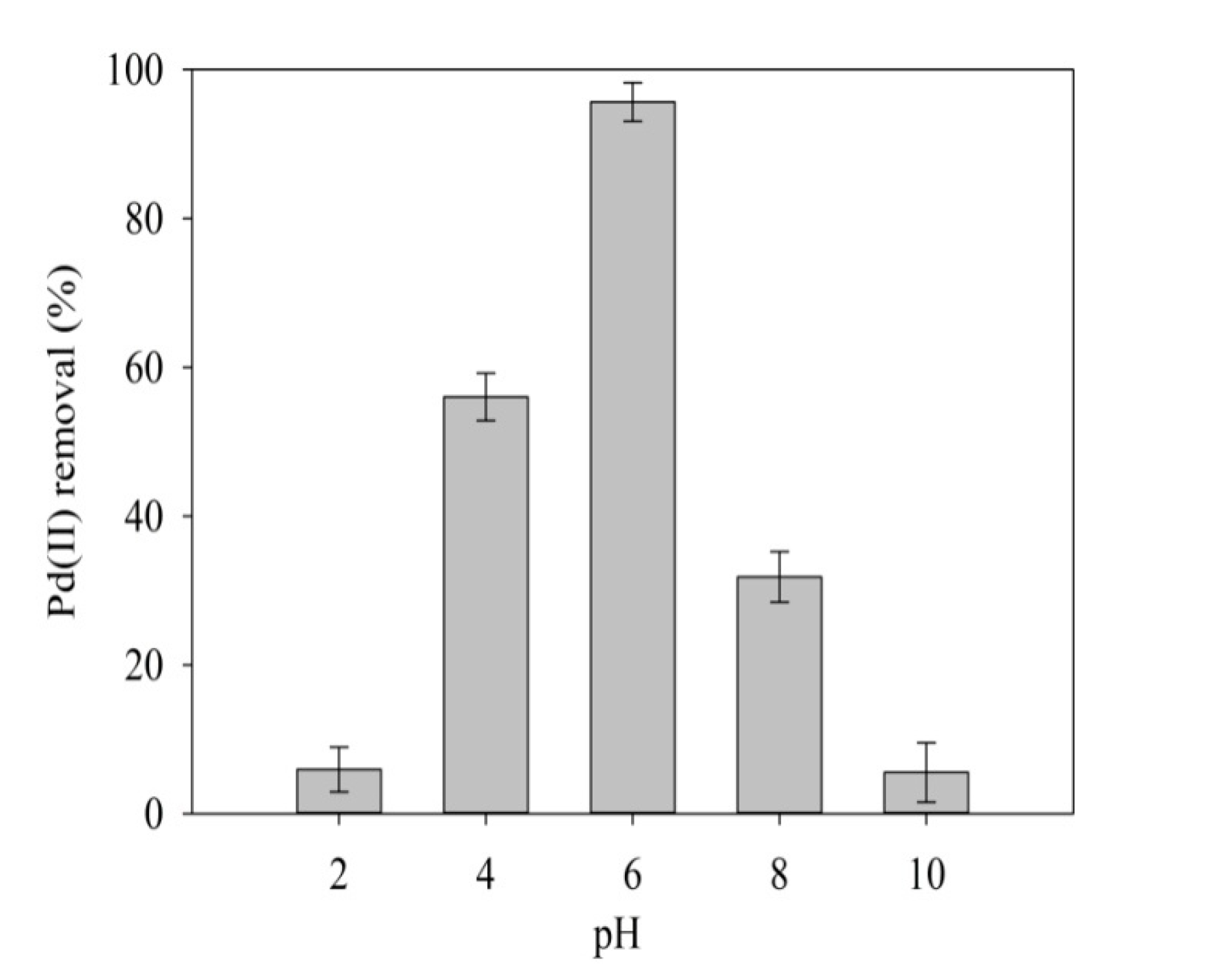
Pd(II) removal by Citrobacter sp. was tested over a wide range of temperatures. Pd(II) removal was above 80% in the temperature range of 30–40 °C, and Pd(II) removal was below 52% for 20 and 60 °C experiments (Figure 3). This is because temperature has been found to affect growth of metal-reducing bacteria and this in turn affects the extent at which metals will be reduced [40]. In most enzyme catalyzed reactions, an increase in temperature will increase enzyme activity and promote microbial growth. However, further increase to very high temperatures will diminish enzyme activity and the protein will denature. On the other hand, lowering the temperature will decrease enzyme activity and reduce microbial growth [41]. Organisms can be classified according to their optimum growth temperature: (i) psychrophiles grow best between −5 °C and 20 °C, (ii) mesophiles grow best between 20 °C and 45 °C, and (iii) thermophiles grow best at temperatures above 45 °C. Since Citrobacter sp. displayed high enzyme activity in the removal of 80% of Pd(II) at temperatures between range of 30–40 °C with the highest removal of 96.8% being at 30 °C (Figure 3), it was concluded that Citrobacter sp. isolated from Brits sludge is a mesophile.

2.3.2. Effect of pH

Acidic (pH 2) and basic (pH 10) conditions exhibited inhibitory effects on Citrobacter sp. removing only 5.9 and 4.2% of Pd(II), respectively, while pH 6 showed the ability to highly remove 96% of Pd(II) (Figure 4). Since Citrobacter sp. has shown biosorption capabilities, the improved removal at pH 6 might have been due to improved ability of Pd(II) to biosorb on Citrobacter sp. bacterial cell walls and be biologically reduced.
Another plausible explanation for the improved removal of Pd(II) at pH 6 is related to the electrophoretic mobility (EPM) of bacteria. The EPM of bacteria is dependent on pH, and an increase in pH increases the EPM towards negative values. Most bacteria start to achieve negative EPM at pH of 3–4 [42,43]. Negative EPM indicates that the surface charge by the shear plane of bacteria is negative, and since Pd(II) in the form of palladium tetra-ammine complex (Pd(NH3)42+) was used in our experiments, the approach between palladium cation specie and Citrobacter sp. negative bacterial surface might have been favored by coulombic interaction at pH 6.
At lower pH values, the bacteria surface charge is normally positive [44]. Therefore, at pH value of 2, the bacterial surface charge might have been positive and the approach between palladium species and Citrobacter sp. bacterial surface might have not been favored by coulombic interaction. At higher pH values (8.0 to 10.0), lower heavy metal uptake, and bioreduction were postulated to be due to increased precipitation as a result of the interaction between the metal ions and hydroxyl ions in the aqueous solution forming insoluble metal precipitates, which prevented biosorption, and subsequently prevented bioreduction. It has previously been reported that at high pH values (pH > 9), palladium hydroxide (Pd(OH)2) is a predominant specie [45].

2.3.3. Effect of Initial Pd(II) Concentration

Heavy metals have displayed the ability to inhibit both microbial growth and metal reduction at high concentrations [25]. In our research, an increase in initial Pd(II) concentration from 100 to 800 mg/L led to a decrease in the removal rate of Pd(II); however, Citrobacter sp. still completed the removal of Pd(II) within 24 h (Figure 5).
A decrease in Pd(II) removal with initial Pd(II) concentration increase indicated the slowing down of enzyme activity. This is due to the inhibition that is caused by the deactivation of Pd(II)-reducing enzymes in Citrobacter sp. bacterial cells since cytotoxic effects due to Pd(II) have been reported on microbial cells [46]. In addition, Pd(II) has been shown to inhibit enzymes such as alkaline phosphatise, carbonic anhydrase, prolyl hydroxylase, succinate dehydrogenase, kinase, creatine, and aldolase [26]. Since this study had already shown that Pd(II) affects cell growth, it was also possible that when increasing initial Pd(II) concentration, microbial growth decreased causing a decrease in Pd(II) removal due to the affinity of Pd(II) to different cellular components in bacteria by leading to complexes that cause toxic effects and cell death [31].

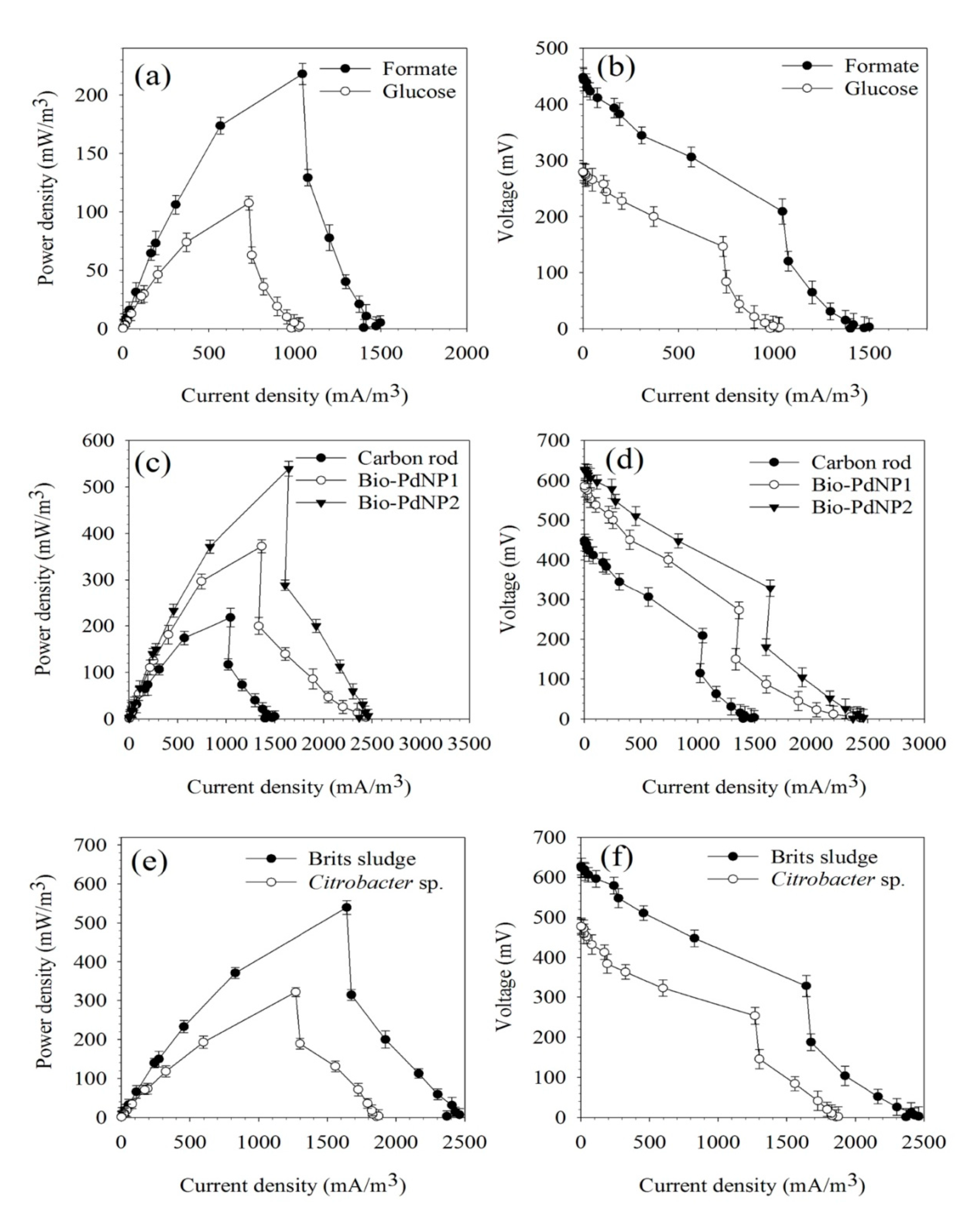
2.3.4. Effect of Carbon Source

Most bacterial species are known to derive their carbon from breaking down organic compounds. These organic compounds are known as carbon sources and drastically differ in terms of energy content and biological routes in which bacteria can degrade them. Sodium bicarbonate failed to serve as a carbon source, while sodium formate and glucose achieved a 99.2% and 53.4% removal of Pd(II), respectively (Figure 6). This was because sodium bicarbonate is an inorganic carbon source and not all microorganisms have the capability of using inorganic substrates as carbon sources. Microorganisms can be classified as chemolithotrophs, which are organisms that obtain their energy from inorganic substrates and chemoorganotrophs, which are organisms that use organic substrate for energy. Therefore, in our case, Citrobacter sp. was able to utilize glucose and formate since they are organic substrates and not sodium bicarbonate, which means Citrobacter sp. is a chemoorganotroph. In addition, a lag-time in Pd(II) removal by glucose signaled an indirect route was involved in Pd(II) removal. Sodium formate provided a direct route to the Pd(II) removal since there was no lag-time. When glucose was used as a carbon source, it is possible that Citrobacter sp. might have reduced Pd(II) through sodium formate production by catalyzed reduction of glucose metabolite (carbon dioxide) using a formate dehydrogenase (FDH) enzyme or through CoA-dependent cleavage of pyruvate [47], and sodium formate as a metabolite of glucose degradation might have activated formate hydrogenlyase (FHL) for Pd(II) removal [39].

2.4. Characterization of Bio-PdNPs Synthesized by Citrobacter sp.

During synthesis of Bio-PdNPs, the first indication that there might be formation of reduced palladium was shown by change in color (Figure 7). This color change to the formation of a black precipitate did not however guarantee the formation of reduced elemental palladium. This is because black palladium precipitates associated with PdO formation have been shown to form under similar conditions [48]. Therefore, it was essential that the formed Pd product be characterized in order to certify if elemental palladium (Pd(0)) was formed through biological reduction using Citrobacter sp. To characterize the Bio-PdNPs, both control cells before the synthesis and the cells after Bio-PdNPs formation were collected, centrifuged, cleaned using ultrapure water, and oven dried at 105 °C.
The elemental composition of Citrobacter sp. bacterial cells was determined before Bio-PdNPs synthesis and after Bio-PdNPs synthesis using EDS analysis. No Pd was detected on the microbial cells before synthesis (Figure 8a, however, Pd peaks were detected on the bacterial cells after Bio-PdNPs synthesis (Figure 8b). The biomass morphology was studied under scanning electron microscopy (SEM). The Citrobacter sp. cell biomass before Bio-PdNPs synthesis showed a relatively smooth surface (Figure 8c), and after synthesis, a rougher surface with particles deposited and anchored on the bacterial cells was observed (Figure 8d). These results indicated the possible presence of Pd(0) by Citrobacter sp.
Citrobacter sp. bacterial cells after Bio-PdNPs synthesis were further subjected to phase identification analysis using XRD. This is because the resulting spectra can be matched to existing databases and the specific crystalline phases present in the Pd precipitate can be identified. Previous studies by Hazarika, et al. [48] characterizing palladium nanoparticles using XRD observed similar distinct peaks as those obtained in the present study (Figure 9). These peaks with 2θ values of 46.54° and 82.18°, and corresponding plane coordinates of (200) and (311), were attributed to the presence of elemental Pd. The distinct peak observed at a 2θ value of 55.86° (Figure 9) showed that PdO as an impurity was formed with pure metallic Pd in biological synthesis of Bio-PdNPs. To ascertain if the formed elemental palladium are nanoparticles, the Scherrer equation was used to calculate the crystallite size. The sizes of Pd at 2θ values of 46.54° and 82.18° were calculated as 15.65 and 11.37 nm, respectively.
Based on the XRD results, the synthesis of Bio-PdNPs by biological reduction of Pd(II) using Citrobacter sp. and sodium formate as electron donor under anaerobic condition was confirmed and the probable general mechanism of the synthesis based on our results is that (i) Pd(II) is biosorbed on the Citrobacter sp. bacterial cells; (ii) bioreduced by enzymatic processes and then redeposited on the bacterial cells to form a rough surface on the cells; (iii) and a side process relating to the chemical reduction of Pd(II) by sodium formate might be taking place, but it is not significant as compared to bioreduction. This mechanism is distinguished from the dissimilatory metal reduction in which the metals are used as electron acceptors without uptake into cells. This general mechanism was also observed by previous studies [27,37]; however, the enzymes involved in the formation of Bio-PdNPs using Citrobacter sp. will need to be fully defined since the Pd(II) bioreduction is an enzymatic process.

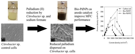
2.5. MFC Performance Using Bio-PdNPs Synthesized by Citrobacter sp. as Anode Catalyst

The effect of the type of substrate (glucose and formate) was evaluated on the basis of MFC performance without Bio-PdNPs. Glucose was chosen as a control substrate since it has been used as the substrate of choice for many MFCs applications. Provided that the glucose performed better than formate in the MFC, then a ratio between formate and glucose would need to be determined so as to enhance oxidation by Bio-PdNPs and microbes in MFC for improved energy generation. This is because the electrocatalytic activity of Bio-PdNPs in the oxidation of glucose has been shown to be slow compared to oxidation of smaller molecules (formate, lactate and ethanol). Also, the use of glucose alone in a Bio-PdNPs/microbes MFC leads to lower performance as compared to using both glucose and formate [16].
However, in our study, formate was better oxidized in absence of Bio-PdNPs by Brits sludge in MFC achieving a maximum power density of 218.1 mW/m3 (2.77 mW/m2) (Figure 10a) and peak voltage of 208.8 mV (Figure 10b). This is because there might have been other bacteria in the sludge which are not electrogens that consumed glucose [49]. The better performance by formate also means that the Brits sludge had microbes that are able to mostly utilize formate as a carbon source for energy generation. Therefore, there was no need to determine a ratio between glucose and formate during Bio-PdNPs experiments because formate was already better oxidised in MFC by microbes for energy production.
The catalytic activity of formate oxidation when using palladium based electrocatalyst (Bio-PdNPs) can improve the performance of the MFC. The use of Bio-PdNPs at different loadings synthesized by Citrobacter sp. to enhance the performance of the MFC was investigated using Bio-PdNP1 (2 mg Bio-PdNP/cm2) and Bio-PdNP2 (4 mg Bio-PdNP/cm2). The MFC with the highest Bio-PdNPs loading achieved a maximum power density of 539.3 mW/m3 (4.01 mW/m2), which was enhanced by 31.1 and 59.6% as compared to Bio-PdNP1 and carbon rod without Bio-PdNPs, respectively (Figure 10c). In addition, Bio-PdNP1 and Bio-PdNP2 improved performance by achieving peak voltage of 272.9 and 328.4 mV, respectively, as compared to 208.8 mV of the carbon rod (Figure 10d).
The improvement in the MFC performance was because in addition to the (i) microbes in the Brits sludge, which were oxidizing formate possibly through an enzyme catalyst formate hydrogenlyase [39], (ii) the Bio-PdNPs were also able to further catalyze the oxidation of formate to improve energy generation [27], (iii) and an increase in Bio-PdNPs loading increased active sites for the oxidation of formate. This meant that in presence of electroactive bacteria, anode electrocatalysts such as Bio-PdNPs can lead to the improvement of anode electrocatalytic activity by enhancing the oxidation of electron donors such as formate in the MFC. This was shown by the improvement in both maximum power density and peak voltage when using Bio-PdNPs as electrocatalysts in the MFC.
The enzyme oxidation of formate by formate hydrogenlyase [39], and catalytic oxidation of formate by palladium nanoparticles [27] has already been previously reported. The modification of anode by Bio-PdNPs has also already been demonstrated for power generation [6], evans blue removal [50], and iohexol degradation in MFCs [16]. However, the study here presents both the synthesis and characterization of Bio-PdNPs using Citrobacter sp. isolated from Brits sludge and demonstrates their usage to act as electrocatalysts in anode modification of MFCs. The synthesized Bio-PdNPs not only show electrocatalytic activity towards degradation of formate to enhance MFC performance but the fabrication requires less chemical agents and reaction under less severe conditions.
In this research, it was already shown that only anaerobic pure isolates (Exigobacterium sp., Bacillus sp., Citrobacter sp.) from Brits sludge can survive exposure of Pd(II), and that Citrobacter sp. better removes Pd(II) as a pure isolate than in a sludge. Since most metal-reducing bacteria are electroactive [51], and with Citrobacter sp. being responsible for the biological reduction of Pd(II), we had expected the MFC performance of Citrobacter sp. pure isolate to be the same if not better than the sludge in the presence of Bio-PdNPs. This is because only Exigobacterium sp., Bacillus sp., Citrobacter sp. survived the exposure of palladium in the Brits sludge, and with Citrobacter sp. being an efficient Pd(II)-reducing bacteria in presence of formate, we expected it to be mostly responsible for energy generation. However, that was not the case in this study. Brits sludge in the presence of Bio-PdNPs performed better than Citrobacter sp. with a maximum power density of 539.3 mW/m3 (4.01 mW/m2) (Figure 10e) and peak voltage of 328.4 mV (Figure 10f). This is because more bacteria which are electroactive in the sludge might have survived exposure to Bio-PdNPs indicating that biogenic Pd nanoparticles are less toxic than Pd(II).

3. Materials and Methods

3.1. Microbial Culture

The microbial consortium was derived from the Brits Wastewater Treatment Plant in South Africa. The samples were stored in sterile containers at 4 °C in the refrigerator for further use.

3.2. Microbial Isolation

Microbial consortium used in this work was enriched for 24 h at 28 °C in Luria-Bertani (LB) medium under both aerobic and anaerobic conditions, and selectively isolated using the method described by Mtimunye [52] to obtain pure cultures that might be capable of producing Bio-PdNPs. The pure cultures were stored at −80 °C in Basal Mineral Medium mixed at a final concentration of 30% with sterile glycerol for further use.

3.3. Culture Characterization

Characterization of microbial cells was done from the 7th to the 10th tube in the serial dilution preparation on individual isolated colonies of bacteria. The classification of colonies was first based on morphology in preparation for the 16S rRNA (16 Svedburg unit ribosomal Ribo-Nucleic-Acid) fingerprint method, which is used to obtain DNA sequences of pure isolated cultures. The extraction of genomic DNA from the pure cultures isolated by morphology was done by using a DNeasy tissue kit (QIAGEN Ltd., West Sussex, UK). The 16S rRNA genes of isolates were amplified by a reverse transcriptase-polymerase chain reaction (RT-PCR) using primers pA and pH1 (Primer pA corresponds to position 8–27; Primer pH to position 1541–1522 of the 16S gene. An internal primer pD was used for sequencing (corresponding to position 519–536 of the 16S gene). The resulting sequences were deposited in the GenBank to be compared to known bacteria using a basic BLAST tool search of the National Centre for Biotechnology Information (NCBI, Bethesda, MD, USA).

3.4. Basal Mineral Media

The mineral medium used in the synthesis was Basal Mineral Medium (BMM). BMM was prepared by dissolving: 10 mM NH4Cl, 30 mM Na2HPO4, 20 mM KH2PO4, 0.8 mM Na2SO4, 0.2 mM MgSO4, 50 µM CaCl2, 25 µM FeSO4, 0.1 µM ZnCl2, 0.2 µM CuCl2, 0.1 µM NaBr, 0.05 µM Na2MoO2, 0.1 µM MnCl2, 0.1 µM KI, 0.2 µM H3BO3, 0.1 µM CoCl2, and 0.1 µM NiCl2 into 1 L of distilled water [52].

3.5. Pd(II) Stock Solution

1000 mg/L palladium (II) [Pd(II)] stock solutionwas made by adding 2.48 g of 99% pure Pd(NH3)4Cl2·H2O (analytical grade) in 1 L ultrapure water, this was then used as the Pd(II) source.

3.6. Anaerobic Bio-PdNPs Synthesis

Brits sludge was first cultured for 24 h at 28 °C in LB medium. Bio-PdNPs were produced in Basal medium in desired Pd(II) concentration and sodium formate (5 g/L) according to a previously described method [53]. The microbial cells after Bio-PdNPs formation were centrifuged at 6000 rpm for 15 min, cleaned with distilled water, and then dried in an oven controlled at 105 °C.

3.7. Pd(II) Concentration Analysis

An AAnalyst 400 Atomic Absorption Spectrometry (AAS) fitted with a S/N 201S8070301 Auto sampler Model 510, was used to determine thePd(II) concentration. All tests were carried out using aPerkin-Elmer Lumina Pd lamp at a wavelength of 244.79 nm [21].

3.8. Bio-PdNPs Characterization

3.8.1. Morphology Analysis

The morphology of the Bio-PdNPs was determined using a Zeiss Ultra Plus field emission scanning electron microscope (FE-SEM) (Zeiss, Germany) at 2 kV [54]. All samples were prepared using standard techniques.

3.8.2. Elemental Composition Analysis

To determine the elemental composition of Bio-PdNPs, Electron Dispersive Spectroscopy (EDS) was used. The carrier sample was air dried and mounted with adhesive carbon tape on aluminum stubs. The EDS analysis was performed using the (AZtecEnergy) software (Oxford Instruments, UK) linked to an Oxford detector (Oxford Instruments, UK) with an 80-mm2 detection window.

3.8.3. XRD Analysis

For phase identification and determining information on the unit dimensions, a PANanalytical X’Pert Pro Powder diffractometer with an X’Celerator detector, variable divergence, and fixed receiving slits with Fe filtered Co-Kα radiation (λ = 1.789 Å) was used. The mineralogy was found by choosing the matching pattern from the ICSD database to the diffraction pattern measured using X’Pert Highscore plus software. The crystallite size was determined using the Scherrer equation, D = kλ / (βcosθ), where k is the shape factor constant (0.9), λ is the X-ray wavelength (0.179 nm), β is the full width at half maximum intensity in radians, and θ is the bragg angle in degrees.

3.9. Anode Preparation

Anode supporting material was a carbon rod (diameter: 2.5 cm, length: 10 cm). Anode was prepared through the spread of Bio-PdNP in 5% Nafion binder solution mixture (Fuel Cell Store, Texas, USA) on the carbon rod [6]. Different Bio-PdNPs loading were fabricated, i.e., anode electrodes loaded with 2 mg Bio-PdNP/cm2 (Bio-PdNP1) and 4 mg Bio-PdNP/cm2 (Bio-PdNP2), and an unaltered carbon rod.

3.10. MFC Set-Up and Operation

Dual-chambered MFC was used to investigate Bio-PdNPs as anode catalyst. Each chamber had an effective volume of 200 mL. Nafion 117 membrane (Fuel Cell Store, TX, USA) was used to separate anode and cathode chambers. The anode electrodes were two carbon rods (diameter: 2.5 cm, length: 10 cm) with or without Bio-PdNP loading. Two carbon rods (diameter: 2.5 cm, length: 10 cm) were also used as cathodes. The anode chambers were inoculated with sludge obtained from Brits Wastewater Treatment Plant in South Africa. The anode chamber was fed with Basal mineral medium, fixed carbon source of either glucose or sodium formate (5 g/L) and phosphate buffer solution (PBS) was used to regulate pH of the cathode. The operation of the MFC was in a batch set-up at 1 kΩ external resistance and room temperature.

3.11. Electrochemical Analysis and Calculation

The output potential difference of the MFC was recorded using a computer-based data acquisition system connected to a UNIT-TREND UT61A multimeter (UNI-TREND TECHNOLOGY Limited, Kowloon, Hong Kong) and UT61 software (UNI-TREND TECHNOLOGY Limited, Kowloon, Hong Kong). Polarization curves were constructed by changing the external resistance (2.7 Ω–1.2 MΩ) at 15-minute time interval, starting from open circuit voltages (OCV). The current density was determined using, I = Um / (Rext∙V), where I denotes current density in mA/m3, Um is the measured output potential difference in mV, V is the effective anode volume in m3, and Rext is the external resistance in Ω. The power density curve was determined using, P = (I∙Um) / 1000, where P is power density in mW/m3.

4. Conclusions

In the current study, Citrobacter sp. was successfully isolated from the Brits sludge for the synthesis of Bio-PdNPs. Citrobacter sp. live cells displayed high enzymatic contribution to the removal of Pd(II) by biological reduction to Pd(0). This was confirmed by the SEM, EDS, and XRD results, which revealed a much rougher surface with biogenic palladium nanoparticles deposited and anchored on the bacterial cells.
The usage of Bio-PdNPs synthesized by Citrobacter sp. for the enhancement of MFC performance was successfully shown. The MFC with the highest Bio-PdNPs loading, Bio-PdNP2 (4 mg Bio-PdNP/cm2) achieved a maximum power density of 539.3 mW/m3 (4.01 mW/m2), which was enhanced by 31.1 and 59.6% as compared to Bio-PdNP1 (2 mg Bio-PdNP/cm2) and carbon rod, respectively. In addition, Bio-PdNP1 and Bio-PdNP2 improved performance by achieving peak voltage of 272.9 and 328.4 mV, respectively, as compared to 208.8 mV of the carbon rod. The improvement in the MFC performance was because in addition to the (i) microbes in the Brits sludge which were oxidizing formate possibly through an enzyme catalyst formate hydrogenlyase, (ii) the Bio-PdNPs were also able to further catalyze the oxidation of formate to improve energy generation, (iii) and an increase in Bio-PdNPs loading increased active sites for the oxidation of formate.
This study demonstrated the ability of Citrobacter sp. isolated from Brits sludge to synthesize Bio-PdNPs, which can be used to modify anode and improve the performance of MFCs.

Author Contributions

Conceptualization, M.T.M. and E.M.N.C.; methodology, M.T.M.; investigation, M.T.M., and S.M.T.; resources, E.M.N.C. and S.M.T.; data curation, M.T.M. and S.M.T.; writing—original draft preparation, M.T.M.; writing—review and editing, M.T.M., S.M.T., and E.M.N.C.; supervision, E.M.N.C.; funding acquisition, E.M.N.C. All authors have read and agreed to the published version of the manuscript.

Funding

This research was funded by National Research Foundation (NRF) for Rated Researchers (IFRR) Grant No. IFR170214222643 awarded to Evans M. N. Chirwa of the University of Pretoria.

Acknowledgments

The authors would like to thank Alette Devega for assistance during this research; Fanus Venter at University of Pretoria for helping with microbial characterization; Erna van Wilpe for assistance with SEM/EDS analysis; and Wiebke Grote for assistance with XRD analysis.

Conflicts of Interest

The authors declare no conflicts of interest.

References

  1. Lee, D.L.; Chang, J.S.; Lai, J.Y. Microalgae–microbial fuel cell: A mini review. Bioresour. Technol. 2015, 198, 891–895. [Google Scholar] [CrossRef] [PubMed]
  2. Palanisamy, G.; Jung, H.Y.; Sadhasivam, T.; Kurkuri, M.D.; Kim, S.C.; Roh, S.H. A comprehensive review on microbial fuel cell technologies: Processes, utilization, and advanced developments in electrodes and membranes. J. Clean. Prod. 2019, 221, 598–621. [Google Scholar] [CrossRef]
  3. Rahimnejad, M.; Adhami, A.; Darvari, S.; Zirepour, A.; Oh, S.E. Microbial fuel cell as new technology for bioelectricity generation: A review. Alex. Eng. J. 2015, 54, 745–756. [Google Scholar] [CrossRef] [Green Version]
  4. Chaturvedi, V.; Verma, P. Microbial fuel cell: A green approach for the utilization of waste for the generation of bioelectricity. Bioresour. Bioprocess. 2016, 3, 38. [Google Scholar] [CrossRef] [Green Version]
  5. Osman, M.H.; Shah, A.A.; Walsh, F.C. Recent progress and continuing challenges in bio-fuel cells. part II: Microbial. Biosens. Bioelectron. 2010, 26, 953–963. [Google Scholar] [CrossRef] [PubMed]
  6. Quan, X.; Sun, B.; Xu, H. Anode decoration with biogenic Pd nanoparticles improved power generation in microbial fuel cells. Electrochim. Acta 2015, 182, 815–820. [Google Scholar] [CrossRef]
  7. Cai, H.; Wang, J.; Bu, Y.; Zhong, Q. Treatment of carbon cloth anodes for improving power generation in a dual-chamber microbial fuel cell. J. Chem. Technol. Biotechnol. 2013, 88, 623–628. [Google Scholar] [CrossRef]
  8. Zhu, N.; Chen, X.; Zhang, T.; Wu, P.; Li, P.; Wu, J. Improved performance of membrane free single-chamber air-cathode microbial fuel cells with nitric acid and ethylenediamine surface modified activated carbon fiber felt anodes. Bioresour. Technol. 2011, 102, 422–426. [Google Scholar] [CrossRef]
  9. Sun, M.; Zhang, F.; Tong, Z.H.; Sheng, G.P.; Chen, Y.Z.; Zhao, Y.; Chen, Y.P.; Zhou, S.Y.; Liu, G.; Tian, Y.C.; et al. A gold-sputtered carbon paper as an anode for improved electricity generation from a microbial fuel cell inoculated with Shewanella oneidensis MR-1. Biosens. Bioelectron. 2010, 26, 338–343. [Google Scholar] [CrossRef]
  10. Fan, Y.; Xu, S.; Schaller, R.; Jiao, J.; Chaplen, F.; Liu, H. Nanoparticle decorated anodes for enhanced current generation in microbial electrochemical cells. Biosens. Bioelectron. 2011, 26, 1908–1912. [Google Scholar] [CrossRef]
  11. Kumar, R.; Singh, L.; Zularisam, A.W. Exoelectrogens: Recent advances in molecular drivers involved in extracellular electron transfer and strategies used to improve it for microbial fuel cell applications. Renew. Sustain. Energy Rev. 2016, 56, 1322–1336. [Google Scholar] [CrossRef] [Green Version]
  12. Rosenbaum, M.; Schröder, U.; Scholz, F. Investigation of the electrocatalytic oxidation of formate and ethanol at platinum black under microbial fuel cell conditions. J. Solid State Electrochem. 2006, 10, 872–878. [Google Scholar] [CrossRef]
  13. Varanasi, J.L.; Nayak, A.K.; Sohn, Y.; Pradhan, D.; Das, D. Improvement of power generation of microbial fuel cell by integrating tungsten oxide electrocatalyst with pure or mixed culture biocatalysts. Electrochim. Acta 2016, 199, 154–163. [Google Scholar] [CrossRef]
  14. Gupta, R.; Guin, S.K.; Aggarwal, S.K. Electrocrystallization of palladium (Pd) nanoparticles on platinum (Pt) electrode and its application for electro-oxidation of formic acid and methanol. Electrochim. Acta 2014, 116, 314–320. [Google Scholar] [CrossRef]
  15. Zhou, Z.H.; Li, W.S.; Fu, Z.; Xiang, X.D. Carbon nanotube-supported Pt-HxMoO3 as electrocatalyst for methanol oxidation. Int. J. Hydrogen Energy 2010, 35, 936–941. [Google Scholar] [CrossRef]
  16. Quan, X.; Zhang, X.; Sun, Y.; Zhao, J. Iohexol degradation by biogenic palladium nanoparticles hosted in anaerobic granular sludge. Front. Microbiol. 2018, 9, 1980. [Google Scholar] [CrossRef]
  17. Yang, G.; Chen, D.; Lv, P.; Kong, X.; Sun, Y.; Wang, Z.; Yuan, Z.; Liu, H.; Yang, J. Core-shell Au-Pd nanoparticles as cathode catalysts for microbial fuel cell applications. Sci. Rep. 2016, 6, 35252. [Google Scholar] [CrossRef] [Green Version]
  18. Moon, J.S.; Lee, Y.W.; Han, S.B.; Park, K.W. Pd nanoparticles on mesoporous tungsten carbide as a non-Pt electrocatalyst for methanol electrooxidation reaction in alkaline solution. Int. J. Hydrogen Energy 2014, 39, 7798–7804. [Google Scholar] [CrossRef]
  19. Hennebel, T.; Verhagen, P.; Simoen, H.; Gusseme, B.D.; Vlaeminck, S.E.; Boon, N.; Verstraete, W. Remediation of trichloroethylene by bio-precipitated and encapsulated palladium nanoparticles in a fixed bed reactor. Chemosphere 2009, 76, 1221–1225. [Google Scholar] [CrossRef]
  20. Baxter-Plant, V.S.; Mikheenko, I.P.; Macaskie, L.E. Sulphate-reducing bacteria, palladium and the reductive dehalogenation of chlorinated aromatic compounds. Biodegradation 2003, 14, 83–90. [Google Scholar] [CrossRef]
  21. Malunga, K.; Chirwa, E. Redox potential and proton demand in an anaerobic palladium (II) reducing culture of Desulfovibrio desulfuricans Seroval. Chem. Eng. Trans. 2019, 76, 1309–1314. [Google Scholar] [CrossRef]
  22. Yates, M.D.; Logan, B.E. Biotemplated palladium catalysts can be stabilized on different support materials. ChemElectroChem 2014, 1, 1867–1873. [Google Scholar] [CrossRef]
  23. Han, R.; Li, P.; Liu, T.; Li, X.; Wu, Y.; Wang, Y.; Chen, D. Effects of incubation conditions on Cr(VI) reduction by c-type cytochromes in intact Shewanella oneidensis MR-1 cells. Front. Microbiol. 2016, 7. [Google Scholar] [CrossRef] [PubMed]
  24. Sharmila, G.; Farzana Fathima, M.; Haries, S.; Geetha, S.; Manoj Kumar, N.; Muthukumaran, C. Green synthesis, characterization and antibacterial efficacy of palladium nanoparticles synthesized using Filicium decipiens leaf extract. J. Mol. Struct. 2017, 1138, 35–40. [Google Scholar] [CrossRef]
  25. Adams, C.P.; Walker, K.A.; Obare, S.O.; Docherty, K.M. Size-dependent antimicrobial effects of novel palladium nanoparticles. PLoS ONE 2014, 9, e85981. [Google Scholar] [CrossRef] [Green Version]
  26. Liu, T.Z.; Lee, S.D.; Bhatnagar, R.S. Toxicity of palladium. Toxicol. Lett. 1979, 4, 469–473. [Google Scholar] [CrossRef]
  27. Yong, P.; Rowson, N.A.; Farr, J.P.G.; Harris, I.R.; Macaskie, L.E. Bioreduction and biocrystallization of palladium by Desulfovibrio desulfuricans NCIMB 8307. Biotechnol. Bioeng. 2002, 80, 369–379. [Google Scholar] [CrossRef]
  28. Hosseinkhani, B.; Hennebel, T.; Van Nevel, S.; Verschuere, S.; Yakimov, M.M.; Cappello, S.; Blaghen, M.; Boon, N. Biogenic nanopalladium based remediation of chlorinated hydrocarbons in marine environments. Environ. Sci. Technol. 2014, 48, 550–557. [Google Scholar] [CrossRef]
  29. Hennebel, T.; Van Nevel, S.; Verschuere, S.; De Corte, S.; De Gusseme, B.; Cuvelier, C.; Fitts, J.P.; van der Lelie, D.; Boon, N.; Verstraete, W. Palladium nanoparticles produced by fermentatively cultivated bacteria as catalyst for diatrizoate removal with biogenic hydrogen. Appl. Microbiol. Biotechnol. 2011, 91, 1435–1445. [Google Scholar] [CrossRef]
  30. Wang, J.; Bi, S.; Chen, Y.; Hu, Y. Electron transfer involved in bio-Pd (0) synthesis by Citrobacter freundii at different growth phases. Ecotoxicol. Environ. Saf. 2020, 190, 110124. [Google Scholar] [CrossRef]
  31. Hobman, J.L.; Crossman, L.C. Bacterial antimicrobial metal ion resistance. J. Med. Microbiol. 2015, 64, 471–497. [Google Scholar] [CrossRef] [PubMed]
  32. Molina, M.d.C.; Bautista, L.F.; Belda, I.; Carmona, M.; Díaz, E.; Durante-Rodríguez, G.; García-Salgado, S.; Jaime, L.-A.; Martínez-Hidalgo, P.; Quijano, M.Á.; et al. Bioremediation of soil contaminated with arsenic. In Microbes and Enzymes in Soil Health and Bioremediation; Kumar, A., Sharma, S., Eds.; Springer: Singapore, 2019; pp. 321–351. [Google Scholar]
  33. Sannasi, P.; Kader, J.; Ismail, B.S.; Salmijah, S. Sorption of Cr (VI), Cu (II) and Pb (II) by growing and non-growing cells of a bacterial consortium. Bioresour. Technol. 2006, 97, 740–747. [Google Scholar] [CrossRef] [PubMed]
  34. Anna, M.; Renata, M.; Jana, K.; Oksana, V.; Magdalena, B. Influence of used bacterial culture on zinc and aluminium bioleaching from printed circuit boards. Nova Biotechnol. Chim. 2015, 14, 45–51. [Google Scholar] [CrossRef] [Green Version]
  35. Simona, K.; Ľudmila, H.; Peter, P. Zinc bioaccumulation by microbial consortium isolated from nickel smelter sludge disposal site. Nova Biotechnol. Chim. 2017, 16, 48–53. [Google Scholar] [CrossRef] [Green Version]
  36. de Vargas, I.; Macaskie, L.E.; Guibal, E. Biosorption of palladium and platinum by sulfate-reducing bacteria. J. Chem. Technol. Biotechnol. 2004, 79, 49–56. [Google Scholar] [CrossRef]
  37. Windt, W.D.; Aelterman, P.; Verstraete, W. Bioreductive deposition of palladium (0) nanoparticles on Shewanella oneidensis with catalytic activity towards reductive dechlorination of polychlorinated biphenyls. Environ. Microbiol. 2005, 7, 314–325. [Google Scholar] [CrossRef] [PubMed]
  38. Bunge, M.; Søbjerg, L.S.; Rotaru, A.-E.; Gauthier, D.; Lindhardt, A.T.; Hause, G.; Finster, K.; Kingshott, P.; Skrydstrup, T.; Meyer, R.L. Formation of palladium (0) nanoparticles at microbial surfaces. Biotechnol. Bioeng. 2010, 107, 206–215. [Google Scholar] [CrossRef]
  39. Deplanche, K.; Caldelari, I.; Mikheenko, I.P.; Sargent, F.; Macaskie, L.E. Involvement of hydrogenases in the formation of highly catalytic Pd (0) nanoparticles by bioreduction of Pd (II) using Escherichia coli mutant strains. Microbiology 2010, 156, 2630–2640. [Google Scholar] [CrossRef] [Green Version]
  40. Camargo, F.A.O.; Bento, F.M.; Okeke, B.C.; Frankenberger, W.T. Chromate reduction by chromium-resistant bacteria isolated from soils contaminated with dichromate. J. Environ. Qual. 2003, 32, 1228–1233. [Google Scholar] [CrossRef]
  41. Robinson, P.K. Enzymes: Principles and biotechnological applications. Essays Biochem. 2015, 59, 1–41. [Google Scholar] [CrossRef]
  42. White, C.P.; Popovici, J.; Lytle, D.A.; Adcock, N.J.; Rice, E.W. Effect of pH on the electrophoretic mobility of spores of bacillus anthracis and its surrogates in aqueous solutions. Appl. Environ. Microbiol. 2012, 78, 8470–8473. [Google Scholar] [CrossRef] [PubMed] [Green Version]
  43. Remoudaki, E.; Tsezos, M.; Hatzikioseyian, A.; Karakoussis, V. Mechanism of palladium biosorption by microbial biomass. The effects of metal ionic speciation and solution co-ions. In Process Metallurgy; Amils, R., Ballester, A., Eds.; Elsevier: Amsterdam, The Netherlands, 1999; Volume 9, pp. 449–462. [Google Scholar]
  44. Jucker, B.A.; Harms, H.; Zehnder, A.J. Adhesion of the positively charged bacterium Stenotrophomonas (Xanthomonas) maltophilia 70401 to glass and Teflon. J. Bacteriol. 1996, 178, 5472–5479. [Google Scholar] [CrossRef] [PubMed] [Green Version]
  45. Wood, S.A. Experimental determination of the hydrolysis constants of Pt2+ and Pd2+ at 25°C from the solubility of Pt and Pd in aqueous hydroxide solutions. Geochim. Cosmochim. Acta 1991, 55, 1759–1767. [Google Scholar] [CrossRef]
  46. Kielhorn, J.; Melber, C.; Keller, D.; Mangelsdorf, I. Palladium—A review of exposure and effects to human health. Int. J. Hyg. Environ. Health 2002, 205, 417–432. [Google Scholar] [CrossRef] [PubMed]
  47. Crable, B.R.; Plugge, C.M.; McInerney, M.J.; Stams, A.J.M. Formate formation and formate conversion in biological fuels production. Enzyme Res. 2011, 2011, 532536. [Google Scholar] [CrossRef] [PubMed] [Green Version]
  48. Hazarika, M.; Borah, D.; Bora, P.; Silva, A.R.; Das, P. Biogenic synthesis of palladium nanoparticles and their applications as catalyst and antimicrobial agent. PLoS ONE 2017, 12, e0184936. [Google Scholar] [CrossRef]
  49. Chae, K.J.; Choi, M.J.; Lee, J.W.; Kim, K.Y.; Kim, I.S. Effect of different substrates on the performance, bacterial diversity, and bacterial viability in microbial fuel cells. Bioresour. Technol. 2009, 100, 3518–3525. [Google Scholar] [CrossRef]
  50. Quan, X.; Xu, H.; Sun, B.; Xiao, Z. Anode modification with palladium nanoparticles enhanced Evans Blue removal and power generation in microbial fuel cells. Int. Biodeterior. Biodegrad. 2018, 132, 94–101. [Google Scholar] [CrossRef]
  51. Ali, J.; Sohail, A.; Wang, L.; Rizwan Haider, M.; Mulk, S.; Pan, G. Electro-microbiology as a promising approach towards renewable energy and environmental sustainability. Energies 2018, 11, 1822. [Google Scholar] [CrossRef] [Green Version]
  52. Mtimunye, P.J. A Steady-State Model for Hexavalent Chromium Reduction in Simulated Biological Reactive Barrier: Microcosm Analysis. Ph.D. Thesis, University of Pretoria, Pretoria, South Africa, 2011. [Google Scholar]
  53. Wang, W.; Zhang, B.; Liu, Q.; Du, P.; Liu, W.; He, Z. Biosynthesis of palladium nanoparticles using Shewanella loihica PV-4 for excellent catalytic reduction of chromium (vi). Environ. Sci. Nano 2018, 5, 730–739. [Google Scholar] [CrossRef]
  54. Rava, E.M.E.; Chirwa, E.M.N. Effect of carrier fill ratio on biofilm properties and performance of a hybrid fixed-film bioreactor treating coal gasification wastewater for the removal of COD, phenols and ammonia-nitrogen. Water Sci. Technol. 2016, 73, 2461–2467. [Google Scholar] [CrossRef] [PubMed]
Figure 1. (a) Microbial growth curve of microbes from Brits sand drying bed sludge exposed to different initial palladium (Pd)(II) concentrations under aerobic and anaerobic conditions; (b) effect of initial Pd(II) on Pd(II) removal under aerobic and anaerobic after 74 h.
Catalysts 10 00838 g001
Figure 2. (a) The removal of Pd(II) using different pure isolates (Exigobacterium sp., Bacillus sp., Citrobacter sp.) under anaerobic conditions, (b) the removal of Pd(II) using a pure culture (Citrobacter sp.) and Brits sludge under anaerobic conditions at initial Pd(II) concentration of 40 mg/L and experimental time of 10 h, and (c) Pd(II) removal using no cells, heat-killed cells, and live cells under Pd only and HCOONa/Pd(II) at initial Pd(II) concentration of 40 mg/L and experimental time of 10 h.
Catalysts 10 00838 g002
Figure 3. Pd(II) removal under different temperatures at initial Pd(II) concentration of 100 mg/L and experimental time of 10 h.
Catalysts 10 00838 g003
Figure 4. Pd(II) removal under different pH (2, 4, 6, 8, 10) at initial Pd(II) concentration of 100 mg/L and experimental time of 10 h.
Catalysts 10 00838 g004
Figure 5. Pd(II) removal under different initial Pd(II) concentrations measured at both experimental time of 6 h and 24 h.
Catalysts 10 00838 g005
Figure 6. Pd(II) removal by Citrobacter sp. under different carbon sources (sodium formate, sodium bicarbonate, glucose).
Catalysts 10 00838 g006
Figure 7. Visual representation of the synthesis of biogenic Pd nanoparticles (Bio-PdNPs) (a) before (b) and after Bio-PdNPs formation.
Catalysts 10 00838 g007
Figure 8. Elemental composition analysis by EDS of (a) Citrobacter sp. cells before Bio-PdNPs synthesis (b) and after Bio-PdNPs synthesis, both samples were oven dried at 105 °C; Scanning Electron Microscope (SEM) images showing morphology of (c) Citrobacter sp. cells before Bio-PdNPs synthesis (d) and after Bio-PdNPs synthesis, both samples were oven dried at 105 °C.
Catalysts 10 00838 g008
Figure 9. X-ray Diffraction (XRD) pattern of Bio-PdNPs.
Catalysts 10 00838 g009
Figure 10. (a) Power output (b) and polarization curve of Microbial Fuel Cell (MFC) at different substrates (formate and glucose) using Brits sludge; (c) power output, (d) and polarization curve of MFC with carbon rod anode as a supporting material modified with Bio-PdNP1 (2 mg Bio-PdNP/cm2) and Bio-PdNP2 (4 mg Bio-PdNP/cm2); (e) power output (f) and polarization curve of MFC under Brits sludge and pure isolate (Citrobacter sp.) using anode modified with Bio-PdNPs (4 mg Bio-PdNP/cm2).
Catalysts 10 00838 g010
Table 1. Culture characterization of anaerobic pure cultures that survived exposure to Pd(II) during culture isolation.
Species IdentifiedQuery Cover %
Exigobacterium sp.100
Bacillus sp.100
Citrobacter sp.100

