
The Added Value of Transcatheter CT Hepatic Angiography (CTHA) Image Guidance in Percutaneous Thermal Liver Ablation: An Experts’ Opinion Pictorial Essay
 , , ,
, , ,  , , ,
, , ,  , and
, and 
Abstract
:Simple Summary
Abstract
1. Introduction
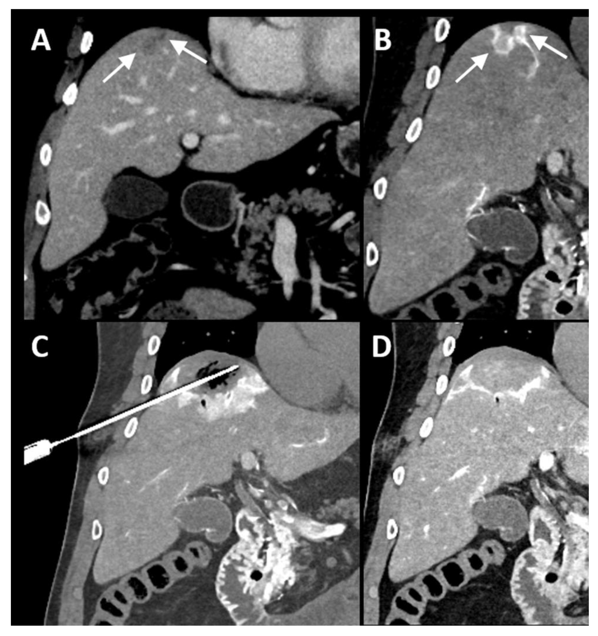
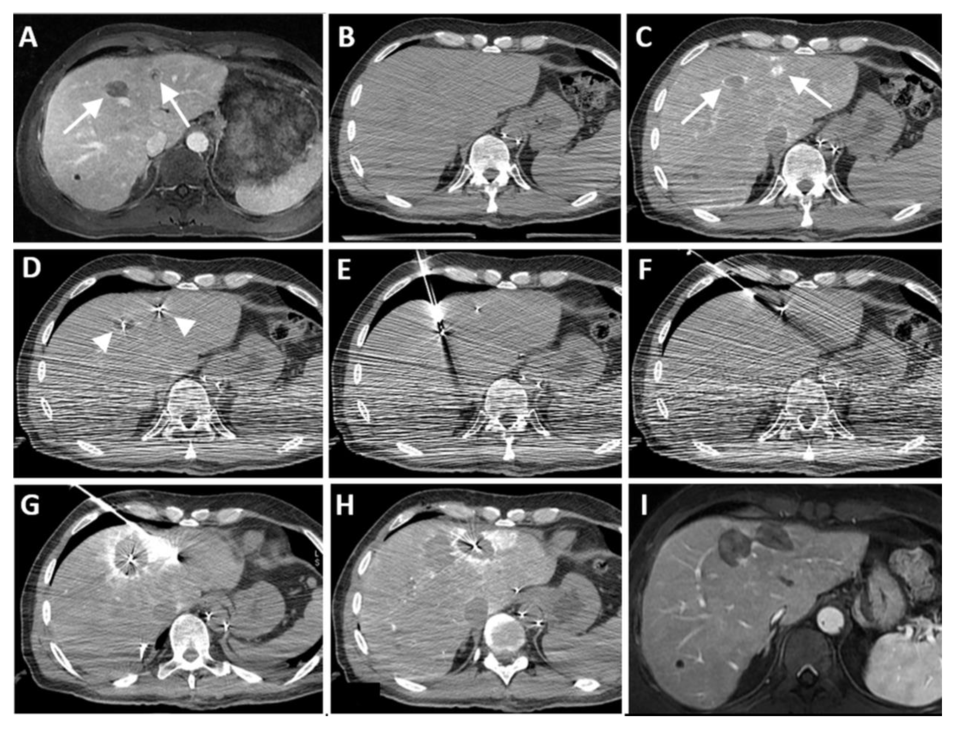
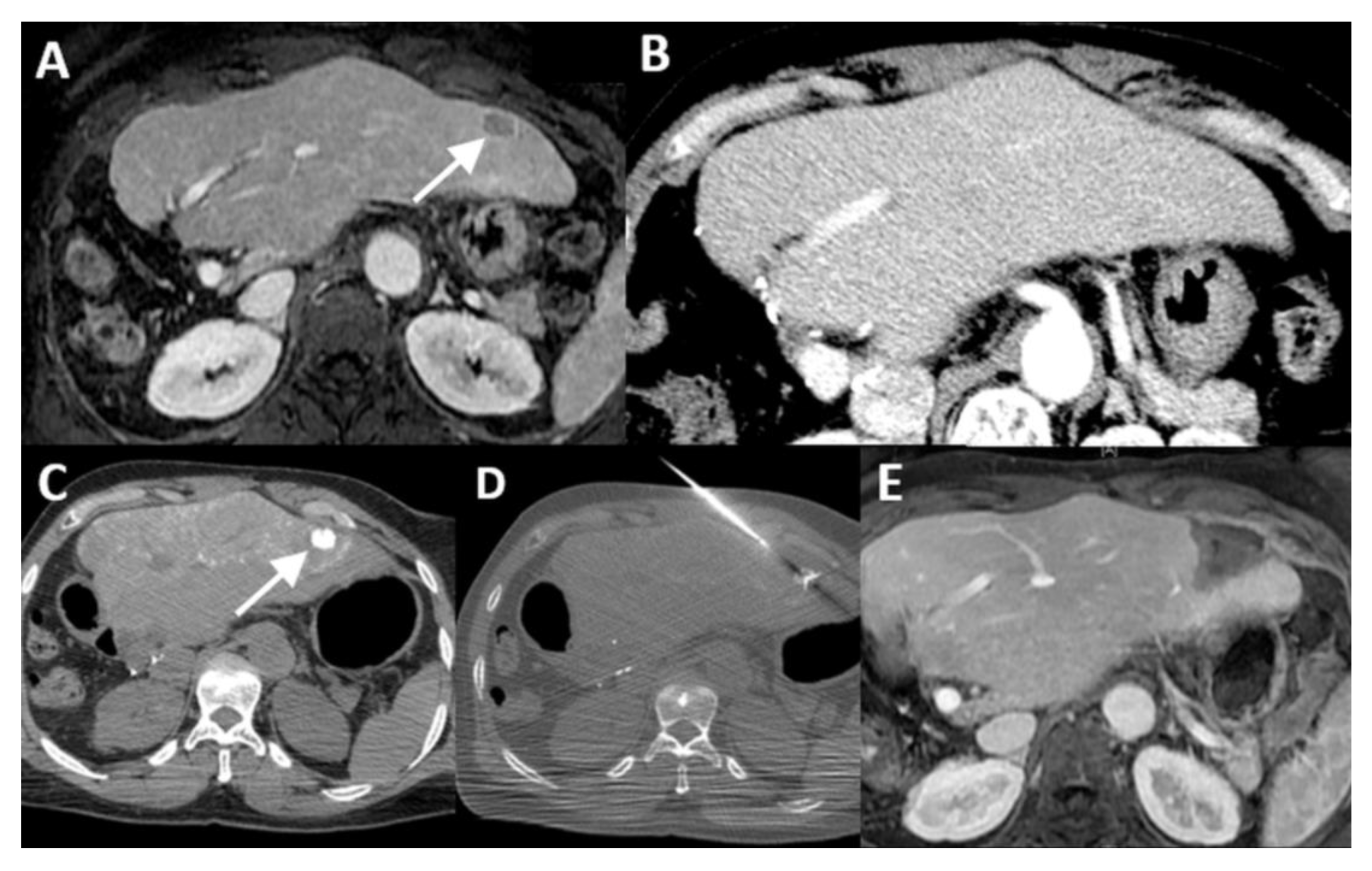
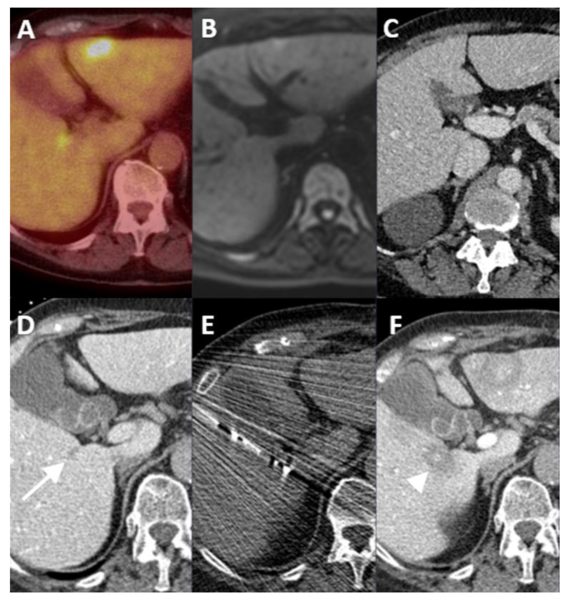
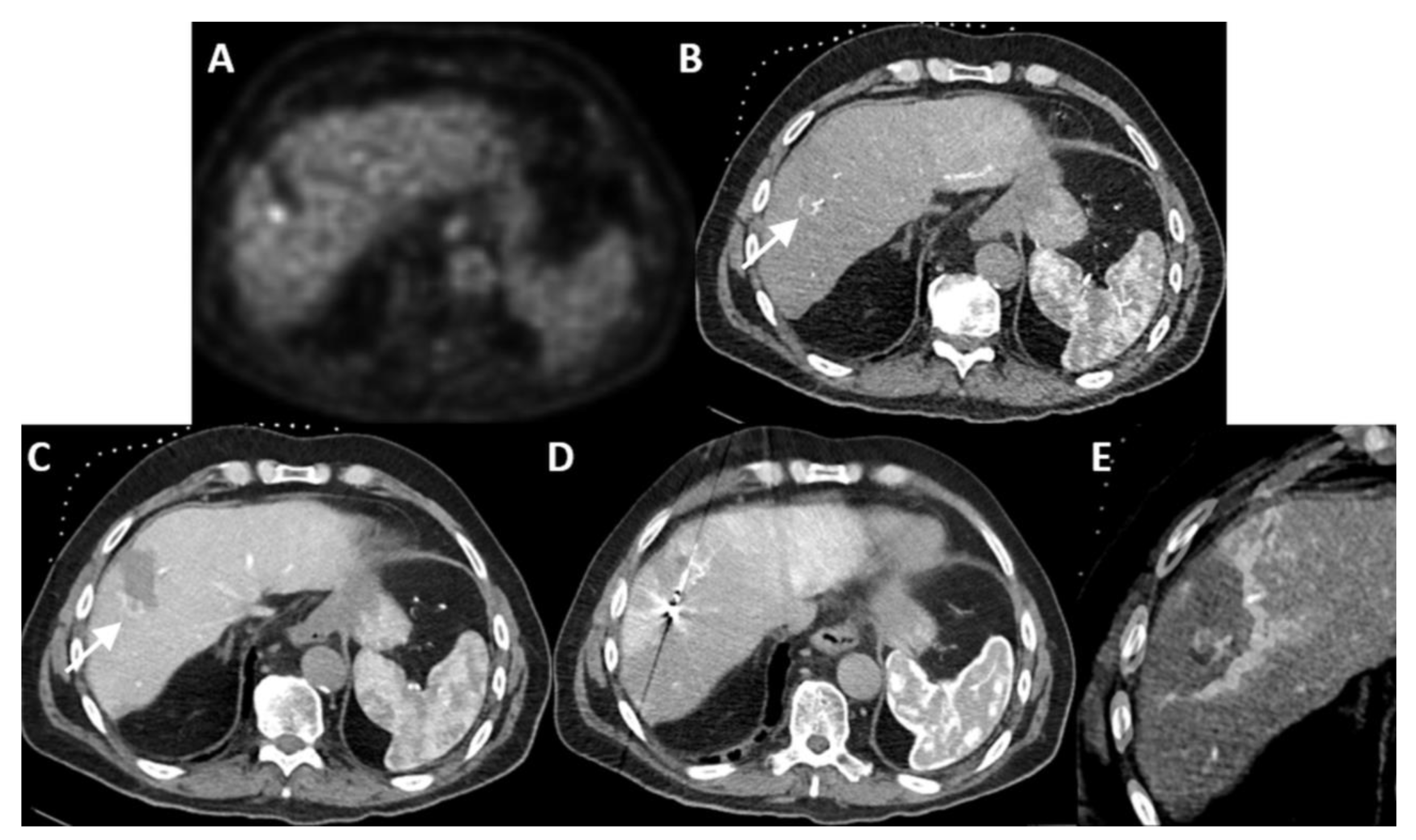
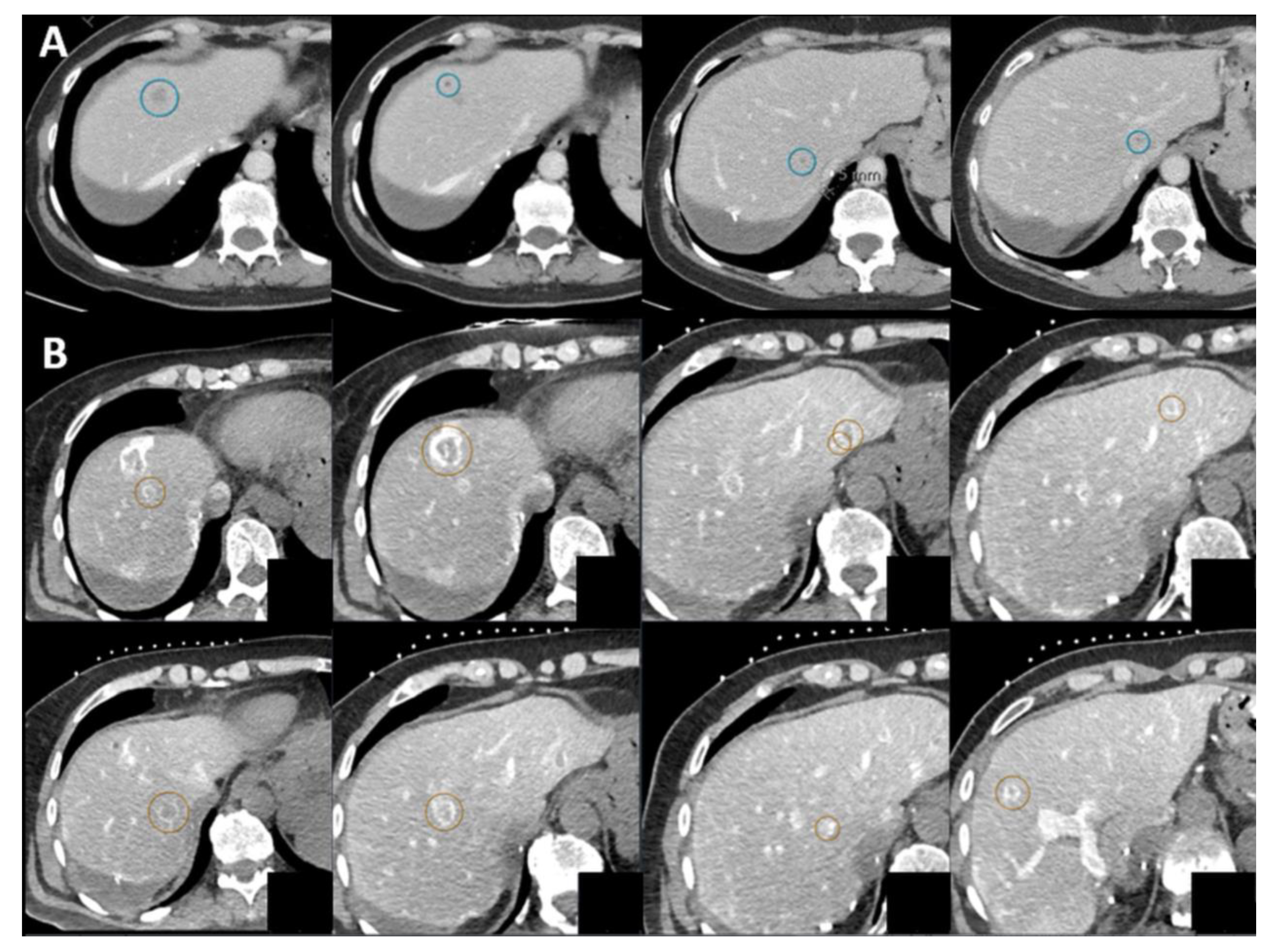
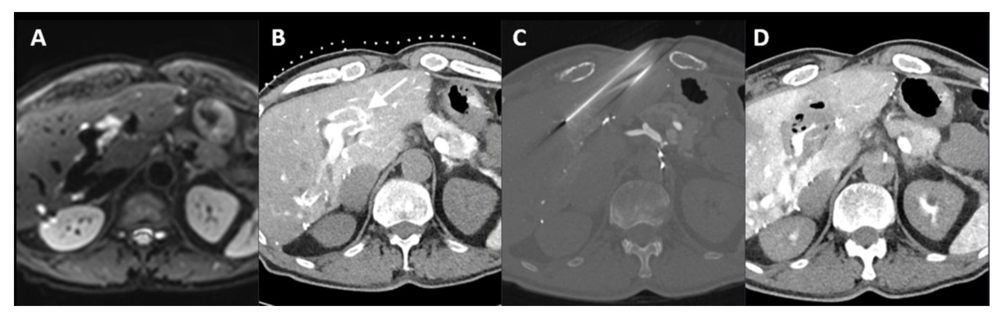
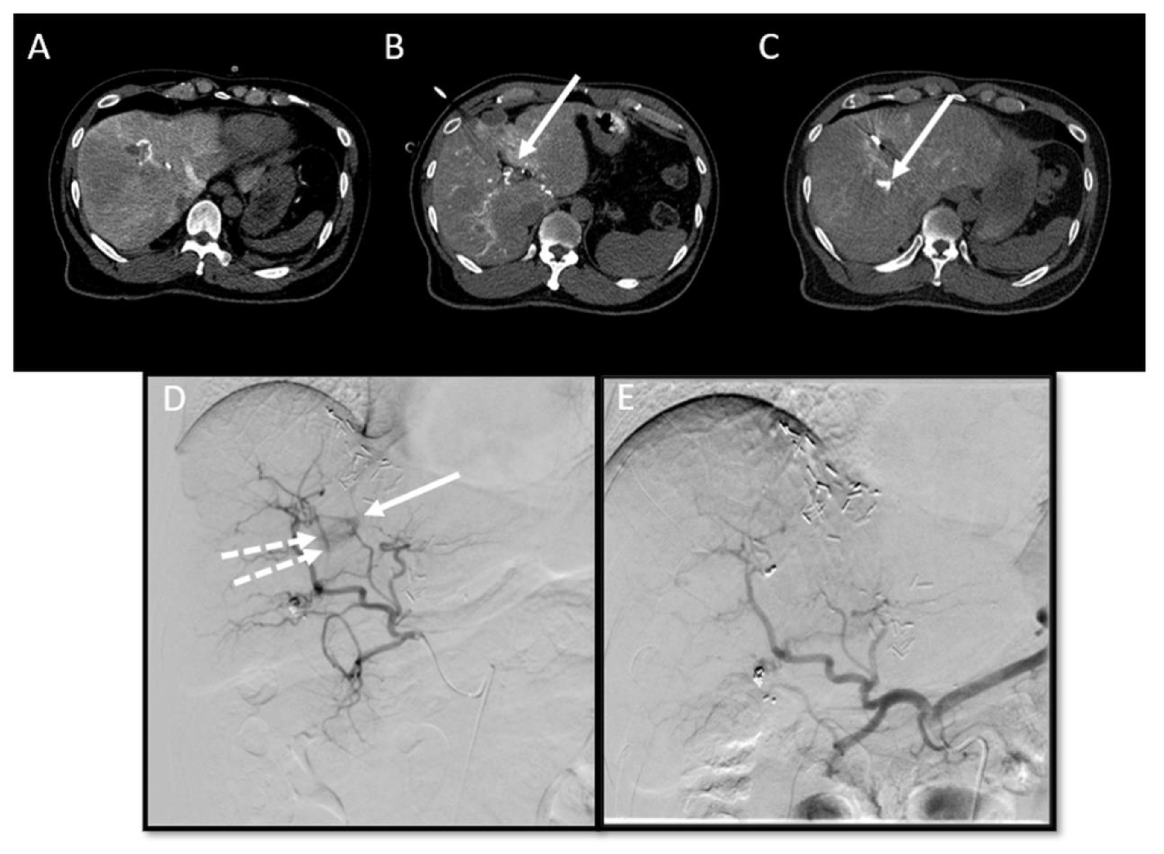
2. Cases
3. Discussion
4. Conclusions
Author Contributions
Funding
Conflicts of Interest
References
- Van Amerongen, M.J.; Jenniskens, S.F.M.; van den Boezem, P.B.; Fütterer, J.J.; de Wilt, J.H.W. Radiofrequency ablation compared to surgical resection for curative treatment of patients with colorectal liver metastases—A meta-analysis. HPB 2017, 19, 749–756. [Google Scholar] [CrossRef] [PubMed]
- Vasiniotis Kamarinos, N.; Kaye, E.A.; Sofocleous, C.T. Image-Guided Thermal Ablation for Colorectal Liver Metastases. Tech. Vasc. Interv. Radiol. 2020, 23, 100672. [Google Scholar] [CrossRef] [PubMed]
- Van Cutsem, E.; Cervantes, A.; Adam, R.; Sobrero, A.; Van Krieken, J.H.; Aderka, D.; Aguilar, E.A.; Bardelli, A.; Benson, A.; Bodoky, G.; et al. ESMO consensus guidelines for the management of patients with metastatic colorectal cancer. Ann. Oncol. 2016, 27, 1386–1422. [Google Scholar] [CrossRef] [PubMed]
- Gillams, A.; Goldberg, N.; Ahmed, M.; Bale, R.; Breen, D.; Callstrom, M.; Chen, M.H.; Choi, B.I.; de Baere, T.; Dupuy, D.; et al. Thermal ablation of colorectal liver metastases: A position paper by an international panel of ablation experts, The Interventional Oncology Sans Frontieres meeting 2013. Eur. Radiol. 2015, 25, 3438–3454. [Google Scholar] [CrossRef] [PubMed]
- Salhab, M.; Canelo, R. An overview of evidence-based management of hepatocellular carcinoma: A meta-analysis. J. Cancer Res. Ther. 2011, 7, 463–475. [Google Scholar] [CrossRef] [PubMed]
- Tsitskari, M.; Filippiadis, D.; Kostantos, C.; Palialexis, K.; Zavridis, P.; Kelekis, N.; Brountzos, E. The role of interventional oncology in the treatment of colorectal cancer liver metastases. Ann. Gastroenterol. 2019, 32, 147–155. [Google Scholar] [CrossRef] [PubMed]
- Shady, W.; Petre, E.N.; Do, K.G.; Gonen, M.; Yarmohammadi, H.; Brown, K.T.; Kemeny, N.E.; D’Angelica, M.; Kingham, P.T.; Solomon, S.B.; et al. Percutaneous Microwave versus Radiofrequency Ablation of Colorectal Liver Metastases: Ablation with Clear Margins (A0) Provides the Best Local Tumor Control. J. Vasc. Interv. Radiol. 2018, 29, 268–275.e261. [Google Scholar] [CrossRef] [PubMed]
- Ahmed, M.; Solbiati, L.; Brace, C.L.; Breen, D.J.; Callstrom, M.R.; Charboneau, J.W.; Chen, M.H.; Choi, B.I.; de Baere, T.; Dodd, G.D., 3rd; et al. Image-guided tumor ablation: Standardization of terminology and reporting criteria—A 10-year update. Radiology 2014, 273, 241–260. [Google Scholar] [CrossRef]
- Wang, X.; Sofocleous, C.T.; Erinjeri, J.P.; Petre, E.N.; Gonen, M.; Do, K.G.; Brown, K.T.; Covey, A.M.; Brody, L.A.; Alago, W.; et al. Margin size is an independent predictor of local tumor progression after ablation of colon cancer liver metastases. Cardiovasc. Intervent. Radiol. 2013, 36, 166–175. [Google Scholar] [CrossRef]
- Kim, Y.S.; Lee, W.J.; Rhim, H.; Lim, H.K.; Choi, D.; Lee, J.Y. The minimal ablative margin of radiofrequency ablation of hepatocellular carcinoma (>2 and <5 cm) needed to prevent local tumor progression: 3D quantitative assessment using CT image fusion. Am. J. Roentgenol. 2010, 195, 758–765. [Google Scholar] [CrossRef]
- Kaye, E.A.; Cornelis, F.H.; Petre, E.N.; Tyagi, N.; Shady, W.; Shi, W.; Zhang, Z.; Solomon, S.B.; Sofocleous, C.T.; Durack, J.C. Volumetric 3D assessment of ablation zones after thermal ablation of colorectal liver metastases to improve prediction of local tumor progression. Eur. Radiol. 2019, 29, 2698–2705. [Google Scholar] [CrossRef] [PubMed]
- Puijk, R.S.; Ahmed, M.; Adam, A.; Arai, Y.; Arellano, R.; de Baere, T.; Bale, R.; Bellera, C.; Binkert, C.A.; Brace, C.L.; et al. Consensus Guidelines for the Definition of Time-to-Event End Points in Image-guided Tumor Ablation: Results of the SIO and DATECAN Initiative. Radiology 2021, 301, 533–540. [Google Scholar] [CrossRef] [PubMed]
- Laimer, G.; Schullian, P.; Bale, R. Stereotactic Thermal Ablation of Liver Tumors: 3D Planning, Multiple Needle Approach, and Intraprocedural Image Fusion Are the Key to Success—A Narrative Review. Biology 2021, 10, 644. [Google Scholar] [CrossRef] [PubMed]
- Laimer, G.; Jaschke, N.; Schullian, P.; Putzer, D.; Eberle, G.; Solbiati, M.; Solbiati, L.; Goldberg, S.N.; Bale, R. Volumetric assessment of the periablational safety margin after thermal ablation of colorectal liver metastases. Eur. Radiol. 2021, 31, 6489–6499. [Google Scholar] [CrossRef] [PubMed]
- Puijk, R.S.; Nieuwenhuizen, S.; van den Bemd, B.A.T.; Ruarus, A.H.; Geboers, B.; Vroomen, L.; Muglia, R.; de Jong, M.C.; de Vries, J.J.J.; Scheffer, H.J.; et al. Transcatheter CT Hepatic Arteriography Compared with Conventional CT Fluoroscopy Guidance in Percutaneous Thermal Ablation to Treat Colorectal Liver Metastases: A Single-Center Comparative Analysis of 2 Historical Cohorts. J. Vasc. Interv. Radiol. 2020, 31, 1772–1783. [Google Scholar] [CrossRef] [PubMed]
- Ohki, T.; Tateishi, R.; Akahane, M.; Mikami, S.; Sato, M.; Uchino, K.; Arano, T.; Enooku, K.; Kondo, Y.; Yamashiki, N.; et al. CT with hepatic arterioportography as a pretreatment examination for hepatocellular carcinoma patients: A randomized controlled trial. Am. J. Gastroenterol. 2013, 108, 1305–1313. [Google Scholar] [CrossRef]
- Tsurusaki, M.; Sugimoto, K.; Fujii, M.; Fukuda, T.; Matsumoto, S.; Sugimura, K. Combination of CT during arterial portography and double-phase CT hepatic arteriography with multi-detector row helical CT for evaluation of hypervascular hepatocellular carcinoma. Clin. Radiol. 2007, 62, 1189–1197. [Google Scholar] [CrossRef] [PubMed]
- van Tilborg, A.A.; Scheffer, H.J.; van der Meijs, B.B.; van Werkum, M.H.; Melenhorst, M.C.; van den Tol, P.M.; Meijerink, M.R. Transcatheter CT hepatic arteriography-guided percutaneous ablation to treat ablation site recurrences of colorectal liver metastases: The incomplete ring sign. J. Vasc. Interv. Radiol. 2015, 26, 583–587.e581. [Google Scholar] [CrossRef]
- van der Lei, S.; Opperman, J.; Dijkstra, M.; Kors, N.; Boon, R.; van den Bemd, B.A.T.; Timmer, F.E.F.; Nota, I.; van den Bergh, J.E.; de Vries, J.J.J.; et al. The Added Diagnostic Value of Transcatheter CT Hepatic Arteriography for Intraprocedural Detection of Previously Unknown Colorectal Liver Metastases During Percutaneous Ablation and Impact on the Definitive Treatment Plan. Cardiovasc. Intervent. Radiol. 2023, 46, 1257–1266. [Google Scholar] [CrossRef]
- Paolucci, I.; Albuquerque Marques Silva, J.; Lin, Y.M.; Fellman, B.M.; Jones, K.A.; Tatsui, C.E.; Weinberg, J.S.; Ruiz, J.; Tan, J.; Brock, K.K.; et al. Study Protocol STEREOLAB: Stereotactic Liver Ablation Assisted with Intra-Arterial CT Hepatic Arteriography and Ablation Confirmation Software Assessment. Cardiovasc. Intervent. Radiol. 2023, 46, 1748–1754. [Google Scholar] [CrossRef]
- Smits, M.L.J.; Bruijnen, R.C.G.; Tetteroo, P.; Vonken, E.P.A.; Meijerink, M.R.; Hagendoorn, J.; de Bruijne, J.; Prevoo, W. Hepatic Arteriography and C-Arm CT-Guided Ablation (HepACAGA) to Improve Tumor Visualization, Navigation and Margin Confirmation in Percutaneous Liver Tumor Ablation. Cardiovasc. Intervent. Radiol. 2023, 46, 1365–1374. [Google Scholar] [CrossRef] [PubMed]
- Farouil, G.; Deschamps, F.; Hakime, A.; de Baere, T. Coil-assisted RFA of poorly visible liver tumors: Effectiveness and risk factors of local tumor progression. Cardiovasc. Intervent. Radiol. 2014, 37, 716–722. [Google Scholar] [CrossRef] [PubMed]
- Solbiati, M.; Passera, K.M.; Rotilio, A.; Oliva, F.; Marre, I.; Goldberg, S.N.; Ierace, T.; Solbiati, L. Augmented reality for interventional oncology: Proof-of-concept study of a novel high-end guidance system platform. Eur. Radiol. Exp. 2018, 2, 18. [Google Scholar] [CrossRef] [PubMed]
- Rhim, H.; Lim, H.K.; Kim, Y.S.; Choi, D.; Lee, W.J. Radiofrequency ablation of hepatic tumors: Lessons learned from 3000 procedures. J. Gastroenterol. Hepatol. 2008, 23, 1492–1500. [Google Scholar] [CrossRef] [PubMed]
- Deschamps, F.; Moine, L.; Isoardo, T.; Tselikas, L.; Paci, A.; Mir, L.M.; Huang, N.; Fattal, E.; de Baere, T. Parameters for Stable Water-in-Oil Lipiodol Emulsion for Liver Trans-Arterial Chemo-Eembolization. Cardiovasc. Intervent. Radiol. 2017, 40, 1927–1932. [Google Scholar] [CrossRef] [PubMed]
- Tsilimigras, D.I.; Ntanasis-Stathopoulos, I.; Paredes, A.Z.; Moris, D.; Gavriatopoulou, M.; Cloyd, J.M.; Pawlik, T.M. Disappearing liver metastases: A systematic review of the current evidence. Surg. Oncol. 2019, 29, 7–13. [Google Scholar] [CrossRef] [PubMed]
- Tanaka, T.; Arai, Y.; Inaba, Y.; Inoue, M.; Nishiofuku, H.; Anai, H.; Hori, S.; Sakaguchi, H.; Kichikawa, K. Current role of hybrid CT/angiography system compared with C-arm cone beam CT for interventional oncology. Br. J. Radiol. 2014, 87, 20140126. [Google Scholar] [CrossRef] [PubMed]
- Yu, Q.; Knight, G.; Karani, K.; Fergus, J.; Leef, J.; Funaki, B.; Ahmed, O. Real-time arteriography-directed percutaneous microwave ablation for small or poorly characterized hepatic lesions using hybrid Angio-CT. Abdom. Radiol. 2022, 47, 1457–1463. [Google Scholar] [CrossRef]
- Yuan, H.; Li, X.; Tian, X.; Ji, K.; Liu, F. Comparison of Angio-CT and cone-beam CT-guided immediate radiofrequency ablation after transcatheter arterial chemoembolization for large hepatocellular carcinoma. Abdom. Radiol. 2020, 45, 2585–2592. [Google Scholar] [CrossRef]
- Solbiati, M.; Muglia, R.; Goldberg, S.N.; Ierace, T.; Rotilio, A.; Passera, K.M.; Marre, I.; Solbiati, L. A novel software platform for volumetric assessment of ablation completeness. Int. J. Hyperth. 2019, 36, 337–343. [Google Scholar] [CrossRef]
- Mauri, G.; Cova, L.; De Beni, S.; Ierace, T.; Tondolo, T.; Cerri, A.; Goldberg, S.N.; Solbiati, L. Real-time US-CT/MRI image fusion for guidance of thermal ablation of liver tumors undetectable with US: Results in 295 cases. Cardiovasc. Intervent. Radiol. 2015, 38, 143–151. [Google Scholar] [CrossRef]
- Kang, T.W.; Lee, M.W.; Choi, S.H.; Rhim, H.; Lim, S.; Song, K.D.; Min, J.H.; Choi, S.Y.; Lim, H.K.; Yang, J. A novel electrode with electromagnetic tip tracking in ultrasonography-guided radiofrequency ablation: A phantom, ex vivo, and in vivo experimental study. Investig. Radiol. 2015, 50, 81–87. [Google Scholar] [CrossRef]
- Taghavi, M.; Staal, F.; Gomez Munoz, F.; Imani, F.; Meek, D.B.; Simoes, R.; Klompenhouwer, L.G.; van der Heide, U.A.; Beets-Tan, R.G.H.; Maas, M. CT-Based Radiomics Analysis Before Thermal Ablation to Predict Local Tumor Progression for Colorectal Liver Metastases. Cardiovasc. Intervent. Radiol. 2021, 44, 913–920. [Google Scholar] [CrossRef]


| Case No. | Tumor Size (mm) | Catheter Tip Position | Amount and Type of Contrast per Injection | Ablation Device | Institute |
|---|---|---|---|---|---|
| 1 | 20 mm | Common hepatic artery | 15–20 mL Visipaque™ | Emprint™ Microwave Ablation System, Medtronic-Covidien, Boulder, CO, USA | MD Anderson, Houston, TX, USA |
| 2 | 20 and 7 mm | Common hepatic artery | 4 mL Xenetix 300® | Cool-tip™ RFA Ablation Aystem, Medtronic-Covidien, Boulder, CO, USA | Gustave Roussy, Villejuif, France |
| 3 | 12 mm | Left-sided hepatic artery | Not specified | Cool-tip™ RFA Ablation System | Gustave Roussy |
| 4 | 9 mm | Common hepatic artery | 10 mL Xenetix 300® | Emprint™ Microwave Ablation System | Amsterdam UMC, Amsterdam, The Netherlands |
| 5 | 15 mm | Coeliac trunk | 20 mL Xenetix 300® | Emprint™ Microwave Ablation System | Amsterdam UMC |
| 6 | Not applicable | Common hepatic artery | 20 mL Xenetix 300® | Emprint™ Microwave Ablation System | Amsterdam UMC |
| 7 | Confluent | Common hepatic artery | 10 mL Xenetix 300® | NanoKnife system under ECG-gating; AccuSync model 72, AngioDynamics, Latham, NY, USA | Amsterdam UMC |
| 8 | 15 mm | Common hepatic artery | 15–20 mL Visipaque™ | Emprint™ Microwave Ablation System | MD Anderson |
Disclaimer/Publisher’s Note: The statements, opinions and data contained in all publications are solely those of the individual author(s) and contributor(s) and not of MDPI and/or the editor(s). MDPI and/or the editor(s) disclaim responsibility for any injury to people or property resulting from any ideas, methods, instructions or products referred to in the content. |
© 2024 by the authors. Licensee MDPI, Basel, Switzerland. This article is an open access article distributed under the terms and conditions of the Creative Commons Attribution (CC BY) license (https://creativecommons.org/licenses/by/4.0/).
Share and Cite
Puijk, R.S.; Dijkstra, M.; van der Lei, S.; Schulz, H.H.; Vos, D.J.W.; Timmer, F.E.F.; Geboers, B.; Scheffer, H.J.; de Vries, J.J.J.; Smits, M.L.J.; et al. The Added Value of Transcatheter CT Hepatic Angiography (CTHA) Image Guidance in Percutaneous Thermal Liver Ablation: An Experts’ Opinion Pictorial Essay. Cancers 2024, 16, 1193. https://doi.org/10.3390/cancers16061193
Puijk RS, Dijkstra M, van der Lei S, Schulz HH, Vos DJW, Timmer FEF, Geboers B, Scheffer HJ, de Vries JJJ, Smits MLJ, et al. The Added Value of Transcatheter CT Hepatic Angiography (CTHA) Image Guidance in Percutaneous Thermal Liver Ablation: An Experts’ Opinion Pictorial Essay. Cancers. 2024; 16(6):1193. https://doi.org/10.3390/cancers16061193
Chicago/Turabian StylePuijk, Robbert S., Madelon Dijkstra, Susan van der Lei, Hannah H. Schulz, Danielle J. W. Vos, Florentine E. F. Timmer, Bart Geboers, Hester J. Scheffer, Jan J. J. de Vries, Maarten L. J. Smits, and et al. 2024. "The Added Value of Transcatheter CT Hepatic Angiography (CTHA) Image Guidance in Percutaneous Thermal Liver Ablation: An Experts’ Opinion Pictorial Essay" Cancers 16, no. 6: 1193. https://doi.org/10.3390/cancers16061193