Article

Imaging GRPr Expression in Metastatic Castration-Resistant Prostate Cancer with [68Ga]Ga-RM2—A Head-to-Head Pilot Comparison with [68Ga]Ga-PSMA-11

1
Nuclear Medicine and PET/CT Center PositronMed, Providencia, Santiago 7501068, Chile
2
Positronpharma SA, Providencia, Santiago 7501068, Chile
3
Department of Radiology, Division of Nuclear Medicine and Molecular Imaging, Stanford University, Stanford, CA 94305, USA
4
Life Molecular Imaging GmbH, 13353 Berlin, Germany
5
Formerly Piramal Imaging GmbH, 13353 Berlin, Germany
6
Merck Healthcare KGaA, 64293 Darmstadt, Germany
7
Fundación Arturo López Pérez, Providencia, Santiago 750069, Chile
*
Author to whom correspondence should be addressed.
Cancers 2024, 16(1), 173; https://doi.org/10.3390/cancers16010173
Submission received: 8 November 2023 / Revised: 18 December 2023 / Accepted: 24 December 2023 / Published: 29 December 2023

Abstract

:

Simple Summary

Prostate cancer is the most prevalent cancer among men. Patients diagnosed with metastatic, castration-resistant prostate cancer (mCRPC) face a highly aggressive disease and reduced overall survival. For these patients, [177Lu]Lu-PSMA-617 has shown promising results. However, this therapy may not benefit patients with low or heterogeneous PSMA expression. The gastrin-releasing peptide receptor (GRPr) is highly expressed in prostate cancer and other cancer cells, and [177Lu]Lu-labeled GRPr-ligands have demonstrated good tumor uptake and retention, with minimal uptake in healthy tissues. However, the level of GRPr expression in advanced mCRPC patients remains elusive. In this study, we compared [68Ga]Ga-RM2 with [68Ga]Ga-PSMA-11 in a Latin American mCRPC cohort to evaluate the clinical utility of [68Ga]Ga-RM2 in this group of patients. Although GRPr is overexpressed in the early stages of prostate cancer, our results indicate that in more advanced stages, such as mCRPC, the expression is lower than PSMA.

Abstract

Background: The gastrin-releasing peptide receptor (GRPr) is highly overexpressed in several solid tumors, including treatment-naïve and recurrent prostate cancer. [68Ga]Ga-RM2 is a well-established radiotracer for PET imaging of GRPr, and [177Lu]Lu-RM2 has been proposed as a therapeutic alternative for patients with heterogeneous and/or low expression of PSMA. In this study, we aimed to evaluate the expression of GRPr and PSMA in a group of patients diagnosed with castration-resistant prostate cancer (mCRPC) by means of PET imaging. Methods: Seventeen mCRPC patients referred for radio-ligand therapy (RLT) were enrolled and underwent [68Ga]Ga-PSMA-11 and [68Ga]Ga-RM2 PET/CT imaging, 8.8 ± 8.6 days apart, to compare the biodistribution of each tracer. Uptake in healthy organs and tumor lesions was assessed by SUV values, and tumor-to-background ratios were analyzed. Results: [68Ga]Ga-PSMA-11 showed significantly higher uptake in tumor lesions in bone, lymph nodes, prostate, and soft tissues and detected 23% more lesions compared to [68Ga]Ga-RM2. In 4/17 patients (23.5%), the biodistribution of both tracers was comparable. Conclusions: Our results show that in our cohort of mCRPC patients, PSMA expression was higher compared to GRPr. Nevertheless, RLT with [177Lu]Lu-RM2 may be an alternative treatment option for selected patients or patients in earlier disease stages, such as biochemical recurrence.

Graphical Abstract

1. Introduction

Prostate cancer (PCa) is the most common cancer in men, with an incidence of approximately 30.7 per 100,000 inhabitants (age standardized) [1]. While the five-year survival rate of localized, low-volume prostate cancer is close to 100%, metastatic, castration-resistant prostate cancer (mCRPC) is a highly aggressive disease with a significantly reduced median overall survival, accounting for 3.8% of all cancer deaths in men [1,2,3]. Although taxane-based chemotherapy and other available treatments can mitigate the effects of the disease, mCRPC can eventually progress, leaving the patients without further treatment options.
Recently, radioligand therapy (RLT) with [177Lu]Lu-PSMA-617, targeting the prostate-specific membrane antigen (PSMA), has emerged as a promising treatment for advanced PCa patients. RLT with [177Lu]Lu-PSMA-617 has demonstrated a remarkable capacity to improve quality of life and overall survival in most patients with mCRPC [4]. Nonetheless, the evidence indicates that approximately 30% of patients already show progression after the first or second treatment cycle [5,6], which might in part be related to a heterogeneous PSMA expression and low, insufficient absorbed doses in individual lesions.
The gastrin-releasing peptide (GRP) can be found in the nervous system and peripheral tissues, such as the gastrointestinal tract. GRP binds to its receptor (gastrin-releasing peptide receptor (GRPr)), a G-coupled protein from the bombesin family. GRPr is overexpressed in different cancers, such as breast cancer, small-cell lung cancer, and gastrointestinal stromal tumors. However, GRPr is also highly expressed in tumoral vessels of urinary tract cancers, particularly treatment-naïve and recurrent prostate cancer [7,8,9], and during early and advanced stages of PCa [10,11]. Therefore, positron emission tomography (PET) imaging with the synthetic GRPr antagonist [68Ga]Ga-RM2 has emerged as a useful tool for biopsy guidance in patients with suspected PCa [12] and for staging and localization of disease in patients with primary PCa and patients with biochemical recurrence (BCR) and negative findings on conventional imaging and evaluation of treatment response [8,13,14,15,16]. Thus both, PSMA and GRPr are relevant diagnostic biomarkers for PET imaging in PCa at different stages of the disease [9,14,17,18,19,20]. Nevertheless, the biological mechanisms underlying PCa progression are complex and PET imaging of PSMA and GRPr might provide different insights into the heterogeneity of the disease. For instance, several studies support the notion that not all prostate cancer lesions present high levels of PSMA expression [20,21,22,23]. Interestingly, some metastases are exclusively detected by GRPr-targeted compounds and others are positive only for PSMA-targeted radiotracers, suggesting a complementary role between PSMA- and GRPR-targeted compounds [19,24,25]. However, determining the expression behavior of GRPr in the advanced stages of PCa remains a challenge. On the other hand, both tracers have renal elimination; however, PSMA presents increased physiological uptake in the liver parenchyma, a feature not observed with RM2. The low hepatobiliary uptake of 68Ga-RM2 enables the detection of liver metastasis.
Due to the high GRPr expression in PCa, [177Lu]Lu-labeled GRPr-ligands have been proposed as a therapeutic alternative for patients with low PSMA expression. This was exemplified in a proof-of-concept study evaluating the biodistribution and dosimetry of [177Lu]Lu-RM2 in mCRPC patients showing good tumor uptake, retention, and rapid clearance from healthy tissues [26]. The low hepatobiliary, salivary, and lacrimal gland uptake might represent an advantage of [177Lu]Lu-labeled GRPr ligands currently under development considering the high frequency of xerostomia as an adverse effect in patients under RLT with [177Lu]Lu-PSMA-617 [27,28]. Furthermore, given the high expression of GRPr in several cancer types, it is a relevant pan-tumor target for RLT [29,30,31]. However, a drawback for RLT may emerge due to the high uptake in the pancreas, leading to undesired side effects attributed to the high radiation dose. Thus, the pancreas is considered a dose-limiting organ for GRPr-mediated treatment. Nonetheless, preliminary data indicate that the uptake is not persistent and cleared within 24 h [26,27,28], and the pancreas is considered radioresistant [32]. However, the extent of GRPr expression in the advanced stages of PCa remains unclear [33,34], and ongoing investigations are evaluating what criteria are appropriate to select patients for GRPr-targeting RLT [35]. In this study, we compared [68Ga]Ga-RM2 and [68Ga]Ga-PSMA-11 in a cohort of Latin American patients diagnosed with mCRCP to further understand the potential clinical utility of GRPr-targeting RLT in these patients.

2. Materials and Methods

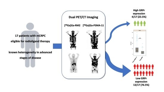
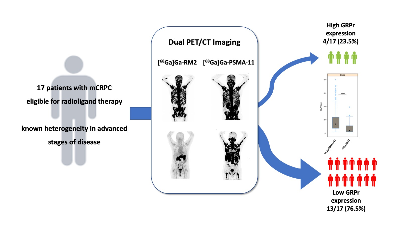
2.1. Patient Population

This prospective study was approved by the regional ethics committee board (Servicio de Salud Metropolitano Oriente, ethics committee, permit 26042016) and was conducted following the Declaration of Helsinki, Good Clinical Practices, and Chilean regulations. Seventeen subjects (median 66, IQR 8 years of age) with biopsy-proven mCRPC, rising PSA > 2 ng/mL, Gleason score of 8 to 10, testosterone < 20 ng/mL, a performance status score ECOG of 0–3, without further conventional treatment options, and who have been referred for RLT with either [177Lu]Lu-PSMA-617 or [177Lu]Lu-RM2, were enrolled in the study and gave written informed consent. Previous treatments included surgery (33%), androgen receptor signaling inhibitors (ARSI) (78%), radiotherapy (RT) (67%), and a combination of systemic therapies (ARSI, RT, and chemotherapy) (33%) (Table 1). Patients had PSA levels of 292 ± 465 ng/mL (range 0.05–1365 ng/mL) measured within 4 ± 3 days (range: 1–7 days) prior to PET imaging. Further blood biomarkers were evaluated as inclusion criteria (alkaline phosphatases > 2.5 upper normal limits in the absence of bone metastases; glutamic-oxaloacetic transaminase (GOT) and glutamic pyruvic transaminase (GPT) < 2.5 upper normal limits and up to 5 times if liver metastases are present; and creatinine clearance ≥ 40 mL/min/1.73 m2) for all patients included in the study and prior to the intervention. Exclusion criteria included the inability to sign the informed consent, not complying with the inclusion criteria, severe claustrophobia, or being diagnosed with a malignancy other than adenocarcinoma of the prostate.

2.2. Radiotracer Preparation

Production of [68Ga]Ga-PSMA-11 and [68Ga]Ga-RM2 was performed in accordance with local GMP regulations and using a similar procedure as published previously [14]. Briefly, radiolabeling of [68Ga]Ga-PSMA-11 and [68Ga]Ga-RM2 was performed using a cassette-based module (Gaia, Elysia-Raytest, Straubenhardt, Germany), PSMA-11, cassettes and reagent kits (Advanced biochemical compounds ABX, Dresden, Germany), RM2 (kindly provided from Life Molecular Imaging, Berlin, Germany), and a 2 GBq 68Ge/68Ga-generator (iThemba Labs, Somerset West 7129, Cape Town, South Africa).
The eluted gallium-68, was trapped on a strong cation exchange cartridge, rinsed with ultrapure water, and eluted with 450 μL eluent (5 M NaCl in 5.5 M HCl) into a mixture of 40 μL precursor (1 mg/mL in ultrapure water) in 3.85 mL buffer (0.08 M ammonium acetate, pH 4.5) and 200 μL ethanol. After radiolabeling at 95 °C for 8 min., the reaction mixture was diluted with 5 mL water. The crude product was extracted using a C18 cartridge and rinsed with water. The purified product was eluted with 1.5 mL 60 vol% ethanol followed by 8.5 mL saline and passed through a 0.22 µm sterile filter (Millex-GV, Merck Millipore, Darmstadt, Germany). Quality control was performed, including controls for visual inspection, pH, radiochemical purity by HPLC, radionuclidic identity, residual solvents, endotoxins, filter integrity (prior release), and sterility (post-release).

2.3. PET/CT Imaging and Analysis

All subjects had two PET/CT scans performed on separate days using a Biograph mCT Flow scanner (Siemens Healthineers, Erlangen, Germany) within 8.8 ± 8.6 days (range 1–33 days), without any medical intervention between the scans. The order of [68Ga]Ga-PSMA-11 and [68Ga]Ga-RM2 PET scans was random and according to the availability of the radiotracer. A contrast-enhanced CT scan and low-dose CT scan were performed for anatomical localization and attenuation correction prior to [68Ga]Ga-PSMA-11 and [68Ga]Ga-RM2 PET scans, respectively. PET/CT images were acquired head-to-mid-thigh at 60 ± 5 min post-injection of 191 ± 25 MBq (range 122–229 MBq) [68Ga]Ga-PSMA-11 and 166 ± 39 MBq (range: 63–243 MBq) [68Ga]Ga-RM2, respectively, starting at the pelvis.
Volumes of interest (VOIs) were drawn around tumor lesions, visually distinguished as regions of increased radiotracer uptake relative to adjacent background uptake and outside areas of expected physiological radiotracer uptake. To perform semi-quantitative analysis, mean and maximum standard uptake values (SUVmean and max, respectively) were calculated using Siemens SyngoVia software (SV60). Tumor-to-background ratios (TBRs) were calculated by dividing the SUVmax of different tumor lesions by the SUVmean of the blood pool in the left ventricle of the heart.

2.4. Statistical Analysis

The normal distribution of continuous variables was determined with Q-Q plots and histograms. In the case of non-parametric quantitative data, the Wilcoxon signed-rank test was used to compare SUVmax values and TBRs between scans. The test was two-sided, and a p-value < 0.05 was considered statistically significant. All statistical analyses were performed using R software version 4.2.0 (22 April 2022) [36]. The sample size for the study considered a minimum of 16 patients (17 were finally included). The sample size calculation was based on the difference between means and standard deviations of SUVmean ratios to the normal background (blood pool) for both tracers (9.2 ± 7.2 for [68Ga]Ga -PSMA-11 and 5.2 ± 3.5 for [68Ga]Ga-RM2), reported by Minamimoto et al. (2016). The calculation considered a confidence of 95% and a power of 80%, with a correlation of 60%. The analysis was performed using G*power [37].

3. Results

3.1. Patient Characteristics

Seventeen participants (65.3 ± 7.4 years of age; range: 53–76 years) were enrolled (Table 1) and both PET/CT scans were performed 8.8 ± 8.6 days (range: 1–33 days) apart.

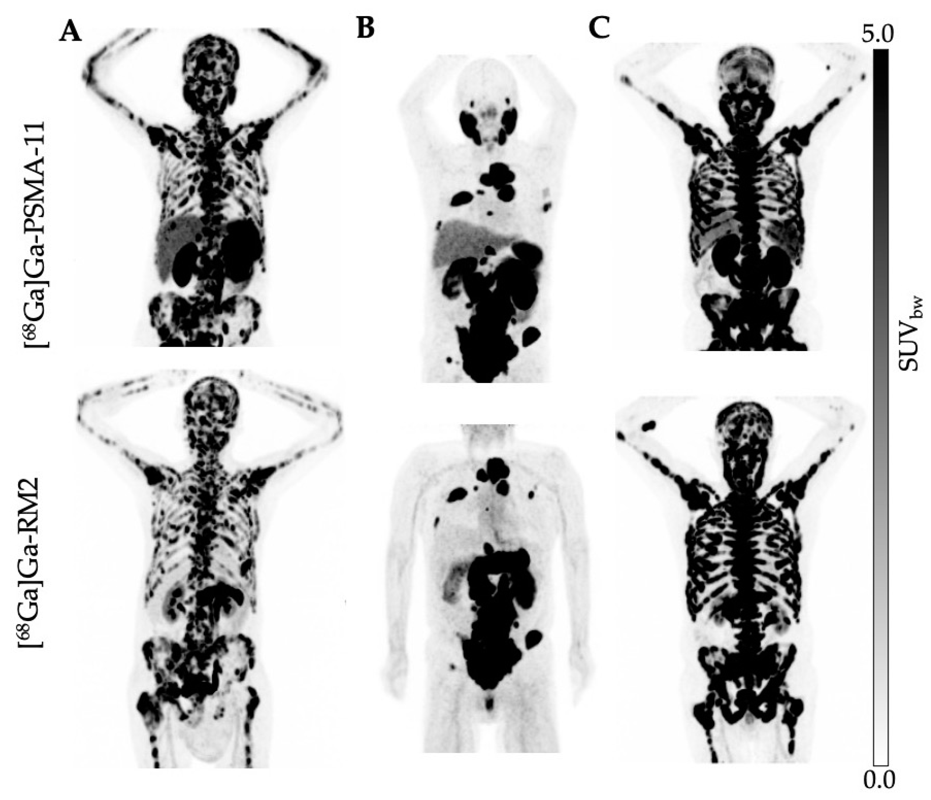
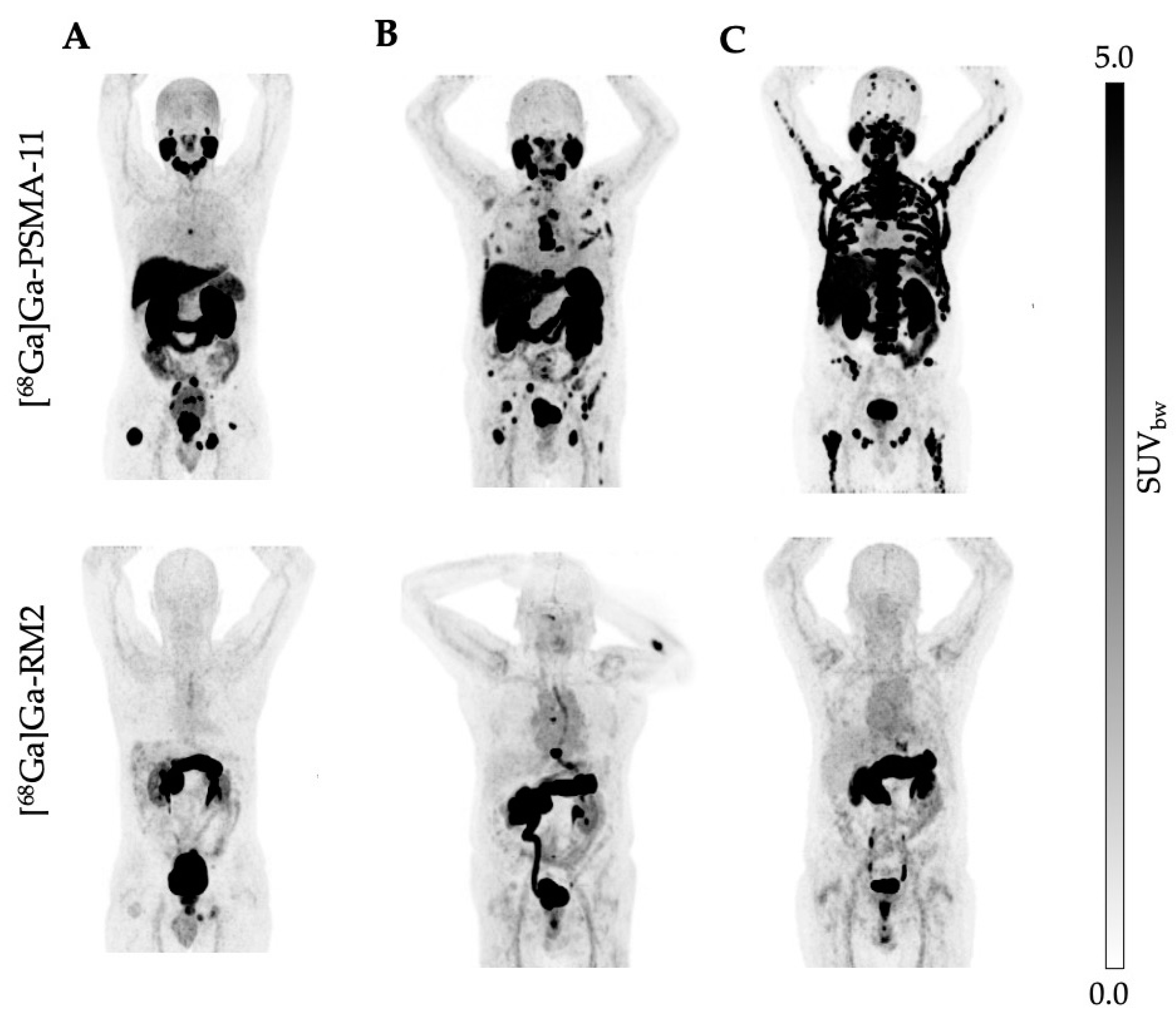
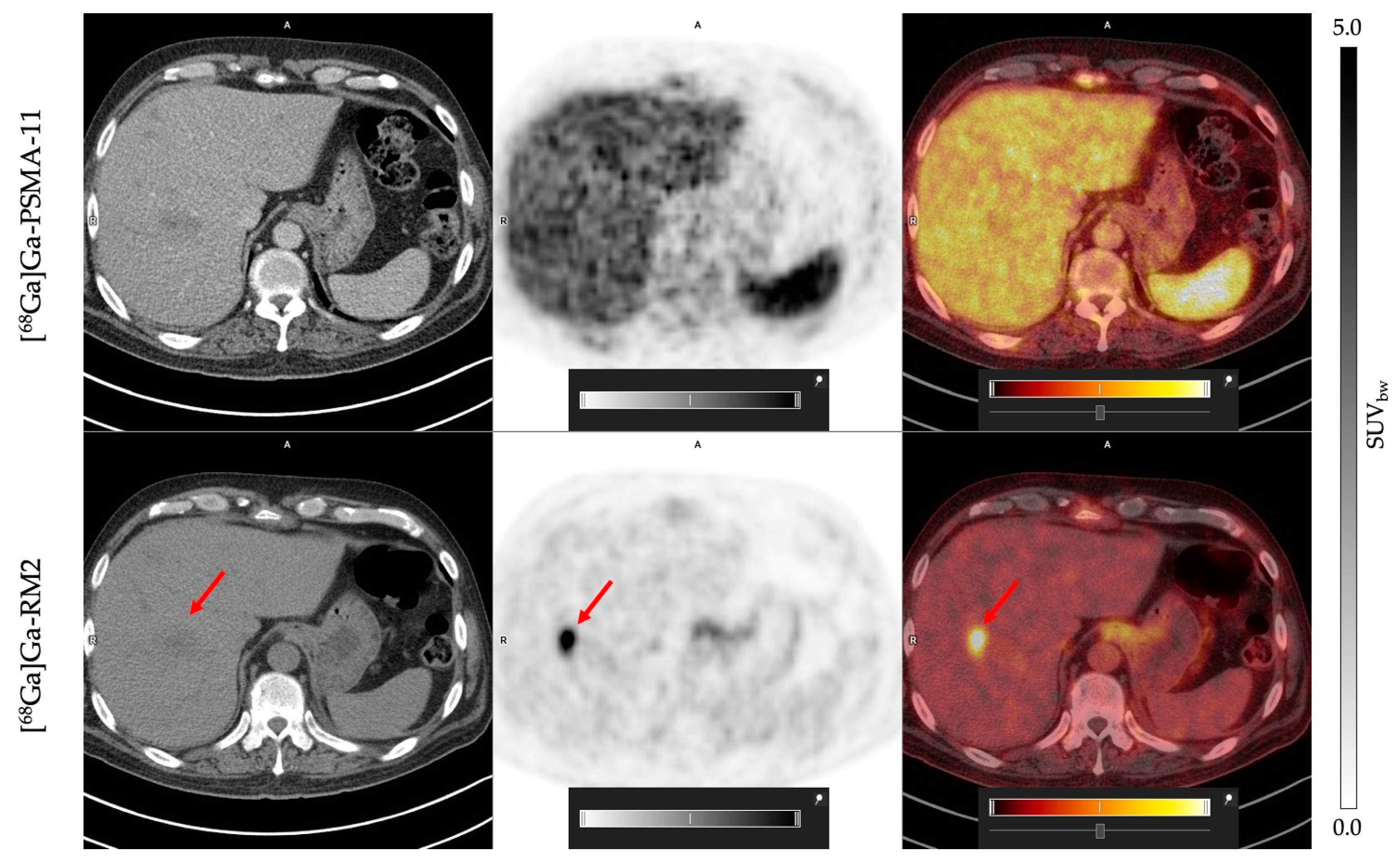
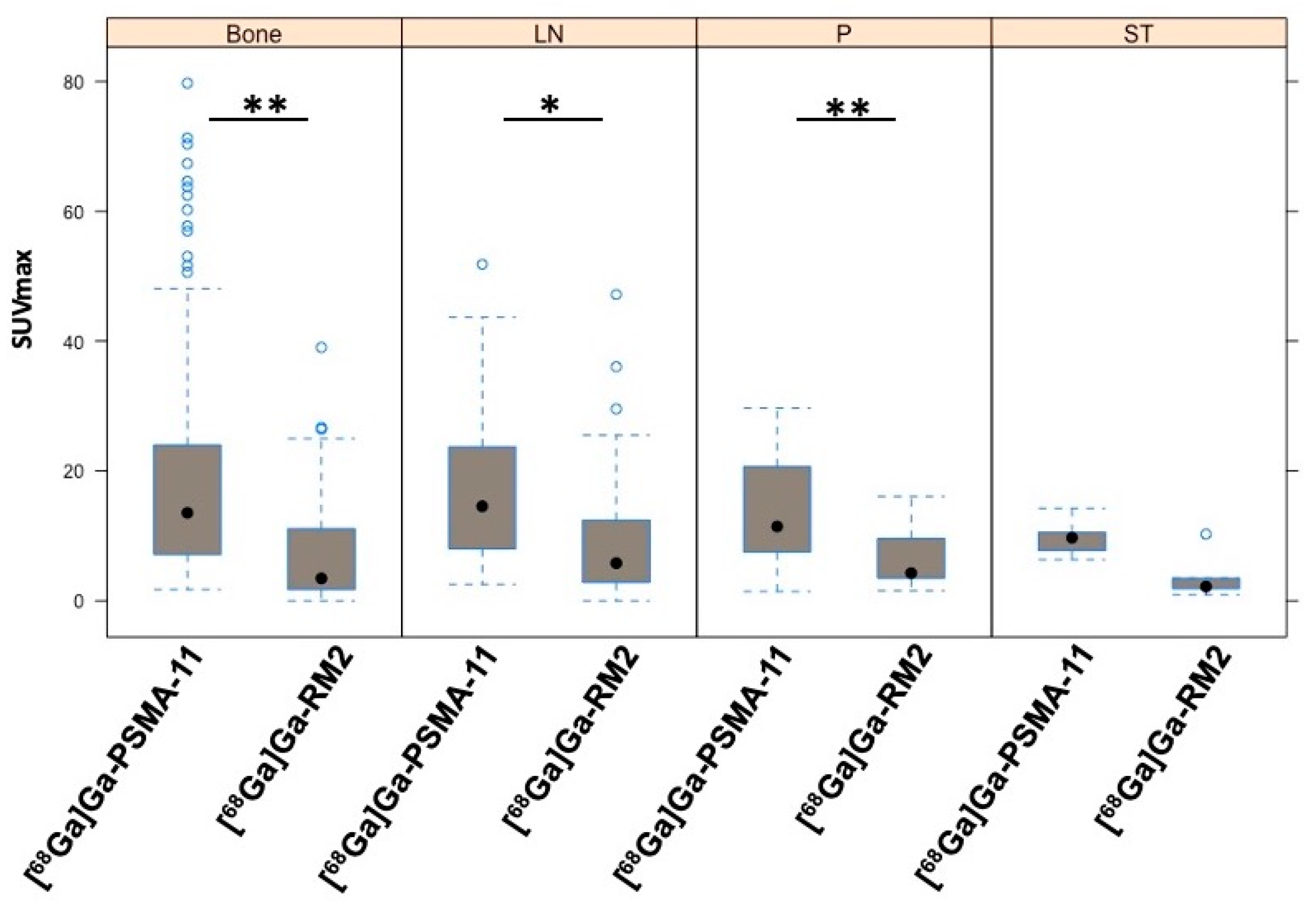
3.2. Uptake Comparison between [68Ga]Ga-PSMA-11 and [68Ga]Ga-RM2

The administration of [68Ga]Ga-PSMA-11, [68Ga]Ga-RM2, and the imaging procedure were well tolerated and no adverse events, discomfort, or change in vital signs was observed. The excretion profile of both tracers was similar with a predominant renal clearance via the kidneys observed for [68Ga]Ga-PSMA-11 and [68Ga]Ga-RM2 (Figure 1 and Figure 2). However, we observed differences in the physiological biodistribution between the two tracers in the submandibular, parotid, and lacrimal glands, liver, and small intestine, where unlike [68Ga]Ga-PSMA-11, [68Ga]Ga-RM2 showed no uptake. In contrast, [68Ga]Ga-RM2 showed high uptake in the pancreas, whereas no uptake of [68Ga]Ga-PSMA-11 was observed.
[68Ga]Ga-RM2 presents an absence of physiological uptake in the liver, contrary to what was observed with [68Ga]Ga-PSMA-11, which favors the detection of possible hepatic metastasis lesions. This was validated in one patient, who exhibited a hepatic lesion with [68Ga]Ga-RM2 that was not visible on [68Ga]Ga-PSMA-11 scan (Figure 3).
Specific uptake in tumor lesions in the prostate, lymph nodes, bone, and soft tissue was evident with both radioligands; however, the SUVmax values of [68Ga]Ga-PSMA-11 were statistically higher compared to [68Ga]Ga-RM2 in most lesions. Indeed, only 23.5% of the patients showed a high GRPr expression (Figure 1, Figure 2 and Figure 4).
As for the SUVmax values, the same trend was observed when evaluating TBRs for bone, lymph node, prostate, and soft tissue lesions, which were significantly higher for [68Ga]Ga-PSMA-11 compared to [68Ga]Ga-RM2 (Table 2).
Next, we analyzed whether both tracers were able to detect the same number of lesions considering the total number of lesions found in each patient. In line with our previous results, [68Ga]Ga-PSMA-11 detected 23.2% more tumor lesions compared to [68Ga]Ga-RM2.

4. Discussion

Prostate cancer, and in particular mCRPC, shows high levels of PSMA expression which also correlates with disease stage and severity [38,39]. However, due to the unstable genomic nature of cancerous cells, a tumor may present a great variability of PSMA expression levels resulting in different grades of malignancy and outcomes [40]. For instance, results from the Vision Trial indicate that 50–60% of patients with mCRPC respond with a PSA decline of >50% and an improvement in their overall survival of 15.3 months compared to 11.3 months in standard care. Likewise, imaging-based progression-free survival is also increased in those patients compared to standard care (median, 8.7 vs. 3.4 months) [4,5]. However, approximately 30–40% of mCRPC patients do not respond to [177Lu]Lu-PSMA therapy [5,6], which might be due to a heterogeneous PSMA expression or a decrease in PSMA triggered by an aggressive trans-differentiation process, resulting in cancerous cells resistant to therapies. Typically, these patients display visceral metastasis, and adenocarcinoma features are reduced or lost [41,42]. This variance or decrease in PSMA expressions affects the patient selection process and subsequently results in low absorbed doses in individual tumor lesions, ultimately reducing the therapeutic efficacy of [177Lu]Lu-PSMA therapy [40,43]. Variability in PSMA expression might depend on many different factors. For example, inflammation NF-κB has been involved in resistance to ADT, contributing to mCRPC progression [44]. Signaling pathways such as PI3K/AKT influence the tumor niche inducing different downstream events, including the expression of the H19 gene [45,46] and hypoxia [47]. The interaction between hypoxia and other pathways is, however, complex. The evidence suggests that hypoxia drives transdifferentiation toward an NE-like phenotype promoting tumor resistance [48].
Similar to PSMA, GRPr is a membrane-bound tumor biomarker, which is found to be overexpressed in 84% of prostate cancer cells [49]. While expression of both PSMA and GRPr is increased in prostate cancer cells, the underlying biological mechanisms responsible for this abnormal behavior are distinct. Previous work has shown that in androgen–dependent prostate cancer xenografts, GRPr is highly expressed, but this expression is drastically reduced after castration. These findings suggested that the expression of GRPr may be regulated by the action of androgen [50] and therefore associated with earlier phases of the disease. In contrast, PSMA expression is higher in later and poorly differentiated stages of the disease [51], suggesting inverse expression profiles of GRPr and PSMA. In a pilot study including six biochemically recurrent prostate cancer patients, Minamimoto et al. (2016) compared [68Ga]Ga-PSMA-11 with [68Ga]Ga-RM2, revealing distinctive biodistribution patterns for both tracers. However, in tumoral tissue, the study concluded that there were no significant differences in uptake between the two tracers [14].
More recently, Minamimoto et al. (2018) demonstrated a detection rate of approximately 72% using [68Ga]Ga-RM2 in a prospective study including 32 patients with biochemical recurrence of prostate cancer and negative findings on conventional imaging [15]. In addition, other reports have shown the expression of GRPr in metastatic lymph nodes, bones, and advanced tumor stages [11,52], suggesting the clinical potential of GRPr as a target for PET imaging and RLT and as an alternative to PSMA.
Consequently, the objective of this study was to compare the uptake and performance of [68Ga]Ga-RM2 and [68Ga]Ga-PSMA-11, with the aim of evaluating their potential as therapeutic targets for RLT in patients with advanced mCRPC.
We and others have shown that PSMA is highly expressed in prostate tumoral lesions and also in kidneys, spleen, lacrimal, parotid, and submandibular glands, small intestine, and bladder [14,40,53]. This is consistent with what we observed in the present study. The physiological expression of GRPr shows a different pattern compared to PSMA and is high in the pancreas, bladder [14], lymph node metastases, and bone lesions of prostate cancer [11]. Interestingly, the low uptake of [68Ga]Ga-RM2 in hepatic tissue allowed the detection of a malignant lesion in the liver, while this lesion was not observed in the [68Ga]Ga-PSMA-11 PET/CT scan (Figure 4). This observation is in line with results reported by Verhoeven et al. (2023) [54].
In our study, [68Ga]Ga-PSMA-11 outperformed [68Ga]Ga-RM2 in terms of lesion detection rate, uptake, and imaging contrast in tumor lesions in bone, lymph nodes, and prostate in patients with advanced mCRPC. The SUVmax values for [68Ga]Ga-PSMA-11 were significantly higher than for [68Ga]Ga-RM2 in most lesions (Table 2). We obtained the same results using tumor-to-background ratios, allowing for the standardization of the image analysis, providing reproducible, consistent and accurate data across different PET scanners and patients [55,56]. Although both tracers detected tumoral lesions in each patient, [68Ga]Ga-PSMA-11 detected 23.2% more lesions than [68Ga]Ga-RM2. Both tracers show a high affinity for their targets and it is unlikely that the lower detection rate and uptake values of are related to differences in affinity (Ki = 9.3 nM for [68Ga]Ga-RM2 and Ki = 7.5 ± 2.2 nM for [68Ga]Ga-RM2, respectively, [57,58]). Furthermore, several reports have shown that GRP derivatives present a high affinity to GRPr, demonstrating its potential in clinical applications [7,57,59].
Interestingly, in some patients with advanced disease, both tracers showed a similar biodistribution in tumor lesions (Figure 1). For those patients, alternating cycles between PSMA- and GRPr-targeted RLT may lead to the same treatment response but with less toxicity from each drug. The expression of PSMA in some healthy tissues, such as salivary and lacrimal glands, the kidney, and bone marrow, produces temporary side effects. Our studies, alongside others, have demonstrated that hematological side effects such as pancytopenia are transient and mainly limited to grade 2. Commonly, patients treated with PSMA-targeted RLT experience xerostomia, fatigue, and nausea [60]. In contrast, GRPr-targeted RLT does not affect salivary or lacrimal glands, and the first-in-human dosimetry study has reported that the treatment was well tolerated and showed no side effects. The most intensive uptake, however, is in the pancreas, which is considered a critical organ. Nonetheless, akin to other RLTs with Lutetium-177, the bone marrow is acknowledged as a critical organ, and no significant differences with PSMA-targeted therapies have been noted [26]. Nevertheless, for mCRPC the clinical benefit of using [177Lu]Lu-RM2 is limited to patients who have high expression of GRPr and experienced xerostomia as a dose-limiting event after PSMA-targeted RLT.
Previous works have suggested that GRPr expression is higher in initial disease stages and that [68Ga]Ga-RM2 may be particularly valuable for detecting well-differentiated, slow-growing prostate cancer lesions [14,34,49,51]. This is further supported by several head-to-head comparison studies with [68Ga]Ga-RM2 and [68Ga]Ga-PSMA-11 in preoperative intermediate and high-risk PCa and biochemical recurrent PCa where both tracers performed equally [8,24]. Furthermore, a recent clinical trial showed that [68Ga]Ga-RM2 is a promising PET tracer to improve the characterization of patients and guide biopsy, particularly in intermediate-risk patients with intraprostatic prostate cancer [61].
While these results support the notion that GRPr expression is reduced in most cases of advanced mCRPC, individual patients with low PSMA but high GRPr expression may still find benefit in GRPr-targeted RLT. In fact, we have recently published a case report series with clinical results of 4 patients included in our study (4 out of 17, 23.5%) who showed high [68Ga]Ga-RM2 uptake and were subsequently treated with a single dose of 5.6 GBq [177Lu]Lu-RM2. The 3D SPECT/CT and planar images revealed high tumor uptake and stable binding for up to seven days. In this report, we showed that [177Lu]Lu-RM2 uptake in pancreatic tissue was high but showed a rapid clearance after 24–48 h. Furthermore, there were no significant differences between baseline levels of red blood cells, leukocytes, platelets, creatinine, or amylase levels pre-therapy and after 1, 4, and 8 weeks of therapy. Two patients showed a partial response during the initial weeks, and no adverse effects were observed, demonstrating the feasibility of [177Lu]Lu-RM2 RLT [62]. These results align with the study conducted by Kurth et al. (2019). In their study, 35 patients with mCRPC without further treatment alternatives were imaged using [68Ga]Ga-RM2. Subsequently, four patients were selected to receive [177Lu]Lu-RM2 treatment. The therapy was well tolerated by all patients, and no side effects were evident. Most of the [177Lu]Lu-RM2 uptake was observed in the pancreas where GRPr expression is high. However, due to the rapid clearance of the radiotracer from this organ, the mean absorbed dose for the pancreas was low [26]. Thus, [177Lu]Lu-RM2 therapy was considered to be safe and tolerable for mCRPC patients without any other treatment options.
A very recent clinical study (LuTectomy Trial) investigated the use of [177Lu]Lu-PSMA-617 as neo-adjuvant therapy prior to radical prostatectomy in patients with localized, high-risk prostate cancer [63]. The study evaluated the dosimetry and safety of [177Lu]Lu-PSMA-617 in this indication and the evaluation of long-term oncological benefits is ongoing and might result in a prolonged time until recurrence. Likewise, considering the high expression of GRPr in early stages of prostate cancer, this could also be a potential, clinical indication for [177Lu]Lu-RM2.

5. Conclusions

In conclusion, our results confirm previous reports [11,14,34,49] that although PSMA and GRPr are both expressed in mCRPC, GRPr expression is reduced in advanced mCRPC patients and [68Ga]Ga-PSMA-11 shows significantly higher uptake compared to [68Ga]Ga-RM2. Nevertheless, the low physiological uptake of [68Ga]Ga-RM2 in the liver allowed the detection of a hepatic lesion in one patient that was not observable with [68Ga]Ga-PSMA-11. GRPr-targeted RLT remains a therapeutic alternative for those patients who have limited treatment options and exhibit high GRPr expression. This might include different oncologic indications such as PCa, mCRPC, and lung and breast cancer.

Author Contributions

Study design and conceptualization, V.K., R.F., A.S., C.S. and H.A.; radiochemistry, V.K., A.M. and H.S.; data collection, image acquisition and reconstruction, R.F.; formal analysis, R.F., C.S. and C.S.-R.; data interpretation and statistical analysis, C.S.-R.; original manuscript preparation, C.S.-R.; review and editing, all; supervision, R.F., A.I. and A.S. All authors have read and agreed to the published version of the manuscript.

Funding

This research received no external funding.

Institutional Review Board Statement

All procedures performed in studies involving human participants were in accordance with the ethical standards of the institutional and national research committee and with the principles of the 1964 Declaration of Helsinki and its later amendments or comparable ethical standards. The study was approved by the regional ethics committee board (CEC SSM Oriente, permit 26042016) and written informed consent has been obtained from all participants.

Informed Consent Statement

Informed consent was obtained from all subjects involved in the study.

Data Availability Statement

The data presented in this study are available on request from the corresponding author. The data are not publicly available due to restrictions regarding the privacy of the patients and ethical reasons.

Acknowledgments

We would like to thank all patients and their families for participating and supporting the study. Further, we would like to thank Johanna Wettlin for coordinating patient and imaging visits.

Conflicts of Interest

A.S. and A.M. are employees of Life Molecular Imaging GmbH and H.S. was employee of Piramal Imaging GmbH during the conduct of this study. The authors declare no further conflict of interest.

References

  1. Ferlay, J.; Ervik, M.; Lam, F.; Colombet, M.; Mery, L.; Piñeros, M.; Znaor, A.; Soerjomataram, I.; Bray, F. Global Cancer Observatory: Cancer Today; International Agency for Research on Cancer: Lyon, France, 2020; Available online: https://gco.iarc.fr/today (accessed on 2 August 2022).
  2. Mateo, J.; McKay, R.; Abida, W.; Aggarwal, R.; Alumkal, J.; Alva, A.; Feng, F.; Gao, X.; Graff, J.; Hussain, M.; et al. Accelerating precision medicine in metastatic prostate cancer. Nat. Cancer 2020, 1, 1041–1053. [Google Scholar] [CrossRef] [PubMed]
  3. Beer, T.M.; Armstrong, A.J.; Rathkopf, D.; Loriot, Y.; Sternberg, C.N.; Higano, C.S.; Iversen, P.; Evans, C.P.; Kim, C.S.; Kimura, G.; et al. Enzalutamide in Men with Chemotherapy-naive Metastatic Castration-resistant Prostate Cancer: Extended Analysis of the Phase 3 PREVAIL Study. Eur. Urol. 2017, 71, 151–154. [Google Scholar] [CrossRef] [PubMed]
  4. Sartor, O.; de Bono, J.; Chi, K.N.; Fizazi, K.; Herrmann, K.; Rahbar, K.; Tagawa, S.T.; Nordquist, L.T.; Vaishampayan, N.; El-Haddad, G.; et al. Lutetium-177-PSMA-617 for Metastatic Castration-Resistant Prostate Cancer. N. Engl. J. Med. 2021, 385, 1091–1103. [Google Scholar] [CrossRef] [PubMed]
  5. Rahbar, K.; Schmidt, M.; Heinzel, A.; Eppard, E.; Bode, A.; Yordanova, A.; Claesener, M.; Ahmadzadehfar, H. Response and Tolerability of a Single Dose of 177Lu-PSMA-617 in Patients with Metastatic Castration-Resistant Prostate Cancer: A Multicenter Retrospective Analysis. J. Nucl. Med. 2016, 57, 1334–1338. [Google Scholar] [CrossRef] [PubMed]
  6. Ahmadzadehfar, H.; Wegen, S.; Yordanova, A.; Fimmers, R.; Kurpig, S.; Eppard, E.; Wei, X.; Schlenkhoff, C.; Hauser, S.; Essler, M. Overall survival and response pattern of castration-resistant metastatic prostate cancer to multiple cycles of radioligand therapy using [(177)Lu]Lu-PSMA-617. Eur. J. Nucl. Med. Mol. Imaging 2017, 44, 1448–1454. [Google Scholar] [CrossRef] [PubMed]
  7. Jensen, R.T.; Battey, J.F.; Spindel, E.R.; Benya, R.V. International Union of Pharmacology. LXVIII. Mammalian bombesin receptors: Nomenclature, distribution, pharmacology, signaling, and functions in normal and disease states. Pharmacol. Rev. 2008, 60, 1–42. [Google Scholar] [CrossRef]
  8. Duan, H.; Baratto, L.; Fan, R.E.; Soerensen, S.J.C.; Liang, T.; Chung, B.I.; Thong, A.E.C.; Gill, H.; Kunder, C.; Stoyanova, T.; et al. Correlation of (68)Ga-RM2 PET with Postsurgery Histopathology Findings in Patients with Newly Diagnosed Intermediate- or High-Risk Prostate Cancer. J. Nucl. Med. 2022, 63, 1829–1835. [Google Scholar] [CrossRef]
  9. Baratto, L.; Jadvar, H.; Iagaru, A. Prostate Cancer Theranostics Targeting Gastrin-Releasing Peptide Receptors. Mol. Imaging Biol. 2018, 20, 501–509. [Google Scholar] [CrossRef]
  10. Maina, T.; Bergsma, H.; Kulkarni, H.R.; Mueller, D.; Charalambidis, D.; Krenning, E.P.; Nock, B.A.; de Jong, M.; Baum, R.P. Preclinical and first clinical experience with the gastrin-releasing peptide receptor-antagonist [(6)(8)Ga]SB3 and PET/CT. Eur. J. Nucl. Med. Mol. Imaging 2016, 43, 964–973. [Google Scholar] [CrossRef]
  11. Ananias, H.J.; van den Heuvel, M.C.; Helfrich, W.; de Jong, I.J. Expression of the gastrin-releasing peptide receptor, the prostate stem cell antigen and the prostate-specific membrane antigen in lymph node and bone metastases of prostate cancer. Prostate 2009, 69, 1101–1108. [Google Scholar] [CrossRef]
  12. Duan, H.; Ghanouni, P.; Daniel, B.; Rosenberg, J.; Thong, A.; Kunder, C.; Mari Aparici, C.; Davidzon, G.A.; Moradi, F.; Sonn, G.A.; et al. A Pilot Study of (68)Ga-PSMA11 and (68)Ga-RM2 PET/MRI for Biopsy Guidance in Patients with Suspected Prostate Cancer. J. Nucl. Med. 2022, 64, 744–750. [Google Scholar] [CrossRef] [PubMed]
  13. Mapelli, P.; Ghezzo, S.; Samanes Gajate, A.M.; Preza, E.; Brembilla, G.; Cucchiara, V.; Ahmed, N.; Bezzi, C.; Presotto, L.; Bettinardi, V.; et al. Preliminary Results of an Ongoing Prospective Clinical Trial on the Use of (68)Ga-PSMA and (68)Ga-DOTA-RM2 PET/MRI in Staging of High-Risk Prostate Cancer Patients. Diagnostics 2021, 11, 2068. [Google Scholar] [CrossRef] [PubMed]
  14. Minamimoto, R.; Hancock, S.; Schneider, B.; Chin, F.T.; Jamali, M.; Loening, A.; Vasanawala, S.; Gambhir, S.S.; Iagaru, A. Pilot Comparison of (6)(8)Ga-RM2 PET and (6)(8)Ga-PSMA-11 PET in Patients with Biochemically Recurrent Prostate Cancer. J. Nucl. Med. 2016, 57, 557–562. [Google Scholar] [CrossRef] [PubMed]
  15. Minamimoto, R.; Sonni, I.; Hancock, S.; Vasanawala, S.; Loening, A.; Gambhir, S.S.; Iagaru, A. Prospective Evaluation of (68)Ga-RM2 PET/MRI in Patients with Biochemical Recurrence of Prostate Cancer and Negative Findings on Conventional Imaging. J. Nucl. Med. 2018, 59, 803–808. [Google Scholar] [CrossRef] [PubMed]
  16. Duan, H.; Ghanouni, P.; Daniel, B.; Rosenberg, J.; Davidzon, G.A.; Aparici, C.M.; Kunder, C.; Sonn, G.A.; Iagaru, A. A Pilot Study of (68)Ga-PSMA11 and (68)Ga-RM2 PET/MRI for Evaluation of Prostate Cancer Response to High-Intensity Focused Ultrasound Therapy. J. Nucl. Med. 2023, 64, 592–597. [Google Scholar] [CrossRef]
  17. Fendler, W.P.; Eiber, M.; Beheshti, M.; Bomanji, J.; Ceci, F.; Cho, S.; Giesel, F.; Haberkorn, U.; Hope, T.A.; Kopka, K.; et al. (68)Ga-PSMA PET/CT: Joint EANM and SNMMI procedure guideline for prostate cancer imaging: Version 1.0. Eur. J. Nucl. Med. Mol. Imaging 2017, 44, 1014–1024. [Google Scholar] [CrossRef]
  18. Hofman, M.S.; Violet, J.; Hicks, R.J.; Ferdinandus, J.; Thang, S.P.; Akhurst, T.; Iravani, A.; Kong, G.; Ravi Kumar, A.; Murphy, D.G.; et al. [(177)Lu]-PSMA-617 radionuclide treatment in patients with metastatic castration-resistant prostate cancer (LuPSMA trial): A single-centre, single-arm, phase 2 study. Lancet Oncol. 2018, 19, 825–833. [Google Scholar] [CrossRef]
  19. Fassbender, T.F.; Schiller, F.; Zamboglou, C.; Drendel, V.; Kiefer, S.; Jilg, C.A.; Grosu, A.L.; Mix, M. Voxel-based comparison of [(68)Ga]Ga-RM2-PET/CT and [(68)Ga]Ga-PSMA-11-PET/CT with histopathology for diagnosis of primary prostate cancer. EJNMMI Res. 2020, 10, 62. [Google Scholar] [CrossRef]
  20. Iagaru, A. Will GRPR Compete with PSMA as a Target in Prostate Cancer? J. Nucl. Med. 2017, 58, 1883–1884. [Google Scholar] [CrossRef]
  21. Eiber, M.; Maurer, T.; Souvatzoglou, M.; Beer, A.J.; Ruffani, A.; Haller, B.; Graner, F.P.; Kubler, H.; Haberkorn, U.; Eisenhut, M.; et al. Evaluation of Hybrid (6)(8)Ga-PSMA Ligand PET/CT in 248 Patients with Biochemical Recurrence After Radical Prostatectomy. J. Nucl. Med. 2015, 56, 668–674. [Google Scholar] [CrossRef]
  22. Rowe, S.P.; Gage, K.L.; Faraj, S.F.; Macura, K.J.; Cornish, T.C.; Gonzalez-Roibon, N.; Guner, G.; Munari, E.; Partin, A.W.; Pavlovich, C.P.; et al. (1)(8)F-DCFBC PET/CT for PSMA-Based Detection and Characterization of Primary Prostate Cancer. J. Nucl. Med. 2015, 56, 1003–1010. [Google Scholar] [CrossRef] [PubMed]
  23. Hermann, R.M.; Djannatian, M.; Czech, N.; Nitsche, M. Prostate-Specific Membrane Antigen PET/CT: False-Positive Results due to Sarcoidosis? Case Rep. Oncol. 2016, 9, 457–463. [Google Scholar] [CrossRef] [PubMed]
  24. Baratto, L.; Song, H.; Duan, H.; Hatami, N.; Bagshaw, H.P.; Buyyounouski, M.; Hancock, S.; Shah, S.; Srinivas, S.; Swift, P.; et al. PSMA- and GRPR-Targeted PET: Results from 50 Patients with Biochemically Recurrent Prostate Cancer. J. Nucl. Med. 2021, 62, 1545–1549. [Google Scholar] [CrossRef] [PubMed]
  25. Koller, L.; Joksch, M.; Schwarzenbock, S.; Kurth, J.; Heuschkel, M.; Holzleitner, N.; Beck, R.; von Amsberg, G.; Wester, H.J.; Krause, B.J.; et al. Preclinical Comparison of the (64)Cu- and (68)Ga-Labeled GRPR-Targeted Compounds RM2 and AMTG, as Well as First-in-Humans [(68)Ga]Ga-AMTG PET/CT. J. Nucl. Med. 2023, 64, 1654–1659. [Google Scholar] [CrossRef] [PubMed]
  26. Kurth, J.; Krause, B.J.; Schwarzenbock, S.M.; Bergner, C.; Hakenberg, O.W.; Heuschkel, M. First-in-human dosimetry of gastrin-releasing peptide receptor antagonist [(177)Lu]Lu-RM2: A radiopharmaceutical for the treatment of metastatic castration-resistant prostate cancer. Eur. J. Nucl. Med. Mol. Imaging 2020, 47, 123–135. [Google Scholar] [CrossRef] [PubMed]
  27. Dalm, S.U.; Bakker, I.L.; de Blois, E.; Doeswijk, G.N.; Konijnenberg, M.W.; Orlandi, F.; Barbato, D.; Tedesco, M.; Maina, T.; Nock, B.A.; et al. 68Ga/177Lu-NeoBOMB1, a Novel Radiolabeled GRPR Antagonist for Theranostic Use in Oncology. J. Nucl. Med. 2017, 58, 293–299. [Google Scholar] [CrossRef]
  28. Nock, B.A.; Kaloudi, A.; Lymperis, E.; Giarika, A.; Kulkarni, H.R.; Klette, I.; Singh, A.; Krenning, E.P.; de Jong, M.; Maina, T.; et al. Theranostic Perspectives in Prostate Cancer with the Gastrin-Releasing Peptide Receptor Antagonist NeoBOMB1: Preclinical and First Clinical Results. J. Nucl. Med. 2017, 58, 75–80. [Google Scholar] [CrossRef]
  29. Mansi, R.; Fleischmann, A.; Macke, H.R.; Reubi, J.C. Targeting GRPR in urological cancers--from basic research to clinical application. Nat. Rev. Urol. 2013, 10, 235–244. [Google Scholar] [CrossRef]
  30. Gruber, L.; Jimenez-Franco, L.D.; Decristoforo, C.; Uprimny, C.; Glatting, G.; Hohenberger, P.; Schoenberg, S.O.; Reindl, W.; Orlandi, F.; Mariani, M.; et al. MITIGATE-NeoBOMB1, a Phase I/IIa Study to Evaluate Safety, Pharmacokinetics, and Preliminary Imaging of (68)Ga-NeoBOMB1, a Gastrin-Releasing Peptide Receptor Antagonist, in GIST Patients. J. Nucl. Med. 2020, 61, 1749–1755. [Google Scholar] [CrossRef]
  31. Michalski, K.; Kemna, L.; Asberger, J.; Grosu, A.L.; Meyer, P.T.; Ruf, J.; Sprave, T. Gastrin-Releasing Peptide Receptor Antagonist [(68)Ga]RM2 PET/CT for Staging of Pre-Treated, Metastasized Breast Cancer. Cancers 2021, 13, 6106. [Google Scholar] [CrossRef]
  32. Colbert, L.E.; Rebueno, N.; Moningi, S.; Beddar, S.; Sawakuchi, G.O.; Herman, J.M.; Koong, A.C.; Das, P.; Holliday, E.B.; Koay, E.J.; et al. Dose escalation for locally advanced pancreatic cancer: How high can we go? Adv. Radiat. Oncol. 2018, 3, 693–700. [Google Scholar] [CrossRef] [PubMed]
  33. Nagasaki, S.; Nakamura, Y.; Maekawa, T.; Akahira, J.; Miki, Y.; Suzuki, T.; Ishidoya, S.; Arai, Y.; Sasano, H. Immunohistochemical analysis of gastrin-releasing peptide receptor (GRPR) and possible regulation by estrogen receptor betacx in human prostate carcinoma. Neoplasma 2012, 59, 224–232. [Google Scholar] [CrossRef] [PubMed]
  34. Beer, M.; Montani, M.; Gerhardt, J.; Wild, P.J.; Hany, T.F.; Hermanns, T.; Muntener, M.; Kristiansen, G. Profiling gastrin-releasing peptide receptor in prostate tissues: Clinical implications and molecular correlates. Prostate 2012, 72, 318–325. [Google Scholar] [CrossRef] [PubMed]
  35. Duan, H.; Davidzon, G.A.; Moradi, F.; Liang, T.; Song, H.; Iagaru, A. Modified PROMISE Criteria for Standardized Interpretation of Gastrin Realising Peptide Receptor (GRPR)-targeted PET. Eur. J. Nucl. Med. Mol. Imaging 2023, 50, 4087–4095. [Google Scholar] [CrossRef] [PubMed]
  36. R Core Team. R: A Language and Environment for Statistical Computing. 2013. Available online: http://www.R-project.org/ (accessed on 31 March 2022).
  37. Faul, F.; Erdfelder, E.; Lang, A.G.; Buchner, A. G*Power 3: A flexible statistical power analysis program for the social, behavioral, and biomedical sciences. Behav. Res. Methods 2007, 39, 175–191. [Google Scholar] [CrossRef]
  38. Perner, S.; Hofer, M.D.; Kim, R.; Shah, R.B.; Li, H.; Moller, P.; Hautmann, R.E.; Gschwend, J.E.; Kuefer, R.; Rubin, M.A. Prostate-specific membrane antigen expression as a predictor of prostate cancer progression. Hum. Pathol. 2007, 38, 696–701. [Google Scholar] [CrossRef]
  39. Silver, D.A.; Pellicer, I.; Fair, W.R.; Heston, W.D.; Cordon-Cardo, C. Prostate-specific membrane antigen expression in normal and malignant human tissues. Clin. Cancer Res. 1997, 3, 81–85. [Google Scholar]
  40. Eder, M.; Schafer, M.; Bauder-Wust, U.; Haberkorn, U.; Eisenhut, M.; Kopka, K. Preclinical evaluation of a bispecific low-molecular heterodimer targeting both PSMA and GRPR for improved PET imaging and therapy of prostate cancer. Prostate 2014, 74, 659–668. [Google Scholar] [CrossRef]
  41. Hofman, M.S.; Emmett, L.; Sandhu, S.; Iravani, A.; Joshua, A.M.; Goh, J.C.; Pattison, D.A.; Tan, T.H.; Kirkwood, I.D.; Francis, R.J.; et al. TheraP: 177Lu-PSMA-617 (LuPSMA) versus cabazitaxel in metastatic castration-resistant prostate cancer (mCRPC) progressing after docetaxel—Overall survival after median follow-up of 3 years (ANZUP 1603). J. Clin. Oncol. 2022, 40, 5000. [Google Scholar] [CrossRef]
  42. Merkens, L.; Sailer, V.; Lessel, D.; Janzen, E.; Greimeier, S.; Kirfel, J.; Perner, S.; Pantel, K.; Werner, S.; von Amsberg, G. Aggressive variants of prostate cancer: Underlying mechanisms of neuroendocrine transdifferentiation. J. Exp. Clin. Cancer Res. 2022, 41, 46. [Google Scholar] [CrossRef]
  43. Mannweiler, S.; Amersdorfer, P.; Trajanoski, S.; Terrett, J.A.; King, D.; Mehes, G. Heterogeneity of prostate-specific membrane antigen (PSMA) expression in prostate carcinoma with distant metastasis. Pathol. Oncol. Res. 2009, 15, 167–172. [Google Scholar] [CrossRef] [PubMed]
  44. Staal, J.; Beyaert, R. Inflammation and NF-kappaB Signaling in Prostate Cancer: Mechanisms and Clinical Implications. Cells 2018, 7, 122. [Google Scholar] [CrossRef] [PubMed]
  45. Matouk, I.; Raveh, E.; Ohana, P.; Lail, R.A.; Gershtain, E.; Gilon, M.; De Groot, N.; Czerniak, A.; Hochberg, A. The increasing complexity of the oncofetal h19 gene locus: Functional dissection and therapeutic intervention. Int. J. Mol. Sci. 2013, 14, 4298–4316. [Google Scholar] [CrossRef] [PubMed]
  46. Liang, W.C.; Fu, W.M.; Wong, C.W.; Wang, Y.; Wang, W.M.; Hu, G.X.; Zhang, L.; Xiao, L.J.; Wan, D.C.; Zhang, J.F.; et al. The lncRNA H19 promotes epithelial to mesenchymal transition by functioning as miRNA sponges in colorectal cancer. Oncotarget 2015, 6, 22513–22525. [Google Scholar] [CrossRef]
  47. Zhang, Z.; Yao, L.; Yang, J.; Wang, Z.; Du, G. PI3K/Akt and HIF-1 signaling pathway in hypoxia-ischemia (Review). Mol. Med. Rep. 2018, 18, 3547–3554. [Google Scholar] [CrossRef]
  48. Danza, G.; Di Serio, C.; Rosati, F.; Lonetto, G.; Sturli, N.; Kacer, D.; Pennella, A.; Ventimiglia, G.; Barucci, R.; Piscazzi, A.; et al. Notch signaling modulates hypoxia-induced neuroendocrine differentiation of human prostate cancer cells. Mol. Cancer Res. 2012, 10, 230–238. [Google Scholar] [CrossRef]
  49. Markwalder, R.; Reubi, J.C. Gastrin-releasing peptide receptors in the human prostate: Relation to neoplastic transformation. Cancer Res. 1999, 59, 1152–1159. [Google Scholar]
  50. de Visser, M.; van Weerden, W.M.; de Ridder, C.M.; Reneman, S.; Melis, M.; Krenning, E.P.; de Jong, M. Androgen-dependent expression of the gastrin-releasing peptide receptor in human prostate tumor xenografts. J. Nucl. Med. 2007, 48, 88–93. [Google Scholar]
  51. Yu, Z.; Ananias, H.J.; Carlucci, G.; Hoving, H.D.; Helfrich, W.; Dierckx, R.A.; Wang, F.; de Jong, I.J.; Elsinga, P.H. An update of radiolabeled bombesin analogs for gastrin-releasing peptide receptor targeting. Curr. Pharm. Des. 2013, 19, 3329–3341. [Google Scholar] [CrossRef]
  52. Constantinides, C.; Lazaris, A.C.; Haritopoulos, K.N.; Pantazopoulos, D.; Chrisofos, M.; Giannopoulos, A. Immunohistochemical detection of gastrin releasing peptide in patients with prostate cancer. World J. Urol. 2003, 21, 183–187. [Google Scholar] [CrossRef]
  53. Giesel, F.L.; Knorr, K.; Spohn, F.; Will, L.; Maurer, T.; Flechsig, P.; Neels, O.; Schiller, K.; Amaral, H.; Weber, W.A.; et al. Detection Efficacy of (18)F-PSMA-1007 PET/CT in 251 Patients with Biochemical Recurrence of Prostate Cancer After Radical Prostatectomy. J. Nucl. Med. 2019, 60, 362–368. [Google Scholar] [CrossRef]
  54. Verhoeven, M.; Ruigrok, E.A.M.; van Leenders, G.; van den Brink, L.; Balcioglu, H.E.; van Weerden, W.M.; Dalm, S.U. GRPR versus PSMA: Expression profiles during prostate cancer progression demonstrate the added value of GRPR-targeting theranostic approaches. Front. Oncol. 2023, 13, 1199432. [Google Scholar] [CrossRef]
  55. Annunziata, S.; Cuccaro, A.; Calcagni, M.L.; Hohaus, S.; Giordano, A.; Rufini, V. Interim FDG-PET/CT in Hodgkin lymphoma: The prognostic role of the ratio between target lesion and liver SUVmax (rPET). Ann. Nucl. Med. 2016, 30, 588–592. [Google Scholar] [CrossRef]
  56. Wang, C.; Zhao, K.; Hu, S.; Huang, Y.; Ma, L.; Li, M.; Song, Y. The PET-Derived Tumor-to-Liver Standard Uptake Ratio (SUV (TLR) ) Is Superior to Tumor SUVmax in Predicting Tumor Response and Survival After Chemoradiotherapy in Patients With Locally Advanced Esophageal Cancer. Front. Oncol. 2020, 10, 1630. [Google Scholar] [CrossRef]
  57. Mansi, R.; Wang, X.; Forrer, F.; Waser, B.; Cescato, R.; Graham, K.; Borkowski, S.; Reubi, J.C.; Maecke, H.R. Development of a potent DOTA-conjugated bombesin antagonist for targeting GRPr-positive tumours. Eur. J. Nucl. Med. Mol. Imaging 2011, 38, 97–107. [Google Scholar] [CrossRef]
  58. Eder, M.; Schafer, M.; Bauder-Wust, U.; Hull, W.E.; Wangler, C.; Mier, W.; Haberkorn, U.; Eisenhut, M. 68Ga-complex lipophilicity and the targeting property of a urea-based PSMA inhibitor for PET imaging. Bioconjug. Chem. 2012, 23, 688–697. [Google Scholar] [CrossRef]
  59. Reile, H.; Armatis, P.E.; Schally, A.V. Characterization of high-affinity receptors for bombesin/gastrin releasing peptide on the human prostate cancer cell lines PC-3 and DU-145: Internalization of receptor bound 125I-(Tyr4) bombesin by tumor cells. Prostate 1994, 25, 29–38. [Google Scholar] [CrossRef]
  60. Yadav, M.P.; Ballal, S.; Sahoo, R.K.; Tripathi, M.; Damle, N.A.; Shamim, S.A.; Kumar, R.; Seth, A.; Bal, C. Long-term outcome of 177Lu-PSMA-617 radioligand therapy in heavily pre-treated metastatic castration-resistant prostate cancer patients. PLoS ONE 2021, 16, e0251375. [Google Scholar] [CrossRef]
  61. Beheshti, M.; Taimen, P.; Kemppainen, J.; Jambor, I.; Muller, A.; Loidl, W.; Kahkonen, E.; Kakela, M.; Berndt, M.; Stephens, A.W.; et al. Value of (68)Ga-labeled bombesin antagonist (RM2) in the detection of primary prostate cancer comparing with [(18)F]fluoromethylcholine PET-CT and multiparametric MRI-a phase I/II study. Eur. Radiol. 2023, 33, 472–482. [Google Scholar] [CrossRef]
  62. Fernández, R.; Kramer, V.; Hurtado de Mendoza, A.; Flores, J.; Amaral, H. Preliminary Evaluation of Tumor Uptake and Laboratory parameters After a Single Dose of 177Lu-RM2 Radioligand therapy in Metastatic Castrate-Resistant Prostate Cancer. Eur. J. Nucl. Med. Mol. Imaging 2019, 46, 952. [Google Scholar] [CrossRef]
  63. Eapen, R.S.; Buteau, J.P.; Jackson, P.; Mitchell, C.; Oon, S.F.; Alghazo, O.; McIntosh, L.; Dhiantravan, N.; Scalzo, M.J.; O’Brien, J.; et al. Administering [(177)Lu]Lu-PSMA-617 Prior to Radical Prostatectomy in Men with High-risk Localised Prostate Cancer (LuTectomy): A Single-centre, Single-arm, Phase 1/2 Study. Eur. Urol. 2023. [Google Scholar] [CrossRef] [PubMed]
Figure 1. (AC) Maximum-intensity projections (MIP) of [68Ga]Ga-PSMA-11 (upper row) and [68Ga]Ga-RM2 (lower row) PET images of patients with similar biodistribution and tumor uptake (n = 14).
Cancers 16 00173 g001
Figure 2. (AC) Maximum-intensity projections (MIP) of [68Ga]Ga-PSMA-11 (upper row) and [68Ga]Ga-RM2 (lower row) PET images of patients with high PSMA but low GRPr expression (n = 13).
Cancers 16 00173 g002
Figure 3. Patient 13: PET/CT [68Ga]Ga-PSMA-11 (upper row) and [68Ga]Ga-RM2 (lower row). Red arrows indicates a liver metastasis visible with [68Ga]Ga-RM2 and not detected with [68Ga]Ga-PSMA-11.
Cancers 16 00173 g003
Figure 4. Average SUVmax values of [68Ga]Ga-PSMA-11 and [68Ga]Ga-RM2 in bone, lymph node (LN), prostate (P), and soft tissue (ST) lesions across all patients. * p < 0.05, ** p < 0.01. Light blue dots represent outlier values. Black dots represent median values.
Cancers 16 00173 g004
Table 1. Patient characteristics, previous treatments, and PET findings in the prostate (P), lymph nodes (LN), bone (B), and soft tissue (ST).
Patient No.Age (y)Gleason ScorePSA (ng/mL)Previous [68Ga]Ga-PSMA-11 *[68Ga]Ga-RM2Delay
Treatments(Days)
163NANAQT + RT + ARSIB + PB + P1
265NA1206RT + ARSIB + LN + P + STB + LN + P2
371NA7.94S + QT + RT + ARSILNLN14
4537NAARSILN + B + PLN + B + P1
576NA88.4S + RT + ARSIBB18
6547(4 + 3)470ARSIB + LN + P + STB + LN + P + ST1
775NANAQT + RT + ARSIB + PB + P7
873NA660QT + RT + ARSIB + LN + PB + LN + P2
95381RT **P + LNP6
107067.11NAP + LNP + LN14
1168NA40.1S + QT + RT + ARSIB + LN + PP14
1264NA1365RT + ARSIB + LN + P + STB + LN3
135580.05S + RT + ARSIB + LN + STB + LN + ST18
1471NANANALN + PP6
1566NA79.37S + ARSIBB4
16644 + 33.6S + RT + ARSIBB33
1771NA18QT + RT + ARSIBNL6
* All patients had metastatic disease at the moment of the study. ** The patient rejected ARSI therapy due to personal reasons. NA: not available.
Table 2. SUVmax values and TBRs for [68Ga]Ga-PSMA-11 and [68Ga]Ga-RM2 in bone, lymph node, prostate, and soft tissue lesions.
RegionParameter[68Ga]Ga-PSMA-11[68Ga]Ga-RM2p-Value
BoneSUVmax17.0 ± 5.211.0 ± 5.90.0029
TBR15.9 ± 10.94.3 ± 5.40.0023
LNSUVmax15.7 ± 10.73.5 ±6.00.028
TBR16.0 ± 12.45.7 ± 5.20.038
ProstateSUVmax16.8 ± 12.24.8 ± 4.20.002
TBR16.5 ± 13.05.9 ± 4.60.002
Soft tissueSUVmax9.8 ± 3.21.7 ± 2.10.06
TBR13.7 ± 11.41.5 ± 1.70.11
TBR: tumor-to-background ratio. LN: lymph node.
Disclaimer/Publisher’s Note: The statements, opinions and data contained in all publications are solely those of the individual author(s) and contributor(s) and not of MDPI and/or the editor(s). MDPI and/or the editor(s) disclaim responsibility for any injury to people or property resulting from any ideas, methods, instructions or products referred to in the content.

