Review

CAR-T Cell Therapy and the Gut Microbiota

by
Sahana Asokan
1,2,†,
Nyssa Cullin
1,†,
Christoph K. Stein-Thoeringer
3,* and
Eran Elinav
1,4,*
1
Division of Microbiome and Cancer, German Cancer Research Center (DKFZ), Im Neuenheimer Feld 280, 69120 Heidelberg, Germany
2
Faculty of Biosciences, Heidelberg University, 69120 Heidelberg, Germany
3
Department of Internal Medicine I, Laboratory of Translational Microbiome Science, University Clinic Tuebingen, Otfried-Mueller-Strasse 10, 72076 Tuebingen, Germany
4
Systems Immunology Department, Weizmann Institute of Science, 234 Herzl Street, Rehovot 7610001, Israel
*
Authors to whom correspondence should be addressed.
These authors contributed equally to this work.
Cancers 2023, 15(3), 794; https://doi.org/10.3390/cancers15030794
Submission received: 20 December 2022 / Revised: 12 January 2023 / Accepted: 18 January 2023 / Published: 28 January 2023
(This article belongs to the Special Issue CAR T-cell Therapy for Lymphoma Research)

Abstract

:

Simple Summary

CAR-T cell therapy has recently revolutionized the field of cancer therapeutics, especially for hematological malignancies, and is also evolving as an experimental therapeutic option for solid tumors. Despite the groundbreaking initial response rates, nearly half of CAR-T cell treated patients have a lower response rate and experience major adverse effects. Recently, the microbiota has been suggested to constitute a contributing factor possibly impacting host antitumor CAR-T cell-mediated immune responses. As such, microbiota signatures may be harnessed to personally predict therapy response or adverse effects in optimizing CAR-T therapy to the individual. Collectively, personalized diagnostic and therapeutic utilization of the microbiota holds vast potential in achieving a safer and more efficacious CAR-T cell-based treatment.

Abstract

Chimeric antigen receptor (CAR) - T cell cancer therapy has yielded promising results in treating hematologic malignancies in clinical studies, and a growing number of CAR-T regimens are approved for clinical usage. While the therapy is considered of great potential in expanding the cancer immunotherapy arsenal, more than half of patients receiving CAR-T infusions do not respond, while others develop significant adverse effects, collectively indicating a need for optimization of CAR-T treatment to the individual. The microbiota is increasingly suggested as a major modulator of immunotherapy responsiveness. Studying causal microbiota roles possibly contributing to CAR-T therapy efficacy, adverse effects reduction, and prediction of patient responsiveness constitutes an exciting area of active research. Herein, we discuss the latest developments implicating human microbiota involvement in CAR-T therapy, while highlighting challenges and promises in harnessing the microbiota as a predictor and modifier of CAR-T treatment towards optimized efficacy and minimization of treatment-related adverse effects.

Graphical Abstract

1. Introduction

Chimeric antigen receptors (CARs) are genetically engineered synthetic receptors expressed in autologous T cells (CAR-Ts). CARs feature a molecular design combining an ectodomain comprising an antigen-binding module, typically a single-chain variable fragment (scFv) derived from a monoclonal antibody, and a T cell signaling module (CD3ζ: CD3 zeta-chain) connected to single/multiple intracellular signaling domain(s) of a co-stimulatory molecule such as CD28, 4-1BB, or OX40 [1,2]. Each of these elements has a distinctive function, which can be optimized by variations of these domains. Due to high CD19 expression in B cell leukemias and lymphomas, CARs targeting CD19 constitute the most widely clinically utilized CAR to date [3]. Over the years, the design of CARs has evolved considerably to enhance specificity, improve efficacy, and reduce adverse effects (Figure 1). The first-generation of CAR-T cells contained a single CD3ζ signaling domain devoid of additional co-stimulatory molecules [4,5]. These complexes were similar to endogenous T cell receptors (TCR) and specifically targeted the antigen but had modest clinical activity and a short in vivo lifespan [6,7,8]. Coupling additional co-stimulatory signaling domains (for instance, CD28, 4-1BB, or OX40) to the antigen-specific scFv led to enhanced activation, improved survival, effective expansion of the modified T cells and sustained response due to longer in vivo half-lives [9]. These second-generation CARs enabled the construction of persistent ‘living drugs’ which form the basis of current CAR-T cell therapies in clinical use. Third-generation CARs were constructed with multiple co-stimulatory signaling domains (for instance, CD3ζ-CD28-OX40 or CD3ζ-CD28-41BB) within the endodomain. A new, fourth-generation of CAR-T cell constructs combines additional T cell activity modulators with tumor-targeted effectors, such as T cells redirected for universal cytokine-mediated killing (TRUCK) CAR-T cells. These CAR-T cells express a constitutive or inducible transgenic protein expression cassette such as a transgene for cytokine secretion (e.g., IL -2) or co-stimulatory ligands to improve the antitumor activity [10,11].

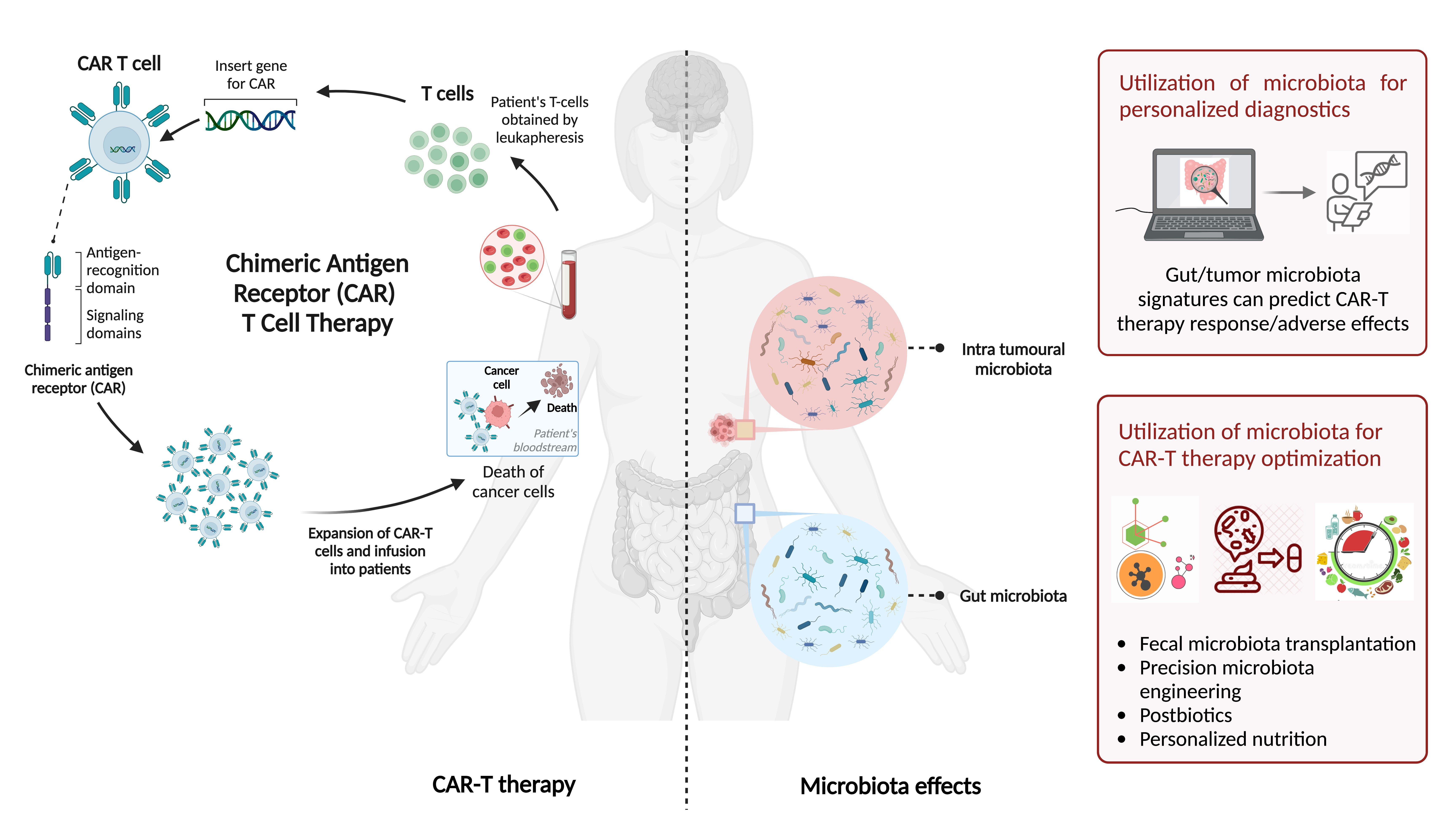
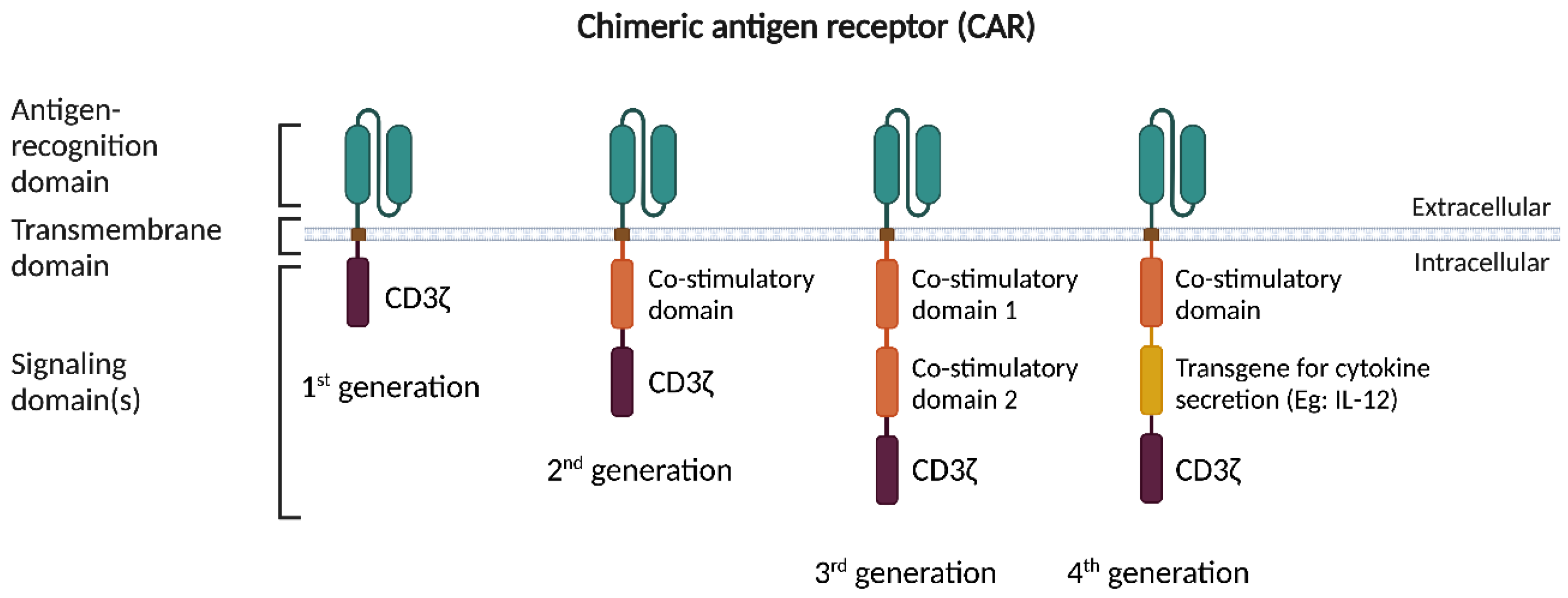
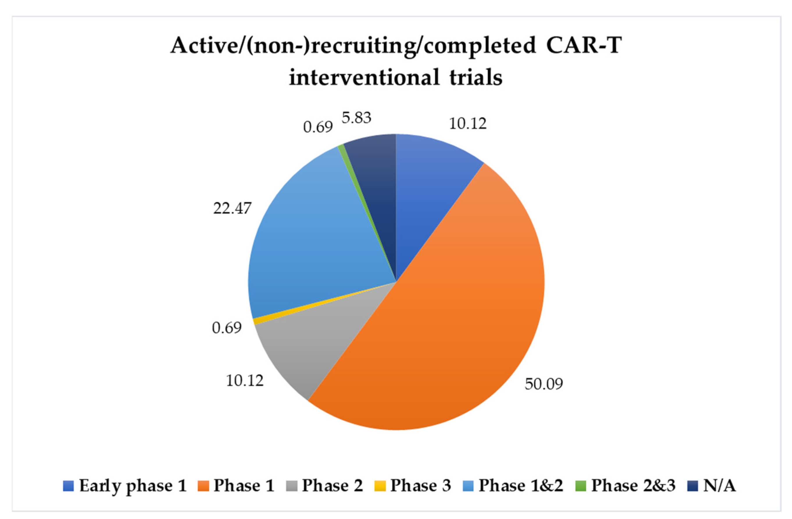
2. CAR-T Cell Therapy

Through the expression of these chimeric receptors, CAR-T cell therapy has recently revolutionized the field of cancer therapeutics by redirecting autologous T lymphocytes, isolated through leukapheresis, towards a tumor-specific antigen using viral and non-viral transfection methods. CAR-T cells constitute a successful ‘adoptive cell immunotherapy’ especially suited for treatment of patients with relapsed or refractory hematological malignancies, which resulted in the U.S. Food and Drug Administration (FDA) and European Medicines Agency (EMA) approving several CAR-T cell medicines as a standard care of hematological cancers. Tisagenlecleucel (Kymriah® by Novartis) was approved in August 2017 by the FDA as a therapeutic modality for treatment of patients younger than 25 years of age with relapsed or refractory B cell acute lymphoblastic leukemia (ALL) and adult patients with relapsed or refractory follicular lymphoma after two or more lines of systemic therapies [12]. Axicabtagene ciloleucel (Yescarta® by Kite) was later authorized as a therapy for adult patients with relapsed or refractory diffuse large B cell lymphoma (DLBCL) after first-line treatment of chemoimmunotherapy in Europe as well as the U.S. in 2018 [13]. Since then, several CAR-T cell therapies (CD19-targeted brexucabtagene autoleucel - Tecartus®, lisocabtagene maraleucel - Breyanzi®, and B cell maturation antigen-targeted idecabtagene vicleucel - Abecma®, ciltacabtagene autoleucel - Carvykti®) have been approved by the FDA for the treatment of hematological malignancies, including lymphomas and some forms of leukemia, and most recently for the treatment of multiple myeloma [14,15,16].
Currently, CAR-T cell therapy is available through clinical trials for several forms of blood cancer. However, its application for solid tumors, which collectively account for ~ 90% of cancer-associated mortality, has remained challenging. Ongoing studies are exploring CAR-T cell therapy in solid tumors while primarily evaluating safety and reporting preliminary research outcomes. Over the years, such solid tumor-focused clinical trials have targeted surface proteins including carcinoembryonic antigen (CEA), Erb-B2 receptor tyrosine kinase 2 (ERBB2), epidermal growth factor receptor (EGFR), fibroblast activation protein (FAP), diganglioside (G2), human epidermal growth factor receptor 2 (Her2), interleukin 13 receptor α (IL-13Rα), L1 cell adhesion molecule (L1CAM), mesothelin, mucin 1 (MUC1), and prostate-specific membrane antigen (PSMA) [17,18,19]. However, the clinical results of CAR-T cell therapy in these solid tumor settings have been much less encouraging until some advances reported recently. For example, Jin et al. demonstrated that naturally expressed or radiation-induced expression of IL-8 enhanced intratumoral T cell trafficking [20]. Indeed, IL-8 upregulation at the tumor invasion front has been demonstrated in several types of human cancers [21]. Consequently, tumor-produced IL-8-guided CAR-T cells facilitated migration into tumors, thereby prompting an enhanced antitumor response in solid tumors [20]. Additionally, multiple novel target antigens are being investigated in preclinical and clinical trials across different types of cancers [22]. Currently, 995 CAR trials are ongoing (results from https://clinicaltrials.gov/; search for CAR cells; accessed on 4 January 2023): nearly 49% of the trials are currently recruiting, while 5% of the trials have been completed. Among the completed/recruiting/active, not-recruiting interventional trials, approximately 10% of the studies are in early phase 1; 50% are in phase 1; 10% are in phase 2; 0.7% are in phase 3; 22% are in both phase 1 and 2; 0.7% are in phase 2 and 3; while in nearly 6% of studies the information on current trial phase was not identifiable. The graphical representation of the completed/recruiting/active, not-recruiting CAR-T trials in different study phases has been illustrated in Figure 2 and has been summarized in Table S1.

3. Toxicity Associated with CAR-T Therapy

In addition to the 50% of CAR-T-treated patients who do not respond to therapy or relapse after therapeutic intervention, a significant number of patients experience severe adverse effects including cytokine release syndrome (CRS) and immune effector cell-associated neurotoxicity syndrome (ICANS). If left unchecked, these lead to adverse outcomes that do not allow for therapy responses, in part due to significant morbidity and mortality.
CRS is marked by overall symptoms of fever, exhaustion, anorexia, myalgia, and arthralgia, which can further progress to more severe forms of the syndrome including cardiac conditions (arrhythmia, tachycardia) and respiratory (tachypnea) and multi-organ failure [23,24,25]. CRS is driven by the release of inflammatory cytokines, including IL-2, IL-6, IL-10, IFNγ, and TNFα [26,27], as a direct consequence of the response to CAR-T cells and additional surrounding immune cells, leading to an overall hyperinflammatory response [26,27,28]. Immune effector cell-associated neurotoxicity syndrome (ICANS) is characterized by initial symptoms relating to impaired cognition and overall confusion including aphagia, lethargy, and delirium [24,29,30]. Over time, ICANS can progress to seizures, coma, and cerebral edema [31,32]. Blood–brain barrier disruptions, influx of cytokines into the central nervous system (CNS), and microglial as well as myeloid activation within the CNS are considered contributors to ICANS in relation to CAR-T infusion and cell migration [32,33,34]; however, the exact causes of ICANS are not fully understood. CRS can be treated by symptomatic treatment, IV hydration and in higher-graded CRS with corticosteroids and anti-cytokine treatments such as tocilizumab targeting IL-6. In more serious cases of ICANS, anti-epileptics may be necessary to manage seizures [35].
CAR-T therapeutic efficacy is also altered by potential off-target effects of the CAR-T cells. In CAR-T therapies targeting CD19-expressing malignant cells, such off-target effects include infection susceptibility driven by non-malignant B cell aplasia and resultant hypogammaglobulinemia. In patients receiving CD19-targeted CAR-T cells to treat DLBCL, more than half of those enrolled in a one-year study developed infections, with the majority of those attributed to bacteria [36]. Strikingly, many of the bacteria identified in blood-stream infections in these patients such as Escherichia, Pseudomonas, and Staphylococcus notably originate in the gut prior to systemic translocation [36,37,38].

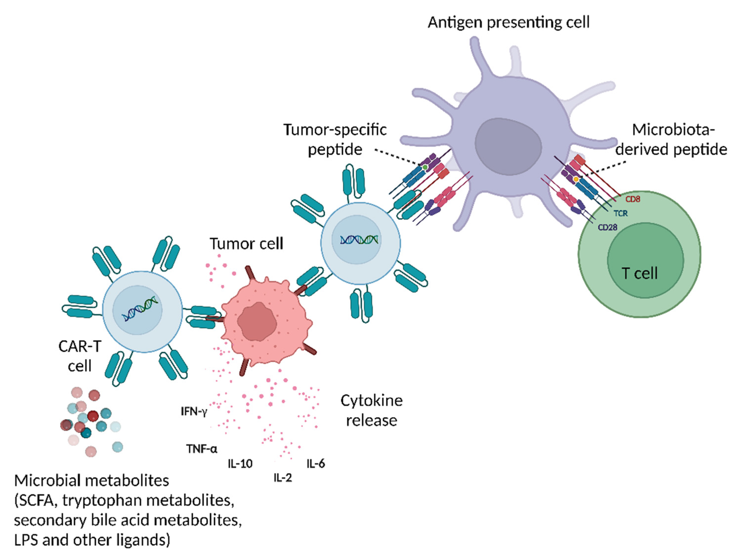
4. Microbiota Involvement in CAR-T Response and Toxicity

Over the last decade, a multitude of preclinical and clinical studies have demonstrated an interplay between the intestinal commensal microbiota and the mammalian immune system development and function [39]. Moreover, the intestinal microbiota was suggested to correlate and even modulate responses to anticancer therapeutics including chemotherapy, radiotherapy, immune checkpoint blockade, and adoptive cellular therapy, potentially by impacting host antitumor immune responses [40,41,42,43]. For example, Paulos et al. demonstrated that microbiota translocation augmented TLR4-mediated activation of the immune system, thereby enhancing the efficacy of adoptively transferred self/tumor-specific CD8+ T cells [44]. Likewise, allogeneic hematopoietic cell transplantation (allo HCT) was correlated to gut microbiota changes induced by dietary and antibiotic exposure [45,46], including an expansion of Enterococcus associated with a higher risk of graft-versus-host disease (GVHD)–related mortality [28,45,47]. Conversely, Eubacterium limosum has been associated with a decrease in cancer relapse/progression after allo HCT in patients with hematologic malignancies [48]. Emerging data also suggest that the gut microbiota may impact immune checkpoint blockade therapies, targeting programmed cell death protein 1 (PD-1) and cytotoxic T lymphocyte-associated protein 4 (CTLA-4), as is extensively reviewed elsewhere [28,49]. Briefly, a variety of taxa including Bacteroides, Akkermansia, Faecalibacterium, and Clostridiales spp. have been identified in murine studies to be associated with PD-1 therapy and its ligand PD-L1 and suggested to enhance the overall antitumor efficacy of checkpoint blockade [49,50,51]. Bacteroides fragilis and Bacteroides thetaiotaomicron were shown to enhance the CTLA-4 inhibitor efficacy in mice [50,51], collectively indicating a potential role for the microbiota in altering responses to CAR-T cell therapy [52].
Similar microbiota impacts were recently suggested to impact CAR-T cancer immunotherapy efficacy. In a first human study, Smith et al. analyzed the fecal microbiota composition of patients receiving second-generation CD19-targeted CAR-T therapy for treatment of B cell malignancies, hypothesizing that the microbiota would have associations with treatment efficacy and toxicity. Baseline stool samples prior to CAR-T therapy were heterogenous for bacteria at the phylum level and present with a decreased Shannon index for alpha diversity as compared to healthy controls. At the genus level, the patient microbiota significantly differed from that of healthy controls [53]. Given that antibiotics are commonly administered to treat secondary infections in patients undergoing anticancer therapies, antibiotics exposure and associated dysbiosis were suggested to adversely affect the overall clinical outcome of immunotherapies [54]. Indeed, Smith et al. also noted that 60% of their patient cohort received antibiotics and 20.6% of the cohort specifically received broad-spectrum antibiotics such as piperacillin/tazobactam, imipenem/cilastatin, and meropenem (PIM) that target anaerobic gut commensal bacteria. PIM exposure prior to CAR-T therapy correlated with worse overall survival and progression-free survival; notably, PIM correlated with a more aggressive disease and a higher lactate dehydrogenase, which is a biomarker of a higher tumor burden. Patients receiving any antibiotics in the weeks preceding CAR-T therapy initiation displayed increased incidence of ICANS. Specifically, exposure to PIM also correlated with a higher ICANS in non-Hodgkin lymphoma (NHL), but not in ALL. CRS was not shown to be correlated with PIM exposure. In all, multiple bacterial species were associated with the absence of toxicity, but microbes associated with toxicity were unidentifiable by a linear discriminant analysis effect size (LEfSe). Complete response rates at day 100 after CAR-T cell infusion were also correlated with a higher abundance of certain taxa, specifically the class Clostridia, further indicating that the gut microbiota may be playing a role in modulating the efficacy of CAR-T therapy. Overall, Smith et al. concluded that antibiotic exposure and its alteration of the gut microbiota prior to CAR-T therapy likely plays a role in its antitumor efficacy and toxicity [53].
Another recent study by Hu et al. [55] investigated CAR-T toxicity in relapsed/refractory multiple myeloma (MM), NHL, and ALL patients receiving second-generation therapy related to changes in the gut microbiota. Microbiota changes were longitudinally monitored throughout CAR-T delivery by stool sampling prior to CAR-T infusion, during CAR-T infusion but prior to development of CRS, during active CRS, and up to fourteen days after CAR-T infusion. Severe CRS was associated with a decreased abundance of Bifidobacteria. Alpha diversity as indicated by the Shannon index significantly decreased after CAR-T infusion and was further associated with an increase specifically in the abundance of Actinomyces and Enterococcus genera. Furthermore, Prevotella, Collinsella, Bifidobacterium, and Sutterella spp. were more abundant in patients experiencing complete response versus partial response [55]. Likewise, Smith et al. reported that patients with a higher abundance of certain bacterial species such as Ruminococcus, Bacteroides, and Faecalibacterium had a better response to CAR-T cell therapy [53]. Overabundance of Enterococcus faecium, post-antibiotics treatment in CAR-T patients, was further negatively correlated with treatment response [56]. In all, antibiotic exposure and the subsequent alteration of the gut microbiota associates with increased toxicities including CRS and ICANS, and with worsened CAR-T responses.
Importantly, these antibiotic effects may represent a causal impact of the antibiotics-perturbed microbiota on CAR-T therapy-related endpoints, or alternatively a reverse causality, in which antibiotics-treated patients present in an a priori clinically worse state predisposed to altered CAR-T therapy responsiveness. To untangle these possibilities, a recent study (Stein-Thoeringer et al., in press) [57] followed a large cohort of lymphoma patients receiving second-generation CD19-targeted CAR-T cells in Germany and the U.S. Like previous studies, an association was noted between an exposure to antibiotics prior to CAR-T cell infusion and an increased prevalence of cancer relapse or disease progression and a decrease in overall survival. However, wide spectrum antibiotics-treated patients suffer from an a priori worse disease state and increased tumor burden, likely accounting for their decreased CAR-T therapy responsiveness. Excluding these patients allowed for the detection of microbiota signatures strongly correlating with CAR-T responsiveness. Moreover, a cross-country evaluation of non-wide spectrum antibiotics-treated patients enabled a machine learning microbiome-based prediction of treatment outcomes, and the identification of Bacteroides, Ruminococcus, Eubacteria, and Akkermansia spp. as major potential drivers of therapy responsiveness.
Microbiota modulation of CAR-T treatment can be also driven by microbially secreted metabolites (Figure 3). A recent in vivo study demonstrated that CAR-T therapy modified to possess a receptor tyrosine kinase-like orphan 1 (ROR1) receptor induced a significant decrease in tumor volume and weight in a subcutaneous mouse model of pancreatic cancer featuring ROR-1-expressing Panc02 cells. The effect of these ROR1 CAR-T cells was further modulated with an addition of the short chain fatty acids (SCFA) butyrate and pentanoate [58]. Indeed, microbial-derived SCFA may favorably impact multiple immunotherapies, through a variety of mechanisms including enhanced TNFα and IFNγ effector responses, as well as upregulating anti-inflammatory T regs and CD8+ T cell functions while minimizing pro-inflammatory macrophage, dendritic cell, and Th1/Th17 activities [58,59]. Whether similar impacts would be observed in the human setting, and whether other bioactive metabolites participate in such interactions merits future studies.

5. Therapeutic Microbiota-Mediated Modulation of CAR-T Efficacy

Growing evidence delineates that modulation of the intestinal microbiota may impact the outcome of a variety of microbiota-contributed diseases, including cancer and immunotherapy. For example, two pilot first-in-human clinical trials recently provided evidence that melanoma immunotherapy responder-derived fecal microbiota transplantation (FMT) in combination with anti-PD-1 may benefit a subset of patients with PD-1-refractory melanoma [60,61].
As noted above, microbiota-modulated bioactive metabolites (termed ‘postbiotics’), such as SCFA, may enhance the antitumor action of cytotoxic lymphocytes and CAR-T cells [58]. These effects could be carried out by metabolic and epigenetic remodeling of CAR-T cells, driving increased expression of effector molecules such as CD25, IFNγ, and TNFα in syngeneic murine melanoma and pancreatic cancer models [58], or by SCFA binding to the G-protein-coupled receptor GPR109A on T cells, promoting T cell killing after high antigen stimulation [62]. Other microbial-derived metabolites, signaling through aryl hydrocarbon receptors (AhR), may also play a role in contributing to CD8+ T cell exhaustion by upregulating inhibitory receptors and downregulating cytokine production, thereby altering the ability of T cells to kill tumor cells [63,64]. Supplementation or inhibition of such microbially secreted bioactive metabolites may potentially be used to reinvigorate the immune response.
Modulating dietary content and timing can alter microbiota community structure and related metabolite secretion profiles, thereby impacting host physiology including CAR-T therapy responsiveness and adverse effect profiles. Such ‘personalized nutrition’ approaches have been shown to impact glycemic response outputs in a reproducible, microbiota-dependent manner [65,66]. For instance, a diet high in fibers induces the production of butyrate, propionate, and acetate, which have been linked to anti-inflammatory pathways in mouse cancer models [67,68,69]. As previously shown in non-cancer contexts, monitoring the glucose levels of hundreds of individuals showed highly variable responses to similar meals and predicting this outcome and establishing an optimal diet using machine learning effectively altered the post-prandial glycemic responses in individuals; thus, it may be feasible to integrate personal microbiota and host features by artificial intelligence and machine learning tools in harnessing dietary responses of the individual towards optimization of CAR-T therapy responses [49,65,70]. Targeted suppression of microbes associated with CAR-T therapy non-responsiveness and higher incidence of CRS may constitute another attractive modality in optimizing treatment. Rationally designed bacteriophage combinations, for instance, have been recently utilized to specifically suppress intestinal pathogens associated with inflammatory bowel disease and therefore may serve as a potential targeted method in suppressing pathobionts in other microbiome-contributing clinical conditions [71]. These modalities merit further consideration in CAR-T-treatment contexts.

6. Limitations and Challenges

Defining the causal effects of the microbiota and associated secreted bioactive compounds, rather than merely relying on associations and correlations, remains a major challenge in microbiota-associated research. Establishing mechanisms proving such causal effects in the CAR-T therapy context will likely require further in vitro and in vivo research, including studies utilizing animal models of cancer and CAR-T therapy. Transferring defined microbiota configurations from human CAR-T responders and non-responders into cancer-bearing germ-free mice would enable such elucidation of the causal contribution of microbial consortia and their bioactive metabolites to treatment responsiveness [49]. Inter-individual microbiota variability represents another formidable challenge in identifying reproducible and generalizable microbes and bioactive compounds impacting CAR-T therapy and adverse effects across large patient populations. Indeed, both Smith et al. and Hu et al. report high variability in microbiota populations between CAR-T-treated patients, characterized by the dominance of different phyla [53,55]. Similarly, varying taxa have been associated with impaired immunotherapy responsiveness in different trials, indicating that defining a generalizable CAR-T therapy optimizing microbial signature will be difficult to achieve [72,73,74]. This further highlights the need to include multicentric clinical trials with high-quality training and validation sets in identifying CAR-T therapy-related microbiota signatures [45,75]. Such efforts will be aided by the use of artificial intelligence technologies, in utilizing heterogenous patient-derived data towards actionable conclusions [65]. Of note, whole microbiota transfers into CAR-T-resistant individuals may optimize responsiveness and even convert non-responders to responders. Such treatment, however, poses risks of introducing potentially harmful bacteria into patients who are already severely immunocompromised, while inducing off-target effects, barrier disruption, and even sepsis [49,76]. In the long term, identification of defined consortia mediating such favorable effects may offer a safer, more reproducible, and universal treatment option. Finally, the focus of this review has been on the influence of the bacterial microbiota in patients receiving CAR-T therapy. The microbiota additionally includes viruses, fungi, and parasites that could all potentially influence the efficacy of CAR-T therapy. Studying the impacts of these insufficiently explored commensal kingdoms holds vast potential for the development of an even larger set of therapeutic microbiota-related modulations.

7. Conclusions

CAR-T therapy has revolutionized the treatment of hematological malignancies and overall advanced our understanding of modern cancer therapeutics. Determining novel methods to enhance CAR-T efficacy and responsiveness is of utmost concern. The human microbiota has shown profound influence as a modulator of immunotherapy response. Here, we review the recent reports that microbiota features such as abundance of various species correlate with CAR-T toxicity and overall response. Furthermore, the potentially confounding nature of antibiotic usage upon microbiota as related to CAR-T therapy should be further studied and defined.

Supplementary Materials

The following supporting information can be downloaded at https://www.mdpi.com/article/10.3390/cancers15030794/s1, Table S1: Summary of the latest CAR-T interventional trials in different study phases.

Author Contributions

Conceptualization, S.A., N.C., C.K.S.-T., E.E.; writing—original draft, S.A. and N.C.; writing—review, editing, S.A., N.C., C.K.S.-T., E.E., final editing and proof reading, S.A., N.C., C.K.S.-T., E.E. All authors have read and agreed to the published version of the manuscript.

Funding

This research received no external funding.

Acknowledgments

We thank the members of the Division of Microbiome and Cancer, DKFZ and ElinavLab, Weizmann Institute of Science, for insightful discussions; C.K.S-T. is supported by the German José Carreras Leukemia Foundation (01 R/2020), the German Research Foundation (DFG) (STE 2964/5-1), and the Baden-Württemberg Stiftung. E.E. is supported by the Leona M. and Harry B. Helmsley Charitable Trust, Adelis Foundation, Pearl Welinsky Merlo Scientific Progress Research Fund, Park Avenue Charitable Fund, Hanna and Dr. Ludwik Wallach Cancer Research Fund, Daniel Morris Trust, Wolfson Family Charitable Trust and Wolfson Foundation, Ben B. and Joyce E. Eisenberg Foundation, White Rose International Foundation, Estate of Malka Moskowitz, Estate of Myron H. Ackerman, Estate of Bernard Bishin for the WIS-Clalit Program, Else Kröener-Fresenius Foundation, Jeanne and Joseph Nissim Center for Life Sciences Research, A. Moussaieff, M. de Botton, Vainboim family, A. Davidoff, the V. R. Schwartz Research Fellow Chair and by grants funded by the European Research Council, Israel Science Foundation, Israel Ministry of Science and Technology, Israel Ministry of Health, Helmholtz Foundation, Garvan Institute of Medical Research, European Crohn’s and Colitis Organization, Deutsch-Israelische Projektkooperation, IDSA Foundation, and Wellcome Trust. E.E. is the incumbent of the Sir Marc and Lady Tania Feldmann Professorial Chair, a senior fellow of the Canadian Institute of Advanced Research, and an international scholar of the Bill & Melinda Gates Foundation and Howard Hughes Medical Institute.

Conflicts of Interest

E.E. is a scientific cofounder of DayTwo and BiomX, and an advisor to Hello Inside, Igen, and Aposense in topics unrelated to this work. The remaining authors declare no competing interests.

Abbreviations

AhRaryl hydrocarbon receptors
ALLacute lymphoblastic leukemia
allo HCTallogeneic hematopoietic cell transplantation
CARChimeric antigen receptor
CD3ζcluster of differentiation 3 zeta-chain
CEAcarcinoembryonic antigen
CNScentral nervous system
CRScytokine release syndrome
CTLA-4cytotoxic T lymphocyte-associated protein 4
DLBCLdiffuse large B cell lymphoma
EGFRepidermal growth factor receptor
EMAEuropean Medicines Agency
ERBB2Erb-B2 receptor tyrosine kinase 2
FAPfibroblast activation protein
FDAU.S. Food and Drug Administration
FMTfecal microbiota transplantation
G2diganglioside
GVDHgraft-versus-host disease
Her2human epidermal growth factor receptor 2
ICANSimmune effector cell-associated neurotoxicity syndrome
IFN-γinterferon gamma
ILinterleukin
IL-13Rαinterleukin 13 receptor α
L1CAML1 cell adhesion molecule
LEfSelinear discriminant analysis effect size
LPSlipopolysaccharide
MMmultiple myeloma
MUC1mucin 1
NHLnon-Hodgkin lymphoma
PD-1programmed cell death protein 1
PSMAprostate-specific membrane antigen
ROR1receptor tyrosine kinase-like orphan 1
SCFAshort chain fatty acids
scFvsingle-chain variable fragment
TCRT cell receptors
TNF-αtumor necrosis factor alpha
TRUCKT cells redirected for universal cytokine-mediated killing

References

  1. Kenderian, S.S.; Porter, D.L.; Gill, S. Chimeric Antigen Receptor T Cells and Hematopoietic Cell Transplantation: How Not to Put the CART Before the Horse. Biol. Blood Marrow Transpl. 2017, 23, 235–246. [Google Scholar] [CrossRef] [PubMed] [Green Version]
  2. Lee, Y.-H.; Kim, C.H. Evolution of Chimeric Antigen Receptor (CAR) T Cell Therapy: Current Status and Future Perspectives. Arch. Pharm. Res. 2019, 42, 607–616. [Google Scholar] [CrossRef]
  3. Subklewe, M.; von Bergwelt-Baildon, M.; Humpe, A. Chimeric Antigen Receptor T Cells: A Race to Revolutionize Cancer Therapy. Transfus. Med. Hemother. Off. Organ Dtsch. Ges. Transfus. Immunhamatol. 2019, 46, 15–24. [Google Scholar] [CrossRef] [PubMed]
  4. Gross, G.; Waks, T.; Eshhar, Z. Expression of Immunoglobulin-T-Cell Receptor Chimeric Molecules as Functional Receptors with Antibody-Type Specificity. Proc. Natl. Acad. Sci. USA 1989, 86, 10024–10028. [Google Scholar] [CrossRef] [PubMed] [Green Version]
  5. Kuwana, Y.; Asakura, Y.; Utsunomiya, N.; Nakanishi, M.; Arata, Y.; Itoh, S.; Nagase, F.; Kurosawa, Y. Expression of Chimeric Receptor Composed of Immunoglobulin-Derived V Regions and T-Cell Receptor-Derived C Regions. Biochem. Biophys. Res. Commun. 1987, 149, 960–968. [Google Scholar] [CrossRef]
  6. Brocker, T.; Karjalainen, K. Signals through T Cell Receptor-Zeta Chain Alone Are Insufficient to Prime Resting T Lymphocytes. J. Exp. Med. 1995, 181, 1653–1659. [Google Scholar] [CrossRef]
  7. Gong, M.C.; Latouche, J.B.; Krause, A.; Heston, W.D.; Bander, N.H.; Sadelain, M. Cancer Patient T Cells Genetically Targeted to Prostate-Specific Membrane Antigen Specifically Lyse Prostate Cancer Cells and Release Cytokines in Response to Prostate-Specific Membrane Antigen. Neoplasia 1999, 1, 123–127. [Google Scholar] [CrossRef] [Green Version]
  8. Till, B.G.; Jensen, M.C.; Wang, J.; Chen, E.Y.; Wood, B.L.; Greisman, H.A.; Qian, X.; James, S.E.; Raubitschek, A.; Forman, S.J.; et al. Adoptive Immunotherapy for Indolent Non-Hodgkin Lymphoma and Mantle Cell Lymphoma Using Genetically Modified Autologous CD20-Specific T Cells. Blood 2008, 112, 2261–2271. [Google Scholar] [CrossRef] [Green Version]
  9. Krause, A.; Guo, H.F.; Latouche, J.B.; Tan, C.; Cheung, N.K.; Sadelain, M. Antigen-Dependent CD28 Signaling Selectively Enhances Survival and Proliferation in Genetically Modified Activated Human Primary T Lymphocytes. J. Exp. Med. 1998, 188, 619–626. [Google Scholar] [CrossRef] [Green Version]
  10. Wang, L.-C.S.; Lo, A.; Scholler, J.; Sun, J.; Majumdar, R.S.; Kapoor, V.; Antzis, M.; Cotner, C.E.; Johnson, L.A.; Durham, A.C.; et al. Targeting Fibroblast Activation Protein in Tumor Stroma with Chimeric Antigen Receptor T Cells Can Inhibit Tumor Growth and Augment Host Immunity without Severe Toxicity. Cancer Immunol. Res. 2014, 2, 154–166. [Google Scholar] [CrossRef]
  11. Brentjens, R.J.; Curran, K.J. Novel Cellular Therapies for Leukemia: CAR-Modified T Cells Targeted to the CD19 Antigen. Hematol. Am. Soc. Hematol. Educ. Program 2012, 2012, 143–151. [Google Scholar] [CrossRef] [Green Version]
  12. Mullard, A. FDA Approves First CAR T Therapy. Nat. Rev. Drug Discov. 2017, 16, 669. [Google Scholar] [CrossRef] [PubMed]
  13. FDA Okays Second CAR-T for Kite. Nat. Biotechnol. 2020, 38, 1012. [CrossRef] [PubMed]
  14. Mullard, A. FDA Approves Fourth CAR-T Cell Therapy. Nat. Rev. Drug Discov. 2021, 20, 166. [Google Scholar] [CrossRef] [PubMed]
  15. Mullard, A. FDA Approves Second BCMA-Targeted CAR-T Cell Therapy. Nat. Rev. Drug Discov. 2022, 21, 249. [Google Scholar] [CrossRef] [PubMed]
  16. First CAR-T Therapy to Target BCMA Gets FDA Nod. Nat. Biotechnol. 2021, 39, 531. [CrossRef]
  17. Gill, S.; Maus, M.V.; Porter, D.L. Chimeric Antigen Receptor T Cell Therapy: 25years in the Making. Blood Rev. 2016, 30, 157–167. [Google Scholar] [CrossRef]
  18. Fousek, K.; Ahmed, N. The Evolution of T-Cell Therapies for Solid Malignancies. Clin. Cancer Res. Off. J. Am. Assoc. Cancer Res. 2015, 21, 3384–3392. [Google Scholar] [CrossRef] [Green Version]
  19. Newick, K.; O’Brien, S.; Moon, E.; Albelda, S.M. CAR T Cell Therapy for Solid Tumors. Annu. Rev. Med. 2017, 68, 139–152. [Google Scholar] [CrossRef]
  20. Jin, L.; Tao, H.; Karachi, A.; Long, Y.; Hou, A.Y.; Na, M.; Dyson, K.A.; Grippin, A.J.; Deleyrolle, L.P.; Zhang, W.; et al. CXCR1- or CXCR2-Modified CAR T Cells Co-Opt IL-8 for Maximal Antitumor Efficacy in Solid Tumors. Nat. Commun. 2019, 10, 4016. [Google Scholar] [CrossRef]
  21. Asokan, S.; Bandapalli, O.R. CXCL8 Signaling in the Tumor Microenvironment. Adv. Exp. Med. Biol. 2021, 1302, 25–39. [Google Scholar] [CrossRef] [PubMed]
  22. Marofi, F.; Motavalli, R.; Safonov, V.A.; Thangavelu, L.; Yumashev, A.V.; Alexander, M.; Shomali, N.; Chartrand, M.S.; Pathak, Y.; Jarahian, M.; et al. CAR T Cells in Solid Tumors: Challenges and Opportunities. Stem Cell Res. Ther. 2021, 12, 81. [Google Scholar] [CrossRef] [PubMed]
  23. Lee, D.W.; Gardner, R.; Porter, D.L.; Louis, C.U.; Ahmed, N.; Jensen, M.; Grupp, S.A.; Mackall, C.L. Current Concepts in the Diagnosis and Management of Cytokine Release Syndrome. Blood 2014, 124, 188–195. [Google Scholar] [CrossRef] [PubMed] [Green Version]
  24. Neelapu, S.S.; Tummala, S.; Kebriaei, P.; Wierda, W.; Gutierrez, C.; Locke, F.L.; Komanduri, K.V.; Lin, Y.; Jain, N.; Daver, N.; et al. Chimeric Antigen Receptor T-Cell Therapy—Assessment and Management of Toxicities. Nat. Rev. Clin. Oncol. 2018, 15, 47–62. [Google Scholar] [CrossRef] [PubMed]
  25. Brudno, J.N.; Kochenderfer, J.N. Toxicities of Chimeric Antigen Receptor T Cells: Recognition and Management. Blood 2016, 127, 3321–3330. [Google Scholar] [CrossRef] [PubMed] [Green Version]
  26. Hay, K.A.; Hanafi, L.-A.; Li, D.; Gust, J.; Liles, W.C.; Wurfel, M.M.; López, J.A.; Chen, J.; Chung, D.; Harju-Baker, S.; et al. Kinetics and Biomarkers of Severe Cytokine Release Syndrome after CD19 Chimeric Antigen Receptor-Modified T-Cell Therapy. Blood 2017, 130, 2295–2306. [Google Scholar] [CrossRef] [Green Version]
  27. Teachey, D.T.; Lacey, S.F.; Shaw, P.A.; Melenhorst, J.J.; Maude, S.L.; Frey, N.; Pequignot, E.; Gonzalez, V.E.; Chen, F.; Finklestein, J.; et al. Identification of Predictive Biomarkers for Cytokine Release Syndrome after Chimeric Antigen Receptor T-Cell Therapy for Acute Lymphoblastic Leukemia. Cancer Discov. 2016, 6, 664–679. [Google Scholar] [CrossRef] [PubMed] [Green Version]
  28. Schubert, M.-L.; Rohrbach, R.; Schmitt, M.; Stein-Thoeringer, C.K. The Potential Role of the Intestinal Micromilieu and Individual Microbes in the Immunobiology of Chimeric Antigen Receptor T-Cell Therapy. Front. Immunol. 2021, 12, 670286. [Google Scholar] [CrossRef] [PubMed]
  29. Maude, S.L.; Frey, N.; Shaw, P.A.; Aplenc, R.; Barrett, D.M.; Bunin, N.J.; Chew, A.; Gonzalez, V.E.; Zheng, Z.; Lacey, S.F.; et al. Chimeric Antigen Receptor T Cells for Sustained Remissions in Leukemia. N. Engl. J. Med. 2014, 371, 1507–1517. [Google Scholar] [CrossRef] [Green Version]
  30. Hunter, B.D.; Jacobson, C.A. CAR T-Cell Associated Neurotoxicity: Mechanisms, Clinicopathologic Correlates, and Future Directions. JNCI J. Natl. Cancer Inst. 2019, 111, 646–654. [Google Scholar] [CrossRef]
  31. Nastoupil, L.J.; Jain, M.D.; Feng, L.; Spiegel, J.Y.; Ghobadi, A.; Lin, Y.; Dahiya, S.; Lunning, M.; Lekakis, L.; Reagan, P.; et al. Standard-of-Care Axicabtagene Ciloleucel for Relapsed or Refractory Large B-Cell Lymphoma: Results From the US Lymphoma CAR T Consortium. J. Clin. Oncol. 2020, 38, 3119–3128. [Google Scholar] [CrossRef] [PubMed]
  32. Gust, J.; Hay, K.A.; Hanafi, L.-A.; Li, D.; Myerson, D.; Gonzalez-Cuyar, L.F.; Yeung, C.; Liles, W.C.; Wurfel, M.; Lopez, J.A.; et al. Endothelial Activation and Blood–Brain Barrier Disruption in Neurotoxicity after Adoptive Immunotherapy with CD19 CAR-T Cells. Cancer Discov. 2017, 7, 1404–1419. [Google Scholar] [CrossRef] [PubMed] [Green Version]
  33. Gust, J.; Finney, O.C.; Li, D.; Brakke, H.M.; Hicks, R.M.; Futrell, R.B.; Gamble, D.N.; Rawlings-Rhea, S.D.; Khalatbari, H.K.; Ishak, G.E.; et al. Glial Injury in Neurotoxicity after Pediatric CD19-directed Chimeric Antigen Receptor T Cell Therapy. Ann. Neurol. 2019; 86, 42–54. [Google Scholar] [CrossRef]
  34. Santomasso, B.D.; Park, J.H.; Salloum, D.; Riviere, I.; Flynn, J.; Mead, E.; Halton, E.; Wang, X.; Senechal, B.; Purdon, T.; et al. Clinical and Biological Correlates of Neurotoxicity Associated with CAR T-Cell Therapy in Patients with B-Cell Acute Lymphoblastic Leukemia. Cancer Discov. 2018, 8, 958–971. [Google Scholar] [CrossRef] [Green Version]
  35. Schubert, M.-L.; Schmitt, M.; Wang, L.; Ramos, C.A.; Jordan, K.; Müller-Tidow, C.; Dreger, P. Side-Effect Management of Chimeric Antigen Receptor (CAR) T-Cell Therapy. Ann. Oncol. 2021, 32, 34–48. [Google Scholar] [CrossRef] [PubMed]
  36. Wudhikarn, K.; Palomba, M.L.; Pennisi, M.; Garcia-Recio, M.; Flynn, J.R.; Devlin, S.M.; Afuye, A.; Silverberg, M.L.; Maloy, M.A.; Shah, G.L.; et al. Infection during the First Year in Patients Treated with CD19 CAR T Cells for Diffuse Large B Cell Lymphoma. Blood Cancer J. 2020, 10, 79. [Google Scholar] [CrossRef] [PubMed]
  37. Tamburini, F.B.; Andermann, T.M.; Tkachenko, E.; Senchyna, F.; Banaei, N.; Bhatt, A.S. Precision Identification of Diverse Bloodstream Pathogens in the Gut Microbiome. Nat. Med. 2018, 24, 1809–1814. [Google Scholar] [CrossRef]
  38. Eshel, A.; Sharon, I.; Nagler, A.; Bomze, D.; Danylesko, I.; Fein, J.A.; Geva, M.; Henig, I.; Shimoni, A.; Zuckerman, T.; et al. Origins of Bloodstream Infections Following Fecal Microbiota Transplantation: A Strain-Level Analysis. Blood Adv. 2022, 6, 568–573. [Google Scholar] [CrossRef]
  39. Zheng, D.; Liwinski, T.; Elinav, E. Interaction between Microbiota and Immunity in Health and Disease. Cell Res. 2020, 30, 492–506. [Google Scholar] [CrossRef]
  40. Ivanov, I.I.; Tuganbaev, T.; Skelly, A.N.; Honda, K. T Cell Responses to the Microbiota. Annu. Rev. Immunol. 2022, 40, 559–587. [Google Scholar] [CrossRef]
  41. Viaud, S.; Saccheri, F.; Mignot, G.; Yamazaki, T.; Daillère, R.; Hannani, D.; Enot, D.P.; Pfirschke, C.; Engblom, C.; Pittet, M.J.; et al. The Intestinal Microbiota Modulates the Anticancer Immune Effects of Cyclophosphamide. Science 2013, 342, 971–976. [Google Scholar] [CrossRef] [Green Version]
  42. Yang, K.; Hou, Y.; Zhang, Y.; Liang, H.; Sharma, A.; Zheng, W.; Wang, L.; Torres, R.; Tatebe, K.; Chmura, S.J.; et al. Suppression of Local Type I Interferon by Gut Microbiota–Derived Butyrate Impairs Antitumor Effects of Ionizing Radiation. J. Exp. Med. 2021, 218, e20201915. [Google Scholar] [CrossRef]
  43. Andrews, M.C.; Duong, C.P.M.; Gopalakrishnan, V.; Iebba, V.; Chen, W.-S.; Derosa, L.; Khan, M.A.W.; Cogdill, A.P.; White, M.G.; Wong, M.C.; et al. Gut Microbiota Signatures Are Associated with Toxicity to Combined CTLA-4 and PD-1 Blockade. Nat. Med. 2021, 27, 1432–1441. [Google Scholar] [CrossRef] [PubMed]
  44. Paulos, C.M.; Wrzesinski, C.; Kaiser, A.; Hinrichs, C.S.; Chieppa, M.; Cassard, L.; Palmer, D.C.; Boni, A.; Muranski, P.; Yu, Z.; et al. Microbial Translocation Augments the Function of Adoptively Transferred Self/Tumor-Specific CD8+ T Cells via TLR4 Signaling. J. Clin. Investig. 2007, 117, 2197–2204. [Google Scholar] [CrossRef] [PubMed] [Green Version]
  45. Peled, J.U.; Gomes, A.L.C.; Devlin, S.M.; Littmann, E.R.; Taur, Y.; Sung, A.D.; Weber, D.; Hashimoto, D.; Slingerland, A.E.; Slingerland, J.B.; et al. Microbiota as Predictor of Mortality in Allogeneic Hematopoietic-Cell Transplantation. N. Engl. J. Med. 2020, 382, 822–834. [Google Scholar] [CrossRef] [PubMed]
  46. Shono, Y.; Docampo, M.D.; Peled, J.U.; Perobelli, S.M.; Velardi, E.; Tsai, J.J.; Slingerland, A.E.; Smith, O.M.; Young, L.F.; Gupta, J.; et al. Increased GVHD-Related Mortality with Broad-Spectrum Antibiotic Use after Allogeneic Hematopoietic Stem Cell Transplantation in Human Patients and Mice. Sci. Transl. Med. 2016, 8, 339ra71. [Google Scholar] [CrossRef] [PubMed] [Green Version]
  47. Stein-Thoeringer, C.K.; Nichols, K.B.; Lazrak, A.; Docampo, M.D.; Slingerland, A.E.; Slingerland, J.B.; Clurman, A.G.; Armijo, G.; Gomes, A.L.C.; Shono, Y.; et al. Lactose Drives Enterococcus Expansion to Promote Graft-versus-Host Disease. Science 2019, 366, 1143–1149. [Google Scholar] [CrossRef]
  48. Peled, J.U.; Devlin, S.M.; Staffas, A.; Lumish, M.; Khanin, R.; Littmann, E.R.; Ling, L.; Kosuri, S.; Maloy, M.; Slingerland, J.B.; et al. Intestinal Microbiota and Relapse After Hematopoietic-Cell Transplantation. J. Clin. Oncol. Off. J. Am. Soc. Clin. Oncol. 2017, 35, 1650–1659. [Google Scholar] [CrossRef] [Green Version]
  49. Cullin, N.; Azevedo Antunes, C.; Straussman, R.; Stein-Thoeringer, C.K.; Elinav, E. Microbiome and Cancer. Cancer Cell 2021, 39, 1317–1341. [Google Scholar] [CrossRef]
  50. Sivan, A.; Corrales, L.; Hubert, N.; Williams, J.B.; Aquino-Michaels, K.; Earley, Z.M.; Benyamin, F.W.; Lei, Y.M.; Jabri, B.; Alegre, M.-L.; et al. Commensal Bifidobacterium Promotes Antitumor Immunity and Facilitates Anti-PD-L1 Efficacy. Science 2015, 350, 1084–1089. [Google Scholar] [CrossRef]
  51. Vétizou, M.; Pitt, J.M.; Daillère, R.; Lepage, P.; Waldschmitt, N.; Flament, C.; Rusakiewicz, S.; Routy, B.; Roberti, M.P.; Duong, C.P.M.; et al. Anticancer Immunotherapy by CTLA-4 Blockade Relies on the Gut Microbiota. Science 2015, 350, 1079–1084. [Google Scholar] [CrossRef] [Green Version]
  52. Tanoue, T.; Morita, S.; Plichta, D.R.; Skelly, A.N.; Suda, W.; Sugiura, Y.; Narushima, S.; Vlamakis, H.; Motoo, I.; Sugita, K.; et al. A Defined Commensal Consortium Elicits CD8 T Cells and Anti-Cancer Immunity. Nature 2019, 565, 600–605. [Google Scholar] [CrossRef] [PubMed]
  53. Smith, M.; Dai, A.; Ghilardi, G.; Amelsberg, K.V.; Devlin, S.M.; Pajarillo, R.; Slingerland, J.B.; Beghi, S.; Herrera, P.S.; Giardina, P.; et al. Gut Microbiome Correlates of Response and Toxicity Following Anti-CD19 CAR T Cell Therapy. Nat. Med. 2022, 28, 713–723. [Google Scholar] [CrossRef] [PubMed]
  54. Kamada, N.; Seo, S.-U.; Chen, G.Y.; Núñez, G. Role of the Gut Microbiota in Immunity and Inflammatory Disease. Nat. Rev. Immunol. 2013, 13, 321–335. [Google Scholar] [CrossRef] [PubMed]
  55. Hu, Y.; Li, J.; Ni, F.; Yang, Z.; Gui, X.; Bao, Z.; Zhao, H.; Wei, G.; Wang, Y.; Zhang, M.; et al. CAR-T Cell Therapy-Related Cytokine Release Syndrome and Therapeutic Response Is Modulated by the Gut Microbiome in Hematologic Malignancies. Nat. Commun. 2022, 13, 5313. [Google Scholar] [CrossRef]
  56. Blumenberg, V.; Schubert, M.-L.; Zamir, E.; Schmidt, S.; Rohrbach, R.; Waldhoff, P.; Bozic, D.; Pock, H.; Elinav, E.; Schmidt, C.; et al. Antibiotic Therapy and Low Gut Microbiome Diversity Is Associated with Decreased Response and High Toxicity in BCP-ALL and DLBCL Patients after Treatment with CD19. CAR T-Cells. Blood 2020, 136, 33–34. [Google Scholar] [CrossRef]
  57. Stein-Thoeringer, C.K.; Saini, N.Y.; Zamir, E.; Blumenberg, V.; Schubert, M.-L.; Mor, U.; Fante, M.A.; Schmidt, S.; Hayase, E.; Hayase, T.; et al. A Non-Antibiotic-Disrupted Gut Microbiome Predicts Clinical Responses to CD19-Targeted CAR-T Cell Cancer Immunotherapy across International Cohorts. Nat. Med. 2023, in press. [Google Scholar]
  58. Luu, M.; Riester, Z.; Baldrich, A.; Reichardt, N.; Yuille, S.; Busetti, A.; Klein, M.; Wempe, A.; Leister, H.; Raifer, H.; et al. Microbial Short-Chain Fatty Acids Modulate CD8+ T Cell Responses and Improve Adoptive Immunotherapy for Cancer. Nat. Commun. 2021, 12, 4077. [Google Scholar] [CrossRef] [PubMed]
  59. Rangan, P.; Mondino, A. Microbial Short-Chain Fatty Acids: A Strategy to Tune Adoptive T Cell Therapy. J. Immunother. Cancer 2022, 10, e004147. [Google Scholar] [CrossRef]
  60. Baruch, E.N.; Youngster, I.; Ben-Betzalel, G.; Ortenberg, R.; Lahat, A.; Katz, L.; Adler, K.; Dick-Necula, D.; Raskin, S.; Bloch, N.; et al. Fecal Microbiota Transplant Promotes Response in Immunotherapy-Refractory Melanoma Patients. Science 2021, 371, 602–609. [Google Scholar] [CrossRef] [PubMed]
  61. Davar, D.; Dzutsev, A.K.; McCulloch, J.A.; Rodrigues, R.R.; Chauvin, J.-M.; Morrison, R.M.; Deblasio, R.N.; Menna, C.; Ding, Q.; Pagliano, O.; et al. Fecal Microbiota Transplant Overcomes Resistance to Anti-PD-1 Therapy in Melanoma Patients. Science 2021, 371, 595–602. [Google Scholar] [CrossRef]
  62. Docampo, M.D.; da Silva, M.B.; Lazrak, A.; Nichols, K.B.; Lieberman, S.R.; Slingerland, A.E.; Armijo, G.K.; Shono, Y.; Nguyen, C.; Monette, S.; et al. Alloreactive T Cells Deficient of the Short-Chain Fatty Acid Receptor GPR109A Induce Less Graft-versus-Host Disease. Blood 2022, 139, 2392–2405. [Google Scholar] [CrossRef]
  63. Liu, Y.; Zhou, N.; Zhou, L.; Wang, J.; Zhou, Y.; Zhang, T.; Fang, Y.; Deng, J.; Gao, Y.; Liang, X.; et al. IL-2 Regulates Tumor-Reactive CD8+ T Cell Exhaustion by Activating the Aryl Hydrocarbon Receptor. Nat. Immunol. 2021, 22, 358–369. [Google Scholar] [CrossRef] [PubMed]
  64. Gargaro, M.; Manni, G.; Scalisi, G.; Puccetti, P.; Fallarino, F. Tryptophan Metabolites at the Crossroad of Immune-Cell Interaction via the Aryl Hydrocarbon Receptor: Implications for Tumor Immunotherapy. Int. J. Mol. Sci. 2021, 22, 4644. [Google Scholar] [CrossRef] [PubMed]
  65. Zeevi, D.; Korem, T.; Zmora, N.; Israeli, D.; Rothschild, D.; Weinberger, A.; Ben-Yacov, O.; Lador, D.; Avnit-Sagi, T.; Lotan-Pompan, M.; et al. Personalized Nutrition by Prediction of Glycemic Responses. Cell 2015, 163, 1079–1094. [Google Scholar] [CrossRef] [PubMed] [Green Version]
  66. Thaiss, C.A.; Zeevi, D.; Levy, M.; Zilberman-Schapira, G.; Suez, J.; Tengeler, A.C.; Abramson, L.; Katz, M.N.; Korem, T.; Zmora, N.; et al. Transkingdom Control of Microbiota Diurnal Oscillations Promotes Metabolic Homeostasis. Cell 2014, 159, 514–529. [Google Scholar] [CrossRef] [Green Version]
  67. Bultman, S.J. Molecular Pathways: Gene-Environment Interactions Regulating Dietary Fiber Induction of Proliferation and Apoptosis via Butyrate for Cancer Prevention. Clin. Cancer Res. Off. J. Am. Assoc. Cancer Res. 2014, 20, 799–803. [Google Scholar] [CrossRef] [PubMed] [Green Version]
  68. Garcia-Mantrana, I.; Selma-Royo, M.; Alcantara, C.; Collado, M.C. Shifts on Gut Microbiota Associated to Mediterranean Diet Adherence and Specific Dietary Intakes on General Adult Population. Front. Microbiol. 2018, 9, 890. [Google Scholar] [CrossRef] [Green Version]
  69. Donohoe, D.R.; Holley, D.; Collins, L.B.; Montgomery, S.A.; Whitmore, A.C.; Hillhouse, A.; Curry, K.P.; Renner, S.W.; Greenwalt, A.; Ryan, E.P.; et al. A Gnotobiotic Mouse Model Demonstrates That Dietary Fiber Protects against Colorectal Tumorigenesis in a Microbiota- and Butyrate-Dependent Manner. Cancer Discov. 2014, 4, 1387–1397. [Google Scholar] [CrossRef] [Green Version]
  70. Berry, S.E.; Valdes, A.M.; Drew, D.A.; Asnicar, F.; Mazidi, M.; Wolf, J.; Capdevila, J.; Hadjigeorgiou, G.; Davies, R.; Al Khatib, H.; et al. Human Postprandial Responses to Food and Potential for Precision Nutrition. Nat. Med. 2020, 26, 964–973. [Google Scholar] [CrossRef]
  71. Federici, S.; Kredo-Russo, S.; Valdés-Mas, R.; Kviatcovsky, D.; Weinstock, E.; Matiuhin, Y.; Silberberg, Y.; Atarashi, K.; Furuichi, M.; Oka, A.; et al. Targeted Suppression of Human IBD-Associated Gut Microbiota Commensals by Phage Consortia for Treatment of Intestinal Inflammation. Cell 2022, 185, 2879–2898.e24. [Google Scholar] [CrossRef]
  72. Gopalakrishnan, V.; Spencer, C.N.; Nezi, L.; Reuben, A.; Andrews, M.C.; Karpinets, T.V.; Prieto, P.A.; Vicente, D.; Hoffman, K.; Wei, S.C.; et al. Gut Microbiome Modulates Response to Anti-PD-1 Immunotherapy in Melanoma Patients. Science 2018, 359, 97–103. [Google Scholar] [CrossRef] [Green Version]
  73. Matson, V.; Fessler, J.; Bao, R.; Chongsuwat, T.; Zha, Y.; Alegre, M.-L.; Luke, J.J.; Gajewski, T.F. The Commensal Microbiome Is Associated with Anti-PD-1 Efficacy in Metastatic Melanoma Patients. Science 2018, 359, 104–108. [Google Scholar] [CrossRef] [PubMed] [Green Version]
  74. Routy, B.; Le Chatelier, E.; Derosa, L.; Duong, C.P.M.; Alou, M.T.; Daillère, R.; Fluckiger, A.; Messaoudene, M.; Rauber, C.; Roberti, M.P.; et al. Gut Microbiome Influences Efficacy of PD-1-Based Immunotherapy against Epithelial Tumors. Science 2018, 359, 91–97. [Google Scholar] [CrossRef] [PubMed] [Green Version]
  75. Lee, K.A.; Thomas, A.M.; Bolte, L.A.; Björk, J.R.; de Ruijter, L.K.; Armanini, F.; Asnicar, F.; Blanco-Miguez, A.; Board, R.; Calbet-Llopart, N.; et al. Cross-Cohort Gut Microbiome Associations with Immune Checkpoint Inhibitor Response in Advanced Melanoma. Nat. Med. 2022, 28, 535–544. [Google Scholar] [CrossRef] [PubMed]
  76. Villéger, R.; Lopès, A.; Carrier, G.; Veziant, J.; Billard, E.; Barnich, N.; Gagnière, J.; Vazeille, E.; Bonnet, M. Intestinal Microbiota: A Novel Target to Improve Anti-Tumor Treatment? Int. J. Mol. Sci. 2019, 20, 4584. [Google Scholar] [CrossRef] [PubMed]
Figure 1. Evolution of CAR constructs. The molecular design of CARs is comprised of three regions: (i) an ectodomain comprising an antigen-binding module; (ii) a transmembrane domain as an anchor; and (iii) a signaling domain for T cell activation. First-generation CARs contain only a single signaling domain (CD3ζ). Second-generation CAR constructs include a co-stimulatory domain along with the signaling domain. Third-generation CARs are comprised of two co-stimulatory domains connected to the intracellular signaling domain. Fourth-generation CARs, also referred to as TRUCKs, have an inducible transgene construct that expresses cytokines, for instance IL-12. CAR-T, chimeric antigen receptor T; CD3ζ, cluster of differentiation 3 zeta-chain); IL-12, interleukin 12. Figure created with BioRender (biorender.com).
Cancers 15 00794 g001
Figure 2. Study phase of CAR-T interventional trials. The percentage of CAR-T interventional trials that have been completed or are currently recruiting in each of the study phases. Data obtained from https://clinicaltrials.gov/; search for CAR cells; accessed on 4 January 2023. CAR-T, chimeric antigen receptor T cell.
Cancers 15 00794 g002
Figure 3. Microbiota involvement in CAR-T therapy efficacy and toxicity. Gut microbiota-derived peptides and metabolites exert their influence on both T cells as well as CAR-T cells, which can be further modulated by modification of diet and/or administration of antibiotics. The abundance of certain species in the gut microbiome facilitates therapeutic efficacy while dysbiosis leads to adverse effects including CRS and ICANS, increased tumor relapse or disease progression, and decreased overall survival. CRS, cytokine release syndrome; ICANS, immune effector cell-associated neurotoxicity syndrome; SCFA, short chain fatty acids; LPS, lipopolysaccharide, IFN-γ, interferon gamma; TNF-α, tumor necrosis factor alpha; IL-10, interleukin 10; IL-2, interleukin 2; IL-6, interleukin 6, CAR-T, chimeric antigen receptor T; TCR, T cell receptor; CD 28, cluster of differentiation 28; CD 8, cluster of differentiation 8. Figure created with BioRender (biorender.com).
Cancers 15 00794 g003
Disclaimer/Publisher’s Note: The statements, opinions and data contained in all publications are solely those of the individual author(s) and contributor(s) and not of MDPI and/or the editor(s). MDPI and/or the editor(s) disclaim responsibility for any injury to people or property resulting from any ideas, methods, instructions or products referred to in the content.

