Review

Type IV P-Type ATPases: Recent Updates in Cancer Development, Progression, and Treatment

by
Eugenia M. Yazlovitskaya
* and
Todd R. Graham
*
Department of Biological Sciences, Vanderbilt University, Nashville, TN 37232, USA
*
Authors to whom correspondence should be addressed.
Cancers 2023, 15(17), 4327; https://doi.org/10.3390/cancers15174327
Submission received: 11 July 2023 / Revised: 15 August 2023 / Accepted: 28 August 2023 / Published: 30 August 2023
(This article belongs to the Topic Advance in Tumorigenesis Research and Cancer Cell Therapy)

Abstract

:

Simple Summary

Lipids are the main structural component of cellular membranes; they play an essential role in cellular functions. For the growth and progression of cancer, tumor cells require lipid-related metabolic and structural adaptations, such as altered lipid membrane composition, which is in part regulated by lipid flippases. Type IV P-type ATPases (P4-ATPases), the main class of flippases, are responsible for membrane lipid asymmetry, and thus they are closely involved in cancer-related lipid changes. Lately, flippases have gained more attention regarding the pathobiology of human disease. This review focuses on recent publications discussing the role of P4-ATPases in cancer development, progression, and prospective treatment.

Abstract

Adaptations of cancer cells for survival are remarkable. One of the most significant properties of cancer cells to prevent the immune system response and resist chemotherapy is the altered lipid metabolism and resulting irregular cell membrane composition. The phospholipid distribution in the plasma membrane of normal animal cells is distinctly asymmetric. Lipid flippases are a family of enzymes regulating membrane asymmetry, and the main class of flippases are type IV P-type ATPases (P4-ATPases). Alteration in the function of flippases results in changes to membrane organization. For some lipids, such as phosphatidylserine, the changes are so drastic that they are considered cancer biomarkers. This review will analyze and discuss recent publications highlighting the role that P4-ATPases play in the development and progression of various cancer types, as well as prospects of targeting P4-ATPases for anti-cancer treatment.

1. Introduction

A typical cellular membrane is a bilayered lipid structure with embedded proteins. The diversity of membrane lipids and asymmetric lipid composition of the membrane leaflets are remarkable [1]. Among major phospholipids, phosphatidylserine (PS) and phosphatidylethanolamine (PE) are mainly localized in the inner leaflet of the plasma membrane, whereas phosphatidylcholine (PC) and sphingomyelin (SM) are concentrated in the outer leaflet [2]. This membrane asymmetry is vital for cellular functions and survival. For example, the increased level of external cellular PS exposure during apoptosis provides an “eat me” signal to surrounding macrophages while suppressing an autoimmune response [3]. Cancer cells may acquire this feature to mimic the anti-inflammatory action of apoptotic cells. This adaptation results in the failure of the immune system to detect cancer cells; therefore, cancer cells are protected from the immune response and continue proliferating.
Membrane lipid asymmetry is maintained by flippase, floppase, and scramblase transmembrane enzymes. Flippases, which translocate lipids from outer to inner membrane leaflet, and floppases, which are responsible for the reverse process, utilize ATP to provide the energy needed for lipid transport. Scramblases transport lipids in both directions across the membrane and are energy-independent. Most flippases belong to the type IV P-type ATPases. P-type ATPases, a superfamily of evolutionarily related ion and lipid pumps, are named based on their ability to catalyze auto-phosphorylation (hence the designation P-type) of a key conserved aspartate residue within the pump and their energy source, ATP [4]. Based on sequence similarity, the P-type ATPases are divided into five types with different transport specificities [5]. Types I-III (P1, P2, P3) P-type ATPases catalyze cation uptake and/or efflux, while type IV P-type ATPases (P4-ATPases) flip phospholipids and type V (P5) P-type ATPases transport polyamines or mediate protein dislocation from membranes [6]. P4-ATPases are expressed in all eukaryotic cells and are the primary class of flippases responsible for establishing and maintaining membrane asymmetry. Their activities are the foundation for establishing and maintaining membrane inner and outer leaflets phospholipid asymmetry, which is essential for membrane protein regulation, polarity maintenance, signaling events, and vesicle trafficking (reviewed in [7,8,9]). While there are a number of well-structured analyses of studies demonstrating the importance of P4-ATPase functions in maintaining blood homeostasis, liver metabolism, neural development, and the immune response [10,11], no comprehensive reviews are available pertinent to the role of P4-ATPases in cancer biology. This review summarizes recent updates on type IV P-type ATPases play in cancer development, progression, and treatment.

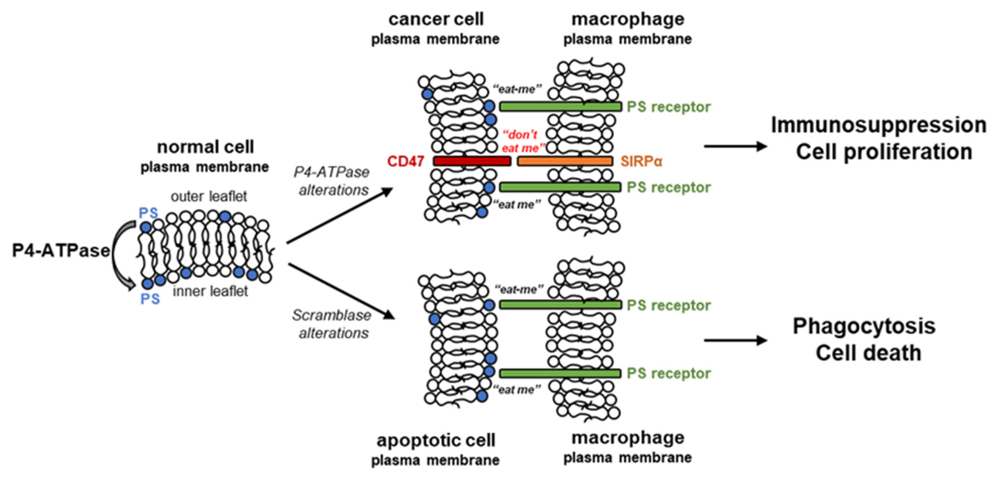
2. Flipping Phospholipids in Cancer Cells

The classification of P4-ATPases is based on phylogenetic similarities of the protein sequences [11]. Classical substrates of the P4-ATPases include aminophospholipids PS and PE. Of the 15 mammalian P4-ATPases, class 1a (ATP8A1, 2) and class 6 (ATP11A, B, C) translocate both PS and PE; class 1b (ATP8B1, 3) translocate PS; and class 1b (ATP8B1, 2) and class 5 (ATP10A, B) translocate PC and/or glucosylceramide (GlcCer) (Table 1).

2.1. Phosphatidylserine

The asymmetry of PS across the plasma membrane plays a crucial signaling role in numerous physiological processes. In normal conditions, PS is primarily localized on the inner leaflet of the cell membranes [2]. Many membrane-associated proteins have PS-binding domains on the cytoplasmic surfaces. In the case of peripheral membrane proteins, their interaction with PS regulates their movements around a cell and their functions in different locations. For integral membrane proteins, the membrane asymmetry, including PS membrane leaflet localization, is critical for their conformation, which regulates their activity and functions [2]. Exposure of PS on the external surface of the cell membrane is a prominent feature of cells undergoing apoptosis. It marks cells to be targeted and engulfed by surrounding macrophages, since apart from other cell types, macrophages have a PS-binding site of the PS-sensing receptors in ectoplasmic domains, allowing macrophages to detect apoptotic cells. Interestingly, viable, non-apoptotic cancer cells display increased surface PS expression compared to normal cells [17]. However, macrophages do not phagocytose tumor cells because of high levels of CD47, a cluster of differentiation 47, in tumor cells, which inhibits phagocytosis vis interaction with SIRPα, signal regulatory protein α [18,19]. A combination of high PS levels on the outer leaflet of the cell membrane and high cellular CD47 levels allows cancer cells to mimic the immunosuppressive anti-inflammatory features of apoptotic cells while not being phagocytized by macrophages, which results in a high rate of cancer cell proliferation. The PS-dependent prevention of immune reaction is caused by the ligation of PS to receptors present on dendritic cells, macrophages, and T cells. The ligation of PS to receptors on macrophages promotes macrophage polarization from a proinflammatory M1-like phenotype towards a protumor M2-like phenotype, allowing the secretion of the anti-inflammatory immunosuppressive cytokines interleukin-10 and TGF-β [20]. While the exposure of PS on the extracellular surface of apoptotic cells is mainly caused by the activation of scramblase, there are data supporting a significant role of flippase deficiency for this process in cancer cells (Figure 1).
A study of abnormally high PS exposure in cancer cells of various origins revealed an inverse correlation between flippase activity and constitutive PS externalization [21]. Elevated external PS exposure in cancer cells was accompanied by high total cellular PS. In addition, steady-state calcium levels, possibly by inhibiting flippase activity, also affect PS exposure. Therefore, a high level of external PS in cancer cells seems to be a result of low flippase activity [21]. The study using tumorigenic N18 and non-tumorigenic HN2 hippocampal neuron-derived cells showed both cell lines express P-type ATPase Atp8a1, but only the neuroblastoma N18 cells use this protein as a plasma membrane flippase. Targeted knockdown of this enzyme caused PS externalization and phagocytic removal of these cells [22]. Another study demonstrated the development of PSout tumor models with tumor cells lacking PS, the flippase component CDC50A, constantly exposing PS but alive [23]. Altogether, these studies suggest that insights into PS exposure mechanisms in cancer cells might facilitate tumor cell-specific induction in surface PS, and thus enhance the effectiveness of PS-targeting drugs. Therefore, flippases, responsible for PS transfer from the outer to the inner membrane surface, represent attractive anti-cancer therapeutic targets.

2.2. Phosphatidylethanolamine

While PS is one of the major studied membrane lipids in cancer biology, closely related aminophospholipid PE is underappreciated [24]. PE comprises 20–50% of total phospholipids, and is the second-most abundant phospholipid in mammalian cells after PC, while PS is a quantitatively minor membrane phospholipid representing 2–10% of total phospholipids [25]. In addition to PS, PE has increased surface representation on the outer membrane of tumor cells [26,27]. In addition, PE has a high degree of surface expression on endothelial cells in tumor vasculature [28], making it an attractive molecular target for future cancer interventions.
PE is highly enriched in mitochondrial inner membranes [29] suggesting the importance of PE for the Warburg effect. The Warburg effect is a classical metabolic phenotype of cancer cells that is characterized by the switch of energy production from oxidative phosphorylation in the mitochondria as observed in normal cells to a less efficient process of aerobic glycolysis even in the presence of abundant oxygen. Indeed, the Warburg effect involves a much more complex combination of multiple factors and can account for oncogenesis, tumor progression, and chemotherapy resistance of cancer cells [30]. A study using hepatocellular carcinoma cells HepG2 showed that ATP8B1 knockdown in HepG2 cells leads to a strong increase in the mitochondrial oxidative phosphorylation without a change in glycolysis, which coincided with increased mitochondrial fragmentation and phosphatidylethanolamine levels [31]. This data could be considered as a “reverse” Warburg effect, suggesting PE flippases are a potential anti-cancer molecular target.

2.3. Phosphatidylcholine

PC is the most abundant compound of the lipid bilayer. In normal cells, in comparison to PS and PE, it is mostly present in the outer leaflet of the plasma membrane [32]. A number of cancers exhibit the impairment of PC biosynthesis and PC metabolism alterations, resulting in a decrease in its membrane level [26]. However, the studies about specific PC distribution between inner and outer leaflets in membranes of cancer cells are very limited. Interestingly, several publications functionally connect multidrug resistance transporter (MDR1; Pgp, P-glycoprotein; ABCB1) with phosphatidylcholine flippase (MDR2) [33,34]. There is also a suggestion that MDR1 functions as a broad-specificity flippase for GlcCer, simple glycosphingolipids, and membrane phospholipids, including PC [35]. Although MDR proteins do not belong to the P4-ATPases family and are beyond the topic of this review, an association of PC membrane asymmetry with the transmembrane drug delivery system is intriguing.

3. P4-ATPases in Cancer Development and Progression

Recent developments in cancer research demonstrate the involvement of P4-ATPases in the development and progression, including metastases, of various types of cancers (Table 2). Most information is related to class 1 and 6 flippases.

3.1. Gastrointestinal Cancers

For colorectal cancer (CRC), the functional role of various P4-ATPases is highly controversial depending on the specific study. ATP8B1 was initially identified as a driver gene for sporadic CRC by Genomic Identification of Significant Targets In Cancer (GISTIC) [44]. Using three-level exon array data (gene, exon, and network) and additional separate differential expression analyses, Significance Analysis of Microarrays (SAM), and Linear Models for MicroArray data (LIMMA), ATP8B1 was found to be a novel gene associated with CRC that shows changes at cytogenetic, gene and exon levels [45]. Additionally, analyses of transcriptomics, genomics, and clinical data of CRC samples from The Cancer Genome Atlas (TCGA) revealed that ATP8B1 was the only potential biomarker of phospholipid transporters in CRC. Its prognostic value was also validated with the data from the Gene Expression Omnibus (GEO) [46]. However, in another study, ATP8B1 was suggested as a tumor suppressor for CRC since the forced reduction in ATP8B1 expression either by CRISPR/Cas9 or shRNA was associated with increased growth and proliferation of the CRC cell line HT29, while an overexpression of ATP8B1 resulted in reduced growth and proliferation of the SW480 CRC cell line [47]. Gene expression for another P4-ATPase, ATP11A, was assessed in 7 colorectal cancer cell lines and 95 paired cases of colorectal cancer and non-cancerous regions. A high level of ATP11A was proposed as a novel predictive marker for metachronous metastasis of colorectal cancer [62]. In addition, epigenome-wide methylation analysis of CpG sites of genes in visceral adipose tissue of CRC patients vs. healthy individuals detected a correlation between high methylation levels of ATP11A with CRC [63].
A deficiency of P4-ATPase ATP8B1 is mainly associated with progressive familial intrahepatic cholestasis type 1. However, a next-generation transcriptome sequencing study of three human hepatocellular carcinoma (HCC) tumor/tumor-adjacent pairs, followed by validation of differential gene expression findings in a large data set consisting of 434 liver normal/tumor sample pairs, revealed non-synonymous mutations in ATP8B1 as liver cancer driver mutations [48]. On the other hand, for patients with HCC, high ATP9A levels predicted a poor outcome [73]. In hepatocellular carcinoma cells, ATP9A was critical for regulating macropinocytosis via interaction with ATP6V1A resulting in plasma membrane cholesterol accumulation. Macropinocytosis, an important process that fuels cancer cell proliferation by scavenging extracellular proteins or necrotic cell debris, is critical for sustaining HCC proliferation and growth under nutrient limitation. The interaction of ATP9A with ATP6V1A facilitated ATP6V1A membrane trafficking, thereby activating plasma membrane cholesterol-dependent RAC1 signaling and initiating macropinocytosis to increase the energy supply and support HCC cell survival and proliferation [73]. Another study detected that miR-103a was one of the most highly expressed microRNAs in HCC tissues and that a high expression of miR-103a was associated with poor patient prognosis. Experiments using HCC cells in culture and subcutaneous xenograft models showed that miR-103a promoted glucose metabolism and directly inhibited cell death by targeting ATP11A and EIF5 transcripts, therefore, suggesting a regulatory mechanism(s) for HCC progression [64].
ATP8B2 was identified as one of the nine macrophage phenotypic switch-related genes (MRGs) with satisfactory prognostic ability in multiple analyses of databases from the Cancer Genome Atlas (TCGA)-pancreatic adenocarcinoma (PAAD), Genotype-Tissue Expression (GTEx)-Pancreas, and Gene Expression Omnibus (GEO) [51]. The critical role of ATP8B2 as one of four N6-methyladenosine (m6A)-associated metabolic genes and prognostic biomarkers of the immune response in cancer progression was confirmed in a study of pancreatic ductal adenocarcinoma. Overall, amplification was the most frequent type of mutation in ATP8B2, as well as the highest rate of genetic alterations (4%) among these four genes [52]. In a comparison of pancreatic cancer and paracancerous tissues, ATP11A mRNA and protein levels were significantly higher in cancer [65]. Moreover, ATP11A promoted the invasion and migration of cultured pancreatic cancer cells via TGFB-dependent epithelial-mesenchymal transition (EMT) [65].

3.2. Prostate Cancer

Most recently, ATP8B1 was identified as a prognostic prostate cancer biomarker. The investigated association of 222 haplotype-tagging SNPs in eight phospholipid-transporting ATPase genes with cancer-specific survival and overall survival of 630 patients treated with androgen-deprivation therapy (ADT) for prostate cancer revealed that ATP8B1 was under-expressed in tumor tissues, and that a higher ATP8B1 expression was associated with a better patient prognosis [49]. Specifically, ATP8B1 rs7239484 was proven useful in predicting the efficacy of ADT. Another study discovered ATP11A as a biomarker that improved discrimination of patients with metastatic-lethal prostate cancer. In this study, epigenome-wide DNA methylation profiling of surgically resected primary tumor tissues was compared between a population-based and a replication cohort of prostate cancer patients with five years follow-up [66]. ATP11A DNA methylation levels were significantly correlated with transcript levels and predicted metastatic-lethal events in the validation cohort. High methylation levels of CpGs in ATP11A also correlated with loss of the tumor suppressor gene PTEN, which has an unfavorable prognosis in prostate cancer [67].

3.3. Endometrial, Cervical, Ovarian and Breast Cancers

In a study of exome-wide rare variant association with endometrial cancer using germline whole exome data from the MyCode community health initiative, ATP8A1 was identified as one of several candidate genes involved in endometrial cancer predisposition, which could help in personalized prognosis [36].
A recent study using integrated bioinformatics analysis for expression and methylation of exocytosis genes from The Cancer Genome Atlas Cervical Squamous Cell Carcinoma and Endocervical Adenocarcinoma (TCGA-CESC) dataset identified ATP8B4 as one of nine genes linked with metastasis in cervical carcinoma [53]. Another study on integrative analysis of the DNA methylome and transcriptome in uterine leiomyoma found hypomethylation/upregulation of ATP8B4 compared with adjacent myometrium [54]. These studies speculated that reversion of this methylation could offer a therapeutic option for these types of cancer. In a genome-wide DNA methylation profiling study of cervical cancer stages, in which CpG sites whose state of methylation correlated with lesion grade were assessed, ATP10A was short-listed as a gene associated with the progression from normal through precancerous lesions and cervical cancer [57].
For ovarian cancer, ATP11B high expression was found to correlate with higher tumor grade in human ovarian carcinoma samples and with cisplatin resistance in human ovarian cancer cell lines [69]. ATP11B gene silencing restored the sensitivity of ovarian cancer cell lines to cisplatin, as well as in mice bearing ovarian tumors derived from cisplatin-sensitive and resistant cells. Mechanistic studies in cell lines suggested that ATP11B contributes to the secretory vesicular transport of cisplatin from the Golgi to the plasma membrane [69]. In contrast, another study reported that when relative gene expression in a set of primary epithelial ovarian cancer and control ovarian tissues were compared with clinical data and survival of patients, levels of ATP11B were decreased in carcinomas [70].
Data from three genome-wide association studies (GWAS) were reanalyzed using a novel computational biostatistics approach (muGWAS) for validation of single nucleotide polymorphisms associated with incurable metastatic breast cancer [37]. Findings from this study included ATP8A1 and ATP8B1 among other proteins that translocate and metabolize phospholipids, which control endo-/exocytosis. These novel findings suggest scavenging phospholipids as a novel intervention to control the local spread of cancer, packaging of exosomes, and endocytosis of specific receptors [37]. Another P4-ATPase implicated in breast cancer is ATP8A2. Differential expression analysis with the subsequent univariate Cox regression and LASSO algorithm were used to uncover key prognostic genes for 611 luminal A breast cancer patients [40]. This analysis resulted in a 5-gene-risk score model for predicting luminal A-invasive lobular breast cancer survival. Specifically, 105 prognostic genes and 9 predictors were identified, allowing the identification of 5-key prognostic genes including ATP8A2 [40]. In another study, analysis of the cells from breast cancer patients demonstrated that no or low expression of ATP11B in conjunction with high expression of PTDSS2, which is negatively regulated by BRCA1, markedly accelerates tumor metastasis and associates with poor prognosis [71].

3.4. Blood Cancers

An under-expression of ATP9A was reported in relapsed follicular lymphoma patients upon the analysis of 38 differentially expressed ion channels and transporter genes identifying ATP9A as one of the novel lymphoma biomarkers related to excitability and metabolic pathways, with particular relevance for drug-resistant, relapsed follicular lymphoma [56].
While childhood acute lymphoblastic leukemia (ALL) typically has a favorable prognosis, a substantial subset of patients with childhood ALL frequently relapses. Recurrent aberrations of 14 genes in patients who subsequently relapsed were detected, including deletions/uniparental isodisomies of ATP10A in B-cell precursor ALL [58]. Additionally, enriched mutations, although infrequent, in ATP10A were linked to relapsed chronic lymphocytic leukemia (CLL) [59]. ATP11A levels of long non-coding RNA were significantly upregulated in ALL [74]. Furthermore, low levels of ATP11A expression/methylation were identified as independent prognostic factors for another blood malignancy, acute myeloid leukemia (AML) using the methylation array data and mRNA array data from the Gene Expression Omnibus (GEO) database [68].

3.5. Lung Cancers

The role of ATP8A1 in the development and progression of non-small-cell lung cancer (NSCLC) was discovered in a study of the tumor suppressor function of microRNA MiR-140-3p. The growth of NSCLC cells in nude mouse models was suppressed by an overexpression of miR-140-3p, which was attenuated by an overexpression of ATP8A1 [39]. In a study of 25 cases of tumor tissues and the adjacent normal tissues from surgeries of NSCLC patients, ATP8A1 was found overexpressed in NSCLC tissues by immunohistochemical staining [38]. Mechanistically, ATP8A1 promoted the expression of MMP-9 and Vimentin, as well as suppressed the expression of E-cadherin, thus resulting in the elevated invasion/migration ability of NSCLC cells [38].
A study of differential gene expression analysis revealed that ATP8A2 and four other genes, each harboring one of the five frequently hypermethylated CpG sites within its promoters, were frequently downregulated in lung adenocarcinoma (LUAD) [41]. Poor expression of ATP8A2 in LUAD was also discovered in the analysis of the LUAD-related mRNA expression profile using univariate and multivariate Cox regression [43]. Additionally, ATP8A2 was found as 1 of 18 prognostic biomarkers in an analysis of the integrated relationship between nuclear mitochondrial genes (NMGs) and the progression of lung adenocarcinoma [42]. Machine learning algorithms were used to investigate the gene expression profiles of lung adenocarcinoma (AC) and lung squamous cell cancer (SCC) samples retrieved from Gene Expression Omnibus [72]. In this study, high levels of ATP11B were specifically detected in AC, but not in SCC.
ATP8B1 was proposed as a novel predictive biomarker in lung squamous cell carcinoma (LUSC). Investigation of the whole genomic expression profiles of LUSC samples from The Cancer Genome Atlas (TCGA) database and Tianjin Medical University Cancer Institute and Hospital (TJMUCH) cohort revealed that the low expression of ATP8B1 was associated with poor prognosis of LUSC patients [50]. In LUSC cell lines, ATP8B1 knockdown using lentiviral infection with shRNA promoted proliferation, inhibited apoptosis, and aggravated invasion and migration [50].
ATP10D was specifically identified as a positive biomarker in individuals presenting extreme phenotypes of low risk of developing tobacco-induced lung cancer [61]. SNPs associated with the risk of developing tobacco-induced NSCLC were analyzed in independent discovery and validation sets of patients followed by assessment of the prognostic value of mRNA expression and protein expression of target genes. A significant correlation of low mRNA expression of ATP10D with shorter survival in patients with stage I–II NSCLC confirmed the prognostic value of ATP10D [61].

3.6. Melanoma

The whole-exome sequencing of 34 melanoma-prone families (119 cases) coupled with coexpression network analyses focused on modules associated with pigmentation processes introduced 36 genes (including ATP10A) as potential melanoma risk genes in the families [60].
Hepatocyte growth factor-overexpressing mice that harbor a deletion of the Ink4a/p16 locus (HP mice) form melanomas with low metastatic potential in response to UV irradiation. However, these tumors become highly metastatic following hemizygous deletion of the Nme1 and Nme2 metastasis suppressor genes (HPN mice) [55]. Interestingly, missense mutations in eight signature genes including Atp8b4 were associated with a striking increase in lung metastatic activity in the melanoma model of HPN mice. Analysis of transcriptome data from The Cancer Genome Atlas revealed that expression profiles of these genes may serve as prognostic markers and/or therapeutic targets for clinical management of metastatic melanoma [55].

4. P4-ATPases as Targets for Anti-Cancer Treatment

4.1. Interplay with the Small Molecule Inhibitors

ATP8B3 was proposed as one of eight genes that regulate oxaliplatin response in the treatment of CRC. The presence of ATP8B3 SNPs correlated with the increased sensitivity of CRC to oxaliplatin therapy. Its predictive value was confirmed in a two-step procedure to comprehensively investigate 1444 single nucleotide polymorphisms (SNPs) from metabolic and transporter enzymes, using 623 stage II–IV CRC patients (of whom 201 patients received oxaliplatin) from a German prospective patient cohort treated with adjuvant or palliative chemotherapy [75]. It was further validated in a follow-up study of 53 SNPs using data from 1502 patients with stage II-IV colorectal cancer who received primary adjuvant chemotherapy and confirmed as predictive functional SNP with significance at p < 0.05 [76].
Greatly increased levels of ATP11A were detected in a study using breakpoint cluster region/Abelson murine leukemia (Bcr/Abl) P190 lymphoblasts with resistance to farnesyltransferase inhibitors (FTI) [77]. The same study demonstrated that the overexpression of ATP11A provided protection against the effects of FTI SCH66336, whereas knockdown of endogenous ATP11A using small interfering RNA (siRNA) made cells more sensitive to this drug. The lymphoblasts that were resistant to SCH66336 were also more resistant to structurally similar FTIs, FTI-276 and GGTI-298, and to imatinib mesylate, the Bcr/Abl tyrosine kinase inhibitor. Since elevated levels of ATP11A can protect malignant lymphoblastic leukemia cells against several novel small molecule signal transduction inhibitors, therefore, ATP11A could serve as a target for chemotherapy effects [77].
MiR-140-3p has a tumor suppressor function via inhibition of cancer cell growth, migration, and invasion. ATP8A1 was demonstrated as a novel direct target of miR-140-3p [39]. NSCLC cells in nude mouse models were suppressed by the overexpression of miR-140-3p. The increased level of intracellular ATP8A1 protein attenuated the inhibitor role of miR-140-3p suggesting ATP8A1 as a potential target for anti-cancer combination treatment.

4.2. Immunotherapy

In cancer treatment, oncolytic immunotherapy is one of the most promising approaches to boost antitumor immune response. The local injection of oncolytic peptides can induce immunogenic cell death leading to the activation of the immune system and the enhancement of the immune response [78]. One such peptide, LTX-315 (oncolytic peptide deriving from bovine lactoferrin), downregulates the expression of PD-L1 (programmed cell death ligand 1), a representative immune-inhibitory checkpoint molecule, and enhances CD8+ T cell infiltration in pancreatic cancer [79]. ATP11B was identified as a potential target of LTX-315 and a critical regulator in maintaining PD-L1 expression in pancreatic cancer cells and a mouse model [79]. Mechanistically, ATP11B interacted with PD-L1 in a CKLF-like MARVEL transmembrane domain containing 6 (CMTM6)-dependent manner. The depletion of ATP11B promoted CMTM6-mediated lysosomal degradation of PD-L1, thus reactivating the immune microenvironment and inducing an antitumor immune response. The significant correlation between ATP11B and PD-L1 was confirmed in clinical samples of pancreatic cancer. Since PD-L1 largely determines the efficacy and effectiveness of cancer immunotherapies, the development of ATP11B-targeting drugs is essential for anti-pancreatic cancer immunity.
The macrophage phenotype switch plays a vital role in the progression of malignancies. As mentioned previously, ATP8B2 was detected as one of nine signature macrophage phenotypic switch-related genes that could be used as an independent prognostic index for pancreatic adenocarcinoma (PDAC) patients [51]. Macrophages are one of the most abundant immune cells in PDAC tumor microenvironment. According to their polarization states, macrophages are roughly categorized into two types: classically activated type 1 (M1 macrophages), and alternatively activated type 2 (M2 macrophages). M2 macrophages exert pro-tumor functions, whereas M1 macrophages exert anti-tumor functions [80]. The macrophage phenotypic switch-related genes indicate a change in macrophage type and provide prognostic information for PDAC treatment. Later, ATP8B2 was confirmed as part of a four-gene signature to predict the immune response to treatment and overall survival of pancreatic adenocarcinoma patients [52] suggesting ATP8B2 as a target for anti-cancer immunotherapy.

5. Conclusions and Future Directions

This review discussed recent literature identifying P4-ATPase alterations associated with cancer predisposition, association, development, and progression. These alterations occur at the genomic, translational, and transcriptional levels, reflecting the variety of possible flippase-dependent regulatory mechanisms involved in cancer pathobiology. Much progress has been made in the P4-ATPase field in the last few years. However, there is still a need for additional research directed towards molecular mechanisms and the development of flippase-targeted anti-cancer therapeutic interventions.

Author Contributions

Conceptualization, E.M.Y. and T.R.G.; data curation, E.M.Y.; writing—original draft preparation, E.M.Y.; writing—review and editing, E.M.Y. and T.R.G.; funding acquisition, T.R.G. All authors have read and agreed to the published version of the manuscript.

Funding

This work was supported by a grant from the National Institutes of Health (1R35GM144123-01) to T.R.G.

Conflicts of Interest

The authors declare no conflict of interest.

References

  1. Harayama, T.; Riezman, H. Understanding the diversity of membrane lipid composition. Nat. Rev. Mol. Cell Biol. 2018, 19, 281–296. [Google Scholar] [CrossRef] [PubMed]
  2. Clarke, R.; Hossain, K.; Cao, K. Physiological roles of transverse lipid asymmetry of animal membranes. Biochim. Biophys. Acta Biomembr. 2020, 1862, 183382. [Google Scholar] [CrossRef]
  3. Nagata, S.; Segawa, K. Sensing and clearance of apoptotic cells. Curr. Opin. Immunol. 2021, 68, 1–8. [Google Scholar] [CrossRef]
  4. Palmgren, M.G.; Nissen, P. P-type ATPases. Annu. Rev. Biophys. 2011, 40, 243–266. [Google Scholar] [CrossRef]
  5. Lopez-Marques, R.L.; Theorin, L.; Palmgren, M.G.; Pomorski, T.G. P4-ATPases: Lipid flippases in cell membranes. Pflug. Arch. 2014, 466, 1227–1240. [Google Scholar] [CrossRef] [PubMed]
  6. Sim, S.I.; Park, E. P5-ATPases: Structure, substrate specificities, and transport mechanisms. Curr. Opin. Struct. Biol. 2023, 79, 102531. [Google Scholar] [CrossRef]
  7. Sebastian, T.T.; Baldridge, R.D.; Xu, P.; Graham, T.R. Phospholipid flippases: Building asymmetric membranes and transport vesicles. Biochim. Biophys. Acta 2012, 1821, 1068–1077. [Google Scholar] [CrossRef]
  8. Best, J.T.; Xu, P.; Graham, T.R. Phospholipid flippases in membrane remodeling and transport carrier biogenesis. Curr. Opin. Cell Biol. 2019, 59, 8–15. [Google Scholar] [CrossRef]
  9. Panatala, R.; Hennrich, H.; Holthuis, J.C.M. Inner workings and biological impact of phospholipid flippases. J. Cell Sci. 2015, 128, 2021–2032. [Google Scholar] [CrossRef] [PubMed]
  10. Folmer, D.E.; Elferink, R.P.O.; Paulusma, C.C. P4 ATPases—Lipid flippases and their role in disease. Biochim. Biophys. Acta 2009, 1791, 628–635. [Google Scholar] [CrossRef] [PubMed]
  11. Van der Mark, V.A.; Elferink, R.P.O.; Paulusma, C.C. P4 ATPases: Flippases in health and disease. Int. J. Mol. Sci. 2013, 14, 7897–7922. [Google Scholar] [CrossRef] [PubMed]
  12. van der Velden, L.M.; Wichers, C.G.; Van Breevoort, A.E.; Coleman, J.A.; Molday, R.S.; Berger, R.; Klomp, L.W.; van De Graaf, S.F. Heteromeric interactions required for abundance and subcellular localization of human CDC50 proteins and class 1 P4-ATPases. J. Biol. Chem. 2010, 285, 40088–40096. [Google Scholar] [CrossRef]
  13. Takatsu, H.; Tanaka, G.; Segawa, K.; Suzuki, J.; Nagata, S.; Nakayama, K.; Shin, H.-W. Phospholipid flippase activities and substrate specificities of human type IV P-type ATPases localized to the plasma membrane. J. Biol. Chem. 2014, 289, 33543–33556. [Google Scholar] [CrossRef] [PubMed]
  14. Gong, E.-Y.; Park, E.; Lee, H.J.; Lee, K. Expression of Atp8b3 in murine testis and its characterization as a testis specific P-type ATPase. Reproduction 2009, 137, 345–351. [Google Scholar] [CrossRef]
  15. Wang, L.; Beserra, C.; Garbers, D.L. A novel aminophospholipid transporter exclusively expressed in spermatozoa is required for membrane lipid asymmetry and normal fertilization. Dev. Biol. 2004, 267, 203–215. [Google Scholar] [CrossRef]
  16. Takatsu, H.; Baba, K.; Shima, T.; Umino, H.; Kato, U.; Umeda, M.; Nakayama, K.; Shin, H.-W. ATP9B, a P4-ATPase (a putative aminophospholipid translocase), localizes to the trans-Golgi network in a CDC50 protein-independent manner. J. Biol. Chem. 2011, 286, 38159–38167. [Google Scholar] [CrossRef]
  17. Utsugi, T.; Schroit, A.J.; Connor, J.; Bucana, C.D.; Fidler, I.J. Elevated expression of phosphatidylserine in the outer membrane leaflet of human tumor cells and recognition by activated human blood monocytes. Cancer Res. 1991, 51, 3062–3066. [Google Scholar] [PubMed]
  18. Tseng, D.; Volkmer, J.-P.; Willingham, S.B.; Contreras-Trujillo, H.; Fathman, J.W.; Fernhoff, N.B.; Seita, J.; Inlay, M.A.; Weiskopf, K.; Miyanishi, M.; et al. Anti-CD47 antibody-mediated phagocytosis of cancer by macrophages primes an effective antitumor T-cell response. Proc. Natl. Acad. Sci. USA 2013, 110, 11103–11108. [Google Scholar] [CrossRef]
  19. Huang, C.-Y.; Ye, Z.-H.; Huang, M.-Y.; Lu, J.-J. Regulation of CD47 expression in cancer cells. Transl. Oncol. 2020, 13, 100862. [Google Scholar] [CrossRef]
  20. Tse, E.; Kwong, Y.-L. T-cell lymphoma: Microenvironment-related biomarkers. Semin. Cancer Biol. 2015, 34, 46–51. [Google Scholar] [CrossRef]
  21. Vallabhapurapu, S.D.; Blanco, V.M.; Sulaiman, M.K.; Vallabhapurapu, S.L.; Chu, Z.; Franco, R.S.; Qi, X. Variation in human cancer cell external phosphatidylserine is regulated by flippase activity and intracellular calcium. Oncotarget 2015, 6, 34375–34388. [Google Scholar] [CrossRef] [PubMed]
  22. Levano, K.; Sobocki, T.; Jayman, F.; Debata, P.R.; Sobocka, M.B.; Banerjee, P. A genetic strategy involving a glycosyltransferase promoter and a lipid translocating enzyme to eliminate cancer cells. Glycoconj. J. 2009, 26, 739–748. [Google Scholar] [CrossRef]
  23. Wang, W.; Wu, S.; Cen, Z.; Zhang, Y.; Chen, Y.; Huang, Y.; Cillo, A.R.; Prokopec, J.S.; Quarato, G.; Vignali, D.A.; et al. Mobilizing phospholipids on tumor plasma membrane implicates phosphatidylserine externalization blockade for cancer immunotherapy. Cell Rep. 2022, 41, 111582. [Google Scholar] [CrossRef] [PubMed]
  24. Chakrabarti, A. Phospholipid Asymmetry in Biological Membranes: Is the Role of Phosphatidylethanolamine Underappreciated? J. Membr. Biol. 2021, 254, 127–132. [Google Scholar] [CrossRef]
  25. Vance, J.E. Phosphatidylserine and phosphatidylethanolamine in mammalian cells: Two metabolically related aminophospholipids. J. Lipid Res. 2008, 49, 1377–1387. [Google Scholar] [CrossRef]
  26. Szlasa, W.; Zendran, I.; Zalesińska, A.; Tarek, M.; Kulbacka, J. Lipid composition of the cancer cell membrane. J. Bioenergy Biomembr. 2020, 52, 321–342. [Google Scholar] [CrossRef] [PubMed]
  27. Broughton, L.J.; Crow, C.; Maraveyas, A.; Madden, L.A. Duramycin-induced calcium release in cancer cells. Anticancer Drugs 2016, 27, 173–182. [Google Scholar] [CrossRef]
  28. Stafford, J.H.; Thorpe, P.E. Increased exposure of phosphatidylethanolamine on the surface of tumor vascular endothelium. Neoplasia 2011, 13, 299–308. [Google Scholar] [CrossRef]
  29. Monteiro, J.P.; Oliveira, P.J.; Jurado, A.S. Mitochondrial membrane lipid remodeling in pathophysiology: A new target for diet and therapeutic interventions. Prog. Lipid Res. 2013, 52, 513–528. [Google Scholar] [CrossRef]
  30. Barbosa, I.A.; Machado, N.G.; Skildum, A.J.; Scott, P.M.; Oliveira, P.J. Mitochondrial remodeling in cancer metabolism and survival: Potential for new therapies. Biochim. Biophys. Acta 2012, 1826, 238–254. [Google Scholar] [CrossRef]
  31. Gómez-Mellado, V.E.; Chang, J.-C.; Ho-Mok, K.S.; Morcillo, C.B.; Kersten, R.H.J.; Elferink, R.P.J.O.; Verhoeven, A.J.; Paulusma, C.C. ATP8B1 Deficiency Results in Elevated Mitochondrial Phosphatidylethanolamine Levels and Increased Mitochondrial Oxidative Phosphorylation in Human Hepatoma Cells. Int. J. Mol. Sci. 2022, 23, 12344. [Google Scholar] [CrossRef] [PubMed]
  32. Zech, T.; Ejsing, C.S.; Gaus, K.; de Wet, B.; Shevchenko, A.; Simons, K.; Harder, T. Accumulation of raft lipids in T-cell plasma membrane domains engaged in TCR signalling. EMBO J. 2009, 28, 466–476. [Google Scholar] [CrossRef] [PubMed]
  33. Zhou, Y.; Gottesman, M.M.; Pastan, I. Domain exchangeability between the multidrug transporter (MDR1) and phosphatidylcholine flippase (MDR2). Mol. Pharmacol. 1999, 56, 997–1004. [Google Scholar] [CrossRef]
  34. Zhou, Y.; Gottesman, M.M.; Pastan, I. Studies of human MDR1-MDR2 chimeras demonstrate the functional exchangeability of a major transmembrane segment of the multidrug transporter and phosphatidylcholine flippase. Mol. Cell Biol. 1999, 19, 1450–1459. [Google Scholar] [CrossRef] [PubMed]
  35. Eckford, P.D.W.; Sharom, F.J. The reconstituted P-glycoprotein multidrug transporter is a flippase for glucosylceramide and other simple glycosphingolipids. Biochem. J. 2005, 389 Pt 2, 517–526. [Google Scholar] [CrossRef]
  36. Shivakumar, M.; Miller, J.E.; Dasari, V.R.; Gogoi, R.; Kim, D. Exome-Wide Rare Variant Analysis from the DiscovEHR Study Identifies Novel Candidate Predisposition Genes for Endometrial Cancer. Front. Oncol. 2019, 9, 574. [Google Scholar] [CrossRef]
  37. Wittkowski, K.M.; Dadurian, C.; Seybold, M.P.; Kim, H.S.; Hoshino, A.; Lyden, D. Complex polymorphisms in endocytosis genes suggest alpha-cyclodextrin as a treatment for breast cancer. PLoS ONE 2018, 13, e0199012. [Google Scholar] [CrossRef]
  38. Li, D.; Xu, T.; Wang, X.; Ma, X.; Liu, T.; Wang, Y.; Jiang, S. The role of ATP8A1 in non-small cell lung cancer. Int. J. Clin. Exp. Pathol. 2017, 10, 7760–7766. [Google Scholar]
  39. Dong, W.; Yao, C.; Teng, X.; Chai, J.; Yang, X.; Li, B. MiR-140-3p suppressed cell growth and invasion by downregulating the expression of ATP8A1 in non-small cell lung cancer. Tumour Biol. 2016, 37, 2973–2985. [Google Scholar] [CrossRef]
  40. Chen, Y.H.; Zhang, T.F.; Liu, Y.Y.; Zheng, J.H.; Lin, W.X.; Chen, Y.K.; Cai, J.H.; Zou, J.; Li, Z.Y. Identification of a 5-gene-risk score model for predicting luminal A-invasive lobular breast cancer survival. Genetica 2022, 150, 299–316. [Google Scholar] [CrossRef]
  41. Yan, H.; Guan, Q.; He, J.; Lin, Y.; Zhang, J.; Li, H.; Liu, H.; Gu, Y.; Guo, Z.; He, F. Individualized analysis reveals CpG sites with methylation aberrations in almost all lung adenocarcinoma tissues. J. Transl. Med. 2017, 15, 26. [Google Scholar] [CrossRef] [PubMed]
  42. Zhang, X.; Dong, W.; Zhang, J.; Liu, W.; Yin, J.; Shi, D.; Ma, W. A Novel Mitochondrial-Related Nuclear Gene Signature Predicts Overall Survival of Lung Adenocarcinoma Patients. Front. Cell Dev. Biol. 2021, 9, 740487. [Google Scholar] [CrossRef] [PubMed]
  43. Wang, X.; Shi, D.; Zhao, D.; Hu, D. Aberrant Methylation and Differential Expression of SLC2A1, TNS4, GAPDH, ATP8A2, and CASZ1 Are Associated with the Prognosis of Lung Adenocarcinoma. Biomed. Res. Int. 2020, 2020, 1807089. [Google Scholar] [CrossRef] [PubMed]
  44. Eldai, H.; Periyasamy, S.; Al Qarni, S.; Al Rodayyan, M.; Muhammed Mustafa, S.; Deeb, A.; Al Sheikh, E.; Afzal Khan, M.; Johani, M.; Yousef, Z.; et al. Novel genes associated with colorectal cancer are revealed by high resolution cytogenetic analysis in a patient specific manner. PLoS ONE 2013, 8, e76251. [Google Scholar] [CrossRef]
  45. Aziz, M.A.; Periyasamy, S.; Al Yousef, Z.; AlAbdulkarim, I.; Al Otaibi, M.; Alfahed, A.; Alasiri, G. Integrated exon level expression analysis of driver genes explain their role in colorectal cancer. PLoS ONE 2014, 9, e110134. [Google Scholar] [CrossRef] [PubMed]
  46. Deng, L.; Niu, G.-M.; Ren, J.; Ke, C.-W.; Loura, L. Identification of ATP8B1 as a Tumor Suppressor Gene for Colorectal Cancer and Its Involvement in Phospholipid Homeostasis. Biomed. Res. Int. 2020, 2020, 2015648. [Google Scholar] [CrossRef] [PubMed]
  47. Althenayyan, S.; AlGhamdi, A.; AlMuhanna, M.H.; Hawsa, E.; Aldeghaither, D.; Iqbal, J.; Mohammad, S.; Aziz, M.A. Modulation of ATP8B1 Gene Expression in Colorectal Cancer Cells Suggest its Role as a Tumor Suppressor. Curr. Cancer Drug Targets 2022, 22, 577–590. [Google Scholar] [CrossRef]
  48. Meerzaman, D.M.; Yan, C.; Chen, Q.-R.; Edmonson, M.N.; Schaefer, C.F.; Clifford, R.J.; Dunn, B.K.; Dong, L.; Finney, R.P.; Cultraro, C.M.; et al. Genome-wide transcriptional sequencing identifies novel mutations in metabolic genes in human hepatocellular carcinoma. Cancer Genom. Proteom. 2014, 11, 1–12. [Google Scholar]
  49. Chen, L.; Huang, S.; Shih, C.; Li, C.; Chen, Y.; Huang, C.; Yu, C.; Lin, V.C.; Lee, C.; Geng, J.; et al. ATP8B1: A prognostic prostate cancer biomarker identified via genetic analysis. Prostate 2023, 83, 602–611. [Google Scholar] [CrossRef]
  50. Zhang, X.; Zhang, R.; Liu, P.; Zhang, R.; Ning, J.; Ye, Y.; Yu, W.; Yu, J. ATP8B1 Knockdown Activated the Choline Metabolism Pathway and Induced High-Level Intracellular REDOX Homeostasis in Lung Squamous Cell Carcinoma. Cancers 2022, 14, 835. [Google Scholar] [CrossRef]
  51. Li, M.-X.; Wang, H.-Y.; Yuan, C.-H.; Ma, Z.-L.; Jiang, B.; Li, L.; Zhang, L.; Xiu, D.-R. Establishment of a Macrophage Phenotypic Switch Related Prognostic Signature in Patients with Pancreatic Cancer. Front. Oncol. 2021, 11, 619517. [Google Scholar] [CrossRef] [PubMed]
  52. Tan, Z.; Liu, J.; Xu, J.; Zhang, B.; Yu, X.; Wang, W.; Liang, C. LDHA is a prognostic biomarker on the immune response in pancreatic adenocarcinoma and associated with m6A modification. J. Cancer Res. Clin. Oncol. 2022, 149, 4853–4865. [Google Scholar] [CrossRef] [PubMed]
  53. Eswaran, S.; Adiga, D.; Khan, N.; Sriharikrishnaa, S.; Kabekkodu, S.P. Comprehensive analysis of the exocytosis pathway genes in cervical cancer. Am. J. Med. Sci. 2022, 363, 526–537. [Google Scholar] [CrossRef] [PubMed]
  54. Carbajo-García, M.C.; Corachán, A.; Juárez-Barber, E.; Monleón, J.; Payá, V.; Trelis, A.; Quiñonero, A.; Pellicer, A.; Ferrero, H. Integrative analysis of the DNA methylome and transcriptome in uterine leiomyoma shows altered regulation of genes involved in metabolism, proliferation, extracellular matrix, and vesicles. J. Pathol. 2022, 257, 663–673. [Google Scholar] [CrossRef]
  55. Leonard, M.K.; Puts, G.S.; Pamidimukkala, N.; Adhikary, G.; Xu, Y.; Kwok, E.; Jin, Y.; Snyder, D.; Matsangos, N.; Novak, M.; et al. Comprehensive molecular profiling of UV-induced metastatic melanoma in Nme1/Nme2-deficient mice reveals novel markers of survival in human patients. Oncogene 2021, 40, 6329–6342. [Google Scholar] [CrossRef]
  56. Magi, A.; Masselli, M.; Sala, C.; Guerriero, A.; Laise, P.; Puccini, B.; Rigacci, L.; Breschi, C.; Crociani, O.; Pillozzi, S.; et al. The ion channels and transporters gene expression profile indicates a shift in excitability and metabolisms during malignant progression of Follicular Lymphoma. Sci. Rep. 2019, 9, 8586. [Google Scholar] [CrossRef]
  57. El-Zein, M.; Cheishvili, D.; Gotlieb, W.; Gilbert, L.; Hemmings, R.; Behr, M.A.; Szyf, M.; Franco, E.L.; for the MARKER Study Group; Bouten, S.; et al. Genome-wide DNA methylation profiling identifies two novel genes in cervical neoplasia. Int. J. Cancer 2020, 147, 1264–1274. [Google Scholar] [CrossRef]
  58. Olsson, L.; Castor, A.; Behrendtz, M.; Biloglav, A.; Forestier, E.; Paulsson, K.; Johansson, B. Deletions of IKZF1 and SPRED1 are associated with poor prognosis in a population-based series of pediatric B-cell precursor acute lymphoblastic leukemia diagnosed between 1992 and 2011. Leukemia 2014, 28, 302–310. [Google Scholar] [CrossRef]
  59. Amin, N.A.; Seymour, E.; Saiya-Cork, K.; Parkin, B.; Shedden, K.; Malek, S.N. A Quantitative Analysis of Subclonal and Clonal Gene Mutations before and after Therapy in Chronic Lymphocytic Leukemia. Clin. Cancer Res. 2016, 22, 4525–4535. [Google Scholar] [CrossRef]
  60. Yepes, S.; Tucker, M.A.; Koka, H.; Xiao, Y.; Zhang, T.; Jones, K.; Vogt, A.; Burdette, L.; Luo, W.; Zhu, B.; et al. Integrated Analysis of Coexpression and Exome Sequencing to Prioritize Susceptibility Genes for Familial Cutaneous Melanoma. J. Investig. Dermatol. 2022, 142, 2464–2475.e5. [Google Scholar] [CrossRef]
  61. Fusco, J.P.; Pita, G.; Pajares, M.J.; Andueza, M.P.; Patiño-García, A.; De-Torres, J.P.; Gurpide, A.; Zulueta, J.; Alonso, R.; Alvarez, N.; et al. Genomic characterization of individuals presenting extreme phenotypes of high and low risk to develop tobacco-induced lung cancer. Cancer Med. 2018, 7, 3474–3483. [Google Scholar] [CrossRef]
  62. Miyoshi, N.; Ishii, H.; Mimori, K.; Tanaka, F.; Nagai, K.; Uemura, M.; Sekimoto, M.; Doki, Y.; Mori, M. ATP11A is a novel predictive marker for metachronous metastasis of colorectal cancer. Oncol. Rep. 2010, 23, 505–510. [Google Scholar]
  63. Izquierdo, A.G.; Boughanem, H.; Diaz-Lagares, A.; Arranz-Salas, I.; Esteller, M.; Tinahones, F.J.; Casanueva, F.F.; Macias-Gonzalez, M.; Crujeiras, A.B. DNA methylome in visceral adipose tissue can discriminate patients with and without colorectal cancer. Epigenetics 2022, 17, 665–676. [Google Scholar] [CrossRef]
  64. Liu, Y.; Zhang, Y.; Xiao, B.; Tang, N.; Hu, J.; Liang, S.; Pang, Y.; Xu, H.; Ao, J.; Yang, J.; et al. MiR-103a promotes tumour growth and influences glucose metabolism in hepatocellular carcinoma. Cell Death Dis. 2021, 12, 618. [Google Scholar] [CrossRef]
  65. Chen, L.; Tang, J.; Sheng, W.; Sun, J.; Ma, Y.; Dong, M. ATP11A promotes EMT by regulating Numb PRR(L) in pancreatic cancer cells. PeerJ 2022, 10, e13172. [Google Scholar] [CrossRef]
  66. Zhao, S.; Geybels, M.S.; Leonardson, A.; Rubicz, R.; Kolb, S.; Yan, Q.; Klotzle, B.; Bibikova, M.; Hurtado-Coll, A.; Troyer, D.; et al. Epigenome-Wide Tumor DNA Methylation Profiling Identifies Novel Prognostic Biomarkers of Metastatic-Lethal Progression in Men Diagnosed with Clinically Localized Prostate Cancer. Clin. Cancer Res. 2017, 23, 311–319. [Google Scholar] [CrossRef]
  67. Geybels, M.S.; Fang, M.; Wright, J.L.; Qu, X.; Bibikova, M.; Klotzle, B.; Fan, J.-B.; Feng, Z.; Ostrander, E.A.; Nelson, P.S.; et al. PTEN loss is associated with prostate cancer recurrence and alterations in tumor DNA methylation profiles. Oncotarget 2017, 8, 84338–84348. [Google Scholar] [CrossRef]
  68. Hu, L.; Gao, Y.; Shi, Z.; Liu, Y.; Zhao, J.; Xiao, Z.; Lou, J.; Xu, Q.; Tong, X. DNA methylation-based prognostic biomarkers of acute myeloid leukemia patients. Ann. Transl. Med. 2019, 7, 737. [Google Scholar] [CrossRef]
  69. Moreno-Smith, M.; Halder, J.B.; Meltzer, P.S.; Gonda, T.A.; Mangala, L.S.; Rupaimoole, R.; Lu, C.; Nagaraja, A.S.; Gharpure, K.M.; Kang, Y.; et al. ATP11B mediates platinum resistance in ovarian cancer. J. Clin. Investig. 2013, 123, 2119–2130. [Google Scholar] [CrossRef]
  70. Elsnerova, K.; Mohelnikova-Duchonova, B.; Cerovska, E.; Ehrlichova, M.; Gut, I.; Rob, L.; Skapa, P.; Hruda, M.; Bartakova, A.; Bouda, J.; et al. Gene expression of membrane transporters: Importance for prognosis and progression of ovarian carcinoma. Oncol. Rep. 2016, 35, 2159–2170. [Google Scholar] [CrossRef]
  71. Xu, J.; Su, S.M.; Zhang, X.; Chan, U.I.; Adhav, R.; Shu, X.; Liu, J.; Li, J.; Mo, L.; Wang, Y.; et al. ATP11B inhibits breast cancer metastasis in a mouse model by suppressing externalization of nonapoptotic phosphatidylserine. J. Clin. Investig. 2022, 132. [Google Scholar] [CrossRef] [PubMed]
  72. Yuan, F.; Lu, L.; Zou, Q. Analysis of gene expression profiles of lung cancer subtypes with machine learning algorithms. Biochim. Biophys. Acta Mol. Basis Dis. 2020, 1866, 165822. [Google Scholar] [CrossRef]
  73. Wang, X.; Li, Y.; Xiao, Y.; Huang, X.; Wu, X.; Zhao, Z.; Yang, M.; Kong, L.; Shi, D.; Chen, X.; et al. The phospholipid flippase ATP9A enhances macropinocytosis to promote nutrient starvation tolerance in hepatocellular carcinoma. J. Pathol. 2023, 260, 17–31. [Google Scholar] [CrossRef] [PubMed]
  74. Wang, W.; Lyu, C.; Wang, F.; Wang, C.; Wu, F.; Li, X.; Gan, S. Identification of Potential Signatures and Their Functions for Acute Lymphoblastic Leukemia: A Study Based on the Cancer Genome Atlas. Front. Genet. 2021, 12, 656042. [Google Scholar] [CrossRef] [PubMed]
  75. Kap, E.J.; Seibold, P.; Scherer, D.; Habermann, N.; Balavarca, Y.; Jansen, L.; Zucknick, M.; Becker, N.; Hoffmeister, M.; Ulrich, A.; et al. SNPs in transporter and metabolizing genes as predictive markers for oxaliplatin treatment in colorectal cancer patients. Int. J. Cancer 2016, 138, 2993–3001. [Google Scholar] [CrossRef]
  76. Park, H.A.; Seibold, P.; Edelmann, D.; Benner, A.; Canzian, F.; Alwers, E.; Jansen, L.; Schneider, M.; Hoffmeister, M.; Brenner, H.; et al. Validation of Genetic Markers Associated with Survival in Colorectal Cancer Patients Treated with Oxaliplatin-Based Chemotherapy. Cancer Epidemiol. Biomark. Prev. 2022, 31, 352–361. [Google Scholar] [CrossRef]
  77. Zhang, B.; Groffen, J.; Heisterkamp, N. Resistance to farnesyltransferase inhibitors in Bcr/Abl-positive lymphoblastic leukemia by increased expression of a novel ABC transporter homolog ATP11a. Blood 2005, 106, 1355–1361. [Google Scholar] [CrossRef]
  78. Tang, T.; Huang, X.; Zhang, G.; Liang, T. Oncolytic immunotherapy: Multiple mechanisms of oncolytic peptides to confer anticancer immunity. J. Immunother. Cancer 2022, 10, e005065. [Google Scholar] [CrossRef]
  79. Tang, T.; Huang, X.; Zhang, G.; Lu, M.; Hong, Z.; Wang, M.; Huang, J.; Zhi, X.; Liang, T. Oncolytic peptide LTX-315 induces anti-pancreatic cancer immunity by targeting the ATP11B-PD-L1 axis. J. Immunother. Cancer 2022, 10, e004129. [Google Scholar] [CrossRef]
  80. Genard, G.; Lucas, S.; Michiels, C. Reprogramming of Tumor-Associated Macrophages with Anticancer Therapies: Radiotherapy versus Chemo- and Immunotherapies. Front. Immunol. 2017, 8, 828. [Google Scholar] [CrossRef]
Figure 1. Altered phosphatidylserine plasma membrane leaflet localization in cancer and apoptotic cells determines cell fate. PS, phosphatidylserine; SIRPα, signal regulatory protein α.
Cancers 15 04327 g001
Table 1. Substrate specificities of mammalian P4-ATPases.
ClassNameLipid SubstrateSub-Cellular LocalizationReferences
1aATP8A1PS, PEPM, TNG, endosome[12]
1aATP8A2PS, PEPM, TNG, endosome[12]
1bATP8B1PS, PC, cardiolipin?PM[12,13]
1bATP8B2PCPM[12,13]
1bATP8B3PS?ER, TNG[12,14,15]
1bATP8B4unknownPM[12]
2ATP9AunknownTNG, endosome[16]
2ATP9BunknownTNG[16]
5ATP10APC, GlcCerPM[16]
5ATP10BPC, GlcCerendosome, lysosome[16]
5ATP10DGlcCerPM[16]
6ATP11APS, PEPM[13,16]
6ATP11BPS, PEendosome[13,16]
6ATP11CPS, PEPM[13,16]
Table 2. P4 ATPases in cancer development and progression.
ATPase Cancer TypeReported FeatureReported EffectCitations
ATP8A1endometrial cancergene expression upcancer predisposition[36]
metastatic breast cancerSNPscancer association[37]
non-small-cell lung cancerprotein expression upcancer association[38]
non-small-cell lung cancer (mouse model)protein expression upcancer cell growth[39]
ATP8A2luminal A-invasive lobular breast cancergene expression uppoor prognosis[40]
lung adenocarcinomagene expression downcancer association[41,42]
lung adenocarcinomamRNA expression downcancer association[43]
ATP8B1colorectal carcinomagene expression upcancer association[44,45,46]
colorectal carcinoma (cell lines)protein expression downincreased cell growth[47]
protein expression updecreased cell growth
hepatocellular carcinomagene mutationcancer association[48]
prostate cancerprotein expression upbetter prognosis[49]
metastatic breast cancerSNPscancer association[37]
lung squamous cell carcinomaprotein expression downpoor prognosis[50]
ATP8B2pancreatic adenocarcinomagene expression uppoor prognosis[51]
pancreatic ductal adenocarcinomagene expression uppoor prognosis[52]
ATP8B4metastasis in cervical carcinomagene methylation downcancer association[53]
uterine leiomyomagene methylation downcancer association[54]
metastasis in melanoma (mouse model)gene mutationscancer progression[55]
ATP9Ahepatocellular carcinomaprotein expression uppoor outcome[43]
relapsed follicular lymphomagene expression downpoor prognosis[56]
ATP10Acervical cancergene methylation upcancer progression[57]
relapsed acute lymphoblastic leukemiagene mutationspoor prognosis[58]
relapsed chronic lymphocytic leukemiagene mutationspoor prognosis[59]
melanomagene expression upcancer predisposition[60]
ATP10Dtobacco-induced lung cancermRNA expression downpoor prognosis[61]
ATP11Ametastasis of colorectal carcinomagene expression upcancer association[62]
colorectal carcinomagene methylation upcancer association[63]
hepatocellular carcinoma (mouse model)mRNA expression downcancer progression[64]
pancreatic cancerprotein expression upcancer association[65]
metastatic-lethal prostate cancergene methylation uppoor outcome[66,67]
acute lymphoblastic leukemianon-coding RNA upcancer association[23]
acute myeloid leukemiagene methylation downpoor outcome[68]
ATP11Bovarian carcinomaprotein expression upcancer progression[69]
ovarian carcinomagene expression downtumor association[70]
breast cancerprotein expression downpoor prognosis[71]
lung adenocarcinomagene expression upcancer association[72]
Disclaimer/Publisher’s Note: The statements, opinions and data contained in all publications are solely those of the individual author(s) and contributor(s) and not of MDPI and/or the editor(s). MDPI and/or the editor(s) disclaim responsibility for any injury to people or property resulting from any ideas, methods, instructions or products referred to in the content.

