Systematic Review

Malignancy Associated with Low-Risk HPV6 and HPV11: A Systematic Review and Implications for Cancer Prevention

by
Leandro Lima da Silva
1,
Amanda Mara Teles
1,2,
Joana M. O. Santos
3,
Marcelo Souza de Andrade
1,
Rui Medeiros
3,
Ana I. Faustino-Rocha
4,5,
Paula A. Oliveira
4,5,
Ana Paula Azevedo dos Santos
1,6,
Fernanda Ferreira Lopes
7,
Geraldo Braz
8,
Haissa O. Brito
1 and
Rui M. Gil da Costa
1,3,4,5,9,10,*
1
Post-Graduate Program in Adult Health (PPGSAD), Federal University of Maranhão (UFMA), São Luís 65080-805, MA, Brazil
2
Post-Graduate Program in Animal Health, State University of Maranhão, São Luís 65099-110, MA, Brazil
3
Molecular Oncology and Viral Pathology Group, Portuguese Institute of Oncology of Porto Research Center (CI-IPOP)/RISE@CI-IPOP (Health Research Network), Portuguese Institute of Oncology of Porto (IPO-Porto)/Porto Comprehensive Cancer Center (Porto.CCC), 4200-072 Porto, Portugal
4
Centre for the Research and Technology of Agro-Environmental and Biological Sciences (CITAB), University of Trás-os-Montes and Alto Douro, 5000-801 Vila Real, Portugal
5
Inov4Agro—Institute for Innovation, Capacity Building and Sustainability of Agri-Food Production, University of Trás-os-Montes and Alto Douro, 5000-801 Vila Real, Portugal
6
Post-Graduate Program in Health Sciences, Federal University of Maranhão (UFMA), São Luís 65080-805, MA, Brazil
7
Post-Graduate Program in Odontology, Federal University of Maranhão (UFMA), São Luís 65080-805, MA, Brazil
8
Post-Graduate Program in Computing Sciences, Federal University of Maranhão (UFMA), São Luís 65080-805, MA, Brazil
9
Laboratory for Process Engineering, Environment, Biotechnology and Energy (LEPABE), Faculty of Engineering, University of Porto, 4200-465 Porto, Portugal
10
Associate Laboratory in Chemical Engineering (ALiCE), Faculty of Engineering, University of Porto, 4200-465 Porto, Portugal
*
Author to whom correspondence should be addressed.
Cancers 2023, 15(16), 4068; https://doi.org/10.3390/cancers15164068
Submission received: 30 June 2023 / Revised: 28 July 2023 / Accepted: 8 August 2023 / Published: 11 August 2023

Abstract

:

Simple Summary

Vaccination against human papillomavirus (HPV) helps prevent cancer caused by this virus. Determining which viral genotypes should be included is key for developing successful vaccination strategies. Low-risk genotypes, especially HPV6 and HPV11, are associated with benign warts. However, some studies also report their presence in cancers. We reviewed the scientific literature to estimate the proportion of cancers that bear single or dual HPV6/11 infections. HPV6 and HPV11 have been reported in up to 5.5% of penile and 87.5% of laryngeal cancers; however, they have not been reported in vulvar, vaginal or oral cancers. Next, we compared the HPV6/11 genomes with HPV16, the most common high-risk HPV genotype, and observed that the similarities mainly involved the E7 gene, suggesting a limited ability to interfere with the differentiation of the host cells. These findings support the use of HPV vaccines that cover HPV6/11 not only for preventing genital warts but also for preventing specific types of cancers.

Abstract

High-risk human papillomavirus (HPV) is etiologically related to cervical cancer, other anogenital cancers and oropharyngeal carcinomas. Low-risk HPV, especially HPV6 and HPV11, cause genital warts and laryngeal papillomas. However, the accumulating data suggests that HPV6 and HPV11 may cause malignant lesions at non-cervical anatomic sites. This review aims to estimate the proportions of single and dual HPV6/11 infections in multiple cancers reported in the last 10 years in the Cochrane, Embasa and PubMed databases. Secondly, the genomes of HPV6/11 were compared with the most common high-risk genotype, HPV16, to determine the similarities and differences. A total of 11 articles were selected, including between one and 334 HPV+ cancer patients. The frequencies of single or dual HPV6/11 infections ranged between 0–5.5% for penile and 0–87.5% for laryngeal cancers and were null for vulvar, vaginal and oral cancers. The genomic similarities between HPV6/11 and HPV16 mainly involved the E7 gene, indicating a limited ability to block cell differentiation. The presence of single or dual HPV6/11 infections in variable proportions of penile and laryngeal cancers support the vaccination strategies that cover these genotypes, not only for preventing genital warts but also for cancer prevention. Other risk factors and co-carcinogens are likely to participate in epithelial carcinogenesis associated with low-risk HPV.

1. Introduction

Human papillomaviruses are host species-specific, double-stranded DNA viruses that exhibit a conserved icosahedral morphology, ranging between 50–55 nm in diameter with a molecular weight of 5 × 10⁶ Da [1,2]. Infection through tissue microdamage allows the virus to gain access to basal keratinocytes in the epidermis and keratinized mucosae [3,4]. The HPV genome contains a set of genes that are expressed early in the viral cycle upon cell entry, designated the “early” (E) genes, and two “late” (L) genes, L1 and L2, which are expressed later in the viral cycle and encode the structural capsid proteins as well as the regulatory regions [5]. Based on their nucleotide sequence of the L1 gene, HPVs are divided into types which are grouped in five genera (alpha, beta, gamma, mu and nu), where alpha is the main genus, comprising the HPV types associated with the development of cervical cancer known as high-risk (HR) HPVs (e.g., HPV16 and HPV18) and the types associated with genital warts, termed low-risk (LR) HPVs (e.g., HPV6 and HPV11) [6,7,8,9,10]. The HPV types are defined based on a 10% variation in their L1 nucleotide sequence and may be further subdivided into variants with different biological properties based on smaller differences [11,12,13,14]. The HPV types may also be divided according to their target epithelial site, i.e., cutaneous versus mucocutaneous [15]. Quadrivalent and nonavalent HPV vaccines are protective against HPV6 and HPV11 infections along with infections by HR-HPVs, while the bivalent vaccine only targets HPV16 and HPV18 [16]. Low-risk genital types are often responsible for benign lesions, such as condyloma acuminata, and may also cause low-grade cervical dysplasia. However, the risk of developing invasive cervical carcinoma is low [17]. HPV6 and HPV11 are most frequently found in genital warts [18] and are also involved in respiratory papillomas [19,20]. In contrast, HR-HPVs are able to establish persistent infections; interfere with cell proliferation, differentiation and survival; and are the etiologic agents of cervical cancer [5]. However, in contrast with cervical cancer, a limited number of studies have found single infections caused by LR-HPV, specifically HPV6 and HPV11, in small proportions of some non-cervical anogenital cancers, such as vulvar [21] and penile [22] cancers. Such observations suggest the hypothesis that these two HPV types may exert a more significant oncogenic effect on those specific anatomic sites than in the uterine cervix. If this is the case, the knowledge of the proportion of cancers potentially associated with HPV6/11 would help tailor vaccination strategies, especially in world regions where such cancers are more common. The present work adopted two complementary approaches to study this hypothesis. First, we performed a systematic review of the scientific literature from the last 10 years to determine the prevalence of single or dual HPV6 and HPV11 infections in the sites of HPV-associated cancers. Secondly, the genomic organization of these viruses was comparatively studied against HPV16, the most common high-risk HPV genotype, to identify meaningful similarities and differences. Finally, based on the results from both these studies, the factors that may contribute to a possible oncogenic role for HPV6 and HPV11 were discussed.

2. Materials and Methods

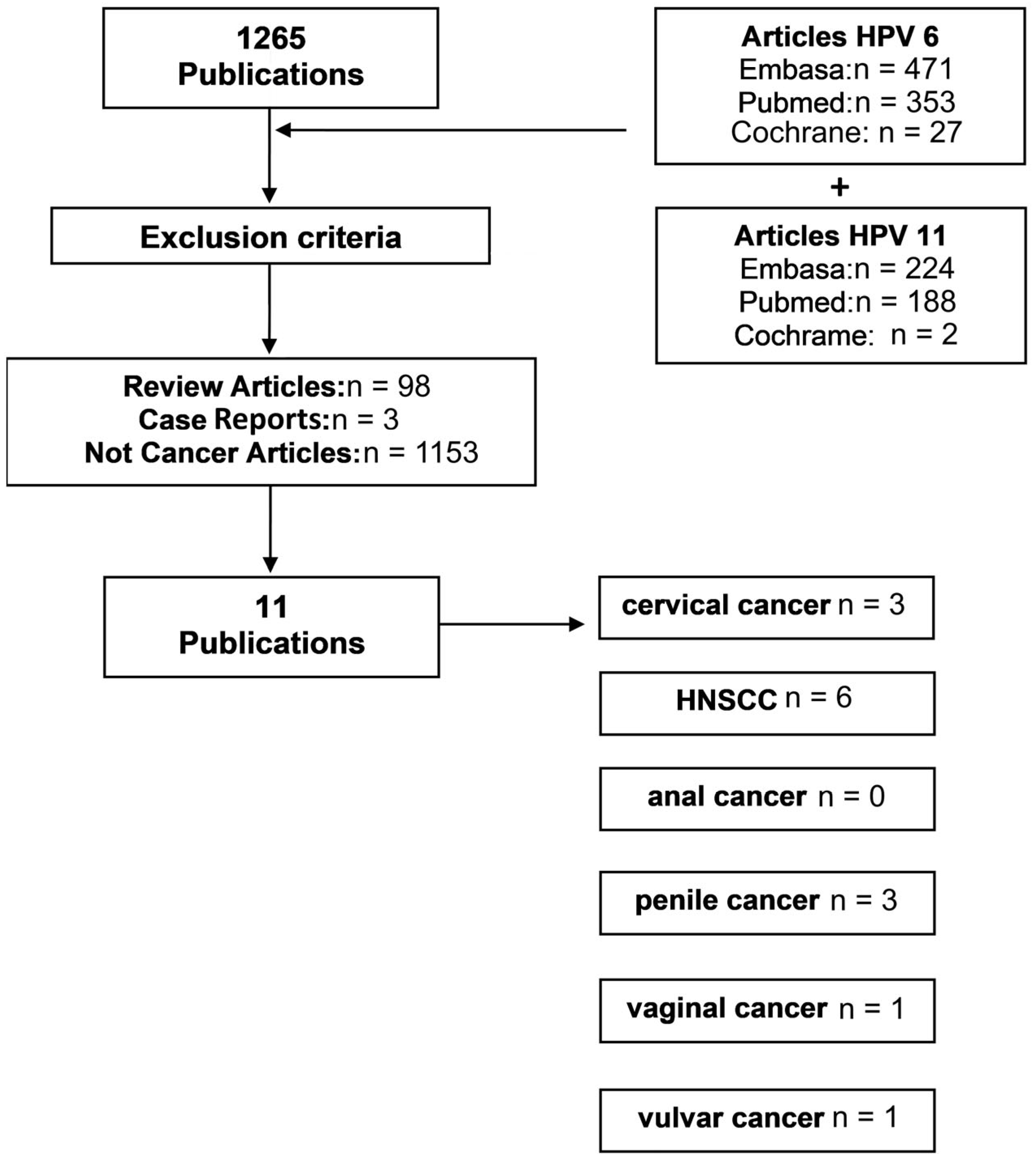
2.1. Systematic Review of HPV6 and HPV11 in Cancer

The systematic review was performed in accordance with the preferred reporting items for systematic reviews and meta-analyses (PRISMA) guidelines [23], according to the following parameters. Population: HNSCCs, anal, cervical, penile, vaginal and vulvar cancer patients. Intervention: the frequency of HPV6 and HPV11 single infections. The search strategy contemplated three standard databases on biomedicine: PubMed, Embase and Cochrane, accessed in March and April 2022. The keywords “cancer AND HPV6” or “cancer AND HPV11” were applied. A total of 541 articles were retrieved from PubMed, 695 articles from Embase and 29 articles from Cochrane. Duplicated records were excluded based on the article’s bibliographic reference. The following inclusion criteria were established concerning the type of study (case series and case–control studies in humans), tumor sample type (formalin-fixed paraffin-embedded and fresh tissue biopsies), tumor location (head and neck, uterine cervix, anorectum, penis, prepuce, vulva and vagina), histological diagnosis (squamous cell carcinoma), HPV detection methodology performed (PCR-based or sequencing techniques) and the type of agents identified (the frequency of single or dual HPV6 and HPV11 infections reported with or without a report of the infections by other LR-HPV types). The exclusion criteria were a lack of histological confirmation of cancer, a lack of identification of single/dual HPV6 or HPV11 infections, case reports, review articles and meta-analyses. The abstracts and, when necessary, the materials and methods were analyzed to apply the inclusion and exclusion criteria.

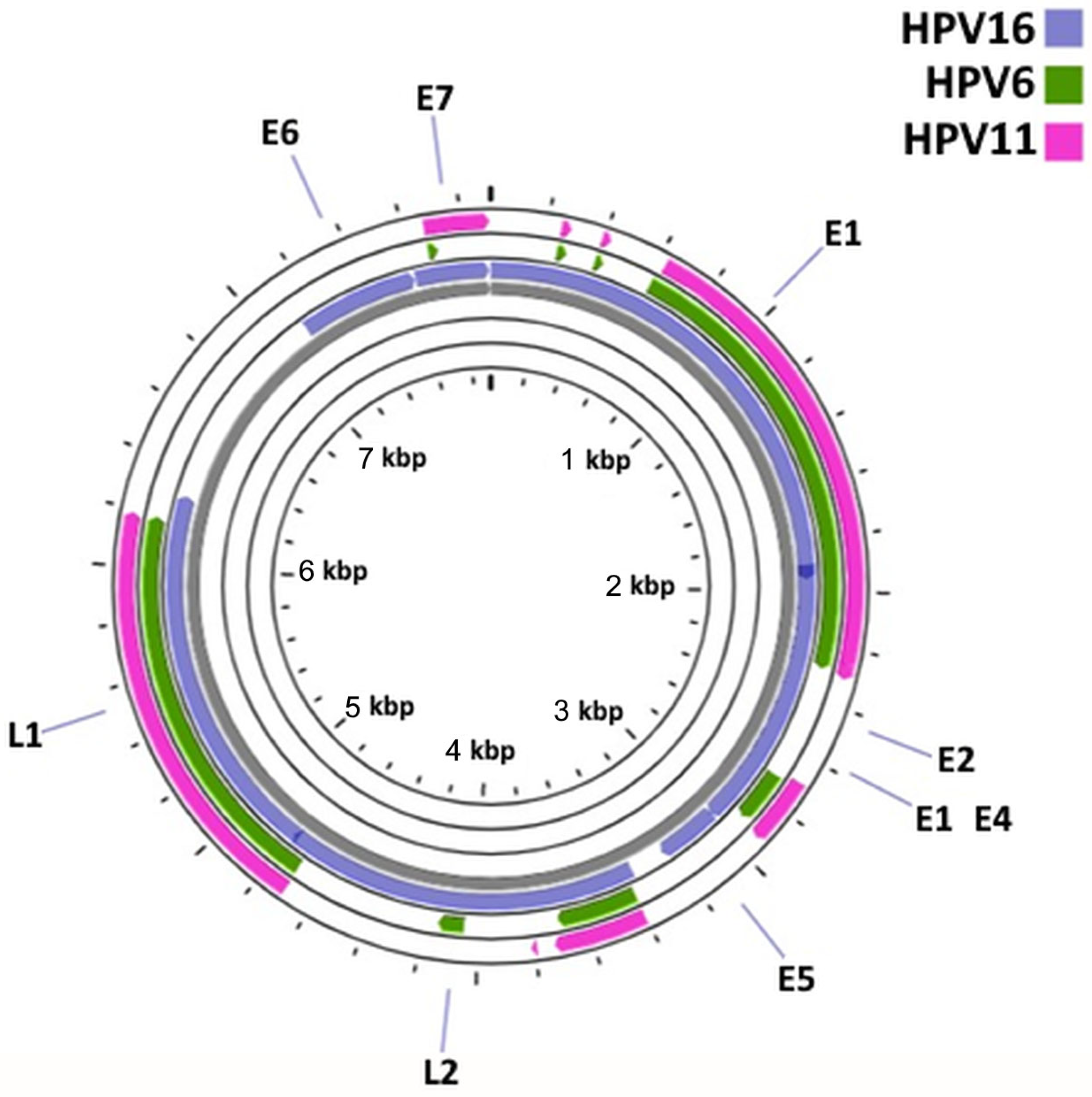
2.2. Comparative Genomic Analysis of HPV6, HPV11 and HPV16

The HPV6 and HPV11 genomes were compared with HPV16, the most commonly identified high-risk HPV in cancer, to identify the similarities and differences in the key genes involved in the cell transformation. The complete genomes of HPV16 (NC_001526.4) and HPV6 (NC_001355.1) were retrieved from the RefSeq database, available at NCBI. The RefSeq database was unavailable for HPV11. Therefore, its complete genome was retrieved from the GeneBank database, available at NCBI (MW404328.1). Then, the complete genomes of HPV16, HPV6 and HVP11 were uploaded to the Proksee/CGView Server online tool, which is a system for genome assembly, annotation and visualization [24]. In this tool, BLAST (blastn) was used to identify the regions of similarity between the genomic sequences. The amino acid sequences of the early proteins E6, E7, E5A and E5B from HPV16 and HPV6 were retrieved from the RefSeq database, available at NCBI, and the proteins from HPV11 were retrieved from the GenePept database, available at NCBI. The blastp tool from NCBI was used to evaluate the similarities between the early protein sequences.

3. Results

3.1. Systematic Review of HPV6 and HPV11 in Cancer

Most of the initially screened publications were excluded since they dealt with benign lesions instead of cancer, which was in line with the known role of HPV6 and HPV11 in warts. Overall, after applying the inclusion and exclusion criteria, 11 articles were selected for further analysis (Figure 1). A total of three articles were analyzed for cervical cancer [25,26,27], six for HNSCCs [28,29,30,31,32,33], none for anal cancer, three for penile cancer [28,34,35], one for vaginal cancer [28] and one for vulvar cancer [28]. The characteristics of all 11 publications that were selected for further analysis are summarized in Table 1.
The selected publications (Table 1) spanned the period between 2012 and 2021 and dealt with patient cohorts varying in size between eight and 1010 total patients. HPV detection was primarily performed using PCR-based methods, except for Aldersley et al. (2021) who used previously obtained whole exome data. The proportion of HPV-positive cases ranged between 1/85 [30] and 142/142 [25]. Seven studies used formalin-fixed paraffin-embedded (FFPE) material [28,29,30,31,32,34,35]; however, one used fresh biopsies [33] and another used tissue stored in RNAlater [26]. HPV genotyping was performed using a variety of commercial and custom methods. Three studies addressed the frequency of HPV6/11 infections in cervical cancer [25,26,27] (Table 2).
These studies generally identified an extremely low prevalence of single HPV6/11 infections or infections with HPV6/11 in the context of other LR-HPV in cervical cancer. A single case of HPV11 mono-infection was reported by [25], which also had the largest caseload of the three. Three studies addressed penile cancer [28,34,35]. Alemany et al. (2016) presented the largest caseload of the three and reported that 3.6% and 1.2% of HPV-positive cases carried HPV6 and HPV11 mono-infections, respectively. Barzon et al. (2014) reported a case showing HPV11 mono-infection and Magaña-León et al. (2015) reported none. In the larynx, the proportions of single infections varied between 0% and 75% cases for HPV6 and between 0% and 12.5% cases for HPV11. No studies observed single HPV6/11 infections in oral, vaginal or vulvar SCCs.
The HPV surrogate marker p16INK4a was studied using immunohistochemistry in five articles. Two studies found that most penile cancers harboring HR-HPV were p16INK4a-positive [34,35]. However, the larger Alemany et al. [35] study found that only a small proportion of cancers with LR-HPV were p16INK4a-positive. Three studies described p16INK4a immunostaining in laryngeal cancers [29,30,31] and reported a poor correlation with the presence of HPV DNA. Weiss et al. found two positive LR-HPV cases [31].

3.2. HPV6/11/16 Comparative Genomic Analysis

HPV16 had a circular dsDNA with a total of 7906 bp and eight coding sequences (Figure 2). When performing a blastn analysis at Proksee/CGView to compare both the HPV16/HPV6 and HPV16/HPV11 genomes, it was possible to observe that the majority of the similarities between the genomic sequences were located in the E1, E2, L2, L1 and E7 coding regions. No similarities between the genomic sequences at the E6 and E5 regions were found using these tools. The early proteins from HPV16 and HPV6/11 were then evaluated regarding the similarity of their amino acid sequence (Supplementary Table S1) using blastp. The identities, positives and expected values are presented in Table 3 and Table 4, where the identity describes the similarities of the sequences (the number of identical amino acids) and positives correspond to the number of amino acids that were either identical or had similar chemical properties.
According to the data obtained, the protein with highest similarity between HPV16 and HPV6 was E7, while E5 was the protein with lowest similarity. Similar results were obtained for HPV16 and HPV11.

4. Discussion

High-risk (HR)-HPVs, particularly HPV16, have been identified as the etiologic agents of multiple anogenital and oropharyngeal cancers, as shown by numerous observational and experimental studies [36,37,38,39,40,41]. LR-HPVs mostly cause benign lesions, such as condylomas, but have also been suggested to be involved in subsets of malignant non-cervical lesions [42,43]. The present work provided a systematic analysis of the data published in the last 10 years to determine the frequencies of HPV6 and HPV11 as single infections in anogenital and head and neck cancers. The 11 selected articles showed significant geographic diversity, including works from four continents, as well as a large international penile cancer study by Alemany et al. (2016). While some studies showed a low HPV-positive caseload for specific sites, such as the Weiss et al. (2015) and Magaña-Leon et al. (2015) reports, others were much larger and included dozens or hundreds of patients, such as the Taberna et al. (2016) or Alemany et al. (2016) articles. These heterogeneous results recommended caution when interpreting the findings from our systematic review. Smaller studies may highlight locally important phenomena, such as a higher HPV6/11 infection rate, while larger studies may dilute those observations and provide a more general picture. One study from Venezuela [33] reported data from multiple head and neck locations. However, it was impossible to ascribe specific HPV genotypes to each anatomical site. We chose to include this study because it provided data on the frequencies of LR-HPVs in head and neck SCCs in general. However, it could not replace the detailed reports ascribing HPV6 and HPV11 to more specific anatomic sites. We began by analyzing the studies focused on cervical cancer. Approx. 95% of women with cervical cancer were infected with one or more HR-HPV subtype, with HPV16 and 18 being the most common [44,45]. LR-HPV was associated with benign neoplasia and scientific data accumulated over decades does not support its involvement in cervical SCCs [46,47]. In line with such observations, our systematic review showed considerably low frequencies for HPV6 and HPV11 mono-infections in cervical cancer. Quadrivalent and nonavalent HPV vaccines conferred protection against these LR-HPV types and their associated lesions [16]. Advanced cervical cancer had dramatic consequences for patients due to cancer invasion and metastasis, but also due to psychological issues and paraneoplastic syndromes [48,49,50]. Even intraepithelial lesions were associated with significant morbidity, which could recur after surgical excision [51,52]. HPV-associated head and neck cancers were most frequently located in the oropharynx, where HPV16 was responsible for approximately 95% of the HPV-related cases [13,36,53]. The head and neck studies in our systematic review mostly reported data from the larynx, which likely reflected the known role of HPV6 and HPV11 in respiratory papillomas in this anatomic area [17]. HPV6 and HPV11 were the main causative agents of laryngeal papillomas, the most frequent benign tumors in the lower respiratory tract [17]. Respiratory lesions associated with the HPV11 type are suggested to be more aggressive compared to those associated with HPV6 [54]. Our systematic review showed that laryngeal SCCs carried HPV6 and HPV11 in varying proportions. While large studies from China and the USA showed frequencies ranging between 1% and 4%, a smaller German study showed much higher figures, with up to 75% of cases showing HPV11 mono-infections. It is likely that these widely varying figures reflected different geographical realities, but the results supported the involvement of HPV6 and HPV11 in a significant proportion of laryngeal SCCs. Other studies showed similar results [55]. The association between HPV6, HPV11 and this anatomic area may reflect local microenvironmental factors and an exposure to chemical carcinogens, as well as immunological impairment. These factors were not screened in this systematic review. Minimal data were available concerning other head and neck locations, with one study indicating null figures for oral SCCs [28] and the [33] study reporting figures for mixed locations. LR-HPV, such as HPV6 and HPV11, were associated with penile condylomas [56]. While it is possible that some penile condylomas may progress to SCCs [57], there is still insufficient data to support this hypothesis. Our systematic review showed that HPV6 and HPV11 infections were found in a significant proportion of penile SCCs, in agreement with the previous reports on penile cancer and penile intraepithelial neoplasia by multiple teams [58,59], including a meta-analysis by [22]. While the Magaña-Leon study with only eight cases did not identify any HPV6 orHPV11 single infections, larger studies such as those of Barzon et al. (2014) and especially Alemany et al. (2016), indicated that HPV6 and HPV11 mono-infections were found in approximately 5% of penile SCCs. Multiple LR-HPV infections were also found in 4% of other cases, according to Alemany et al. (2016). Taken together, these observations support the involvement of the LR-HPV types in a significant proportion of penile SCCs, suggesting that the penis and prepuce are anatomical sites with a particular susceptibility to carcinogenesis induced by these agents. Vaccines covering HPV6 and HPV11 may be more adequate for preventing penile neoplasia than bivalent vaccines targetting only HPV16 and HPV18. In the vulva and vagina, HPV6 and HPV11 were commonly associated with benign neoplasia, most often condylomas [47,60,61]. In our systematic review, a single study [28] addressed the frequency of HPV6 and HPV11 mono-infections in the vagina and vulva, limiting our ability to draw conclusions. This study suggested that a low frequency of infection caused by these LR-HPV types could be associated with vaginal SCCs. However, no cases of vulva SCCs with HPV6/11 were identified. These results were in agreement with the previous reports [62]. In our 10-year study period, no studies focused on anal cancer fulfilled the inclusion criteria, and we could not conclude the involvement of HPV6 and HPV11 mono-infections in this type of cancer. This was regrettable, as other studies identified the presence of a small proportion of anal SCCs associated with those LR-HPV types, especially in the context of immunosuppression induced by HIV [63,64,65,66].
It is possible that genomic similarities with high-risk HPV allow for HPV6 and HPV11 to interact with important cellular targets, conferring a limited carcinogenic potential. The HPV early proteins had regulatory functions and could be found in both high- and low-risk HPVs [67,68,69,70,71,72]. Among these, the E5, E6 and E7 oncoproteins were believed to be the main transforming proteins of HR-HPV [5]. While E5 may have a low transforming activity when expressed alone in a cell culture, it could play important roles in carcinogenesis induced by high-risk HPV [73,74]. The E6 protein was able to inactivate the p53 tumor suppressor protein and also perform p53-independent functions, thus playing a major role in the HPV-induced cell transformation [75,76]. The E7 protein played several major roles in carcinogenesis, especially by driving the degradation of the retinoblastoma protein (pRb) and thereby promoting cell proliferation [77,78]. It has been previously suggested that low-risk HPV types do not use their E6 and E7 gene products to drive extensive cell proliferation in the basal and parabasal cell layers, thereby drastically reducing their ability to induce cancer [79]. Indeed, our genomic analysis showed that the HPV6/11 and HPV16 genomes shared important differences concerning the E5, E6 and E7 oncogenes. However, they also exhibited some similarities. The E7 coding region was most conserved among all three viruses, while no similarities were found between the genomic sequences at the E6 and E5 regions. This suggests that the lower oncogenic potential of HPV6 and HPV11 compared with HPV16 was at least partly related to the differences on their E5 and E6 oncogenes. Conversely, the similarities observed in the E7 oncogene could help explain why HPV6 and HPV11 seemed to show some carcinogenic potential towards non-cervical tissues. Indeed, the HPV6 and HPV11 E7 proteins interacted with the pRb family member p130, inducing its proteasomal degradation. This mechanism could contribute to deregulating cell differentiation and proliferation in the suprabasal epithelial layers [80,81,82]. Classically, the accumulation of the p16INK4a protein was assessed immunohistochemically in squamous cell carcinomas as a surrogate marker for pRb downregulation to confirm viral activity [83]. Two of the studies included in this review [34,35] reported results for p16INK4a immunostaining in penile cancer, suggesting that only a minority of cases with LR-HPV were positive for this marker, which was in line with their limited ability to induce the degradation of pRb family proteins. Three studies of laryngeal cancer [29,30,31] reported that p16INK4a immunostaining had a poor correlation with the HPV DNA status in this type of cancer and, as observed for penile cancer, some LR-HPV-positive cases were p16INK4a-positive [29,31].

5. Conclusions

Overall, the present review combined and analyzed the data concerning the frequency of HPV6 and HPV11 mono-infections across multiple types of cancer. This analysis was limited by the small caseload of some studies and also by the absence of data concerning possible carcinogenic co-factors that could synergize with HPV6 and HPV11 to promote their tumorigenic potential. HPV6 and HPV11 mono-infections were mostly associated with SCCs of the larynx and penis. SCCs of the cervix, vagina, vulva and the head and neck (apart from the pharynx) showed the lowest frequencies of HPV6 and HPV11 mono-infections. It is plausible that factors such as immune suppression and specific changes to the local microbiome may contribute to the enhancement of viral persistence, while chemical agents may also act as co-carcinogens in the pharyngeal and penile mucosae. Establishing the etiologic role of these LR-HPVs in the penis and pharynx and the contributions of other co-factors will require additional studies and experimental demonstrations.

Supplementary Materials

The following supporting information can be downloaded at: https://www.mdpi.com/article/10.3390/cancers15164068/s1, Table S1: HPV6, HPV11 and HPV16 E5, E6 and E7 protein sequences.

Author Contributions

Conceptualization, M.S.d.A., H.O.B. and R.M.G.d.C.; methodology, M.S.d.A., G.B., R.M. and J.M.O.S.; validation, F.F.L. and A.P.A.d.S.; investigation, L.L.d.S. and A.M.T.; resources, P.A.O., R.M. and R.M.G.d.C.; data curation, A.I.F.-R., P.A.O. and R.M.G.d.C.; writing—original draft preparation, A.M.T. and R.M.G.d.C.; writing—review and editing, L.L.d.S., J.M.O.S., M.S.d.A., R.M., A.I.F.-R., P.A.O., A.P.A.d.S., F.F.L., G.B. and H.O.B.; supervision, R.M., R.M.G.d.C. and H.O.B.; project administration, R.M.G.d.C. and R.M.; funding acquisition, R.M., P.A.O. and R.M.G.d.C. All authors have read and agreed to the published version of the manuscript.

Funding

This research was funded by the following institutions: CAPES (finance code 001 and grant 13/2020), PDPG Amazônia Legal 0810/2020/88881.510244/2020-01 (grants IECT-FAPEMA-05796/18 and FAPEMA IECT 30/2018), IECT Saúde (grant PPSUS-02160/20 financed by FAPEMA, CNPq and the Brazilian Ministry of Health), the Research Center of the Portuguese Oncology Institute of Porto (project no. PI86-CI-IPOP-66-2017), the European Investment Fund, the FEDER/COMPETE/POCI—Operational Competitiveness and Internationalization Program, and national funds from the FCT—Portuguese Foundation for Science and Technology under projects UID/AGR/04033/2020 and UIDB/CVT/00772/2020. This work was also supported by LA/P/0045/2020 (ALiCE), UIDB/00511/2020 and UIDP/00511/2020 (LEPABE), funded by national funds through FCT/MCTES (PIDDAC) and 2SMART (NORTE-01-0145-FEDER-000054) and supported by the Norte Portugal Regional Operational Programme (NORTE 2020) under the PORTUGAL 2020 Partnership Agreement through the European Regional Development Fund (ERDF). Rui Gil da Costa received a FAPEMA postdoctoral grant (BPD-01343/23). Leando Lima da Silva and Amanda Teles were supported by CAPES research grants under the project PDPG Amazônia Legal 0810/2020/88881.510244/2020-01.

Institutional Review Board Statement

Not applicable.

Informed Consent Statement

Not applicable.

Data Availability Statement

The data produced in this study are available in this article and in Supplementary Table S1.

Conflicts of Interest

The authors declare no conflict of interest. The funders had no role in the design of the study; in the collection, analyses, or interpretation of the data; in the writing of the manuscript; or in the decision to publish the results.

References

  1. Tommasino, M. The human papillomavirus family and its role in carcinogenesis. Semin. Cancer Biol. 2014, 26, 13–21. [Google Scholar] [CrossRef] [PubMed]
  2. Vashisht, S.; Mishra, H.; Mishra, P.K.; Ekielski, A.; Talegaonkar, S. Structure, genome, infection cycle and clinical manifestations associated with human papillomavirus. Curr. Pharm. Biotechnol. 2019, 20, 1260–1280. [Google Scholar] [CrossRef] [PubMed]
  3. Liu, Y.; Baleja, J. Structure and function of the papillomavirus E6 protein and its interacting proteins. Front. Biosci. 2008, 1, 121–134. [Google Scholar] [CrossRef] [Green Version]
  4. De Sanjose, S.; Brotons, M.; Pavon, M.A. The natural history of human papillomavirus infection. Best Pract. Res. Clin. Obstet. Gynaecol. 2018, 47, 2–13. [Google Scholar] [CrossRef]
  5. Estêvão, D.; Costa, N.R.; da Costa RM, G.; Medeiros, R. Hallmarks of HPV carcinogenesis: The role of E6, E7 and E5 oncoproteins in cellular malignancy. Biochim. Biophys. Acta–Gene Regul. Mech. 2019, 1862, 153–162. [Google Scholar] [CrossRef]
  6. Zur Hausen, H. Papillomaviruses and cancer: From basic studies to clinical application. Nat. Rev. Cancer 2002, 2, 342–350. [Google Scholar] [CrossRef]
  7. Zur Hausen, H. Papillomavirus infections: A major cause of human cancers. In Infections Causing Human Cancer; Zur Hausen, H., Ed.; Wiley–VCH: Weinheim, Germany, 2006; pp. 145–243. [Google Scholar]
  8. Bernard, H.U.; Burk, R.D.; Chen, Z.; van Doorslaer, K.; Zur Hausen, H.; de Villiers, E.M. Classification of papillomaviruses (PVs) based on 189 PV types and proposal of taxonomic amendments. Virology 2010, 401, 70–79. [Google Scholar] [CrossRef] [Green Version]
  9. De Villiers, E.M.; Fauquet, C.; Broker, T.R.; Bernard, H.U.; Zur Hausen, H. Classification of papillomaviruses. Virology 2004, 324, 17–27. [Google Scholar] [CrossRef] [Green Version]
  10. Muñoz, N.; Bosch, F.X.; De Sanjosé, S.; Herrero, R.; Castellsagué, X.; Shah, K.V.; Snijders, P.J.F.; Meijer, C.J.L.M. Epidemiologic classifcation of human papillomavirus types associated with cervical cancer. N. Engl. J. Med. 2003, 348, 518–527. [Google Scholar] [CrossRef] [Green Version]
  11. Salgado, A.H.; Martín-Gámez, D.C.; Moreno, P.; Murillo, R.; Bravo, M.M.; Villa, L.; Molano, M. E6 molecular variants of human papillomavirus (HPV) type 16: An updated and unified criterion for clustering and nomenclature. Virology 2011, 410, 201–215. [Google Scholar] [CrossRef] [Green Version]
  12. Sichero, L.; Sobrinho, J.S.; Villa, L.L. Oncogenic potential diverge among human papillomavirus type 16 natural variants. Virology 2012, 432, 127–132. [Google Scholar] [CrossRef]
  13. Cochicho, D.; da Costa, R.G.; Felix, A. Exploring the roles of HPV16 variants in head and neck squamous cell carcinoma: Current challenges and opportunities. Virol. J. 2021, 18, 217. [Google Scholar] [CrossRef]
  14. Leto, M.D.G.P.; Santos Júnior, G.F.D.; Porro, A.M.; Tomimori, J. Human papillomavirus infection: Etiopathogenesis, molecular biology and clinical manifestations. An. Bras. Dermatol. 2011, 86, 306–317. [Google Scholar] [CrossRef] [PubMed] [Green Version]
  15. Kroupis, C.; Vourlidis, N. Human papilloma virus (HPV) molecular diagnostics. Clin. Chem. Lab. Med. 2011, 49, 1783–1799. [Google Scholar] [CrossRef] [PubMed]
  16. Hampson, I.N. Effects of the prophylactic HPV vaccines on HPV type prevalence and cervical pathology. Viruses 2022, 14, 757. [Google Scholar] [CrossRef] [PubMed]
  17. Handisurya, A.; Schellenbacher, C.; Kirnbauer, R. Diseases caused by human papillomaviruses (HPV). J. Dtsch. Dermatol. Ges. 2009, 7, 453–666, quiz 466, 467. [Google Scholar] [CrossRef] [PubMed]
  18. Forman, D.; de Martel, C.; Lacey, C.J.; Soerjomataram, I.; Lortet-Tieulent, J.; Bruni, L.; Vignat, J.; Ferlay, J.; Bray, F.; Plummer, M.; et al. Global burden of human papillomavirus and related diseases. Vaccine 2012, 30, F12–F23. [Google Scholar] [CrossRef] [Green Version]
  19. Yuan, H.; Zhou, D.; Wang, J.; Schlegel, R. Divergent human papillomavirus associated with recurrent respiratory papillomatosis with lung involvement. Genome Announc. 2013, 1, 10. [Google Scholar] [CrossRef] [Green Version]
  20. Donne, A.; Hampson, L.; Homer, J.; Hampson, I. The role of HPV type in recurrent respiratory papillomatosis. Int. J. Pediatr. Otorhinolaryngol. 2010, 74, 7–14. [Google Scholar] [CrossRef]
  21. Faber, M.T.; Sand, F.L.; Albieri, V.; Norrild, B.; Kjær, S.K.; Verdoodt, F. Prevalence and type distribution of human papillomavirus in squamous cell carcinoma and intraepithelial neoplasia of the vulva. Int. J. Cancer 2017, 141, 1161–1169. [Google Scholar] [CrossRef] [Green Version]
  22. Olesen, T.B.; Sand, F.L.; Rasmussen, C.L.; Albieri, V.; Toft, B.G.; Norrild, B.; Munk, C.; Kjær, S.K. Prevalence of human papillomavirus DNA and p16INK4a in penile cancer and penile intraepithelial neoplasia: A systematic review and meta-analysis. Lancet Oncol. 2019, 20, 145–158. [Google Scholar] [CrossRef]
  23. Page, M.J.; McKenzie, J.E.; Bossuyt, P.M.; Boutron, I.; Hoffmann, T.C.; Mulrow, C.D.; Shamseer, L.; Tetzlaff, J.M.; Akl, E.A.; Brennan, S.E.; et al. The PRISMA 2020 statement: An updated guideline for reporting systematic reviews. Int. J. Surg. 2021, 88, 105906. [Google Scholar] [CrossRef]
  24. Grant, J.R.; Stothard, P. The CGView server: A comparative genomics tool for circular genomes. Nucleic Acids Res. 2008, 36, W181–W184. [Google Scholar] [CrossRef] [PubMed]
  25. Tao, G.; Yaling, G.; Zhan, G.; Pu, L.; Miao, H. Human papillomavirus genotype distribution among HPV-positive women in Sichuan province, Southwest China. Arch. Virol. 2017, 163, 65–72. [Google Scholar] [CrossRef]
  26. Das, D.; Rai, A.K.; Kataki, A.C.; Barmon, D.; Deka, P.; Sharma, J.D.; Sarma, A.; Shrivastava, S.; Bhattacharyya, M.; Kalita, A.K.; et al. Nested multiplex PCR based detection of human papillomavirus in cervical carcinoma patients of North-East India. Asian Pac. J. Cancer Prev. 2013, 14, 785–790. [Google Scholar] [CrossRef] [PubMed] [Green Version]
  27. Aldersley, J.; Lorenz, D.R.; Mouw, K.W.; D’Andrea, A.D.; Gabuzda, D. Genomic landscape of primary and recurrent anal squamous cell carcinomas in relation to HPV integration, copy-number variation, and DNA damage response genes. Mol. Cancer Res. 2021, 19, 1308–1321. [Google Scholar] [CrossRef]
  28. Magaña-León, C.; Oros, C.; López-Revilla, R. Human papillomavirus types in non-cervical high-grade intraepithelial neoplasias and invasive carcinomas from San Luis Potosí, Mexico: A retrospective cross-sectional study. Infect. Agents Cancer 2015, 10, 33. [Google Scholar] [CrossRef] [PubMed] [Green Version]
  29. Taberna, M.; Resteghini, C.; Swanson, B.; Pickard, R.K.; Jiang, B.; Xiao, W.; Mena, M.; Kreinbrink, P.; Chio, E.; Gillison, M.L. Low etiologic fraction for human papillomavirus in larynx squamous cell carcinoma. Oral. Oncol. 2016, 61, 55–61. [Google Scholar] [CrossRef]
  30. Lam, E.W.H.; Chan, M.M.H.; Wai, C.K.C.; Ngai, C.M.; Chen, Z.; Wong, M.C.S.; Yeung, A.C.M.; Tong, J.H.M.; Chan, A.B.W.; To, K.F.; et al. The role of human papillomavirus in laryngeal cancer in Southern China. J. Med. Virol. 2018, 90, 1150–1159. [Google Scholar] [CrossRef] [PubMed]
  31. Weiss, D.; Heinkele, T.; Rudack, C. Reliable detection of human papillomavirus in recurrent laryngeal papillomatosis and associated carcinoma of archival tissue. J. Med. Virol. 2015, 87, 860–870. [Google Scholar] [CrossRef]
  32. Sun, J.; Xiong, J.; Zhen, Y.; Chen, Z.L.; Zhang, H. P53 and PCNA is positively correlated with HPV infection in laryngeal epitheliopapillomatous lesions in patiets with different ethnic backgrounds in Xinjiang. Asian Pac. J. Cancer Prev. 2012, 13, 5439–5444. [Google Scholar] [CrossRef]
  33. Vietía, D.; Liuzzi, J.; Avila, M.; De Guglielmo, Z.; Prado, Y.; Correnti, M. Human papillomavirus detection in head and neck squamous cell carcinoma. Ecancermedicalscience 2014, 8, 475. [Google Scholar] [CrossRef] [Green Version]
  34. Barzon, L.; Cappellesso, R.; Peta, E.; Militello, V.; Sinigaglia, A.; Fassan, M.; Simonato, F.; Guzzardo, V.; Ventura, L.; Blandamura, S.; et al. Profiling of expression of human papillomavirus-related cancer miRNAs in penile squamous cell carcinomas. Am. J. Pathol. 2014, 184, 3376–3383. [Google Scholar] [CrossRef]
  35. Alemany, L.; Cubilla, A.; Halec, G.; Kasamatsu, E.; Quirós, B.; Masferrer, E.; Tous, S.; Lloveras, B.; Hernández-Suarez, G.; Lonsdale, R.; et al. Role of human papillomavirus in penile carcinomas worldwide. Eur. Urol. 2016, 69, 953–961. [Google Scholar] [CrossRef]
  36. Lechner, M.; Liu, J.; Masterson, L.; Fenton, T.R. HPV-associated oropharyngeal cancer: Epidemiology, molecular biology and clinical mangement. Nat. Rev. Clin. Oncol. 2022, 19, 306–327. [Google Scholar] [CrossRef]
  37. Serrano, B.; Brotons, M.; Bosch, F.X.; Bruni, L. Epidemiology and burden of HPV-related disease. Clin. Obstet. Gynaecol. 2018, 47, 14–26. [Google Scholar] [CrossRef]
  38. Mestre, V.F.; Medeiros-Fonseca, B.; Estêvão, D.; Casaca, F.; Silva, S.; Félix, A.; Silva, F.; Colaço, B.; Seixas, F.; Bastos, M.M.; et al. HPV16 is sufficient to induce squamous cell carcinoma specifically in the tongue base in transgenic mice. J. Pathol. 2020, 251, 4–11. [Google Scholar] [CrossRef]
  39. Medeiros-Fonseca, B.; Mestre, V.F.; Estêvão, D.; Sánchez, D.F.; Cañete-Portillo, S.; Fernández-Nestosa, M.J.; Casaca, F.; Silva, S.; Brito, H.; Félix, A.; et al. HPV16 induces penile intraepithelial neoplasia and squamous cell carcinoma in transgenic mice: First mpouse model for HPV-related penile cancer. J. Pathol. 2020, 251, 411–419. [Google Scholar] [CrossRef]
  40. Stelzer, M.K.; Pitot, H.C.; Liem, A.; Schweizer, J.; Mahoney, C.; Lambert, P.F. A mouse model for human anal cancer. Cancer Prev. Res. 2010, 3, 1534–1541. [Google Scholar] [CrossRef] [Green Version]
  41. Cochicho, D.; Nunes, A.; Gomes, J.P.; Martins, L.; Cunha, M.; Medeiros-Fonseca, B.; Oliveira, P.; Bastos, M.M.S.M.; Medeiros, R.; Mendonça, J.; et al. Characterization of the human papillomavirus 16 oncogenes in K14HPV16 mice: Sublineage A1 drives multi-organ carcinogenesis. Int. J. Mol. Sci. 2022, 23, 12371. [Google Scholar] [CrossRef]
  42. Trottier, H.; Franco, E.L. The epidemiology of genital human papillomavirus infection. Vaccine 2006, 24, S4–S15. [Google Scholar] [CrossRef]
  43. Garbuglia, A.R.; Gentile, M.; Del Nonno, F.; Lorenzini, P.; Lapa, D.; Lupi, F.; Pinnetti, C.; Baiocchini, A.; Libertone, R.; Cicalini, S.; et al. An anal cancer screening program for MSM in Italy: Prevalence of multiple HPV types and vaccine-targeted infections. J. Clin. Virol. 2015, 72, 49–54. [Google Scholar] [CrossRef]
  44. Jain, M.A.; Limaiem, F. Cervical Intraepithelial Squamous Cell Lesion; StatPearls Publishing: Treasure Island, FL, USA, 2021. [Google Scholar]
  45. Cohen, P.A.; Jhingran, A.; Oaknin, A.; Denny, L. Cervical cancer. Lancet 2019, 393, 169–182. [Google Scholar] [CrossRef]
  46. Li, N.; Franceschi, S.; Howell-Jones, R.; Snijders, P.J.; Clifford, G.M. Human papillomavirus type distribution in 30,848 invasive cervical cancers worldwide: Variation by geographical region, histological type and year of publication. Int. J. Cancer 2011, 128, 927–935. [Google Scholar] [CrossRef]
  47. Manyere, N.R.; Dube Mandishora, R.S.; Magwali, T.; Mtisi, F.; Mataruka, K.; Mtede, B.; Palefsky, J.M.; Chirenje, Z.M. Human papillomavirus genotype distribution in genital warts among women in Harare-Zimbabwe. J. Obstet. Gynaecol. 2020, 40, 830–836. [Google Scholar] [CrossRef]
  48. Viau, M.; Renaud, M.C.; Grégoire, J.; Sebastianelli, A.; Plante, M. Paraneoplastic syndromes associated with gynecological cancers: A systematic review. Gynecol. Oncol. 2017, 146, P661–P671. [Google Scholar] [CrossRef]
  49. Pang, S.S.; Murphy, M.; Markham, M.J. Current management of locally advanced and metastatic cervical cancer in the United States. JCO Oncol. Pract. 2022, 18, 417–422. [Google Scholar] [CrossRef]
  50. Peixoto da Silva, S.; Santos, J.M.; Costa e Silva, M.P.; Gil da Costa, R.M.; Medeiros, R. Cancer cachexia and its pathophysiology: Links with sarcopenia, anorexia and asthenia. J. Cachexia Sarcopenia Muscle 2020, 11, 619–635. [Google Scholar] [CrossRef]
  51. Monti, M.; D’Aniello, D.; Scopelliti, A.; Tibaldi, V.; Santangelo, G.; Colagiovanni, V.; Giannini, A.; DI Donato, V.; Palaia, I.; Perniola, G.; et al. Relationship between cervical excisional treatment for cervical intraepithelial neoplasia and obstectrical outcome. Minerva Obstet. Gynecol. 2020, 73, 233–246. [Google Scholar]
  52. Giannini, A.; Di Donato, V.; Sopracordevole, F.; Ciavattini, A.; Ghelardi, A.; Vizza, E.; D’Oria, O.; Simoncini, T.; Plotti, F.; Casarin, J.; et al. Outcomes of high-grade cervical dysplasia with positive margins anf HPV persistence after cervical conization. Vaccines 2022, 11, 698. [Google Scholar] [CrossRef]
  53. Cochicho, D.; Esteves, S.; Rito, M.; Silva, F.; Martins, L.; Montalvão, P.; Cunha, M.; Magalhães, M.; Gil da Costa, R.M.; Felix, A. PIK3CA gene mutations in HNSCC: Systematic review and correlations with HPV status and patient survival. Cancers 2022, 14, 1286. [Google Scholar] [CrossRef]
  54. Rabah, R.; Lancaster, W.D.; Thomas, R.; Gregoire, L. Human papillomavirus-11-associated recurrent respiratory papillomatosis is more aggressive than human papillomavirus-6-associated disease. Pediatr. Dev. Pathol. 2001, 4, 68–72. [Google Scholar] [CrossRef]
  55. Lee, L.-A.; Cheng, A.-J.; Fang, T.-J.; Huang, C.-G.; Liao, C.-T.; Chang, J.T.-C.; Li, H.-Y. High incidence of malignant transformation of laryngeal papilloma in Taiwan. Laryngoscope 2008, 118, 50–55. [Google Scholar] [CrossRef]
  56. Wieland, U.; Kreuter, A. HPV-induced anal lesions. Hautarzt 2015, 66, 439–445. [Google Scholar] [CrossRef]
  57. Zaouak, A.; Ebdelli, W.; Bacha, T.; Koubaa, W.; Hammami, H.; Fenniche, S. Verrucous carcinoma arising in an extended giant condyloma acuminatum. Skinmed 2023, 21, 53–54. [Google Scholar]
  58. De Sousa, I.D.B.; Vidal, F.C.B.; Vidal, J.P.C.B.; de Mello, G.C.F.; Nascimento, M.D.D.S.B.; Brito, L.M.O. Prevalence of human papillomavirus in penile malignant tumors: Viral genotyping and clinical aspects. BMC Urol. 2015, 15, 13. [Google Scholar] [CrossRef] [Green Version]
  59. Fernández-Nestosa, M.J.; Guimerà, N.; Sanchez, D.F.; Cañete-Portillo, S.; Velazquez, E.F.; Jenkins, D.; Quint, W.; Cubilla, A.L. Human papillomavirus (HPV) genotypes in condylomas, intraepithelial neoplasia, and invasive carcinoma of the penis using laser capture microdissection (LCM)-PCR: A study of 191 lesions in 43 patients. Am. J. Surg. Pathol. 2017, 41, 820–832. [Google Scholar] [CrossRef]
  60. Srodon, M.; Stoler, M.H.; Baber, G.B.; Kurman, R.J. The distribution of low and high-risk HPV types in vulvar and vaginal intraepithelial neoplasia (VIN and VaIN). Am. J. Surg. Pathol. 2006, 30, 1513–1518. [Google Scholar] [CrossRef]
  61. Facio, F.N., Jr.; Facio, M.F.W.; Spessoto, A.C.N.; Godoy, M.; Tessaro, H.; Campos, R.; Zanatto, D.; Calmon, M.; Rahal, P.; Fava, L.C.; et al. Clinical and molecular profile of patients with condyloma acuminatum treated in the Brazilian public healthcare system. Cureus 2022, 14, e21961. [Google Scholar] [CrossRef]
  62. Horn, L.C.; Klostermann, K.; Hautmann, S.; Höhn, A.K.; Beckmann, M.W.; Mehlhorn, G. HPV-associated alterations of the vulva and vagina. Morphology and molecular pathology. Pathologe 2011, 32, 467–475. [Google Scholar] [CrossRef]
  63. Cornall, A.M.; Roberts, J.M.; Garland, S.M.; Hillman, R.J.; Grulich, A.E.; Tabrizi, S.N. Anal and perianal squamous carcinomas and high-grade intraepithelial lesions exclusively associated with “low-risk” HPV genotypes 6 and 11. Int. J. Cancer 2013, 133, 2253–2258. [Google Scholar] [CrossRef]
  64. De Pokomandy, A.; Rouleau, D.; Ghattas, G.; Vézina, S.; Coté, P.; Macleod, J.; Allaire, G.; Franco, E.L.; HIPVIRG Study Group. Prevalence, clearance, and incidence of anal human papillomavirus infection in HIV-infected men: The HIPVIRG Cohort Study. J. Infect. Dis. 2009, 199, 965–973. [Google Scholar] [CrossRef]
  65. Goldstone, S.; Palefsky, J.M.; Giuliano, A.R.; Moreira, E.D.; Aranda, C.; Jessen, H.; Hillman, R.J.; Ferris, D.G.; Coutlee, F.; Liaw, K.-L.; et al. Prevalence of and risk factors for human papillomavirus (HPV) infection among HIV-seronegative men who have sex with men. J. Infect. Dis. 2011, 203, 66–74. [Google Scholar] [CrossRef]
  66. Alexandrou, A.; Dimitriou, N.; Levidou, G.; Griniatsos, J.; Sougioultzis, S.; Korkolopoulou, P.; Felekouras, E.; Pikoulis, E.; Diamantis, T.; Tsigris, C.; et al. The incidence of HPV infection in anal cancer patients in Greece. Acta Gastroenterol. Belg. 2014, 77, 213–216. [Google Scholar] [PubMed]
  67. Egawa, N.; Nakahara, T.; Ohno, S.; Narisawa-Saito, M.; Yugawa, T.; Fujita, M.; Yamato, K.; Natori, Y.; Kiyono, T. The E1 protein of human papillomavirus type 16 is dispensable for maintenance replication of the viral genome. J. Virol. 2012, 86, 3276–3283. [Google Scholar] [CrossRef] [PubMed] [Green Version]
  68. Hughes, F.J.; Romanos, M.A. E1 protein of human papillomavirus is a DNA helicase/ATPase. Nucleic Acids Res. 1993, 21, 5817–5823. [Google Scholar] [CrossRef] [PubMed] [Green Version]
  69. Doorbar, J.; Egawa, N.; Griffin, H.; Kranjec, C.; Murakami, I. Human papillomavirus molecular biology and disease association. Rev. Med. Virol. 2015, 25 (Suppl. S1), 2–23. [Google Scholar] [CrossRef] [Green Version]
  70. Sanders, C.M.; Stenlund, A. Transcription factor-dependent loading of the E1 initiator reveals modular assembly of the papillomavirus origin melting complex. J. Biol. Chem. 2000, 275, 3522–3534. [Google Scholar] [CrossRef] [Green Version]
  71. Võsa, L.; Sudakov, A.; Remm, M.; Ustav, M.; Kurg, R. Identification and analysis of papillomavirus E2 protein binding sites in the human genome. J. Virol. 2012, 86, 348–357. [Google Scholar] [CrossRef] [Green Version]
  72. Wang, X.; Meyers, C.; Wang, H.-K.; Chow, L.T.; Zheng, Z.-M. Construction of a full transcription map of human papillomavirus type 18 during productive viral infection. J. Virol. 2011, 85, 8080–8092. [Google Scholar] [CrossRef] [Green Version]
  73. Müller, M.; Prescott, E.L.; Wasson, C.W.; Macdonald, A. Human papillomavirus E5 oncoprotein: Function and potential target for antiviral therapeutics. Future Virol. 2015, 10, 27–39. [Google Scholar] [CrossRef]
  74. DiMaio, D.; Petti, L.M. The E5 proteins. Virology 2013, 445, 99–114. [Google Scholar] [CrossRef] [PubMed]
  75. Filippova, M.; Johnson, M.M.; Bautista, M.; Filippov, V.; Fodor, N.; Tungteakkhun, S.S.; Williams, K.; Duerksen-Hughes, P.J. The large and small isoforms of human papillomavirus type 16 E6 bind to and differentially affect procaspase 8 stability and activity. J. Virol. 2007, 81, 4116–4129. [Google Scholar] [CrossRef] [Green Version]
  76. Genther Williams, S.M.; Disbrow, G.L.; Schlegel, R.; Lee, D.; Threadgill, D.W.; Lambert, P.F. Requirement of epidermal growth factor receptor for hyperplasia induced by E5, a high-risk human papillomavirus oncogene. Cancer Res. 2005, 65, 6534–6542. [Google Scholar] [CrossRef] [PubMed] [Green Version]
  77. Roman, A.; Munger, K. The papillomavirus E7 proteins. Virology 2013, 445, 138–168. [Google Scholar] [CrossRef] [PubMed] [Green Version]
  78. McLaughlin-Drubin, M.E.; Bromberg-White, J.L.; Meyers, C. The role of the human papillomavirus type 18 E7 oncoprotein during the complete viral life cycle. Virology 2005, 338, 61–68. [Google Scholar] [CrossRef] [Green Version]
  79. Egawa, N.; Doorbar, J. The low-risk papillomaviruses. Virus Res. 2017, 231, 119–127. [Google Scholar] [CrossRef]
  80. Barrow-Laing, L.; Chen, W.; Romão, A. Low- and high-risk human papillomavirusE7 proteins regulate p130 differently. Virology 2010, 400, 233–239. [Google Scholar] [CrossRef] [Green Version]
  81. Zhang, B.; Chen, W.; Roman, A. The E7 proteins of low- and high-risk human papillomaviruses share the ability to target the pRB family member p130 for degradation. Proc. Natl. Acad. Sci. USA 2006, 103, 437–442. [Google Scholar] [CrossRef]
  82. Genovese, N.J.; Broker, T.R.; Chow, L.T. Nonconserved lysine residues attenuate the biological function of the low-risk human papillomavirus E7 protein. J. Virol. 2011, 85, 5546–5554. [Google Scholar] [CrossRef] [Green Version]
  83. Singhi, A.D.; Westra, W.H. Comparison of human papillomavirus in situ hybridization and p16 immunohistochemistry in the detection of human papillomavirus-associated head and neck cancer based on a prospective clinical experience. Cancer 2010, 116, 2166–2173. [Google Scholar] [CrossRef] [PubMed]
Figure 1. Systematic review of HPV6 and HPV11 in cancer and the resulting publications selected for analysis.
Cancers 15 04068 g001
Figure 2. Comparative genomic organization of HPV6, HV11 and HPV16.
Cancers 15 04068 g002
Table 1. Characteristics of the 11 articles included in the HPV6 and HPV11 systematic review.
ReferencesType of SampleDetection MethodGenotyping MethodTotal Sample and
(HPV + Sample)
Tao et al., 2017
[25]
Cervical scrappingsPCR/Luminex 200 (Tellgen, Shanghai, China)PCR/Luminex 200 (Tellgen, China)142 (142)
Das et al., 2013
[26]
Tissue biopsy in RNAlater (Qiagen, Hilden, Germany)Nested PCRDigene HPV Hybrid Capture II Test (Qiagen, Germany)107 (105)
Aldersley et al., 2021
[27]
Whole exome data from previous publicationsSureSelect Exon Capture (Agilent, Santa Clara, CA, USA) and HiSeq sequencing (Illumina, San Diego, CA, USA)SureSelect Exon Capture (Agilent) and HiSeq sequencing (Illumina, USA)72 (62)
Barzon et al., 2014 [34]FFPEPCR (Inno-LiPa, Tokyo, Japan)Real-time PCR59 (18)
Alemany et al., 2016
[35]
FFPESPF-10/DEIA/LIPA25 (Laboratory Biomedical Products, Rijswijk, The Netherlands)LIPA25 (Laboratory Biomedical Products, The Netherlands) and Sanger sequencing1010 (334)
Vietía et al., 2014
[33]
Fresh biopsiesPCR (Inno-LiPa, Japan)PCR (Inno-LiPa, Japan)71 (48)
Taberna et al., 2016
[29]
FFPEPCR (Inno-LiPa, Japan)Real-Time PCR404 (54)
Lam et al., 2018
[30]
FFPENested PCRSanger sequncing85 (1)
Weiss et al., 2015
[31]
FFPEReal-Time PCR GP5+/6+ and In Situ HybridizationReal-Time PCR8 (6)
Sun et al., 2012
[32]
FFPEPCR for HPV 6/11 and HPV 16/18PCR for HPV 6/11 and HPV 16/1883 (42)
Magaña-León et al., 2015
[28]
FFPESPF-10/DEIA/LIPA25 (Laboratory Biomedical Products, The Netherlands)PCR (Inno-LiPa, Japan)35 (10)
Table 2. Prevalence of HPV6 and HPV11 single infections in different types of HPV-associated cancers.
Anatomic LocationHPV6
% (n/N)
HPV11
% (n/N)
Multiple Low-Risk% (n/N)Geographical LocationReferences
Uterine cervix0/1421/1421/142ChinaTao et al., 2017
[25]
0/1050/1050/105IndiaDas et al., 2013
[26]
1.4% (1/62)0/621/62Republic of Korea/United States/FranceAldersley et al., 2021
[27]
Penis0/185.5% (1/18)1/18ItalyBarzon et al., 2014
[34]
3.6% (12/334)1.2% (4/334)4.8% (16/334)25 CountriesAlemany et al., 2016
[35]
0/80/80/8MexicoMagaña-León et al., 2015
[28]
Head and neckOral cavity0/90/90/9MexicoMagaña-León et al., 2015
[28]
Larynx3.7% (2/54)3.7% (2/54)4/54United StatesTaberna et al., 2016
[29]
1.2% (1/85)0/851/85ChinaLam et al., 2018
[30]
75.0% (6/8)12.5% (1/8)7/8GermanyWeiss et al., 2015
[31]
Not testedNot tested9.6% (8/42)ChinaSun et al., 2012
[32]
0/90/90/9MexicoMagaña-León et al., 2015
[28]
Mixed locations12.5% (6/48) 0% (0/48)16.67% (8/48)VenezuelaVietía et al., 2014
[33]
Vagina0/70/71/7 *MexicoMagaña-León et al., 2015
[28]
Vulva0/10/10/1MexicoMagaña-León et al., 2015
[28]
* One single HPV54 infection among seven vaginal SCC cases in the Magaña-Leon et al. (2015) study, which included 35 SCCs from multiple locations.
Table 3. A comparative analysis of the HPV6 and HPV16 E6, E7 and E5 oncoproteins.
HPV6
E6 (NP_040296.1)E7 (NP_040297.1)E5A (NP_040301.1)E5B (NP_040302.1)
HPV16E6 (NP_041325.1)Identities: 39%;
Positives: 60%;
Expect: 2 × 10−41
E7 (NP_041326.1) Identities: 57%;
Positives: 69%;
Expect: 3 × 10⁻³⁴
E5 (NP_041330.2) Identities: 24%;
Positives: 58%;
Expect: 0.018
No significant similarity found.
Table 4. A comparative analysis of the HPV11 and HPV16 E6, E7 and E5 oncoproteins.
HPV11
E6 (QXM18822.1)E7 (QXM18823.1)E5A (QXM18827.1)E5B (QXM18828.1)
HPV16E6 (NP_041325.1)Identities: 37%;
Positives: 61%;
Expect: 2 × 10−40
E7 (NP_041326.1) Identities: 55%;
Positives: 70%;
Expect: 1 × 10⁻³³
E5 (NP_041330.2) No significant similarity found.No significant similarity found.
Disclaimer/Publisher’s Note: The statements, opinions and data contained in all publications are solely those of the individual author(s) and contributor(s) and not of MDPI and/or the editor(s). MDPI and/or the editor(s) disclaim responsibility for any injury to people or property resulting from any ideas, methods, instructions or products referred to in the content.

