
Pre- and Post-Neoadjuvant Clinicopathological Parameters Can Help in the Prognosis and the Prediction of Response in HER2+ and Triple Negative Breast Cancer
 , , , , , , and
Abstract
:Simple Summary
Abstract
1. Introduction
2. Materials and Methods
2.1. Study Design
2.2. Pathology Assessment
2.3. Statistical Analyses
3. Results
3.1. Clinicopathological Parameters
3.2. Response to Neoadjuvant Treatment
3.3. Comparision of Primary and Residual Tumors in Non-Responder Patients
4. Discussion
4.1. Predictive and Prognostic Biomarkers in HER2+ and TN Tumours
4.1.1. Histological Grade
4.1.2. Hormone Receptors
4.1.3. Ki67 LI
4.1.4. TILs
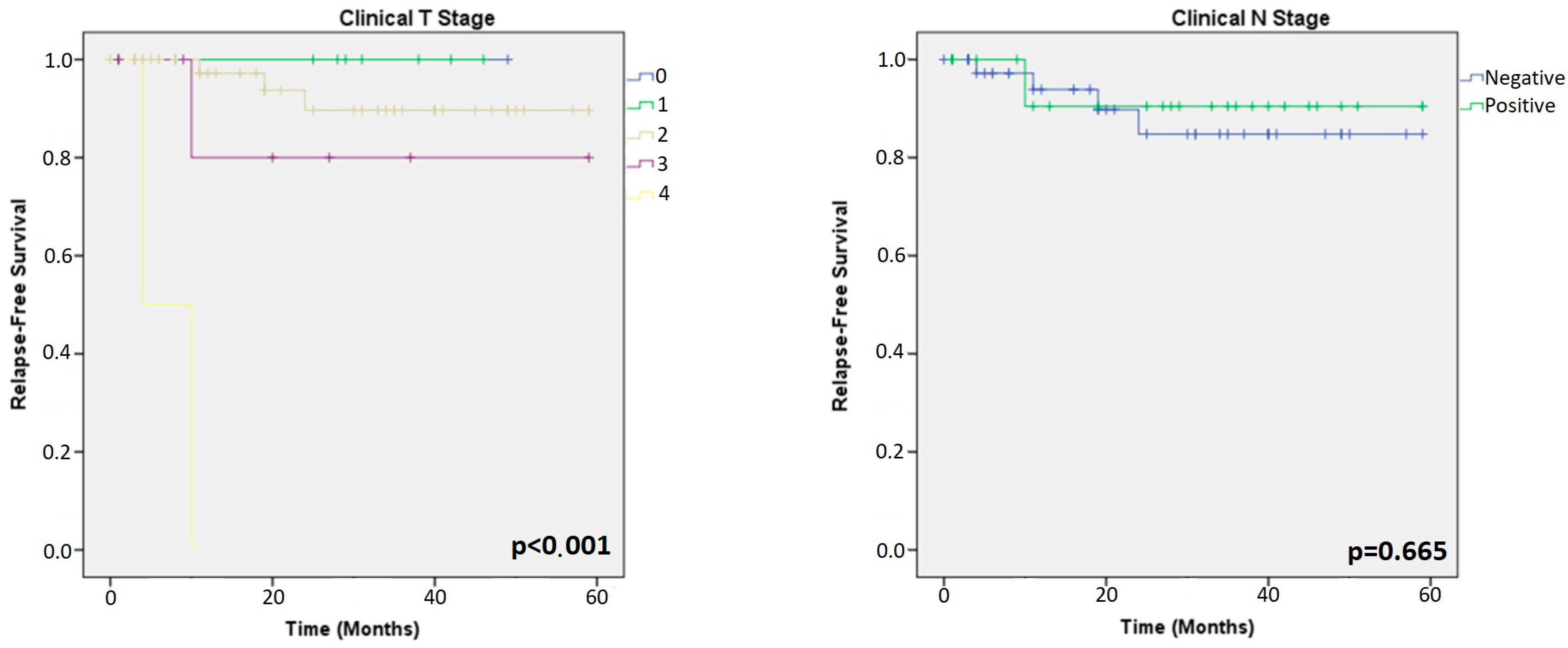
4.1.5. T-Stage
4.2. Differences in Biomarkers in Primary and Residual Tumors
4.3. Limitations
5. Conclusions
Author Contributions
Funding
Institutional Review Board Statement
Informed Consent Statement
Data Availability Statement
Acknowledgments
Conflicts of Interest
References
- Derks, M.G.M.; van de Velde, C.J.H. Neoadjuvant chemotherapy in breast cancer: More than just downsizing. Lancet Oncol. 2018, 19, 2–3. [Google Scholar] [CrossRef] [Green Version]
- Mougalian, S.S.; Soulos, P.R.; Killelea, B.K.; Lannin, D.R.; Abu-Khalaf, M.M.; DiGiovanna, M.P.; Sanft, T.B.; Pusztai, L.; Gross, C.P.; Chagpar, A.B. Use of neoadjuvant chemotherapy for patients with stage I to III breast cancer in the United States. Cancer 2015, 121, 2544–2552. [Google Scholar] [CrossRef] [PubMed]
- Clough, K.B.; Acosta-Marín, V.; Nos, C.; Alran, S.; Rouanet, P.; Garbay, J.-R.; Giard, S.; Verhaeghe, J.-L.; Houvenaeghel, G.; Flipo, B.; et al. Rates of Neoadjuvant Chemotherapy and Oncoplastic Surgery for Breast Cancer Surgery: A French National Survey. Ann. Surg. Oncol. 2015, 22, 3504–3511. [Google Scholar] [CrossRef] [PubMed]
- Vugts, G.; Maaskant-Braat, A.J.G.; Nieuwenhuijzen, G.A.P.; Roumen, R.M.H.; Luiten, E.J.T.; Voogd, A.C. Patterns of Care in the Administration of Neo-adjuvant Chemotherapy for Breast Cancer. A Population-Based Study. Breast J. 2016, 22, 316–321. [Google Scholar] [CrossRef] [PubMed]
- Von Minckwitz, G.; Untch, M.; Blohmer, J.-U.; Costa, S.D.; Eidtmann, H.; Fasching, P.A.; Gerber, B.; Eiermann, W.; Hilfrich, J.; Huober, J.; et al. Definition and Impact of Pathologic Complete Response on Prognosis After Neoadjuvant Chemotherapy in Various Intrinsic Breast Cancer Subtypes. J. Clin. Oncol. 2012, 30, 1796–1804. [Google Scholar] [CrossRef] [Green Version]
- Cortazar, P.; Geyer, C.E. Pathological Complete Response in Neoadjuvant Treatment of Breast Cancer. Ann. Surg. Oncol. 2015, 22, 1441–1446. [Google Scholar] [CrossRef]
- Spring, L.; Greenup, R.; Niemierko, A.; Schapira, L.; Haddad, S.; Jimenez, R.; Coopey, S.; Taghian, A.; Hughes, K.S.; Isakoff, S.J.; et al. Pathologic Complete Response After Neoadjuvant Chemotherapy and Long-Term Outcomes Among Young Women with Breast Cancer. J. Natl. Compr. Cancer Netw. 2017, 15, 1216–1223. [Google Scholar] [CrossRef]
- Untch, M.; Möbus, V.; Kuhn, W.; Muck, B.R.; Thomssen, C.; Bauerfeind, I.; Harbeck, N.; Werner, C.; Lebeau, A.; Schneeweiss, A.; et al. Intensive Dose-Dense Compared with Conventionally Scheduled Preoperative Chemotherapy for High-Risk Primary Breast Cancer. J. Clin. Oncol. 2009, 27, 2938–2945. [Google Scholar] [CrossRef]
- Schneeweiss, A.; Chia, S.; Hickish, T.; Harvey, V.; Eniu, A.; Hegg, R.; Tausch, C.; Seo, J.H.; Tsai, Y.-F.; Ratnayake, J.; et al. Pertuzumab plus trastuzumab in combination with standard neoadjuvant anthracycline-containing and anthracycline-free chemotherapy regimens in patients with HER2-positive early breast cancer: A randomized phase II cardiac safety study (TRYPHAENA). Ann. Oncol. 2013, 24, 2278–2284. [Google Scholar] [CrossRef]
- Swain, S.M.; Ewer, M.S.; Viale, G.; Delaloge, S.; Ferrero, J.-M.; Verrill, M.; Colomer, R.; Vieira, C.; Werner, T.L.; Douthwaite, H.; et al. Pertuzumab, trastuzumab, and standard anthracycline- and taxane-based chemotherapy for the neoadjuvant treatment of patients with HER2-positive localized breast cancer (BERENICE): A phase II, open-label, multicenter, multinational cardiac safety study. Ann. Oncol. 2018, 29, 646–653. [Google Scholar] [CrossRef]
- Denkert, C.; Von Minckwitz, G.; Darb-Esfahani, S.; Lederer, B.; Heppner, B.I.; Weber, K.E.; Budczies, J.; Huober, J.; Klauschen, F.; Furlanetto, J.; et al. Tumour-infiltrating lymphocytes and prognosis in different subtypes of breast cancer: A pooled analysis of 3771 patients treated with neoadjuvant therapy. Lancet Oncol. 2018, 19, 40–50. [Google Scholar] [CrossRef] [PubMed]
- Kurozumi, S.; Inoue, K.; Takei, H.; Matsumoto, H.; Kurosumi, M.; Horiguchi, J.; Takeyoshi, I.; Oyama, T. ER, PgR, Ki67, p27Kip1, and histological grade as predictors of pathological complete response in patients with HER2-positive breast cancer receiving neoadjuvant chemotherapy using taxanes followed by fluorouracil, epirubicin, and cyclophosphamide concomitant with trastuzumab. BMC Cancer 2015, 15, 622. [Google Scholar] [CrossRef] [Green Version]
- Alba, E.; Lluch, A.; Ribelles, N.; Anton-Torres, A.; Sanchez-Rovira, P.; Albanell, J.; Calvo, L.; García-Asenjo, J.A.L.; Palacios, J.; Chacon, J.I.; et al. High Proliferation Predicts Pathological Complete Response to Neoadjuvant Chemotherapy in Early Breast Cancer. Oncologist 2016, 21, 150–155. [Google Scholar] [CrossRef] [Green Version]
- Goorts, B.; Van Nijnatten, T.J.A.; De Munck, L.; Moossdorff, M.; Heuts, E.M.; De Boer, M.; Lobbes, M.B.I.; Smidt, M.L. Clinical tumor stage is the most important predictor of pathological complete response rate after neoadjuvant chemotherapy in breast cancer patients. Breast Cancer Res. Treat. 2017, 163, 83–91. [Google Scholar] [CrossRef] [Green Version]
- Amin, M.B.; Greene, F.L.; Edge, S.B.; Compton, C.C.; Gershenwald, J.E.; Brookland, R.K.; Meyer, L.; Gress, D.M.; Byrd, D.R.; Winchester, D.P. The Eighth Edition AJCC Cancer Staging Manual: Continuing to build a bridge from a population-based to a more “personalized” approach to cancer staging. CA Cancer J. Clin. 2017, 67, 93–99. [Google Scholar] [CrossRef]
- Symmans, W.F.; Wei, C.; Gould, R.; Yu, X.; Zhang, Y.; Liu, M.; Walls, A.; Bousamra, A.; Ramineni, M.; Sinn, B.; et al. Long-Term Prognostic Risk After Neoadjuvant Chemotherapy Associated with Residual Cancer Burden and Breast Cancer Subtype. J. Clin. Oncol. 2017, 35, 1049–1060. [Google Scholar] [CrossRef] [Green Version]
- Symmans, W.F.; Peintinger, F.; Hatzis, C.; Rajan, R.; Kuerer, H.; Valero, V.; Assad, L.; Poniecka, A.; Hennessy, B.; Green, M.; et al. Measurement of Residual Breast Cancer Burden to Predict Survival After Neoadjuvant Chemotherapy. J. Clin. Oncol. 2007, 25, 4414–4422. [Google Scholar] [CrossRef] [PubMed]
- Wolff, A.C.; Hammond, M.E.H.; Allison, K.H.; Harvey, B.E.; Mangu, P.B.; Bartlett, J.M.S.; Bilous, M.; Ellis, I.O.; Fitzgibbons, P.; Hanna, W.; et al. Human Epidermal Growth Factor Receptor 2 Testing in Breast Cancer: American Society of Clinical Oncology/College of American Pathologists Clinical Practice Guideline Focused Update. J. Clin. Oncol. 2018, 36, 2105–2122. [Google Scholar] [CrossRef] [Green Version]
- Nielsen, T.O.; Leung, S.C.Y.; Rimm, D.L.; Dodson, A.; Acs, B.; Badve, S.; Denkert, C.; Ellis, M.J.; Fineberg, S.; Flowers, M.; et al. Assessment of Ki67 in Breast Cancer: Updated Recommendations from the International Ki67 in Breast Cancer Working Group. Gynecol. Oncol. 2021, 113, 808–819. [Google Scholar] [CrossRef]
- Curigliano, G.; Burstein, H.J.; Winer, E.P.; Gnant, M.; Dubsky, P.; Loibl, S.; Colleoni, M.; Regan, M.M.; Piccart-Gebhart, M.; Senn, H.-J.; et al. De-escalating and escalating treatments for early-stage breast cancer: The St. Gallen International Expert Consensus Conference on the Primary Therapy of Early Breast Cancer 2017. Ann. Oncol. Off. J. Eur. Soc. Med. Oncol. 2017, 28, 1700–1712. [Google Scholar] [CrossRef]
- Zhu, X.; Chen, L.; Huang, B.; Wang, Y.; Ji, L.; Wu, J.; Di, G.; Liu, G.; Yu, K.; Shao, Z.; et al. The prognostic and predictive potential of Ki-67 in triple-negative breast cancer. Sci. Rep. 2020, 10, 225. [Google Scholar] [CrossRef] [PubMed] [Green Version]
- Salgado, R.; Denkert, C.; Demaria, S.; Sirtaine, N.; Klauschen, F.; Pruneri, G.; Wienert, S.; Van den Eynden, G.; Baehner, F.L.; Penault-Llorca, F.; et al. The evaluation of tumor-infiltrating lymphocytes (TILs) in breast cancer: Recommendations by an International TILs Working Group 2014. Ann. Oncol. 2015, 26, 259–271. [Google Scholar] [CrossRef] [PubMed]
- Dieci, M.V.; Radosevic-Robin, N.; Fineberg, S.; Eynden, G.v.D.; Ternes, N.; Penault-Llorca, F.; Pruneri, G.; D’alfonso, T.M.; Demaria, S.; Castaneda, C.; et al. Update on tumor-infiltrating lymphocytes (TILs) in breast cancer, including recommendations to assess TILs in residual disease after neoadjuvant therapy and in carcinoma in situ: A report of the International Immuno-Oncology Biomarker Working Group on Breast Cancer. Semin. Cancer Biol. 2017, 52, 16–25. [Google Scholar] [CrossRef]
- Early Breast Cancer Trialists’ Collaborative Group (EBCTCG) Effects of chemotherapy and hormonal therapy for early breast cancer on recurrence and 15-year survival: An overview of the randomised trials. Lancet 2005, 365, 1687–1717. [CrossRef] [PubMed]
- Jarząb, M.; Stobiecka, E.; Badora-Rybicka, A.; Chmielik, E.; Kowalska, M.; Bal, W.; Polakiewicz-Gilowska, A.; Bobek-Billewicz, B.; Lange, D.; Tarnawski, R. Association of breast cancer grade with response to neoadjuvant chemotherapy assessed postoperatively. Pol. J. Pathol. 2019, 70, 91–99. [Google Scholar] [CrossRef]
- Díaz-Redondo, T.; Lavado-Valenzuela, R.; Jimenez, B.; Pascual, T.; Gálvez, F.; Falcón, A.; Alamo, M.D.C.; Morales, C.; Amerigo, M.; Pascual, J.; et al. Different Pathological Complete Response Rates According to PAM50 Subtype in HER2+ Breast Cancer Patients Treated With Neoadjuvant Pertuzumab/Trastuzumab vs. Trastuzumab Plus Standard Chemotherapy: An Analysis of Real-World Data. Front. Oncol. 2019, 9, 1178. [Google Scholar] [CrossRef]
- Gass, P.; Lux, M.P.; Rauh, C.; Hein, A.; Bani, M.R.; Fiessler, C.; Hartmann, A.; Häberle, L.; Pretscher, J.; Erber, R.; et al. Prediction of pathological complete response and prognosis in patients with neoadjuvant treatment for triple-negative breast cancer. BMC Cancer 2018, 18, 1051. [Google Scholar] [CrossRef] [Green Version]
- Meisel, J.L.; Zhao, J.; Suo, A.; Zhang, C.; Wei, Z.; Taylor, C.; Aneja, R.; Krishnamurti, U.; Li, Z.; Nahta, R.; et al. Clinicopathologic Factors Associated with Response to Neoadjuvant Anti-HER2–Directed Chemotherapy in HER2-Positive Breast Cancer. Clin. Breast Cancer 2020, 20, 19–24. [Google Scholar] [CrossRef]
- Ács, B.; Zámbó, V.; Vízkeleti, L.; Szász, A.M.; Madaras, L.; Szentmártoni, G.; Tőkés, T.; Molnár, B.; Molnár, I.A.; Vári-Kakas, S.; et al. Ki-67 as a controversial predictive and prognostic marker in breast cancer patients treated with neoadjuvant chemotherapy. Diagn. Pathol. 2017, 12, 20. [Google Scholar] [CrossRef] [Green Version]
- Chen, X.; He, C.; Han, D.; Zhou, M.; Wang, Q.; Tian, J.; Li, L.; Xu, F.; Zhou, E.; Yang, K. The predictive value of Ki-67 before neoadjuvant chemotherapy for breast cancer: A systematic review and meta-analysis. Futur. Oncol. 2017, 13, 843–857. [Google Scholar] [CrossRef]
- Zhang, G.; Xie, W.; Liu, Z.; Lin, C.; Piao, Y.; Xu, L.; Guo, F.; Xie, X. Prognostic function of Ki-67 for pathological complete response rate of neoadjuvant chemotherapy in triple-negative breast cancer. Tumori J. 2014, 100, 136–142. [Google Scholar] [CrossRef] [PubMed]
- Sánchez-Muñoz, A.; Plata-Fernández, Y.M.; Fernández, M.; Jaén-Morago, A.; Fernández-Navarro, M.; de la Torre-Cabrera, C.; Ramirez-Tortosa, C.; Lomas-Garrido, M.; Llácer, C.; Navarro-Perez, V.; et al. The Role of Immunohistochemistry in Breast Cancer Patients Treated With Neoadjuvant Chemotherapy: An Old Tool With an Enduring Prognostic Value. Clin. Breast Cancer 2013, 13, 146–152. [Google Scholar] [CrossRef]
- Wajid, S.; Samad, F.A.; Syed, A.S.; Kazi, F. Ki-67 and Its Relation with Complete Pathological Response in Patients With Breast Cancer. Cureus 2021, 13, e16788. [Google Scholar] [CrossRef]
- Tao, M.; Chen, S.; Zhang, X.; Zhou, Q. Ki-67 labeling index is a predictive marker for a pathological complete response to neoadjuvant chemotherapy in breast cancer. Medicine 2017, 96, e9384. [Google Scholar] [CrossRef] [PubMed]
- Tan, S.; Fu, X.; Xu, S.; Qiu, P.; Lv, Z.; Xu, Y.; Zhang, Q. Quantification of Ki67 Change as a Valid Prognostic Indicator of Luminal B Type Breast Cancer After Neoadjuvant Therapy. Pathol. Oncol. Res. 2021, 27, 1609972. [Google Scholar] [CrossRef] [PubMed]
- Pistelli, M.; Merloni, F.; Crocetti, S.; Scortichini, L.; Tassone, L.; Cantini, L.; Agostinelli, V.; Bastianelli, L.; Savini, A.; Berardi, R. Prognostic Impact of Ki-67 Change in Locally Advanced and Early Breast Cancer after Neoadjuvant Chemotherapy: A Single Institution Experience. J. Oncol. 2021, 2021, 5548252. [Google Scholar] [CrossRef]
- Li, L.; Han, N.; Wang, X.; Wang, Q.; Tian, J.; Yao, J.; Yuan, L.; Qian, K.; Zou, Q.; Yi, W.; et al. Prognostic values of Ki-67 in neoadjuvant setting for breast cancer: A systematic review and meta-analysis. Futur. Oncol. 2017, 13, 1021–1034. [Google Scholar] [CrossRef]
- Miglietta, L.; Morabito, F.; Provinciali, N.; Canobbio, L.; Meszaros, P.; Naso, C.; Murialdo, R.; Boitano, M.; Salvi, S.; Ferrarini, M. A prognostic model based on combining estrogen receptor expression and Ki-67 value after neoadjuvant chemotherapy predicts clinical outcome in locally advanced breast cancer: Extension and analysis of a previously reported cohort of patients. Eur. J. Surg. Oncol. (EJSO) 2013, 39, 1046–1052. [Google Scholar] [CrossRef]
- Liu, Y.; Yin, W.; Yan, T.; Du, Y.; Shao, Z.; Lu, J. The clinical significance of Ki-67 as a marker of prognostic value and chemosensitivity prediction in hormone-receptor-positive breast cancer: A meta-analysis of the published literature. Curr. Med. Res. Opin. 2013, 29, 1453–1461. [Google Scholar] [CrossRef]
- Denkert, C.; Loibl, S.; Noske, A.; Roller, M.; Müller, B.M.; Komor, M.; Budczies, J.; Darb-Esfahani, S.; Kronenwett, R.; Hanusch, C.; et al. Tumor-Associated Lymphocytes as an Independent Predictor of Response to Neoadjuvant Chemotherapy in Breast Cancer. J. Clin. Oncol. 2010, 28, 105–113. [Google Scholar] [CrossRef]
- Lee, H.; Lee, M.; Seo, J.-H.; Gong, G.; Lee, H.J. Changes in Tumor-infiltrating Lymphocytes After Neoadjuvant Chemotherapy and Clinical Significance in Triple Negative Breast Cancer. Anticancer Res. 2020, 40, 1883–1890. [Google Scholar] [CrossRef] [PubMed]
- Caudle, A.S.; Gonzalez-Angulo, A.M.; Hunt, K.K.; Liu, P.; Pusztai, L.; Symmans, W.F.; Kuerer, H.M.; Mittendorf, E.A.; Hortobagyi, G.N.; Meric-Bernstam, F. Predictors of Tumor Progression During Neoadjuvant Chemotherapy in Breast Cancer. J. Clin. Oncol. 2010, 28, 1821–1828. [Google Scholar] [CrossRef] [Green Version]
- Jin, X.; Jiang, Y.-Z.; Chen, S.; Yu, K.-D.; Ma, D.; Sun, W.; Shao, Z.-M.; Di, G.-H. A nomogram for predicting pathological complete response in patients with human epidermal growth factor receptor 2 negative breast cancer. BMC Cancer 2016, 16, 606. [Google Scholar] [CrossRef] [PubMed] [Green Version]
- Prat, A.; Fan, C.; Fernández, A.; Hoadley, K.A.; Martinello, R.; Vidal, M.; Viladot, M.; Pineda, E.; Arance, A.; Muñoz, M.; et al. Response and survival of breast cancer intrinsic subtypes following multi-agent neoadjuvant chemotherapy. BMC Med. 2015, 13, 303. [Google Scholar] [CrossRef] [PubMed] [Green Version]
- Taucher, S.; Rudas, M.; Gnant, M.; Thomanek, K.; Dubsky, P.; Roka, S.; Bachleitner-Hofmann, T.; Kandioler, D.; Wenzel, C.; Steger, G.; et al. Sequential steroid hormone receptor measurements in primary breast cancer with and without intervening primary chemotherapy. Endocr. Relat. Cancer 2003, 10, 91–98. [Google Scholar] [CrossRef] [PubMed] [Green Version]
- Kasami, M.; Uematsu, T.; Honda, M.; Yabuzaki, T.; Sanuki, J.; Uchida, Y.; Sugimura, H. Comparison of estrogen receptor, progesterone receptor and Her-2 status in breast cancer pre- and post-neoadjuvant chemotherapy. Breast 2008, 17, 523–527. [Google Scholar] [CrossRef]
- Mittendorf, E.A.; Wu, Y.; Scaltriti, M.; Meric-Bernstam, F.; Hunt, K.K.; Dawood, S.; Esteva, F.J.; Buzdar, A.U.; Chen, H.; Eksambi, S.; et al. Loss of HER2 Amplification Following Trastuzumab-Based Neoadjuvant Systemic Therapy and Survival Outcomes. Clin. Cancer Res. 2009, 15, 7381–7388. [Google Scholar] [CrossRef] [Green Version]
- Hurley, J.; Doliny, P.; Reis, I.; Silva, O.; Gomez-Fernandez, C.; Velez, P.; Pauletti, G.; Powell, J.E.; Pegram, M.D.; Slamon, D.J. Docetaxel, Cisplatin, and Trastuzumab as Primary Systemic Therapy for Human Epidermal Growth Factor Receptor 2–Positive Locally Advanced Breast Cancer. J. Clin. Oncol. 2006, 24, 1831–1838. [Google Scholar] [CrossRef]
- Harris, L.N.; You, F.; Schnitt, S.J.; Witkiewicz, A.; Lu, X.; Sgroi, D.; Ryan, P.D.; Come, S.E.; Burstein, H.J.; Lesnikoski, B.-A.; et al. Predictors of Resistance to Preoperative Trastuzumab and Vinorelbine for HER2-Positive Early Breast Cancer. Clin. Cancer Res. 2007, 13, 1198–1207. [Google Scholar] [CrossRef] [Green Version]
- Grassini, D.; Cascardi, E.; Sarotto, I.; Annaratone, L.; Sapino, A.; Berrino, E.; Marchiò, C. Unusual Patterns of HER2 Expression in Breast Cancer: Insights and Perspectives. Pathobiology 2022, 89, 278–296. [Google Scholar] [CrossRef]
- Matsubara, N.; Mukai, H.; Fujii, S.; Wada, N. Different prognostic significance of Ki-67 change between pre- and post-neoadjuvant chemotherapy in various subtypes of breast cancer. Breast Cancer Res. Treat. 2012, 137, 203–212. [Google Scholar] [CrossRef] [PubMed]
- Chen, C.; Zhang, Y.; Huang, Z.; Wu, J.; Huang, W.; Zhang, G. Decrease in the Ki67 index during neoadjuvant chemotherapy predicts favorable relapse-free survival in patients with locally advanced breast cancer. Cancer Biol. Med. 2019, 16, 575–586. [Google Scholar] [CrossRef]
- Ding, Y.; Ding, K.; Qian, H.; Yu, X.; Zou, D.; Yang, H.; Mo, W.; He, X.; Zhang, F.; Qin, C.; et al. Impact on survival of estrogen receptor, progesterone receptor and Ki-67 expression discordance pre- and post-neoadjuvant chemotherapy in breast cancer. PLoS ONE 2020, 15, e0231895. [Google Scholar] [CrossRef] [PubMed] [Green Version]
- Wang, Y.; Zong, B.; Yu, Y.; Wang, Y.; Tang, Z.; Chen, R.; Huang, M.; Liu, S. Ki67 Index Changes and Tumor-Infiltrating Lymphocyte Levels Impact the Prognosis of Triple-Negative Breast Cancer Patients with Residual Disease After Neoadjuvant Chemotherapy. Front. Oncol. 2021, 11, 668610. [Google Scholar] [CrossRef]
- Ochi, T.; Bianchini, G.; Ando, M.; Nozaki, F.; Kobayashi, D.; Criscitiello, C.; Curigliano, G.; Iwamoto, T.; Niikura, N.; Takei, H.; et al. Predictive and prognostic value of stromal tumour-infiltrating lymphocytes before and after neoadjuvant therapy in triple negative and HER2-positive breast cancer. Eur. J. Cancer 2019, 118, 41–48. [Google Scholar] [CrossRef] [PubMed]
- Jin, X.; Jiang, Y.-Z.; Chen, S.; Yu, K.-D.; Shao, Z.-M.; Di, G.-H. Prognostic value of receptor conversion after neoadjuvant chemotherapy in breast cancer patients: A prospective observational study. Oncotarget 2015, 6, 9600–9611. [Google Scholar] [CrossRef] [Green Version]
- Yang, Y.-F.; Liao, Y.-Y.; Li, L.-Q.; Xie, S.-R.; Xie, Y.-F.; Peng, N.-F. Changes in ER, PR and HER2 receptors status after neoadjuvant chemotherapy in breast cancer. Pathol. Res. Pract. 2013, 209, 797–802. [Google Scholar] [CrossRef]
- Van De Ven, S.; Smit, V.T.H.B.M.; Dekker, T.J.A.; Nortier, J.W.R.; Kroep, J.R. Discordances in ER, PR and HER2 receptors after neoadjuvant chemotherapy in breast cancer. Cancer Treat. Rev. 2011, 37, 422–430. [Google Scholar] [CrossRef]


| pCR | No pCR | ||||
|---|---|---|---|---|---|
| N | % | N | % | P | |
| HER2+ (n = 81) | 0.068 | ||||
| 39 | 48.1 | 42 | 51.9 | ||
| Anthracycline + THP | 27 | 69.2 | 35 | 83.3 | |
| Docetaxel + CB + TH | 6 | 15.4 | 1 | 2.4 | |
| THP | 4 | 10.3 | 6 | 14.3 | |
| THP + anti-PD-L1 | 2 | 5.1 | 0 | 0 | |
| TN (n = 69) | 0.794 | ||||
| 37 | 53.6 | 32 | 46.4 | ||
| P + CB + ACdd | 16 | 43.2 | 15 | 46.9 | |
| P + EC | 18 | 48.6 | 16 | 50 | |
| P + anti-PD-L1 | 3 | 8.1 | 1 | 3.1 | |
| HER2+ (n = 81) | TN (n = 69) | |||||||||
|---|---|---|---|---|---|---|---|---|---|---|
| pCR | No pCR | P | pCR | No pCR | P | |||||
| N | % | N | % | N | % | N | % | |||
| 39 | 48.1 | 42 | 51.9 | 37 | 53.6 | 32 | 46.6 | |||
| Median age [range] | 0.114 | 0.105 | ||||||||
| 57.3 [35–79] | 53.6 [31–77] | 49.4 [26–69] | 53.7 [35–77] | |||||||
| Histological subtype | >0.999 | >0.999 | ||||||||
| No special | 38 | 97.4 | 41 | 97.6 | 36 | 97.3 | 31 | 96.9 | ||
| Other | 1 | 2.6 | 1 | 2.4 | 1 | 2.7 | 1 | 3.1 | ||
| Histological grade | 0.254 | 0.097 | ||||||||
| Grade 1/2 | 18 | 46.2 | 26 | 61.9 | 4 | 5.8 | 8 | 25 | ||
| Grade 3 | 19 | 48.7 | 15 | 35.7 | 32 | 86.5 | 23 | 71.9 | ||
| Not available | 2 | 5.1 | 1 | 2.4 | 1 | 2.7 | 1 | 3.1 | ||
| ER | 0.022 | >0.999 | ||||||||
| Negative (<1%) | 19 | 48.7 | 10 | 23.8 | 35 | 94.6 | 31 | 96.9 | ||
| Positive (>1%) | 20 | 51.3 | 32 | 76.2 | 2 | 5.4 | 1 | 3.1 | ||
| PR | 0.471 | 0.743 | ||||||||
| Negative (<1%) | 14 | 35.9 | 11 | 26.2 | 32 | 86.5 | 26 | 81.3 | ||
| Positive (>1%) | 25 | 64.1 | 31 | 73.8 | 5 | 13.5 | 6 | 18.7 | ||
| Mean Ki67 | 0.022 | 0.224 | ||||||||
| 41.7 | 33.9 | 65 | 58.7 | |||||||
| Ki67 LI | 0.195 | 0.201 | ||||||||
| Low 0–29% | 8 | 20.5 | 13 | 30.9 | 0 | 0 | 2 | 6.3 | ||
| High ≥ 30% | 30 | 76.9 | 22 | 52.4 | 35 | 94.6 | 27 | 84.4 | ||
| Not available | 1 | 2.6 | 7 | 16.7 | 2 | 5.4 | 3 | 9.3 | ||
| Mean TILs | 0.027 | 0.111 | ||||||||
| 23.3 | 14.1 | 27.8 | 18.6 | |||||||
| Percentage of TILs | 0.032 | 0.617 | ||||||||
| Low ≤ 20% | 25 | 64.1 | 35 | 83.3 | 22 | 59.5 | 21 | 65.6 | ||
| High > 20% | 13 | 33.3 | 5 | 11.9 | 14 | 37.8 | 10 | 31.3 | ||
| Not available | 1 | 2.6 | 2 | 4.8 | 1 | 2.7 | 1 | 3.1 | ||
| Clinical T Stage | 0.005 | 0.496 | ||||||||
| T0/T1/T2 | 35 | 89.7 | 26 | 61.9 | 33 | 89.2 | 26 | 81.3 | ||
| T3/T4 | 4 | 10.3 | 16 | 38.1 | 4 | 10.8 | 6 | 18.7 | ||
| Clinical N Stage | 0.505 | 0.450 | ||||||||
| Negative | 18 | 46.2 | 16 | 38.1 | 25 | 67.6 | 18 | 56.3 | ||
| Positive | 21 | 53.8 | 26 | 61.9 | 12 | 32.4 | 14 | 43.7 | ||
| RCB-0/I | RCB-II/III | ||||
|---|---|---|---|---|---|
| N | % | N | % | P | |
| 55 | 67.9 | 26 | 32.1 | ||
| Histological grade | 0.807 | ||||
| Grade 1/2 | 29 | 52.7 | 15 | 57.7 | |
| Grade 3 | 24 | 43.6 | 10 | 38.5 | |
| Not available | 2 | 7.2 | 1 | 3.8 | |
| ER | 0.046 | ||||
| Negative (<1%) | 24 | 43.6 | 5 | 19.2 | |
| Positive (>1%) | 31 | 56.4 | 21 | 80.8 | |
| Not available | 0 | 0 | 0 | 0 | |
| PR | 0.620 | ||||
| Negative (<1%) | 18 | 32.7 | 7 | 26.9 | |
| Positive (>1%) | 37 | 67.3 | 19 | 73.1 | |
| Not available | 0 | 0 | 0 | 0 | |
| Mean Ki67 | 0.278 | ||||
| 39.2 | 35.3 | ||||
| Ki67 LI | 0.395 | ||||
| Low (0–29%) | 11 | 20 | 10 | 38.5 | |
| High (≥30%) | 39 | 70.9 | 13 | 50 | |
| Not available | 5 | 9.1 | 3 | 11.5 | |
| Mean TILs | 0.188 | ||||
| 20.4 | 14.7 | ||||
| Percentage of TILs | 0.395 | ||||
| Low (≤20%) | 39 | 70.9 | 21 | 80.8 | |
| High (>20%) | 14 | 25.5 | 4 | 15.4 | |
| Not available | 2 | 3.6 | 1 | 3.8 | |
| Clinical T Stage | 0.058 | ||||
| T0/T1/T2 | 45 | 81.8 | 16 | 61.5 | |
| T3/T4 | 10 | 18.2 | 10 | 38.5 | |
| Clinical N Stage | 0.473 | ||||
| Negative | 25 | 45.5 | 9 | 34.6 | |
| Positive | 30 | 54.5 | 17 | 65.4 | |
| RCB-0/I | RCB-II/III | ||||
|---|---|---|---|---|---|
| N | % | N | % | P | |
| 50 | 72.5 | 19 | 27.5 | ||
| Histological grade | 0.281 | ||||
| Grade 1/2 | 7 | 14 | 5 | 26.3 | |
| Grade 3 | 42 | 84 | 13 | 68.4 | |
| Not available | 1 | 2 | 1 | 5.3 | |
| ER | 0.555 | ||||
| Negative (<1%) | 47 | 94 | 19 | 100 | |
| ER-low (1–10%) | 3 | 6 | 0 | 0 | |
| Not available | 0 | 0 | 0 | 0 | |
| PR | <0.001 | ||||
| Negative (<1%) | 44 | 88 | 8 | 42.1 | |
| PR-low (1–10%) | 6 | 12 | 11 | 57.9 | |
| Not available | 0 | 0 | 0 | 0 | |
| Mean Ki67 | 0.706 | ||||
| 62.8 | 60.4 | ||||
| Ki67 LI | 0.076 | ||||
| Low (0–29%) | 0 | 0 | 2 | 10.5 | |
| High (≥30%) | 46 | 92 | 16 | 84.2 | |
| Not available | 4 | 8 | 1 | 5.3 | |
| Mean TILs | 0.483 | ||||
| 24.7 | 20.3 | ||||
| Percentage of TILs | 0.779 | ||||
| Low (≤20%) | 32 | 64 | 11 | 57.9 | |
| High (>20%) | 17 | 34 | 7 | 36.8 | |
| Not available | 1 | 2 | 1 | 5.3 | |
| Clinical T Stage | 0.445 | ||||
| T0/T1/T2 | 44 | 88 | 15 | 78.9 | |
| T3/T4 | 6 | 12 | 4 | 21.1 | |
| Clinical N Stage | 0.406 | ||||
| Negative | 33 | 66 | 10 | 52.6 | |
| Positive | 17 | 34 | 9 | 47.4 | |
| HER2 (n = 42) | TN (n = 32) | ||||||||||
|---|---|---|---|---|---|---|---|---|---|---|---|
| Primary | Residual | P | Primary | Residual | P | ||||||
| N | % | N | % | N | % | N | % | ||||
| Histological grade | 0.206 | 0.086 | |||||||||
| Grade 1/2 | 26 | 61.9 | 25 | 59.5 | 8 | 25 | 12 | 37.5 | |||
| Grade 3 | 15 | 35.7 | 7 | 16.7 | 23 | 71.9 | 11 | 34.4 | |||
| Not available | 1 | 2.4 | 10 | 23.8 | 1 | 3.1 | 9 | 28.1 | |||
| ER | 0.021 | >0.999 | |||||||||
| Negative (<1%) | 10 | 23.8 | 11 | 26.2 | 31 | 96.9 | 28 | 87.5 | |||
| Positive (>1%) | 32 | 76.2 | 25 | 59.5 | 1 | 3.1 | 1 | 3.1 | |||
| Not available | 0 | 0 | 6 | 14.3 | 0 | 0 | 3 | 9.4 | |||
| PR | 0.021 | 0.477 | |||||||||
| Negative (<1%) | 11 | 26.2 | 19 | 45.5 | 26 | 81.3 | 26 | 81.3 | |||
| Positive (>1%) | 31 | 73.8 | 17 | 40.5 | 6 | 18.7 | 3 | 9.4 | |||
| Not available | 0 | 0 | 6 | 14.3 | 0 | 0 | 3 | 9.4 | |||
| HER2 Status | 0.001 | 1 | |||||||||
| Negative | 0 | 0 | 8 | 19.1 | 32 | 100 | 31 | 96.9 | |||
| Positive | 42 | 100 | 28 | 66.6 | 0 | 0 | 0 | 0 | |||
| Not available | 0 | 0 | 6 | 14.3 | 0 | 0 | 1 | 3.1 | |||
| Mean Ki67 | <0.001 | <0.001 | |||||||||
| 33.9 | 18.7 | 56 | 17.8 | ||||||||
| Ki67 LI | 0.007 | 0.201 | |||||||||
| Low (0–29%) | 13 | 30.9 | 22 | 52.4 | 2 | 5.4 | 20 | 6.3 | |||
| High (≥30%) | 22 | 52.4 | 9 | 21.4 | 27 | 84.4 | 6 | 84.4 | |||
| Not available | 7 | 16.7 | 11 | 26.2 | 3 | 8.1 | 6 | 9.3 | |||
| Mean TILs | 0.003 | 0.668 | |||||||||
| 14.1 | 5.2 | 18.6 | 16.5 | ||||||||
| Percentage of TILs | 0.441 | 1 | |||||||||
| Low (≤20%) | 35 | 32 | 21 | 16 | |||||||
| High (>20%) | 5 | 2 | 10 | 7 | |||||||
| Not available | 2 | 8 | 1 | 9 | |||||||
| cT and ypT Stage | <0.001 | <0.001 | |||||||||
| Tis/T0/T1 | 3 | 7.1 | 32 | 76.2 | 8 | 21.6 | 25 | 78.1 | |||
| T2 | 23 | 54.8 | 5 | 11.9 | 25 | 67.6 | 5 | 15.6 | |||
| T3 | 8 | 19 | 4 | 9.5 | 3 | 8.1 | 2 | 6.3 | |||
| T4 | 8 | 19 | 1 | 2.4 | 1 | 2.7 | 0 | 0 | |||
| cN and ypN Stage | 0.122 | 0.583 | |||||||||
| Negative | 16 | 38.1 | 22 | 52.4 | 18 | 56.3 | 15 | 46.9 | |||
| Positive | 26 | 61.9 | 17 | 40.5 | 14 | 43.7 | 8 | 25 | |||
| Not available | 0 | 0 | 3 | 7.1 | 0 | 0 | 9 | 28.1 | |||
Disclaimer/Publisher’s Note: The statements, opinions and data contained in all publications are solely those of the individual author(s) and contributor(s) and not of MDPI and/or the editor(s). MDPI and/or the editor(s) disclaim responsibility for any injury to people or property resulting from any ideas, methods, instructions or products referred to in the content. |
© 2023 by the authors. Licensee MDPI, Basel, Switzerland. This article is an open access article distributed under the terms and conditions of the Creative Commons Attribution (CC BY) license (https://creativecommons.org/licenses/by/4.0/).
Share and Cite
Pons, L.; Hernández, L.; Urbizu, A.; Osorio, P.; Rodríguez-Martínez, P.; Castella, E.; Muñoz, A.; Sanz, C.; Arnaldo, L.; Felip, E.; et al. Pre- and Post-Neoadjuvant Clinicopathological Parameters Can Help in the Prognosis and the Prediction of Response in HER2+ and Triple Negative Breast Cancer. Cancers 2023, 15, 3068. https://doi.org/10.3390/cancers15123068
Pons L, Hernández L, Urbizu A, Osorio P, Rodríguez-Martínez P, Castella E, Muñoz A, Sanz C, Arnaldo L, Felip E, et al. Pre- and Post-Neoadjuvant Clinicopathological Parameters Can Help in the Prognosis and the Prediction of Response in HER2+ and Triple Negative Breast Cancer. Cancers. 2023; 15(12):3068. https://doi.org/10.3390/cancers15123068
Chicago/Turabian StylePons, Laura, Laura Hernández, Aintzane Urbizu, Paula Osorio, Paula Rodríguez-Martínez, Eva Castella, Ana Muñoz, Carolina Sanz, Laura Arnaldo, Eudald Felip, and et al. 2023. "Pre- and Post-Neoadjuvant Clinicopathological Parameters Can Help in the Prognosis and the Prediction of Response in HER2+ and Triple Negative Breast Cancer" Cancers 15, no. 12: 3068. https://doi.org/10.3390/cancers15123068