

Evaluation of the Oesophagogastric Cancer-Associated Microbiome: A Systematic Review and Quality Assessment
Abstract
:Simple Summary
Abstract
1. Introduction
2. Methods
2.1. Search Strategy
2.2. Eligibility Assessment and Data Extraction
2.3. Definitions of Groups
2.4. Outcomes
2.5. Quality Assessment
3. Results
3.1. Study Selection
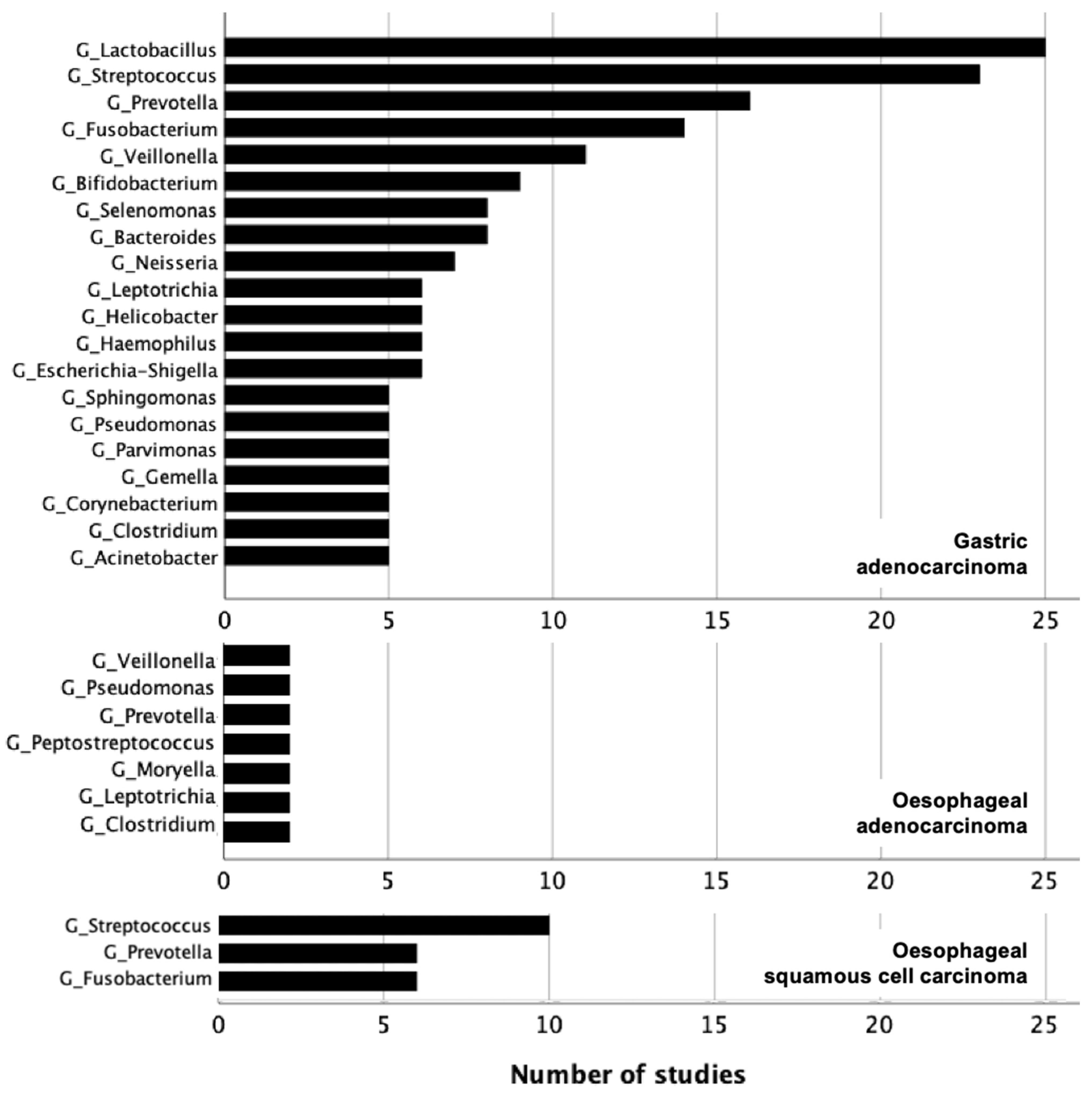
3.2. Gastric Adenocarcinoma
3.3. Oesophageal Adenocarcinoma
3.4. Oesophageal Squamous Cell Carcinoma
4. Evaluation of Diagnostic Performance
5. Influence of Sample Origin
6. Quality of Assessment
7. Discussion
Supplementary Materials
Author Contributions
Funding
Conflicts of Interest
References
- Available online: https://gco.iarc.fr/today/fact-sheets-cancers (accessed on 21 March 2021).
- Flemer, B.; Warren, R.D.; Barrett, M.P.; Cisek, K.; Das, A.; Jeffery, I.B.; Hurley, E.; O‘Riordain, M.; Shanahan, F.; O’Toole, P.W. The oral microbiota in colorectal cancer is distinctive and predictive. Gut 2018, 67, 1454–1463. [Google Scholar] [CrossRef] [PubMed]
- Sun, J.; Li, X.; Yin, J.; Li, Y.; Hou, B.; Zhang, Z. A screening method for gastric cancer by oral microbiome detection. Oncol. Rep. 2018, 39, 2217–2224. [Google Scholar] [CrossRef] [PubMed]
- Fan, X.; Alekseyenko, A.V.; Wu, J.; Peters, B.A.; Jacobs, E.J.; Gapstur, S.M.; Purdue, M.P.; Abnet, C.C.; Stolzenberg-Solomon, R.; Miller, G.; et al. Human oral microbiome and prospective risk for pancreatic cancer: A population-based nested case-control study. Gut 2018, 67, 120–127. [Google Scholar] [CrossRef] [PubMed]
- Hosgood, H.D.; Cai, Q.; Hua, X.; Long, J.; Shi, J.; Wan, Y.; Yang, Y.; Abnet, C.; Bassig, B.A.; Hu, W.; et al. Variation in oral microbiome is associated with future risk of lung cancer among never-smokers. Thorax 2020, 76, 256–263. [Google Scholar] [CrossRef] [PubMed]
- Xu, L.; Qi, Y.; Jiang, Y.; Ji, Y.; Zhao, Q.; Wu, J.; Lu, W.; Wang, Y.; Chen, Q.; Wang, C. Crosstalk between the gut microbiome and clinical response in locally advanced thoracic esophageal squamous cell carcinoma during neoadjuvant camrelizumab and chemotherapy. Ann. Transl. Med. 2022, 10, 325. [Google Scholar] [CrossRef]
- Xu, X.; Lv, J.; Guo, F.; Li, J.; Jia, Y.; Jiang, D.; Wang, N.; Zhang, C.; Kong, L.; Liu, Y.; et al. Gut Microbiome Influences the Efficacy of PD-1 Antibody Immunotherapy on MSS-Type Colorectal Cancer via Metabolic Pathway. Front. Microbiol. 2020, 11, 814. [Google Scholar] [CrossRef]
- Jin, Y.; Dong, H.; Xia, L.; Yang, Y.; Zhu, Y.; Shen, Y.; Zheng, H.; Yao, C.; Wang, Y.; Lu, S. The Diversity of Gut Microbiome is Associated with Favorable Responses to Anti–Programmed Death 1 Immunotherapy in Chinese Patients with NSCLC. J. Thorac. Oncol. 2019, 14, 1378–1389. [Google Scholar] [CrossRef]
- Routy, B.; Gopalakrishnan, V.; Daillère, R.; Zitvogel, L.; Wargo, J.A.; Kroemer, G. The gut microbiota influences anticancer immunosurveillance and general health. Nat. Rev. Clin. Oncol. 2018, 15, 382–396. [Google Scholar] [CrossRef]
- Peng, Z.; Cheng, S.; Kou, Y.; Wang, Z.; Jin, R.; Hu, H.; Zhang, X.; Gong, J.F.; Li, J.; Lu, M.; et al. The Gut Microbiome Is Associated with Clinical Response to Anti-PD-1/PD-L1 Immunotherapy in Gastrointestinal Cancer. Cancer Immunol. Res. 2020, 8, 1251–1261. [Google Scholar] [CrossRef]
- Reddy, R.M.; Weir, W.B.; Barnett, S.; Heiden, B.; Orringer, M.B.; Lin, J.; Chang, A.; Carrott, P.W.; Lynch, W.R.; Etherton-Beer, C.; et al. Increased Variance in Oral and Gastric Microbiome Correlates with Esophagectomy Anastomotic Leak. Ann. Thorac. Surg. 2018, 105, 865–870. [Google Scholar] [CrossRef]
- Yuda, M.; Yamashita, K.; Okamura, A.; Hayami, M.; Fukudome, I.; Toihata, T.; Imamura, Y.; Mine, S.; Ishizuka, N.; Watanabe, M. Influence of Preoperative Oropharyngeal Microflora on the Occurrence of Postoperative Pneumonia and Survival in Patients Undergoing Esophagectomy for Esophageal Cancer. Ann. Surg. 2020, 272, 1035–1043. [Google Scholar] [CrossRef]
- Pimentel, M.; Saad, R.J.; Long, M.D.; Rao, S.S.C. ACG Clinical Guideline: Small Intestinal Bacterial Overgrowth. Am. J. Gastroenterol. 2020, 115, 165–178. [Google Scholar] [CrossRef]
- Ianiro, G.; Rossi, E.; Thomas, A.M.; Schinzari, G.; Masucci, L.; Quaranta, G.; Settanni, C.R.; Lopetuso, L.R.; Armanini, F.; Blanco-Miguez, A.; et al. Faecal microbiota transplantation for the treatment of diarrhoea induced by tyrosine-kinase inhibitors in patients with metastatic renal cell carcinoma. Nat. Commun. 2020, 11, 4333. [Google Scholar] [CrossRef]
- IARC Working Group on the Evaluation of Carcinogenic Risks to Humans. Schistosomes, Liver Flukes and Helicobacter pylori; IARC: Lyon, France, 1994; Volume 61, pp. 1–241.
- Liu, X.; Shao, L.; Liu, X.; Ji, F.; Mei, Y.; Cheng, Y.; Liu, F.; Yan, C.; Li, L.; Ling, Z. Alterations of gastric mucosal microbiota across different stomach microhabitats in a cohort of 276 patients with gastric cancer. Ebiomedicine 2019, 40, 336–348. [Google Scholar] [CrossRef]
- Pittayanon, R.; Lau, J.T.; Leontiadis, G.I.; Tse, F.; Yuan, Y.; Surette, M.; Moayyedi, P. Differences in Gut Microbiota in Patients with vs. without Inflammatory Bowel Diseases: A Systematic Review. Gastroenterology 2020, 158, 930–946.e1. [Google Scholar] [CrossRef]
- Zhang, X.; Liu, Q.; Liao, Q.; Zhao, Y. Pancreatic Cancer, Gut Microbiota, and Therapeutic Efficacy. J. Cancer 2020, 11, 2749–2758. [Google Scholar] [CrossRef]
- Mirzayi, C.; Renson, A.; Genomic Standards Consortium; Massive Analysis and Quality Control Society; Zohra, F.; Elsafoury, S.; Geistlinger, L.; Kasselman, L.J.; Eckenrode, K.; van de Wijgert, J.; et al. Reporting guidelines for human microbiome research: The STORMS checklist. Nat. Med. 2021, 27, 1885–1892. [Google Scholar] [CrossRef]
- Sjöstedt, S.; Heimdahl, A.; Kager, L.; Nord, C.E. Microbial colonization of the oropharynx, esophagus and stomach in patients with gastric diseases. Eur. J. Clin. Microbiol. Infect. Dis. 1985, 4, 49–51. [Google Scholar] [CrossRef]
- Dicksved, J.; Lindberg, M.; Rosenquist, M.; Enroth, H.; Jansson, J.; Engstrand, L. Molecular characterization of the stomach microbiota in patients with gastric cancer and in controls. J. Med. Microbiol. 2009, 58, 509–516. [Google Scholar] [CrossRef]
- Seo, I.; Jha, B.K.; Suh, S.-I.; Suh, M.-H.; Baek, W.-K. Microbial Profile of the Stomach: Comparison between Normal Mucosa and Cancer Tissue in the Same Patient. J. Bacteriol. Virol. 2014, 44, 162–169. [Google Scholar] [CrossRef]
- Aviles-Jimenez, F.; Vazquez-Jimenez, F.; Medrano-Guzman, R.; Mantilla, A.; Torres, J. Stomach microbiota composition varies between patients with non-atrophic gastritis and patients with intestinal type of gastric cancer. Sci. Rep. 2014, 4, 4202. [Google Scholar] [CrossRef] [PubMed]
- Eun, C.S.; Kim, B.K.; Han, D.S.; Kim, S.Y.; Kim, K.M.; Choi, B.Y.; Song, K.S.; Kim, Y.S.; Kim, J.F. Differences in Gastric Mucosal Microbiota Profiling in Patients with Chronic Gastritis, Intestinal Metaplasia, and Gastric Cancer Using Pyrosequencing Methods. Helicobacter 2014, 19, 407–416. [Google Scholar] [CrossRef] [PubMed]
- Hu, J.; Han, S.; Chen, Y.; Ji, Z. Variations of Tongue Coating Microbiota in Patients with Gastric Cancer. BioMed Res. Int. 2015, 2015, 173729. [Google Scholar] [CrossRef] [PubMed]
- Wang, L.; Zhou, J.; Xin, Y.; Geng, C.; Tian, Z.; Yu, X.; Dong, Q. Bacterial overgrowth and diversification of microbiota in gastric cancer. Eur. J. Gastroenterol. Hepatol. 2016, 28, 261–266. [Google Scholar] [CrossRef] [PubMed]
- Yu, G.; Torres, J.; Hu, N.; Medrano-Guzman, R.; Herrera-Goepfert, R.; Humphrys, M.S.; Wang, L.; Wang, C.; Ding, T.; Ravel, J.; et al. Molecular Characterization of the Human Stomach Microbiota in Gastric Cancer Patients. Front. Cell. Infect. Microbiol. 2017, 7, 302. [Google Scholar] [CrossRef]
- Li, T.H.; Qin, Y.; Sham, P.C.; Lau, K.; Chu, K.-M.; Leung, W.K. Alterations in Gastric Microbiota after H. Pylori Eradication and in Different Histological Stages of Gastric Carcinogenesis. Sci. Rep. 2017, 7, 44935. [Google Scholar] [CrossRef]
- Castaño-Rodríguez, N.; Goh, K.-L.; Fock, K.M.; Mitchell, H.M.; Kaakoush, N.O. Dysbiosis of the microbiome in gastric carcinogenesis. Sci. Rep. 2017, 7, 15957. [Google Scholar] [CrossRef]
- Ferreira, R.M.; Pereira-Marques, J.; Pinto-Ribeiro, I.; Costa, J.L.; Carneiro, F.; Machado, J.C.; Figueiredo, C. Gastric microbial community profiling reveals a dysbiotic cancer-associated microbiota. Gut 2017, 67, 226–236. [Google Scholar] [CrossRef]
- Hu, Y.-L.; Pang, W.; Huang, Y.; Zhang, Y.; Zhang, C.-J. The Gastric Microbiome Is Perturbed in Advanced Gastric Adenocarcinoma Identified through Shotgun Metagenomics. Front. Cell. Infect. Microbiol. 2018, 8, 433. [Google Scholar] [CrossRef]
- Wu, J.; Xu, S.; Xiang, C.; Cao, Q.; Li, Q.; Huang, J.; Shi, L.; Zhang, J.; Zhan, Z. Tongue Coating Microbiota Community and Risk Effect on Gastric Cancer. J. Cancer 2018, 9, 4039–4048. [Google Scholar] [CrossRef]
- Hsieh, Y.-Y.; Tung, S.-Y.; Pan, H.-Y.; Yen, C.-W.; Xu, H.-W.; Lin, Y.-J.; Deng, Y.-F.; Hsu, W.-T.; Wu, C.-S.; Li, C. Increased Abundance of Clostridium and Fusobacterium in Gastric Microbiota of Patients with Gastric Cancer in Taiwan. Sci. Rep. 2018, 8, 158. [Google Scholar] [CrossRef]
- Coker, O.O.; Dai, Z.; Nie, Y.; Zhao, G.; Cao, L.; Nakatsu, G.; Wu, W.K.; Wong, S.H.; Chen, Z.; Sung, J.J.Y.; et al. Mucosal microbiome dysbiosis in gastric carcinogenesis. Gut 2017, 67, 1024–1032. [Google Scholar] [CrossRef]
- Shao, D.; Vogtmann, E.; Liu, A.; Qin, J.; Chen, W.; Abnet, C.C.; Wei, W. Microbial characterization of esophageal squamous cell carcinoma and gastric cardia adenocarcinoma from a high-risk region of China. Cancer 2019, 125, 3993–4002. [Google Scholar] [CrossRef]
- Gunathilake, M.N.; Lee, J.; Choi, I.J.; Kim, Y.-I.; Ahn, Y.; Park, C.; Kim, J. Association between the relative abundance of gastric microbiota and the risk of gastric cancer: A case-control study. Sci. Rep. 2019, 9, 13589. [Google Scholar] [CrossRef]
- Liang, W.; Yang, Y.; Wang, H.; Wang, H.; Yu, X.; Lu, Y.; Shen, S.; Teng, L. Gut microbiota shifts in patients with gastric cancer in perioperative period. Medicine 2019, 98, e16626. [Google Scholar] [CrossRef]
- Kageyama, S.; Takeshita, T.; Takeuchi, K.; Asakawa, M.; Matsumi, R.; Furuta, M.; Shibata, Y.; Nagai, K.; Ikebe, M.; Morita, M.; et al. Characteristics of the Salivary Microbiota in Patients with Various Digestive Tract Cancers. Front. Microbiol. 2019, 10, 1780. [Google Scholar] [CrossRef]
- Chen, X.-H.; Wang, A.; Chu, A.-N.; Gong, Y.-H.; Yuan, Y. Mucosa-Associated Microbiota in Gastric Cancer Tissues Compared with Non-cancer Tissues. Front. Microbiol. 2019, 10, 1261. [Google Scholar] [CrossRef]
- Dong, Z.; Chen, B.; Pan, H.; Wang, D.; Liu, M.; Yang, Y.; Zou, M.; Yang, J.; Xiao, K.; Zhao, R.; et al. Detection of Microbial 16S rRNA Gene in the Serum of Patients with Gastric Cancer. Front. Oncol. 2019, 9, 608. [Google Scholar] [CrossRef]
- Qi, Y.-F.; Sun, J.-N.; Ren, L.-F.; Cao, X.-L.; Dong, J.-H.; Tao, K.; Guan, X.-M.; Cui, Y.-N.; Su, W. Intestinal Microbiota Is Altered in Patients with Gastric Cancer from Shanxi Province, China. Dig. Dis. Sci. 2019, 64, 1193–1203. [Google Scholar] [CrossRef]
- Wang, Z.; Gao, X.; Zeng, R.; Wu, Q.; Sun, H.; Wu, W.; Zhang, X.; Sun, G.; Yan, B.; Wu, L.; et al. Changes of the Gastric Mucosal Microbiome Associated with Histological Stages of Gastric Carcinogenesis. Front. Microbiol. 2020, 11, 997. [Google Scholar] [CrossRef]
- Wang, L.; Xin, Y.; Zhou, J.; Tian, Z.; Liu, C.; Yu, X.; Meng, X.; Jiang, W.; Zhao, S.; Dong, Q. Gastric Mucosa-Associated Microbial Signatures of Early Gastric Cancer. Front. Microbiol. 2020, 11, 1548. [Google Scholar] [CrossRef]
- Spiegelhauer, M.R.; Kupcinskas, J.; Johannesen, T.B.; Urba, M.; Skieceviciene, J.; Jonaitis, L.; Frandsen, T.H.; Kupcinskas, L.; Fuursted, K.; Andersen, L.P. Transient and Persistent Gastric Microbiome: Adherence of Bacteria in Gastric Cancer and Dyspeptic Patient Biopsies after Washing. J. Clin. Med. 2020, 9, 1882. [Google Scholar] [CrossRef]
- Gantuya, B.; El Serag, H.B.; Matsumoto, T.; Ajami, N.J.; Uchida, T.; Oyuntsetseg, K.; Bolor, D.; Yamaoka, Y. Gastric mucosal microbiota in a Mongolian population with gastric cancer and precursor conditions. Aliment. Pharmacol. Ther. 2020, 51, 770–780. [Google Scholar] [CrossRef] [PubMed]
- Wu, Z.-F.; Zou, K.; Wu, G.-N.; Jin, Z.-J.; Xiang, C.-J.; Xu, S.; Wang, Y.-H.; Wu, X.-Y.; Chen, C.; Xu, Z.; et al. A Comparison of Tumor-Associated and Non-Tumor-Associated Gastric Microbiota in Gastric Cancer Patients. Dig. Dis. Sci. 2020, 66, 1673–1682. [Google Scholar] [CrossRef] [PubMed]
- Xu, S.; Xiang, C.; Wu, J.; Teng, Y.; Wu, Z.; Wang, R.; Lu, B.; Zhan, Z.; Wu, H.; Zhang, J. Tongue Coating Bacteria as a Potential Stable Biomarker for Gastric Cancer Independent of Lifestyle. Dig. Dis. Sci. 2020, 66, 2964–2980. [Google Scholar] [CrossRef] [PubMed]
- Dang, Y.-N.; Dong, Y.; Mu, Y.-Z.; Yan, J.; Lu, M.; Zhu, Y.-L.; Zhang, G.-X. Identification of gastric microbiota biomarker for gastric cancer. Chin. Med. J. 2020, 133, 2765–2767. [Google Scholar] [CrossRef]
- Chen, X.; Winckler, B.; Lu, M.; Cheng, H.; Yuan, Z.; Yang, Y.; Jin, L.; Ye, W. Oral Microbiota and Risk for Esophageal Squamous Cell Carcinoma in a High-Risk Area of China. PLoS ONE 2015, 10, e0143603. [Google Scholar] [CrossRef]
- Nasrollahzadeh, D.; Malekzadeh, R.; Ploner, A.; Shakeri, R.; Sotoudeh, M.; Fahimi, S.; Nasseri-Moghaddam, S.; Kamangar, F.; Abnet, C.C.; Winckler, B.; et al. Variations of gastric corpus microbiota are associated with early esophageal squamous cell carcinoma and squamous dysplasia. Sci. Rep. 2015, 5, 8820. [Google Scholar] [CrossRef]
- Yamamura, K.; Baba, Y.; Nakagawa, S.; Mima, K.; Miyake, K.; Nakamura, K.; Sawayama, H.; Kinoshita, K.; Ishimoto, T.; Iwatsuki, M.; et al. Human Microbiome Fusobacterium Nucleatum in Esophageal Cancer Tissue Is Associated with Prognosis. Clin. Cancer Res. 2016, 22, 5574–5581. [Google Scholar] [CrossRef]
- Elliott, D.R.F.; Walker, A.W.; O’Donovan, M.; Parkhill, J.; Fitzgerald, R.C. A non-endoscopic device to sample the oesophageal microbiota: A case-control study. Lancet Gastroenterol. Hepatol. 2017, 2, 32–42. [Google Scholar] [CrossRef]
- Peters, B.A.; Wu, J.; Pei, Z.; Yang, L.; Purdue, M.P.; Freedman, N.D.; Jacobs, E.J.; Gapstur, S.M.; Hayes, R.B.; Ahn, J. Oral Microbiome Composition Reflects Prospective Risk for Esophageal Cancers. Cancer Res. 2017, 77, 6777–6787. [Google Scholar] [CrossRef]
- Wang, Q.; Rao, Y.; Guo, X.; Liu, N.; Liu, S.; Wen, P.; Li, S.; Li, Y. Oral Microbiome in Patients with Oesophageal Squamous Cell Carcinoma. Sci. Rep. 2019, 9, 19055. [Google Scholar] [CrossRef]
- Yamamura, K.; Izumi, D.; Kandimalla, R.; Sonohara, F.; Baba, Y.; Yoshida, N.; Kodera, Y.; Baba, H.; Goel, A. Intratumoral Fusobacterium Nucleatum Levels Predict Therapeutic Response to Neoadjuvant Chemotherapy in Esophageal Squamous Cell Carcinoma. Clin. Cancer Res. 2019, 25, 6170–6179. [Google Scholar] [CrossRef]
- Xu, L.; Li, Y.; Sun, S.; Yue, J. Decrease of oral microbial diversity might correlate with radiation esophagitis in patients with esophageal cancer undergoing chemoradiation: A pilot study. Precis. Radiat. Oncol. 2020, 4, 81–88. [Google Scholar] [CrossRef]
- Peter, S.; Pendergraft, A.; VanDerPol, W.; Wilcox, C.M.; Baig, K.R.K.K.; Morrow, C.; Izard, J.; Mannon, P.J. Mucosa-Associated Microbiota in Barrett’s Esophagus, Dysplasia, and Esophageal Adenocarcinoma Differ Similarly Compared with Healthy Controls. Clin. Transl. Gastroenterol. 2020, 11, e00199. [Google Scholar] [CrossRef]
- Li, D.; He, R.; Hou, G.; Ming, W.; Fan, T.; Chen, L.; Zhang, L.; Jiang, W.; Wang, W.; Lu, Z.; et al. Characterization of the Esophageal Microbiota and Prediction of the Metabolic Pathways Involved in Esophageal Cancer. Front. Cell. Infect. Microbiol. 2020, 10, 268. [Google Scholar] [CrossRef]
- Zhao, Q.; Yang, T.; Yan, Y.; Zhang, Y.; Li, Z.; Wang, Y.; Yang, J.; Xia, Y.; Xiao, H.; Han, H.; et al. Alterations of Oral Microbiota in Chinese Patients with Esophageal Cancer. Front. Cell. Infect. Microbiol. 2020, 10, 541144. [Google Scholar] [CrossRef]
- Zhou, J.; Shrestha, P.; Qiu, Z.; Harman, D.G.; Teoh, W.-C.; Al-Sohaily, S.; Liem, H.; Turner, I.; Ho, V. Distinct Microbiota Dysbiosis in Patients with Non-Erosive Reflux Disease and Esophageal Adenocarcinoma. J. Clin. Med. 2020, 9, 2162. [Google Scholar] [CrossRef]
- Lopetuso, L.R.; Severgnini, M.; Pecere, S.; Ponziani, F.R.; Boskoski, I.; Larghi, A.; Quaranta, G.; Masucci, L.; Ianiro, G.; Camboni, T.; et al. Esophageal microbiome signature in patients with Barrett’s esophagus and esophageal adenocarcinoma. PLoS ONE 2020, 15, e0231789. [Google Scholar] [CrossRef]
- Kawasaki, M.; Ikeda, Y.; Ikeda, E.; Takahashi, M.; Tanaka, D.; Nakajima, Y.; Arakawa, S.; Izumi, Y.; Miyake, S. Oral infectious bacteria in dental plaque and saliva as risk factors in patients with esophageal cancer. Cancer 2020, 127, 512–519. [Google Scholar] [CrossRef] [PubMed]
- Yu, D.; Yang, J.; Jin, M.; Zhou, B.; Shi, L.; Zhao, L.; Zhang, J.; Lin, Z.; Ren, J.; Liu, L.; et al. Fecal Streptococcus Alteration Is Associated with Gastric Cancer Occurrence and Liver Metastasis. mBio 2021, 12, e0299421. [Google Scholar] [CrossRef]
- Abate, M.M.; Vos, E.; Gonen, M.; Janjigian, Y.Y.; Schattner, M.; Laszkowska, M.; Tang, L.; Maron, S.B.; Coit, D.G.; Vardhana, S.; et al. A Novel Microbiome Signature in Gastric Cancer. Ann. Surg. 2022, 276, 605–615. [Google Scholar] [CrossRef] [PubMed]
- Liu, D.; Zhang, R.; Chen, S.; Sun, B.; Zhang, K. Analysis of gastric microbiome reveals three distinctive microbial communities associated with the occurrence of gastric cancer. BMC Microbiol. 2022, 22, 184. [Google Scholar] [CrossRef] [PubMed]
- Park, J.-Y.; Kang, C.-S.; Seo, H.-C.; Shin, J.-C.; Kym, S.-M.; Park, Y.-S.; Shin, T.-S.; Kim, J.-G.; Kim, Y.-K. Bacteria-Derived Extracellular Vesicles in Urine as a Novel Biomarker for Gastric Cancer: Integration of Liquid Biopsy and Metagenome Analysis. Cancers 2021, 13, 4687. [Google Scholar] [CrossRef]
- Pimentel-Nunes, P.; Barros, A.; Pita, I.; Miranda, I.; Conceição, G.; Borges-Canha, M.; Leite-Moreira, A.F.; Libânio, D.; Dinis-Ribeiro, M. Gastric microbiome profile throughout gastric carcinogenesis: Beyond helicobacter. Scand. J. Gastroenterol. 2021, 56, 708–716. [Google Scholar] [CrossRef]
- Yang, Y.; Long, J.; Wang, C.; Blot, W.J.; Pei, Z.; Shu, X.; Wu, F.; Rothman, N.; Wu, J.; Lan, Q.; et al. Prospective study of oral microbiome and gastric cancer risk among Asian, African American and European American populations. Int. J. Cancer 2021, 150, 916–927. [Google Scholar] [CrossRef]
- Dai, D.; Yang, Y.; Yu, J.; Dang, T.; Qin, W.; Teng, L.; Ye, J.; Jiang, H. Interactions between gastric microbiota and metabolites in gastric cancer. Cell Death Dis. 2021, 12, 1104. [Google Scholar] [CrossRef]
- Li, F.; Zhu, H.; Tao, K.; Xia, Y.; Liu, M.; Wang, Y.; Sun, Y.; Cao, T.; Chai, J.; Ni, F.; et al. Mucosal microbial microenvironment in early gastric neoplasia and non-neoplastic gastric disease. J. Gastroenterol. Hepatol. 2021, 36, 3092–3101. [Google Scholar] [CrossRef]
- Gunathilake, M.; Lee, J.; Choi, I.J.; Kim, Y.; Kim, J. Association between bacteria other than Helicobacter pylori and the risk of gastric cancer. Helicobacter 2021, 26, e12836. [Google Scholar] [CrossRef]
- Huang, K.; Gao, X.; Wu, L.; Yan, B.; Wang, Z.; Zhang, X.; Peng, L.; Yu, J.; Sun, G.; Yang, Y. Salivary Microbiota for Gastric Cancer Prediction: An Exploratory Study. Front. Cell. Infect. Microbiol. 2021, 11, 640309. [Google Scholar] [CrossRef] [PubMed]
- Oliveira, G.R.D.C.; Anna, C.D.C.S.; Lamarão, L.M.; Guimarães, A.C.; da Rocha, C.M.; Bahia, M.D.O.; de Souza, C.R.; Calcagno, D.Q.; de Assumpção, P.P.; Burbano, R.R. Quantitative difference of oral pathogen between individuals with gastric cancer and individuals without cancer. Oncotarget 2021, 12, 1677–1686. [Google Scholar] [CrossRef] [PubMed]
- Sarhadi, V.; Mathew, B.; Kokkola, A.; Karla, T.; Tikkanen, M.; Rautelin, H.; Lahti, L.; Puolakkainen, P.; Knuutila, S. Gut microbiota of patients with different subtypes of gastric cancer and gastrointestinal stromal tumors. Gut Pathog. 2021, 13, 11. [Google Scholar] [CrossRef] [PubMed]
- Zhang, X.; Li, C.; Cao, W.; Zhang, Z. Alterations of Gastric Microbiota in Gastric Cancer and Precancerous Stages. Front. Cell. Infect. Microbiol. 2021, 11, 559148. [Google Scholar] [CrossRef] [PubMed]
- Liu, S.; Dai, J.; Lan, X.; Fan, B.; Dong, T.; Zhang, Y.; Han, M. Intestinal bacteria are potential biomarkers and therapeutic targets for gastric cancer. Microb. Pathog. 2021, 151, 104747. [Google Scholar] [CrossRef]
- Zhang, Y.; Shen, J.; Shi, X.; Du, Y.; Niu, Y.; Jin, G.; Wang, Z.; Lyu, J. Gut microbiome analysis as a predictive marker for the gastric cancer patients. Appl. Microbiol. Biotechnol. 2021, 105, 803–814. [Google Scholar] [CrossRef]
- Zhang, C.; Hu, A.; Li, J.; Zhang, F.; Zhong, P.; Li, Y.; Li, Y. Combined Non-Invasive Prediction and New Biomarkers of Oral and Fecal Microbiota in Patients with Gastric and Colorectal Cancer. Front. Cell. Infect. Microbiol. 2022, 12, 830684. [Google Scholar] [CrossRef]
- He, C.; Peng, C.; Shu, X.; Wang, H.; Zhu, Z.; Ouyang, Y.; Yang, X.; Xie, C.; Hu, Y.; Li, N.; et al. Convergent dysbiosis of gastric mucosa and fluid microbiome during stomach carcinogenesis. Gastric Cancer 2022, 25, 837–849. [Google Scholar] [CrossRef]
- Ding, J.; Man, Y.-G.; Deng, X.; Chen, T. Differences in community structure of gastrointestinal tract between Helicobacter pylori positive patients and negative patients with gastric cancer. J. Cancer 2022, 13, 1905–1913. [Google Scholar] [CrossRef]
- Park, J.Y.; Seo, H.; Kang, C.-S.; Shin, T.-S.; Kim, J.W.; Park, J.-M.; Kim, J.G.; Kim, Y.-K. Dysbiotic change in gastric microbiome and its functional implication in gastric carcinogenesis. Sci. Rep. 2022, 12, 4285. [Google Scholar] [CrossRef]
- Zhou, C.-B.; Pan, S.-Y.; Jin, P.; Deng, J.-W.; Xue, J.-H.; Ma, X.-Y.; Xie, Y.-H.; Cao, H.; Liu, Q.; Xie, W.-F.; et al. Fecal Signatures of Streptococcus anginosus and Streptococcus constellatus for Noninvasive Screening and Early Warning of Gastric Cancer. Gastroenterology 2022, 162, 1933–1947.e18. [Google Scholar] [CrossRef]
- Sun, Q.-H.; Zhang, J.; Shi, Y.-Y.; Fu, W.-W.; Ding, S.-G. Microbiome changes in the gastric mucosa and gastric juice in different histological stages of Helicobacter pylori-negative gastric cancers. World J. Gastroenterol. 2022, 28, 365–380. [Google Scholar] [CrossRef]
- Png, C.W.; Lee, W.J.J.; Chua, S.J.; Zhu, F.; Gastric Consortium; Yeoh, K.G.; Zhang, Y. Mucosal microbiome associates with progression to gastric cancer. Theranostics 2022, 12, 48–58. [Google Scholar] [CrossRef] [PubMed]
- Shu, J.; Yu, H.; Ren, X.; Wang, Y.; Zhang, K.; Tang, Z.; Dang, L.; Chen, W.; Li, B.; Xie, H.; et al. Role of salivary glycopatterns for oral microbiota associated with gastric cancer. Int. J. Biol. Macromol. 2022, 209, 1368–1378. [Google Scholar] [CrossRef] [PubMed]
- Zhang, Z.; Zhu, L.; Ma, Y.; Wang, B.; Ci, C.; Zhang, J.; Zhou, Y.; Dou, C.; Gu, Q.; An, Y.; et al. Study on the Characteristics of Intestinal Flora Composition in Gastric Cancer Patients and Healthy People in the Qinghai-Tibet Plateau. Appl. Biochem. Biotechnol. 2022, 194, 1510–1526. [Google Scholar] [CrossRef]
- Shi, L.; Fan, Q.; Zhou, B.; Wu, J.; Jin, M.; Yu, D.; Zhang, T.; Song, J.; Liu, H. The composition and functional profile of the microbial communities in human gastric cancer tissues and adjacent normal tissues. Acta Biochim. Biophys. Sin. 2021, 54, 47–54. [Google Scholar] [CrossRef]
- Ishaq, H.M.; Mohammad, I.S.; Muhammad, K.S.; Li, H.; Abbas, R.Z.; Sindhu, Z.U.D.; Ullah, S.; Fan, Y.; Sadiq, A.; Raza, M.A.; et al. Gut microbial dysbiosis and its association with esophageal cancer. J. Appl. Biomed. 2021, 19, 1–13. [Google Scholar] [CrossRef]
- Wang, Y.; Guo, H.; Gao, X.; Wang, J. The Intratumor Microbiota Signatures Associate with Subtype, Tumor Stage, and Survival Status of Esophageal Carcinoma. Front. Oncol. 2021, 11, 754788. [Google Scholar] [CrossRef]
- Deng, Y.; Tang, D.; Hou, P.; Shen, W.; Li, H.; Wang, T.; Liu, R. Dysbiosis of gut microbiota in patients with esophageal cancer. Microb. Pathog. 2021, 150, 104709. [Google Scholar] [CrossRef] [PubMed]
- Hao, Y.; Karaoz, U.; Yang, L.; Yachimski, P.S.; Tseng, W.; Nossa, C.W.; Ye, W.; Tseng, M.; Poles, M.; Francois, F.; et al. Progressive dysbiosis of human orodigestive microbiota along the sequence of gastroesophageal reflux, Barrett’s esophagus and esophageal adenocarcinoma. Int. J. Cancer 2022, 151, 1703–1716. [Google Scholar] [CrossRef]
- Li, Z.; Shi, C.; Zheng, J.; Guo, Y.; Fan, T.; Zhao, H.; Jian, D.; Cheng, X.; Tang, H.; Ma, J. Fusobacterium nucleatum predicts a high risk of metastasis for esophageal squamous cell carcinoma. BMC Microbiol. 2021, 21, 301. [Google Scholar] [CrossRef]
- Li, Z.; Dou, L.; Zhang, Y.; He, S.; Zhao, D.; Hao, C.; Song, G.; Zhang, W.; Liu, Y.; Wang, G. Characterization of the Oral and Esophageal Microbiota in Esophageal Precancerous Lesions and Squamous Cell Carcinoma. Front. Cell. Infect. Microbiol. 2021, 11, 714162. [Google Scholar] [CrossRef]
- Wei, J.; Li, R.; Lu, Y.; Meng, F.; Xian, B.; Lai, X.; Lin, X.; Deng, Y.; Yang, D.; Zhang, H.; et al. Salivary microbiota may predict the presence of esophageal squamous cell carcinoma. Genes Dis. 2021, 9, 1143–1151. [Google Scholar] [CrossRef]
- Jiang, Z.; Wang, J.; Shen, Z.; Zhang, Z.; Wang, S. Characterization of Esophageal Microbiota in Patients with Esophagitis and Esophageal Squamous Cell Carcinoma. Front. Cell. Infect. Microbiol. 2021, 11, 774330. [Google Scholar] [CrossRef]
- Shen, W.; Tang, D.; Deng, Y.; Li, H.; Wang, T.; Wan, P.; Liu, R. Association of gut microbiomes with lung and esophageal cancer: A pilot study. World J. Microbiol. Biotechnol. 2021, 37, 128. [Google Scholar] [CrossRef]
- Chen, M.-F.; Lu, M.-S.; Hsieh, C.-C.; Chen, W.-C. Porphyromonas gingivalis promotes tumor progression in esophageal squamous cell carcinoma. Cell. Oncol. 2021, 44, 373–384. [Google Scholar] [CrossRef]
- Kovaleva, O.; Podlesnaya, P.; Rashidova, M.; Samoilova, D.; Petrenko, A.; Mochalnikova, V.; Kataev, V.; Khlopko, Y.; Plotnikov, A.; Gratchev, A. Prognostic Significance of the Microbiome and Stromal Cells Phenotype in Esophagus Squamous Cell Carcinoma. Biomedicines 2021, 9, 743. [Google Scholar] [CrossRef]
- Yang, W.; Chen, C.-H.; Jia, M.; Xing, X.; Gao, L.; Tsai, H.-T.; Zhang, Z.; Liu, Z.; Zeng, B.; Yeung, S.-C.J.; et al. Tumor-Associated Microbiota in Esophageal Squamous Cell Carcinoma. Front. Cell Dev. Biol. 2021, 9, 641270. [Google Scholar] [CrossRef] [PubMed]
- Cheung, M.K.; Yue, G.G.L.; Lauw, S.; Li, C.S.Y.; Yung, M.Y.; Ng, S.C.; Yip, H.C.; Kwan, H.S.; Chiu, P.W.Y.; Lau, C.B.S. Alterations in gut microbiota of esophageal squamous cell carcinoma patients. J. Gastroenterol. Hepatol. 2022, 37, 1919–1927. [Google Scholar] [CrossRef] [PubMed]
- Wu, C.; Wang, M.; Zhou, Q.; Shi, H. Associations of Changes in Intestinal Flora and Inflammatory Factors with Prognosis of Patients with Esophageal Cancer. J. Health Eng. 2022, 2022, 2426301. [Google Scholar] [CrossRef] [PubMed]
- Lin, Z.; Rao, W.; Xiang, Z.; Zeng, Q.; Liu, S.; Yu, K.; Zhou, J.; Wang, J.; Chen, W.; Chen, Y.; et al. Characteristics and interplay of esophageal microbiota in esophageal squamous cell carcinoma. BMC Cancer 2022, 22, 696. [Google Scholar] [CrossRef]
- Shen, W.; Tang, D.; Wan, P.; Peng, Z.; Sun, M.; Guo, X.; Liu, R. Identification of tissue-specific microbial profile of esophageal squamous cell carcinoma by full-length 16S rDNA sequencing. Appl. Microbiol. Biotechnol. 2022, 106, 3215–3229. [Google Scholar] [CrossRef]
- Wells, G.A.; Shea, B.; O-Connell, D.; Peterson, J.; Welch, V.; Losos, M.; Tugwell, P. The Newcastle-Ottawa Scale (NOS) for Assessing the Quality of Nonrandomised Studies in Meta-Analyses. Available online: http://www.ohri.ca/programs/clinical_epidemiology/oxford.asp (accessed on 30 September 2022).
- Muszyński, D.; Kudra, A.; Sobocki, B.K.; Folwarski, M.; Vitale, E.; Filetti, V.; Dudzic, W.; Kaźmierczak-Siedlecka, K.; Połom, K. Esophageal cancer and bacterial part of gut microbiota—A multidisciplinary point of view. Front. Cell. Infect. Microbiol. 2022, 12, 1057668. [Google Scholar] [CrossRef] [PubMed]
- Tarazi, M.; Jamel, S.; Mullish, B.H.; Markar, S.R.; Hanna, G.B. Impact of gastrointestinal surgery upon the gut microbiome: A systematic review. Surgery 2022, 171, 1331–1340. [Google Scholar] [CrossRef]
- Arai, J.; Niikura, R.; Hayakawa, Y.; Suzuki, N.; Hirata, Y.; Ushiku, T.; Fujishiro, M. Clinicopathological Features of Gastric Cancer with Autoimmune Gastritis. Biomedicines 2022, 10, 884. [Google Scholar] [CrossRef] [PubMed]
- Duncan, S.H.; Iyer, A.; Russell, W.R. Impact of protein on the composition and metabolism of the human gut microbiota and health. Proc. Nutr. Soc. 2021, 80, 173–185. [Google Scholar] [CrossRef]
- Li, N.; Bai, C.; Zhao, L.; Ge, Y.; Li, X. Characterization of the fecal microbiota in gastrointestinal cancer patients and healthy people. Clin. Transl. Oncol. 2022, 24, 1134–1147. [Google Scholar] [CrossRef] [PubMed]
- Mirzayi, C.; Renson, A.; Zohra, F.; Elsafoury, S.; Kasselman, L.; van de Wijgert, J.; Segata, N.; Beghini, F.; Eckenrode, K.; Dowd, J.; et al. Strengthening The Organizing and Reporting of Microbiome Studies (STORMS). bioRxiv 2020. [Google Scholar] [CrossRef]
- Arnold, M.; Soerjomataram, I.; Ferlay, J.; Forman, D. Global incidence of oesophageal cancer by histological subtype in 2012. Gut 2015, 64, 381–387. [Google Scholar] [CrossRef]


| Author | Year | Country | Specimen Type | Microbiome Assessment Method | NAT | PPI | Abx | Ref. |
|---|---|---|---|---|---|---|---|---|
| Sjostedt | 1985 | Sweden | Saliva, OG fluid | Culture | - | No | No | [20] |
| Dicksved | 2009 | Sweden | Tissue | T-RFLP | - | - | No | [21] |
| Seo | 2014 | Korea | Tissue | RNA seq database | - | - | - | [22] |
| Aviles-Jimenez | 2014 | Mexico City | Tissue | 16S, microarray hybridization | - | No | No | [23] |
| Eun | 2014 | Korea | Tissue | 16S V5, pyroseq | - | No | No | [24] |
| Hu | 2015 | China | Tongue coating | 16S V2-4, Illumina seq | No | - | No | [25] |
| Wang | 2016 | China | Tissue | 16S V1-3, pyrosequencing | - | No | No | [26] |
| Yu | 2017 | China/Mexico | Tissue | 16S V3-4, MiSeq | No | - | - | [27] |
| Li | 2017 | Hong Kong | Tissue | 16S V3-4, Solexa Illumina seq | - | No | No | [28] |
| Castano-Rodriguez | 2017 | Malaysia | Tissue | 16S, MiSeq | No | No | No | [29] |
| Ferreira | 2018 | Portugal | Tissue | 16S V5-6, NGS | - | No | No | [30] |
| Sun | 2018 | China | Saliva, SP | 16S, MiSeq | - | - | No | [3] |
| Hu | 2018 | China | Gastric fluid | Shotgun, HiSeq | No | No | No | [31] |
| Wu | 2018 | China | Tongue coating | 16S V4, pyrosequencing | No | No | No | [32] |
| Hsieh | 2018 | Taiwan | Tissue | 16S V3-4, MiSeq | - | - | - | [33] |
| Coker | 2018 | China | Tissue | 16S V4, N-W algorithm | No | No | No | [34] |
| Shao | 2019 | China | Tissue | 16S V4 + miniseq | - | - | - | [35] |
| Gunathilake | 2019 | South Korea | Tissue | 16S V3-4 + MiSeq | - | - | - | [36] |
| Liang | 2019 | China | Faeces | 16S +MiSeq | No | No | No | [37] |
| Kageyama | 2019 | Japan | Saliva | 16S V1-2 + Ion PGM Hi-Q Seq | No | - | No | [38] |
| Chen | 2019 | China | Tissue | 16S V4-5 HiSeq | No | No | No | [39] |
| Dong | 2019 | China | Serum | 16S V1-2 HiSeq | - | No | No | [40] |
| Liu | 2019 | China | Tissue | 16S V3-4 MiSeq | No | No | No | [16] |
| Qi | 2019 | China | Faeces | 16S V3-4 MiSeq | No | - | No | [41] |
| Wang | 2020 | China | Tissue | 16S V4 MiSeq | No | No | No | [42] |
| Wang | 2020 | China | Tissue | 16S V3-4 HiSeq | - | No | No | [43] |
| Spiegelhauer | 2020 | Denmark | Tissue | Culture + 16S V3-4 HiSeq | No | No | No | [44] |
| Gantuya | 2020 | Mongolia | Tissue | 16S V3-4 + MiSeq | - | No | No | [45] |
| Wu | 2020 | China | Tissue | 16S + HiSeq | No | No | No | [46] |
| Xu | 2020 | China | Tongue coating | 16S V3-4 + MiSeq | No | No | No | [47] |
| Dang | 2020 | China | Tissue | 16S V3-4 + MiSeq | - | - | - | [48] |
| Park | 2021 | Korea | Faeces, serum, urine | 16S V3-4 + MiSeq | No | No | No | [66] |
| Pimenetel-Nunes | 2021 | Portugal | Tissue | 16S V1-8 | - | No | - | [67] |
| Yu | 2021 | China | Faeces | 16S V3-4 + TruSeq nano | No | - | No | [63] |
| Yang | 2021 | USA | Buccal | Shotgun | - | - | No | [68] |
| Dai | 2021 | China | Tissue | 16S V3-4 + Ion plus fragment | - | - | - | [69] |
| Li | 2021 | China | Tissue | 16S V3-4 + MiSeq | - | No | No | [70] |
| Gunathilake | 2021 | Korea | Tissue | 16S V4 + MiSeq | - | - | - | [71] |
| Huang | 2021 | China | Saliva | 16S V3-4 + MiSeq | No | No | No | [72] |
| Oliveira | 2021 | North Brazil | Saliva, dental plaque | qPCR | No | - | No | [73] |
| Sarhadi | 2021 | Finland | Faeces | 16S V2-4, V6-9 + Ion Chip | No | - | No | [74] |
| Zhang | 2021 | China | Tissue | 16S V3-4 + MiSeq | - | - | - | [75] |
| Liu | 2021 | China | Faeces | 16S V3-4 + 454 GS-FLX | - | - | - | [76] |
| Zhang Y | 2021 | China | Faeces | 16S V4 + HiSeq | No | No | No | [77] |
| Abate | 2022 | FFPE tissue | MSKCC and TCGA | - | - | - | [64] | |
| Liu | 2022 | China | Tissue | 10 public datasets 16S | - | No | No | [65] |
| Zhang C | 2022 | China | Faeces, tissue, oral mucosal | 16S V4 + Novoseq/MiSeq | No | - | No | [78] |
| He | 2022 | China | Tissue, gastric juice | 16S V4 + MiSeq | - | No | No | [79] |
| Ding | 2022 | China | Faeces, gastric juice | 16S V4 + NovoSeq | - | - | No | [80] |
| Park | 2022 | China | Gastric juice | 16S V3-4 + MiSeq | - | - | No | [81] |
| Zhou | 2022 | China | Tissue, faeces | 16S V3-4 + MiSeq | No | No | No | [82] |
| Sun | 2022 | China | Tissue | 16S V3-4 | No | No | No | [83] |
| Png | 2022 | Singapore | Tissue | 16S V3-4 + MiSeq | - | - | - | [84] |
| Shu | 2022 | China | Saliva | 16S V3-4 + Ion S5TM XL | No | - | No | [85] |
| Zhang Z | 2022 | China | Faecal | 16S V3-4 + MiSeq | No | No | No | [86] |
| Shi | 2022 | China | Tissue | 16S V3-4 + MiSeq | No | - | No | [87] |
| Author | Year | Country | Specimen Type | Microbiome Assessment Method | NAT | PPI | Abx | Ref. |
|---|---|---|---|---|---|---|---|---|
| Yamamura | 2016 | Japan | Tissue | qPCR | No | - | No | [51] |
| Elliott | 2017 | UK | Tissue | 16S V1-2 + MiSeq | Mixed | - | - | [52] |
| Peters | 2017 | USA | Mouthwash | 16S V4 + MiSeq | - | - | - | [53] |
| Kageyama | 2019 | Japan | Saliva | 16S V1-2 + Ion PGM Hi-Q Seq | No | - | No | [38] |
| Yuda | 2020 | Japan | Saliva | Culture | Mixed | - | No | [12] |
| Peter | 2020 | USA | Tissue | 16S V4 + Miseq | - | Mixed | No | [57] |
| Li | 2020 | China | Tissue | 16S V3-4 + Miseq | No | - | No | [58] |
| Zhou | 2020 | Australia | Tissue | 16S V1-3 + Miseq | No | - | No | [60] |
| Lopetsu | 2020 | Italy | Tissue | 16S V3-4 + Miseq | No | N | No | [61] |
| Kawasaki | 2020 | Japan | Subgingival plaque | RT-PCR | No | - | No | [62] |
| Ishaq | 2021 | China | Faeces | 16S V3-4 + Hiseq, qPCR | - | - | No | [88] |
| Wang | 2021 | - | Tissue | TCMA database | - | - | - | [89] |
| Deng | 2021 | China | Faeces | 16S V4 + Miseq | No | No | No | [90] |
| Hao | 2022 | USA | Tissue, oral mucosal | - | - | - | No | [91] |
| Author | Year | Country | Specimen Type | Microbiome Assessment Method | NAT | PPI | Abx | Ref. |
|---|---|---|---|---|---|---|---|---|
| Chen | 2015 | China | Saliva | 16S V3-4 + pyrosequencing | No | - | No | [49] |
| Nasrollahzadeh | 2015 | Iran | Tissue | 16S V3-4 + GS-FLX | - | - | - | [50] |
| Yamamura | 2016 | Japan | Tissue | qPCR | No | - | No | [51] |
| Shao | 2019 | China | Tissue | 16S V4 + miniSeq | - | - | - | [35] |
| Kageyama | 2019 | Japan | Saliva | 16S V1-2 + Ion PGM Hi-Q Seq | No | - | No | [38] |
| Wang | 2019 | China | Saliva | 16S V3-4 + MiSeq | No | - | No | [54] |
| Yamamura | 2019 | Japan | Tissue | qPCR | Mixed | - | - | [55] |
| Xu | 2020 | China | Oral mucosal swab | 16S V3-4 + Ion S5 TM XL | Yes | - | No | [56] |
| Yuda | 2020 | Japan | Saliva | Culture | Mixed | - | No | [12] |
| Li | 2020 | China | Tissue | 16S V3-4 + Miseq | No | - | No | [58] |
| Zhao | 2020 | China | Saliva | 16S V3-4 + Miseq | No | - | No | [59] |
| Kawasaki | 2020 | Japan | Subgingival plaque | RT-PCR | No | - | No | [62] |
| Li Z | 2021 | China | Tissue | 16S V3-4 + Miseq, qPCR | - | - | - | [92] |
| Li Z | 2021 | China | Saliva, tissue | 16S V4 + Ion S5TM XL | - | No | No | [93] |
| Wei | 2021 | China | Saliva | 16S V4 + Hiseq | - | No | No | [94] |
| Ishaq | 2021 | China | Faeces | 16S V3-4 + Hiseq, qPCR | - | - | No | [88] |
| Jiang | 2021 | China | Tissue | 16S V3-4 | No | No | No | [95] |
| Wang | 2021 | - | Tissue | TCMA database | - | - | - | [89] |
| Shen | 2021 | China | Tissue | 16S + qPCR | - | - | - | [96] |
| Chen | 2021 | China | Oral mucosal swab | 16S V3-4 | - | - | - | [97] |
| Kovaleva | 2021 | Russia | FFPE tissue | 16S V3-4 + qPCR | - | - | - | [98] |
| Yang | 2021 | China | Tissue | 16S V4 | - | No | No | [99] |
| Deng | 2021 | China | Faeces | 16S V4 + Miseq | No | No | No | [90] |
| Cheung | 2022 | Hong Kong | Faeces | 16S V4 + Miseq | No | No | No | [100] |
| Wu | 2022 | China | Faeces | - | No | No | No | [101] |
| Lin | 2022 | China | Tissue | 16S V3-4 + Hiseq | No | No | No | [102] |
| Shen | 2022 | China | Tissue | 16S V1-9 + Miseq | No | - | - | [103] |
| Workflow | Considerations | |
|---|---|---|
| Study design | Prospective case-control | Cross-sectional observational studies to determine microbial–disease associations Longitudinal studies: premalignant conditions to identify causative factors for diagnostic purposes, therapeutic response, prognostication |
| Patient factors influencing the microbiome | Neoadjuvant treatment naive Use of proton pump inhibitors and/or histamine-2 antagonists Use of antibiotics (consider time interval between use and microbiome assessment) Immunosuppressive states Synchronous cancer Smoking status Previous gastrointestinal surgery | |
| Geographical location | Genetic and lifestyle factors such as diet and exercise | |
| Matching groups | Matched age and gender as a minimum | |
| Sample size calculation | ||
| Sub-group analysis | E.g., tumour stage, ethnicity, geographical location and its association with therapeutic response and prognostication—homogeneity will allow for a more accurate microbiome assessment. | |
| Sampling process | Sample weight | Endoscopic tissue biopsies can be of variable size. Establish a minimum weight of tissue for adequate analysis. |
| Positive control | Consider adjacent healthy tissue. | |
| Negative control | Consider storing an empty tube and/or storage medium at the same time as the sample. | |
| Replicates | (where possible) | |
| Minimise freeze–thaw cycles | ||
| Laboratory techniques | This is comprehensively covered by the STORMS reporting checklist [110]. |
Disclaimer/Publisher’s Note: The statements, opinions and data contained in all publications are solely those of the individual author(s) and contributor(s) and not of MDPI and/or the editor(s). MDPI and/or the editor(s) disclaim responsibility for any injury to people or property resulting from any ideas, methods, instructions or products referred to in the content. |
© 2023 by the authors. Licensee MDPI, Basel, Switzerland. This article is an open access article distributed under the terms and conditions of the Creative Commons Attribution (CC BY) license (https://creativecommons.org/licenses/by/4.0/).
Share and Cite
Vadhwana, B.; Tarazi, M.; Boshier, P.R.; Hanna, G.B. Evaluation of the Oesophagogastric Cancer-Associated Microbiome: A Systematic Review and Quality Assessment. Cancers 2023, 15, 2668. https://doi.org/10.3390/cancers15102668
Vadhwana B, Tarazi M, Boshier PR, Hanna GB. Evaluation of the Oesophagogastric Cancer-Associated Microbiome: A Systematic Review and Quality Assessment. Cancers. 2023; 15(10):2668. https://doi.org/10.3390/cancers15102668
Chicago/Turabian StyleVadhwana, Bhamini, Munir Tarazi, Piers R. Boshier, and George B. Hanna. 2023. "Evaluation of the Oesophagogastric Cancer-Associated Microbiome: A Systematic Review and Quality Assessment" Cancers 15, no. 10: 2668. https://doi.org/10.3390/cancers15102668