Article

Long Non-Coding RNAs Associated with Mitogen-Activated Protein Kinase in Human Pancreatic Cancer

1
Department of Investigative Pathology, Tohoku University Graduate School of Medicine, Sendai 980-8575, Japan
2
Department of Otolaryngology-Head and Neck Surgery, Tohoku University Graduate School of Medicine, Sendai 980-8574, Japan
3
Department of Otolaryngology, Gifu University Graduate School of Medicine, Gifu 501-1194, Japan
*
Author to whom correspondence should be addressed.
Cancers 2023, 15(1), 303; https://doi.org/10.3390/cancers15010303
Submission received: 7 December 2022 / Revised: 26 December 2022 / Accepted: 29 December 2022 / Published: 2 January 2023

Abstract

:

Simple Summary

Long non-coding RNAs (lncRNAs) have emerged as a significant player in various cancers, including pancreatic cancer. However, how lncRNAs are aberrantly expressed in cancers is largely unknown. We hypothesized that lncRNAs would be regulated by signaling pathways and contribute to malignant phenotypes of cancer. In this study, to understand the significance of mitogen-activated protein kinase/extracellular signal-regulated kinase (MAPK/ERK) for the expression of lncRNAs in pancreatic cancer, we performed comparative transcriptome analyses between pancreatic cancer cell lines with or without activation of MAPK. We identified 45 lncRNAs presumably associated with MAPK in pancreatic cancer cells; among these, LINC00941 was consistently upregulated by MAPK. The promoter of LINC00941 was determined and found to be preferentially associated with MAPK activity via ETS-1 binding site. TCGA data analysis indicated that high expression of LINC00941 was associated with poor prognosis of pancreatic cancer patients. Downstream targets of LINC00941 were involved in 44 biological processes, including the glycoprotein biosynthetic process, beta-catenin-TCF complex assembly, and histone modification. These results indicate that MAPK mediates the aberrant expression of lncRNAs, and LINC00941 is the most consistently promoted lncRNA by MAPK in pancreatic cancer. Therefore, MAPK-associated lncRNAs might represent both potentially valid therapeutic targets and diagnostic biomarkers.

Abstract

Long non-coding RNAs (lncRNAs) have emerged as a significant player in various cancers, including pancreatic cancer. However, how lncRNAs are aberrantly expressed in cancers is largely unknown. We hypothesized that lncRNAs would be regulated by signaling pathways and contribute to malignant phenotypes of cancer. In this study, to understand the significance of mitogen-activated protein kinase/extracellular signal-regulated kinase (MAPK/ERK), which is a major aberrant signaling pathway in pancreatic cancer, for the expression of lncRNAs, we performed comparative transcriptome analyses between pancreatic cancer cell lines with or without activation of MAPK. We identified 45 lncRNAs presumably associated with MAPK in pancreatic cancer cells; among these, LINC00941 was consistently upregulated by MAPK. The immediate genomic upstream region flanking LINC00941 was identified as a promoter region, the activity of which was found to be preferentially associated with MAPK activity via ETS-1 binding site. LINC00941 promoted cell proliferation in vitro. Moreover, TCGA data analysis indicated that high expression of LINC00941 was associated with poor prognosis of patients with pancreatic cancer. Transcriptomes comparing transcriptions between cells with and without LINC00941 knockdown revealed 3229 differentially expressed genes involved in 44 biological processes, including the glycoprotein biosynthetic process, beta-catenin-TCF complex assembly, and histone modification. These results indicate that MAPK mediates the aberrant expression of lncRNAs. LINC00941 is the lncRNA by MAPK most consistently promoted, and is implicated in the dismal prognosis of pancreatic cancer. MAPK-associated lncRNAs may play pivotal roles in malignant phenotypes of pancreatic cancer, and as such might represent both potentially valid therapeutic targets and diagnostic biomarkers.

1. Introduction

Pancreatic cancer is one of the leading causes of cancer death [1,2]. Despite advancements in diagnostic and therapeutic techniques, the 5-year survival rate of patients with pancreatic cancer remains less than 10% [1,2]. This dismal prognosis highlights an urgent need to develop sensitive diagnostic biomarkers as well as effective therapeutics. It is essential to understand the molecular pathogenesis of cancer in order to develop a molecular-oriented method of diagnosis and/or targeted therapy. Pancreatic cancer is characterized by constitutive activation of the mitogen-activated protein kinase/extracellular signal-regulated kinase (MAPK/ERK) pathway provoked by the prevailed KRAS mutation. The activated MAPK translocates into the nucleus, phosphorylate various transcription factors, and alter the expression of downstream genes, which leads to diverse cellular responses [3]. The MAPK pathway is recognized as an important target for targeted molecular drugs for which many drugs have been developed and clinically applied, such as sorafenib, trametinib, cobimetinib, and selumetinib, although unfortunately these drugs have not proven clinically effective for pancreatic cancer [4,5]. To overcome this refractoriness and to develop efficient therapies for pancreatic cancer, more knowledge of pathobiological effects of MAPK activation is needed.
Long non-coding RNAs (lncRNAs) are non-coding RNAs greater than 200 nucleotides [6]. Recent advancements in high-throughput genomic technologies have elucidated the implications of previously unannotated non-protein coding transcripts, namely, small nuclear RNAs, small nucleolar RNAs, microRNAs (miRNAs), and lncRNAs, in various disease settings. LncRNAs have been recognized as critical regulators for various biological processes, including DNA replication, transcription, RNA splicing, translation, and epigenetic regulation [6,7]. Moreover, many studies have demonstrated that lncRNAs play important roles in the development and progression of various cancers, including pancreatic cancer, and the aberrant expression of lncRNAs can be a prognostic biomarker [6,7]. However, the mechanism of aberrant expression of lncRNAs in cancer is largely unknown. The expression of genes is primarily controlled by the functions of signal transduction pathways. We previously uncovered that a number of miRNAs are downstream targets of MAPK, and that these are implicated in cell proliferation and survival by modulating certain effector genes in pancreatic cancer [8]. Furthermore, these miRNAs represent useful serum biomarkers to discriminate pancreatic cancer from benign diseases [9]. Hence, we hypothesized that the MAPK signaling pathway would play a significant role in the aberrant expression of lncRNAs involved in pancreatic cancer. In this study, we explore lncRNAs associated with MAPK activity in pancreatic cancer cells, their clinical impacts, and the mechanism of their implications.

2. Materials and Methods

2.1. Methodological Flowchart

A methodological flowchart of this study is depicted in Supplementary Figure S1.

2.2. Cell Lines

Established human pancreatic cancer cell lines MIA PaCa-2, AsPC-1, and PCI-35 were used in this study. MIA PaCa-2 and AsPC-1 were obtained from American Type Culture Collection (Manassas, VA, USA) and cultured as instructed by the supplier. PCI-35 was provided by Dr. Hiroshi Ishikura and cultured as described in the original publication [10].

2.3. Inhibition of MAPK

The pancreatic cancer cells were seeded in 100-mm culture dishes at 5 × 10⁵ cells per dish with an appropriate culture medium containing 10% FBS, then cultured under 37 °C in 5% CO2 with the appropriate humidity. After 24 h, the medium was replaced with a medium containing U0126 (Sigma-Aldrich, St. Louis, MO, USA), a MAP2K inhibitor, dissolved in dimethyl sulfoxide (DMSO) at a final concentration of 10 μmol/L, or replaced with a medium containing the same volume of DMSO without U0126 as a control. After 24 h, RNA isolation and protein extraction were carried out.

2.4. Immunoblotting

Cells were lysed in RIPA buffer (Sigma-Aldrich) supplemented with inhibitors of proteinases and phosphatases (Complete Mini and PhosSTOP; Roche Diagnostics, Rotkreuz, Switzerland). The cell lysate with 20 μg of protein was electrophoresed in a 10–20% gradient polyacrylamide gel (DRC, Tokyo, Japan) and transferred to Clear Blot Membrane-p (ATTO, Tokyo, Japan). The blot was blocked using PBS with 0.1% Tween-20 containing 2% bovine serum albumin or 0.8% ECL Prime Blocking Reagent (GE Healthcare, Chicago, IL, USA). Primary antibodies employed were monoclonal anti-MAP kinase, activated (diphosphorylated ERK1 and 2) (clone MAPK-YT; 1:3000; Sigma-Aldrich), a monoclonal anti-p44/42 MAPK (Erk1/2) (clone 137F5; 1:1500; Cell Signaling Technology, Danvers, MA, USA), and monoclonal anti-beta actin (clone AC-15; 1:2000; Sigma-Aldrich). The secondary antibodies employed were a horseradish peroxidase (HRP)-conjugated anti-mouse immunoglobulin (1:30,000; GE Healthcare) or an HRP-conjugated anti-rabbit immunoglobulin (1:10,000; Cell Signaling Technology). These antibodies were diluted with Can Get Signal (TOYOBO, Osaka, Japan) according to the manufacturer’s instructions. Signals were visualized by the reaction with ECL Prime Detection Reagent (GE Healthcare) and digitally processed using an LAS 4000 mini-CCD camera system (Fuji Photo Film, Tokyo, Japan).

2.5. Whole Transcriptome Sequencing

Total RNA was isolated using the RNeasy Mini Kit (QIAGEN, Hilden, Germany) according to the manufacturer’s instruction. The isolated RNA was constructed into a fragment library using TruSeq Stranded Total RNA LT Sample Prep Kit Gold (Illumina, San Diego, CA, USA). Constructed libraries were subjected to total transcriptome enrichment using a NovaSeq 6000 S4 Reagent Kit. The prepared transcriptome libraries were sequenced on an Illumina NovaSeq 6000 platform using the paired-end sequencing method. All procedures were performed according to the manufacturer’s instructions. Reads were aligned to GRCh38 using HISAT2. Around one hundred million reads were mapped per sample. Known genes and transcripts were assembled with StringTie based on the reference genome model. We utilized the FPKM value of known genes obtained through StringTie analysis to detect the differentially expressed lncRNA. During data preprocessing, low-quality transcripts were filtered out. Afterward, log2 transformation of FPKM+1 and quantile normalization were performed. Genes showing two-fold or more changes in expression by MAPK attenuation or LINC00941 knockdown were interpreted as significant.

2.6. Quantitative Reverse Transcription-Polymerase Chain Reaction (qRT-PCR)

Complementary DNA (cDNA) was synthesized using the total RNA and High-Capacity cDNA Reverse Transcription Kit (Applied Biosystems, Foster City, CA, USA). Then, the quantitative reverse transcription-polymerase chain reaction (qRT-PCR) of LINC00941 was performed using TaqMan Gene Expression Assay, TaqMan Fast Advanced Mix, and ABI 7500 Sequence Detection System (Applied Biosystems). Expression levels were calculated by the ∆∆Ct method using GAPDH level as an endogenous control. Similarly, qRT-PCR of E2F7, F3, and CD82 with an endogenous control B2M was performed using primers listed in Supplementary Table S1 and the PowerUp SYBR Green Master Mix (Applied Biosystems). All procedures were performed according to the manufacturer’s instructions.

2.7. TCGA Open-Access Database Analysis

RNA-Seq data were downloaded from The Cancer Genome Atlas (TCGA) portal (https://portal.gdc.cancer.gov accessed on 4 October 2020) for pancreatic cancer patients whose clinical data were available. The data of 177 primary tumor tissues and four normal pancreatic tissues were analyzed. To investigate the correlation between the expression level and overall survival, we divided 177 cases into a high expression group (n = 86) and a low expression group (n = 86), excluding cases with a median value, for each gene.

2.8. Promoter Assay

A candidate promoter region spanning 3720 base pairs (bp) adjacently upstream of LINC00941 (NR_040245 in Refseq (https://www.ncbi.nlm.nih.gov/refseq/ accessed on 18 March 2020)) was amplified using a KOD-FX NEO DNA Polymerase Kit (TOYOBO) with the paired primers 5′-TACGCGTGCTAGCCCTTCTGTCCAACAAATTCCTCTCC-3′ and 5′-GCAGATCTCGAGCCCCCATCCGGCTCTCAGAAGTG-3′, including a portion of vector sequence for in-fusion cloning and human genomic DNA as a template in the following condition: initial denaturation for 2 min at 94 °C, 40 cycles of reactions comprising 40 s at 98 °C, 30 s at 65 °C, and 3 min and 42 s at 68 °C, then a final extension for 2 min at 68 °C. The amplified product was purified with a High-Pure PCR Product Purification Kit (Roche) and cloned into the reporter vector, pGL3-Basic (Promega, Madison, WI, USA), with an In-Fusion HD Cloning Kit (TaKaRa, Shiga, Japan) according to the manufacturer’s instructions. The cloned vector was confirmed by DNA sequencing.
Reporter vectors harboring mutated sequences of the consensus binding sites of ELK-1 and ETS-1 transcription factors were generated by site-directed mutagenesis performed with a QuikChange Ⅱ Site-directed Mutagenesis kit (Agilent Technologies, Santa Clara, CA, USA) and the mutagenic primers 5′-GGATAAGAATACTATTTATGAGCTTTTTGTTTGTGAATGGC-3′ for ELK-1 and 5′-CCTTGGCTTCTACCATCCAAAAGTCTTCTGCCAACCCC-3′ for ETS-1, respectively, with mutations at the underlined two bases according to the manufacturer’s instructions. The constructed vectors were verified by DNA sequencing.
AsPC-1 cells were seeded in a 6-well plate at 3 × 10⁴ cells per well and cultured with RPMI1640 with 10% FBS. Subsequently, the cells were subjected to culture in the following two conditions to activate MAPK pathway. (1) Transfection with the active MAP2K vector assay: 24 h after seeding, 0.5 μg of either of the constructed reporter vectors, 0.05 μg of phRL-TK vector (Promega), and 0.5 μg of a plasmid expressing the active MAP2K1/MEK (MAP2K1D44-51/S218E/S222E) [11] or pcDNA3.1 V5/HisA (empty vector) were co-transfected using Lipofectamine 3000 (Invitrogen, Carlsbad, CA, USA) reagent according to the manufacturer’s instruction. The transfected cells were maintained for 48 h. (2) FBS starvation assay: 24 h after seeding, the medium was changed to RPMI1640 without FBS. After an additional 24 h, 0.5 μg of either cloned reporter vectors and 0.05 μg of phRL-TK vector (Promega) were co-transfected. Then, 24 h after the transfection (48 h after FBS starvation), the 10% FBS was added or not added. The transfected cells were maintained for 24 h.
The cells were then washed with PBS and lysed in Lysis buffer (Promega). A dual luciferase assay using a Dual Luciferase Assay Kit (Promega) and Centro LB960 (Berthold, Bad Wildbad, Germany) was performed according to the manufacturers’ instructions.

2.9. Cell Proliferation Assay with Altering LINC00941 Expression

Cells were seeded in 96-well plates at 3 × 10³ per well for MIA PaCa-2 and AsPC-1 or at 1 × 10³ per well for PCI-35. Twenty-four hours after seeding, the cells were transfected with small interfering RNAs (siRNAs) for LINC00941 (si#1, sense: 5′-GCCUCCAUAUUCAUGAACUtt-3′, antisense: 5′-AGUUCAUGAAUAUGGAGGCtg-3′, si#2, sense: 5′-CCAUUCAGCCUUGAACAUUtt-3′, antisense: 5′-AAUGUUCAAGGCUGAAUGGtc-3′, Thermo Fisher Scientific, Waltham, MA, USA) or Silencer Select Negative Control (Thermo Fisher Scientific) at 200 nmol/L using Oligofectamine Transfection Reagent (Invitrogen) according to the manufacturer’s instructions. Then, 24 h after transfection, the expression of LINC00941 was assayed by qRT-PCR. A colorimetric cell proliferation assay employing 0.05% MTT (Sigma) was carried out as described previously [12]. For the proliferation assay with forced expression of LINC00941, full-length LINC00941 cDNA was cloned into the pcDNA6/myc-HisA vector (Invitrogen). The cloned vectors were verified by DNA sequencing. The cells were transfected with the cloned vector or the empty vector at 1 ng/μL using Lipofectamine3000 Transfection Reagent (Invitrogen) according to the manufacturer’s instructions. Finally, 24 h after transfection, the expression of LINC00941 was assayed by qRT-PCR, and an MTT assay was carried out in the same way.

2.10. RNA-Seq Data Analysis

iDEP (http://bioinformatics.sdstate.edu/idep/ accessed on 3 December 2020) was used for hierarchical clustering [13]. Metascape (http://metascape.org/ accessed on 3 December 2020) was used for the gene set enrichment analysis [14]. A gene list for analysis was generated from transcriptome sequencing analysis with the knockdown of LINC00941, with a total of 3229 genes showing aberrant expression and 994 genes showing aberrant expression in common between two siRNA conditions compared with the control condition.

2.11. Statistical Analysis

Every experiment was conducted at least twice. Statistical analyses were performed using Microsoft Excel for Mac (ver.16.43 (20110804)) and R for Mac (R 3.5.2 GUI 1.70 El Capitan build (7612)). A Student’s t-test was performed for comparisons between homoscedastic and parametric two groups. A Welch’s t-test was performed for comparisons between non-homoscedastic and parametric two groups. Kaplan–Meier survival curves were drawn, and a Log-rank test was performed to compare overall survivals between two groups. Spearman’s rank correlation test was used to determine the significance of two nonparametric variables, and p values less than 0.05 were considered statistically significant.

2.12. Data Availability

The RNA-Seq data discussed in this study have been deposited in NCBI’s Gene Expression Omnibus and are accessible through GEO Series accession numbers GSE216708 (https://www.ncbi.nlm.nih.gov/geo/query/acc.cgi?acc=GSE216708 accessed on 18 March 2020) and GSE216710 (https://www.ncbi.nlm.nih.gov/geo/query/acc.cgi?acc=GSE216710 accessed on 18 March 2020).

3. Results

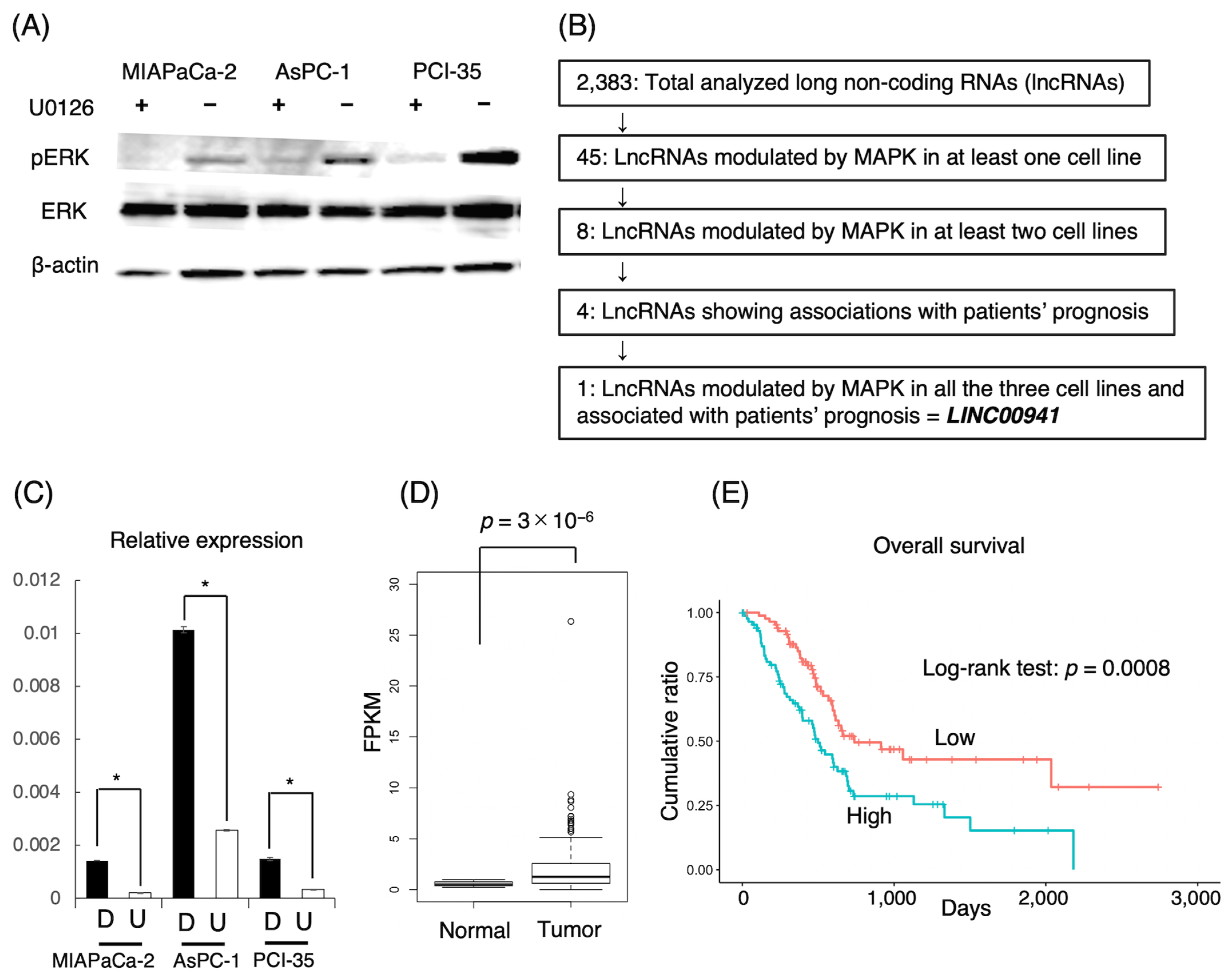
3.1. LncRNAs Associated with MAPK

To uncover lncRNAs modulated by the MAPK/ERK pathway in pancreatic cancer, we carried out comparative transcriptome sequencing assays using the pancreatic cancer cell lines MIA PaCa-2, AsPC-1, and PCI-35 with or without attenuation of MAPK by U0126, a MAP2K inhibitor (Figure 1A). The transcriptome sequencing revealed that 2383 lncRNAs were evidently expressed in the cell lines in total (Supplementary Table S2). Among them, 45 lncRNAs revealed two-fold or more changes in expression level upon the attenuation of MAPK in at least one of the three cell lines to the level of statistical significance (Supplementary Table S3). Among these, eight lncRNAs, namely, CCDC18-AS1, LINC00941, LINC-PINT, NEAT1, PRR7-AS1, SNAI3-AS1, SNHG4, and SPRY4-IT1, revealed two-fold or more changes in two or three cell lines. Then, LINC00941 was found to be significantly down-regulated in all the three cell lines (Figure 1B). The downregulation of LINC00941 by MAPK attenuation was validated by qRT-PCR (Figure 1C). The expression of LINC00941 was higher in AsPC-1 than in MIA PaCa-2 or PCI-35 despite the modest level of phosphorylated ERK, which suggests that the expression of LINC00941 might depend only on both MAPK activity and on other molecules.

3.2. Prognostic Impacts of lncRNAs Associated with MAPK

We examined the expression of lncRNAs uncovered to be associated with MAPK activity in pancreatic cancer tissues using the TCGA portal open-access database. We focused on the eight lncRNAs showing aberrant expression in two or three cell lines, in which LINC00941 was found to be expressed significantly higher in tumor tissues than in normal tissues (Table 1, Figure 1D). In an analysis of the association with survival, the expression levels of LINC00941, LINC-PINT, CCDC18-AS1, and SNAI3-AS1 were significantly correlated with overall survival of patients. LINC00941 indicated poorer prognosis at higher expression, while the others indicated poorer prognosis at lower expression (Table 1, Figure 1E).

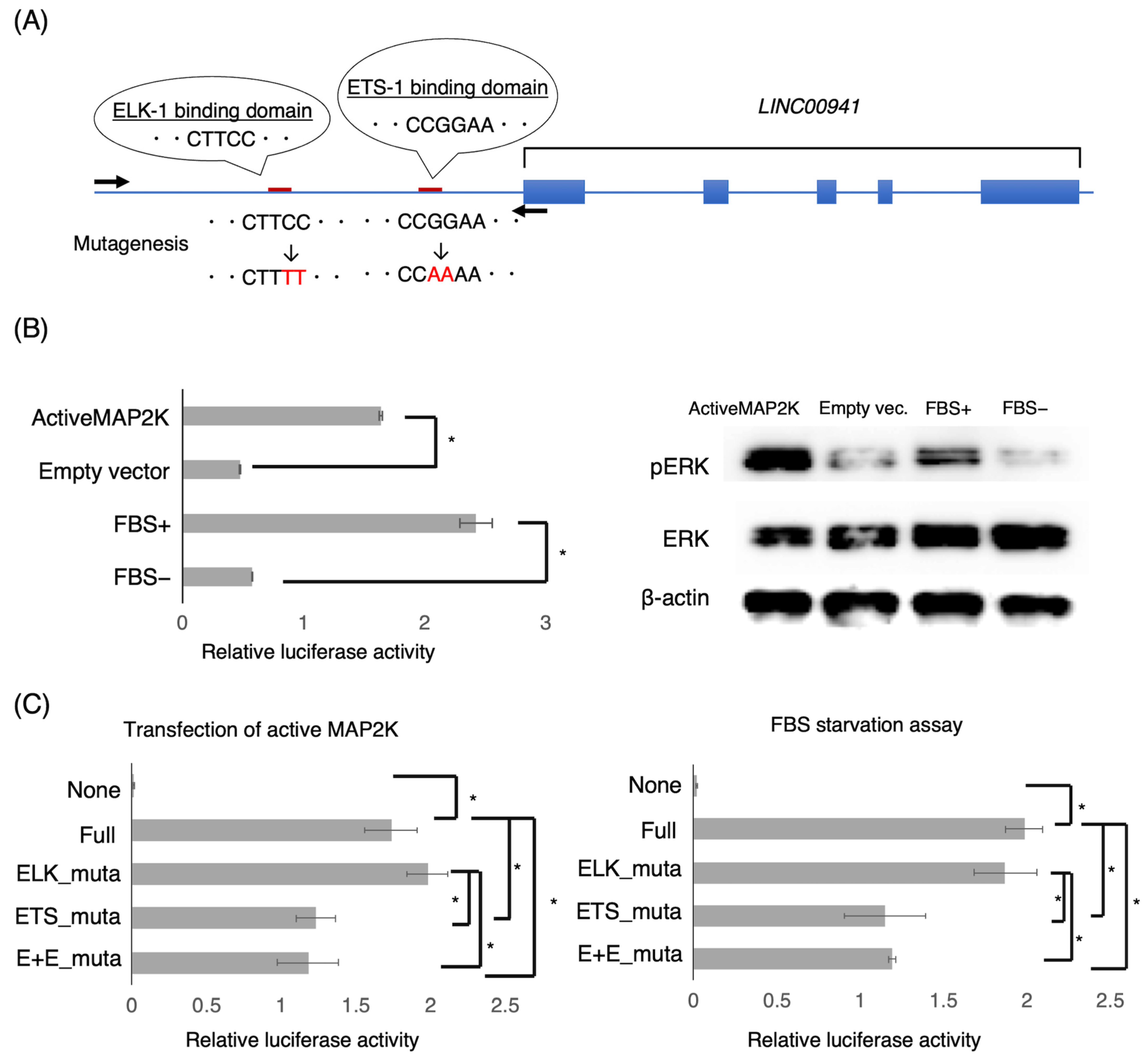
3.3. LINC00941 Promoter Activity Associated with MAPK

The transcriptome sequencing analysis suggested that MAPK may positively regulate LINC00941. To determine whether LINC00941 is directly promoted by MAPK, we performed a promoter assay. We hypothesized that an upstream CpG-rich region of LINC00941 would play a promoter role; hence, we cloned the upstream region spanning 3720 bp of LINC00941 into a reporter vector (Figure 2A). The reporter vector was transfected into the pancreatic cancer cell lines. Then, the activity of MAPK was modulated by the transfection of the constitutively active MAP2K vector or FBS starvation. We found that the upstream region had promoter activity tightly concordant with MAPK activity (Figure 2B).
Next, we searched for possible binding sites of transcription factors associated with MAPK in the active promoter region. With help from a web-based searching program for transcription factor-binding sites, Match (http://www.gene-regulation.com/cgi-bin/pub/programs/match/bin/match.cgi accessed on 18 March 2020), we found consensus binding sequences of the ELK-1 transcription factor CTTCC and ETS-1 transcription factor CGGAA in the region. We then constructed reporter vectors containing mutated sequences of the consensus binding site, i.e., CTTCC and CGGAA into CTTTT and CAAAA, respectively, and analyzed their promoter activities (Figure 2A). While the ETS-1 mutant vector showed significantly less activity, it did show a certain promoter activity compared to the wild-type sequence vector, while the ELK-1 mutant vector showed no significant alteration, indicating that ETS-1 is one of transcription factors significantly associated with the promoter activity of LINC00941 (Figure 2C).

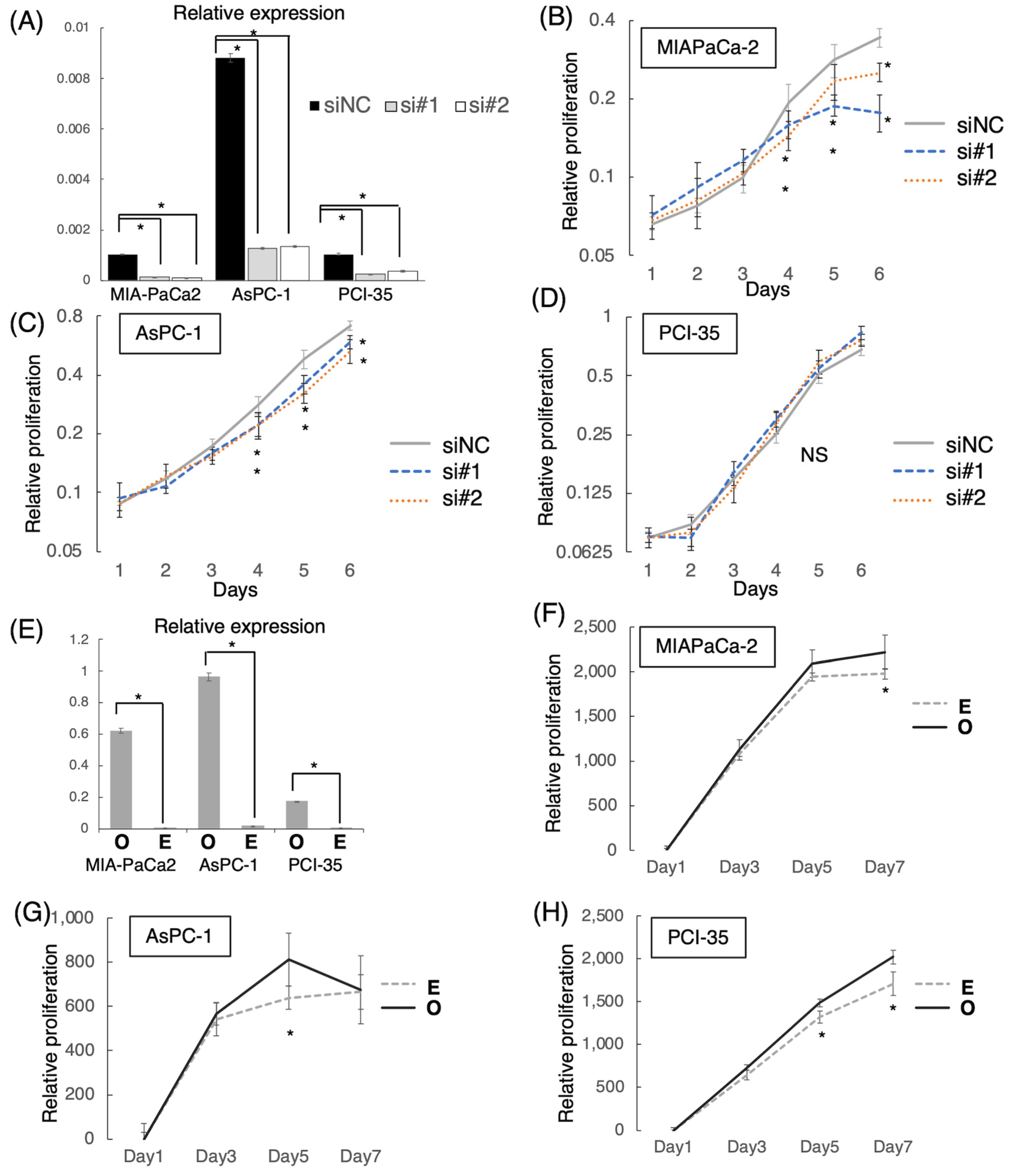
3.4. Effect of LINC00941 on Cell Proliferation

To determine the functional significance of LINC00941 in pancreatic cancer, we examined whether cellular proliferation is altered by altering the expression of LINC00941 in cultured pancreatic cancer cells. Transfection of siRNAs targeting LINC00941 induced significant suppression of endogenous expression of LINC00941 in all examined cell lines (Figure 3A). In vitro proliferation assays showed that proliferation of MIA PaCa-2 and AsPC-1 were inhibited by the suppression of LINC00941, while that of PCI-35 was not changed (Figure 3B–D). On the other hand, transfection of the pcDNA vector harboring the full-length LINC00941 cDNA induced overexpression of LINC00941 and upregulation of cell proliferation in all of the examined cell lines (Figure 3E–H). The inconsistent results in PCI-35 might be due to feedback effects involving unknown molecules.

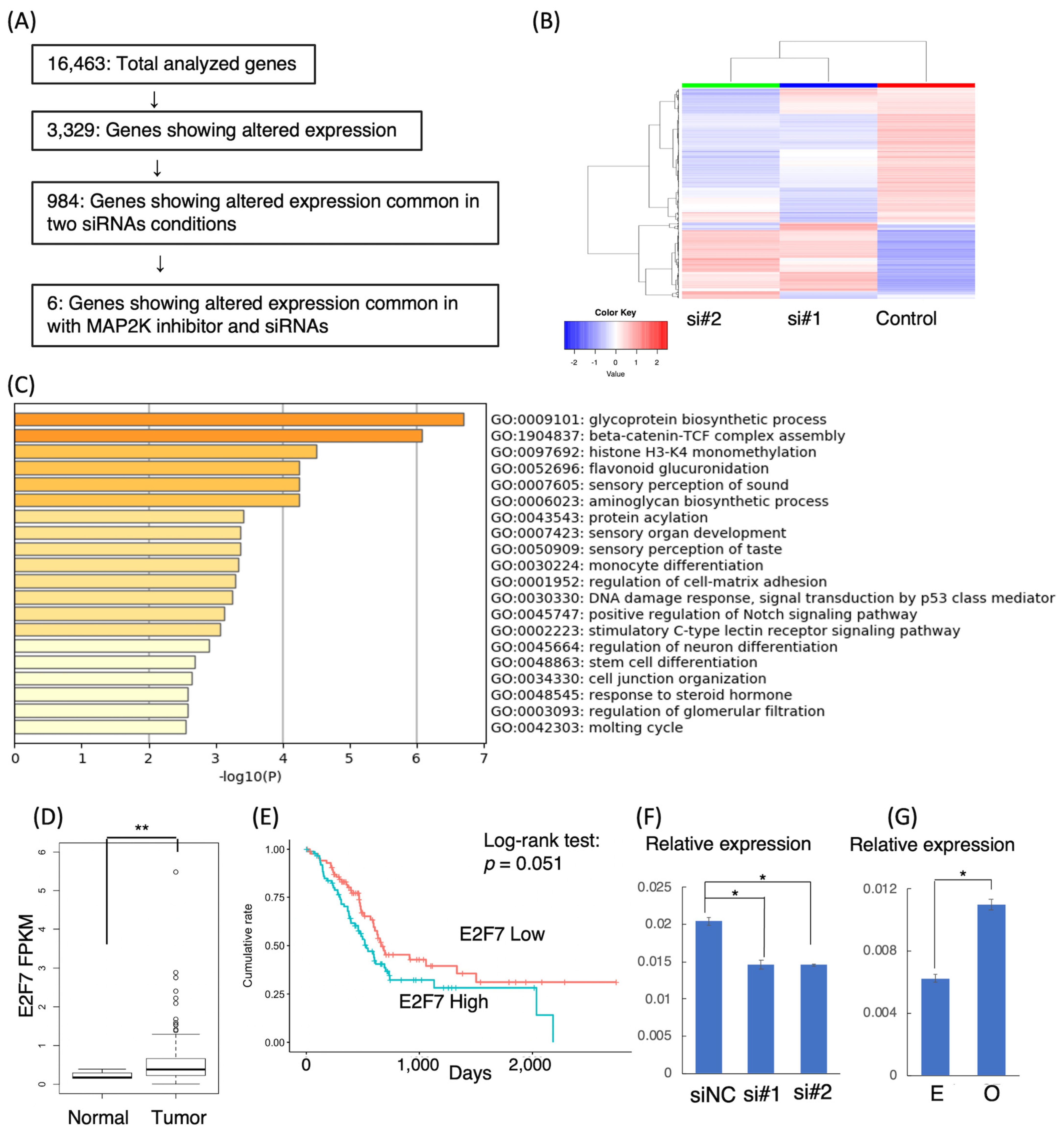
3.5. Target Genes of LINC00941

To investigate downstream targets of LINC00941, we performed a comparative transcriptome sequencing analysis of AsPC-1 with or without LINC00941 knockdown, because AsPC-1 showed the most robust endogenous expression of LINC00941 with the largest difference in LINC00941 expression by siRNA-mediated knockdown. We analyzed genes differentially expressed between a negative control (non-specific siRNA) and the knockdown of LINC00941 with si#1 or si#2. The scheme of processing data obtained by total transcriptome sequencing is drawn in Figure 4A. In 16,463 genes expressed in AsPC-1, we found that 3229 genes showed two-fold or more changes with modulation of LINC00941. A hierarchical clustering heatmap suggests the similarity between two siRNA conditions (Figure 4B). Among them, 416 and 578 genes were significantly up-regulated and down-regulated in common between the two siRNA conditions, respectively, compared with the negative control (Supplementary Tables S4 and S5). Metascape enrichment analysis using GO BP terms showed that the genes modulated by LINC00941 were associated with 44 biological processes, including glycoprotein biosynthetic process, beta-catenin-TCF complex assembly, and histone H3-K4 monomethylation. The top twenty GO BP terms are shown in Figure 4C and Supplementary Table S6.
We compared the data from the transcriptome analyses of MAPK-attenuated and LINC00941-attenuated in AsPC-1. We found that six genes showed concordant alterations in expression in the two conditions: Downregulated genes were E2F7 and SPRY4-IT1, while upregulated genes were CD82, F3, LOC100507291, and TNNC1 (Table 2). TCGA database analysis revealed that E2F7, CD82, and TNNC1 showed significantly higher expression in tumor tissues than in normal tissues (Table 2, Figure 4D). In addition, higher expression of E2F7, F3, and CD82 were associated with poor prognosis (Table 2, Figure 4E). In addition, E2F7, F3, and CD82 showed higher expression levels than SPRY4-IT1, LOC100507291, and TNNC1 (Supplementary Tables S4 and S5). We focused on E2F7, F3, and CD82 and validated the transcriptome sequencing result by qRT-PCR. E2F7 showed tightly allied expression with LINC00941 expression, indicating that LINC00941 directly regulates E2F7 (Figure 4F,G). On the other hand, CD82 and F3 were upregulated in both knockdown and forced expression of LINC00941, suggesting that LINC00941 may not directly regulate them (Supplementary Figure S2). The expression levels of LINC00941 and E2F7 were positively correlated in the TCGA data (Spearman’s rank correlation rho = 0.57, p < 2.2 × 10⁻¹⁶; Supplementary Figure S3). When the patients were divided into four groups according to levels of expression of LINC00941 and E2F7, namely, low-LINC00941 and low-E2F7 (low/low), low-LINC00941 and high-E2F7 (low/high), high-LINC00941 and low-E2F7 (high/low), and high-LINC00941 and high-E2F7 (high/high), the overall survival was more favorable in the low/low group than the rest of groups, which suggests that low expression of both LINC00941 and E2F7 can be a biomarker predicting the favorable prognosis (LINC00941 level/E2F7 level, Log-rank test: low/low vs. low/high, p = 0.04; low/low vs. high/low, p = 3 × 10⁻⁴; low/low vs. high/high, p = 0.002, Supplementary Figure S3).

4. Discussions

This study identified lncRNAs associated with MAPK activity in pancreatic cancer cells. Transcriptome sequencing showed that 2383 lncRNAs were evidently expressed; among these, 45 lncRNAs were found to be significantly associated with MAPK activity. We determined that LINC00941 was commonly promoted by MAPK in the examined pancreatic cancer cell lines. In TCGA data analysis, the high expression of LINC00941 was associated with poor prognosis of patients with pancreatic cancer. The in vitro proliferation analysis showed that LINC00941 promoted the proliferation of pancreatic cancer cells. Moreover, we identified that the 5′-upstream region spanning 3700-bp adjacent to exon 1 of LINC00941 had promoter activity, which was indeed associated with MAPK activity. ETS-1, the downstream target gene of MAPK, was suggested to be one of the transcription factors involved in the expression of LINC00941. Furthermore, we identified potential target genes of LINC00941 associated with various biological processes. These results indicate that MAPK regulates lncRNAs, and that LINC00941 is one such regulated lncRNA associated with the poor prognosis of pancreatic cancer.
LncRNAs are a group of non-protein coding RNA molecules greater than 200 nucleotides [6], and have been proven to play important roles in cancer pathogenesis. LncRNAs function as guides of transcription factors, scaffolds of multiple molecules, and decoys of miRNA intervening interactions with target genes [6,7]. In cancers, lncRNAs are supposed to act as oncogenes and/or tumor suppressor genes, which may contribute to the development and progression of cancers [6,7]. There is great interest in identifying lncRNAs applicable to cancer biomarkers and/or therapeutic targets [15]. However, how lncRNAs are aberrantly expressed in cancer cells is largely unknown. Cancer-associated aberrant signaling pathways play a central role in the development and progression of cancer [16]. These aberrant signals modulate the expression of genes and alter the fate of cells. LncRNAs in cancers might be regulated by aberrant signaling pathways as well; therefore, we explored lncRNAs associated with MAPK in pancreatic cancer. Pancreatic cancer is characterized by constitutive activation of the MAPK pathway. Mutations of KRAS or BRAF and epigenetic abrogation of DUSP6 contribute synergistically to the constitutive activation of MAPK [3]. Active MAPK induces the expression of various genes that are thought to play roles in malignant phenotypes of pancreatic cancer. The downstream genes of MAPK are involved in the cell cycle and mitosis, DNA replication, receptor signaling, and transport. The downstream target genes of MAPK are not only protein-coding genes, they are noncoding RNA genes. We previously identified miRNAs preferentially associated with MAPK, namely, miR-7-3, miR-34a, miR-181d, and miR-193b, in which miR-7-3 is the oncogenic miRNAs, whereas miR-34a, miR-181d, and miR-193b are tumor-suppressive miRNAs. These miRNAs are useful as serum biomarkers to discriminate pancreatic cancer from benign diseases [8,9].
We uncovered 45 lncRNAs significantly associated with MAPK activity. Among them, LINC00941 was consistently promoted by MAPK in the examined pancreatic cancer cell lines. LINC00941 was associated with cell proliferation. Moreover, TCGA data analysis indicated that LINC00941 was found to be upregulated in pancreatic cancer tissues and associated with poor survival of patients. Recent studies have demonstrated that LINC00941 is involved in tumorigenesis and malignant phenotypes of several kinds of cancers, including pancreatic cancer. In pancreatic cancer, it has been reported that that LINC00941 promotes cancer cell growth by enhancing aerobic glycolysis through modulation of the Hippo pathway [17]. Another study has demonstrated that LINC00941 is associated with epithelial-mesenchymal transition (EMT), acting as a competitive endogenous RNA for miR-335-5p [18]. In other cancers, LINC00941 is often highly expressed in tumor tissues compared to normal tissues. This higher expression is associated with poorer prognosis in hepatocellular carcinoma, lung adenocarcinoma, papillary thyroid carcinoma, gastric cancer, colorectal cancer, and oral squamous cell carcinoma [19,20,21,22,23,24,25]. Several studies in other cancers have suggested that LINC00941 promotes the epithelial-to-mesenchymal transition (EMT) [18,19,23,24]. Yan et al. demonstrated that lncRNA-MUF/LINC00941 activated EMT in hepatocellular carcinoma cells by interaction with the Annexin A2 (ANXA2) and miR-34a [19]. Wu et al. demonstrated that LINC00941 activated EMT in colorectal cancer cells via activating the TGF-β/SMAD2/3 axis by directly binding the SMAD4 protein [23]. Furthermore, Ai et al. demonstrated that LINC00941 promoted the growth of oral squamous cell carcinoma cells. They showed that EP300 activated LINC00941 transcription and LINC00941 promoted Wnt/β-catenin signaling with CAPRIN2 activation [24]. Therefore, LINC00941 is indicated as one of the major onco-lncRNAs in various cancers, including pancreatic cancer. However, how the expression of LINC00941 is modulated in cancer tissues has been unknown. In this study, we prove that the expression of LINC00941 is tightly associated with MAPK activity in pancreatic cancer cells.
We identified the promoter region of LINC00941, which was actually associated with MAPK activity via ETS-1, not ELK-1, consensus binding site. ETS-1 is a member of the ETS transcription factor family, which is known to be a critical player in cancer pathogenesis as a mediator of MAPK signaling activity [26,27,28,29]. The relationship between the ETS family and lncRNAs has been proven by several studies, and it has been reported that ELK-1 induces the upregulation of lncRNAs and is associated with the growth of cancer cells [30,31]. On the other hand, lncRNAs are reported to be involved in regulating the ETS family [32,33]. Therefore, ETS-family molecules and lncRNAs seem to make up a feedback network, and our results consistently indicate that LINC00941 can be modulated by ETS-1.
E2F7 is likely to be one of the downstream targets of LINC00941. E2F7 encodes transcription factor E2F7, which has been reported to be essential in regulating cell cycle progression [34]. E2F7 has been reported to be highly expressed in pancreatic cancer tissues, and higher expression of E2F7 was associated with the poorer prognosis [35,36]. Lu et al. suggested that lncRNA CASC19 contributes to the progression of pancreatic cancer by modulating miR148b/E2F7 axis [37]. Xu and Qi suggested that miR-10b inhibits invasion by migration of pancreatic cancer cells by regulating E2F7 expression, and that the high expression of E2F7 is associated with poor prognosis [38]. These previous studies and our study show that various ncRNAs are associated with regulating E2F7, which may play an important role in the malignant phenotype of pancreatic cancer.
The 45 lncRNAs associated with MAPK activity are likely to be implicated in the pathogenesis of pancreatic cancer. Indeed, BLACAT1, CASC8, HCP5, LINC-PINT, SNHG1, SNHG9, SNHG12, and SPRY4-IT1 were detected as MAPK-promoted lncRNAs, and NEAT1, PSMB8-AS1, THAP9-AS1, and TP53TG1 were detected as MAPK-inhibited lncRNAs reported to be involved in pancreatic cancer [39,40,41,42,43,44,45,46,47,48,49,50,51]. For example, previous studies reported that LINC-PINT levels in plasma are lower in pancreatic cancer patients than healthy controls [42,51], and that LINC-PINT suppresses the growth of pancreatic cancer cells through TGF-β pathway activation [51], which suggests that LINC-PINT can be a diagnostic and a prognostic biomarker of pancreatic cancer. NEAT1 was reported to be highly expressed in pancreatic cancer tissues, and the knockdown of NEAT1 suppressed the growth of pancreatic cancer cells [32]. Interestingly, the activation of MAPK is considered to play a role in promoting malignant phenotypes of pancreatic cancer [3]. However, the MAPK-promoted lncRNAs include both those reported as oncogenic and tumor-suppressive and vice versa for the MAPK-inhibited lncRNAs. Several of these lncRNAs might have an unappreciated function or show a conflicting result of a feedback loop of MAPK signaling pathway in pancreatic cancer. Nevertheless, these MAPK-associated lncRNAs could be therapeutic targets as well as diagnostic biomarkers, as already shown in LINC-PINT and NEAT1 [32,51]. Further research is needed in order to understand the functions of lncRNAs associated with MAPK.

Supplementary Materials

The following supporting information can be downloaded at: https://www.mdpi.com/article/10.3390/cancers15010303/s1, Supplementary Figure S1: Methodological flowchart of this study; Supplementary Figure S2: qRT-PCR of LINC00941-associated genes in AsPC-1; Supplementary Figure S3: Correlation between LINC00941 and E2F7 based on TCGA database analysis; Table S1: Primers used in this study; Table S2: Transcriptome data of long non-coding RNAs in pancreatic cancer cell lines treated with U0126 or DMSO (abbreviations are A, AsPC-1; D, DMSO; M, MIA PaCa-2; P, PCI-35; and U, U0126); Table S3: Comparative transcriptome data of long non-coding RNAs with 2-fold or more changes in any of cell line by attenuation of MAPK (abbreviations are A, AsPC-1; D, DMSO; M, MIA PaCa-2; P, PCI-35; and U, U0126); Table S4: Upregulated genes in 2-fold or more in transcriptome data of AsPC-1 with siRNA for LINC00941 (si-1 or si-2) or negative control (siNC); Table S5: Downregulated genes in 2-fold or more in transcriptome data of AsPC-1 with siRNA for LINC00941 (si-1 or si-2) or negative control (siNC); Table S6: Metascape analysis results.

Author Contributions

Conceptualization, T.F.; Data curation, T.I. and T.F.; Formal analysis, T.I. and T.F.; Funding acquisition, T.F.; Investigation, T.I., S.F., Y.S., K.H., T.H., T.O. and T.F.; Methodology, T.I. and T.F.; Resources, T.F.; Supervision, T.O., Y.K. and T.F.; Validation, T.I. and T.F.; Writing—original draft, T.I.; Writing—review and editing, T.F. All authors have read and agreed to the published version of the manuscript.

Funding

This research was funded by JSPS KAKENHI, Grant Number JP22H02839 to T.F.

Institutional Review Board Statement

Not applicable.

Informed Consent Statement

Not applicable.

Data Availability Statement

The RNA-Seq data discussed in this study have been deposited in NCBI’s Gene Expression Omnibus and are accessible through GEO Series accession numbers GSE216708 (https://www.ncbi.nlm.nih.gov/geo/query/acc.cgi?acc=GSE216708 accessed on 18 March 2020) and GSE216710 (https://www.ncbi.nlm.nih.gov/geo/query/acc.cgi?acc=GSE216710 accessed on 18 March 2020).

Acknowledgments

We would like to thank Miki Yoshizawa (Biomedical Research Core of Tohoku University Graduate School of Medicine) for technical support in immunoblotting and cell culture.

Conflicts of Interest

The authors declare no potential conflict of interest.

References

  1. Matsuda, T.; Ajiki, W.; Marugame, T.; Ioka, A.; Tsukuma, H.; Sobue, T. Population-based survival of cancer patients diagnosed between 1993 and 1999 in Japan: A chronological and international comparative study. Jpn. J. Clin. Oncol. 2011, 41, 40–51. [Google Scholar] [CrossRef] [PubMed]
  2. Siegel, R.L.; Miller, K.D.; Jemal, A. Cancer statistics, 2020. CA Cancer J. Clin. 2020, 70, 7–30. [Google Scholar] [CrossRef] [PubMed]
  3. Furukawa, T. Impacts of activation of the mitogen-activated protein kinase pathway in pancreatic cancer. Front. Oncol. 2015, 5, 23. [Google Scholar] [CrossRef] [PubMed]
  4. Cox, A.D.; Fesik, S.W.; Kimmelman, A.C.; Luo, J.; Der, C.J. Drugging the undruggable RAS: Mission possible? Nat. Rev. Drug Discov. 2014, 13, 828–851. [Google Scholar] [CrossRef] [PubMed] [Green Version]
  5. Jänne, P.A.; van den Heuvel, M.M.; Barlesi, F.; Cobo, M.; Mazieres, J.; Crinò, L.; Orlov, S.; Blackhall, F.; Wolf, J.; Garrido, P.; et al. Selumetinib Plus Docetaxel Compared With Docetaxel Alone and Progression-Free Survival in Patients With KRAS-Mutant Advanced Non-Small Cell Lung Cancer: The SELECT-1 Randomized Clinical Trial. JAMA 2017, 317, 1844–1853. [Google Scholar] [CrossRef] [Green Version]
  6. Esteller, M. Non-coding RNAs in human disease. Nat. Rev. Genet. 2011, 12, 861–874. [Google Scholar] [CrossRef]
  7. Sharma, G.G.; Okada, Y.; Von Hoff, D.; Goel, A. Non-coding RNA biomarkers in pancreatic ductal adenocarcinoma. Semin. Cancer Biol. 2021, 75, 153–168. [Google Scholar] [CrossRef]
  8. Ikeda, Y.; Tanji, E.; Makino, N.; Kawata, S.; Furukawa, T. MicroRNAs associated with mitogen-activated protein kinase in human pancreatic cancer. Mol. Cancer Res. 2012, 10, 259–269. [Google Scholar] [CrossRef] [Green Version]
  9. Akamatsu, M.; Makino, N.; Ikeda, Y.; Matsuda, A.; Ito, M.; Kakizaki, Y.; Saito, Y.; Ishizawa, T.; Kobayashi, T.; Furukawa, T.; et al. Specific MAPK-Associated MicroRNAs in Serum Differentiate Pancreatic Cancer from Autoimmune Pancreatitis. PLoS ONE 2016, 11, e0158669. [Google Scholar] [CrossRef] [Green Version]
  10. Iwai, K.; Ishikura, H.; Kaji, M.; Sugiura, H.; Ishizu, A.; Takahashi, C.; Kato, H.; Tanabe, T.; Yoshiki, T. Importance of E-selectin (ELAM-1) and sialyl Lewis(a) in the adhesion of pancreatic carcinoma cells to activated endothelium. Int. J. Cancer 1993, 54, 972–977. [Google Scholar] [CrossRef]
  11. Mansour, S.J.; Candia, J.M.; Matsuura, J.E.; Manning, M.C.; Ahn, N.G. Interdependent domains controlling the enzymatic activity of mitogen-activated protein kinase kinase 1. Biochemistry 1996, 35, 15529–15536. [Google Scholar] [CrossRef] [PubMed]
  12. Hata, T.; Furukawa, T.; Sunamura, M.; Egawa, S.; Motoi, F.; Ohmura, N.; Marumoto, T.; Saya, H.; Horii, A. RNA interference targeting aurora kinase a suppresses tumor growth and enhances the taxane chemosensitivity in human pancreatic cancer cells. Cancer Res. 2005, 65, 2899–2905. [Google Scholar] [CrossRef] [PubMed] [Green Version]
  13. Ge, S.X.; Son, E.W.; Yao, R. iDEP: An integrated web application for differential expression and pathway analysis of RNA-Seq data. BMC Bioinform. 2018, 19, 534. [Google Scholar] [CrossRef] [PubMed] [Green Version]
  14. Zhou, Y.; Zhou, B.; Pache, L.; Chang, M.; Khodabakhshi, A.H.; Tanaseichuk, O.; Benner, C.; Chanda, S.K. Metascape provides a biologist-oriented resource for the analysis of systems-level datasets. Nat. Commun. 2019, 10, 1523. [Google Scholar] [CrossRef] [PubMed]
  15. Matsui, M.; Corey, D.R. Non-coding RNAs as drug targets. Nat. Rev. Drug Discov. 2017, 16, 167–179. [Google Scholar] [CrossRef] [PubMed] [Green Version]
  16. Hanahan, D.; Weinberg, R.A. The hallmarks of cancer. Cell 2000, 100, 57–70. [Google Scholar] [CrossRef] [PubMed] [Green Version]
  17. Xu, M.; Cui, R.; Ye, L.; Wang, Y.; Wang, X.; Zhang, Q.; Wang, K.; Dong, C.; Le, W.; Chen, B. LINC00941 promotes glycolysis in pancreatic cancer by modulating the Hippo pathway. Mol. Ther. Nucleic Acids. 2021, 26, 280–294. [Google Scholar] [CrossRef]
  18. Wang, J.; He, Z.; Xu, J.; Chen, P.; Jiang, J. Long noncoding RNA LINC00941 promotes pancreatic cancer progression by competitively binding miR-335-5p to regulate ROCK1-mediated LIMK1/Cofilin-1 signaling. Cell Death Dis. 2021, 12, 36. [Google Scholar] [CrossRef]
  19. Yan, X.; Zhang, D.; Wu, W.; Wu, S.; Qian, J.; Hao, Y.; Yan, F.; Zhu, P.; Wu, J.; Huang, G.; et al. Mesenchymal Stem Cells Promote Hepatocarcinogenesis via lncRNA-MUF Interaction with ANXA2 and miR-34a. Cancer Res. 2017, 77, 6704–6716. [Google Scholar] [CrossRef] [PubMed] [Green Version]
  20. Wang, L.; Zhao, H.; Xu, Y.; Li, J.; Deng, C.; Deng, Y.; Bai, J.; Li, X.; Xiao, Y.; Zhang, Y. Systematic identification of lincRNA-based prognostic biomarkers by integrating lincRNA expression and copy number variation in lung adenocarcinoma. Int. J. Cancer 2019, 144, 1723–1734. [Google Scholar] [CrossRef]
  21. Gugnoni, M.; Manicardi, V.; Torricelli, F.; Sauta, E.; Bellazzi, R.; Manzotti, G.; Vitale, E.; de Biase, D.; Piana, S.; Ciarrocchi, A. Linc00941 Is a Novel Transforming Growth Factor β Target That Primes Papillary Thyroid Cancer Metastatic Behavior by Regulating the Expression of Cadherin 6. Thyroid 2021, 31, 247–263. [Google Scholar] [CrossRef] [PubMed]
  22. Luo, C.; Tao, Y.; Zhang, Y.; Zhu, Y.; Minyao, D.N.; Haleem, M.; Dong, C.; Zhang, L.; Zhang, X.; Zhao, J.; et al. Regulatory network analysis of high expressed long non-coding RNA LINC00941 in gastric cancer. Gene 2018, 662, 103–109. [Google Scholar] [CrossRef] [PubMed]
  23. Wu, N.; Jiang, M.; Liu, H.; Chu, Y.; Wang, D.; Cao, J.; Wang, Z.; Xie, X.; Han, Y.; Xu, B. LINC00941 promotes CRC metastasis through preventing SMAD4 protein degradation and activating the TGF-beta/SMAD2/3 signaling pathway. Cell Death Differ. 2021, 28, 219–232. [Google Scholar] [CrossRef] [PubMed]
  24. Ai, Y.; Wu, S.; Zou, C.; Wei, H. LINC00941 promotes oral squamous cell carcinoma progression via activating CAPRIN2 and canonical WNT/beta-catenin signaling pathway. J. Cell Mol. Med. 2020, 24, 10512–10524. [Google Scholar] [CrossRef] [PubMed]
  25. Hu, Y.; Guo, G.; Li, J.; Chen, J.; Tan, P. Screening key lncRNAs with diagnostic and prognostic value for head and neck squamous cell carcinoma based on machine learning and mRNA-lncRNA co-expression network analysis. Cancer Biomark 2020, 27, 195–206. [Google Scholar] [CrossRef]
  26. Furukawa, T.; Kanai, N.; Shiwaku, H.O.; Soga, N.; Uehara, A.; Horii, A. AURKA is one of the downstream targets of MAPK1/ERK2 in pancreatic cancer. Oncogene 2006, 25, 4831–4839. [Google Scholar] [CrossRef] [PubMed] [Green Version]
  27. Foulds, C.E.; Nelson, M.L.; Blaszczak, A.G.; Graves, B.J. Ras/mitogen-activated protein kinase signaling activates Ets-1 and Ets-2 by CBP/p300 recruitment. Mol. Cell Biol. 2004, 24, 10954–10964. [Google Scholar] [CrossRef] [Green Version]
  28. Kobberup, S.; Nyeng, P.; Juhl, K.; Hutton, J.; Jensen, J. ETS-family genes in pancreatic development. Dev. Dyn. 2007, 236, 3100–3110. [Google Scholar] [CrossRef]
  29. Furukawa, T.; Tanji, E.; Xu, S.; Horii, A. Feedback regulation of DUSP6 transcription responding to MAPK1 via ETS2 in human cells. Biochem. Biophys. Res. Commun. 2008, 377, 317–320. [Google Scholar] [CrossRef]
  30. Huang, B.; Chang, C.; Wang, B.L.; Li, H. ELK1-induced upregulation of lncRNA TRPM2-AS promotes tumor progression in gastric cancer by regulating miR-195/ HMGA1 axis. J. Cell Biochem. 2019, 120, 16921–16933. [Google Scholar] [CrossRef]
  31. Su, X.; Teng, J.; Jin, G.; Li, J.; Zhao, Z.; Cao, X.; Guo, Y.; Guo, M.; Li, X.; Wu, J.; et al. ELK1-induced upregulation of long non-coding RNA MIR100HG predicts poor prognosis and promotes the progression of osteosarcoma by epigenetically silencing LATS1 and LATS2. Biomed. Pharmacother. 2019, 109, 788–797. [Google Scholar] [CrossRef] [PubMed]
  32. Feng, Y.; Gao, L.; Cui, G.; Cao, Y. LncRNA NEAT1 facilitates pancreatic cancer growth and metastasis through stabilizing ELF3 mRNA. Am. J. Cancer Res. 2020, 10, 237–248. [Google Scholar] [PubMed]
  33. Luo, Y.; Liu, F.; Yan, C.; Qu, W.; Zhu, L.; Guo, Z.; Zhou, F.; Zhang, W. Long Non-Coding RNA CASC19 Sponges microRNA-532 and Promotes Oncogenicity of Clear Cell Renal Cell Carcinoma by Increasing ETS1 Expression. Cancer Manag. Res. 2020, 12, 2195–2207. [Google Scholar] [CrossRef] [PubMed] [Green Version]
  34. Di Stefano, L.; Jensen, M.R.; Helin, K. E2F7, a novel E2F featuring DP-independent repression of a subset of E2F-regulated genes. EMBO J. 2003, 22, 6289–6298. [Google Scholar] [CrossRef] [PubMed] [Green Version]
  35. Kim, J.; Jo, Y.H.; Jang, M.; Nguyen, N.N.Y.; Yun, H.R.; Ko, S.H.; Shin, Y.; Lee, J.S.; Kang, I.; Ha, J.; et al. PAC-5 Gene Expression Signature for Predicting Prognosis of Patients with Pancreatic Adenocarcinoma. Cancers 2019, 11, 1749. [Google Scholar] [CrossRef] [Green Version]
  36. Raman, P.; Maddipati, R.; Lim, K.H.; Tozeren, A. Pancreatic cancer survival analysis defines a signature that predicts outcome. PLoS ONE 2018, 13, e0201751. [Google Scholar] [CrossRef]
  37. Lu, T.; Wei, G.H.; Wang, J.; Shen, J. LncRNA CASC19 contributed to the progression of pancreatic cancer through modulating miR-148b/E2F7 axis. Eur. Rev. Med. Pharmacol. Sci. 2020, 24, 10462–10471. [Google Scholar] [CrossRef]
  38. Xu, C.; Qi, X. MiR-10b inhibits migration and invasion of pancreatic ductal adenocarcinoma via regulating E2F7. J. Clin. Lab. Anal. 2020, 34, e23442. [Google Scholar] [CrossRef]
  39. Zhou, X.; Gao, W.; Hua, H.; Ji, Z. LncRNA-BLACAT1 Facilitates Proliferation, Migration and Aerobic Glycolysis of Pancreatic Cancer Cells by Repressing CDKN1C via EZH2-Induced H3K27me3. Front. Oncol. 2020, 10, 539805. [Google Scholar] [CrossRef]
  40. Wang, Y.; Yang, Y.; Wang, Y.; Li, X.; Xiao, Y.; Wang, W. High Cancer Susceptibility Candidate 8 Expression Is Associated With Poor Prognosis of Pancreatic Adenocarcinoma: Validated Analysis Based on Four Cancer Databases. Front. Cell Dev. Biol. 2020, 8, 392. [Google Scholar] [CrossRef]
  41. Liu, Y.; Wang, J.; Dong, L.; Xia, L.; Zhu, H.; Li, Z.; Yu, X. Long Noncoding RNA HCP5 Regulates Pancreatic Cancer Gemcitabine (GEM) Resistance By Sponging Hsa-miR-214-3p To Target HDGF. Onco. Targets Ther. 2019, 12, 8207–8216. [Google Scholar] [CrossRef] [PubMed] [Green Version]
  42. Li, L.; Zhang, G.Q.; Chen, H.; Zhao, Z.J.; Chen, H.Z.; Liu, H.; Wang, G.; Jia, Y.H.; Pan, S.H.; Kong, R.; et al. Plasma and tumor levels of Linc-pint are diagnostic and prognostic biomarkers for pancreatic cancer. Oncotarget 2016, 7, 71773–71781. [Google Scholar] [CrossRef] [PubMed] [Green Version]
  43. Cui, L.; Dong, Y.; Wang, X.; Zhao, X.; Kong, C.; Liu, Y.; Jiang, X.; Zhang, X. Downregulation of long noncoding RNA SNHG1 inhibits cell proliferation, metastasis, and invasion by suppressing the Notch-1 signaling pathway in pancreatic cancer. J. Cell Biochem. 2019, 120, 6106–6112. [Google Scholar] [CrossRef] [PubMed]
  44. Zhang, B.; Li, C.; Sun, Z. Long non-coding RNA LINC00346, LINC00578, LINC00673, LINC00671, LINC00261, and SNHG9 are novel prognostic markers for pancreatic cancer. Am. J. Transl. Res. 2018, 10, 2648–2658. [Google Scholar]
  45. Cao, W.; Zhou, G. LncRNA SNHG12 contributes proliferation, invasion and epithelial-mesenchymal transition of pancreatic cancer cells by absorbing miRNA-320b. Biosci. Rep. 2020, 40. [Google Scholar] [CrossRef]
  46. Guo, W.; Zhong, K.; Wei, H.; Nie, C.; Yuan, Z. Long non-coding RNA SPRY4-IT1 promotes cell proliferation and invasion by regulation of Cdc20 in pancreatic cancer cells. PLoS ONE 2018, 13, e0193483. [Google Scholar] [CrossRef] [PubMed]
  47. Cao, J.; Zhang, Y.; Yang, J.; He, S.; Li, M.; Yan, S.; Chen, Y.; Qu, C.; Xu, L. NEAT1 regulates pancreatic cancer cell growth, invasion and migration though mircroRNA-335-5p/c-met axis. Am. J. Cancer Res. 2016, 6, 2361–2374. [Google Scholar] [PubMed]
  48. Zhang, H.; Zhu, C.; He, Z.; Chen, S.; Li, L.; Sun, C. LncRNA PSMB8-AS1 contributes to pancreatic cancer progression via modulating miR-382-3p/STAT1/PD-L1 axis. J. Exp. Clin. Cancer Res. 2020, 39, 179. [Google Scholar] [CrossRef]
  49. Li, N.; Yang, G.; Luo, L.; Ling, L.; Wang, X.; Shi, L.; Lan, J.; Jia, X.; Zhang, Q.; Long, Z.; et al. lncRNA THAP9-AS1 Promotes Pancreatic Ductal Adenocarcinoma Growth and Leads to a Poor Clinical Outcome via Sponging miR-484 and Interacting with YAP. Clin. Cancer Res. 2020, 26, 1736–1748. [Google Scholar] [CrossRef] [Green Version]
  50. Zhang, Y.; Yang, H.; Du, Y.; Liu, P.; Zhang, J.; Li, Y.; Shen, H.; Xing, L.; Xue, X.; Chen, J.; et al. Long noncoding RNA TP53TG1 promotes pancreatic ductal adenocarcinoma development by acting as a molecular sponge of microRNA-96. Cancer Sci. 2019, 110, 2760–2772. [Google Scholar] [CrossRef] [Green Version]
  51. Lu, H.; Yang, D.; Zhang, L.; Lu, S.; Ye, J.; Li, M.; Hu, W. Linc-pint inhibits early stage pancreatic ductal adenocarcinoma growth through TGF-β pathway activation. Oncol. Lett. 2019, 17, 4633–4639. [Google Scholar] [CrossRef] [PubMed]
Figure 1. MAPK-associated lncRNAs identified by a comparative transcriptome sequencing assay of pancreatic cancer cells with or without attenuation of MAPK activity. (A) The expression of phosphorylated ERK was inhibited by U0126, a MAP2K inhibitor, in pancreatic cancer cell lines. (B) Stringent processing of transcriptome sequencing data resulted in identification of MAPK-associated lncRNAs. (C) qRT-PCR assay validated the transcriptome data revealing downregulation of LINC00941 by the attenuation of MAPK in pancreatic cancer cell lines (* p < 0.05). Abbreviations: D, DMSO; U, U0126. (D) TCGA data analysis showed that LINC00941 was expressed significantly more highly in pancreatic cancer tissues than in normal tissues (p = 3 × 10⁻⁶). (E) TCGA data analysis showed that highly expressed LINC00941 in pancreatic cancer tissues was associated with patients’ poor overall survival (p = 0.0008).
Cancers 15 00303 g001
Figure 2. LINC00941 promoter activity is associated with MAPK activity. (A) A candidate promoter region of LINC00941 harboring consensus sequences of biding sites for ELK-1 and ETS-1. Arrows indicate relative positions of primers spanning the 3720-bp genomic region immediately upstream of exon 1 of LINC00941 used for construction of reporter vectors for the promoter assay. Mutagenic disruptions of the consensus binding sites used in experiments shown in (C) are indicated. (B) Reporter assays with modulation of MAPK activity. Left panel: the candidate promoter region reveals the promoter activity significantly associated with MAPK activity modulated by the active MEK/MAP2K transfection or FBS starvation in AsPC-1 (* p < 0.05). Right panel: Immunoblots showing upregulation and downregulation of MAPK activity in AsPC-1 by the active MEK transfection and FBS starvation, respectively. (C) Mutagenesis of the consensus binding sites in the promoter of LINC00941 results in a significant reduction of promoter activity by disruption of the binding site of ETS-1, and not that of ELK-1, in both active MEK transfection and FBS starvation in AsPC-1 (* p < 0.05). E+E means disruption of both binding sites of ETS-1 and ELK-1.
Cancers 15 00303 g002
Figure 3. MTT proliferation assays with the knockdown or overexpression of LINC00941. (A) qRT-PCR validation of downregulation of LINC00941 expression by siRNAs. NC, negative control. (BD) The knockdown of LINC00941 suppressed cell proliferation of MIAPaCa-2 and AsPC-1 (* p < 0.05), and did not suppress PCI-35. (E) qRT-PCR validation of the overexpression of LINC00941 by cDNA vector transfection. Abbreviations: E, empty vector; O, overexpression. (FH) The overexpression of LINC00941 promoted the cell proliferation of MIAPaCa-2, AsPC-1, and PCI-35. Please note that the values of negative controls of experiments in (A,E) were comparable in each cell line: 0.0010 and 0.0015, 0.0088 and 0.017, and 0.0010 and 0.0019 for siNC and E in MIA PaCa-2, AsPC-1, and PCI-35, respectively.
Cancers 15 00303 g003
Figure 4. Identification of genes associated with LINC00941. (A) Stringent processing of comparative transcriptome sequencing data of AsPC-1 with or without attenuation of the expression of LINC00941. (B) Heatmap with hierarchical clustering showing comparison of transcriptome data between cells with siRNAs for LINC00941 and negative control. (C) Top 20 Gene Ontology Biological Process terms of genes associated with LINC00941. (D) TCGA data analysis showed that pancreatic cancer tissues revealed higher expression of E2F7 than normal tissues (** p < 0.001). (E) TCGA data analysis showed the marginal survival difference between patients with pancreatic cancer with higher and lower expression of E2F7 (p = 0.051 by log-rank test). (F) qRT-PCR validated the transcriptome data showing downregulation of E2F7 by knockdown of LINC00941 in AsPC-1 (* p < 0.05). (G) qRT-PCR showed that overexpression of LINC00941 in AsPC-1 resulted in upregulation of E2F7 (* p < 0.05). Abbreviations: E, empty vector; O, overexpression.
Cancers 15 00303 g004
Table 1. MAPK-regulated lncRNAs.
Gene SymbolTranscript IDEffect of MAPKp-Value for the Difference of Expression in Tumor (T) vs. Normal (N) in TCGAp-Value (Log-Rank) for Overall Survival in TCGAExpression Level Associated with Poor Prognosis
CCDC18-AS1NR_034089Downregulation0.61 (N > T)0.009Low
LINC00941NR_040245Upregulation3 × 10−6 (T > N)0.0008High
LINC-PINTNR_015431Upregulation0.55 (N > T)0.01Low
NEAT1NR_028272Downregulation0.86 (N > T)0.8-
PRR7-AS1NR_038915Upregulation0.65 (N > T)0.07-
SNAI3-AS1NR_024399Downregulation0.29 (N > T)0.00001Low
SNHG4NR_003141Upregulation0.99 (T > N)0.7-
SPRY4-IT1NR_131221Upregulation0.22 (T > N)0.2-
Table 2. Concordantly altered genes between MAPK attenuation and knockdown of LINC00941.
Gene SymbolTranscript IDEffect of MAPK and LINC00941p-Value for the Difference of Expression in Tumor (T) vs. Normal (N) in TCGAp-Value (Log-Rank) for Overall Survivalin TCGAExpression Level Associated with Poor Prognosis
CD82NM_001024844Downregulation0.01 (T > N)0.04High
E2F7NM_203394Upregulation0.001 (T > N)0.05High
F3NM_001178096Downregulation0.36 (T > N)0.0001High
LOC100507291NR_121608Downregulation0.10 (T > N)0.3-
SPRY4-IT1NR_131221Upregulation0.22 (T > N)0.2-
TNNC1NM_003280Downregulation7 × 10−6 (T > N)0.5-
Disclaimer/Publisher’s Note: The statements, opinions and data contained in all publications are solely those of the individual author(s) and contributor(s) and not of MDPI and/or the editor(s). MDPI and/or the editor(s) disclaim responsibility for any injury to people or property resulting from any ideas, methods, instructions or products referred to in the content.

