Article

TP53 Co-Mutation Status Association with Clinical Outcomes in Patients with EGFR-Mutant Non-Small Cell Lung Cancer

1
MD Anderson Cancer Center, University of Texas, Houston, TX 77030, USA
2
Eli Lilly and Company, Lilly Corporate Center, Indianapolis, IN 46225, USA
3
Eli Lilly and Company, 3528 BJ Utrecht, The Netherlands
*
Authors to whom correspondence should be addressed.
Current address: Novartis Services Incorporated, 9965 Buffalo Speedway, Houston, TX 77054, USA.
Cancers 2022, 14(24), 6127; https://doi.org/10.3390/cancers14246127
Submission received: 3 October 2022 / Revised: 30 November 2022 / Accepted: 2 December 2022 / Published: 12 December 2022
(This article belongs to the Special Issue Actionable Mutations in Lung Cancer)

Abstract

:

Simple Summary

There is conflicting evidence on the impact of TP53 co-mutations on survival in patients with EGFR-mutated advanced non-small cell lung cancer (aNSCLC). This observational study used data from a de-identified database to assess the role of TP53 co-mutation on survival in patients with EGFR-mutated aNSCLC (n = 356). We observed a significant decrease in survival outcomes (namely real-world progression-free survival [rwPFS] and overall survival [OS]) duration in the group of aNSCLC patients with presence of TP53 co-mutation compared to those with wild-type TP53 tumors. TP53 co-mutations were also associated with worse outcomes by subgroups, including type of EGFR-mutation and first-line treatment. Together, these findings indicated negative impact of TP53 co-mutation in outcomes for patients with EGFR-mutated aNSCLC.

Abstract

TP53 co-mutations have shown association with poor prognosis in various solid tumors. For EGFR-mutated advanced non-small cell lung cancer (aNSCLC), conflicting results exist regarding its impact on survival. Clinical outcomes and genomic data were obtained retrospectively from the real-world (rw) de-identified clinicogenomic database. Patients who initiated therapy for EGFR-mutated aNSCLC between January 2014 and December 2020 were identified. Clinical outcomes were evaluated by TP53-mutational status. In 356 eligible EGFR-mutated aNSCLC patients (median age 68 years), 210 (59.0%) had TP53 co-mutation and 146 (41.0%) had TP53 wild-type tumor. Unadjusted analysis showed significantly shorter survival in patients with TP53 co-mutation versus TP53 wild-type (rw progression-free survival [rwPFS]: HR = 1.4, 95% CI 1.1–1.9, p = 0.0196; overall survival [OS]: HR = 1.6, 95% CI 1.1–2.2, p = 0.0088). Multivariable analysis confirmed independent association between TP53 co-mutation and worse rwPFS (HR = 1.4, 95% CI 1.0–0.9, p = 0.0280) and OS (HR = 1.4, 95% CI 1.0–2.0, p = 0.0345). Directionally consistent findings were observed for response rates, and subgroups by EGFR-activating mutation and first-line (1 L) therapy, with more pronounced negative effect in 1 L EGFR-TKI subgroup. TP53 co-mutations negatively affected survival in patients with EGFR-mutated aNSCLC receiving standard 1 L therapy in real-world practice.

1. Introduction

Epidermal growth factor receptor (EGFR) mutations are among the most common oncogenic drivers in advanced or metastatic non-small cell lung cancer (aNSCLC) [1,2], which continues to be the most common cause of cancer deaths in the world [3]. Most (~85%) EGFR-activating mutations involve deletions in exon 19 (ex19del) or a substitution mutation in exon 21, specifically Leu858Arg (ex21L858R) [4] and they are classical EGFR mutations [5]. The current NCCN guideline recommendation and standard of care for aNSCLC with an activating EGFR mutation is targeted therapy with EGFR-Tyrosine Kinase Inhibitors (EGFR-TKIs) alone or in combination with antiangiogenics, ramucirumab or bevacizumab [4,6,7,8,9]. EGFR-TKIs are initially effective with progression-free survival ranging between 9 and 19 months [10,11,12,13,14,15]. However, resistance inevitably develops after initial benefit to EGFR-TKIs, leading to treatment failure. A few mechanisms of primary or acquired resistance have been identified, which include on-target EGFR-dependent mechanisms of resistance (e.g., exon 20 T790M and C797S mutations) and off-target EGFR-independent mechanisms (e.g., histologic transformation to small cell lung cancer, or acquisition of other driver alterations such as MET, HER2, p53, or PI3KCA amplification) [16,17,18,19,20,21,22,23,24,25]. However, more investigation is needed to understand and overcome resistance to EGFR-TKIs.
Various co-occurring genetic alterations in EGFR-mutated aNSCLC have been linked to poorer outcomes compared with EGFR-mutated aNSCLC without co-occurring alterations [26]. TP53, also known as the “guardian of the genome”, encodes the p53 transcription factor and plays a pivotal role in controlling and maintaining the overall integrity and stability of the genome [27]. It is the most frequently co-mutated gene in EGFR-mutated aNSCLC (up to 72%) [28], and dysfunction of the p53 protein due to mutations contributes to tumor development, progression, and metastasis [27]. Furthermore, the presence of mutations that disrupt the normal function of p53 predisposes patients to primary or acquired resistance to EGFR-TKI therapy [29].
Previous studies examining the impact of co-mutations in EGFR-mutated aNSCLC treated with EGFR-TKIs demonstrated that the presence of TP53 co-mutation might be a marker of poor prognosis [28,30,31,32,33]. However, other studies found no association between the presence of TP53 co-mutations and clinical outcomes [34,35,36,37,38]. The conflicting results were likely due to small cohort sizes, for example n = 37 in one study [34], or due to differences in statistical analyses (e.g., univariate vs. multivariable analyses accounting for known prognostic factors such as age, baseline metastases, and ECOG performance status) [35]. In early disease stage lung cancers (stage I–III), the role of TP53 mutation is more controversial [36,37]. To provide a further body of evidence by building on previous work [30,31,32,33,34,35,36,37,38], this study examined the impact of TP53 co-mutations on the treatment outcomes in a cohort with EGFR-mutated aNSCLC using a nationally representative electronic medical records (EMR)-derived database linked with next-generation sequencing data. Subgroup analyses are reported based on EGFR mutation subtype and EGFR-TKI generation.

2. Materials and Methods

2.1. Study Design and Patients

This retrospective observational study used the nationwide de-identified Flatiron Health Foundation Medicine (FH-FMI) aNSCLC clinicogenomic database to select adults (≥18 years) with aNSCLC harboring an EGFR mutation, with or without the TP53 gene mutation, who initiated first-line (1 L) therapy between January 2014 and September 2020 in routine clinical practice. The EGFR mutations included were the exon 19 deletions or the exon 21L858R mutation, evaluated in comprehensive genomic profiling baitsets DX1, T7, T5a, or T4b. The patient data originated from approximately 280 cancer clinics (~800 sites of care). Patients with evidence of other primary tumors, other than non-melanoma skin cancer, and those with no confirmed EMR activity within 90 days of aNSCLC diagnosis, were excluded. Patients were followed from the date of initiation of 1 L therapy (index date) until death, loss to follow-up, or end of the database (December 2020), whichever occurred first. Eligible patients were divided into two groups based on the status of the TP53 gene mutation: TP53 wild-type and TP53 mutant groups.
The development, structure, and validation of the linked, de-identified FH-FMI clinicogenomic database have been described previously [39]. Briefly, the database contains retrospective longitudinal clinical data derived from EMR data, comprising patient-level structured and unstructured data (e.g., demographics, tumor stage and histology, performance status, tumor progression, death), curated via technology-enabled abstraction and linked to genomic data derived from FMI comprehensive genomic profiling (CGP) tests (e.g., next generation sequencing) in the FH-FMI clinicogenomic database by de-identified, deterministic matching. The clinicogenomic database included 12,726 patients with chart-confirmed aNSCLC receiving care at Flatiron Health’s network of community (~75%) and academic (~25%) oncology practices [40].
The study was exempt from institutional review board (IRB) approval because it was a retrospective study and used de-identified data in compliance with the Health Insurance Portability and Accountability Act. Approval from WCG IRB for data collection for this real-world cohort was obtained prior to study conduct and included a waiver of informed consent.

2.2. End Points

The main end point was real-world progression-free survival (rwPFS, the time until the first documented tumor progression, death, or loss to follow-up), which was chosen because it measures treatment effects without the cofounding influence of subsequent therapies. Secondary end points included overall survival (OS, time until death or loss to follow-up), real-world overall response rate (rwORR, documented complete response or partial response), real-world disease control rate (rwDCR, documented complete response or partial response or stable disease), and real-world duration of response (rwDoR: time from initial tumor response to disease progression or death, whichever occurred first). Real-world best overall response (rwBOR) was defined as best tumor response (from progressive disease, stable disease, partial response, to complete response) during treatment. All end points were indexed to the start of 1 L therapy unless otherwise noted.

2.3. Statistical Analysis

Baseline characteristics and time-to-event endpoints were analyzed and presented for all eligible patients, whereas analysis of tumor response end points was restricted to patients with post-baseline tumor response assessment data. Descriptive statistics, including frequencies and percentages for categorical variables and median and range for continuous variables, were used to summarize the demographic and clinical characteristics as well as treatments received. Differences between groups were assessed using tests appropriate for the distribution of each measure, including t-tests for continuous variables and chi-squared tests for categorical variables. The Kaplan–Meier method was used to estimate rwPFS and other time-to-event endpoints, along with median values and 95% confidence intervals (CIs). The difference between groups for time-to-event endpoints was assessed using the log-rank test.
To evaluate and identify the baseline characteristics potentially associated with outcome in the dataset, hazard ratios (HRs) for time-to-event endpoints and odds ratios (ORs) for response rates, along with the corresponding 95% CIs, were computed using univariate Cox-proportional hazards and logistic regression models, respectively. Independent variables for the univariate models (including age, sex, smoking history, Eastern Cooperative Oncology Group performance status [ECOG PS], presence of brain or bone metastases, EGFR mutation type, and TP53 mutation status) were selected a priori as having a potential prognostic impact in EGFR-mutated NSCLC based on the literature [41,42,43,44,45,46,47]. Any factor found to be statistically significant for any endpoint, p < 0.05 in a univariate model, was then included as a covariate in all final multivariable Cox and logistic regression models, with one final multivariable model for each endpoint. The final multivariable models were the primary basis for investigating the independent prognostic effect of the TP53 co-mutations. No data imputation was performed for missing values.
For rwPFS and rwDoR, patients who were alive and progression-free at the end of the dataset (December 2020) were censored at the date of their last clinic visit. For patients still alive at the end of the observation period, OS was censored at their last activity date in the EMR. Subgroup analyses were performed to assess the effect of TP53 co-occurring mutations in subsets with EGFR exon 19 deletions, EGFR exon 21L858R mutations, and those treated with EGFR-TKI monotherapy, first-, second-, and third-generation EGFR-TKIs, and other non-EGFR-TKI therapy (including chemotherapy and immunotherapy). All statistical tests were two-sided, with a p value < 0.05 considered statistically significant. Statistical analyses were performed using SAS version 9.4 software (SAS Institute Inc., Cary, NC, USA).

3. Results

3.1. Baseline Demographics and Clinical Characteristics

A total of 356 patients with EGFR-mutated aNSCLC were eligible for the study (Figure 1), 210 (59.0%) of whom had a TP53 co-mutation and 146 (41.0%) had TP53 wild-type tumors. Baseline patient characteristics overall and by TP53 co-mutation status are summarized in Table 1. Overall, the median age was 68 years (interquartile range [IQR] 60.5–76.1) and the majority were white (60.4%) and female (68.5%) with stage IV disease (86.2%) at initial diagnosis, no history of smoking (56.7%), and ECOG PS ≥ 1 (70.2%). The proportion of patients with a TP53 co-mutation with exon 19 deletion or exon 21L858R mutation was 57.0% and 43.0%, respectively. Distribution of baseline characteristics was generally similar by TP53 co-mutation status, although patients with a TP53 co-mutation tended to be younger (<65 years: 43.3% vs. 31.5%) and white (62.3% vs. 57.5%).

3.2. Treatments Received

By inclusion criteria, all patients received 1 L systemic therapy (Table 2). However, 169 (47.5%) also received second-line (2 L) treatment during the observation period of the study. Treatment patterns were generally similar between the TP53 co-mutation and TP53 wild-type groups (Table 2). Overall, the majority received targeted therapy with EGFR-TKI monotherapy in 1 L (75.0%), with chemotherapy alone (10.0%) and a chemotherapy combination with immunotherapy (5.6%) as the next two common treatments administered in 1 L. In 2 L, EGFR-TKI monotherapy (67.5%) was most received treatment, with chemotherapy in combination with immunotherapy (7.3%), and chemotherapy alone (4.7%) as the second and third most common treatments administered in 2 L. Osimertinib was the most used EGFR-TKI in both 1 L and 2 L (43.3% and 47.9%, respectively), followed by erlotinib (18.3% and 8.9%), and afatinib (12.4% and 8.3%) (Supplementary Table S1).

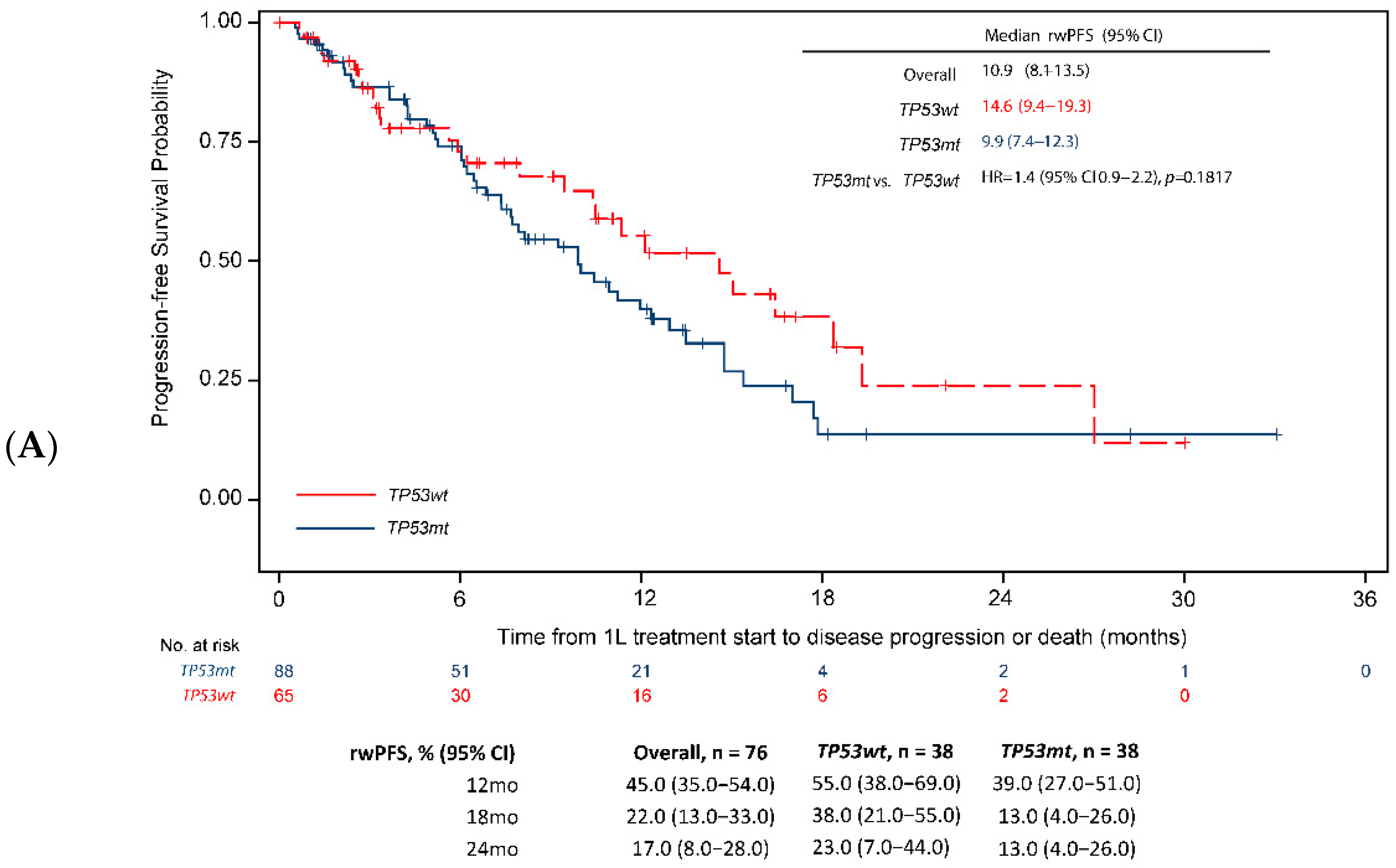
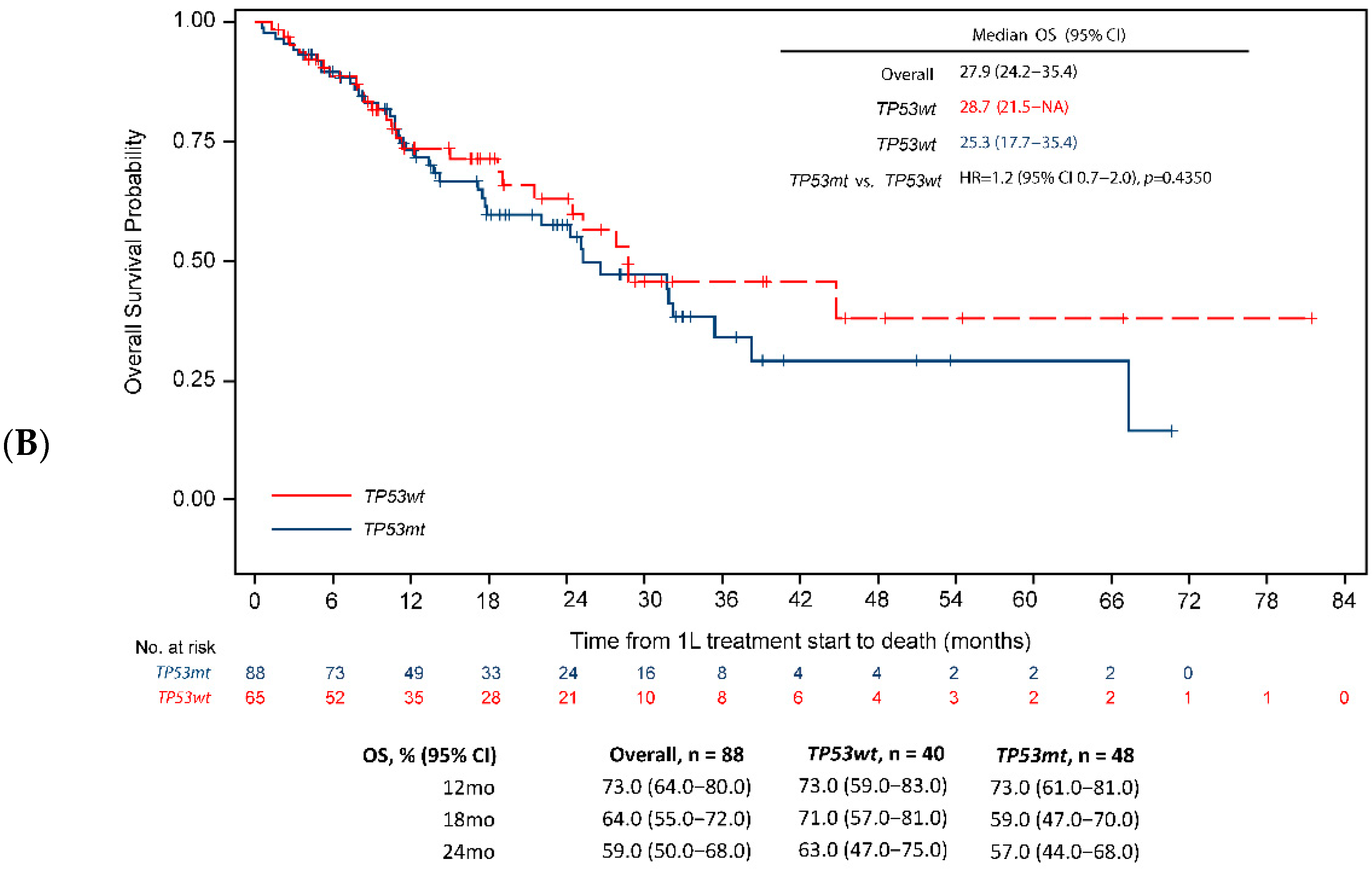
3.3. Survival End Points

Through December 2020, the mean (SD) follow-up for all eligible patients was 19.9 (15.8) months (median: 15.9 months [IQR, 8.3–26.7]). In the overall population, the median rwPFS and median OS were 12.4 months (95% CI 10.6–14.1) and 28.7 months (95% CI 25.3–33.2) respectively. The rwPFS was significantly shorter in patients with TP53 co-mutations than those with TP53 wild-type tumors (median rwPFS 10.6 vs. 14.6 months, unadjusted HR = 1.4, 95% CI 1.1–1.9, p = 0.0196), as was OS (median OS 25.6 vs. 46.9 months, unadjusted HR = 1.6, 95% CI 1.1–2.2, p = 0.0088). One- and two-year survival probabilities were consistently lower for patients with TP53 co-mutations (rwPFS: 47.0% and 18.0%; OS: 77.0% and 56.0%) than those with TP53 wild-type tumors (rwPFS: 57.0% and 27.0%; OS: 77.0% and 65.0%) (Figure 2A,B).
Based on univariate analysis, age ≥ 75 years, male sex, brain metastases, bone metastases, ECOG PS ≥ 1, EGFR exon 21L858R mutation, smoking history, and the interaction between the EGFR mutation subtype and TP53 co-mutation status were significantly associated with worse survival outcomes (rwPFS and OS, Table 3) in the overall population. After adjustment for these factors in multivariable analysis, the TP53 co-mutations remained independently associated with a significantly greater risk for disease progression (rwPFS: HR = 1.4, 95% CI 1.0–1.9, p = 0.0280, Figure 2A) or death (OS: HR = 1.4, 95% CI 1.0–2.0, p = 0.0345, Figure 2B).

3.4. Tumor Response

Among the 275 patients with evaluable tumor response (77.2% of overall study cohort), the rwORR in the overall population was 78.0%. Univariate analysis (Supplementary Table S2) showed lower response rates in patients with a TP53 co-mutation than in those with TP53 wild-type tumors, although differences were not statistically significant (rwORR: 75.0% vs. 83.0% odds ratio (OR) = 0.6, 95% CI 0.3–1.1, p = 0.1262; rwDCR: 91.0% vs. 94.0%, OR = 0.6, 95% CI 0.2–1.6, p = 0.3324). A similar trend was observed for rwDoR (median 9.0 [95% CI 6.9–11.1] vs. 11.9 [95% CI 8.4–15.5] months), best response of CR (4.5% vs. 10.8%) and best response of progressive disease (8.9% vs. 5.4%), (Supplementary Table S3).

3.5. Subgroup Analysis

3.5.1. EGFR Mutation Subtype

When analysis was restricted to the type of EGFR-activating mutation, the negative effect of TP53 co-mutations on the rwPFS in the overall population (HR = 1.4, 95% CI 1.0–1.9, Figure 2) was directionally consistent across the subgroups by activating the EGFR mutation subtype, but was not statistically significant in the subgroups of patients with exon 19 deletion (13.2 months [95% CI 8.7–15.3] vs. 15.6 months [95% CI 11.2–19.9], HR = 1.5 [95% CI 1.1–2.2], p = 0.0536, Figure 3A) or those with exon 21L858R mutations (9.9 months [95% CI 7.3–12.3] vs. 14.6 months [95% CI 9.4–19.3], HR = 1.4 [95% CI 0.0–2.2], p = 0.1817, Figure 4A). Similarly, the negative effect of TP53 co-mutations on OS in the overall population (HR = 1.4, 95% CI 1.0–2.0, p = 0.0345, Figure 2B) was directionally consistent with the EGFR-activating mutation subtype. However, the difference in OS was statistically significant only in the subgroup of patients with exon 19 deletions (median 25.6 [95% CI 21.7–29.7] vs. 47.1 [28.7-not achieved, NA], HR = 1.9, 95% CI 1.2–2.9, p = 0.0072, Figure 3B) and not significant in those with exon 21L858R mutations (median 25.3 [95% CI 17.7–35.4] vs. 28.7 [95% CI 21.5-NA], HR = 1.2, 95% CI 0.7–2.0, p = 0.4350), Figure 4B).
Supplementary Figure S1 presents the four-way comparison between the survival values for the included groups. Survival analysis for patients with EGFR exon 21L858R mutations and TP53 co-mutations had the shortest median rwPFS (9.9 months) compared with 15.6 months, 14.6 months, and 13.2 months for patients with TP53 wild-type plus EGFR exon 19 deletions, TP53 wild-type plus EGFR exon 21L858R mutations, and TP53 co-mutations plus EGFR exon 19 deletions, respectively (p = 0.0243, Supplementary Figure S1A). Similarly, patients with EGFR exon 21L858R and TP53 co-mutations had the shortest median OS (25.3 months) compared with 47.1 months, 28.7 months, and 25.6 months for patients with TP53 wild-type plus EGFR exon 19 deletions, TP53 wild-type plus EGFR exon 21L858R mutations, and TP53 co-mutations plus EGFR exon 19 deletions, respectively (p = 0.0388; Supplementary Figure S1B).

3.5.2. EGFR-TKI Monotherapy

When analysis was focused on the subset of patients receiving targeted therapy with EGFR-TKI monotherapy (75.0% of overall study population), the median rwPFS was 14.0 months (95% CI 11.3–15.6), the median OS was 29.2 months (95% CI 25.5–36.6), and the rwORR was 77.0% (95% CI 72.0–83.0%). The rwPFS was significantly shorter for patients with TP53 co-mutations than those with TP53 wild-type tumors (median 12.0 vs. 16.4 months, HR = 1.6, 95% CI 1.1–2.3, p = 0.0069), as was OS (median 25.3 vs. 44.7 months, HR = 1.7, 95% CI 1.1–2.6, p = 0.0121). In contrast, there was no significant difference in the rwORR between the TP53 co-mutation group and the TP53 wild-type group (rwORR 73.0% vs. 84.0%, OR 0.53, 95% CI 0.27–1.03) (Supplementary Table S4). After adjustment for significant prognostic factors in multivariable analysis, TP53 co-mutations remained significantly associated with a worse rwPFS (HR = 1.5, 95% CI 1.1–2.2, p = 0.0168) but not with OS (HR = 1.5, 95% CI 1.0–2.3, p = 0.0669).

3.5.3. EGFR-TKI Generation

When patients were grouped according to generation of EGFR-TKIs, analyses showed generally similar trends to those observed in the overall population, i.e., shorter rwPFS and OS in patients with TP53 co-mutations than in patients with TP53 wild-type tumor (Supplementary Table S5). Specifically, the negative effect of a TP53 co-mutation on survival outcomes was directionally consistent across the first- (median rwPFS: 9.0 vs. 11.3 months, HR = 1.9, 95% CI 1.0–3.5; median OS: 29.2 vs. 42.0 months, HR 1.6, 95% CI 0.9–3.0) and third-generation (median rwPFS: 17.0 vs. 18.4 months, HR = 1.3, 95% CI 0.8–2.2; median OS 31.2 vs. not reported [NR], HR 1.1, 95% CI 0.6–2.3) EGFR-TKI subgroups, but not in the second-generation EGFR-TKI subgroup (median rwPFS: 13.9 vs. 11.8 months, HR = 1.3, 95% CI 0.6–3.0; median OS 25.3 vs. NR months, HR = 3.1, 95% CI 1.2–8.4), (Supplementary Table S5). Except for the median rwPFS for first generation (p = 0.0399) and OS for second generation (p = 0.0236), the differences did not reach statistical significance, possibly owing to the small number of patients in these subgroups.

3.5.4. Non-EGFR-TKI Monotherapy Treatment

Analysis of the subpopulation who received other treatments (i.e., non-TKI monotherapy, including chemotherapy alone or in combination with immunotherapy, as well as EGFR-TKI combinations) showed no significant differences between the TP53 co-mutation and TP53 wild-type groups in rwPFS (median 7.4 vs. 10.0 months, HR = 0.9, 95% CI 0.5–1.6), and OS (median 25.6 vs. 24.5 months, HR = 1.3, 95% CI 0.7–2.3), although the results are limited by the relatively small sample size (n = 89 patients, 50 with TP53 co-mutation and 39 with TP53 wild-type tumor), Supplementary Table S6.

4. Discussion

In this large retrospective clinicogenomic dataset, real-world progression-free and overall survival were significantly shorter among treated EGFR-mutated aNSCLC patients with a TP53 co-mutation versus those with a TP53 wild-type tumor, even after adjustment for known clinicopathologic prognostic factors. A non-significant trend toward lower tumor response rates was observed among patients with a TP53 co-mutation. This negative contribution was generally independent of the type of 1 L treatment or EGFR mutation subtype. These findings confirm and strengthen available evidence from other datasets with limited follow-up [28,30,31,32,33,48,49], supporting the independent negative prognostic value of TP53 co-mutation status. This may also help to identify a subset of patients with anticipated worse outcomes on 1 L aNSCLC treatment, particularly with EGFR-TKI monotherapy. While this study represents a large dataset, larger cohorts with EGFR and TP53 co-mutated aNSCLC are needed to validate the current findings and to further clarify the contribution and underlying mechanism of a TP53 co-mutation to primary or acquired resistance to EGFR-TKIs.
The study dataset was drawn predominantly from community oncology centers and restricted to patients with genomic testing. However, there was limited representation from academic practices where broad-based genomic sequencing may be part of research protocols, and from vulnerable patients with disproportionate access to physicians with distinct testing and practice patterns. The dataset is nationally illustrative of demographic (e.g., age, sex, and geographic location) and clinical (e.g., stage of disease) characteristics and distribution of patients across community and academic practices [50,51,52]. For example, the incidence of a TP53 co-mutation in our study (59%) was within the range reported in other observational and clinical datasets (30–72%) of patients with EGFR-mutated aNSCLC, using various methods of EGFR detection with different sensitivity [28]. As also previously reported [31,48], TP53 co-mutations were generally not associated with other measured prognostic factors (e.g., age, sex, smoking status, ECOG PS, baseline brain metastases), EGFR mutation subtype, or treatment assignment. Lastly, consistent with guideline recommendations [7,8] and prior studies [53], most patients with EGFR-mutated aNSCLC received EGFR-TKI monotherapy (predominantly osimertinib) in the 1 L and 2 L settings in current study, with few patients receiving 1 L EGFR-TKI combinations with either anti-angiogenics or chemotherapy.
In line with previous studies linking a TP53 co-mutation with reduced sensitivity to EGFR-TKIs [29,49], a TP53 co-mutation was associated with worse outcomes in the current dataset, even after adjustment for known aNSCLC prognostic factors including age, sex, smoking history, performance status, and baseline brain or bone metastases. Our findings also suggest that TP53 co-mutations in the context of the EGFR mutation subtype may impact response to treatment and the survival outcomes, as the negative survival effect associated with TP53 co-mutations has been observed to occur less among patients with EGFR exon 19 deletions than exon 21L858R mutations [31,32] and is likely because EGFR exon 19 deletions are known to be more responsive to EGFR-TKIs [53,54], as also demonstrated by our results. Notably, this finding appears to be driven by the robust OS in patients with TP53 wild-type tumors plus the exon 19 deletion subgroup (47.1 months), as the OS was similar between the exon 19 deletion subgroup plus a TP53 co-mutation (25.6 months) and an exon 21L858R mutation plus the TP53 co-mutation subgroup (28.7 months). Sufficiently powered subgroup analyses on the effect of TP53 co-mutations by the EGFR mutation subtype are warranted to validate these associations.
One of the strengths of this study is that it was conducted between 2014–2020, where the treatment landscape for EGFR-mutated NSCLC had dramatically changed, spanning the approval of multiple first-, second-, and third-generation EGFR-TKIs, and allowing for selection of a relatively large cohort with a long follow-up to assess the prognostic impact TP53 co-mutations in this setting. Despite the large sample size and directional consistency in survival endpoints and response, as well as subgroup analysis, the relatively small numbers and limited follow-up among some subgroups may have (1) underestimated the number of patients (<50% in the current dataset) who ultimately did receive 2 L treatment, and (2) limited statistical power to detect significant differences. For instance, as FDA approval of osimertinib in the frontline setting was in April 2018 [55], findings from subgroup analysis by third-generation EGFR-TKIs had limited follow-up (less than 2 years) and should be validated in future studies as cohorts of patients treated with 1 L osimertinib mature in routine practice.
TP53 co-mutation status did not appear to influence treatment selection of therapy for EGFR-mutated aNSCLC, likely because there are no approved therapies targeting TP53 mutations. Although treatments targeting TP53 co-mutations remain under investigation, our findings may have therapeutic implications given the expansion of precision oncology and increased adoption of next generation sequencing and multi-gene panels [7,8,56], resulting in increased availability of TP53 status in this EGFR-mutated aNSCLC patient population. Specifically, these findings strengthen the rationale for clinicians to consider available or emerging EGFR-TKI combination strategies with other therapies such as antiangiogenics to delay resistance and prolong duration of targeted therapy for EGFR-mutated aNSCLC. Notably, in contrast to the body of evidence [28,30,31,32,33,48], including our findings, showing attenuated benefit with EGFR-TKI monotherapy in patients with EGFR and TP53 co-mutated aNSCLC, a biomarker and subgroup analysis of the RELAY trial demonstrated that the addition of ramucirumab (anti-VEGF receptor inhibitor) to erlotinib (first-generation EGFR-TKI) was associated with prolonged PFS for EGFR and TP53 co-mutated aNSCLC, both in patients with EGFR exon 19 deletions or exon 21L858R mutations [57,58]. TP53 mutations have also been linked to VEGF expression across different solid tumor cancers therefore increasing the sensitivity of these tumors to anti-VEGF therapies [59]. While promising [57,60], however, the efficacy of EGFR-TKI combinations in EGFR and TP53 co-mutated NSCLC is not established and needs further study in larger datasets.
The limitations of this study are inherent in the nature of the analyzed data, collected during routine care mainly for clinical documentation, management, and billing, and not for research. The potential for bias exists from: (1) misclassification due to data entry and reporting errors in the EMR and use of database algorithms (e.g., line of therapy, adherence to oral EGFR-TKIs), (2) missing or incomplete data (e.g., ECOG performance status, comorbidities, tumor response to therapy), and (3) unmeasured or unknown confounding bias despite the use of multivariable regression to control for measured prognostic factors. However, there is no reason to believe that the validity of the dataset would be systematically biased according to TP53 co-mutation status, although the association in this study should not imply causation. In addition, this study did not capture information on different types of TP53 co-mutations and only TP53 co-mutation status at baseline was considered. This is because the information on TP53 mutation subtypes was not collected or available in the Flatiron database. As earlier reports suggest that mutations in TP53 may induce resistance through different mechanisms in the cancer cell [61], further classification of distinct TP53 mutation subtypes (e.g., disruptive vs. non-disruptive, missense vs. non-missense, at primary vs. metastatic lesions), both alone and in combination with other EGFR mutation subtypes (e.g., EGFR exon 20 T790M, the most common mechanism of resistance for 1st and 2nd generation EGFR-TKIs), may predict more precisely the impact of TP53 co-mutation on prognosis and survival on EGFR-TKIs.

5. Conclusions

This large observational study confirms and strengthens the available evidence demonstrating that a TP53 co-mutation identifies a subset of EGFR-mutated aNSCLC patients with worse outcomes on EGFR-TKI monotherapy as compared to those with TP53 wild-type EGFR-mutated aNSCLC who might benefit from more intensified treatment schedules. Tailoring the treatment to individual patient characteristics (e.g., EGFR mutation subtype and co-occurring mutations) at baseline might be helpful to further improve treatment outcomes for patients with EGFR-mutated aNSCLC.

Supplementary Materials

The following supporting information can be downloaded at: https://www.mdpi.com/article/10.3390/cancers14246127/s1, Figure S1: rwPFS and OS–TP53 EGFR Exon Groups; Table S1: Treatment Patterns in 1L and 2L–By Drugs; Table S2: Univariate Analysis–rwORR and rwDCR; Table S3: rwORR, rwDCR, and rwBOR–TP53mt vs. TP53wt; Table S4: Efficacy Outcomes–EGFR-TKI Monotherapy; Table S5: Efficacy Outcomes–By EGFR-TKI Generation; Table S6: Efficacy Outcomes–Non-EGFR-TKI Monotherapy.

Author Contributions

Study design: C.M., M.S.L., M.T.R., C.V.-G. and N.C. Data collection and Analysis: M.T.R., P.M.P., C.V.-G., Y.C. and N.C. Data interpretation: X.L., M.S.L., M.T.R., P.M.P., C.V.-G., E.G.G. and N.C. Manuscript writing: CM, M.S.L., M.T.R. and C.V.-G. Critical revision of the manuscript: X.L., C.M., M.S.L., M.T.R., P.M.P., C.V.-G., Y.C. and E.G.G. Final approval for submission: X.L., C.M., M.S.L., M.T.R., P.M.P., C.V.-G., Y.C., E.G.G. and N.C. All authors have read and agreed to the published version of the manuscript.

Funding

This study was funded by Eli Lilly and Company.

Institutional Review Board Statement

The study was exempt from institutional review board (IRB) approval because it was a retrospective study and used de-identified data in compliance with the Health Insurance Portability and Accountability Act.

Informed Consent Statement

Approval from WCG IRB for data collection for this real-world cohort was obtained prior to study conduct and included a waiver of informed consent.

Data Availability Statement

The data that support the findings of this study have been originated by Flatiron Health, Inc. and Foundation Medicine, Inc. These de-identified data may be made available upon request and are subject to a license agreement with Flatiron Health and Foundation Medicine; interested researchers should contact cgdb-fmi@flatiron.com and dataaccess@flatiron.com to determine licensing terms.

Acknowledgments

The authors would like to thank Karan Sharma from Eli Lilly and Company for providing medical writing and editorial support.

Conflicts of Interest

X.L. is employee at MD Anderson Cancer Center. She has received: (i) Grants: from Eli Lilly and Company, EMD Serono, Boehringer Ingelheim, Regeneron; (ii) Consulting fee: EMD Serono (Merck KGaA), AstraZeneca, Spectrum Pharmaceutics, Novartis, Eli Lilly, and Company, Boehringer Ingelheim, Hengrui Therapeutics, AstraZeneca, Janssen, Blueprint Medicines, Sensei Biotherapeutics, Abbvie and ArriVent; (iii) Support for attending meetings: Spectrum Therapeutics. CM, M.S.L., M.T.R., P.M.P., C.V.-G., Y.C., and EEG, are employees, and stockholders at Eli Lilly and Company. N.C. is a former employee at Eli Lilly and Company.

References

  1. Dearden, S.; Stevens, J.; Wu, Y.L.; Blowers, D. Mutation incidence and coincidence in non small-cell lung cancer: Meta-analyses by ethnicity and histology (mutMap). Ann. Oncol. 2013, 24, 2371–2376. [Google Scholar] [CrossRef] [PubMed]
  2. Midha, A.; Dearden, S.; McCormack, R. EGFR mutation incidence in non-small-cell lung cancer of adenocarcinoma histology: A systematic review and global map by ethnicity (mutMapII). Am. J. Cancer Res. 2015, 5, 2892–2911. [Google Scholar] [PubMed]
  3. Sung, H.; Ferlay, J.; Siegel, R.L.; Laversanne, M.; Soerjomataram, I.; Jemal, A.; Bray, F. Global Cancer Statistics 2020: GLOBOCAN Estimates of Incidence and Mortality Worldwide for 36 Cancers in 185 Countries. CA Cancer J. Clin. 2021, 71, 209–249. [Google Scholar] [CrossRef]
  4. Harrison, P.T.; Vyse, S.; Huang, P.H. Rare epidermal growth factor receptor (EGFR) mutations in non-small cell lung cancer. Semin. Cancer Biol. 2020, 61, 167–179. [Google Scholar] [CrossRef]
  5. Robichaux, J.P.; Le, X.; Vijayan, R.S.K.; Hicks, J.K.; Heeke, S.; Elamin, Y.Y.; Lin, H.Y.; Udagawa, H.; Skoulidis, F.; Tran, H.; et al. Structure-based classification predicts drug response in EGFR-mutant NSCLC. Nature 2021, 597, 732–737. [Google Scholar] [CrossRef]
  6. Langer, C.J. Epidermal growth factor receptor inhibition in mutation-positive non-small-cell lung cancer: Is afatinib better or simply newer? J. Clin. Oncol. 2013, 31, 3303–3306. [Google Scholar] [CrossRef]
  7. National Comprehensive Cancer Network. Non-Small Cell Lung Cancer (Version 3.2022). Available online: https://www.nccn.org/professionals/physician_gls/pdf/nscl.pdf (accessed on 3 May 2022).
  8. Hanna, N.H.; Robinson, A.G.; Temin, S.; Baker, S., Jr.; Brahmer, J.R.; Ellis, P.M.; Gaspar, L.E.; Haddad, R.Y.; Hesketh, P.J.; Jain, D.; et al. Therapy for Stage IV Non-Small-Cell Lung Cancer With Driver Alterations: ASCO and OH (CCO) Joint Guideline Update. J. Clin. Oncol. 2021, 39, 1040–1091. [Google Scholar] [CrossRef]
  9. Nakagawa, K.; Garon, E.B.; Seto, T.; Nishio, M.; Aix, S.P.; Paz-Ares, L.; Chiu, C.H.; Park, K.; Novello, S.; Nadal, E.; et al. Ramucirumab plus erlotinib in patients with untreated, EGFR-mutated, advanced non-small-cell lung cancer (RELAY): A randomised, double-blind, placebo-controlled, phase 3 trial. Lancet Oncol. 2019, 20, 1655–1669. [Google Scholar] [CrossRef] [Green Version]
  10. Passaro, A.; Janne, P.A.; Mok, T.; Peters, S. Overcoming therapy resistance in EGFR-mutant lung cancer. Nat. Cancer 2021, 2, 377–391. [Google Scholar] [CrossRef]
  11. Yu, H.A.; Arcila, M.E.; Rekhtman, N.; Sima, C.S.; Zakowski, M.F.; Pao, W.; Kris, M.G.; Miller, V.A.; Ladanyi, M.; Riely, G.J. Analysis of tumor specimens at the time of acquired resistance to EGFR-TKI therapy in 155 patients with EGFR-mutant lung cancers. Clin. Cancer Res. 2013, 19, 2240–2247. [Google Scholar] [CrossRef]
  12. Rosell, R.; Carcereny, E.; Gervais, R.; Vergnenegre, A.; Massuti, B.; Felip, E.; Palmero, R.; Garcia-Gomez, R.; Pallares, C.; Sanchez, J.M.; et al. Erlotinib versus standard chemotherapy as first-line treatment for European patients with advanced EGFR mutation-positive non-small-cell lung cancer (EURTAC): A multicentre, open-label, randomised phase 3 trial. Lancet Oncol. 2012, 13, 239–246. [Google Scholar] [CrossRef] [PubMed]
  13. Mok, T.S.; Wu, Y.L.; Thongprasert, S.; Yang, C.H.; Chu, D.T.; Saijo, N.; Sunpaweravong, P.; Han, B.; Margono, B.; Ichinose, Y.; et al. Gefitinib or carboplatin-paclitaxel in pulmonary adenocarcinoma. N. Engl. J. Med. 2009, 361, 947–957. [Google Scholar] [CrossRef] [PubMed]
  14. Yang, J.C.; Wu, Y.L.; Schuler, M.; Sebastian, M.; Popat, S.; Yamamoto, N.; Zhou, C.; Hu, C.P.; O’Byrne, K.; Feng, J.; et al. Afatinib versus cisplatin-based chemotherapy for EGFR mutation-positive lung adenocarcinoma (LUX-Lung 3 and LUX-Lung 6): Analysis of overall survival data from two randomised, phase 3 trials. Lancet Oncol. 2015, 16, 141–151. [Google Scholar] [CrossRef] [PubMed] [Green Version]
  15. Soria, J.C.; Ohe, Y.; Vansteenkiste, J.; Reungwetwattana, T.; Chewaskulyong, B.; Lee, K.H.; Dechaphunkul, A.; Imamura, F.; Nogami, N.; Kurata, T.; et al. Osimertinib in Untreated EGFR-Mutated Advanced Non-Small-Cell Lung Cancer. N. Engl. J. Med. 2018, 378, 113–125. [Google Scholar] [CrossRef] [PubMed]
  16. Jackman, D.; Pao, W.; Riely, G.J.; Engelman, J.A.; Kris, M.G.; Janne, P.A.; Lynch, T.; Johnson, B.E.; Miller, V.A. Clinical definition of acquired resistance to epidermal growth factor receptor tyrosine kinase inhibitors in non-small-cell lung cancer. J. Clin. Oncol. 2010, 28, 357–360. [Google Scholar] [CrossRef] [PubMed]
  17. Takezawa, K.; Pirazzoli, V.; Arcila, M.E.; Nebhan, C.A.; Song, X.; De Stanchina, E.; Ohashi, K.; Janjigian, Y.Y.; Spitzler, P.J.; Melnick, M.A.; et al. HER2 amplification: A potential mechanism of acquired resistance to EGFR inhibition in EGFR-mutant lung cancers that lack the second-site EGFRT790M mutation. Cancer Discov. 2012, 2, 922–933. [Google Scholar] [CrossRef] [Green Version]
  18. Planchard, D.; Loriot, Y.; Andre, F.; Gobert, A.; Auger, N.; Lacroix, L.; Soria, J.C. EGFR-independent mechanisms of acquired resistance to AZD9291 in EGFR T790M-positive NSCLC patients. Ann. Oncol. 2015, 26, 2073–2078. [Google Scholar] [CrossRef]
  19. Papadimitrakopoulou, V.A.; Mok, T.S.; Han, J.Y.; Ahn, M.J.; Delmonte, A.; Ramalingam, S.S.; Kim, S.W.; Shepherd, F.A.; Laskin, J.; He, Y.; et al. Osimertinib versus platinum-pemetrexed for patients with EGFR T790M advanced NSCLC and progression on a prior EGFR-tyrosine kinase inhibitor: AURA3 overall survival analysis. Ann. Oncol. 2020, 31, 1536–1544. [Google Scholar] [CrossRef]
  20. Ramalingam, S.S.; Vansteenkiste, J.; Planchard, D.; Cho, B.C.; Gray, J.E.; Ohe, Y.; Zhou, C.; Reungwetwattana, T.; Cheng, Y.; Chewaskulyong, B.; et al. Overall Survival with Osimertinib in Untreated, EGFR-Mutated Advanced NSCLC. N. Engl. J. Med. 2020, 382, 41–50. [Google Scholar] [CrossRef]
  21. Le, X.; Puri, S.; Negrao, M.V.; Nilsson, M.B.; Robichaux, J.; Boyle, T.; Hicks, J.K.; Lovinger, K.L.; Roarty, E.; Rinsurongkawong, W.; et al. Landscape of EGFR-Dependent and -Independent Resistance Mechanisms to Osimertinib and Continuation Therapy Beyond Progression in EGFR-Mutant NSCLC. Clin. Cancer Res. 2018, 24, 6195–6203. [Google Scholar] [CrossRef] [PubMed]
  22. Teodoro, J.G.; Evans, S.K.; Green, M.R. Inhibition of tumor angiogenesis by p53: A new role for the guardian of the genome. J. Mol. Med. 2007, 85, 1175–1186. [Google Scholar] [CrossRef] [PubMed]
  23. Boutelle, A.M.; Attardi, L.D. p53 and Tumor Suppression: It Takes a Network. Trends Cell Biol. 2021, 31, 298–310. [Google Scholar] [CrossRef] [PubMed]
  24. Poeta, M.L.; Manola, J.; Goldwasser, M.A.; Forastiere, A.; Benoit, N.; Califano, J.A.; Ridge, J.A.; Goodwin, J.; Kenady, D.; Saunders, J.; et al. TP53 mutations and survival in squamous-cell carcinoma of the head and neck. N. Engl. J. Med. 2007, 357, 2552–2561. [Google Scholar] [CrossRef] [PubMed] [Green Version]
  25. Hamid, A.B.; Petreaca, R.C. Secondary Resistant Mutations to Small Molecule Inhibitors in Cancer Cells. Cancers 2020, 12, 927. [Google Scholar] [CrossRef] [Green Version]
  26. Jao, K.; Tomasini, P.; Kamel-Reid, S.; Korpanty, G.J.; Mascaux, C.; Sakashita, S.; Labbe, C.; Leighl, N.B.; Liu, G.; Feld, R.; et al. The prognostic effect of single and multiple cancer-related somatic mutations in resected non-small-cell lung cancer. Lung Cancer 2018, 123, 22–29. [Google Scholar] [CrossRef]
  27. Hafner, A.; Bulyk, M.L.; Jambhekar, A.; Lahav, G. The multiple mechanisms that regulate p53 activity and cell fate. Nat. Rev. Mol. Cell. Biol. 2019, 20, 199–210. [Google Scholar] [CrossRef]
  28. Qin, K.; Hou, H.; Liang, Y.; Zhang, X. Prognostic value of TP53 concurrent mutations for EGFR- TKIs and ALK-TKIs based targeted therapy in advanced non-small cell lung cancer: A meta-analysis. BMC Cancer 2020, 20, 328. [Google Scholar] [CrossRef]
  29. Zhou, G.; Liu, Z.; Myers, J.N. TP53 Mutations in Head and Neck Squamous Cell Carcinoma and Their Impact on Disease Progression and Treatment Response. J. Cell. Biochem. 2016, 117, 2682–2692. [Google Scholar] [CrossRef] [Green Version]
  30. Molina-Vila, M.A.; Bertran-Alamillo, J.; Gasco, A.; Mayo-de-las-Casas, C.; Sanchez-Ronco, M.; Pujantell-Pastor, L.; Bonanno, L.; Favaretto, A.G.; Cardona, A.F.; Vergnenegre, A.; et al. Nondisruptive p53 mutations are associated with shorter survival in patients with advanced non-small cell lung cancer. Clin. Cancer Res. 2014, 20, 4647–4659. [Google Scholar] [CrossRef] [Green Version]
  31. Canale, M.; Petracci, E.; Delmonte, A.; Chiadini, E.; Dazzi, C.; Papi, M.; Capelli, L.; Casanova, C.; De Luigi, N.; Mariotti, M.; et al. Impact of TP53 Mutations on Outcome in EGFR-Mutated Patients Treated with First-Line Tyrosine Kinase Inhibitors. Clin. Cancer Res. 2017, 23, 2195–2202. [Google Scholar] [CrossRef]
  32. Canale, M.; Petracci, E.; Delmonte, A.; Bronte, G.; Chiadini, E.; Ludovini, V.; Dubini, A.; Papi, M.; Baglivo, S.; De Luigi, N.; et al. Concomitant TP53 Mutation Confers Worse Prognosis in EGFR-Mutated Non-Small Cell Lung Cancer Patients Treated with TKIs. J. Clin. Med. 2020, 9, 1047. [Google Scholar] [CrossRef] [PubMed] [Green Version]
  33. Aggarwal, C.; Davis, C.W.; Mick, R.; Thompson, J.C.; Ahmed, S.; Jeffries, S.; Bagley, S.; Gabriel, P.; Evans, T.L.; Bauml, J.M.; et al. Influence of TP53 Mutation on Survival in Patients with Advanced EGFR-Mutant Non-Small-Cell Lung Cancer. JCO Precis Oncol. 2018, 2, 1–29. [Google Scholar] [CrossRef] [PubMed]
  34. Scoccianti, C.; Vesin, A.; Martel, G.; Olivier, M.; Brambilla, E.; Timsit, J.F.; Tavecchio, L.; Brambilla, C.; Field, J.K.; Hainaut, P.; et al. Prognostic value of TP53, KRAS and EGFR mutations in nonsmall cell lung cancer: The EUELC cohort. Eur. Respir. J. 2012, 40, 177–184. [Google Scholar] [CrossRef] [Green Version]
  35. Ma, X.; Le Teuff, G.; Lacas, B.; Tsao, M.S.; Graziano, S.; Pignon, J.P.; Douillard, J.Y.; Le Chevalier, T.; Seymour, L.; Filipits, M.; et al. Prognostic and Predictive Effect of TP53 Mutations in Patients with Non-Small Cell Lung Cancer from Adjuvant Cisplatin-Based Therapy Randomized Trials: A LACE-Bio Pooled Analysis. J. Thorac. Oncol. 2016, 11, 850–861. [Google Scholar] [CrossRef] [Green Version]
  36. Ma, X.; Rousseau, V.; Sun, H.; Lantuejoul, S.; Filipits, M.; Pirker, R.; Popper, H.; Mendiboure, J.; Vataire, A.L.; Le Chevalier, T.; et al. Significance of TP53 mutations as predictive markers of adjuvant cisplatin-based chemotherapy in completely resected non-small-cell lung cancer. Mol. Oncol. 2014, 8, 555–564. [Google Scholar] [CrossRef]
  37. Shepherd, F.A.; Lacas, B.; Le Teuff, G.; Hainaut, P.; Janne, P.A.; Pignon, J.P.; Le Chevalier, T.; Seymour, L.; Douillard, J.Y.; Graziano, S.; et al. Pooled Analysis of the Prognostic and Predictive Effects of TP53 Comutation Status Combined With KRAS or EGFR Mutation in Early-Stage Resected Non-Small-Cell Lung Cancer in Four Trials of Adjuvant Chemotherapy. J. Clin. Oncol. 2017, 35, 2018–2027. [Google Scholar] [CrossRef] [PubMed]
  38. Labbe, C.; Cabanero, M.; Korpanty, G.J.; Tomasini, P.; Doherty, M.K.; Mascaux, C.; Jao, K.; Pitcher, B.; Wang, R.; Pintilie, M.; et al. Prognostic and predictive effects of TP53 co-mutation in patients with EGFR-mutated non-small cell lung cancer (NSCLC). Lung Cancer 2017, 111, 23–29. [Google Scholar] [CrossRef] [PubMed]
  39. Singal, G.; Miller, P.G.; Agarwala, V.; Li, G.; Kaushik, G.; Backenroth, D.; Gossai, A.; Frampton, G.M.; Torres, A.Z.; Lehnert, E.M.; et al. Association of Patient Characteristics and Tumor Genomics with Clinical Outcomes among Patients with Non-Small Cell Lung Cancer Using a Clinicogenomic Database. JAMA 2019, 321, 1391–1399. [Google Scholar] [CrossRef] [Green Version]
  40. Flatiron. About US. Available online: https://flatiron.com/about-us/ (accessed on 5 May 2022).
  41. Paesmans, M.; Sculier, J.P.; Libert, P.; Bureau, G.; Dabouis, G.; Thiriaux, J.; Michel, J.; Van Cutsem, O.; Sergysels, R.; Mommen, P.; et al. Prognostic factors for survival in advanced non-small-cell lung cancer: Univariate and multivariate analyses including recursive partitioning and amalgamation algorithms in 1,052 patients. The European Lung Cancer Working Party. J. Clin. Oncol. 1995, 13, 1221–1230. [Google Scholar] [CrossRef]
  42. O’Connell, J.P.; Kris, M.G.; Gralla, R.J.; Groshen, S.; Trust, A.; Fiore, J.J.; Kelsen, D.P.; Heelan, R.T.; Golbey, R.B. Frequency and prognostic importance of pretreatment clinical characteristics in patients with advanced non-small-cell lung cancer treated with combination chemotherapy. J. Clin. Oncol. 1986, 4, 1604–1614. [Google Scholar] [CrossRef]
  43. Janjigian, Y.Y.; McDonnell, K.; Kris, M.G.; Shen, R.; Sima, C.S.; Bach, P.B.; Rizvi, N.A.; Riely, G.J. Pack-years of cigarette smoking as a prognostic factor in patients with stage IIIB/IV nonsmall cell lung cancer. Cancer 2010, 116, 670–675. [Google Scholar] [CrossRef] [PubMed] [Green Version]
  44. Bergqvist, M.; Christensen, H.N.; Wiklund, F.; Bergstrom, S. Real world utilization of EGFR TKIs and prognostic factors for survival in NSCLC during 2010-2016 in Sweden: A nationwide observational study. Int. J. Cancer 2020, 146, 2510–2517. [Google Scholar] [CrossRef]
  45. Yu, X.; Sheng, J.; Pan, G.; Fan, Y. Real-world utilization of EGFR TKIs and prognostic factors for survival in EGFR-mutated non-small cell lung cancer patients with brain metastases. Int. J. Cancer 2021, 149, 1121–1128. [Google Scholar] [CrossRef] [PubMed]
  46. Cuyun Carter, G.; Barrett, A.M.; Kaye, J.A.; Liepa, A.M.; Winfree, K.B.; John, W.J. A comprehensive review of nongenetic prognostic and predictive factors influencing the heterogeneity of outcomes in advanced non-small-cell lung cancer. Cancer Manag. Res. 2014, 6, 437–449. [Google Scholar] [CrossRef] [PubMed] [Green Version]
  47. Kawaguchi, T.; Takada, M.; Kubo, A.; Matsumura, A.; Fukai, S.; Tamura, A.; Saito, R.; Maruyama, Y.; Kawahara, M.; Ou, S.H.I. Performance status and smoking status are independent favorable prognostic factors for survival in non-small cell lung cancer: A comprehensive analysis of 26,957 patients with NSCLC. J. Thorac. Oncol. 2010, 5, 620–630. [Google Scholar] [CrossRef] [PubMed] [Green Version]
  48. Roeper, J.; Falk, M.; Chalaris-Rissmann, A.; Lueers, A.C.; Ramdani, H.; Wedeken, K.; Stropiep, U.; Diehl, L.; Tiemann, M.; Heukamp, L.C.; et al. TP53 co-mutations in EGFR mutated patients in NSCLC stage IV: A strong predictive factor of ORR, PFS and OS in EGFR mt+ NSCLC. Oncotarget 2020, 11, 250–264. [Google Scholar] [CrossRef] [Green Version]
  49. Vokes, N.I.; Chambers, E.; Nguyen, T.; Coolidge, A.; Lydon, C.A.; Le, X.; Sholl, L.; Heymach, J.V.; Nishino, M.; Van Allen, E.M.; et al. Concurrent TP53 Mutations Facilitate Resistance Evolution in EGFR-Mutant Lung Adenocarcinoma. J. Thorac. Oncol. 2022, 17, 779–792. [Google Scholar] [CrossRef]
  50. Ma, X.; Long, L.; Moon, S.; Adamson, B.J.S.; Baxi, S.S. Comparison of population characteristics in real-world clinical oncology databases in the US: Flatiron Health, SEER, and NPCR. medRxiv 2020. [Google Scholar] [CrossRef] [Green Version]
  51. Berger, M.L.; Curtis, M.D.; Smith, G.; Harnett, J.; Abernethy, A.P. Opportunities and challenges in leveraging electronic health record data in oncology. Future Oncol. 2016, 12, 1261–1274. [Google Scholar] [CrossRef]
  52. Stewart, M.; Norden, A.D.; Dreyer, N.; Henk, H.J.; Abernethy, A.P.; Chrischilles, E.; Kushi, L.; Mansfield, A.S.; Khozin, S.; Sharon, E.; et al. An Exploratory Analysis of Real-World End Points for Assessing Outcomes Among Immunotherapy-Treated Patients With Advanced Non-Small-Cell Lung Cancer. JCO Clin. Cancer Inform. 2019, 3, 1–15. [Google Scholar] [CrossRef]
  53. Winfree, K.B.; Molife, C.; Peterson, P.M.; Chen, Y.; Visseren-Grul, C.M.; Leusch, M.S.; Beyrer, J.; Dimou, A. Real-world characteristics and outcomes of advanced non-small-cell lung cancer patients with EGFR exon 19 deletions or exon 21 mutations. Future Oncol. 2021, 17, 2867–2881. [Google Scholar] [CrossRef] [PubMed]
  54. Sheng, M.; Wang, F.; Zhao, Y.; Li, S.; Wang, X.; Shou, T.; Luo, Y.; Tang, W. Comparison of clinical outcomes of patients with non-small-cell lung cancer harbouring epidermal growth factor receptor exon 19 or exon 21 mutations after tyrosine kinase inhibitors treatment: A meta-analysis. Eur. J. Clin. Pharmacol. 2016, 72, 1–11. [Google Scholar] [CrossRef] [PubMed]
  55. Department of Health and Human Services, US Food and Drug Administration. 2018. Available online: https://www.fda.gov/drugs/resources-information-approved-drugs/fda-approves-osimertinib-first-line-treatment-metastatic-nsclc-most-common-egfr-mutations (accessed on 5 May 2022).
  56. Sheinson, D.M.; Wong, W.B.; Meyer, C.S.; Stergiopoulos, S.; Lofgren, K.T.; Flores, C.; Adams, D.V.; Fleury, M.E. Trends in Use of Next-Generation Sequencing in Patients With Solid Tumors by Race and Ethnicity After Implementation of the Medicare National Coverage Determination. JAMA Netw. Open 2021, 4, e2138219. [Google Scholar] [CrossRef] [PubMed]
  57. Nakagawa, K.; Nadal, E.; Garon, E.B.; Nishio, M.; Seto, T.; Yamamoto, N.; Park, K.; Shih, J.Y.; Paz-Ares, L.; Frimodt-Moller, B.; et al. RELAY Subgroup Analyses by EGFR Ex19del and Ex21L858R Mutations for Ramucirumab Plus Erlotinib in Metastatic Non-Small Cell Lung Cancer. Clin. Cancer Res. 2021, 27, 5258–5271. [Google Scholar] [CrossRef]
  58. Zhang, L.; Zhao, H.; Zhang, Z.; Yao, W.; Min, X.; Gu, K.; Yu, G.; Cheng, C.; Cui, J.; Miao, L.; et al. ACTIVE: Apatinib Plus Gefitinib versus Placebo Plus Gefitinib as First-Line Treatment for Advanced Epidermal Growth Factor Receptor-Mutant (EGFRm) Non-Small-Cell Lung Cancer (NSCLC): A Multicentered, Randomized, Double-Blind, Placebo-Controlled Phase III Trial (CTONG1706). Available online: https://www.annalsofoncology.org/article/S0923-7534(20)42365-8/pdf (accessed on 14 September 2022).
  59. Li, A.M.; Boichard, A.; Kurzrock, R. Mutated TP53 is a marker of increased VEGF expression: Analysis of 7525 pan-cancer tissues. Cancer Biol. Ther. 2020, 21, 95–100. [Google Scholar] [CrossRef] [Green Version]
  60. Shi, Y.K.; Wang, L.; Han, B.H.; Li, W.; Yu, P.; Liu, Y.P.; Ding, C.M.; Song, X.; Ma, Z.Y.; Ren, X.L.; et al. First-line icotinib versus cisplatin/pemetrexed plus pemetrexed maintenance therapy for patients with advanced EGFR mutation-positive lung adenocarcinoma (CONVINCE): A phase 3, open-label, randomized study. Ann. Oncol. 2017, 28, 2443–2450. [Google Scholar] [CrossRef]
  61. Sabapathy, K.; Lane, D.P. Therapeutic targeting of p53: All mutants are equal, but some mutants are more equal than others. Nat. Rev. Clin. Oncol. 2018, 15, 13–30. [Google Scholar] [CrossRef]
Figure 1. Patient inclusion/exclusion criteria and attrition.
Cancers 14 06127 g001
Figure 2. rwPFS and OS—TP53 co-mutation vs. TP53 wild-type. (A) rwPFS. (B) OS.CI = confidence interval; HR = hazard ratio; OS = overall survival; rwPFS = real-world progression-free survival; TP53mt = TP53 co-mutation; TP53wt = TP53 wild-type. EGFR mutation includes both exon 19 and exon 21 mutations.
Cancers 14 06127 g002
Figure 3. rwPFS and OS—TP53 co-mutation vs. TP53 wild-type by Exon19del mutation. (A) rwPFS. (B) OS. Unadjusted HR and 95% CI. CI = confidence interval; HR = hazard ratio; OS = overall survival; rwPFS = real-world progression-free survival; TP53mt = TP53 co-mutation; TP53wt = TP53 wild-type; exon 19 = exon 19 deletions.
Cancers 14 06127 g003aCancers 14 06127 g003b
Figure 4. rwPFS and OS—TP53 co-mutation vs. TP53 wild-type by Exon21L858R mutation. (A) rwPFS. (B) OS. Unadjusted HR and 95% CI. CI = confidence interval; HR = hazard ratio; OS = overall survival; rwPFS = real-world progression-free survival; TP53mt = TP53 co-mutation; TP53wt = TP53 wild-type; exon 21 = exon 21L858R mutations.
Cancers 14 06127 g004aCancers 14 06127 g004b
Table 1. Patient demographic and clinical characteristics.
VariableOverall
(n = 356)
TP53wt
(n = 146)
TP53mt
(n = 210)
Follow-up in months from 1 L start, mean (SD) 19.9 (15.8)21.5 (17.5)18.8 (14.4)
Age at adv. Dx, median (IQR)68.0 (60.5–76.1)69.6 (63.5–77.7)66.7 (58.2–75.2)
Age group, n (%)
 <65 years137 (38.5)46 (31.5)91 (43.3)
 65–74 years115 (32.3)51 (34.9)64 (30.5)
 75+ years104 (29.2)49 (33.6)55 (26.2)
Sex, n (%)
 Female244 (68.5)103 (70.5)141 (67.1)
 Male112 (31.5)43 (29.5)69 (32.9)
Race, n (%)
 Missing26 (7.3)9 (6.2)17 (8.1)
 Asian41 (11.5)22 (15.1)19 (9.0)
 Black or African American15 (4.2)5 (3.4)10 (4.8)
 Other Race59 (16.6)26 (17.8)33 (15.7)
 White215 (60.4)84 (57.5)131 (62.4)
Smoking history, yes, n (%)154 (43.3)61 (41.8)93 (44.3)
NSCLC stage at initial diagnosis, n (%)
 I–II20 (5.6)11 (7.5)9 (4.3)
 III29 (8.1)11 (7.5)18 (8.6)
 IV307 (86.2)124 (84.9)183 (87.1)
ECOG performance status, n (%)
 0107 (30.1)43 (29.5)64 (30.5)
 1+145 (40.7)59 (40.4)86 (41.0)
 Missing104 (29.2)44 (30.1)60 (28.6)
EGFR mutation type, n (%)
 Exon19203 (57.0)81 (55.5)122 (58.1)
 Exon21153 (43.0)65 (44.5)88 (41.9)
Metastatic site, n (%)
 Brain77 (21.6)28 (19.2)49 (23.3)
 Bone97 (27.2)34 (23.3)63 (30.0)
 Liver14 (3.9)8 (5.5)6 (2.9)
 Lung51 (14.3)17 (11.6)34 (16.2)
 Lymph Node23 (6.5)13 (8.9)10 (4.8)
 Adrenal21 (5.9)7 (4.8)14 (6.7)
 Other5 (1.4)3 (2.1)2 (1.0)
Practice type, n (%)
 Academic41 (11.5)18 (12.3)23 (11.0)
 Community315 (88.5)128 (87.7)187 (89.0)
Adv. Dx = advanced diagnosis; ECOG = Eastern Cooperative Oncology Group; EGFR = epidermal growth factor receptor; IQR = interquartile range; NSCLC = non-small cell lung cancer; SD = standard deviation; TP53 mt = TP53 co-mutation; TP53 wt = TP53 wild-type. Smoking history includes both current and past smokers. EGFR mutation includes both exon 19 and exon 21 mutations.
Table 2. Treatment patterns in 1 L and 2 L—by drug class.
1 L2 L
Drug ClassOverallTP53wtTP53mtDrug ClassOverallTP53wtTP53mt
n = 356n = 146n = 210 n = 169n = 61n = 108
EGFR-TKI monotherapy267 (75.0)107 (73.6)160 (76.2)EGFR-TKI monotherapy114 (67.5)43 (70.5)71 (70.3)
Chemotherapy alone37 (10.0)19 (13.1)18 (8.6)Chemotherapy + Immune Checkpoint Inhibitor13 (7.3)4 (6.6)9 (7.4)
Chemotherapy +
Immune Checkpoint
Inhibitor
20 (5.6)7 (4.3)13 (6.2)Chemotherapy alone8 (4.7)2 (2.9)6 (5.9)
Other8 (2.5)3 (2.1)5 (2.4)Immune Checkpoint Inhibitor monotherapy7 (4.1)5 (8.2)2 (1.9)
Chemotherapy + VEGF Inhibitor8 (2.2)3 (2.1)5 (2.4)EGFR-TKI + Chemotherapy + Immune Checkpoint
Inhibitor
4 (2.4)0 (0)4 (3.9)
EGFR-TKI + VEGF
Inhibitor
6 (1.7)2 (1.4)4 (1.9)Other + Chemotherapy3 (1.8)2 (2.9)1 (0.9)
EGFR-TKI + Chemotherapy3 (0.9)1 (0.7)2 (0.9)EGFR-TKI + Immune
Checkpoint Inhibitor
3 (1.8)0 (0)3 (3.0)
Immune Checkpoint
Inhibitor monotherapy
3 (0.9)2 (1.4)1 (0.5)Chemotherapy + VEGF
Inhibitor
3 (1.7)0 (0)3 (2.8)
EGFR-TKI + Chemotherapy + VEGF Inhibitor1 (0.3)0 (0)1 (0.5)EGFR-TKI + Chemotherapy2 (1.2)2 (3.3)0 (0)
EGFR-TKI + Chemotherapy +
Immune Checkpoint Inhibitor
1 (0.3)1 (0.7)0 (0)EGFR-TKI + Chemotherapy + VEGF Inhibitor2 (1.2)0 (0)2 (1.9)
EGFR-TKI + Immune Checkpoint Inhibitor1 (0.3)0 (0)1 (0.5)EGFR-TKI + VEGF Inhibitor2 (1.2)1 (1.64)1 (0.9)
Other + EGFR-TKI1 (0.3)1 (0.7)0 (0)Hormone receptor therapy1 (0.6)0 (0)1 (0.9)
Immunosuppressant1 (0.6)0 (0)1 (0.9)
Other + EGFR-TKI1 (0.6)0 (0)1 (0.9)
All data are presented as n (%). EGFR-TKI = epidermal growth factor receptor-tyrosine kinase inhibitor; Other = clinical study drug; TP53mt = TP53 co-mutation; TP53wt = TP53 wild-type; VEGF = vascular endothelial growth factor. EGFR mutation includes both exon 19 and exon 21 mutations.
Table 3. Univariate analysis—rwPFS and OS.
VariableCategoryrwPFS,
HR (95% CI)
OS,
HR (95% CI)
Overall
 Age at adv dx≥75 vs. <751.2 (0.9–1.6)2.0 (1.4–2.7) d
 SexMale vs. Female1.1 (0.8–1.5)1.4 (1.0–2.0) a
 ECOG PS1+ vs. 01.5 (1.1–2.2) a1.7 (1.1–2.5) b
Unknown vs. 01.5 (1.0–2.2) a0.9 (0.6–1.5)
 Smoking historyYes vs. No1.6 (1.2–2.1) b1.4 (0.99–1.87)
TP53 statusmt vs. wt1.4 (1.1–1.9) a1.6 (1.1–2.2) b
 TP53 status/EGFR mutation type TP53mt/Exon19 vs. TP53wt/Exon191.5 (1.0–2.2)1.9 (1.2–2.9) b
TP53mt/Exon21 vs. TP53wt/Exon191.9 (1.3–2.9) b1.8 (1.1–3.0) a
TP53wt/Exon21 vs. TP53wt/Exon191.4 (0.9–2.3)1.44 (0.8–2.5)
 Bone metsYes vs. No1.0 (0.7–1.4)1.4 (1.0–1.9)
 Brain metsYes vs. No1.8 (1.3–2.5) c1.7 (1.2–2.4) b
 Exon groupExon21 vs. Exon191.3 (1.0–1.8) a1.1 (0.8–1.5)
TKI Monotherapy
 Age at adv dx≥75 vs. <751.1 (0.8–1.6)1.7 (1.1–2.5) b
 SexMale vs. Female1.2 (0.8–1.7)1.5 (1.0–2.3) a
 ECOG PS1+ vs. 02.1 (1.3–3.3) b2.4 (1.4–4.1) b
Unknown vs. 02.2 (1.4–3.5) b1.3 (0.7–2.3)
 Smoking historyYes vs. No1.6 (1.2–2.2) b1.6 (1.1–2.3) a
TP53mt vs. wt1.6 (1.1–2.3) b1.7 (1.1–2.6) a
TP53 status/EGFR mutation typeTP53mt/Exon19 vs. TP53wt/Exon191.6 (1.0–2.4) a2.2 (1.3–3.7) b
TP53mt/Exon21 vs. TP53wt/Exon192.0 (1.2–3.2) b2.1 (1.1–3.9) a
TP53wt/Exon21 vs. TP53wt/Exon191.2 (0.6–2.1)1.9 (0.9–3.7)
 Bone metsYes vs. No1.1 (0.8–1.6)1.4 (0.9–2.1)
 Brain metsYes vs. No1.8 (1.2–2.6) b1.7 (1.1–2.6) b
 Exon groupExon21 vs. Exon191.2 (0.9–1.7)1.2 (0.8–1.8)
Other Monotherapy
 Age at adv dx≥75 vs. <751.6 (0.8–3.0)3.1 (1.7–5.6) c
 SexMale vs. Female0.7 (0.4–1.4)1.1 (0.6–2.1)
 ECOG PS1+ vs. 00.8 (0.4–1.6)1.1 (0.6–2.0)
Unknown vs. 00.6 (0.3–1.3)0.5 (0.2–1.3)
 Smoking historyYes vs. No1.4 (0.8–2.6)1.0 (0.6–1.8)
TP53mt vs. wt0.9 (0.5–1.6)1.3 (0.7–2.3)
TP53 status/EGFR mutation typeTP53mt/Exon19 vs. TP53wt/Exon190.9 (0.4–2.3)1.2 (0.5–2.9)
TP53mt/Exon21 vs. TP53wt/Exon191.2 (0.5–3.0)1.2 (0.5–2.7)
TP53wt/Exon21 vs. TP53wt/Exon191.5 (0.6–3.7)0.9 (0.4–2.1)
 Bone metsYes vs. No0.9 (0.3–2.3)1.5 (0.8–2.9)
 Brain metsYes vs. No3.5 (1.7–7.3) c1.8 (0.9–3.5)
 Exon groupExon21 vs. Exon191.4 (0.8–2.6)0.9 (0.5–1.6)
ap ≤ 0.05; b p ≤ 0.01; c p ≤ 0.001; d p ≤ 0.0001. Adv dx = advanced diagnosis; ECOG PS = Eastern Cooperative Oncology Group performance status; EGFR = epidermal growth factor receptor; mets = metastases; NSCLC = non-small cell lung cancer; SD = standard deviation; TP53mt = TP53 co-mutation; TP53wt = TP53 wild-type. Cox analysis for time-to-event outcomes, and logistic regression analysis for tumor response. Other monotherapy: non-TKI monotherapies. exon 19 = exon 19 deletions, and exon 21 = exon 21L858R mutations.
Publisher’s Note: MDPI stays neutral with regard to jurisdictional claims in published maps and institutional affiliations.

