Article

Tumour Colonisation of Parvimonas micra Is Associated with Decreased Survival in Colorectal Cancer Patients

1
Department of Medical Biosciences, Pathology, Umeå University, SE-90185 Umeå, Sweden
2
Department of Radiation Sciences, Oncology, Umeå University, SE-90185 Umeå, Sweden
3
Department of Surgical and Perioperative Sciences, Surgery, Umeå University, SE-90185 Umeå, Sweden
*
Author to whom correspondence should be addressed.
Cancers 2022, 14(23), 5937; https://doi.org/10.3390/cancers14235937
Submission received: 28 October 2022 / Revised: 24 November 2022 / Accepted: 28 November 2022 / Published: 30 November 2022
(This article belongs to the Special Issue Bacterial, Viral and Parasitic Pathogens and Colorectal Cancer)

Abstract

:

Simple Summary

The gut microbiota has been suggested to affect tumour development and progression in colorectal cancer. In this study, we investigated the associations of two colorectal cancer-related bacteria, Parvimonas micra and Fusobacterium nucleatum, with survival in colorectal cancer patients. Our results suggest that patients with high tumoural levels of Parvimonas micra have decreased survival. In addition, we found associations for Parvimonas micra and Fusobacterium nucleatum with different clinicopathological and tumour molecular traits. A better understanding of the role of the gut microbiota in colorectal cancer may contribute to improved cancer care.

Abstract

Increasing evidence suggests that the gut microbiota may impact colorectal cancer (CRC) development and progression. In this study, the tumour colonisation of two CRC-associated bacteria, Parvimonas micra and Fusobacterium nucleatum, was studied in relation to patient survival in a cohort of 257 CRC patients. Colonisation of P. micra and F. nucleatum was analysed in fresh frozen tumour tissue (n = 112) and in faeces (n = 250) by qPCR. When analysing tumour tissues, both P. micra and F. nucleatum were found to be associated with decreased five-year cancer-specific survival, an association that remained significant in multivariable analysis for P. micra. Furthermore, we found significant associations of high levels of P. micra and F. nucleatum with tumour molecular characteristics, i.e., tumours mutated in BRAFV600E, and tumours of the MSI subtype. The analysis of faecal samples showed weaker associations with prognosis and tumour molecular characteristics. In conclusion, our findings support a novel association of tumour colonisation of P. micra with decreased patient survival. A better understanding of the role of the gut microbiota in CRC might contribute to the advancement of prognostic tools and new targets for therapy.

1. Introduction

Colorectal cancer (CRC) is one of the most deadly cancers in both men and women worldwide with close to 1 million deaths per year, and the number is expected to rise due to an increasing incidence in younger adults and transitioning countries [1]. CRC is a disease with high heterogeneity where patient prognosis depends on a complex interplay between environmental, molecular and genetic factors. Rising evidence suggests gut dysbiosis plays a crucial role in CRC development and progression and metagenomic studies have revealed a structural segregation in microbial composition between CRC patients and healthy individuals [2]. A driver-passenger theory has been introduced, where pro-oncogenic ‘alpha-bugs’ possess the potential to outcompete other cancer-protective species and drive tumorigenesis through immune response remodulation and the production of toxins, leading to cell damage. Disease progression in turn changes the gut microenvironment, resulting in a shift in the microbial community [3]. As a result, the initial bacterial drivers are gradually replaced with gut commensals that can promote tumour progression through inflammatory processes [3]. The driver-passenger theory has been supported by numerous studies on the role of the gut microbiota in CRC, including mechanisms of genotoxin-induced tumourigenesis, and mechanisms controlling barrier function and inflammation [4]. Inflammation in fact plays dual roles in CRC carcinogensis, where on the one hand it may act protective in response to acute tissue damage and display anti-tumourigenic effects, and on the other hand, chronic inflammatory processes are known to induce tumour growth and progression [4,5].
Together, this makes the gut microbiota a potential and interesting target for future cancer therapy. Indeed, recent studies have suggested that the intestinal microecological composition is related to CRC prognosis [2] and specific gut microbial species have been associated with a deteriorated response to cancer therapy [6]. However, the relationship between the spatio-temporal presence of CRC-related microbes and the functional aspects behind clinical outcome is still largely unknown.
Interestingly, many of the bacterial species suggested to be associated with CRC are often found colonising the oral microflora, one of the more well-studied being Fusobacterium nucleatum [7,8,9,10,11,12]. F. nucleatum has been suggested to contribute to colorectal tumour progression through various mechanisms, including expression of the virulence factor FadA on the cell surface, which induces E-cadherin mediated activation of Wnt/β-catenin signalling [13]. Previous studies have also proposed an association between gut colonisation of F. nucleatum and decreased CRC survival [14,15,16,17,18,19,20,21]. Another oral pathogen suggested to be associated with CRC is Parvimonas micra [9,10,11,12,22,23,24]. In a previous study using specific qPCR, we found both P. micra and F. nucleatum to be more abundant in stool samples from CRC patients compared with healthy individuals [23]. The analysis of fresh frozen tumour tissues enabled us to link P. micra to tumour immunological traits [25]. A recent study on mice suggests P. micra has a tumour-promoting effect through colonocyte proliferation and the alteration of the Th17 immune response [26]. Still, in contrast to F. nucleatum, very little is known about the potential role of P. micra in CRC tumourgenesis and prognosis.
In this study, we investigated if the levels of P. micra and F. nucleatum in tumour tissue and faeces of CRC patients affect cancer-specific survival. A potential link between microbial colonisation in CRC and patient prognosis would facilitate the development of new prognostic and predictive tools, as well as future targeted cancer therapies.

2. Materials and Methods

2.1. Study Cohort

The study was performed on patient samples collected within the Uppsala-Umeå Comprehensive Cancer Consortium (U-CAN), which has been described previously [27]. The project longitudinally collects blood, tissue, faeces, radiological data and clinical data from patients diagnosed with CRC. The fresh frozen tissue, formalin-fixed paraffin-embedded (FFPE) tissue and faecal samples included in this study were collected at Umeå University Hospital between the years 2010–2014. During this period, a total of 684 patients were included in the study, of which 260 Stage I–IV CRC patients (38%) provided a stool sample before start of treatment. The procedure for the faecal sample collection has been described previously [23]. Since that time, a few patients (n = 3) decided to withdraw their participation in the study and their samples have not been included, leaving in total 257 CRC patients with collected faecal samples. Of the patients with collected faecal samples, fresh frozen tumour tissues were available from 115 patients, and FFPE tissues from an additional 72 patients biopsied or surgically resected at Umeå University Hospital. No FFPE material was accessible for patients included in the study but operated on at another hospital (n = 59).

2.2. Detection of Microbial Markers in Fresh Frozen Tumour Tissue and Faeces Using Quantitative Real-Time PCR (qPCR)

Detection of P. micra and F. nucleatum in fresh frozen tumour tissue and faeces was conducted using qPCR as described previously [23,25]. In brief, a 2–3 mm cube of fresh frozen tumour tissue was homogenised using the Precellys® Soft Tissue Homogenizing CK14 Kit (Bertin Techologies, Rockville, MD, USA). The AllPrep DNA/RNA/miRNA Universal kit (Qiagen, Sollentuna, Sweden) was next used for DNA extraction. For faeces, the QIAamp PowerFecal DNA kit (Qiagen) was used to extract DNA from about 0.2 g stool. The Qubit dsDNA BR Assay Kit (Invitrogen, Carlsbad, CA, USA) was used to measure DNA concentration. The specific qPCR assays used to detect P. micra, F. nucleatum, and relevant reference genes were previously established [23,25]. The Quant-Studio™ 6 Flex Real-Time PCR System (Applied Biosystems, Foster City, CA, USA) was used for qPCR reactions. Data from stable duplicates was presented. Markers not amplified within 38 cycles were defined as negative. Samples with discrepancies in Cq values (standard deviation > 0.5) between duplicates were rerun, but after three discordant runs excluded. Further exclusions included depleted samples or poor sample quality. For fresh frozen tumour tissues, 3 samples were excluded after the control PGT assay, leaving 112 samples for further analyses. Additional exclusions included 2 samples for P. micra and 2 samples for F. nucleatum. For the faecal samples, 7 samples were excluded after the 16S rRNA gene analysis, leaving 250 samples for further analysis. For P. micra analysis, 4 additional samples were excluded and for F. nucleatum, 6 additional samples were excluded from analyses. The levels of P. micra and F. nucleatum were presented as a relative quantification using the 2−ΔCq method. The human gene PGT was used as reference for fresh frozen tissue and the 16S rRNA gene was used as reference for faeces.

2.3. Molecular Analyses

2.3.1. Microsatellite Instability (MSI) Assessment

Molecular analyses were performed on DNA extracted from fresh frozen tumour tissues as described above. For patients lacking fresh frozen tumour tissue specimens, DNA was instead extracted from FFPE tumour tissues (five sections of 10 µm) using the AllPrep DNA/RNA FFPE kit (Qiagen) (n = 72). For determination of MSI status, the MSI Analysis System Version 1.2 (Promega) was used, based on the analyses of mononucleotide repeat markers BAT-25, BAT-26, NR-21, NR-24, MONO-27, as was previously described [28]. The Peak Scanner™ Software v1.0 (Applied Biosystems) was used for analyses of data. When two or more markers were altered, the tumours were classified as MSI. Remaining samples were classified as MSS.

2.3.2. Analyses of BRAF and KRAS Mutation

For analyses of BRAFV600E mutation, digital droplet PCR (ddPCR, Bio-Rad Laboratories) was used, as described previously [28]. In brief, PCR was performed with 900 nM of the primers and 250 nM of each probe in a total volume of 20 µL divided into 20,000 nanolitre droplets using a T100 Thermal Cycler (Bio-Rad Laboratories, Hercules, CA, USA). The following programme was used for amplification: 95 °C for 10 min; 40× cycles of 95 °C for 15 s; 56 °C for 1 min (ramp rate 2 °C/s) and 98 °C for 10 min. For KRAS mutation analysis, codon 12 and 13 were sequenced using Big Dye v.3.1 (Applied Biosystems). Primers and probes for BRAF and KRAS mutation analyses were described previously [28].

2.4. Statistical Methods

IBM SPSS Statistics 28 (SPSS Inc., Chicago, IL, USA) was used for statistical analyses. p-values < 0.05 were considered statistically significant. Correlations between continuous variables were analysed using the Spearman’s rank correlation tests. For comparisons of categorical variables, Fisher’s exact test was used.
Included patients were followed from the date of surgery until the date of death or end of follow-up (October 2021). Cancer-specific survival was defined as death with disseminated or recurrent disease. Patients not undergoing resection of the primary tumour, and patients who died from post-operative complications within 90 days, were excluded from survival analyses. ROC curves were created using the relative levels of P. micra or F. nucleatum as test variable and cancer-specific survival as state variable. Youden’s index was then used to calculate the optimal cut-off to identify samples with high or low levels of the markers.
Kaplan–Meier plots were used to estimate cancer-specific survival over time and the log-rank test was used to calculate statistical differences in outcome between groups. Survival analyses were truncated at 5 years to fulfil the assumption of proportional hazards. Uni- and multivariable survival analyses were performed using the Cox Proportional Hazards model. Variables included in the multivariable analyses were selected using (1) Backward Stepwise Conditional model and (2) only including variables with a significant effect on survival in univariable analysis, including all available clinicopathological and molecular variables. Both methods resulted in the same variables being included in the multivariable analyses (stage, grade and levels of P. micra or F. nucleatum). Since the microbial variables included in the study were correlated, separate multivariable analyses were conducted for these.

3. Results

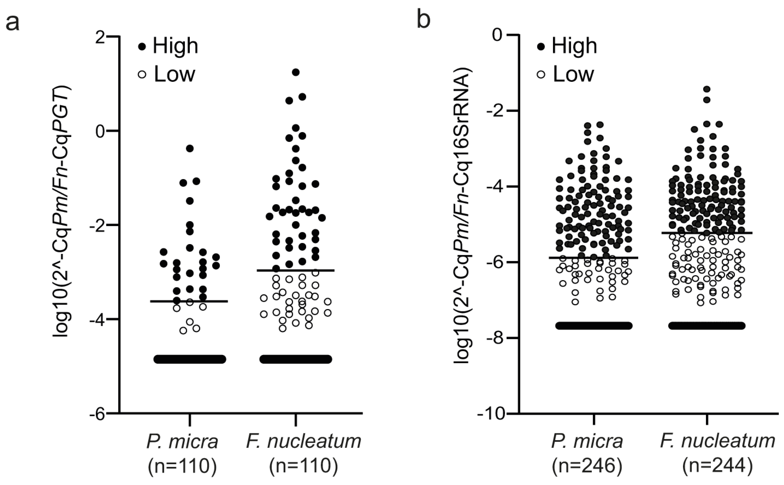
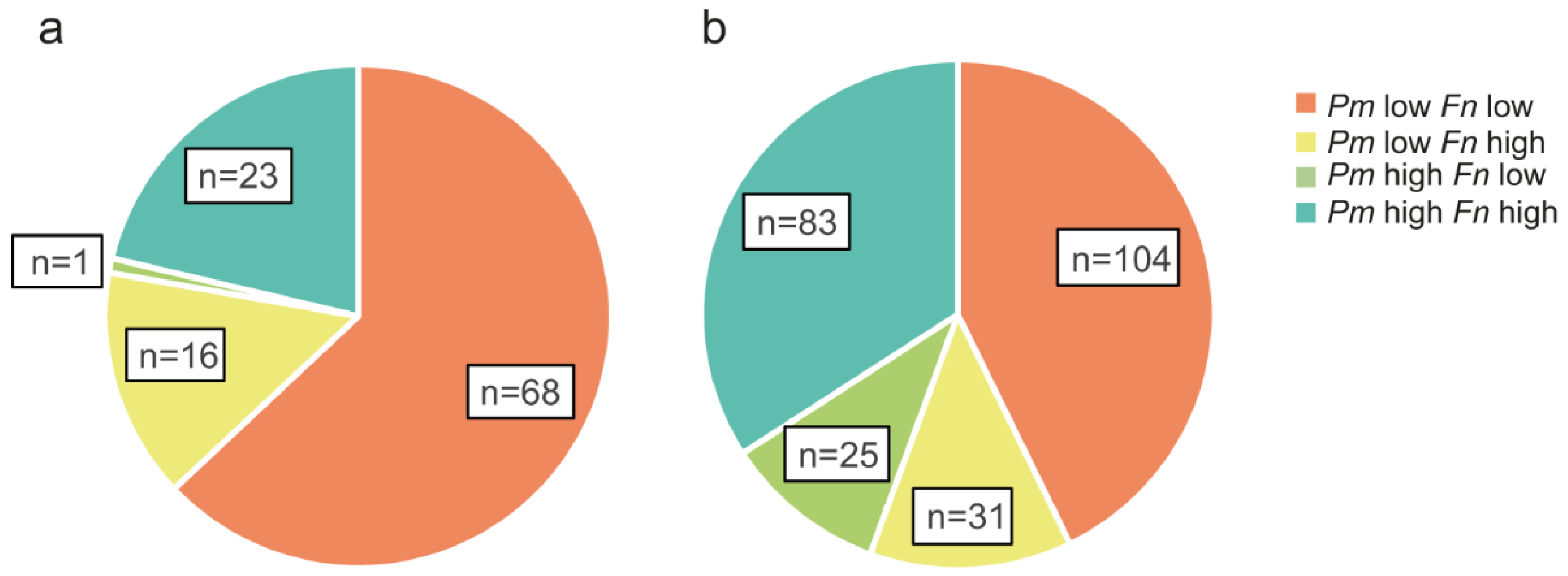
3.1. Distribution of P. micra and F. nucleatum in Tumour Tissue and Faeces of CRC Patients

The levels of P. micra and F. nucleatum were evaluated by qPCR in fresh frozen tumour tissues (n = 112) and in faecal samples (n = 250) from a CRC patient cohort. For fresh frozen tumour tissues, relative quantification of P. micra and F. nucleatum was calculated using the human gene PGT as reference. P. micra was detected in tumour tissue from 30 out of 110 (27.3%) patients and F. nucleatum in 69 out of 110 patients (62.7%). For faecal samples, the relative levels of P. micra and F. nucleatum were calculated using the 16S rRNA gene as reference. P. micra and F. nucleatum were detected in faecal samples from 143 out of 246 (58.1%) and 185 out of 244 patients (75.8%), respectively.
ROC curve analyses based on cancer-specific survival were used to calculate an optimal cut-off value for high or low levels of P. micra and F. nucleatum. The distribution of samples classified as high and low can be found in Figure 1 and Table 1. Utilising this cut-off, 24 out of 110 (21.8%) patients were identified with high levels of P. micra and 40 out of 110 patients (36.4%) with high levels of F. nucleatum in tumour tissue. In faeces, 108 out of 246 patients (43.9%) had high levels of P. micra and 114 out of 244 patients (46.7%) had high levels of F. nucleatum. Of the 24 patients with high levels of P. micra in tumour tissue, 20 patients also had high levels of P. micra in faeces (Table 1). For F. nucleatum analysis in patients with both tumour tissue and faeces available, 24 out of 39 patients with high levels in tumour tissue also had high levels in faeces (Table 1). Furthermore, there was a significant correlation between the relative levels of P. micra or F. nucleatum in tumour tissue and in faeces (rs = 0.533, p < 0.001 for P. micra; rs = 0.468, p < 0.001 for F. nucleatum).
Interestingly, 23 of the 24 tumour tissues with high levels of P. micra also had high levels of F. nucleatum (Figure 2). In faeces, 83 of the 108 samples with high levels of P. micra also had high levels of F. nucleatum (Figure 2). Moreover, there was a significant positive correlation between the levels of P. micra and F. nucleatum in tumour tissue (rs = 0.561, p < 0.001) and in faeces (rs = 0.640, p < 0.001).

3.2. Association of P. micra and F. nucleatum in Tumour Tissue and Faeces with Clinicopathological and Molecular Parameters

The relations of P. micra and F. nucleatum to clinicopathological and molecular characteristics of the study patients were analysed using the cut-off for high/low levels (Table 2 and Table 3, respectively, for fresh frozen tissue). High levels of P. micra in tumour tissue were significantly associated with age and female gender (Table 2), as well as BRAF-mutated tumours and tumours of the MSI subtype (Table 3). As with P. micra, high levels of F. nucleatum were significantly associated with age (Table 2), as well as BRAF-mutated tumours and tumours of the MSI subtype (Table 3). In addition, F. nucleatum was significantly associated with right-sided tumours (Table 2). In faeces, when all included clinical and molecular parameters were analysed, the only significant associations found were those of P. micra and F. nucleatum with tumours of the MSI subtype (Supplementary Tables S1 and S2).

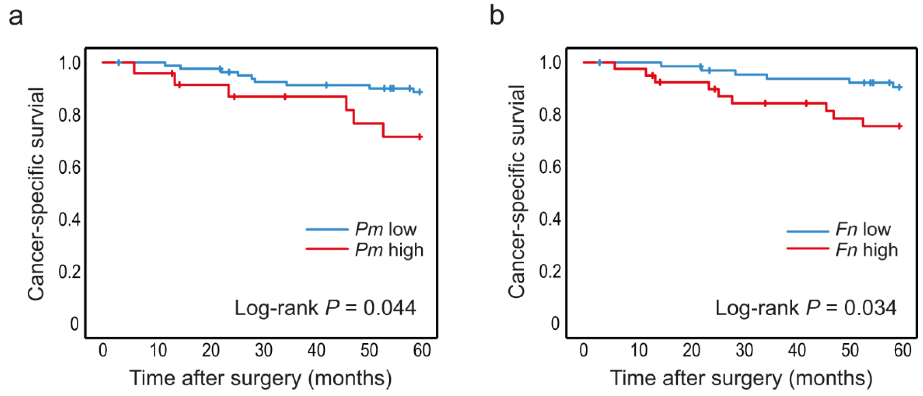
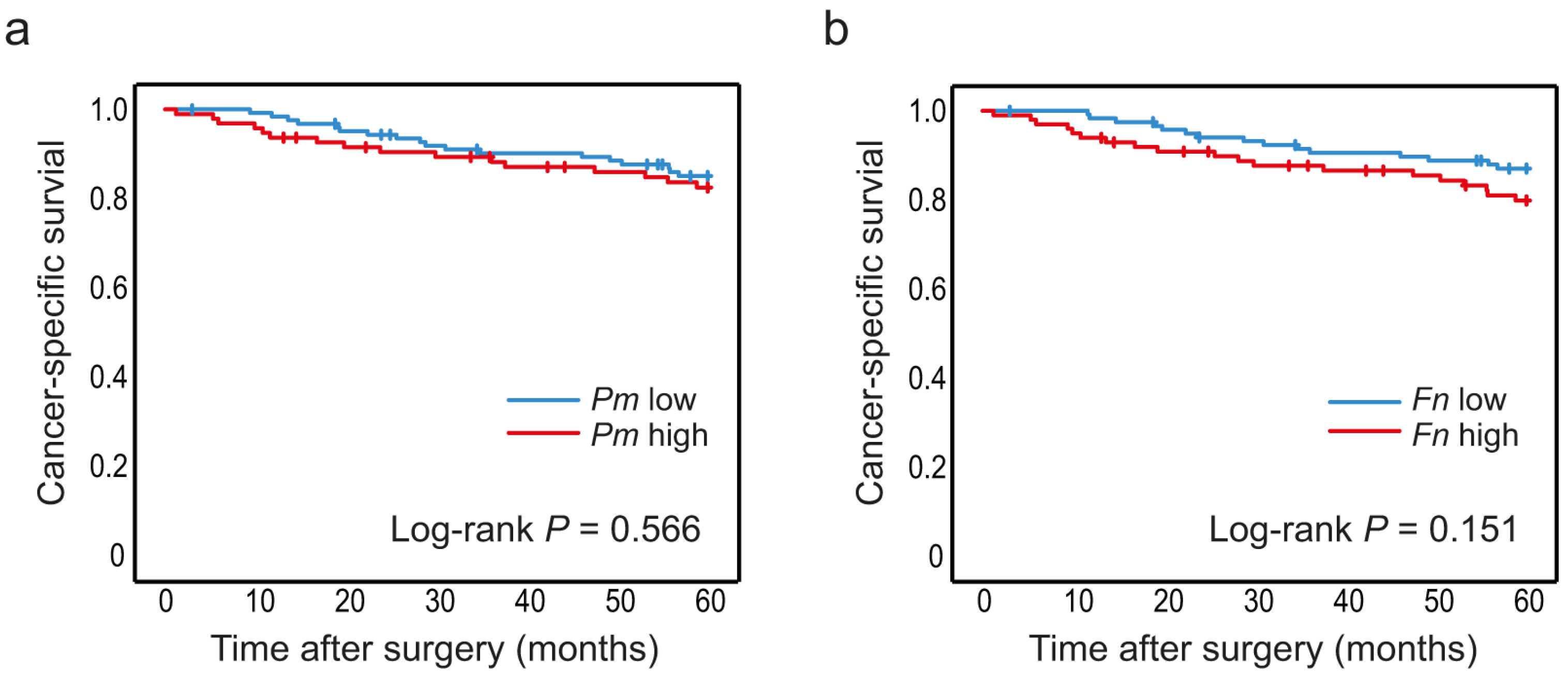
3.3. High Levels of P. micra and F. nucleatum in Tumour Tissue Are Associated with Decreased Patient Survival

The levels of P. micra and F. nucleatum were analysed according to cancer-specific survival in CRC patients. Since no associations were found between the studied bacteria and stage, all stages (I–IV) of CRC patients were included to increase sample size. The median follow-up time from date of cancer surgery was 93.2 (1.2–134) months. Kaplan–Meier curves truncated at 5 years were estimated for patients with high and low levels of P. micra or F. nucleatum in tumour tissue (Figure 3) and in faeces (Figure 4). High levels of P. micra or F. nucleatum in tumour tissue were significantly associated with decreased patient survival (Figure 3). No significant differences in survival were found between patients with high and low levels of P. micra or F. nucleatum in faeces (Figure 4). When excluding stage IV CRC patiens from the analyses, P. micra continued to have a significant association with decreased survival (p = 0.031). For F. nucleatum, no significant association was found (p = 0.090).
High levels of P. micra in tumour tissue were also found to have a significant effect on survival in a multivariable analysis including tumour stage and grade (HR 3.82 (CI 1.12–13.0), p = 0.032), further strengthening the association between increased levels of p. micra in tumour tissue and decreased patient survival (Table 4). For F. nucleatum, the prognostic effect did not remain significant in the multivariable analysis (HR 2.70 (CI 0.83–8.80), p = 0.099) (Table 4).

4. Discussion

In this study, we investigated the presence of two CRC-associated microbes from the oral microflora, P. micra and F. nucleatum, in tumour tissue and in faeces of CRC patients in relation to prognosis. We found a novel association between high tumoural levels of P. micra and a decreased patient survival rate in CRC. High tumoural levels of F. nucleatum were also associated with decreased patient survival. We could further associate high levels of P. micra and/or F. nucleatum in tumour tissue with different patient clinicopathological and tumour molecular traits. In addition, we concluded that analyses of the tumour microbiota were more significant in terms of clinical and molecular associations than analyses of the faecal microflora.
When analysing levels of P. micra and F. nucleatum in tumour tissue in relation to tumour molecular features, high levels of both bacteria were found to be associated with BRAF-mutated tumours and tumours of the MSI subtype. Furthermore, F. nucleatum was significantly associated with right-sided tumours. The results are in line with earlier findings for F. nucleatum [19,29]. In a previous study by our group, as well as in a study by Purcell et al., associations between high levels of P. micra and F. nucleatum in tumour tissue and tumours of CMS subgroup 1 were found [22,25]. CMS 1 tumours are characterised by high immune infiltration and are commonly MSI, BRAF-mutated and right-sided, which is in line with the current findings [30]. High levels of P. micra and F. nucleatum in tumour tissue were further associated both with age under 59 and age above 80. In a study by Bhem et al., an association between high age and high levels of F. nucleatum in tumour tissue was found in gastric cancer patients [31]. However, F. nucleatum has also been associated with young onset CRC [32]. The non-linear association with age found in this study could partly depend on the association with tumours of the MSI subtype. These tumours are known to be more common in patients with hereditary Lynch syndrome and are likely to occur at a younger age [33]. In contrast, sporadic tumours of the MSI subtype are more common in older individuals, having had a longer time to develop mutations leading to MSI [33]. For P. micra, there was a significant association with the female sex, whereas for F. nucleatum there was a trend towards an association, although it was not significant. The association with the female sex has not previously been described and needs to be verified in other studies. However, right-sided tumours, tumours of the MSI subtype and BRAF-mutated tumours are known to be more frequent in older women [34].
Furthermore, we investigated associations between cancer-specific survival in colorectal cancer and levels of P. micra and F. nucleatum in tumour tissue. High levels of P. micra or F. nucleatum in tumour tissue were significantly associated with decreased five-year survival in stage I-IV CRC. This association remained significant in multivariable Cox regression analysis for P. micra. When excluding stage IV CRC patients, the association with decreased survival was still significant for P. micra. To our knowledge, this is the first study showing an association of P. micra in tumour tissue with decreased patient survival. However, one recent study has suggested a prognostic role of faecal P. micra in CRC [26]. F. nucleatum, on the other hand, has been studied more extensively and has previously been associated with decreased survival [14,15,16,17,18,19,20,21]. For example, in a large study by Mima et al. on tumour tissue samples from 1069 CRC cases within the Nurses’ Health Study and the Health Professionals Follow-Up Study, high levels of F. nucleatum were associated with decreased CRC-specific survival (HR 1.58, 95% CI 1.04–2.39) [19]. Interestingly, in this study, both P. micra and F. nucleatum were associated with tumours of the MSI subtype, generally known to have a better prognosis [35]. Associations were also found for P. micra and F. nucleatum with BRAF mutations, which have been proposed to partially mitigate the positive prognostic effect of MSI [35]. It is thus possible that tumours with P. micra and/or F. nucleatum colonisation represent a subgroup of MSI tumours with a worse prognosis. To strengthen this hypothesis, further subgroup analyses are needed in larger patient cohorts. In a study by Wei Z et al., high levels of F. nucleatum in tumour tissue were associated with a decreased survival rate and increased expression of TNF-α, suggesting that dysbiosis might worsen the patient’s prognosis by upregulating gut inflammation [14]. F. nucleatum has also been suggested to be associated with the development of tumour metastasis [36], and has in fact been detected in CRC liver metastases as well as the primary tumour [37]. Chen et al. have further proposed F. nucleatum to promote CRC metastasis through the regulation of caspase activation and recruitment domain 3 (CARD3, a prometastatic kinase), as well as activation of the nuclear factor-kappa B pathway resulting in migration of CRC cells [38,39]. In line with these findings, patients with tumours of the MSI subtype with metastatic spread are known to have decreased survival [35]. In addition, F. nucleatum has been associated with chemotherapy resistance through the regulation of autophagy pathways [40]. It is thus possible that the association with poor survival could depend on proinflammatory factors and the promotion of metastatic formation, as well as chemoresistance.
The levels of P. micra were found to be strongly associated with the levels of F. nucleatum in both tumour tissue and faeces, which is in line with two of our previous studies, as well as a study by Jun Yu et al. [12,23,25]. Interestingly, in a study by Horiuchi A et al., P. micra and F. nucleatum were shown to display synergistic effects in bacterial biofilm formation [41]. Drewes et al. also revealed an abundance of the human oral microbiota in right-sided tumours, including both P. micra and F. nucleatum [10], and right-sided tumours have been suggested to be associated with bacterial biofilm formation [42]. Thus, it is possible that high levels of the studied bacterial species are markers for a higher degree of dysbiosis and bacterial biofilm formation. In a study by Johnson et al., colonic mucosal biofilms were indeed suggested to affect cellular proliferation and tumour growth [43]. Further studies on the mechanistic role of P. micra and F. nucleatum in CRC prognosis are needed to support these theories.
In faecal samples, the associations of P. micra and F. nucleatum with clinical and molecular parameters were much weaker. P. micra and F. nucleatum were still associated with tumours of the MSI subtype. However, no association was found between P. micra or F. nucleatum and cancer-specific survival. One explanation for this could be that tumour tissue represents the local tumour microenvironment better than faecal samples do. In this study, we found significant associations between levels of P. micra and F. nucleatum in both tumour tissue and in faeces, respectively, suggesting that the faecal microflora still partly reflects the local tumour microflora. However, for more detailed analyses of the molecular role of P. micra and F. nucleatum in tumour progression and patient survival, tumour tissue appears to be the preferred choice.
One strength of this study is the inclusion of both tumour tissue and faecal samples originating from the same individuals, giving a better insight into the spatial presence of the examined species in association with studied traits. However, the lack of a validation cohort calls for further studies in order to confirm our results. Furthermore, our study does not include any mechanistic insights for the found associations. Thus, studies on the underlying mechanisms, including inflammatory and immunological processes, are needed in order to better understand the cause and effect relationships. Another area of interest is the association with dietary factors, which may directly and indirectly affect the constitution of the gut microbial content.

5. Conclusions

Our study suggests associations between high levels of P. micra and F. nucleatum in tumour tissue and decreased patient survival in CRC. We found that P. micra and F. nucleatum often colonise the same tumours, and that tumours colonised with P. micra and/or F. nucleatum more often are BRAF-mutated and of the MSI subtype. However, further studies on the role of the gut microbiota in CRC formation and progression, including underlying mechanisms, are needed. A better understanding of the role of gut microbial composition in CRC progression might contribute to future putative targets for therapy.

Supplementary Materials

The following supporting information can be downloaded at: https://www.mdpi.com/article/10.3390/cancers14235937/s1, Table S1: clinicopathological characteristics of study patients in relation to P. micra and F. nucleatum in faeces; Table S2: molecular characteristics of study patients in relation to P. micra and F. nucleatum in faeces.

Author Contributions

Conceptualization, T.L., I.L., S.E. and R.P.; Methodology, T.L., A.L.-B. and C.Z.; Formal Analysis, T.L., S.E. and R.P.; Investigation, T.L., S.E. and R.P.; Data Curation, T.L., I.L., S.E. and R.P.; Writing—Original Draft Preparation, T.L. and S.E.; Writing—Review and Editing, T.L., A.L.-B., C.Z., I.L., M.D., S.E. and R.P.; Visualization, T.L. and S.E.; Project Administration, R.P.; Funding Acquisition, T.L. and R.P. All authors have read and agreed to the published version of the manuscript.

Funding

This work was supported by grants from the Swedish Cancer Society (20 1271 PjF, R.P.), The Sjöberg Foundation (2021, R.P.), Lion´s Cancer Research Foundation (AMP 21-1048, T.L.), the Cancer Research Foundation in Northern Sweden (LP 22-2318, T.L.), and the County Council of Västerbotten (2021, R.P.). The funders had no role in study design, data collection, analysis, or interpretation of the data.

Institutional Review Board Statement

The study was conducted according to the guidelines of the Declaration of Helsinki, and approved by the Regional Ethical Review Board in Umeå, Sweden (dnr 2016/219–31), in accordance with relevant guidelines and regulations.

Informed Consent Statement

Written informed consent was obtained from all subjects involved in the study.

Data Availability Statement

The data presented in this study are available on reasonable request from the corresponding author.

Acknowledgments

The authors are grateful to all the patients who participated in the study. We also thank Åsa Stenberg for valuable technical assistance.

Conflicts of Interest

The authors declare no conflict of interest.

References

  1. Morgan, E.; Arnold, M.; Gina, A.; Lorenzoni, V.; Cabasag, M.L.; Laversanne, M.; Vignat, J.; Ferlay, J.; Murphy, N.; Bray, F. Global burden of colorectal cancer in 2020 and 2040: Incidence and mortality estimates from GLOBOCAN. Gut 2022. [Google Scholar] [CrossRef]
  2. Wong, S.H.; Yu, J. Gut microbiota in colorectal cancer: Mechanisms of action and clinical applications. Nat. Rev. Gastroenterol. Hepatol. 2019, 16, 690–704. [Google Scholar] [CrossRef] [PubMed]
  3. Tjalsma, H.; Boleij, A.; Marchesi, J.R.; Dutilh, B.E. A bacterial driver-passenger model for colorectal cancer: Beyond the usual suspects. Nat. Rev. Microbiol. 2012, 10, 575–582. [Google Scholar] [CrossRef] [PubMed]
  4. Chen, J.; Pitmon, E.; Wang, K. Microbiome, inflammation and colorectal cancer. Semin. Immunol. 2017, 32, 43–53. [Google Scholar] [CrossRef]
  5. Long, A.G.; Lundsmith, E.T.; Hamilton, K.E. Inflammation and colorectal cancer. Curr. Colorectal. Cancer Rep. 2017, 13, 341–351. [Google Scholar] [CrossRef] [PubMed]
  6. Zitvogel, L.; Ma, Y.; Raoult, D.; Kroemer, G.; Gajewski, T.F. The microbiome in cancer immunotherapy: Diagnostic tools and therapeutic strategies. Science 2018, 359, 1366–1370. [Google Scholar] [CrossRef] [Green Version]
  7. Dai, Z.; Coker, O.O.; Nakatsu, G.; Wu, W.K.K.; Zhao, L.; Chen, Z.; Chan, F.K.L.; Kristiansen, K.; Sung, J.J.Y.; Wong, S.H.; et al. Multi-cohort analysis of colorectal cancer metagenome identified altered bacteria across populations and universal bacterial markers. Microbiome 2018, 6, 70. [Google Scholar] [CrossRef]
  8. Wirbel, J.; Pyl, P.T.; Kartal, E.; Zych, K.; Kashani, A.; Milanese, A.; Fleck, J.S.; Voigt, A.Y.; Palleja, A.; Ponnudurai, R.; et al. Meta-analysis of fecal metagenomes reveals global microbial signatures that are specific for colorectal cancer. Nat. Med. 2019, 25, 679–689. [Google Scholar] [CrossRef] [Green Version]
  9. Baxter, N.T.; Ruffin, M.T.T.; Rogers, M.A.; Schloss, P.D. Microbiota-based model improves the sensitivity of fecal immunochemical test for detecting colonic lesions. Genome Med. 2016, 8, 37. [Google Scholar] [CrossRef] [Green Version]
  10. Drewes, J.L.; White, J.R.; Dejea, C.M.; Fathi, P.; Iyadorai, T.; Vadivelu, J.; Roslani, A.C.; Wick, E.C.; Mongodin, E.F.; Loke, M.F.; et al. High-resolution bacterial 16S rRNA gene profile meta-analysis and biofilm status reveal common colorectal cancer consortia. NPJ Biofilms Microbiomes 2017, 3, 34. [Google Scholar] [CrossRef]
  11. Shah, M.S.; DeSantis, T.Z.; Weinmaier, T.; McMurdie, P.J.; Cope, J.L.; Altrichter, A.; Yamal, J.M.; Hollister, E.B. Leveraging sequence-based faecal microbial community survey data to identify a composite biomarker for colorectal cancer. Gut 2018, 67, 882–891. [Google Scholar] [CrossRef]
  12. Yu, J.; Feng, Q.; Wong, S.H.; Zhang, D.; Liang, Q.Y.; Qin, Y.; Tang, L.; Zhao, H.; Stenvang, J.; Li, Y.; et al. Metagenomic analysis of faecal microbiome as a tool towards targeted non-invasive biomarkers for colorectal cancer. Gut 2017, 66, 70–78. [Google Scholar] [CrossRef] [PubMed]
  13. Rubinstein, M.R.; Baik, J.E.; Lagana, S.M.; Han, R.P.; Raab, W.J.; Sahoo, D.; Dalerba, P.; Wang, T.C.; Han, Y.W. Fusobacterium nucleatum promotes colorectal cancer by inducing Wnt/beta-catenin modulator Annexin A1. EMBO Rep. 2019, 20, e47638. [Google Scholar] [CrossRef] [PubMed]
  14. Wei, Z.; Cao, S.; Liu, S.; Yao, Z.; Sun, T.; Li, Y.; Li, J.; Zhang, D.; Zhou, Y. Could gut microbiota serve as prognostic biomarker associated with colorectal cancer patients’ survival? A pilot study on relevant mechanism. Oncotarget 2016, 7, 46158–46172. [Google Scholar] [CrossRef] [PubMed] [Green Version]
  15. Kim, Y.; Cho, N.Y.; Kang, G.H. Prognostic and clinicopathological significance of Fusobacterium nucleatum in colorectal cancer: A systemic review and meta-analysis. J. Pathol. Transl. Med. 2022, 56, 144–151. [Google Scholar] [CrossRef]
  16. Chen, Y.; Lu, Y.; Ke, Y.; Li, Y. Prognostic impact of the Fusobacterium nucleatum status in colorectal cancers. Medicine 2019, 98, e17221. [Google Scholar] [CrossRef]
  17. de Carvalho, A.C.; de Mattos Pereira, L.; Datorre, J.G.; Dos Santos, W.; Berardinelli, G.N.; Matsushita, M.M.; Oliveira, M.A.; Durães, R.O.; Guimarães, D.P.; Reis, R.M. Microbiota Profile and Impact of Fusobacterium nucleatum in Colorectal Cancer Patients of Barretos Cancer Hospital. Front. Oncol. 2019, 9, 813. [Google Scholar] [CrossRef] [Green Version]
  18. Kunzmann, A.T.; Proença, M.A.; Jordao, H.W.; Jiraskova, K.; Schneiderova, M.; Levy, M.; Liska, V.; Buchler, T.; Vodickova, L.; Vymetalkova, V.; et al. Fusobacterium nucleatum tumor DNA levels are associated with survival in colorectal cancer patients. Eur. J. Clin. Microbiol. Infect. Dis. 2019, 38, 1891–1899. [Google Scholar] [CrossRef] [Green Version]
  19. Mima, K.; Nishihara, R.; Qian, Z.R.; Cao, Y.; Sukawa, Y.; Nowak, J.A.; Yang, J.; Dou, R.; Masugi, Y.; Song, M.; et al. Fusobacterium nucleatum in colorectal carcinoma tissue and patient prognosis. Gut 2016, 65, 1973–1980. [Google Scholar] [CrossRef] [Green Version]
  20. Lee, D.W.; Han, S.W.; Kang, J.K.; Bae, J.M.; Kim, H.P.; Won, J.K.; Jeong, S.Y.; Park, K.J.; Kang, G.H.; Kim, T.Y. Association Between Fusobacterium nucleatum, Pathway Mutation, and Patient Prognosis in Colorectal Cancer. Ann. Surg. Oncol. 2018, 25, 3389–3395. [Google Scholar] [CrossRef]
  21. Yamaoka, Y.; Suehiro, Y.; Hashimoto, S.; Hoshida, T.; Fujimoto, M.; Watanabe, M.; Imanaga, D.; Sakai, K.; Matsumoto, T.; Nishioka, M.; et al. Fusobacterium nucleatum as a prognostic marker of colorectal cancer in a Japanese population. J. Gastroenterol. 2018, 53, 517–524. [Google Scholar] [CrossRef] [PubMed]
  22. Purcell, R.V.; Visnovska, M.; Biggs, P.J.; Schmeier, S.; Frizelle, F.A. Distinct gut microbiome patterns associate with consensus molecular subtypes of colorectal cancer. Sci. Rep. 2017, 7, 11590. [Google Scholar] [CrossRef] [PubMed] [Green Version]
  23. Löwenmark, T.; Löfgren-Burström, A.; Zingmark, C.; Eklöf, V.; Dahlberg, M.; Wai, S.N.; Larsson, P.; Ljuslinder, I.; Edin, S.; Palmqvist, R. Parvimonas micra as a putative non-invasive faecal biomarker for colorectal cancer. Sci. Rep. 2020, 10, 15250. [Google Scholar] [CrossRef] [PubMed]
  24. Xu, J.; Yang, M.; Wang, D.; Zhang, S.; Yan, S.; Zhu, Y.; Chen, W. Alteration of the abundance of Parvimonas micra in the gut along the adenoma-carcinoma sequence. Oncol. Lett. 2020, 20, 106. [Google Scholar] [CrossRef] [PubMed]
  25. Löwenmark, T.; Li, X.; Löfgren-Burström, A.; Zingmark, C.; Ling, A.; Kellgren, T.G.; Larsson, P.; Ljuslinder, I.; Wai, S.N.; Edin, S.; et al. Parvimonas micra is associated with tumour immune profiles in molecular subtypes of colorectal cancer. Cancer Immunol. Immunother. 2022, 71, 2565–2575. [Google Scholar] [CrossRef]
  26. Zhao, L.; Zhang, X.; Zhou, Y.; Fu, K.; Lau, H.C.; Chun, T.W.; Cheung, A.H.; Coker, O.O.; Wei, H.; Wu, W.K.; et al. Parvimonas micra promotes colorectal tumorigenesis and is associated with prognosis of colorectal cancer patients. Oncogene 2022, 41, 4200–4210. [Google Scholar] [CrossRef]
  27. Glimelius, B.; Melin, B.; Enblad, G.; Alafuzoff, I.; Beskow, A.; Ahlström, H.; Bill-Axelson, A.; Birgisson, H.; Björ, O.; Edqvist, P.H.; et al. U-CAN: A prospective longitudinal collection of biomaterials and clinical information from adult cancer patients in Sweden. Acta Oncol. 2018, 57, 187–194. [Google Scholar] [CrossRef] [Green Version]
  28. Li, X.; Larsson, P.; Ljuslinder, I.; Öhlund, D.; Myte, R.; Löfgren-Burström, A.; Zingmark, C.; Ling, A.; Edin, S.; Palmqvist, R. Ex Vivo Organoid Cultures Reveal the Importance of the Tumor Microenvironment for Maintenance of Colorectal Cancer Stem Cells. Cancers 2020, 12, 923. [Google Scholar] [CrossRef] [Green Version]
  29. Mima, K.; Cao, Y.; Chan, A.T.; Qian, Z.R.; Nowak, J.A.; Masugi, Y.; Shi, Y.; Song, M.; da Silva, A.; Gu, M.; et al. Fusobacterium nucleatum in Colorectal Carcinoma Tissue According to Tumor Location. Clin. Transl. Gastroenterol. 2016, 7, e200. [Google Scholar] [CrossRef]
  30. Guinney, J.; Dienstmann, R.; Wang, X.; de Reyniès, A.; Schlicker, A.; Soneson, C.; Marisa, L.; Roepman, P.; Nyamundanda, G.; Angelino, P.; et al. The consensus molecular subtypes of colorectal cancer. Nat. Med. 2015, 21, 1350–1356. [Google Scholar] [CrossRef]
  31. Boehm, E.T.; Thon, C.; Kupcinskas, J.; Steponaitiene, R.; Skieceviciene, J.; Canbay, A.; Malfertheiner, P.; Link, A. Fusobacterium nucleatum is associated with worse prognosis in Lauren’s diffuse type gastric cancer patients. Sci. Rep. 2020, 10, 16240. [Google Scholar] [CrossRef] [PubMed]
  32. Yang, Y.; Du, L.; Shi, D.; Kong, C.; Liu, J.; Liu, G.; Li, X.; Ma, Y. Dysbiosis of human gut microbiome in young-onset colorectal cancer. Nat. Commun. 2021, 12, 6757. [Google Scholar] [CrossRef] [PubMed]
  33. Boland, C.R.; Goel, A. Microsatellite instability in colorectal cancer. Gastroenterology 2010, 138, 2073–2087.e2073. [Google Scholar] [CrossRef] [PubMed]
  34. Murphy, G.; Devesa, S.S.; Cross, A.J.; Inskip, P.D.; McGlynn, K.A.; Cook, M.B. Sex disparities in colorectal cancer incidence by anatomic subsite, race and age. Int. J. Cancer 2011, 128, 1668–1675. [Google Scholar] [CrossRef] [PubMed] [Green Version]
  35. Gelsomino, F.; Barbolini, M.; Spallanzani, A.; Pugliese, G.; Cascinu, S. The evolving role of microsatellite instability in colorectal cancer: A review. Cancer Treat. Rev. 2016, 51, 19–26. [Google Scholar] [CrossRef] [Green Version]
  36. Chen, W.D.; Zhang, X.; Zhang, Y.P.; Yue, C.B.; Wang, Y.L.; Pan, H.W.; Zhang, Y.L.; Liu, H.; Zhang, Y. Fusobacterium Nucleatum is a Risk Factor for Metastatic Colorectal Cancer. Curr. Med. Sci. 2022, 42, 538–547. [Google Scholar] [CrossRef]
  37. Bullman, S.; Pedamallu, C.S.; Sicinska, E.; Clancy, T.E.; Zhang, X.; Cai, D.; Neuberg, D.; Huang, K.; Guevara, F.; Nelson, T.; et al. Analysis of Fusobacterium persistence and antibiotic response in colorectal cancer. Science 2017, 358, 1443–1448. [Google Scholar] [CrossRef] [Green Version]
  38. Chen, Y.; Chen, Y.; Zhang, J.; Cao, P.; Su, W.; Deng, Y.; Zhan, N.; Fu, X.; Huang, Y.; Dong, W. Fusobacterium nucleatum Promotes Metastasis in Colorectal Cancer by Activating Autophagy Signaling via the Upregulation of CARD3 Expression. Theranostics 2020, 10, 323–339. [Google Scholar] [CrossRef]
  39. Chen, S.; Su, T.; Zhang, Y.; Lee, A.; He, J.; Ge, Q.; Wang, L.; Si, J.; Zhuo, W.; Wang, L. Fusobacterium nucleatum promotes colorectal cancer metastasis by modulating KRT7-AS/KRT7. Gut Microbes 2020, 11, 511–525. [Google Scholar] [CrossRef]
  40. Yu, T.; Guo, F.; Yu, Y.; Sun, T.; Ma, D.; Han, J.; Qian, Y.; Kryczek, I.; Sun, D.; Nagarsheth, N.; et al. Fusobacterium nucleatum Promotes Chemoresistance to Colorectal Cancer by Modulating Autophagy. Cell 2017, 170, 548–563.e516. [Google Scholar] [CrossRef]
  41. Horiuchi, A.; Kokubu, E.; Warita, T.; Ishihara, K. Synergistic biofilm formation by Parvimonas micra and Fusobacterium nucleatum. Anaerobe 2020, 62, 102100. [Google Scholar] [CrossRef] [PubMed]
  42. Dejea, C.M.; Wick, E.C.; Hechenbleikner, E.M.; White, J.R.; Mark Welch, J.L.; Rossetti, B.J.; Peterson, S.N.; Snesrud, E.C.; Borisy, G.G.; Lazarev, M.; et al. Microbiota organization is a distinct feature of proximal colorectal cancers. Proc. Natl. Acad. Sci. USA 2014, 111, 18321–18326. [Google Scholar] [CrossRef] [PubMed] [Green Version]
  43. Johnson, C.H.; Dejea, C.M.; Edler, D.; Hoang, L.T.; Santidrian, A.F.; Felding, B.H.; Ivanisevic, J.; Cho, K.; Wick, E.C.; Hechenbleikner, E.M.; et al. Metabolism links bacterial biofilms and colon carcinogenesis. Cell Metab. 2015, 21, 891–897. [Google Scholar] [CrossRef] [PubMed]
Figure 1. The levels of P. micra and F. nucleatum in CRC patients. Scatter plots illustrating the relative levels of P. micra (Pm) and F. nucleatum (Fn) in (a) fresh frozen tumour tissue and (b) faeces. Horizontal lines indicate the cut-off between high and low levels of the bacteria as calculated by the 2−ΔCq method using either PGT (for fresh frozen tumour tissue) or 16s RNA (for faeces) as reference.
Cancers 14 05937 g001
Figure 2. The relative distribution of P. micra and F. nucleatum in CRC patients. Pie charts are used to illustrate groups of patients with high or low levels of P. micra (Pm) and/or F. nucleatum (Fn) in (a) fresh frozen tumour tissue (n = 108) or (b) faeces (n = 243) of patients analysed for both microbial markers.
Cancers 14 05937 g002
Figure 3. Levels of P. micra and F. nucleatum in tumour tissue in relation to CRC patient survival. Kaplan–Meier plots of 5-year cancer-specific survival for patients with high or low levels of (a) P. micra and (b) F. nucleatum in tumour tissue. Log-rank tests were used to calculate p values.
Cancers 14 05937 g003
Figure 4. Levels of P. micra and F. nucleatum in faeces in relation to CRC patient survival. Kaplan–Meier plots of 5-year cancer-specific survival for patients with high or low levels of (a) P. micra and (b) F. nucleatum in faecal samples. Log-rank tests were used to calculate p values.
Cancers 14 05937 g004
Table 1. The distribution of patients with high and low levels of P. micra and F. nucleatum in tumour tissue and in faeces.
Faeces
P. micrap ValueF. nucleatump Value
Fresh frozen tissueHighLow HighLow
P. micra, n (%)
High20 (83.3)4 (16.7)<0.00118 (75.0)6 (25.0)<0.001
Low22 (26.2)62 (73.8) 28 (34.1)54 (65.9)
F. nucleatum, n (%)
High22 (55.0)18 (45.0)0.01424 (61.5)15 (38.5)0.009
Low20 (29.4)48 (70.6) 23 (34.3)44 (65.7)
Fischer’s exact test was used to compare categorical variables.
Table 2. Clinicopathological characteristics of study patients in relation to P. micra and F. nucleatum in tumour tissue.
P. micrap ValueF. nucleatump Value
LowHigh LowHigh
Age, n (%)
≤5911 (61.1)7 (38.9)0.0237 (38.9)11 (61.1)0.017
60–6932 (91.4)3 (8.6) 28 (80.0)7 (20.0)
70–7933 (80.5)8 (19.5) 27 (65.9)14 (34.1)
≥8010 (62.5)6 (37.5) 8 (50.0)8 (50.0)
Gender, n (%)
Male53 (89.8)6 (10.2)0.00243 (71.7)17 (28.3)0.073
Female33 (64.7)18 (35.3) 27 (54.0)23 (46.0)
Location, n (%)
Right colon23 (69.7)10 (30.3)0.26912 (37.5)20 (62.5)0.002
Left colon16 (76.2)5 (23.8) 16 (76.2)5 (23.8)
Rectum47 (83.9)9 (16.1) 42 (73.7)15 (26.3)
Stage, n (%)
I29 (90.6)3 (9.4)0.17123 (74.2)8 (25.8)0.425
II28 (73.7)10 (26.3) 24 (63.2)14 (36.8)
III21 (70.0)9 (30.0) 17 (54.8)14 (45.2)
IV7 (77.8)2 (22.2) 5 (55.6)4 (44.4)
Tumour grade, n (%)
Low grade71 (80.7)17 (19.3)0.14358 (65.9)30 (34.1)0.207
High grade13 (65.0)7 (35.0) 10 (50.0)10 (50.0)
Tumour type, n (%)
Non-mucinous72 (79.1)19 (20.9)0.52559 (64.8)32(35.2)0.416
Mucinous12 (70.6)5 (29.4) 9 (52.9)8 (47.1)
Fischer’s exact test was used to compare categorical variables.
Table 3. Molecular characteristics of study patients in relation to P. micra and F. nucleatum in tumour tissue.
P. micrap ValueF. nucleatump Value
LowHigh LowHigh
KRASstatus, n (%)
Wild type51 (76.1)16 (23.9)0.63843 (65.2)23 (34.8)0.691
Mutant35 (81.4)8 (18.6) 27 (61.4)17 (38.6)
BRAFstatus, n (%)
Wild type75 (82.4)16 (17.6)0.03062 (68.1)29 (31.9)0.039
Mutant11 (57.9)8 (42.1) 8 (42.1)11 (57.9)
MSI status, n (%)
MSS80 (83.3)16 (16.7)0.00266 (68.8)30 (31.3)0.006
MSI6 (42.9)8 (57.1) 4 (28.6)10 (71.4)
Fischer’s exact test was used to compare categorical variables. Abbreviations: MSI, microsatellite. instable; MSS, microsatellite stable.
Table 4. 5-year survival uni- and multi-variable Cox analyses. The multivariable analyses were performed separately for P. micra and F. nucleatum.
UnivariableMultivariable P. micraMultivariable F. nucleatum
HR95.0% CIp ValueHR95.0% CIp ValueHR95.0% CIp Value
Stage
 I+II1 1 1
 III6.26(2.04–19.2)0.00110.2(1.23–84.62)0.03210.3(1.23–86.2)0.032
 IV34.07(11.26–103.0)<0.001119(12.8–1106)<0.00191.2(10.0–828)<0.001
Gender
 Female1
 Male1.21(0.61–2.42)0.584NA -NA -
Age
 ≤591
 60–690.42(0.18–1.02)0.055NA -NA -
 70–790.51(0.21–1.22)0.129NA -NA -
 ≥800.56(0.18–1.80)0.333NA -NA -
Location
 Right colon1
 Left colon0.81(0.24–2.75)0.731NA -NA -
 Rectum0.98(0.42–2.27)0.954NA -NA -
Tumour grade
 Low grade1 1 1
 High grade4.80(2.24–10.28)<0.0013.16(1.01–9.93)0.0482.73(0.86–8.69)0.089
Tumour type
 Non-mucinous1
 Mucinous2.10(0.86–5.15)0.103NA -NA -
KRAS
 Wild type1
 Mutant1.36(0.58–3.17)0.483NA -NA -
BRAF
 Wild type1
 Mutant1.35(0.46–3.99)0.589NA -NA -
MSI
 MSS1
 MSI0.78(0.18–3.34)0.739NA -NA -
Tumour tissue
P. micra low1 1
P. micra high2.77(0.98–7.78)0.0543.82(1.12–13.0)0.032NA -
F. nucleatum low1 1
F. nucleatum high2.91(1.04–8.19)0.043NA -2.70(0.83–8.80)0.099
Faeces
P. micra low1
P. micra high1.22(0.62–2.39)0.567NA -NA -
F. nucleatum low1
F. nucleatum high1.64(0.83–3.22)0.155NA -NA -
Variables included in the multivariable analyses were selected using 1. Backward Stepwise Conditional model and 2. only including variables with a significant effect on survival in the univariable analysis. Both methods resulted in the same variables being included in the multivariable analyses (stage, grade and levels of P. micra or F. nucleatum in tumour tissue). Abbreviation: NA, not applicable.
Publisher’s Note: MDPI stays neutral with regard to jurisdictional claims in published maps and institutional affiliations.

