Next Article in Journal
Design and Validation of a Handheld Optical Polarization Imager for Preoperative Delineation of Basal Cell Carcinoma
Previous Article in Journal
Neuroendocrine Tumors: Treatment and Management
 
 
cancers-logo
Article Menu
Article

Stereotactic Radiosurgery for Benign Cavernous Sinus Meningiomas: A Multicentre Study and Review of the Literature

by
Antonio Santacroce
1,2,*,†,
Constantin Tuleasca
3,4,5,6,7,†,
Roman Liščák
8,
Enrico Motti
9,
Christer Lindquist
10,
Matthias Radatz
11,
Brigitte Gatterbauer
12,
Bodo E. Lippitz
13,
Roberto Martínez Álvarez
14,
Nuria Martínez Moreno
14,
Marcel A. Kamp
15,
Bente Sandvei Skeie
16,
Stephanie Schipmann
16,
Michele Longhi
17,
Frank Unger
18,
Ian Sabin
19,
Thomas Mindermann
20,
Otto Bundschuh
21,
Gerhard A. Horstmann
22,
A.T.C. J. van Eck
22,
Maja Walier
23,24,
Manfred Berres
23,24,
Makoto Nakamura
2,25,
Hans Jakob Steiger
26,
Daniel Hänggi
26,
Thomas Fortmann
1,2,
Samer Zawy Alsofy
1,2,
Jean Régis
27 and
Christian Ewelt
1,28
add Show full author list remove Hide full author list
1
Department of Neurosurgery, St. Barbara-Klinik Hamm-Heessen, 59073 Hamm, Germany
2
Department of Medicine, Faculty of Health, Witten/Herdecke University, 58455 Witten, Germany
3
Neurosurgery Service and Gamma Knife Center, Lausanne University Hospital (CHUV), 1011 Lausanne, Switzerland
4
Faculté de Biologie et de Médecine (FBM), Université de Lausanne (Unil), 1005 Lausanne, Switzerland
5
Signal Processing Laboratory (LTS 5), Swiss Federal Institute of Technology (EPFL), 1015 Lausanne, Switzerland
6
Faculté de Médecine, Sorbonné Université, 70513 Paris, France
7
Assisstance Publique-Hôpitaux de Paris, Hôpitaux Universitaires Paris Sud, Centre Hospitalier Universitaire Bicêtre, Service de Neurochirurgie, 94270 Le Kremlin-Bicêtre, France
8
Department of Stereotactic and Radiation Neurosurgery, Na Homolce Hospital, 15000 Prague, Czech Republic
9
Dipartimento di Neuroscienze, Neurochirurgia, Università degli Studi di Milano, 20122 Milano, Italy; Villa Maria Cecilia Hospital, 48033 Cotignola, Italy
10
Gamma Knife Centre, Bupa Cromwell Hospital, London SW5 0TU, UK
11
National Centre for Stereotactic Radiosurgery, Royal Hallamshire Hospital, Sheffield S10 2JF, UK
12
Department of Neurosurgery, Medical University of Vienna, 1090 Vienna, Austria
13
Interdisciplinary Centre for Radiosurgery (ICERA), Radiological Alliance, 22767 Hamburg, Germany
14
Department of Radiosurgery, Rúber International Hospital, 28034 Madrid, Spain
15
Department of Neurosurgery, Jena University Hospital, Friedrich-Schiller-University Jena, 07747 Jena, Germany
16
Department of Neurosurgery, Haukeland University Hospital, 5021 Bergen, Norway
17
Unit of Radiosurgery and Stereotactic Neurosurgery, Department of Neurosciences, Azienda Ospedaliera Universitaria, 37126 Verona, Italy
18
Department of Neurosurgery, Medical University Graz, 8036 Graz, Austria
19
Gamma Knife Unit, Wellington Hospital (Platinum Medical Centre), London NW8 7JA, UK
20
Gamma Knife Center Zurich, Klinik Im Park Hirslanden, 8002 Zurich, Switzerland
21
Gamma Knife Zentrum Hannover, 30167 Hannover, Germany
22
Gamma Knife Zentrum Krefeld, 47805 Krefeld, Germany
23
Institute of Medical Biometry, Epidemiology and Informatics, University Medical Center of Mainz, Langenbeckstrasse 1, 55131 Mainz, Germany
24
Department of Mathematics and Technology, University of Applied Sciences Koblenz, Joseph-Rovan-Allee 2, 53424 Remagen, Germany
25
Department of Neurosurgery, Academic Hospital Köln-Merheim, 51058 Köln, Germany
26
Department of Neurosurgery, Heinrich-Heine-Universität Düsseldorf, 40225 Düsseldorf, Germany
27
Service de Neurochirurgie Fonctionnelle et Stereotaxique, Hôpital D’adulte de la Timone, 13354 Marseille, France
28
Department of Neurosurgery, University Hospital Münster, Albert-Schweitzer-Campus 1, A1, 48149 Munster, Germany
*
Author to whom correspondence should be addressed.
These authors contributed equally to this work.
Cancers 2022, 14(16), 4047; https://doi.org/10.3390/cancers14164047
Submission received: 17 April 2022 / Revised: 17 June 2022 / Accepted: 2 July 2022 / Published: 22 August 2022
(This article belongs to the Section Methods and Technologies Development)

Abstract

:

Simple Summary

Meningiomas are the most common tumours of the central nervous system (CNS). Despite their benign histology, proximity to critical neurovascular structures may lead to significant morbidity with tumour growth. This is the case for cavernous sinus meningiomas (CSMs), as their growth may surround critical neuro-vascular structures and cause significant morbidity. Radical microsurgical resection carries a high risk of additional neurological deficits, as well as the risk of death. Current management of these tumours, where treatment is indicated, has moved away from radical surgery towards radiotherapy/radiosurgery. This is particularly the case for patients who have residual or recurring tumours after previous surgery. There are many reports that describe the effectiveness of using stereotactic radiosurgery (SRS) for CSMs; however, large cohort analyses are lacking. This multicentre analysis reports the outcome data of over 1000 patients with CSMs who were treated with SRS. SRS shows a high local tumour control rate with few complications. These results agree with previous reports in the literature. SRS is a valuable primary or adjuvant treatment option for CSMs.

Abstract

Cavernous sinus meningiomas (CSMs) remain a surgical challenge due to the intimate involvement of their contained nerves and blood vessels. Stereotactic radiosurgery (SRS) is a safe and effective minimally invasive alternative for the treatment of small- to medium-sized CSMs. Objective: To assess the medium- to long-term outcomes of SRS for CSMs with respect to tumour growth, prevention of further neurological deterioration and improvement of existing neurological deficits. This multicentric study included data from 15 European institutions. We performed a retrospective observational analysis of 1222 consecutive patients harbouring 1272 benign CSMs. All were treated with Gamma Knife stereotactic radiosurgery (SRS). Clinical and imaging data were retrieved from each centre and entered into a common database. All tumours with imaging follow-up of less than 24 months were excluded. Detailed results from 945 meningiomas (86%) were then analysed. Clinical neurological outcomes were available for 1042 patients (85%). Median imaging follow-up was 67 months (mean 73.4, range 24–233). Median tumour volume was 6.2 cc (+/−7), and the median marginal dose was 14 Gy (+/−3). The post-treatment tumour volume decreased in 549 (58.1%), remained stable in 336 (35.6%) and increased in only 60 lesions (6.3%), yielding a local tumour control rate of 93.7%. Only 27 (2.8%) of the 60 enlarging tumours required further treatment. Five- and ten-year actuarial progression-free survival (PFS) rates were 96.7% and 90.1%, respectively. Tumour control rates were higher for women than men (p = 0.0031), and also for solitary sporadic meningiomas (p = 0.0201). There was no statistically significant difference in outcome for imaging-defined meningiomas when compared with histologically proven WHO Grade-I meningiomas (p = 0.1212). Median clinical follow up was 61 months (mean 64, range 6–233). Permanent morbidity occurred in 5.9% of cases at last follow-up. Stereotactic radiosurgery is a safe and effective method for treating benign CSM in the medium term to long term.

1. Introduction

The management of cavernous sinus lesions remains a major challenge, despite the latest advances in microsurgery and operating room facilities. The ultimate goal of microsurgery is complete tumor resection, with minimal morbidity and no mortality. Benign meningiomas are the most common lesions arising within the cavernous sinus. Until the introduction of MRI and CT, cavernous sinus meningiomas (CSMs) were often diagnosed late, and the prognosis was considered poor [1,2,3,4]. Despite results published from specialised centres [5,6,7,8], surgery within the cavernous sinus often increases neurological deficits and carries a risk of death [8,9,10,11,12,13]. This is due to the complex anatomy of CSMs, including a segment of the internal carotid artery (ICA); the sympathetic plexus and cranial nerves (CN) III, IV, Va, Vb and VI; and the high blood flow within the venous plexus [14,15,16,17]. For small- and medium-sized CSMs, stereotactic radiosurgery (SRS) is an alternative that provides high local tumour control and significant improvement in CN deficits [1,18,19]. Large tumours, however, with extra-cavernous extension or compression of the optic pathways, are generally treated currently using subtotal microsurgical resection, followed by adjuvant SRS or simple observation over time [1,2,4,20,21,22,23,24,25,26,27,28,29,30,31,32,33,34,35,36]. In order to enable informed decisions about treatment options, this series documents local control rates and morbidity of a large series of CSMs treated by SRS.
Subject to analysis is a subgroup of patients diagnosed with CSM and treated with SRS in 15 European Gamma Knife centres.
The aim of the study is to assess the long-term efficacy and safety of SRS for benign CSMs with respect to tumor growth and prevention of associated neurological deterioration. Medium- to long-term outcomes have been widely reported, but no large multicentre series studies with long-term follow-up have been published.

2. Materials and Methods

2.1. Conceptualization

This study was initiated by the European Leksell Gamma Knife Society. The Gamma Knife Centre in Krefeld, Germany, defined the study protocol and coordinated the data retrieval. The study was approved by the local ethical committee of the Heinrich Heine University of Düsseldorf (ethical approval number 4002) [24].

2.2. Multicentric Database

We initially defined a database for data retrieval and further analysis, which was then divided into 2 sections. The first section reported about clinical and neurological statuses before SRS, treatment parameters, and tumour features. The second section included imaging and clinical neurological follow-up over after treatment [24].
All centres contributed a minimum of 50 meningiomas, the first tumour in each centre having been treated before the year 2000. An agreement was made with all treating physicians that cohort details, diagnostic, treatment, and follow-up protocols and other necessary information would be made accessible to the first author as required. After the protocol was defined, data were retrieved [24].

2.3. Data Retrieval

Data were retrieved by the first author and entered into a structured database, including the clinical neurological examination before treatment and at last follow-up, treatment parameters, and tumour characteristics before treatment and at the last available follow-up. For the current sub-analysis, we included all patients harbouring a cavernous sinus meningioma [24].
All treatment protocols and follow-up material were retrospectively reviewed personally by the first author [24]. Each centre was visited at least once over a period of 18 months; data were drawn from imaging studies, clinical notes and the local databases in all 15 centres under the supervision of the referring physician [24].
The data were then unified into a single database and completed in accordance with the defined model. Each referring physician received a copy of the local database adapted to the model of the mean database in order to monitor the quality of retrieval [24]. The first author had free access to clinical and personal data of patients. No patient names or other forms of personal identification were entered into the database [24]. A single unique identification number was attributed to each centre, and another unique number was allocated to each individual patient in order to allow for a centre-stratified analysis [24]. No member had a copy of the main database, as such a copy was generated solely for the purpose of statistical analysis [24].

2.4. Cohort Description

Between May 1988 and November 2003, 1206 patients harbouring 1256 CSMs were treated in these participating centres. Sixteen patients undergoing repeat SRS for 16 enlarging tumours were considered to be new cases. A cohort of 1222 patients harbouring 1272 CSMs was finally reviewed (Figure 1).
Of 1222 patients treated, 150 (12%) were lost to follow-up. There were further detailed results for 1116 tumours (87.7%), which were collected in the follow-up section of the database. A neurological examination was available for 1024 patients (85%).
All tumours were either histologically confirmed as WHO grade I, or were considered benign from radiological appearances. A total of 677 patients harbouring 703 tumours underwent SRS in definitive setting. The remaining 545 patients were treated for WHO Grade 1 meningiomas (569 tumours).
SRS was performed using the Leksell Gamma Knife (Elekta Instruments AB, Sweden), and the most current planning software at the time of treatment.
Sporadic lesion was defined as a single lesion treated in the cavernous sinus in a single patient. Multiple lesions were defined as two or more meningiomas non-recurring from the same surgical field, with one of the targets arising from the cavernous sinus. Within the second group, we further identified and subdivided lesions into those that were associated with type II neurofibromatosis (NF2) and those patients with frank meningiomatosis [24].
Indications for SRS were tumours with no histological confirmation, remnant or recurrence following microsurgical resection, with maximum major tumour diameters of less than 3 cm and with acceptable dose delivery to adjacent eloquent structures. Specifically, in patients with intact vision, the marginal dose to any part of the optic apparatus was restricted to 10 Gy [37]. Tumours with a clear arising origin from the cavernous sinus loge with infiltration of the surrounding tentorium, i.e., meningeal tissue in middle cranial fossa were defined as cavernous sinus meningioma.

2.5. Endpoints

The primary endpoint was to estimate progression-free survival rate (PFS) after treatment, and to assess the influence of several variables on this outcome. Imaging tumour control was defined as a stability or shrinkage of at least 10% of the target volume assessed by volumetric measurement. A tumour enlargement of 10% or more compared to the target volume was defined as progress.
The secondary endpoint was to confirm SRS safety by establishing clinical neurological stability and complication rates (either clinical and/or radiation adverse events) after SRS [24].

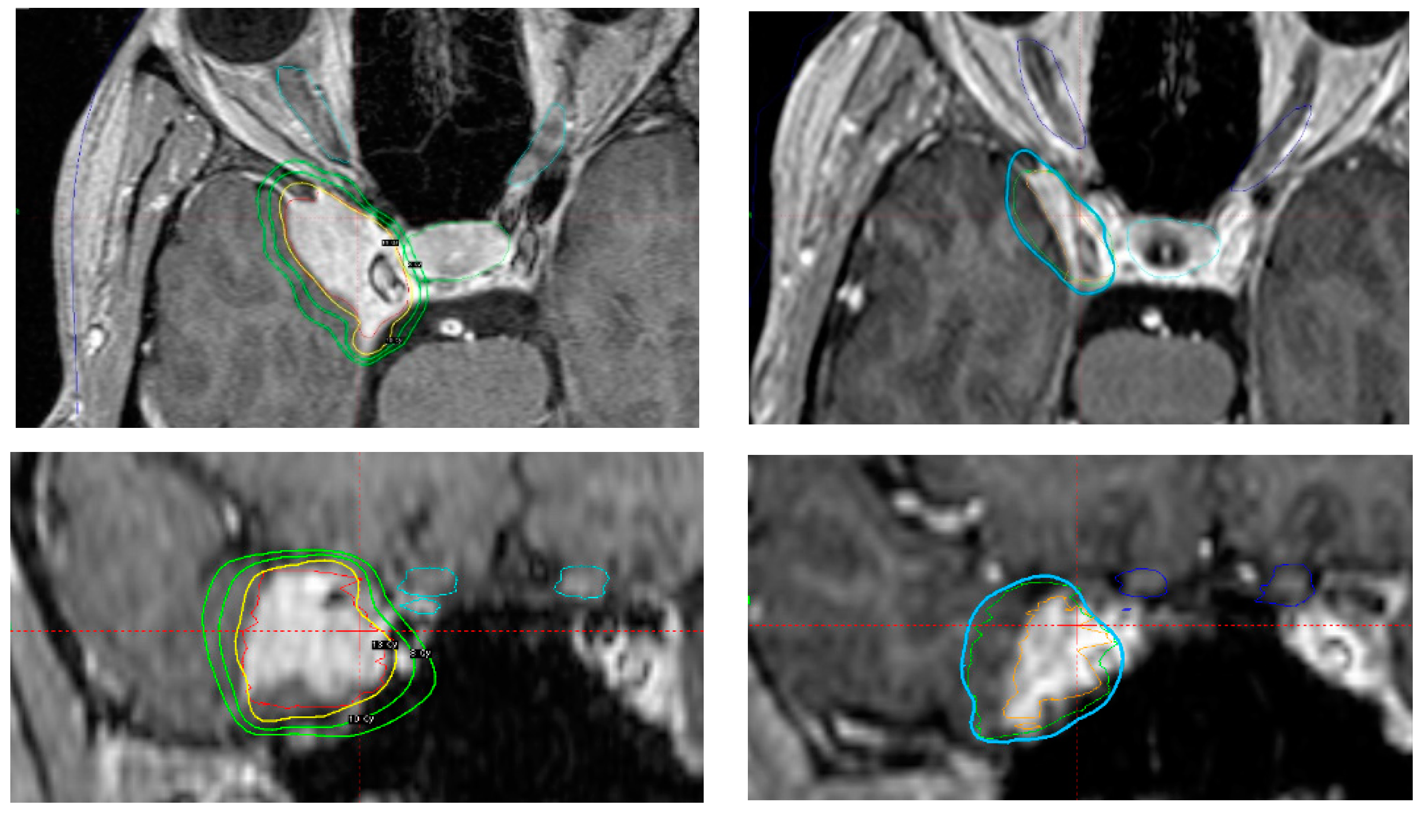
2.6. Imaging Follow-Up

Serial imaging (MRI or CT when MRI was contraindicated) was performed at various times according to each centre, and the results were collated in the follow-up section of the main database. Qualitative and quantitative evaluations of tumour size were performed on each examination by the local staff and independently reviewed by the first author, who also compared reported outcomes. Tumour volume on each scan was compared with the tumour volume before SRS. If the imaging material was digitalized, a volumetric measurement was performed with dedicated software. When imaging was available only on celluloid film, the comparison was made by measuring and comparing the 3 major diameters. Shrinkage or enlargement was defined as an imaging-assessed change in tumour volume of at least 10% determined (as described above) either by direct volumetric measurement or by calculation bas ed on dimensions (Table 1).

2.7. Neurological Assessment and Clinical Follow-Up

Before treatment, all patients underwent neurological examination, which was available for all patients. Clinical data were standardized according to the following uniform classification [24]: 0 = no neurological deficit; 1 = mild or intermittent neurological deficit; 2 = persisting neurological deficit but not affecting performance in daily life; and 3 = permanent/severe neurological deficit affecting performance in daily life. Epileptic seizures were classified as follows: 0 = no seizure activity; 1 = partial seizures; and 2 = generalized seizures. Each epileptic category was subdivided into temporary and permanent subgroups. Clinical and imaging follow-up were performed thereafter (Table 2).
Any neurological deficit newly reported or reported to be worsening after SRS was carefully evaluated and defined as a complication [24].
Complications were then further divided into temporary and permanent subgroups. The latest clinical follow-up is reported separately from the imaging follow-up. In the event of death, date of death was considered to be the date of latest clinical follow-up, and the cause of death was classified as treatment related (treatment), tumour related (meningioma), unrelated (other) or unclear (unknown). Complications are described separately in patients who died of unclear causes. Patients without clinical control were defined as lost to follow-up, and patients without imaging follow-up were defined as lost to imaging follow-up. They are not included in the statistical analysis [24].

2.8. Survey of Patients

Thus, all patients had been treated at least 5 years before this retrieval visit, but it does not follow that all tumours had an effective follow-up of 5 years [24]. A survey of patients lost to follow-up was conducted at 5 years in 11 centers by sending a follow-up letter to the patient or referring physician [24]. Patients were asked to undergo further imaging and neurological evaluation at the treating centre, and physicians were asked to provide updates based on this imaging and to plan any necessary further imaging accordingly [24]. They were then requested to return such results to the treating centre; these results were then subsequently collected by the local research fellow and finally reviewed by the first author [24].

2.9. Statistics

Statistics were performed as detailed in our previous publication [24]. Analysis was performed to evaluate the imaging outcome. Time to enlargement of target volume was estimated with the Kaplan–Meier method, was defined by date of the first scan to show tumour growth, rather than by time to effective regrowth [24]. All target volumes with less than 24 months imaging follow-up were excluded. Independent variables were evaluated separately in order to estimate their influence on imaging outcome. Univariate comparisons between non-continuous variables (histology, patients’ gender, multiple vs. sporadic lesions) were performed with the log rank test [24]. Univariate analysis for continuous variables (prescription margin dose, maximum dose, age, volume and isodose distribution) was performed by applying the Cox proportional hazard model [24]. Multivariate analysis was performed using stepwise Cox regression [24]. For the purposes of this analysis, we used only those variables that had been demonstrated by the prior univariate analysis to be significant [24]. We included patients with more than one meningioma, with frank meningiomatosis and neurofibromatosis type 2 [24]. These cases were analysed separately and singularly compared with sporadic cases, in order to evaluate a statistically significant difference in outcome with respect to PFS rate. Statistical analyses were performed with the SAS software package (SAS for Windows, version 9.1; SAS Institute Inc., Cary, NC, USA) [24].

3. Results

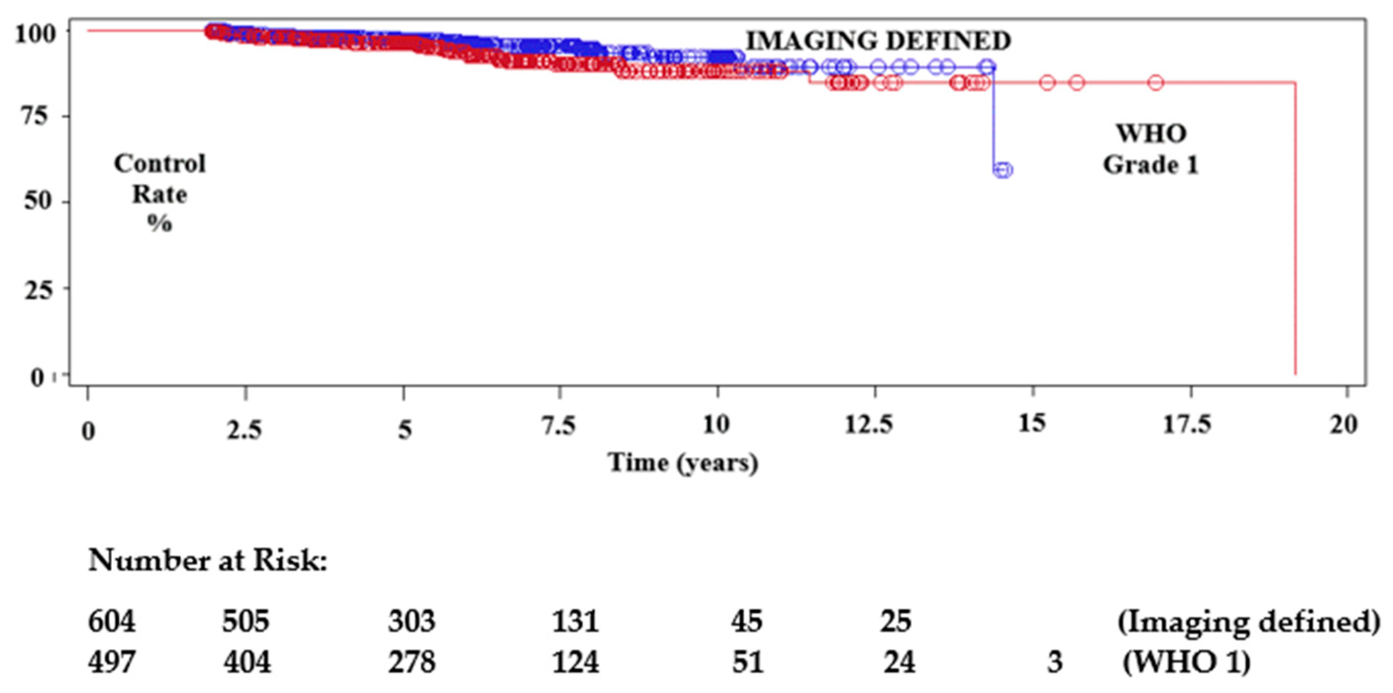
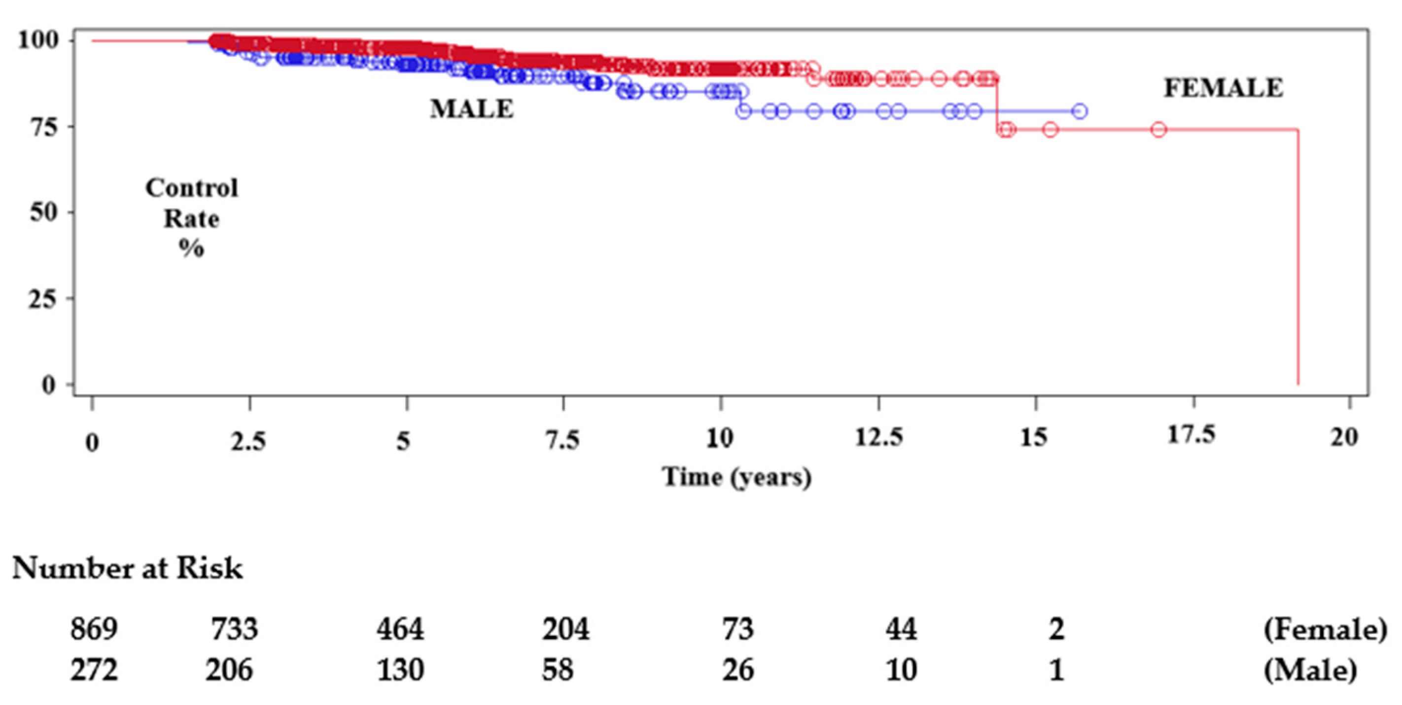
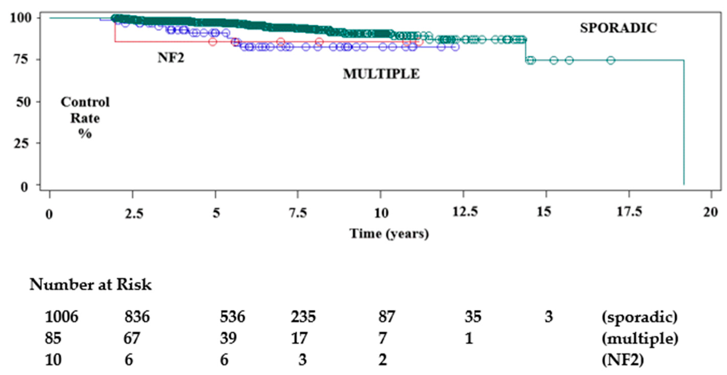
3.1. Local Tumour Control Assessment

Median imaging follow-up was 67 months (mean 73.4, range 24–333). A total of 171 tumours, for whom follow-up was less than 24 months, were excluded. From this cohort we extracted data for follow-up of greater than 5 years (595), 7.5 years (261) and 10 years (103). We found that 549 (58%) meningiomas had regressed (533 patients) and 336 (35.6%) tumours had remained unchanged (325 patients) during follow-up, giving a tumour control rate of 93.6%. Tumour progression occurred in 60 (6.4%) lesions, after a median interval of 57.7 months (mean 62 months). Of the former, 16 patients with 16 enlarging tumours underwent further treatment. Conventional radiotherapy or repeat SRS was performed in four of these patients, and eight underwent surgery. The remaining four tumours had not required further treatment by the time of last follow-up. The Kaplan–Meier estimations of progression-free survival rate (PFS) at 5 years, 7.5 years and 10 years showed control rates of 96.7%, 92.7% and 90.1%, respectively, for the overall data. We observed PFS rates of 97.2%, 90.1% and 87.9% for those tumours initially treated with microsurgery, versus 97.2%, 95.3% and 92.3%, respectively, for tumours without histological confirmation (Figure 2, Figure 3 and Figure 4). Tumour control was better in females than males (p = 0.0079), and for patients with single rather than multiple meningiomas (p = 0.0201) (Table 3, Table 4 and Table 5). A statistically significant difference in tumour control between centres was also observed (p = 0.0160). A representative case is shown in Figure 5.

3.2. Neurological Assessment

Neurological assessment was available for 1042 (85.2%) patients, with follow-up ranging from 6 to 233 months. Clinical improvement was reported in 460 (44.2%) patients at a median follow-up of 62 months (mean 65.6), and complete resolution of symptoms was reported in 241 (23.2%) cases. In particular, we observed an improvement in the symptoms related to the cranial nerves in 27.6% of cases with pre-existing deficits (288 cases), and complete resolution of symptoms in 144 cases (13.8%). Of note, we observed neurological improvement of 52.8% in patients undergoing primary SRS and 28.6% in post-operative patients with WHO Grade I CSMs. Complete resolution of symptoms in these two groups was seen in 26.2% and 14.1% of patients, respectively (Table 6).
Complications were observed after SRS in 113 (11.3%) patients, as detailed in Table 7. Transient morbidity rates were 5.4% and permanent were 5.9%. No radiation-induced tumours were seen, but in two patients that were initially treated with SRS and subsequently operated on for post-treatment tumour enlargement, histology revealed a craniopharyngioma WHO Grade 1 and an anaplastic meningioma WHO Grade 3, respectively.

4. Discussion

4.1. Our Results

To our knowledge, the present study includes the largest consecutive series of patients treated for CSM with SRS. The median imaging follow-up was over five years. Five- and ten-year actuarial PFS rates were 96.6% and 90%, respectively, which are comparable to previous publications [1,2,3,4,18,28,31,34,38].
Tumour control was higher for solitary tumours (p = 0.0097). Although there is no data about SRS for CSMs in multiple lesions or NF2, the sub-analysis confirms our previous results [24].
In our previous analysis we found, contrary to other series [18,39], that previous surgery significantly reduced tumour control [24,39]. This was not confirmed in the current sub-analysis (p = 0.1212) (Table 4), thus implying that better dose planning was possible despite distortion of anatomy by prior surgery [1].
We reported poorer control in males in our previous paper (p = 0.0031), and although the reason is not known, hormonal influences may be a factor [8,24].
Although dose concepts and treatment planning are quite standardized for SRS of CSMs [1,2,28,38,40], a statistically significant difference between centres seems to be still an issue, thus confirming our previous finding [24].

4.2. CSM and Microsurgical Management

CSMs have an estimated incidence of 0.5 per 100,000 [5,6,7,41,42], and their management remains controversial. The complex anatomy of this area, with its contained cranial nerves and carotid siphon makes radical surgical resection challenging, as it carries a high risk of neurological deficits [18]. Dolenc [43,44,45], whose work led to a better appreciation of the surgical anatomy of the cavernous sinus, was an early pioneer of microsurgery; however, as patients have become better informed and less tolerant of surgical morbidity, enthusiasm has waned. Subtotal microsurgical resection leads to fewer complications but is associated with high recurrence rates in the medium to long term. Sindou et al. [12] published a series of 100 patients with CSMs who showed extra-cavernous extensions in follow-ups ranging from 3 to 20 years (mean 8.3 years). Gross-total removal of both the extra- and intra-cavernous portions was achieved in 12 patients (12%); removal of the extra-cavernous portions with only a partial resection of the intra-cavernous portion was achieved in 28 patients (28%); and removal only of the extra-cavernous portions was achieved in 60 patients (60%). Significant additional deficits were reported after surgery, with a higher rate of complications when resection was performed within the cavernous sinus. Five patients died (5%). Tumour regrowth was observed only in 11 of the 82 surviving patients with subtotal removal, however this recurrence rate may increase with additional follow-up in these slowly growing tumours, and with better quality post-operative imaging. The authors concluded that there was no oncological benefit to performing radical resection on the intra-cavernous tumour. Heth et al. [42] reported a series of 163 patients. Total removal was achieved in only 71 cases (44%) and tumour recurrence was observed in 7%. In the cases with partial resection, further growth was seen in 57%.

4.3. Radiation Therapy for CSMs

The role of stereotactic radiation therapy delivered in a single session (SRS) for the management of intracranial benign meningiomas has been well established, as either a primary or adjuvant treatment [1,2,4,18,20,24,25,27,28,29,30,31,32,33,34,40,46]. There is also evidence for the use of stereotactic dose fractionated radiotherapy (SFRT) [25,46,47,48,49,50,51,52]. In a recent systematic review of the role of SRS and SFRT for treating CSM using different platforms, PFS rate at 5 and 10 years after GK-SRS was 93.6% and 87% respectively. PFS rate after Linac based SRS was 95.6% at 5 years and 87.4% at 10 years. PFS rate at 5 and 10 years after Linac based SFRT were 97.4% and 95.5% respectively [38]. However, the indication for treatment and the method of defining target volumes were not uniform. Over 70% of the patient data were from gamma knife centres, with a longer median follow-up, whereas the 10-year statistical results for Linac SRS and SFRT were generally extrapolated from actuarial tables and may have overestimated the imaging outcome [8,10,53,54].
The recent International Stereotactic Radiosurgery Society Guidelines suggest that the literature is limited to level III evidence with respect to the outcomes of SRS for CSMs; however, SRS appears to have more benefits than risks for patients with CSMs [55].
The recent report of the North American Gamma Knife Consortium [40] stated that 61% of patients showed tumour shrinkage, 2% were unchanged, and 15% showed increases in tumour volume after a follow-up period of 101 months. Actuarial tumour control rates at the 5-, 10-, and 15-year follow-ups were 92%, 84%, and 75%, respectively. Of the 120 patients who had undergone SRS as a primary treatment, tumour progression was observed in 14 (11.7%) patients at a median of 48.9 months (range 4.8–120.0 months). The actuarial tumour control rates were 98%, 93%, 85%, and 85% at the 1-, 5-, 10-, and 15-year follow-ups post-SRS, respectively. Fifteen (7.5%) patients experienced permanent CN deficits without evidence of tumour progression at a median onset of 9 months (range 2.3–85 months) after SRS. Patients with larger tumour volumes (≥10 cm3) were more likely to develop permanent CN complications. Three patients (1.5%) developed delayed pituitary dysfunction after SRS.
It is now recognised that the higher radiation dose does not significantly influence PFS rate. Kondziolka et al. reported no improvement in PFS with single fraction doses greater than 15 Gy [56]. Lee and co-authors [4] reported the outcomes of 159 patients with CSMs treated either with adjuvant or primary SRS and found no statistical difference. More recently, Pollock et al. [1] reported 115 cases of CSMs treated with the Gamma Knife between 1990 and 2008. Grade II and III CSMs were excluded. Patients with multiple meningiomas and NF2 were excluded, as well as previously irradiated lesions. Histology was available in 46 patients who had undergone microsurgery. The local tumour control rate was 99% at 5 years and 93% at 10 years following SRS. Prior surgery did not influence the outcome, and the authors suggested that those patients with large tumours, or those who present with visual loss generally should undergo microsurgical debulking, followed by adjuvant SRS to achieve tumour control.
Fariselli et al. reported on the efficacy of hypofractionated, stereotactic radiotherapy HSFRT (25 Gy, delivered in 3 to 5 fractions), using Cyberknife™, in order to avoid radiation-induced optic neuropathy (RION—5.1% risk of visual worsening after treatment) while achieving a high PFS rate (93% at 5 years) [57]. Fractionated radiotherapy is also possible with the Gamma Knife model ICON™ (Elekta Instruments, AB, Stockholm, Sweden) [58], which also allows HSFRT for large-volume tumours in patients with major comorbidities and contraindications to surgery, or for tumours in close contact with, or surrounding the optic apparatus.
Although tumour control with primary SRS is good for small- to medium-sized tumours, surgery for these and for larger tumours remains an option for adequately counselled patients who understand the risks and benefits of both treatments [35].

4.4. Challenging Aspects Related to SRS

One of the challenges of SRS for CSM remains the proximity of the optic nerves and chiasm. However, the gamma knife has the ability to plan for a low isodose with a very steep dose gradient [59]. A distance of 2–4 mm between the optic apparatus and the target is enough to avoid delivering more than 10 Gy to the optic apparatus [60]. If there is contact between the target volume and the optic pathways, SFRT is recommended [57]. In our series, four patients suffering from optic nerve neuropathy after SRS were treated with am margin dose more than 15 Gy.
Pituitary insufficiency is also a risk, with recent studies showing that a single fraction dose of more than 15 Gy to the pituitary stalk should be avoided [61]. Interestingly, we observed in our series two cases of delayed transitory partial pituitary deficit two years after SRS, which regressed after temporary medication. A margin dose of more than 16 Gy was delivered.
ICA Stenosis after SRS is a potential risk and is well described in the literature [1,2,29,30,34]. We observed five cases of symptomatic ICA stenosis; in one patient the stenosis was remote to the target volume and in three patients, the tumour continued to grow.

4.5. Further Aspects of CSM Management

It is now clear that SRS is an effective treatment for CSMs [2,34,62,63,64,65,66,67,68]. The question of the timing of adjuvant SRS after subtotal continues to divide opinion. It is now common practice to monitor residual tumours after surgery using serial MRI scans, and to offer adjuvant radiotherapy or SRS only when growth is confirmed. For vestibular schwannomas [69], subtotal excision followed by SRS as a planned combined approach is an increasingly advocated treatment strategy [70,71]. In our previous report, we commented that “active surveillance” requires ongoing evaluation [24]. Untreated meningiomas, either from the time of diagnosis or after subtotal resection may lead to additional morbidity due to tumour growth [1,2,39,61,72,73,74,75,76,77,78,79,80,81,82,83]. A recent consensus statement on the treatment of CSM from the skull base section of the European Association of Neurological Surgery (EANS) recommends SRS as first-line treatment for small, well defined, pauci-symptomatic lesions in elderly patients, and for larger CSMs not amenable to resection. Microsurgery is recommended for rapidly progressing lesions in young patients presenting with oculomotor, visual, or endocrinological impairments. In order to reduce surgical morbidity, the authors recommend subtotal resection followed by adjuvant SRS in accordance with our outcomes. Conservative treatment with serial imaging follow-up should be proposed in patients with a newly diagnosed asymptomatic CSM that has no mass effect on the adjacent temporal lobe [35].
Complications after SRS are relatively few, but there is a theoretical risk of radiation-induced secondary tumours, or malignant transformation of the treated benign tumour [25,39,56], although this was not seen in our review. Wolf et al. [14] suggest that this risk remains low at long-term follow-up and is similar to the risk of the general population developing primary CNS tumours.

4.6. Limitations of the Study

This outcome study has several limitations. It is retrospective and multicentre, with each unit having slightly different selection criteria, target contouring and dose planning. Nevertheless, the large volume of data collected by the first author was organised in a structured database agreed to by each participating centre, resulting in a uniform data set. There is also potential bias with regard to recurrence/failure rates due to the relatively short follow-up of some patients (24 months). The median time from treatment to tumour growth in our study is four years and nine months (and a mean of five years). Bias is also potentially introduced by loss of patients to follow-up. We elected only to include patients with more than two years of post-treatment imaging, while reducing the clinical follow-up to a minimum of 6 months in order to capture any early adverse treatment effects [24]. As this is a historical study, it includes a range of different planning systems and imaging techniques. All centres treating before 1993 used CT imaging. Between 1994 and 1996, Leksell Gamma Plan software was introduced along with MRI-defined targeting. Since 1998, all treatments were planned with LGP from high-definition MRI images [24].

5. Conclusions

We believe SRS is a valuable treatment option for CSM, both as a primary treatment, and as an adjunct following surgery. Imaging diagnosis is reliable in the absence of histology. The risk of pituitary insufficiency is low. Cranial nerve neuropathies improve after SRS in over 20% of patients treated primarily. For those patients best treated surgically, we support the strategy of subtotal microsurgical resection, followed by adjuvant SRS, with or without a period of “active surveillance”, as this clearly reduces surgical morbidity for patients.

Author Contributions

Conceptualization, A.S., C.T., A.T.C.J.v.E., J.R. and C.E.; data curation, A.S., C.T., R.L., E.M., C.L., M.R., B.G., B.E.L., R.M.Á., N.M.M., B.S.S., M.L., F.U., I.S., T.M., O.B., G.A.H., A.T.C.J.v.E., M.W., J.R. and C.E.; formal analysis, A.S., C.T., S.S., M.W. and M.B.; funding acquisition, A.S.; investigation, A.S. and C.T.; methodology, A.S., C.T., G.A.H. and J.R.; supervision, A.S., C.T., M.N., J.R. and C.E.; validation, A.S., C.T., E.M., M.R., B.G., B.E.L., R.M.Á., N.M.M., M.A.K., B.S.S., S.S., M.L., F.U., I.S., T.M., O.B., G.A.H., A.T.C.J.v.E., M.N., H.J.S., D.H., T.F., S.Z.A., J.R. and C.E.; visualization, C.T., R.L., E.M., C.L., M.R., B.G., B.E.L., R.M.Á., N.M.M., M.A.K., B.S.S., S.S., M.L., F.U., I.S., T.M., O.B., G.A.H., A.T.C.J.v.E., M.W., M.B., M.N., H.J.S., D.H., T.F., S.Z.A. and J.R.; writing—original draft, A.S. and C.T.; writing—review and editing, A.S., C.T., R.L., M.R., M.A.K., B.S.S., S.S., H.J.S., T.F. and C.E. All authors have read and agreed to the published version of the manuscript.

Funding

As we have already disclosed [24], the main observational retrospective study was supported in part by Grant 75-6075 from Elekta Instruments AB, to Santacroce and in Part from GKZ GmbH Krefeld, Germany. No further funding was received for the current sub-analysis.

Institutional Review Board Statement

The study was conducted according to the guidelines of the Declaration of Helsinki and approved by the local ethical committee of the Heinrich Heine University of Düsseldorf (Number of the ethical approval, 4002; date of approval, 29 October 2012).

Informed Consent Statement

Patient consent was waived by the Institutional Review Board.

Data Availability Statement

The data that support the findings of this study are available from the corresponding author, A.S., upon reasonable request.

Acknowledgments

Antonio Santacroce is indebted to the physicians, physicists and administrative staff members of the 15 European centres recruited for the study as follows: Gamma Knife Zentrum Krefeld, Germany; Department of Stereotactic and Radiation Neurosurgery, Na Homolce Hospital, Prague Czech Republic; Centro Gamma Knife Villa Maria Cecilia Hospital, Cotignola (RA) Italy; Gamma Knife Centre, Bupa Cromwell Hospital, London, UK; National Centre for Stereotactic Radiosurgery, Royal Hallamshire Hospital, Sheffield, UK; Department of Neurosurgery, Medical University of Vienna, Vienna, Austria; Department of Neurosurgery, Karolinska University Hospital, Stockholm, Sweden; Department of Functional Neurosurgery and Radiation Oncology, Hospital Ruber Internacional, Madrid, Spain; Department of Neurosurgery, Haukeland University Hospital, Bergen, Norway; Department of Neurosurgery, University Hospital, Verona, Italy; Department of Neurosurgery, Medical University Graz, Graz, Austria; Gamma Knife Centre at Harley Street London, UK; Gamma Knife Centre Zurich, Klinik Im Park Hirslanden, Zurich, Switzerland; Gamma Knife Zentrum Hannover, Germany; Service de Neurochirurgie Fonctionnelle et Stereotaxique, Hôpital d’adulte de la Timone Marseille, France. Special thanks to Romain Carron, Shoji Yomo, Aurelia Kollova’, Lee Walton, Massimo Gerosa, Antonio Nicolato, H.J. Kreiner, Danny Leksell and the late R.I. Foroni, whose contributions, made the main data retrieval feasible.

Conflicts of Interest

Régis received research grants, travel grants and consulting fees from Elekta Instrument AB. Motti has served as an expert witness for Elekta Instruments AB. Lindquist has received consulting fees from, and is a stockholder in Elekta Instruments AB. Lippitz has received consulting fees from Elekta Instruments AB. Santacroce received travel costs refunding to present the results of the first main analysis [24], and in part for the current sub-analysis. The authors have no personal financial or institutional interest in any of the drugs, materials or devices described in this article.

References

  1. Pollock, B.E.; Stafford, S.L.; Link, M.J.; Garces, Y.I.; Foote, R.L. Single-fraction radiosurgery of benign cavernous sinus meningiomas. Comparative Study. J. Neurosurg. 2013, 119, 675–682. [Google Scholar] [CrossRef]
  2. Pollock, B.E.; Stafford, S.L. Results of stereotactic radiosurgery for patients with imaging defined cavernous sinus meningiomas. Int. J. Radiat. Oncol. Biol. Phys. 2005, 62, 1427–1431. [Google Scholar] [CrossRef]
  3. Selch, M.T.; Ahn, E.; Laskari, A.; Lee, S.P.; Agazaryan, N.; Solberg, T.D.; Cabatan-Awang, C.; Frighetto, L.; Desalles, A.A. Stereotactic radiotherapy for treatment of cavernous sinus meningiomas. Int. J. Radiat. Oncol. Biol. Phys. 2004, 59, 101–111. [Google Scholar] [CrossRef]
  4. Lee, J.Y.; Niranjan, A.; McInerney, J.; Kondziolka, D.; Flickinger, J.C.; Lunsford, L.D. Stereotactic radiosurgery providing long-term tumor control of cavernous sinus meningiomas. J. Neurosurg. 2002, 97, 65–72. [Google Scholar] [CrossRef]
  5. Al-Mefty, O. Management of the cavernous sinus and carotid siphon. Otolaryngol. Clin. N. Am. 1991, 24, 1523–1533. [Google Scholar] [CrossRef]
  6. Al-Mefty, O.; Ayoubi, S.; Smith, R.R. Direct surgery of the cavernous sinus: Patient selection. Acta Neurochir. Suppl. 1991, 53, 117–121. [Google Scholar]
  7. Al-Mefty, O.; Holoubi, A.; Rifai, A.; Fox, J.L. Microsurgical removal of suprasellar meningiomas. Neurosurgery 1985, 16, 364–372. [Google Scholar] [CrossRef]
  8. De Jesus, O.; Sekhar, L.N.; Parikh, H.K.; Wright, D.C.; Wagner, D.P. Long-term follow-up of patients with meningiomas involving the cavernous sinus: Recurrence, progression, and quality of life. Neurosurgery 1996, 39, 915–919; discussion 919–920. [Google Scholar] [CrossRef]
  9. Cusimano, M.D.; Sekhar, L.N.; Sen, C.N.; Pomonis, S.; Wright, D.C.; Biglan, A.W.; Jannetta, P.J. The results of surgery for benign tumors of the cavernous sinus. Comparative Study. Neurosurgery 1995, 37, 1–9; discussion 9–10. [Google Scholar] [CrossRef]
  10. O’Sullivan, M.G.; van Loveren, H.R.; Tew, J.M., Jr. The surgical resectability of meningiomas of the cavernous sinus. Neurosurgery 1997, 40, 238–244; discussion 245–247. [Google Scholar] [CrossRef]
  11. Jacob, M.; Wydh, E.; Vighetto, A.; Sindou, M. Visual outcome after surgery for cavernous sinus meningioma. Acta Neurochir. 2008, 150, 421–429; discussion 429. [Google Scholar] [CrossRef]
  12. Sindou, M.; Wydh, E.; Jouanneau, E.; Nebbal, M.; Lieutaud, T. Long-term follow-up of meningiomas of the cavernous sinus after surgical treatment alone. J. Neurosurg. 2007, 107, 937–944. [Google Scholar] [CrossRef]
  13. Sindou, M.; Chavez, J.M.; Pierre, G.S.; Jouvet, A. Percutaneous biopsy of cavernous sinus tumors through the foramen ovale. Neurosurgery 1997, 40, 106–110. [Google Scholar]
  14. Patel, C.R.; Fernandez-Miranda, J.C.; Wang, W.H.; Wang, E.W. Skull Base Anatomy. Otolaryngol. Clin. N. Am. 2016, 49, 9–20. [Google Scholar] [CrossRef]
  15. Raheja, A.; Couldwell, W.T. Cavernous sinus meningioma. Handb. Clin. Neurol. 2020, 170, 69–85. [Google Scholar] [CrossRef]
  16. Miyazaki, Y.; Yamamoto, I.; Shinozuka, S.; Sato, O. Microsurgical anatomy of the cavernous sinus. Neurol. Med. Chir. 1994, 34, 150–163. [Google Scholar] [CrossRef] [PubMed]
  17. Iwanaga, J.; Anand, M.K.; Camacho, A.; Rodriguez, F.; Watson, C.; Caskey, E.L.; Dumont, A.S.; Tubbs, R.S. Surgical anatomy of the internal carotid plexus branches to the abducens nerve in the cavernous sinus. Clin. Neurol. Neurosurg. 2020, 191, 105690. [Google Scholar] [CrossRef]
  18. Kano, H.; Park, K.-J.; Kondziolka, D.; Iyer, A.; Liu, X.; Tonetti, D.; Flickinger, J.C.; Lunsford, L.D. Does prior microsurgery improve or worsen the outcomes of stereotactic radiosurgery for cavernous sinus meningiomas? Research Support, Non-U.S. Gov’t. Neurosurgery 2013, 73, 401–410. [Google Scholar] [CrossRef]
  19. Franzin, A.; Vimercati, A.; Medone, M.; Serra, C.; Marzoli, S.B.; Forti, M.; Gioia, L.; Valle, M.; Picozzi, P. Neuroophthalmological evaluation after Gamma Knife surgery for cavernous sinus meningiomas. Neurosurg. Focus 2007, 23, E10. [Google Scholar] [CrossRef]
  20. Dufour, H.; Muracciole, X.; Metellus, P.; Regis, J.; Chinot, O.; Grisoli, F. Long-term tumor control and functional outcome in patients with cavernous sinus meningiomas treated by radiotherapy with or without previous surgery: Is there an alternative to aggressive tumor removal? Neurosurgery 2001, 48, 285–294; discussion 294–296. [Google Scholar]
  21. Goldbrunner, R.; Minniti, G.; Preusser, M.; Jenkinson, M.D.; Sallabanda, K.; Houdart, E.; von Deimling, A.; Stavrinou, P.; Lefranc, F.; Lund-Johansen, M.; et al. EANO guidelines for the diagnosis and treatment of meningiomas. Lancet Oncol. 2016, 17, e383–e391. [Google Scholar] [CrossRef]
  22. Kuo, J.S.; Yu, C.; Giannotta, S.L.; Petrovich, Z.; Apuzzo, M.L. The Leksell gamma knife Model U versus Model C: A quantitative comparison of radiosurgical treatment parameters. Neurosurgery 2004, 55, 168–172; discussion 172–173. [Google Scholar] [CrossRef]
  23. Larsson, B.; Leksell, L.; Rexed, B.; Sourander, P.; Mair, W.; Andersson, B. The high-energy proton beam as a neurosurgical tool. Nature 1958, 182, 1222–1223. [Google Scholar] [CrossRef]
  24. Santacroce, A.; Walier, M.; Régis, J.; Liščák, R.; Motti, E.; Lindquist, C.; Kemeny, A.; Kitz, K.; Lippitz, B.; Álvarez, R.M.; et al. Long-term tumor control of benign intracranial meningiomas after radiosurgery in a series of 4565 patients. Research Support, Non-U.S. Gov’t. Neurosurgery 2012, 70, 32–39; discussion 39. [Google Scholar] [CrossRef]
  25. Kondziolka, D.; Mathieu, D.; Lunsford, L.D.; Martin, J.J.; Madhok, R.; Niranjan, A.; Flickinger, J.C. Radiosurgery as definitive management of intracranial meningiomas. Clin. Trial Neurosurg. 2008, 62, 53–58; discussion 58–60. [Google Scholar] [CrossRef]
  26. Subach, B.R.; Lunsford, L.D.; Kondziolka, D.; Maitz, A.H.; Flickinger, J.C. Management of petroclival meningiomas by stereotactic radiosurgery. Neurosurgery 1998, 42, 437–443; discussion 443–445. [Google Scholar]
  27. Duma, C.M.; Lunsford, L.D.; Kondziolka, D.; Harsh GRt Flickinger, J.C. Stereotactic radiosurgery of cavernous sinus meningiomas as an addition or alternative to microsurgery. Neurosurgery 1993, 32, 699–704; discussion 704–705. [Google Scholar] [CrossRef]
  28. Skeie, B.S.; Enger, P.O.; Skeie, G.O.; Thorsen, F.; Pedersen, P.H. Gamma knife surgery of meningiomas involving the cavernous sinus: Long-term follow-up of 100 patients. Neurosurgery 2010, 66, 661–668; discussion 668–669. [Google Scholar] [CrossRef]
  29. Spiegelmann, R.; Nissim, O.; Menhel, J.; Alezra, D.; Pfeffer, M.R. Linear accelerator radiosurgery for meningiomas in and around the cavernous sinus. Neurosurgery 2002, 51, 1373–1379; discussion 1379–1380. [Google Scholar] [CrossRef]
  30. Spiegelmann, R.; Cohen, Z.R.; Nissim, O.; Alezra, D.; Pfeffer, R. Cavernous sinus meningiomas: A large LINAC radiosurgery series. J. Neurooncol. 2010, 98, 195–202. [Google Scholar] [CrossRef]
  31. Nicolato, A.; Foroni, R.; Alessandrini, F.; Bricolo, A.; Gerosa, M. Radiosurgical treatment of cavernous sinus meningiomas: Experience with 122 treated patients. Neurosurgery 2002, 51, 1153–1159; discussion 1159–1161. [Google Scholar] [CrossRef] [PubMed]
  32. Nicolato, A.; Foroni, R.; Alessandrini, F.; Maluta, S.; Bricolo, A.; Gerosa, M. The role of Gamma Knife radiosurgery in the management of cavernous sinus meningiomas. Int. J. Radiat. Oncol. Biol. Phys. 2002, 53, 992–1000. [Google Scholar] [CrossRef]
  33. Metellus, P.; Regis, J.; Muracciole, X.; Fuentes, S.; Dufour, H.; Nanni, I.; Chinot, O.; Martin, P.-M.; Grisoli, F. Evaluation of fractionated radiotherapy and gamma knife radiosurgery in cavernous sinus meningiomas: Treatment strategy. Neurosurgery 2005, 57, 873–886; discussion 873–886. [Google Scholar] [CrossRef] [PubMed]
  34. Roche, P.H.; Régis, J.; Dufour, H.; Fournier, H.D.; Delsanti, C.; Pellet, W.; Grisoli, F.; Peragut, J.C. Gamma knife radiosurgery in the management of cavernous sinus meningiomas. J. Neurosurg. 2000, 93 (Suppl. 3), 68–73. [Google Scholar] [CrossRef]
  35. Corniola, M.; Roche, P.H.; Brunea, M.; Cavallo, L.M.; Daniel, R.T.; Messerer, M.; Froelich, S.; Gardner, P.A.; Gentili, F.; Kawase, T.; et al. Management of cavernous sinus meningiomas: Consensus statement on behalf of the EANS skull base section. Brain Spine 2022, 2, 100864. [Google Scholar] [CrossRef]
  36. Fariselli, L.; Biroli, A.; Signorelli, A.; Broggi, M.; Marchetti, M.; Biroli, F. The cavernous sinus meningiomas’ dilemma: Surgery or stereotactic radiosurgery? Rep. Pract. Oncol. Radiother. 2016, 21, 379–385. [Google Scholar] [CrossRef]
  37. Tanaka, S.; Pollock, B.E.; Stafford, S.L.; Link, M.J. Stereotactic radiosurgery for trigeminal pain secondary to benign skull base tumors. World Neurosurg. 2013, 80, 371–377. [Google Scholar] [CrossRef]
  38. Leroy, H.A.; Tuleasca, C.; Reyns, N.; Levivier, M. Radiosurgery and fractionated radiotherapy for cavernous sinus meningioma: A systematic review and meta-analysis. Acta Neurochir. 2018, 160, 2367–2378. [Google Scholar] [CrossRef]
  39. Kollova, A.; Liscak, R.; Novotny, J., Jr.; Vladyka, V.; Simonova, G.; Janouskova, L. Gamma Knife surgery for benign meningioma. J. Neurosurg. 2007, 107, 325–336. [Google Scholar] [CrossRef]
  40. Park, K.-J.; Kano, H.; Iyer, A.; Liu, X.; Tonetti, D.A.; Lehocky, C.; Faramand, A.; Niranjan, A.; Flickinger, J.C.; Kondziolka, D.; et al. Gamma Knife stereotactic radiosurgery for cavernous sinus meningioma: Long-term follow-up in 200 patients. J. Neurosurg. 2018, 130, 1799–1808. [Google Scholar] [CrossRef]
  41. Al-Mefty, O.; Topsakal, C.; Pravdenkova, S.; Sawyer, J.R.; Harrison, M.J. Radiation-induced meningiomas: Clinical, pathological, cytokinetic, and cytogenetic characteristics. J. Neurosurg. 2004, 100, 1002–1013. [Google Scholar] [CrossRef] [PubMed]
  42. Heth, J.A.; Al-Mefty, O. Cavernous sinus meningiomas. Review. Neurosurg. Focus 2003, 14, e3. [Google Scholar] [CrossRef] [PubMed]
  43. Couldwell, W.T.; Heros, R.; Dolenc, V. Skull base meningiomas. Introductory. Neurosurg. Focus 2011, 30, 1p prior to E1. [Google Scholar] [CrossRef]
  44. Shaffrey, M.E.; Dolenc, V.V.; Lanzino, G.; Wolcott, W.P.; Shaffrey, C.I. Invasion of the internal carotid artery by cavernous sinus meningiomas. Surg. Neurol. 1999, 52, 167–171. [Google Scholar] [CrossRef]
  45. Dolenc, V. Microsurgical removal of large sphenoidal bone meningiomas. Acta Neurochir. Suppl. 1979, 28, 391–396. [Google Scholar]
  46. Metellus, P.; Batra, S.; Karkar, S.; Kapoor, S.; Weiss, S.; Kleinberg, L.; Rigamonti, D. Fractionated conformal radiotherapy in the management of cavernous sinus meningiomas: Long-term functional outcome and tumor control at a single institution. Int. J. Radiat. Oncol. Biol. Phys. 2010, 78, 836–843. [Google Scholar] [CrossRef]
  47. Litré, C.F.; Colin, P.; Noudel, R.; Peruzzi, P.; Bazin, A.; Sherpereel, B.; Bernard, M.H.; Rousseaux, P. Fractionated stereotactic radiotherapy treatment of cavernous sinus meningiomas: A study of 100 cases. Int. J. Radiat. Oncol. Biol. Phys. 2009, 74, 1012–1017. [Google Scholar] [CrossRef]
  48. Milker-Zabel, S.; Zabel-du Bois, A.; Huber, P.; Schlegel, W.; Debus, J. Fractionated stereotactic radiation therapy in the management of benign cavernous sinus meningiomas: Long-term experience and review of the literature. Strahlenther. Onkol. 2006, 182, 635–640. [Google Scholar] [CrossRef]
  49. Brell, M.; Villa, S.; Teixidor, P.; Lucas, A.; Ferrán, E.; Marín, S.; Acebes, J.J. Fractionated stereotactic radiotherapy in the treatment of exclusive cavernous sinus meningioma: Functional outcome, local control, and tolerance. Surg. Neurol. 2006, 65, 28–33; discussion 33–34. [Google Scholar] [CrossRef]
  50. Sibtain, A.; Plowman, P.N. Stereotactic radiosurgery. VII. Radiosurgery versus conventionally-fractionated radiotherapy in the treatment of cavernous sinus meningiomas. Br. J. Neurosurg. 1999, 13, 158–166. [Google Scholar] [CrossRef]
  51. Sims, E.; Doughty, D.; Macaulay, E.; Royle, N.; Wraith, C.; Darlison, R.; Plowman, P. Stereotactically delivered cranial radiation therapy: A ten-year experience of linac-based radiosurgery in the UK. Clin. Oncol. (R Coll Radiol.) 1999, 11, 303–320. [Google Scholar] [CrossRef] [PubMed]
  52. Maguire, P.D.; Clough, R.; Friedman, A.H.; Halperin, E.C. Fractionated external-beam radiation therapy for meningiomas of the cavernous sinus. Int. J. Radiat. Oncol. Biol. Phys. 1999, 44, 75–79. [Google Scholar] [CrossRef]
  53. DeMonte, F.; Smith, H.K.; al-Mefty, O. Outcome of aggressive removal of cavernous sinus meningiomas. J. Neurosurg. 1994, 81, 245–251. [Google Scholar] [CrossRef]
  54. Aziz, K.M.A.; Froelich, S.C.; Dagnew, E.; Jean, W.; Breneman, J.C.; Zuccarello, M.; van Loveren, H.R.; Tew, J.M. Large sphenoid wing meningiomas involving the cavernous sinus: Conservative surgical strategies for better functional outcomes. Neurosurgery 2004, 54, 1375–1383; discussion 1383–1384. [Google Scholar] [CrossRef]
  55. Lee, C.-C.; Trifiletti, D.M.; Sahgal, A.; DeSalles, A.; Fariselli, L.; Hayashi, M.; Levivier, M.; Ma, L.; Álvarez, R.M.; Paddick, I.; et al. Stereotactic Radiosurgery for Benign (World Health Organization Grade I) Cavernous Sinus Meningiomas-International Stereotactic Radiosurgery Society (ISRS) Practice Guideline: A Systematic Review. Neurosurgery 2018, 83, 1128–1142. [Google Scholar] [CrossRef]
  56. Kondziolka, D.; Flickinger, J.C.; Perez, B. Judicious resection and/or radiosurgery for parasagittal meningiomas: Outcomes from a multicenter review. Gamma Knife Meningioma Study Group. Neurosurgery 1998, 43, 405–413; discussion 413–414. [Google Scholar] [CrossRef]
  57. Marchetti, M.; Bianchi, S.; Pinzi, V.; Tramacere, I.; Fumagalli, M.L.; Milanesi, I.M.; Ferroli, P.; Franzini, A.; Saini, M.; DiMeco, F.; et al. Multisession Radiosurgery for Sellar and Parasellar Benign Meningiomas: Long-term Tumor Growth Control and Visual Outcome. Neurosurgery 2016, 78, 638–646. [Google Scholar] [CrossRef]
  58. Tuleasca, C.; Leroy, H.A.; Regis, J.; Levivier, M. Gamma Knife radiosurgery for cervical spine lesions: Expanding the indications in the new era of Icon. Acta Neurochir. 2016, 158, 2235–2236. [Google Scholar] [CrossRef]
  59. Kaul, D.; Badakhshi, H.; Gevaert, T.; Pasemann, D.; Budach, V.; Tuleasca, C.; Gruen, A.; Prasad, V.; Levivier, M.; Kufeld, M. Erratum to: Dosimetric comparison of different treatment modalities for stereotactic radiosurgery of meningioma. Acta Neurochir. 2015, 157, 565. [Google Scholar] [CrossRef]
  60. Leavitt, J.A.; Stafford, S.L.; Link, M.J.; Pollock, B.E. Long-term evaluation of radiation-induced optic neuropathy after single-fraction stereotactic radiosurgery. Int. J. Radiat. Oncol. Biol. Phys. 2013, 87, 524–527. [Google Scholar] [CrossRef]
  61. Sheehan, J.P.; Starke, R.M.; Kano, H.; Kaufmann, A.M.; Mathieu, D.; Zeiler, F.A.; West, M.; Chao, S.T.; Varma, G.; Chiang, V.L.S.; et al. Gamma Knife radiosurgery for sellar and parasellar meningiomas: A multicenter study. J. Neurosurg. 2014, 120, 1268–1277. [Google Scholar] [CrossRef] [PubMed]
  62. Friedman, W.A.; Murad, G.J.; Bradshaw, P.; Amdur, R.J.; Mendenhall, W.M.; Foote, K.; Bova, F.J. Linear accelerator surgery for meningiomas. J. Neurosurg. 2005, 103, 206–209. [Google Scholar] [CrossRef] [PubMed]
  63. Hakim, R.; Alexander, E., 3rd; Loeffler, J.S.; Shrieve, D.C.; Wen, P.; Fallon, M.P.; Stieg, P.E.; Black, P.M. Results of linear accelerator-based radiosurgery for intracranial meningiomas. Neurosurgery 1998, 42, 446–453; discussion 453–454. [Google Scholar] [CrossRef] [PubMed]
  64. Kobayashi, T.; Kida, Y.; Mori, Y. Long-term results of stereotactic gamma radiosurgery of meningiomas. Surg. Neurol. 2001, 55, 325–331. [Google Scholar] [CrossRef]
  65. Kida, Y.; Kobayashi, T.; Tanaka, T.; Oyama, H.; Niwa, M.; Maesawa, S. Radiosurgery of cavernous sinus meningiomas with gamma-knife. No Shinkei Geka 1996, 24, 529–533. [Google Scholar]
  66. Kondziolka, D.; Levy, E.I.; Niranjan, A.; Flickinger, J.C.; Lunsford, L.D. Long-term outcomes after meningioma radiosurgery: Physician and patient perspectives. J. Neurosurg. 1999, 91, 44–50. [Google Scholar] [CrossRef]
  67. Kondziolka, D.; Nathoo, N.; Flickinger, J.C.; Niranjan, A.; Maitz, A.H.; Lunsford, L.D. Long-term results after radiosurgery for benign intracranial tumors. Neurosurgery 2003, 53, 815–821; discussion 821–822. [Google Scholar] [CrossRef]
  68. Kreil, W.; Luggin, J.; Fuchs, I.; Weigl, V.; Eustacchio, S.; Papaefthymiou, G. Long term experience of gamma knife radiosurgery for benign skull base meningiomas. J. Neurol. Neurosurg. Psychiatry 2005, 76, 1425–1430. [Google Scholar] [CrossRef]
  69. Tuleasca, C.; Levivier, M. A virtual musician quartet with vestibular schwannoma. Lancet Oncol. 2018, 19, 1025. [Google Scholar] [CrossRef]
  70. Daniel, R.T.; Tuleasca, C.; George, M.; Pralong, E.; Schiappacasse, L.; Zeverino, M.; Maire, R.; Levivier, M. Preserving normal facial nerve function and improving hearing outcome in large vestibular schwannomas with a combined approach: Planned subtotal resection followed by gamma knife radiosurgery. Acta Neurochir. 2017, 159, 1197–1211. [Google Scholar] [CrossRef]
  71. Starnoni, D.; Berthiller, J.; Idriceanu, T.-M.; Meyronet, D.; D’Hombres, A.; Ducray, F.; Guyotat, J. Returning to work after multimodal treatment in glioblastoma patients. Neurosurg. Focus 2018, 44, E17. [Google Scholar] [CrossRef] [PubMed]
  72. Simpson, D. The recurrence of intracranial meningiomas after surgical treatment. J. Neurol. Neurosurg. Psychiatry 1957, 20, 22–39. [Google Scholar] [CrossRef]
  73. Condra, K.S.; Buatti, J.M.; Mendenhall, W.M.; Friedman, W.A.; Marcus, R.B., Jr.; Rhoton, A.L. Benign meningiomas: Primary treatment selection affects survival. Int. J. Radiat. Oncol. Biol. Phys. 1997, 39, 427–436. [Google Scholar] [CrossRef]
  74. Pollock, B.E.; Stafford, S.L.; Utter, A.; Giannini, C.; Schreiner, S.A. Stereotactic radiosurgery provides equivalent tumor control to Simpson Grade 1 resection for patients with small- to medium-size meningiomas. Int. J. Radiat. Oncol. Biol. Phys. 2003, 55, 1000–1005. [Google Scholar] [CrossRef]
  75. Sankila, R.; Kallio, M.; Jaaskelainen, J.; Hakulinen, T. Long-term survival of 1986 patients with intracranial meningioma diagnosed from 1953 to 1984 in Finland. Comparison of the observed and expected survival rates in a population-based series. Cancer 1992, 70, 1568–1576. [Google Scholar] [CrossRef]
  76. Kallio, M.; Sankila, R.; Hakulinen, T.; Jaaskelainen, J. Factors affecting operative and excess long-term mortality in 935 patients with intracranial meningioma. Neurosurgery 1992, 31, 2–12. [Google Scholar] [CrossRef] [PubMed]
  77. Vernooij, M.W.; Ikram, M.A.; Tanghe, H.L.; Vincent, A.J.; Hofman, A.; Krestin, G.P.; Niessen, W.J.; Breteler, M.M.; van der Lugt, A. Incidental findings on brain MRI in the general population. N. Engl. J. Med. 2007, 357, 1821–1828. [Google Scholar] [CrossRef]
  78. Olivero, W.C.; Lister, J.R.; Elwood, P.W. The natural history and growth rate of asymptomatic meningiomas: A review of 60 patients. J. Neurosurg. 1995, 83, 222–224. [Google Scholar] [CrossRef]
  79. Van Havenbergh, T.; Carvalho, G.; Tatagiba, M.; Plets, C.; Samii, M. Natural history of petroclival meningiomas. Neurosurgery 2003, 52, 55–62; discussion 62–64. [Google Scholar] [CrossRef]
  80. Yoneoka, Y.; Fujii, Y.; Tanaka, R. Growth of incidental meningiomas. Acta Neurochir. 2000, 142, 507–511. [Google Scholar] [CrossRef]
  81. Nakamura, M.; Roser, F.; Michel, J.; Jacobs, C.; Samii, M. The natural history of incidental meningiomas. Neurosurgery 2003, 53, 62–70; discussion 70–71. [Google Scholar] [CrossRef] [PubMed]
  82. Niiro, M.; Yatsushiro, K.; Nakamura, K.; Kawahara, Y.; Kuratsu, J. Natural history of elderly patients with asymptomatic meningiomas. J. Neurol. Neurosurg. Psychiatry 2000, 68, 25–28. [Google Scholar] [CrossRef] [PubMed]
  83. Wolf, A.; Naylor, K.; Tam, M.; Habibi, A.; Novotny, J.; Liščák, R.; Martinez-Moreno, N.; Martinez-Alvarez, R.; Sisterson, N.; Golfinos, J.G.; et al. Risk of radiation-associated intracranial malignancy after stereotactic radiosurgery: A retrospective, multicentre, cohort study. Lancet Oncol. 2019, 20, 159–164. [Google Scholar] [CrossRef]
Figure 1. Flow chart of analysed cohort.
Cancers 14 04047 g001
Figure 2. Kaplan–Meier test showing local imaging tumour control after SRS plotted by tumours without histological confirmation as well as histologically confirmed benign meningiomas (WHO Grade 1).
Cancers 14 04047 g002
Figure 3. Kaplan–Meier test showing local imaging tumour control after SRS plotted by gender.
Cancers 14 04047 g003
Figure 4. Kaplan–Meier test showing local imaging tumour control after SRS plotted by number of meningiomas treated.
Cancers 14 04047 g004
Figure 5. Axial and coronal contrast-enhanced MRI-based treatment (left top and bottom) and imaging follow-up 9 years and 6 months after treatment (right top and bottom). Imaging-defined CSM treated in a 44 year-old woman reporting right periorbital dysesthesia. No further neurological defi cits. No previous surgery. In order to avoid future tumour-associated complications due to the eloquent location, definitive SRS was performed with 13 Gy prescribed to the 65% isodose line. After a follow-up of 114 months, target volume decreased from 5.65 cc to 2.38 cc. The patient developed no new neurological deficits and the periorbital dysesthesia had regressed.
Cancers 14 04047 g005
Table 1. Characteristics of 1222 patients treated with SRS.
Patient GroupValue
Age at treatment (years) *55 (±12.0)
Female **932 (76.3)
Male **290 (24.7)
Patients with sporadic meningiomas **1150 (94.1)
Patients with multiple meningiomas ***, **62 (5.1)
Patients with NF 2 ***, **10 (0.8)
Neurological Status *****
Headache **173 (14.1)
Seizures **23 (1.8)
Cranial nerve deficit **973 (79.6)
Hemiparesis, hypoesthesia **56 (4.6)
Imbalance, ataxia-vertigo **69 (5.6)
Details of Stereotactic Radiosurgery
Volume (cm3) *, ****6.3 (±7.0)
Imaging-defined benign meningiomas **703 (55.2)
Histologically proven WHO Gr. 1 meningiomas **569 (44.7)
Sporadic meningiomas ***, **1164 (91.5)
Multiple meningiomas ***, ** 95 (7.5)
NF2 meningiomas ***, **12 (1.0)
Maximal dose (Gy) *28 (±7.0)
Marginal dose (Gy) *14.0 (±3.0)
Treatment marginal isodose (%) *50 (±5.0)
Isocentres *, **** 11 (±9.0)
Dose to optic pathways * 8.0 (±4.0)
Follow-up
Imaging follow-up (months from treatment) *61 (±38) 67(±33) *****
Clinical follow-up (months from treatment) *62 (±39)
Patients lost to follow-up **134 (10.9%)
Tumours lost to follow-up **156 (12.2%)
Tumours with follow-up > 5 years **595 (46.7%)
Tumours with follow-up > 7.5 years **261 (12.6%)
Tumours with follow-up > 10 years **103 (8.4%)
* Median and standard deviation. ** Number—(%). *** Multiple meningiomas were defined as more than one tumour treated in the same or different session and not having recurred within the surgical field. All patients diagnosed with neurofibromatosis 2 regardless of tumour-count treatments were analysed separately. NF2-related meningioma was defined as meningioma linked to multiple inherited schwannomas, meningiomas, and ependymomas. **** Volume was available at time of treatment for 1115 tumours and isocentre number for 1148 treatments. ***** The number of symptomatic cases does not correspond to the total number of patients, as some had more than one symptom. A total of 164 patients (13.4%) were asymptomatic.
Table 2. Cranial nerve deficits in 1222 patients undergoing SRS for CSMs *.
Neurological DeficitNumber of Patients (%)
Cranial nerveDefinitive SRSAdjuvant SRS
I0 (0%)1 (0.1%)
II315 (81%)294 (51.6%)
III-IV-VI360 (51%)261 (45.8%)
V188 (26.7%)204(35.8%)
VII45 (6.4%)56 (9.8%)
VIII25 (3.5%)36 (6.3%)
IX-X-XI5 (0.7%)1 (0.1%)
XII1 (0.1%)1(0.1%)
* Some patients harboured more than 1 target lesion outside the cavernous sinus. The figures relate to neurological deficits before treatment.
Table 3. Progression-free survival rate at 5, 7.5 and 10 years after SRS *.
Variable/Follow-Up
Years
5 years7.5 years10 years
Imaging Defined Meningiomas 97.2% (95.6–98.4)95.3% (92.2–97.2)92.3% (86.8–95.5)
WHO Grade 1 Meningiomas96.2% (93.6–97.7)90.1% (85.3–93.3)87.9% (82.1–91.9)
Female97.7% (96.1–98.6)93.6% (90.6–95.7)91.6% (87.4–94.3)
Male92.9% (87.7–95.9)89.6% (82.8–93.7)85.1% (75.0–91.3)
Sporadic Meningiomas **97.2% (95.7–98.2)93.7% (91.0–95.6)90.8% (86.8–93.6)
Multiple Meningiomas **90.8% (79.3–96.1)85.3% (71.1–92.9)82.4% (67.2–91.0)
NF2 Meningiomas **85.7% (33.4–97.8)
** Multiple meningiomas were encoded as one (no) or more than one (yes) meningioma treated. NF2 Meningiomas were analysed separately. * Progression-free survival rate (PFS) and lower-upper confidence limit at 95% (LCL-UCL).
Table 4. Univariate analysis of tumour control defined as stable or reduced volume of the tumour irradiated.
Univariate Chi-Squares for the Log-Rank Test
VariableChi-SquareProb. > Chi-Square
Previous surgery *2.40200.1212
Gender7.06740.0079
Multiple Meningiomas **7.81100.0201
Unifactorial Cox Proportional Hazard Model
VariablePr. > Chi SquareHazard Ratio95% Hazard Ratio Confidence Limits
Centre0.0148
Previous surgery *0.12420.643(0.366–1.129)
Age0.63510.994(0.972–1.018)
Gender0.00952.149(1.206–3.830)
Volume0.63070.988(0.940–1.038)
Prescription dose0.19291.056(0.973–1.145)
Sporadic tumour vs. Meningiomatosis **0.00972.725(1.275–5.824)
Sporadic tumour vs. NF2 **0.37762.446(0.335–17.851)
* Previous surgery implies histological confirmation of WHO Grade 1 meningioma. ** Multiple meningiomas were encoded as one (no) or more than one (yes) meningioma treated. NF2 meningiomas were analysed separately.
Table 5. Multivariate analysis of tumour control defined as stable or reduced tumour volume on follow-up imaging.
Mutifactorial Cox Regression
VariablePr. > Chi SquareHazard Ratio95% Hazard Ratio Confidence Limits
Centre0.0160
Gender0.00312.467(1.356–4.486)
Sporadic tumour vs. Meningiomatosis *0.00563.082(1.389–6.840)
Sporadic tumour vs.
NF2 tumour *
0.63591.630(0.216–12.309)
* Multiple meningiomas were encoded as one (no) or more than one (yes) meningioma treated. NF2 meningiomas were analysed separately.
Table 6. Clinical neurological picture before SRS (left columns) and at last follow-up (right columns) *, **.
Sign-SymptomPatients with No SymptomsPatients with SymptomsPatients with No Symptoms at Last Follow-UpPatients with Symptoms at Last Follow-Up
Headache889
85.3%
153
14.7%
940
90.2%
102
9.8%
Cranial nerves deficit196
18.8%
909
81.2%
340
32.3%
702
67.7%
Hemiplegia Hemiparesis1003
96.3%
39
3.7%
1006
96.6%
36
3.4%
Dizziness Imbalance Vertigo993
94.3%
59
5.7%
998
95.4%
54
4.6%
Dysesthesia Hypoesthesia1030
98.8%
12
1.2%
1030
98.9%
12
1.1%
Seizures ***1025
98.4%
17
1.6%
1028
98.7%
14
1.3%
Clinical improvement: 460 cases (44.2%)
Resolution of symptoms: 241 cases (23.2%)
Improvement in cranial nerve neuropathy: 288 cases (27.6%)
Resolution of symptoms related to cranial nerve neuropathy: 144 cases (13.5%)
* Improvement rate is calculated from those patients with a detailed neurological examination (1042 patients, 85.2%). ** Some patients exhibited more than one sign/symptom. *** Epilepsy classification: 0—no symptoms, 1—partial seizure, 2—generalized seizures.
Table 7. Complications after SRS in 113 patients *, **.
Sign-SymptomMildContinuous Not DisablingContinuous
Disabling
TemporaryPermanent
Imbalance ataxia Vertigo dizziness11011
Vision troubles32308
3rd, 4th or 6th nerve palsy2139519
Trigeminal symptoms151421714
Facial palsy22031
Hearing loss tinnitus22031
Symptomatic oedema24051
Seizures ***44---62
Headache111011012
Hemiplegia Hemiparesis10010
Other02341
Pituitary deficit02020
Permanent mild morbidity rate: 1.13% (15 cases)
Permanent continuous (not disabling) morbidity rate: 2.78% (31 cases)
Permanent continuous (disabling) morbidity rate: 1.34% (14 cases)
* Morbidity rate is calculated from those patients with a detailed neurological examination (1042 patients); ** some patients exhibited more than one sign/symptom. *** Epilepsy classification: 0—no symptoms, 1—partial seizure, 2—generalized seizures. Each category was then divided into temporary or permanent sub-groups.
Publisher’s Note: MDPI stays neutral with regard to jurisdictional claims in published maps and institutional affiliations.

