Article

Adapter Chimeric Antigen Receptor (AdCAR)-Engineered NK-92 Cells for the Multiplex Targeting of Bone Metastases

1
Department of Hematology and Oncology, University Hospital Tuebingen, Children’s Hospital, 72076 Tuebingen, Germany
2
Department of Orthopedics and Traumatology, University Medical Center Mainz, Johannes Gutenberg University Mainz, 55131 Mainz, Germany
3
Miltenyi Biotec GmbH, 51429 Bergisch Gladbach, Germany
*
Author to whom correspondence should be addressed.
Cancers 2021, 13(5), 1124; https://doi.org/10.3390/cancers13051124
Submission received: 10 January 2021 / Revised: 15 February 2021 / Accepted: 4 March 2021 / Published: 5 March 2021
(This article belongs to the Special Issue Natural Killer Cells in Cancers)

Abstract

:

Simple Summary

Metastatic disease remains one of the biggest challenges for tumor therapy. The aim of our study was the preclinical evaluation of adapter chimeric antigen receptor (AdCAR)-engineered NK-92 cell efficacy as a possible treatment strategy for various types of bone metastatic cancers. We confirmed that AdCAR NK-92 cells successfully induces tumor cell lysis in bone metastasis cell lines derived from mammary, renal cell and colorectal carcinoma as well as melanoma in a specific and controllable manner, thus, establishing a potent cellular product with universal applicability and quick clinical translation potential for the treatment of solid tumors, including metastases.

Abstract

Background: Since metastatic spreading of solid tumor cells often leads to a fatal outcome for most cancer patients, new approaches for patient-individualized, targeted immunotherapy are urgently needed. Methods: Here, we established cell lines from four bone metastases of different tumor entities. We assessed AdCAR NK-92-mediated cytotoxicity in vitro in standard cytotoxicity assays as well as 3D spheroid models Results: AdCAR-engineered NK-92 cells successfully demonstrated distinct and specific cytotoxic potential targeting different tumor antigens expressed on cell lines established from bone metastases of mammary, renal cell and colorectal carcinoma as well as melanomas. In that process AdCAR NK-92 cells produced a multitude of NK effector molecules as well as pro inflammatory cytokines. Furthermore, AdCAR NK-92 showed increased cytotoxicity in 3D spheroid models which can recapitulate in vivo architecture, thereby bridging the gap between in vitro and in vivo models. Conclusions: AdCAR NK-92 cells may provide an interesting and promising “off-the-shelf” cellular product for the targeted therapy of cancers metastasizing to the bone, while utilization of clinically approved, therapeutic antibodies, as exchangeable adapter molecules can facilitate quick clinical translation.

Graphical Abstract

1. Introduction

Cancer is one of the leading causes of death worldwide [1]. Despite the development of new therapeutic approaches and significant improvement of survival rates in the last 20 years, metastatic disease, primarily to the bone, lungs and brain, remains incurable and is the main cause of cancer-associated mortality [2]. Bone is the third most common site for tumor metastasis after the lungs and the liver [3,4]. Depending on the origin of the primary tumor, bone metastases are diagnosed in approximately 75% of patients with breast or prostate cancer and to a lesser extent in other cancers, including lung, kidney, liver and melanoma [5].
The most frequent site affected by bone metastasis is spine, including thoracic spine (63.6%) and lumbar spine (53.8%), followed by ribs (57.5%), pelvis (54.1%), sternum (44.3%), scapula (25.1%), and femur (24.8%) [6]. Bone metastases are often associated with hypercalcemia, severe bone pain, pathological fractures and spinal cord compression, leading to increased morbidity in cancer patients [7].
The treatment of bone metastases in patients with solid tumors is generally palliative, with very limited opportunities for complete eradication. Given the limited success of standard therapies at preventing or treating bone metastatic cancer novel therapeutic strategies designed to destroy dormant disseminated tumor cells and existing cancer metastases is an objective of paramount importance. Adoptive immunotherapy may represent such an innovative treatment option for bone metastases but has not been assessed in detail yet.
Chimeric antigen receptor (CAR)-modified lymphocytes represent a promising immunotherapeutic approach that involves the genetic modification of immune cells to express synthetic recombinant receptors on the cell surface, leading to predefined target specificity [8]. The CAR fusion protein typically comprises an extracellular single-chain variable fragment (scFv) of an antibody for target recognition, a hinge region to provide flexibility, a transmembrane region, and an intracellular activation domain for signal transduction. The core component of the CAR endodomain contains either the CD3ζ portion of the TCR complex or the γ-chain of the high-affinity IgE Fc receptor (first generation CAR), whereas the addition of one or two costimulatory domains derived from CD28, 4-1BB, OX40, ICOS or CD27 for example resulted in second and third generation CAR T cells with sustained activation, persistence and improved functions. Upon expression in lymphocytes, the CAR can engage its target antigen and thereby activating a variety of effector responses resulting in targeted cell killing [9].
Immunotherapy using autologous CD19 CAR T cells has resulted in impressive clinical response rates in patients with relapsed or refractory B cell malignancies [10,11,12]. Recently, the US Food and Drug Administration (FDA) and the European Medicines Agency (EMA) approved two CD19 CAR T cell therapeutics, Kymriah (Tisagenlecleucel) and Yescarta (Axicabtagene ciloleucel), for patients with acute lymphoblastic leukemia and certain types of relapsed or refractory large B cell lymphoma. However, the patient-specific nature of this cell therapy, complex manufacturing workflows and the substantial risk of severe side effects, including cytokine release syndrome (CRS) and immune effector cell–associated neurotoxicity syndrome (ICANS) have led to concerns over costs and safety [13]. In addition, difficulties in obtaining sufficient numbers of autologous T cells for CAR production from heavily pretreated, lymphopenic patients may pose a further problem, illustrating the clinical need for alternative CAR effector cell sources.
There is a rapidly growing interest in NK cells for CAR engineering due to their potent anti-tumor activity and safety in an allogeneic, “off-the-shelf” format which could overcome some of the limitations associated with autologous CAR T cell therapies. In a recent phase 1 and 2 trials, CD19-specific CAR-engineered primary NK cells have shown a tremendous clinical response in patients with relapsed or refractory CD19-positive non-Hodgkin’s lymphoma (NHL) or chronic lymphocytic leukemia (CLL) without the induction of typical CAR T cell-associated side effects such as cytokine release syndrome, neurotoxicity or graft-versus-host disease [14]. The majority of CAR NK cell studies to date, however, have been performed with NK-92 cells, a FDA-approved human cell line, which can be effectively expanded to high cell numbers and easily manufactured in a GMP-compliant manner. More importantly, early phase clinical trials have demonstrated the safety of irradiated NK-92 cells as an allogeneic cell therapeutic in patients with advanced hematological malignancies and solid tumors [15,16,17]. These properties make NK-92 cells an interesting option for CAR engineering and the development of standardized “off-the-shelf“ cell products with enhanced antitumor activity for adoptive cancer immunotherapy [18]. Despite the progress in treating hematological malignancies, CAR T cells in patients with solid tumors have demonstrated only limited antitumor activity [19]. Antigen escape is a key barrier for expanding the use of CAR-modified immune effector cells towards solid cancers with their more diverse surface antigen repertoires which are likely to fail single-targeted CAR therapy [20].
Since antigen heterogeneity and phenotypic plasticity of tumor cells present additional obstacles to the current development of CAR-based immunotherapies, efforts are being made to boost flexibility and improve the effectiveness by engineering modular chimeric antigen receptors so that the antigen recognition domain is split from the signaling domain of a conventional CAR, hence the target antigen can be switched or re-directed more readily without the requirement of re-engineering the CAR-modified immune effector cells [21].
We previously established a modular adapter CAR (AdCAR) platform which consists of AdCAR-expressing NK-92 cells that cannot recognize target antigens directly but are redirected to a target structure referred to as linker-label epitope, which consists of the endogenous vitamin biotin, conjugated to an adapter molecule (AM), e.g., a monoclonal antibody, in the context of special linker moiety, thereby allowing an on/off switch of CAR activity, and facilitating flexible targeting of various tumor antigens depending on the presence and specificity of the biotinylated AM [22,23,24,25,26]. Since novel or preexisting therapeutic antibodies can be easily labeled with biotin, there are almost unlimited possibilities in tumor antigen targeting using the AdCAR technology.
Bringing together the advantages of NK-92 cells as an “off-the-shelf” therapeutic and the controllable multiplex targeting capacity of the AdCAR system, led to the generation of a universal, on-demand CAR NK product which can be maintained and expanded at low cost in a GMP compliant manner for clinical use. Here, we outline the preclinical approach using AdCAR NK-92 cells in combination with therapeutic antibodies for targeting and elimination of bone metastatic cells in vitro using newly established bone metastatic cancer cell lines from different tumor entities, including mammary carcinoma, colorectal carcinoma, renal cell carcinoma and melanoma.

2. Results

2.1. Establishment and Characterization of Newly Developed Bone Metastasis Cell Lines

Tumor material from resected bone metastases and blood samples of the patients were provided by the Department of Orthopedic Surgery, University Hospital Tuebingen (UKT) (Table 1).
Cell lines were established from outgrowth cultures and successfully cultivated for more than 30 passages. All assays conducted throughout the present study were performed with cell lines in early passages between 5 and 7 to prevent potential, cell culture-induced mutational changes. Cell line authentication was performed by Eurofins Scientific using short tandem repeat (STR) analysis. Tumor cells were immunophenotyped for extracellular expression of tumor antigens that can be targeted by therapeutic antibodies using flow cytometry (Figure 1 and Table 2).
All cell lines shared high expression of B7-H3 (CD276), a recently emerging immune checkpoint molecule of the B7 superfamily. The epithelial growth factor receptor (EGFR), an important oncogene in the development of lung and colorectal cancer, is expressed in the mammary carcinoma MAC, the renal cell carcinoma MAM and the colorectal carcinoma MCK83 but not in the melanoma MeGa17. Interestingly, the melanoma cell adhesion molecule MCAM (CD146) was not only expressed in the MeGa17 cell line but also in the MAM cell line.
Further characterization of the tumor cell lines was conducted by flow cytometric assessment of cell surface expression of well-known NK cell ligands (Table 3). The colorectal carcinoma MAC, renal cell carcinoma MAM and melanoma MeGa17 showed comparable NK ligand expression profiles. Uniformly, all cell lines expressed human leukocyte antigen E (HLA-E), a major ligand for the inhibitory receptor complex CD94/NKG2A on NK cells, as well as the major histocompatibility complex (MHC) class I chain-related protein A and B (MICA/B), a protein that acts as an activating signal for NK cells through the natural killer group 2, member D (NKG2D or CD314) receptor. Moreover, all four cell lines expressed death receptor 5 (CD262), Nectin 2 (CD112) and PVR (CD155). CD262 is a cell surface receptor of the TNF-receptor superfamily that binds the tumor necrosis factor (TNF)-related apoptosis-inducing ligand (TRAIL) and mediates apoptosis. CD262 and CD112 are important ligands for DNAM-1 (CD226) triggering activating signaling cascades. They can also trigger an inhibitory NK response by activation of the immune checkpoint receptor TIGIT. Interestingly, the NK ligand HLA-ABC was completely absent on MCK83 cells while MAC, MAM and MeGa17 cells expressed it widely.

2.2. AdCAR NK-92 Cells Specifically Lyse Bone Metastasis Cell Lines In Vitro

NK-92 cells were transduced with lentiviral vectors encoding the second generation adapter CAR with a CD28 co-signaling domain and an intracellular immunoreceptor tyrosine-based activation motif (ITAM) from the CD3 zeta chain (CD3ζ). Cells were subsequently single-cell sorted for highest CAR expression and functionality. Transduction process and AdCAR NK-92 cell characterization was previously described [23]. AdCAR NK-92 cells showed stable AdCAR surface expression for at least 150 days after cell sorting with an average viability of >90%. Proliferation rate was not impaired by the transduction process compared to untransduced, parental NK-92 cells.
Functional assessment of AdCAR NK-92-mediated cytotoxicity was conducted using the previously established metastatic tumor cell lines earlier described in this manuscript as target cells. Calcein-labeled tumor cells were co-incubated with AdCAR NK-92 as well as parental NK-92 cells in the presence and/or absence of biotinylated antibodies (bAb) targeting antigens highly expressed on the tumor cells. AdCAR NK-92 but not untransduced NK-92 cells significantly induced cellular cytotoxicity against all tumor cell lines but only in the presence of a bAb targeting antigens sufficiently expressed on the cell surface (Figure 2a–d), thus, underscoring the controllability of the AdCAR system. Correlation of median fluorescence index (MFI) as an indicator of antigen density on tumor cell surface and specific cell lysis of MAC, MAM, MCK83 and MeGa17 cells resulted in R2 values of 0.5157, 0.1161, 0.7650 and 0.5375, respectively.
Next, we examined the kinetics of AdCAR-mediated cytotoxicity after addition of specific biotinylated antibodies. Utilizing the xCELLigence real-time label-free live cell analysis (RTCA) system based in cell impedance measurement, tumor cells were co-incubated with AdCAR and parental NK-92 cells with and without bAb and monitored for over 12 h. The dimensionless cell index is proportional to the amount of live tumor cells. NK-mediated cytotoxicity is assessed by measurement of cell index decrease. AdCAR NK-92 cells but not parental NK-92 cells successfully lysed the tumor cells of renal cell carcinoma MAM and melanoma MeGa17 in less than 4 h, but only in the presence of a specific bAb (Figure 3a,b). Specific tumor cell lysis correlated with surface expression of the respective antigen and no long-term tumor regrowth was observed with adapter molecules targeting highly expressed antigens.
To further examine NK-92-mediated lysis, a cytokine secretion profile was established to screen for secretion of a variety of cytokines, including NK cell effector molecules. Various cytokines were significantly increased after co-incubation of AdCAR-transduced NK-92 cells with MAC cells (Figure 4). GM-CSF (22-fold; p < 0.002), IL-10 (10-fold, p < 0.0002), granulysin (24-fold; p < 0.0006), granzyme B (6-fold, p < 0.0001), IFN-γ (10-fold; p < 0.0009), MIP-1b (2-fold; p < 0.008) and TNF-α (32-fold; p < 0.0001) showed significantly elevated levels but only upon AdCAR induction via specific biotinylated antibodies. While enhanced secretion of granulysin and granzyme B directly account for increased tumor lysis, IFN-γ and TNF-α stimulate the endogenous immune system and indirectly enhance anti-tumor activity. Secretion of MCP-1 and perforin was not significantly augmented after AdCAR activation (1.7-fold and 1.4-fold, respectively).

2.3. NK-92 Cells Exhibit Successful AdCAR-Mediated Cytotoxicity in a Three-Dimensional Tumor Cell Model

While the majority of in vitro studies about cellular immunotherapy are still based on tumor cell monolayer culture systems, examination of three-dimensional (3D) tumor models allows for limited translation to the in vivo situation. Thus, we generated multicellular spheroids of the GFP-transduced cell lines. Since just one out of the four cell lines successfully grew as a solid spheroid, only the renal cell carcinoma MAM was used to assess cytotoxic potential of AdCAR NK-92 cells in a 3D model.
After four days of culture tumor spheroids were co-incubated with either AdCAR-transduced or parental NK-92 cells in the presence or absence of biotinylated antibodies and monitored for over 96 h. Fluorescence signals of MAM spheroids co-incubated with NK-92 cells were correlated with untreated control spheroids (Figure 5a,b). After 48 h AdCAR NK-92 cells in combination with bCD146, bCD276 or bEGFR successfully increased NK-mediated lysis of MAM tumor cells to 76.9%, 81.1% and 80.3%, while AdCAR NK-92 cells in combination with bCD171 showed specific lysis of 51.4% of tumor cells. After 96 h AdCAR-mediated cell lysis increased to 82.3% (bCD146), 57.0% (bCD171), 83.5% (bCD276), 93.3% (bEGFR).

3. Discussion

To date, conventional therapies have limited success in preventing or treating bone metastasis due to the complex nature of the bone microenvironment, tumor heterogeneity, and the therapeutic resistance of dormant tumor cells. Despite significant advancement of conventional therapies, such as surgery, chemotherapy, hormone or radio therapy, metastatic disease remains virtually incurable and still is one of the most common causes for cancer-associated mortality [2].
Our recently developed AdCAR platform combines a modular adapter chimeric antigen receptor with the universal applicability of the NK-92 cell line, thus creating an “off-the-shelf” cellular product for the treatment of cancer [23]. Currently, universal CAR approaches with T or NK cells using adapter molecules that are foreign to the human body like the affinity-enhanced monomeric streptavidin 2 (mSA2) can potentially cause immunogenic reactions and make clinical translation difficult [27,28]. Even adapter molecules tagged with endogenous molecules such as biotin could potentially cause adverse reactions due to the immunogenicity of avidin and streptavidin [29]. The adapter system used herein is based on a scFv targeting a “neo”-epitope-like structure, the linker label epitope, consisting of biotin in the context of a mAb, instead of biotin itself [26]. Hence, application of AdCAR NK-92 cells can circumvent adverse immunogenic reactions of other CAR systems and decrease the risk of severe side effects during therapy.
The flexibility of the presented AdCAR system can further counteract drawbacks of conventional CAR T cell therapy such as off-tumor toxicity or tumor evasion strategies. Previous CAR T cell studies showed downregulation of the target antigen as a reaction to therapy which ultimately leads to immunotherapy resistance [30,31,32]. Additionally, since only few antigens are uniquely specific for solid tumors, eradication of tumor cells while simultaneously sparing healthy tissue is a major concern of CAR T cell therapy [33]. Utilizing a standardized flow cytometry screening panel enables target options that are tailored on a patient-individualized basis. In case of tumor antigen evasion during therapy, the target structure can be easily switched by application of a different bAb while still retaining therapeutic efficacy. Moreover, simultaneous or consecutive use of different biotinylated antibodies in combination with AdCAR NK-92 cells may provide a possible treatment strategy for highly heterogeneous tumors and is able to counteract tumor antigen loss and off-tumor/on-target toxicity. Utilization of biotinylated antibodies, which are already approved by the FDA, such as cetuximab or trastuzumab generates functional adapter molecules with a known safety profile for AdCAR NK-92 therapy and facilitates translation into clinical settings.
In this study, we demonstrated that CAR-modification of the NK-92 cell line with our AdCAR system can enable promising therapeutic opportunities for the treatment of a variety of metastatic tumor entities. AdCAR NK-92 cells effectively eliminated tumor cells from the newly established cell lines derived from bone metastases of renal cell, mammary and colorectal carcinomas, as well as melanomas in standard in vitro cytotoxicity assays within two hours. Specificity of cytolytic activity was achieved by prior screening for suitable target antigen structures on tumor cell lines and utilization of respective biotinylated antibodies as adapter molecules. Due to the lack of FcγRIII (CD16) expression on NK-92 cells, the formation of the immunological synapse is simply dependent on bAb titration [34]. AdCAR NK-92 cells that were co-incubated without bAb or with bAb without specificity of interest lead to little to no tumor lysis. Additional controllability of AdCAR NK-mediated cytotoxicity is underscored by the fact that cytolytic activity occurred in a concentration-dependent manner [23]. Moreover, AdCAR NK-92 cells are functionally independent of the target cells’ NK ligand profile. Specific AdCAR-mediated cytotoxicity could be demonstrated regardless of tumor cell surface expression of inhibitory ligands such as HLA-E which was shown to have negative effects cytotoxic activity of primary NK cells [35,36].
As shown in first in vivo and clinical CAR NK-92 trials the NK cell line was less likely to induce severe side effects such as neurotoxicity and cytokine release syndrome (CRS) [37,38]. Likewise, in the present study, IL-6, a driving causation of CRS, was not produced by AdCAR NK-92 cells after co-incubation with either tumor cell line or a specific bAb. The secretion of the pro-inflammatory cytokines IFN-γ and TNF-α, as shown by AdCAR NK-92 cells co-incubated with the target cell line and a specific bAb, may, additionally, stimulate the endogenous immune system and enhance anti-tumor activity [18].
One of the key necessities of CAR-immune cell therapies to be successful for the treatment of solid tumors and, especially, in the metastatic setting is their capability of immune cell homing and tumor infiltration [39,40,41]. Establishment of three-dimensional in vitro spheroids enables accurate assessment of a range of in vivo biological processes [42]. Here, AdCAR NK-92 cells are clearly able to efficiently lyse tumor spheroids in an antibody-dependent manner. Furthermore, they have been shown to be resistant to the tumor microenvironmental influences such as the secretion of transforming growth factor beta (TGFβ) which was reported to account for tumor resistance to immunotherapy [18,43,44]. Future research needs to evaluate the therapeutic potential of AdCAR NK-92 cells in pre-clinical in vivo and clinical settings. Particularly, mouse bone metastasis models will be of major importance to assess AdCAR NK-92 biodistribution, homing and infiltration.
As previously described, frozen CAR-engineered NK-92 cells can be thawed and expanded as a batch culture in gas permeable cell culture bags. Doubling time of CAR NK-92 cells is approximately 32 to 36 h and culture yields can be individually scaled according to treatment doses and number of patients treated [45]. Safety was proven in a recent phase I clinical trial with CD33-specific CAR NK-92 cells. No dose-limiting toxicities were observed upon repeated intravenous infusions of up to 5 × 10⁹ irradiated cells per dose [37]. Several other early phase clinical trials with CAR-engineered NK-92 cells are also currently being carried out in Europe, China and the US (clinicaltrials.gov; NCT 02742727, 03383978, 04050709).
For clinical translation, metastatic tumor cells isolated from bone marrow biopsies or surgery can be quickly screened for their target antigen expression profile with the implemented, standardized antibody panel using flow cytometry or ultra-high content imaging techniques. AdCAR-engineered NK-92 cells could be utilized to eradicate disseminated, dormant metastatic tumor cells within the bone marrow as well as micrometastases. Furthermore, for the treatment of large bone metastases, AdCAR NK-92 cells could be given as intratumoral injections or also be applied after surgery at the resection site to eliminate non-resectable tumor residues. A well-characterized, GMP-compliant qualified master cell bank of CAR NK-92 cells as a reliable source for subsequent production of patient doses, enables therapeutic translation within hours of the surgery [38,46]. Together with biotinylated therapeutic antibodies, universal “off-the-shelf” AdCAR NK-92 cells can be utilized for flexible and patient-individualized therapy, thus, enabling broadly available and more affordable immunotherapy for cancer patients independent of specialized facilities. Their specific properties make AdCAR NK-92 cells a promising treatment option for bone metastases.

4. Materials and Methods

4.1. Cell lines and Culturing Conditions

NK-92 cells were purchased from ATCC and maintained at a concentration of 105 cells/mL in MEM Alpha Medium containing stable L-glutamine (GlutaMAX, Thermo Fisher Scientific, Waltham, MA, USA) supplemented with 20% FBS and 100 U/mL IL 2 (Proleukin, Aldesleukin, Chiron, Emeryville, CA, USA) referred to as NK-92 complete medium. MAC, MAM, MCK83 and MeGa17 cell lines were established from biopsy tumor samples from resected bone metastases provided by the Department of Orthopedic Surgery, University Hospital Tuebingen (UKT). Newly established cell lines and matched tumor samples, as well as patient’s blood lymphocytes for comparison, were authenticated by short-tandem-repeat analysis (Eurofins Scientific, Luxembourg City, Luxembourg) to verify cell origin and identity. Tumor cell lines were cultivated in RPMI 1640 medium (Thermo Fisher Scientific, Waltham, MA, USA) supplemented with 10% heat-inactivated fetal bovine serum (FBS) (Thermo Fisher Scientific, Waltham, MA, USA) containing GlutaMAX, referred to as RPMI complete medium, maintained at 37 °C in a humidified 5% CO2 atmosphere and regularly tested for mycoplasma contamination. The present study was approved by the ethics committee at the Medical Faculty of the Eberhard Karls University and the University Hospital Tuebingen (reference number 008/20114/BO2). Human material was collected after obtaining informed consent in accordance with the Helsinki protocol. All media contained 1x antibiotic-antimycotic solution (Thermo Fisher Scientific, Waltham, MA, USA).

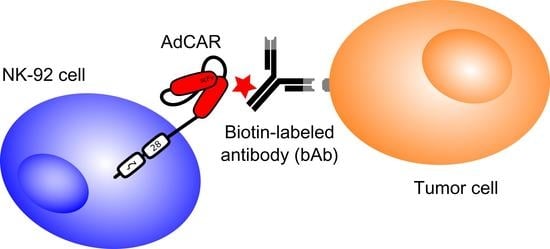
4.2. Design of the AdCAR System

The second-generation adapter CAR is based on the mAb “mBio3”-derived single-chain variable fragment (scFv) targeting a “neo”-epitope-like structure, the linker label epitope, consisting of biotin in the context of a mAb. Exact constitution of the AdCAR construct and generation of AdCAR-engineered NK-92 cells was previously described [23].

4.3. Biotinylated Antibodies

Antibodies were either purchased as bAb from Miltenyi Biotec (Bergisch-Gladbach, Germany) or acquired from the UKT pharmacy and biotinylated by Davids Biotechnologie (Regensburg, Germany).
AntigenCloneAntibodyOrder #LotSupplier
CD146541-10B2n/a130-092-8525190627154Miltenyi Biotec, Bergisch Gladbach, Germany
CD171REA163n/a130-100-7025190607129Miltenyi Biotec, Bergisch Gladbach, Germany
CD200OX-104n/a130-106-0645191021606Miltenyi Biotec, Bergisch Gladbach, Germany
CD221REA271n/a130-103-9735190627184Miltenyi Biotec, Bergisch Gladbach, Germany
CD271REA844n/a130-112-6085190627191Miltenyi Biotec, Bergisch Gladbach, Germany
CD274n/aAtezolizumabn/an/aHoffmann-La Roche, Basel, Switzerland
CD276FM276n/a130-095-5145190627174Miltenyi Biotec, Bergisch Gladbach, Germany
CD340n/aTrastuzumabn/an/aHoffmann-La Roche, Basel, Switzerland
EGFRn/aCetuximabn/an/aMerck KgaA, Darmstadt, Germany
GD2n/aDinutuximab betan/an/aEusa Pharma, Hertfordshire, Great Britain

4.4. Flow Cytometry

Staining of cells was conducted using primary biotinylated mAb with antigen specificity of interest. Cells were incubated with specific antibodies at 4 °C in flow cytometry buffer containing PBS (Sigma-Aldrich, St. Louis, MO, USA) supplemented with 2% FBS and 0.5 M EDTA (Sigma-Aldrich, St. Louis, MO, USA) for 15 min. Unbound antibody was washed off by centrifugation (4 °C, 350 g, 5 min) and stained with a secondary, fluorophore-labeled anti-biotin antibody (Anti-Biotin PE, clone: REA746, Miltenyi Biotec, Bergisch Gladbach, Germany) for 15 min followed by another washing step. Surface antigen expression was analyzed with the secondary antibody alone as a negative control using a BD FACSCanto II flow cytometer (BD, Franklin Lakes, NJ, USA).

4.5. Calcein Release-Based Cytotoxicity Assay (CRA)

Target cell staining with Calcein AM (Thermo Fisher Scientific, Waltham, MA, USA) as well as the protocol for the calcein release-based cytotoxicity assay (CRA) was described previously [23,45].

4.6. Real-Time Label-Free Live Cell Analysis

Bone metastasis cell lines were adjusted to a concentration of 105 cells/mL in RPMI complete medium and seeded in E-Plate 96 VIEW (OLS, Bremen, Germany) micro-well plates. Effector AdCAR NK-92 cells were adjusted to an E:T ratio of 5:1 in NK-92 complete medium without IL-2 and co-incubated with the target cells in the presence or absence of specific bAb. Utilizing the xCELLigence real-time cell analysis (RTCA, OLS, Bremen, Germany) system, cells were monitored for over 12 h. Tumor cell viability was calculated using the RTCA 2.0 software (OLS, Bremen, Germany) and AdCAR-mediated cytotoxicity was subsequently determined.

4.7. Quantification of Cytokine Release

Cytokine release of AdCAR NK-92 cells upon AdCAR induction was determined using the Bio-Plex Pro human cytokine 17-plex assay (Bio Rad, Hercules, CA, USA). The respective protocol was described previously [23].

4.8. 3D Spheroid Cytotoxicity Assay

GFP-transduced metastatic tumor cells were grown as three-dimensional spheroids and co-incubated with NK-92 as well as AdCAR NK-92 cells in the presence or absence of biotinylated antibodies. The respective protocol was described previously [23].

4.9. Data Analysis

All statistical analyses were performed with GraphPad Prism 8 software (GraphPad Software Inc., San Diego, CA, USA). Flow cytometry data were analyzed using FlowJo software V10.0.8 (FlowJo LLC, Ashford, OR, USA).

5. Conclusions

Innovative immunotherapy for the treatment of solid cancers and especially metastatic disease is urgently needed. Adapter CAR-engineered NK-92 cells are able to combine their “off-the-shelf” availability with personalized and controllable elimination of metastatic tumor cells, thus, establishing a potent cellular product with universal applicability and quick clinical translation potential for the treatment of solid tumors, including metastases.

Author Contributions

Conceptualization, S.G. and S.S.; validation, S.G. and S.S.; formal analysis, S.G.; investigation, S.G., F.T., J.M., C.S. and A.K.; writing—original draft preparation, S.G. and S.S.; writing—review and editing, S.G. and S.S.; visualization, S.G.; supervision, S.S.; project administration, S.G. and S.S.; funding acquisition, R.H. All authors have read and agreed to the published version of the manuscript.

Funding

The employing institution of S.G., C.S., R.H. and S.S. (University Children’s Hospital Tuebingen) received research funding by Miltenyi Biotec GmbH on the basis of a collaboration research agreement.

Institutional Review Board Statement

The study was conducted according to the guidelines of the Declaration of Helsinki, and approved by ethics committee at the Medical Faculty of the Eberhard Karls University and the University Hospital Tuebingen (reference number 008/2014BO2).

Informed Consent Statement

Informed consent was obtained from all subjects involved in the study.

Data Availability Statement

The data presented in this study are available from the corresponding author upon reasonable request.

Conflicts of Interest

J.M., C.S., A.K. and R.H. are co-inventors of a patent application focusing on adapter CAR technology.

References

  1. Siegel, R.L.; Miller, K.D.; Jemal, A. Cancer statistics, 2019. Cancer J. Clin. 2019, 69, 7–34. [Google Scholar] [CrossRef] [Green Version]
  2. Chaffer, C.L.; Weinberg, R.A. A perspective on cancer cell metastasis. Science 2011, 331, 1559–1564. [Google Scholar] [CrossRef] [PubMed]
  3. Hage, W.D.; Aboulafia, A.J.; Aboulafia, D.M. Incidence, location, and diagnostic evaluation of metastatic bone disease. Orthop. Clin. North Am. 2000, 31, 515–528. [Google Scholar] [CrossRef]
  4. Mundy, G.R. Metastasis to bone: Causes, consequences and therapeutic opportunities. Nat. Rev. Cancer 2002, 2, 584–593. [Google Scholar] [CrossRef] [PubMed]
  5. Macedo, F.; Ladeira, K.; Pinho, F.; Saraiva, N.; Bonito, N.; Pinto, L.; Gonçalves, F. Bone metastases: An overview. Oncol. Rev. 2017, 11, 321. [Google Scholar] [CrossRef] [PubMed]
  6. Chen, W.-Z.; Shen, J.-F.; Zhou, Y.; Chen, X.-Y.; Liu, J.-M.; Liu, Z.-L. Clinical characteristics and risk factors for developing bone metastases in patients with breast cancer. Sci. Rep. 2017, 7, 1–7. [Google Scholar] [CrossRef] [Green Version]
  7. Coleman, R.E. Clinical features of metastatic bone disease and risk of skeletal morbidity. Clin. Cancer Res. 2006, 12, 6243s–6249s. [Google Scholar] [CrossRef] [Green Version]
  8. Gross, G.; Waks, T.; Eshhar, Z. Expression of immunoglobulin-T-cell receptor chimeric molecules as functional receptors with antibody-type specificity (chimeric genes/antibody variable region). Immunology 1989, 86, 10024–10028. [Google Scholar]
  9. Sadelain, M.; Brentjens, R.; Rivière, I. The basic principles of chimeric antigen receptor design. Cancer Discov. 2013, 3, 388–398. [Google Scholar] [CrossRef] [Green Version]
  10. Maude, S.L.; Frey, N.; Shaw, P.A.; Aplenc, R.; Barrett, D.M.; Bunin, N.J.; Chew, A.; Gonzalez, V.E.; Zheng, Z.; Lacey, S.F.; et al. Chimeric antigen receptor T cells for sustained remissions in leukemia. N. Engl. J. Med. 2014, 371, 1507–1517. [Google Scholar] [CrossRef] [Green Version]
  11. Lee, D.W.; Kochenderfer, J.N.; Stetler-Stevenson, M.; Cui, Y.K.; Delbrook, C.; Feldman, S.A.; Fry, T.J.; Orentas, R.; Sabatino, M.; Shah, N.N.; et al. T cells expressing CD19 chimeric antigen receptors for acute lymphoblastic leukaemia in children and young adults: A phase 1 dose-escalation trial. Lancet 2015, 385, 517–528. [Google Scholar] [CrossRef]
  12. Gardner, R.A.; Finney, O.; Annesley, C.; Brakke, H.; Summers, C.; Leger, K.; Bleakley, M.; Brown, C.; Mgebroff, S.; Kelly-Spratt, K.S.; et al. Intent-to-treat leukemia remission by CD19 CAR T cells of defined formulation and dose in children and young adults. Blood 2017, 129, 3322–3331. [Google Scholar] [CrossRef] [PubMed]
  13. Srivastava, S.; Riddell, S.R. Chimeric antigen receptor T cell therapy: Challenges to bench-to-bedside efficacy. J. Immunol. 2018, 200, 459–468. [Google Scholar] [CrossRef] [PubMed]
  14. Liu, E.; Marin, D.; Banerjee, P.; Macapinlac, H.A.; Thompson, P.; Basar, R.; Kerbauy, L.N.; Overman, B.; Thall, P.; Kaplan, M.; et al. Use of CAR-transduced natural killer cells in CD19-positive lymphoid tumors. N. Engl. J. Med. 2020, 382, 545–553. [Google Scholar] [CrossRef]
  15. Tonn, T.; Schwabe, D.; Klingemann, H.G.; Becker, S.; Esser, R.; Koehl, U.; Suttorp, M.; Seifried, E.; Ottmann, O.G.; Bug, G. Treatment of patients with advanced cancer with the natural killer cell line NK-92. Cytotherapy 2013, 15, 1563–1570. [Google Scholar] [CrossRef] [PubMed]
  16. Williams, B.A.; Law, A.D.; Routy, B.; Denhollander, N.; Gupta, V.; Wang, X.-H.; Chaboureau, A.; Viswanathan, S.; Keating, A. A phase I trial of NK-92 cells for refractory hematological malignancies relapsing after autologous hematopoietic cell transplantation shows safety and evidence of efficacy. Oncotarget 2017, 8, 89256–89268. [Google Scholar] [CrossRef] [PubMed] [Green Version]
  17. Boyiadzis, M.; Agha, M.; Redner, R.L.; Sehgal, A.; Im, A.; Hou, J.-Z.; Farah, R.; Dorritie, K.A.; Raptis, A.; Lim, S.H.; et al. Phase 1 clinical trial of adoptive immunotherapy using “off-the-shelf” activated natural killer cells in patients with refractory and relapsed acute myeloid leukemia. Cytotherapy 2017, 19, 1225–1232. [Google Scholar] [CrossRef]
  18. Zhang, C.; Oberoi, P.; Oelsner, S.; Waldmann, A.; Lindner, A.; Tonn, T.; Wels, W.S. Chimeric antigen receptor-engineered NK-92 cells: An off-the-shelf cellular therapeutic for targeted elimination of cancer cells and induction of protective antitumor immunity. Front. Immunol. 2017, 8, 533. [Google Scholar] [CrossRef]
  19. Bin, H.; Tang, Y.; Li, W.; Zeng, Q.; Chang, D. Efficiency of CAR-T therapy for treatment of solid tumor in clinical trials: A meta-analysis. Dis. Markers 2019. [Google Scholar] [CrossRef] [Green Version]
  20. O’Rourke, D.M.; Nasrallah, M.P.; Desai, A.; Melenhorst, J.J.; Mansfield, K.; Morrissette, J.J.D.; Martinez-Lage, M.; Brem, S.; Maloney, E.; Shen, A.; et al. A single dose of peripherally infused EGFRvIII-directed CAR T cells mediates antigen loss and induces adaptive resistance in patients with recurrent glioblastoma. Sci. Transl. Med. 2017. [Google Scholar] [CrossRef] [Green Version]
  21. Darowski, D.; Kobold, S.; Jost, C.; Klein, C. Combining the best of two worlds: Highly flexible chimeric antigen receptor adaptor molecules (CAR-adaptors) for the recruitment of chimeric antigen receptor T cells. mAbs 2019, 11, 621–631. [Google Scholar] [CrossRef] [PubMed] [Green Version]
  22. Grote, S.; Seitz, C.M.; Diepold, S.; Buchner, M.; Baden, C.; Malenke, E.; Dieckmann, S.M.; Schwaemmle, H.; Mittelstaet, J.; Kaiser, A.; et al. Adapter chimeric antigen receptor (aCAR)-engineered NK-92 cells: An off-the-shelf cellular therapeutic for universal tumor targeting. Blood 2018, 132, 3331. [Google Scholar] [CrossRef]
  23. Grote, S.; Mittelstaet, J.; Baden, C.; Chan, K.C.-H.; Seitz, C.; Schlegel, P.; Kaiser, A.; Handgretinger, R.; Schleicher, S. Adapter chimeric antigen receptor (AdCAR)-engineered NK-92 cells: An off-the-shelf cellular therapeutic for universal tumor targeting. Oncoimmunology 2020, 9, 1825177. [Google Scholar] [CrossRef]
  24. Seitz, C.M.; Schlegel, P.; Hau, J.; Krahl, A.-C.; Schroeder, S.; Bender, G.; Reiter, S.; Schleicher, S.; Schilbach, K.; Ebinger, M.; et al. Novel adapter chimeric antigen receptor (aCAR) T cells for temporally controllable targeting of single and multiple tumor antigens. Blood 2017, 130, 1912. [Google Scholar]
  25. Seitz, C.M.; Kieble, V.; Illi, C.; Reiter, S.; Grote, S.; Mittelstaet, J.; Lock, D.; Kaiser, A.; Schleicher, S.; Handgretinger, R.; et al. Combinatorial targeting of multiple shared antigens by adapter-CAR-T cells (aCAR-Ts) allows target cell discrimination and specific lysis based on differential expression profiles. Blood 2018, 132, 4543. [Google Scholar] [CrossRef]
  26. Kaiser, A.; Mittelstaet, J.; Huppert, V.; Miltenyi, S.; Schlegel, P.; Seitz, C.; Lang, P.; Handgretinger, R. Adapter chimeric antigen receptor expressing cells for targeting of multiple antigens. Europe Patent No. 3315511A1, 29 October 2016. [Google Scholar]
  27. Mitwasi, N.; Feldmann, A.; Arndt, C.; Koristka, S.; Berndt, N.; Jureczek, J.; Loureiro, L.R.; Bergmann, R.; Máthé, D.; Hegedüs, N.; et al. “UniCAR”-modified off-the-shelf NK-92 cells for targeting of GD2-expressing tumour cells. Sci. Rep. 2020, 10, 1–16. [Google Scholar] [CrossRef] [Green Version]
  28. Lohmueller, J.J.; Ham, J.D.; Kvorjak, M.; Finn, O.J. mSA2 affinity-enhanced biotin-binding CAR T cells for universal tumor targeting. OncoImmunology 2017, 7, e1368604. [Google Scholar] [CrossRef] [PubMed]
  29. Urbanska, K.; Lanitis, E.; Poussin, M.; Lynn, R.C.; Gavin, B.P.; Kelderman, S.; Yu, J.; Scholler, N.; Powell, D.J. A universal strategy for adoptive immunotherapy of cancer through use of a novel T-cell antigen receptor. Cancer Res. 2012, 72, 1844–1852. [Google Scholar] [CrossRef] [Green Version]
  30. Topp, M.S.; Gökbuget, N.; Zugmaier, G.; Degenhard, E.; Goebeler, M.-E.; Klinger, M.; Neumann, S.A.; Horst, H.A.; Raff, T.; Viardot, A.; et al. Long-term follow-up of hematologic relapse-free survival in a phase 2 study of blinatumomab in patients with MRD in B-lineage ALL. Blood 2012, 120, 5185–5187. [Google Scholar] [CrossRef] [PubMed] [Green Version]
  31. Topp, M.S.; Gökbuget, N.; Zugmaier, G.; Klappers, P.; Stelljes, M.; Neumann, S.; Viardot, A.; Marks, R.; Diedrich, H.; Faul, C.; et al. Phase II trial of the anti-CD19 bispecific T cell–engager blinatumomab shows hematologic and molecular remissions in patients with relapsed or refractory B-precursor acute lymphoblastic leukemia. J. Clin. Oncol. 2014, 32, 4134–4140. [Google Scholar] [CrossRef] [PubMed]
  32. Maude, S.L.; Laetsch, T.W.; Buechner, J.; Rives, S.; Boyer, M.; Bittencourt, H.; Bader, P.; Verneris, M.R.; Stefanski, H.E.; Myers, G.D.; et al. Tisagenlecleucel in children and young adults with B-cell lymphoblastic leukemia. N. Engl. J. Med. 2018, 378, 439–448. [Google Scholar] [CrossRef]
  33. Guo, Y.; Wang, Y.; Han, W. Chimeric antigen receptor-modified T cells for solid tumors: Challenges and prospects. J. Immunol. Res. 2016, 2016, 1–11. [Google Scholar] [CrossRef] [PubMed] [Green Version]
  34. Gong, J.H.; Maki, G.; Klingemann, H.G. Characterization of a human cell line (NK-92) with phenotypical and functional characteristics of activated natural killer cells. Leukemia 1994, 4, 652–658. [Google Scholar]
  35. Ward, J.P.; Bonaparte, M.I.; Barker, E. HLA-C and HLA-E reduce antibody-dependent natural killer cell-mediated cytotoxicity of HIV-infected primary T cell blasts. AIDS 2004, 18, 1769–1779. [Google Scholar] [CrossRef]
  36. Sarkar, S.; Van Gelder, M.; Noort, W.; Xu, Y.; Rouschop, K.M.A.; Groen, R.; Schouten, H.C.; Tilanus, M.G.J.; Germeraad, W.T.V.; Martens, A.C.M.; et al. Optimal selection of natural killer cells to kill myeloma: The role of HLA-E and NKG2A. Cancer Immunol. Immunother. 2015, 64, 951–963. [Google Scholar] [CrossRef] [PubMed] [Green Version]
  37. Tang, X.; Yang, L.; Li, Z.; Nalin, A.P.; Dai, H.; Xu, T.; Yin, J.; You, F.; Zhu, M.; Shen, W.; et al. First-in-man clinical trial of CAR NK-92 cells: Safety test of CD33-CAR NK-92 cells in patients with relapsed and refractory acute myeloid leukemia. Am. J. Cancer Res. 2018, 8, 1899. [Google Scholar] [PubMed]
  38. Nowakowska, P.; Romanski, A.; Miller, N.; Odendahl, M.; Bonig, H.; Zhang, C.; Seifried, E.; Wels, W.S.; Tonn, T. Clinical grade manufacturing of genetically modified, CAR-expressing NK-92 cells for the treatment of ErbB2-positive malignancies. Cancer Immunol. Immunother. 2017, 67, 25–38. [Google Scholar] [CrossRef] [PubMed]
  39. Schürch, C.M. CARving up colorectal cancer organoids in vitro. Genes Immun. 2019, 21, 1–3. [Google Scholar] [CrossRef]
  40. Wallstabe, L.; Göttlich, C.; Nelke, L.C.; Kühnemundt, J.; Schwarz, T.; Nerreter, T.; Einsele, H.; Walles, H.; Dandekar, G.; Nietzer, S.L.; et al. ROR1-CAR T cells are effective against lung and breast cancer in advanced microphysiologic 3D tumor models. JCI Insight 2019, 4. [Google Scholar] [CrossRef] [Green Version]
  41. Hart, A.L.; Ng, S.C.; Mann, E.; Al-Hassi, H.O.; Bernardo, D.; Knight, S.C. Homing of immune cells: Role in homeostasis and intestinal inflammation. Inflamm. Bowel Dis. 2010, 16, 1969–1977. [Google Scholar] [CrossRef]
  42. Fatehullah, A.; Tan, S.H.; Barker, N. Organoids as an in vitro model of human development and disease. Nat. Cell Biol. 2016, 18, 246–254. [Google Scholar] [CrossRef] [PubMed] [Green Version]
  43. Zhang, C.; Burger, M.C.; Jennewein, L.; Genβler, S.; Schönfeld, K.; Zeiner, P.; Hattingen, E.; Harter, P.N.; Mittelbronn, M.; Tonn, T.; et al. ErbB2/HER2-specific NK cells for targeted therapy of glioblastoma. J. Natl. Cancer Inst. 2015, 108. [Google Scholar] [CrossRef]
  44. Jiao, S.; Subudhi, S.K.; Aparicio, A.; Ge, Z.; Guan, B.; Miura, Y.; Sharma, P. Differences in tumor microenvironment dictate T helper lineage polarization and response to immune checkpoint therapy. Cell 2019, 179, 1177–1190. [Google Scholar] [CrossRef] [PubMed]
  45. Grote, S.; Chan, K.C.; Baden, C.; Bösmüller, H.; Sulyok, M.; Frauenfeld, L.; Ebinger, M.; Handgretinger, R.; Schleicher, S. CD276 as a novel CAR NK-92 therapeutic target for neuroblastoma. Adv. Cell Gene Ther. 2020, 4. [Google Scholar] [CrossRef]
  46. Burger, M.C.; Zhang, C.; Harter, P.N.; Romanski, A.; Strassheimer, F.; Senft, C.; Tonn, T.; Steinbach, J.P.; Wels, W.S. CAR-Engineered NK cells for the treatment of glioblastoma: Turning innate effectors into precision tools for cancer immunotherapy. Front. Immunol. 2019, 10, 2683. [Google Scholar] [CrossRef] [PubMed] [Green Version]
Figure 1. Cell line immunophenotyping. Bone metastatic cell lines were screened for tumor antigen surface expression using flow cytometry. Cells were co-incubated with primary biotinylated antibodies for 15 min at 4 °C. Antigen expression was detected using a secondary PE-coupled anti-biotin antibody (black line) using staining with the secondary antibody alone as negative control (grey area).
Cancers 13 01124 g001
Figure 2. AdCAR NK-92-mediated metastatic tumor cell lysis. AdCAR NK-92 cells were co-incubated with calcein-labeled tumor cell lines MAC (a), MAM (b), MCK83 (c) and MeGa17 (d) in the presence or absence of indicated biotinylated antibodies for 2 h at indicated E:T ratios. Specific lysis is shown as mean ± SD, n = 3. ****: p < 0.0001; ***: p < 0.001; **: p < 0.01; *: p < 0.1; ns: p ≥ 0.1.
Cancers 13 01124 g002
Figure 3. Kinetics of AdCAR-mediated tumor cell lysis. AdCAR NK-92 cells were co incubated with unlabeled tumor cell lines MAM (a) and MeGa17 (b) in the presence or absence of indicated biotinylated antibodies and constantly monitored over time using the xCELLigence real time cell analysis system. NK-mediated tumor cell lysis is depicted as decrease in the dimensionless “cell index”, n = 3.
Cancers 13 01124 g003
Figure 4. Cytokine secretion profile of AdCAR NK-92 cells. AdCAR NK-92 cells as well as parental NK-92 cells were co-incubated with the tumor cell line MAC in the presence and absence of bEGFR for 6 h at an E:T ratio oif 5:1. The release of cytokines was measured using the Bio-Plex Pro human cytokine 17-plex assay and is shown as a heatmap. PMA/Ionomycin was used as control to induce maximum cytokine secretion.
Cancers 13 01124 g004
Figure 5. AdCAR NK-92-mediated lysis of 3D tumor spheroids. GFP-transduced cell lines MAM was grown as 3D spheroids in ultra-low attachment plates and subsequently co-incubated with AdCAR NK-92 or parental NK-92 cells and indicated biotinylated antibodies for 96 h in at least three individual experiments. Fluorescence images show representative MAM spheroids at indicated time points (a). Integrated fluorescence intensity of tumor spheroids was measured regularly using the Celigo S Imaging Cytometer (Nexcelom, Lawrence, MA, USA), compared to untreated control spheroids and is shown as mean ± SD, n = 3 (b).
Cancers 13 01124 g005
Table 1. Patient data for tumor cells from bone metastasis resections.
Patient Age [years]Patient SexMetastatic SiteTumor EntityDesignation
Patient 163fScapulaMammary carcinomaMAC
Patient 217mSpine L3/L4Renal cell carcinomaMAM
Patient 354mSpine C7Colorectal carcinomaMCK83
Patient 447mAcetabulumMelanomaMeGa17
Table 2. Cell line immunophentoyping. Tumor cells were co-incubated with primary biotinylated antibodies. Antigen expression was detected using a secondary PE-coupled anti-biotin antibody. Percentage of stained cells and median fluorescence index (MFI) values were calculated using staining with the secondary antibody alone as negative control.
Cell LinePercentage of Stained Cells
CD146CD171CD200CD221CD271CD274CD276CD340EGFRGD2
MAC97.70%11.80%13.90%68.50%44.60%0.40%100.00%97.90%99.90%1.78%
MAM99.80%99.90%5.29%99.40%2.06%0.65%99.90%99.70%100.00%4.04%
MCK835.62%0.45%0.31%59.60%0.13%0.46%99.50%81.20%85.80%0.44%
MeGa1799.20%42.90%62.10%70.40%22.80%1.23%99.00%44.10%27.20%1.91%
Median Fluorescence Index (MFI)
CD146CD171CD200CD221CD271CD274CD276CD340EGFRGD2
MAC45.481.151.234.101.911.03106.659.39183.421.21
MAM194.2079.541.1611.270.991.03113.9211.5992.861.35
MCK831.221.000.966.260.770.9731.3910.0812.411.14
MeGa17172.523.497.719.061.960.9667.705.002.781.18
Table 3. Characterization of tumor cell lines for NK ligand expression. Tumor cells were co-incubated with fluorescently labeled antibodies. MFI values were calculated using staining with the respective isotype control antibody.
Cell LineCD48CD50CD54CD58CD95CD102CD112CD155CD261CD262
MAC1.471.695.4673.2223.351.2734.41128.963.0116.87
MAM1.361.787.3130.559.541.1369.73192.656.0659.84
MCK831.841.751.295.544.261.2715.3289.624.1514.91
MeGa171.251.589.6631.322.181.1316.2750.201.2215.03
HLA-ABCHLA-DRHLA-EHLA-GMICA/BULBP1ULBP2/5/6ULBP3ULBP4
MAC52.410.909.180.986.533.451.260.680.75
MAM19.070.878.070.8121.253.151.001.671.53
MCK830.470.9210.010.829.076.091.091.251.41
MeGa1723.3312.107.551.347.272.451.370.920.77
Publisher’s Note: MDPI stays neutral with regard to jurisdictional claims in published maps and institutional affiliations.

