Article

Characteristics of a Novel ATP2B3 K416_F418delinsN Mutation in a Classical Aldosterone-Producing Adenoma

1
Chinru Clinic, Taipei 116, Taiwan
2
Department of Internal Medicine, National Taiwan University Hospital, Taipei 110, Taiwan
3
Department of Pathology, National Taiwan University Hospital, National Taiwan University College of Medicine, Taipei 100, Taiwan
4
Department of Urology, College of Medicine, National Taiwan University, National Taiwan University Hospital, Taipei 110, Taiwan
*
Author to whom correspondence should be addressed.
These authors contributed equally to this work.
Membership of the (TAIPAI) Study Group is provided in the Acknowledgments.
Cancers 2021, 13(18), 4729; https://doi.org/10.3390/cancers13184729
Submission received: 23 August 2021 / Revised: 14 September 2021 / Accepted: 15 September 2021 / Published: 21 September 2021

Abstract

:

Simple Summary

The ATP2B3 channel mutation is a rare cause of primary aldosteronism (PA). ATP2B3 gene mutation leads to the dysfunction of calcium channel that pumps calcium ion out of the cell and accumulates intracellular calcium signal to stimulate aldosterone synthesis. In the present study, we found a novel somatic ATP2B3 K416_F418delinsN mutation in a PA patient, and proved its functionality by demonstrating aldosterone hyper-function in the mutant-transfected adrenal cell-line. The ATP2B3 K416_F418delinsN mutation resulted from the deletion from nucleotides 1248 to 1253. The translated amino acid sequence from 416 to 418 as lysine-phenylalanine-phenylalanine was deleted and an asparagine was inserted due to the merging of residual nucleotide sequences.

Abstract

In patients with primary aldosteronism (PA), the prevalence of ATP2B3 mutation is rare. The aim of this study is to report a novel ATP2B3 mutation in a PA patient. Based on our tissue bank of aldosterone-producing adenomas (APA), we identified a novel somatic ATP2B3 K416_F418delinsN mutation. The affected individual was a 53 year-old man with a 4 year history of hypertension. Computed tomography (CT) showed bilateral adrenal masses of 1.6 (left) and 0.5 cm (right) in size. An adrenal venous sampling (AVS) showed a lateralization index (LI) of 2.2 and a contralateral suppression index (CLS) of 0.12; indicating left functional predominance. After a left unilateral adrenalectomy, he achieved partial biochemical and hypertension–remission. This classical adenoma harbored a novel ATP2B3 K416_F418delinsN somatic mutation, which is a deletion from nucleotides 1248 to 1253. The translated amino acid sequence from 416 to 418, reading as lysine-phenylalanine-phenylalanine, was deleted; however, an asparagine was inserted due to merging of residual nucleotide sequences. The CYP11B2 immunohistochemistry staining demonstrated strong immunoreactivity in this classical adenoma. The ATP2B3 K416_F418delinsN mutation is a functional mutation in APA, since HAC15 cells, a human adrenal cell line, transfected with the mutant gene showed increased CYP11B2 expression and aldosterone production.

Graphical Abstract

1. Introduction

Primary aldosteronism (PA) is originally classified into unilateral hyperaldosteronism and bilateral hyperaldosteronism (BHA) [1]. BHA is mainly related to bilateral idiopathic hyperplasia, which could not be detected by computed tomography (CT) [1,2,3]. Bilateral aldosterone producing adenoma (APA) [4] could be detected as bilateral detectable mass by CT but is a rare finding [1]. The pathogenesis of patients affected by bilateral PA, thought related to BHA, could not be confirmed, because few patients with bilateral PA underwent adrenalectomy, and no such adrenal tissues could be obtained for further investigation. Many somatic mutant genes have been identified that enhance aldosterone secretion; in particular, mutations in KCNJ5 [5], CACNA1D [6], CACNA1H [7], CLCN2 [8], ATP1A1 [9], and ATP2B3 [9] genes have been identified. These mutant genes are often related to changes in the function or permeability of ion channels or ion pumps across cell membranes [10].
The aldosterone synthase (CYP11B2) transcription and aldosterone production rely on increased intracellular calcium signaling [10]. After stimulation, zona glomerulosa cells are depolarized and voltage-gated calcium (Ca2+) channels on cellular membrane are activated. Subsequently, an influx of extracellular calcium occurs and increases intracellular calcium concentrations and downstream signaling. The ion channel ATP2B3, a Ca2+ ATPase type 3, is a protein pump over cellular membrane that exports intracellular calcium out of cells [11]. The mutated ATP2B3 in aldosterone-producing cells may reduce efflux of calcium ions from cytoplasm, accumulate intracellular calcium, and stimulate aldosterone production [12].
The prevalence of ATP2B3 mutation in PA is quite low [9]; ranging 1.6% [9]~0.9% [13] from European cohorts. In our previous report, the prevalence of mutated ATP2B3 gene among our PA patients in Taiwan was 0.5% [14]. In this research, we described a novel mutation of ATP2B3 K416_F418delinsN in an APA patient who underwent ipsilateral adrenalectomy, and illustrated his clinical characteristics.

2. Materials and Methods

2.1. Ethics Statement

Ethics approval (approval number 200611031R) was approved by the Institutional Review Committee of National Taiwan University Hospital. Before participating in the study, we obtained a written informed consent form from all participants to collect and study clinical data.

2.2. Diagnosis of PA

Based on the Taiwan standard TAIPAI protocol and the consensus on hyperaldosteronism, the referral of patients with hypertension was screened, confirmed and subtyped for PA patients [15,16]. Prior to the PA screening and confirmation test, all original antihypertensive drugs were discontinued for at least 21 days. We prescribed doxazosin and/or diltiazem during the evaluation phase as needed to control markedly hypertension. The diagnosis of PA in patients with hypertension was according to the abnormal hypersecretion of aldosterone and met the criteria [16,17,18,19,20,21,22,23,24,25,26].

2.3. Genomic DNA Extraction

Tumoral and adjacent adrenal tissue’s genomic DNA was extracted by using QIAamp DNA mini kit (Qiagen, Hilden, Germany); genomic DNA from peripheral whole blood was extracted by using Blood DNA Isolation Kit (Geneaid Biotech; New Taipei City, Taiwan) according to the manufacturer’s instructions.

2.4. ATP2B3 Gene Sequencing

The coding regions containing well- characterized mutations of ATP2B3 gene were amplified and sequenced by using gene-specific primers and the BigDye® Terminator v3.1 Cycle Sequencing Kit (Applied Biosystems Inc., Foster City, CA, USA) on the 3730 DNA Analyzer (Applied Biosystems, Foster City, CA, USA). The primers of polymerase chain reaction (PCR) used to amplify fragments for ATP2B3 direct sequencing followed that from a previous report [21] (forward CCTGGGCTGTTTATCCTGAA, reverse CCCCAGTTTCCGAGTCTGTA). The sequences analysis was performed by using the DNAStar Lasergene SeqMan Pro 7.1.0 software (DNAStar Inc., Madison, WI, USA).

2.5. Immunohistochemistry of Resected Tissues

The CYP11B2 and 17α-hydroxylase (CYP17A1) mouse monoclonal antibody, CYP11B1 rat monoclonal antibody (generous gifts from Professor Celso Gomez-Sanchez [27]), and HSD3B mouse monoclonal antibody (Abnova, Taipei, Taiwan) were used for immunohistochemistry (IHC) [28]. The polymerized horseradish peroxidase (HRP)-anti-mouse conjugate method (Novolink; Novocastra Laboratories Ltd., Newcastle Upon Tyne, UK) was used to stain sections of paraffin-embedded adrenal tumors and surrounding tissues according to the manufacturer’s instructions [14]. The images were captured by Olympus BX51 fluorescence microscope combined with Olympus DP72 camera and cell Sens Standard 1.14 software (Olympus, Hamburg, Germany) was used for image analysis.

2.6. Culture of Cell Line

We used HAC15 cell, a human adrenocortical cell line, which express aldosterone synthase, CYP11B2, and secrete aldosterone for aldosterone production study. The HAC 15 cell line was obtained from generous Dr. Silvia Monticone [29]. HAC15 cells were cultured in HAC15 complete media containing DMEM:F12 (1:1) supplemented with 10% cosmic calf serum, 1× ITS, 1% penicillin–streptomycin and 100 μg/mL primocin at 37 °C. We used humidified incubator with 5% CO2 to incubate the cultured cells, as previously reported [17].

2.7. Plasmid and Transfection

We used PCR-assisted, site-directed mutagenesis for the plasmids, expressing the wild-type and mutant ATP2B3 genes and cloning into the pIRES-GFP-puro vector. PCR-based direct sequencing confirmed that the mutation was successfully cloned into the vector. Moreover, 3 × 10⁶ cells HAC15 cells were transiently transfected with 3 μg pIRES-GFP empty vector, pIRES-GFP-wild-type ATP2B3 or pIRES-GFP ATP2B3 K416_F418delinsN using the Amaxa Cell Line Nucleofector Kit R (Lonza, Cologne, Germany) and the Nucleofector I (program X-005), according to the instructions of the manufacturer. After transfection, we seeded the HAC15 cells with a density of 1 × 10⁶ cells/well into a 6-well plate. Furthermore, 72 h after the transfection, the cells and culture supernatants were harvested for Western blot analysis and aldosterone measurement.

2.8. Western Blot Analysis

Using RIPA buffer (50 mM Tris base pH 8, 150 mM NaCl, 1% NP40, 0.10% SDS) containing a protease inhibitor (Roche Diagnostics, Indianapolis, IN, USA), proteins were isolated from whole cell extracts. After we centrifuged the cell lysates, the supernatants were mixed with 3× sample buffer (30% glycerol, 15% 2-mercaptoethanol and 1% bromophenol blue). The proteins were separated through 12% SDS-PAGE gels and electrophoretic transferred to PVDF membranes. The membranes were then blocked by incubating in the BlockPRO™ blocking buffer (Visual Protein Biotechnology, Taipei, Taiwan) for 1 h blocking buffer containing anti-CYP11B2 mouse monoclonal antibody (a kind gift from Professor Celso Gomez-Sanchez) and anti-GAPDH antibody were incubated overnight at 4 °C. Extensive washing was conducted by Tris-buffered saline containing 0.1% Tween-20 (TBST) buffer. We further incubated the transfer membranes in blocking buffer that contained HRP-conjugated secondary antibodies for 1.5 h. Subsequently, the membranes were washed with TBST three times. Enhanced chemiluminescent reagent (Thermo Scientific, Rockford, IL, USA) was applied at a ratio of 1:1. Reagents for Chemiluminescence detection (Millipore, Billerica, MA, USA) were used to detect protein levels, and UVP Biospectrum 810 imaging system (Ultra Violet Products Ltd., Cambridge, UK) was used for visualization. We quantified protein expression in each sample by using UVP software (Ultra Violet Products Ltd., Cambridge, UK). A densitometry analysis of each protein band was normalized to GAPDH levels and expressed as a relative fold change when compared with the non-transfected control.

2.9. Analysis of Aldosterone

The culture supernatants were collected 72 h after cells transfected with ATP2B3 K416_F418delinsN mutant or wild type plasmids to measure aldosterone concentration (ALDO-RIACT RIA kit, Cisbio Bioassays, Codolet, France) [28].

2.10. Statistical Analysis

The experimental differences between the transfected gene groups were analyzed by one-way ANOVA with post hoc least significant difference test (LSD) tests. A two-sided p value < 0.05 was defined as statistically significant. All of the statistical analyses were performed using IBM SPSS statistics version 19 (IBM Corp, Armonk, NY, USA) software.

3. Results

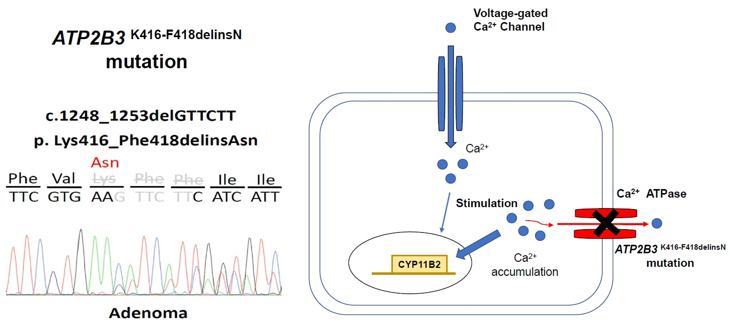
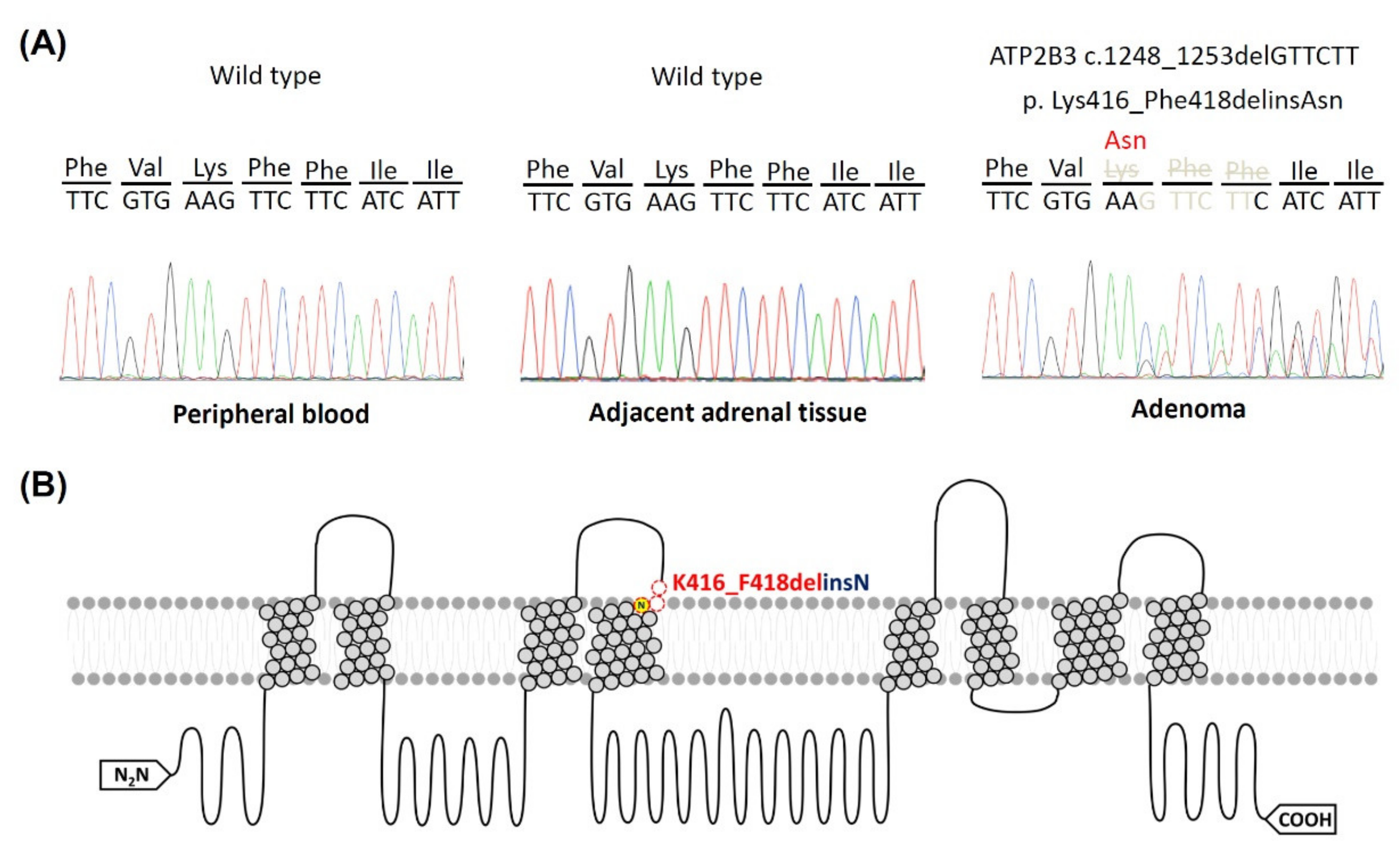
3.1. Identifying the ATP2B3 K416_F418delinsN Gene and Demographics of the Specific Patient

In DNA samples extracted from APA tumor tissues, we identified the mutant ATP2B3 gene in a left adrenal adenoma. The affected individual was a 53 year-old man. He had a history of hypertension for more than 4 years and presented with uncontrollable hypertension and hypokalemia (2.8 mEq/L) for further survey. After the standardized screening and confirmation tests, his PA was diagnosed. A computer tomography scan showed a left 1.6 cm and a right 0.5 cm adrenal masses. An adrenal venous sampling (AVS) showed functionally predominant aldosterone hypersecretion over his left adrenal gland (Table 1). The lateralization index (LI) was 2.2 and the contralateral suppression index (CLS) was 0.12. The result of the iodine-131 6-beta-iodomethyl-19-norcholesterol adrenal scintigraphy (NP-59 scan) was also compatible with a functional left adrenal adenoma. After a left adrenalectomy, his hypertension achieved clinical partial remission, with reduced doses of anti-hypertensive medications (Table 1). In the postoperative biochemical tests, hypokalemia was resolved; however, the aldosterone to renin ratio (ARR) remained high. Therefore, biochemical outcome reached only partial success [30].
This novel ATP2B3 K416_F418delinsN somatic mutation was identified only in the resected adrenal tissue, but not in peripheral blood cells and adjacent adrenal tissue. The ATP2B3 K416_F418delinsN somatic mutation resulted from the deletion at nucleotide 1248 to 1253 as GTTCTT (Figure 1). The resulting amino acid sequence showed the deletion of 416 lysine (Lys, K), 417 phenylalanine (Phe, F) and 418 phenylalanine (Phe, F) due to the deletion of the nucleotide from 1248 to 1253 (Figure 1). However, a new amino acid, asparagine, was found due to merged nucleotides from 1246, 1247 and 1254. Therefore, the original amino acid sequence as lysine-phenylalanine- phenylalanine was not encoded, but instead a new amino acid, asparagine, was inserted.

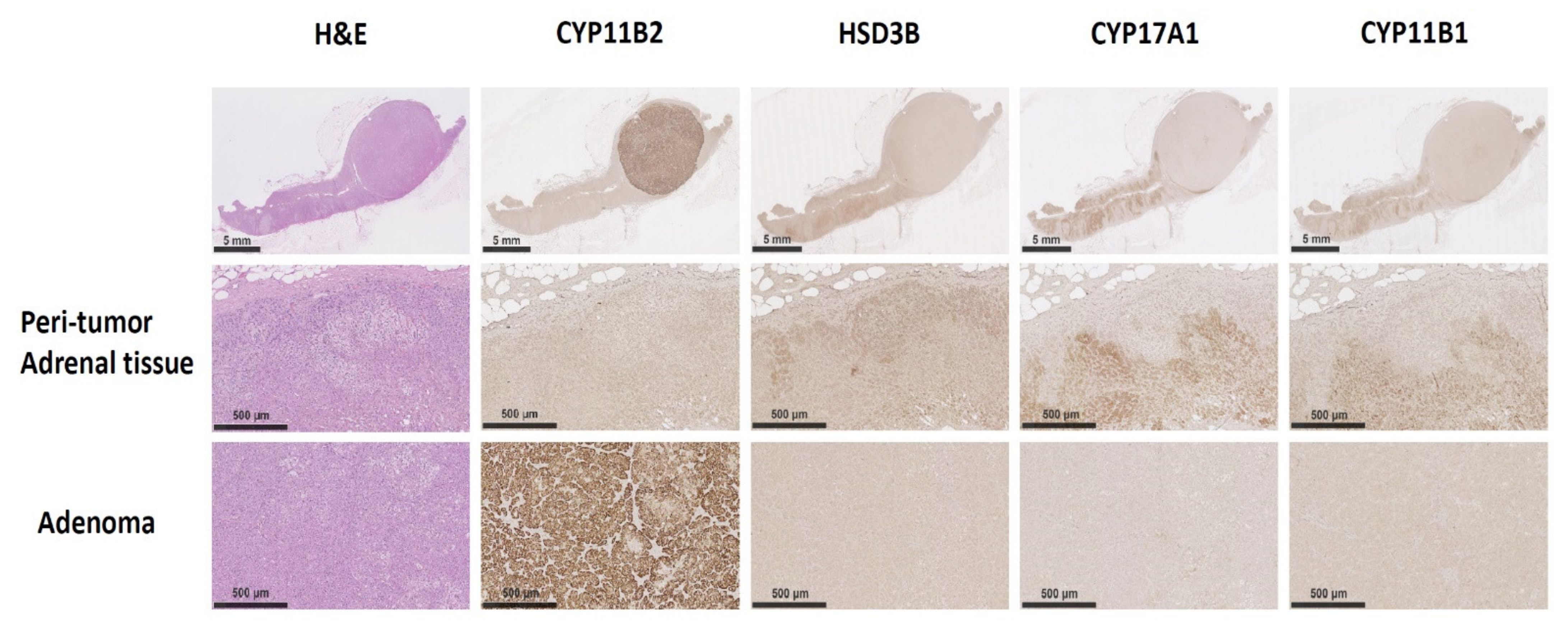
3.2. The Immunochemistry Staining of CYP11B2 on Excised Adrenal Tissue

The gross slide section showed a well-demarcated and easily identified classical APA in the excised adrenal gland. The steroid 18-hydroxylase (CYP11B2; aldosterone synthase) IHC staining showed intense density within that compact zona glomerulosa (ZG)-like adenoma (Figure 2). No CYP11B2 IHC staining was found in the adjacent adrenal cortical tissues. For other steroidogenesis related enzymes, such as 3β-Hydroxysteroid dehydrogenase (HSD3B), 17α-hydroxylase (CYP17A1), and 11β-hydroxylase (CYP11B1), their IHC staining was not enhanced in the adenoma, but was observed in the adjacent adrenal gland tissues.

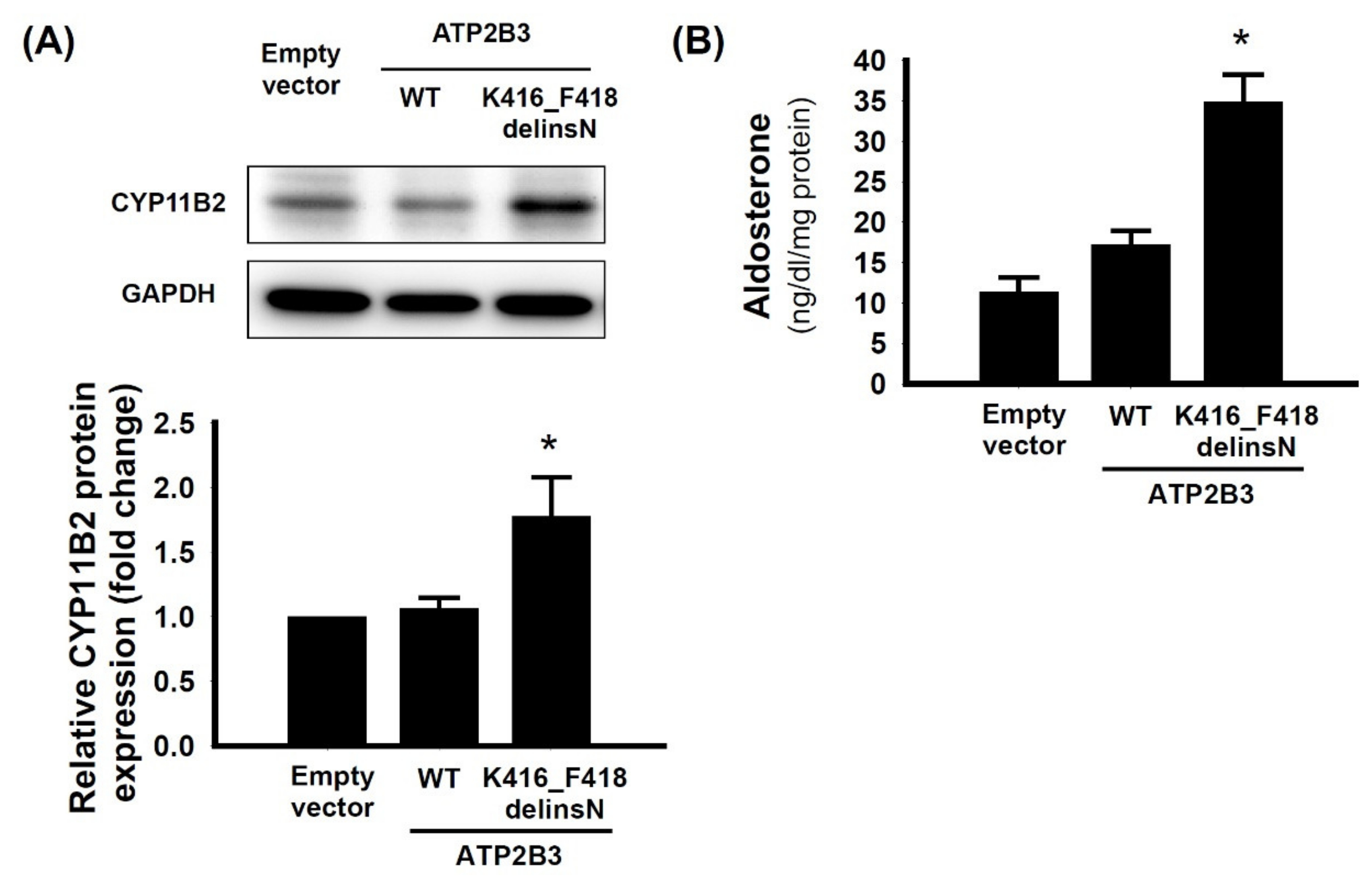
3.3. The Aldosterone Synthase of ATP2B3 K416-F418delinsN Mutation

We transfected the mutant ATP2B3 K416_F418delinsN gene to HAC15 cells and investigated the physiological effects of this novel mutation. The expression of CYP11B2 in mutant-gene transfected cells was increased compared to that of control cells transfected with empty vector or wild type ATP2B3 (Figure 3). The aldosterone levels in the supernatant of the culture medium were higher in mutant-gene transfected cells compared to that from control vector or the wild type cells. Thus, the deletion of amino acid expression of lysine, and two phenylalanine residues from 416 to 418, along with an insertion of asparagine, showed a gain-of-function mutation at ATP2B3 channel for aldosterone over-production.

4. Discussion

In this study, we found a novel functional somatic ATP2B3 K416_F418delinsN mutant gene from our APA tissue bank. The histopathological examination showed a well-defined compact, ZG-like classical adenoma and intense immunoreactivity to CYP11B2 staining. The cells transfected with the indicated mutant gene demonstrated increased CYP11B2 expression and elevated aldosterone levels in the culture supernatant when compared with that of the wild-type cells. Moreover, we have used Sanger sequencing to confirm that there were no other conventional and well-characterized aldosterone-driving gene mutations, including KCNJ5, ATP1A1, CACNA1D, and CTNNB1, in the adenoma harboring with ATP2B3 K416_F418delinsN mutation (Figure S1).

4.1. Calcium Channel and Somatic Mutations in APA

The transcription of CYP11B2 could be activated by intracellular calcium signaling [33]. Consistently increased cytoplasmic calcium concentration or stimulation may lead to the excessive production of aldosterone [34], which is the main mechanism of PA. Mutant KCNJ5, and CLCN2 ion channels are involved in cellular membrane depolarization and subsequently activate voltage-gated Ca2+ channels to increase Ca2+ influx [34,35] into cytoplasm. However, ion channels of CACNA1D and CACNA1H control the entry of extracellular calcium, and enhance the Ca2+ permeability with their mutant voltage-gated Ca2+ channels. Unlike other channels that increase intracellular calcium by affecting Ca2+ entry, the mutated ATP2B3 ion channel reduces Ca2+ export from cytoplasm. Thus, there are two possible ways to increase stimulating signaling in ATP2B3 ion channel [11]: (1) reduce clearance of intracellular calcium and directly stimulating CYP11B2 transcription, and (2) accumulation of cation leads to Na+ influx and subsequently depolarizes the cellular membrane and activates a downstream reaction [11].

4.2. Mutant ATP2B3 and APA

ATP2B3 ion channel belongs to plasma membrane Ca2+ ATPase (PMCA) transporter. The ATP2B3 mutant APAs have higher serum aldosterone levels and lower potassium levels compared to other mutant genes related to APA [36].
Our index patient with ATP2B3 K416_F418delinsN had uncontrollable hypertension and hypokalemia at presentation. In accordance with our finding, most identified ATP2B3 mutation in APA were expressed mainly in ZG-like cells [37]. The IHC showed condensed CYP11B2 staining in the adenoma, but not in the peri-tumoral region. Therefore, the source of excess aldosterone could arise from the classical adenoma, in concordance with the location of the mutant gene. We have also confirmed that the adrenal tissue adjacent to the adenoma, besides that from white blood cells, carried wild-type ATP2B3 gene by using Sanger sequencing (showed in the Figure 1A and Table S1).

4.3. Bilateral Asymmetric Manifestations of the APA

This patient had bilateral adrenal masses, and CT showed a larger mass on the left side. The LI of AVS was 2.2 (>2.0) [38], indicating a left functional predominance. The CLS was 0.12 (<1.0) [39,40], indicating the suppression of the right adrenal gland. The preoperative diagnosis was unilateral PA over the left adrenal gland. However, 1 year after left unilateral adrenalectomy, blood pressure control only achieved partial success. The serum potassium level was normalized from 2.8 to 4.0 mEq/L. However, the ARR after unilateral adrenalectomy remained high. According to the PASO consensus, the biochemical outcome achieved only partial success [30]. Thus, from the functional and clinical responses, this patient with ATP2B3 K416_F418delinsN mutation may have abnormal contralateral adrenal gland and bilateral asymmetric aldosterone secretion. However, before the left total adrenalectomy, the aldosterone secretion from the right adrenal gland was suppressed by the left functional adenoma initially; once the patient underwent left adrenalectomy, the suppression from the left adrenal was gone, and the aldosterone secretion from the right adrenal gland, probably in the form of multiple aldosterone-producing micronodules (mAPM) or APA, may take over and contribute to the bilateral asymmetric disease, and led to his incomplete blood pressure and biochemical recovery.

4.4. Clinical Implication and Study Limitations

Different mutant genes of PA have specific pathophysiological, clinical and biochemical manifestations [36]. Identifying functional genes in PA would help physicians determine the course of the disease and make decision on treatment and follow-up. Patients with ATP2B3 mutant APA have obvious aldosterone and potassium abnormalities [36]. Most reported that APA harboring ATP2B3 mutant was unilateral APA. Our finding of this new mutation will help researchers in this field to incorporate this mutation in their future routine screening of the possible mutation spots, and the actual prevalence of this novel mutation will be further assured. Although left APA was confirmed by the AVS and resected, the pathophysiological characteristics in the contralateral adenoma could not be obtained. Furthermore, we have only one APA patient harboring this novel ATP2B3 K416_F418delinsN gene and could not conclude a general relationship between the genotype and phenotype.

5. Conclusions

In conclusion, we identified a patient with an APA harboring a novel ATP2B3 K416_F418delinsN somatic mutation. He became a partial hypertension-remission and biochemical success after unilateral adrenalectomy. HAC15 cells harboring this ATP2B3 K416_F418delinsN somatic mutation increased CYP11B2 synthesis and aldosterone production. The immunohistochemistry staining showed a compact and well demarcated ZG-like adenoma, with intense CYP11B2 expression. Thus, this novel somatic mutation of ATP2B3 K416_F418delinsN functionally increased aldosterone secretion, and it also showed a distinct histopathologic pattern, as well as an important clinical signature.

Supplementary Materials

The following are available online at https://www.mdpi.com/article/10.3390/cancers13184729/s1, Figure S1. Sanger sequencing analysis of tumor DNAs for conventional and well-characterized aldosterone-driving gene mutations, including KCNJ5, ATP1A1, CACNA1D, and CTNNB1, in the adenoma harboring with ATP2B3 K416_F418delinsN mutation, Figure S2. Uncropped Western Blot images from Figure 3 in the main text, Table S1. Primers used for Sanger sequencing.

Author Contributions

Conceptualization, K.-Y.P. and V.-C.W.; methodology, K.-Y.P.; software, H.-W.L.; validation, Y.-H.L., S.-L.L. and W.-C.L.; formal analysis, H.-W.L. and K.-Y.P.; investigation, V.-C.W.; resources, V.-C.W.; data curation, K.-Y.P.; writing—original draft preparation, H.-W.L. and K.-Y.P.; writing—review and editing, V.-C.W. and J.S.C.; visualization, K.-Y.P. and V.-C.W.; supervision, J.S.C.; project administration, V.-C.W.; funding acquisition, V.-C.W. All authors have read and agreed to the published version of the manuscript.

Funding

This project was supported by National Health Research Institutes [PH-102-SP-09)], Ministry of Science and Technology, Taiwan, R.O.C. [MOST107-2314-B-002-026-MY3, 108-2314-B-002-058, 109-2314-B-002-174-MY3], National Taiwan University Hospital [109-S4634, PC-1264, PC-1309, VN109-09, UN109-041, UN110-030], Grant MOHW110-TDU-B-212-124005 and Mrs. Hsiu-Chin Lee Kidney Research Fund.

Institutional Review Board Statement

Ethics approval was approved by the Institutional Review Committee of National Taiwan University Hospital (approval number 200611031R; extended approval date: 3 August 2020).

Informed Consent Statement

Before participating in the study, a written informed consent statement for clinical data collection and research was obtained from all participants.

Data Availability Statement

The data are all included in the manuscript or can be acquired from the corresponding author.

Acknowledgments

The authors thank the staff of the Second Core Lab in the Department of Medical Research of National Taiwan University Hospital for technical assistance. Membership of the Taiwan Primary Aldosteronism Investigation (TAIPAI) Study Group: Tai-Shuan Lai; Vin-Cent Wu; Shao-Yu Yang; Kao-Lang Liu; Chin-Chen Chang; Bo-Chiag Lee; Shuo-Meng Wang; Kuo-How Huang; Po-Chih Lin; Yen-Hung Lin; Lian-Yu Lin; Shih-Cheng Liao; Ruoh-Fang Yen; Ching-Chu Lu; Shih-Chieh Jeff Chueh (National Taiwan University Hospital ,Taipei, Taiwan); Chieh-Kai Chan (NTUH Hsin-Chu branch); Leay-Kiaw Er; Ya-Hui Hu; Chia-Hui Chang; Che-Hsiung Wu; Yao-Chou Tsai (Taipei Tzu Chi Hospital, Buddhist Tzu Chi Medical Foundation, Taipei, Taiwan); Chen-Hsun Ho (Taipei Medical University-Shuang Ho Hospital, Ministry of Health and Welfare); Wei-Chieh Huang(Taipei Veterans General Hospital); Ying-Ying Chen (MacKay Memorial Hospital); Kwan-Dun Wu (National Taiwan University Hospital ,Taipei, Taiwan NTUH, Director of Coordinating Center).

Conflicts of Interest

No conflict of interest is declared. No financial conflict of interest exists.

References

  1. Omata, K.; Satoh, F.; Morimoto, R.; Ito, S.; Yamazaki, Y.; Nakamura, Y.; Anand, S.K.; Guo, Z.; Stowasser, M.; Sasano, H.; et al. Cellular and Genetic Causes of Idiopathic Hyperaldosteronism. Hypertension 2018, 72, 874–880. [Google Scholar] [CrossRef]
  2. Young, W.F. Diagnosis and treatment of primary aldosteronism: Practical clinical perspectives. J. Intern. Med. 2019, 285, 126–148. [Google Scholar] [CrossRef] [Green Version]
  3. Kuo, C.-C.; Wu, V.-C.; Huang, K.-H.; Wang, S.-M.; Chang, C.-C.; Lu, C.-C.; Yang, W.-S.; Tsai, C.-W.; Lai, C.-F.; Lee, T.-Y.; et al. Verification and evaluation of aldosteronism demographics in the Taiwan Primary Aldosteronism Investigation Group (TAIPAI Group). J. Renin-Angiotensin-Aldosterone Syst. 2011, 12, 348–357. [Google Scholar] [CrossRef] [PubMed] [Green Version]
  4. Wu, V.-C.; Chueh, S.; Chang, H.; Lin, W.-C.; Liu, K.-L.; Li, H.; Lin, Y.-H.; Wu, K.-D.; Hsieh, B. Bilateral aldosterone-producing adenomas: Differentiation from bilateral adrenal hyperplasia. Qjm 2007, 101, 13–22. [Google Scholar] [CrossRef]
  5. Choi, M.; Scholl, U.I.; Yue, P.; Björklund, P.; Zhao, B.; Nelson-Williams, C.; Ji, W.; Cho, Y.; Patel, A.; Men, C.J.; et al. K+ Channel Mutations in Adrenal Aldosterone-Producing Adenomas and Hereditary Hypertension. Science 2011, 331, 768–772. [Google Scholar] [CrossRef] [PubMed] [Green Version]
  6. Scholl, U.I.; Goh, G.; Stölting, G.; De Oliveira, R.C.; Choi, M.; Overton, J.D.; Fonseca, A.L.; Korah, R.; Starker, L.F.; Kunstman, J.; et al. Somatic and germline CACNA1D calcium channel mutations in aldosterone-producing adenomas and primary aldosteronism. Nat. Genet. 2013, 45, 1050–1054. [Google Scholar] [CrossRef] [PubMed]
  7. Daniil, G.; Fernandes-Rosa, F.L.; Chemin, J.; Blesneac, I.; Beltrand, J.; Polak, M.; Jeunemaitre, X.; Boulkroun, S.; Amar, L.; Strom, T.M.; et al. CACNA1H Mutations Are Associated with Different Forms of Primary Aldosteronism. EBioMedicine 2016, 13, 225–236. [Google Scholar] [CrossRef] [PubMed] [Green Version]
  8. Fernandes-Rosa, F.L.; Daniil, G.; Orozco, I.J.; Göppner, C.; El Zein, R.; Jain, V.; Boulkroun, S.; Jeunemaitre, X.; Amar, L.; Lefebvre, H.; et al. A gain-of-function mutation in the CLCN2 chloride channel gene causes primary aldosteronism. Nat. Genet. 2018, 50, 355–361. [Google Scholar] [CrossRef] [Green Version]
  9. Beuschlein, F.; Boulkroun, S.; Osswald, A.; Wieland, T.; Nielsen, H.N.; Lichtenauer, U.D.; Penton, D.; Schack, V.R.; Amar, L.; Fischer, E.; et al. Somatic mutations in ATP1A1 and ATP2B3 lead to aldosterone-producing adenomas and secondary hypertension. Nat. Genet. 2013, 45, 441–442. [Google Scholar] [CrossRef]
  10. Seccia, T.M.; Caroccia, B.; Gomez-Sanchez, E.P.; Gomez-Sanchez, C.E.; Rossi, G.P. The Biology of Normal Zona Glomerulosa And Aldosterone-Producing Adenoma: Pathological Implications. Endocr. Rev. 2018, 39, 1029–1056. [Google Scholar] [CrossRef] [Green Version]
  11. Tauber, P.; Aichinger, B.; Christ, C.; Stindl, J.; Rhayem, Y.; Beuschlein, F.; Warth, R.; Bandulik, S. Cellular Pathophysiology of an Adrenal Adenoma-Associated Mutant of the Plasma Membrane Ca2+-ATPase ATP2B3. Endocrinology 2016, 157, 2489–2499. [Google Scholar] [CrossRef] [PubMed] [Green Version]
  12. Fernandes-Rosa, F.L.; Boulkroun, S.; Zennaro, M.-C. Genetic and Genomic Mechanisms of Primary Aldosteronism. Trends Mol. Med. 2020, 26, 819–832. [Google Scholar] [CrossRef] [PubMed]
  13. Williams, T.A.; Monticone, S.; Schack, V.R.; Stindl, J.; Burrello, J.; Buffolo, F.; Annaratone, L.; Castellano, I.; Beuschlein, F.; Reincke, M.; et al. Somatic ATP1A1, ATP2B3, and KCNJ5 Mutations in Aldosterone-Producing Adenomas. Hypertension 2014, 63, 188–195. [Google Scholar] [CrossRef] [Green Version]
  14. Wu, V.-C.; Wang, S.-M.; Chueh, S.-C.J.; Yang, S.-Y.; Huang, K.-H.; Lin, Y.-H.; Wang, J.-J.; Connolly, R.; Hu, Y.-H.; Gomez-Sanchez, C.E.; et al. The prevalence of CTNNB1 mutations in primary aldosteronism and consequences for clinical outcomes. Sci. Rep. 2017, 7, 39121. [Google Scholar] [CrossRef] [PubMed]
  15. Chao, C.-T.; Wu, V.-C.; Kuo, C.-C.; Lin, Y.-H.; Chang, C.-C.; Chueh, S.J.; Wu, K.-D.; Pimenta, E.; Stowasser, M. Diagnosis and management of primary aldosteronism: An updated review. Ann. Med. 2013, 45, 375–383. [Google Scholar] [CrossRef] [PubMed]
  16. Wu, V.-C.; Hu, Y.-H.; Er, L.K.; Yen, R.-F.; Chang, C.-H.; Chang, Y.-L.; Lu, C.-C.; Chang, C.-C.; Lin, J.-H.; Lin, Y.-H.; et al. Case detection and diagnosis of primary aldosteronism—The consensus of Taiwan Society of Aldosteronism. J. Formos. Med. Assoc. 2017, 116, 993–1005. [Google Scholar] [CrossRef] [PubMed]
  17. Peng, K.-Y.; Chang, H.-M.; Lin, Y.-F.; Chan, C.-K.; Chang, C.-H.; Chueh, S.-C.J.; Yang, S.-Y.; Huang, K.-H.; Lin, Y.-H.; Wu, V.-C.; et al. miRNA-203 Modulates Aldosterone Levels and Cell Proliferation by Targeting Wnt5a in Aldosterone-Producing Adenomas. J. Clin. Endocrinol. Metab. 2018, 103, 3737–3747. [Google Scholar] [CrossRef] [PubMed] [Green Version]
  18. Peng, K.-Y.; Liao, H.-W.; Chan, C.-K.; Lin, W.-C.; Yang, S.-Y.; Tsai, Y.-C.; Huang, K.-H.; Lin, Y.-H.; Chueh, J.S.; Wu, V.-C. Presence of Subclinical Hypercortisolism in Clinical Aldosterone-Producing Adenomas Predicts Lower Clinical Success. Hypertension 2020, 76, 1537–1544. [Google Scholar] [CrossRef] [PubMed]
  19. Wu, V.-C.; Kuo, C.-C.; Wang, S.-M.; Liu, K.-L.; Huang, K.-H.; Lin, Y.-H.; Chu, T.-S.; Chang, H.-W.; Lin, C.-Y.; Tsai, C.-T.; et al. Primary aldosteronism: Changes in cystatin C-based kidney filtration, proteinuria, and renal duplex indices with treatment. J. Hypertens. 2011, 29, 1778–1786. [Google Scholar] [CrossRef]
  20. Wu, V.-C.; Lo, S.-C.; Chen, Y.-L.; Huang, P.-H.; Tsai, C.-T.; Liang, C.-J.; Kuo, C.-C.; Kuo, Y.-S.; Lee, B.-C.; Wu, E.-L.; et al. Endothelial Progenitor Cells in Primary Aldosteronism: A Biomarker of Severity for Aldosterone Vasculopathy and Prognosis. J. Clin. Endocrinol. Metab. 2011, 96, 3175–3183. [Google Scholar] [CrossRef] [Green Version]
  21. Wu, V.-C.; Huang, K.-H.; Peng, K.-Y.; Tsai, Y.-C.; Wu, C.-H.; Wang, S.-M.; Yang, S.-Y.; Lin, L.-Y.; Chang, C.-C.; Lin, Y.-H.; et al. Prevalence and clinical correlates of somatic mutation in aldosterone producing adenoma-Taiwanese population. Sci. Rep. 2015, 5, 11396. [Google Scholar] [CrossRef]
  22. Wu, V.-C.; Chueh, S.-C.; Chang, H.-W.; Lin, L.-Y.; Liu, K.-L.; Lin, Y.-H.; Ho, Y.-L.; Lin, W.-C.; Wang, S.-M.; Huang, K.-H.; et al. Association of Kidney Function With Residual Hypertension After Treatment of Aldosterone-Producing Adenoma. Am. J. Kidney Dis. 2009, 54, 665–673. [Google Scholar] [CrossRef] [PubMed]
  23. Wu, V.-C.; Yang, S.-Y.; Lin, J.-W.; Cheng, B.-W.; Kuo, C.-C.; Tsai, C.-T.; Chu, T.-S.; Huang, K.-H.; Wang, S.-M.; Lin, Y.-H.; et al. Kidney impairment in primary aldosteronism. Clin. Chim. Acta 2011, 412, 1319–1325. [Google Scholar] [CrossRef] [PubMed]
  24. Wu, V.-C.; Chang, H.-W.; Liu, K.-L.; Lin, Y.-H.; Chueh, S.-C.; Lin, W.-C.; Ho, Y.-L.; Huang, J.-W.; Chiang, C.-K.; Yang, S.-Y.; et al. Primary Aldosteronism: Diagnostic Accuracy of the Losartan and Captopril Tests. Am. J. Hypertens. 2009, 22, 821–827. [Google Scholar] [CrossRef] [PubMed] [Green Version]
  25. Wu, V.-C.; Wang, S.-M.; Chang, C.-H.; Hu, Y.-H.; Lin, L.-Y.; Lin, Y.-H.; Chueh, S.-C.J.; Chen, L.; Wu, K.-D. Long term outcome of Aldosteronism after target treatments. Sci. Rep. 2016, 6, 32103. [Google Scholar] [CrossRef] [PubMed]
  26. Chan, C.-K.; Yang, W.-S.; Lin, Y.-H.; Huang, K.-H.; Lu, C.-C.; Hu, Y.-H.; Wu, V.-C.; Chueh, J.S.; Chu, T.-S.; Chen, Y.-M. Arterial Stiffness is Associated with Clinical Outcome and Cardiorenal Injury in Lateralized Primary Aldosteronism. J. Clin. Endocrinol. Metab. 2020, 105. [Google Scholar] [CrossRef]
  27. Gomez-Sanchez, C.E.; Qi, X.; Velarde-Miranda, C.; Plonczynski, M.W.; Parker, C.R.; Rainey, W.; Satoh, F.; Maekawa, T.; Nakamura, Y.; Sasano, H.; et al. Development of monoclonal antibodies against human CYP11B1 and CYP11B2. Mol. Cell. Endocrinol. 2014, 383, 111–117. [Google Scholar] [CrossRef] [Green Version]
  28. Peng, K.-Y.; Liao, H.-W.; Chueh, J.S.; Pan, C.-Y.; Lin, Y.-H.; Chen, Y.-M.; Chen, P.-Y.; Huang, C.-L.; Wu, V.-C. Pathophysiological and Pharmacological Characteristics of KCNJ5 157-159delITE Somatic Mutation in Aldosterone-Producing Adenomas. Biomedicines 2021, 9, 1026. [Google Scholar] [CrossRef]
  29. Monticone, S.; Hattangady, N.G.; Penton, D.; Isales, C.M.; Edwards, M.A.; Williams, T.A.; Sterner, C.; Warth, R.; Mulatero, P.; Rainey, W.E. a Novel Y152C KCNJ5 mutation responsible for familial hyperaldosteronism type III. J. Clin. Endocrinol. Metab. 2013, 98, E1861–E1865. [Google Scholar] [CrossRef] [Green Version]
  30. Williams, T.A.; Lenders, J.W.M.; Mulatero, P.; Burrello, J.; Rottenkolber, M.; Adolf, C.; Satoh, F.; Amar, L.; Quinkler, M.; Deinum, J.; et al. Outcomes after adrenalectomy for unilateral primary aldosteronism: An international consensus on outcome measures and analysis of remission rates in an international cohort. Lancet Diabetes Endocrinol. 2017, 5, 689–699. [Google Scholar] [CrossRef] [Green Version]
  31. Omasits, U.; Ahrens, C.; Müller, S.; Wollscheid, B. Protter: Interactive protein feature visualization and integration with experimental proteomic data. Bioinformatics 2014, 30, 884–886. [Google Scholar] [CrossRef] [PubMed] [Green Version]
  32. Protter Software Application. Available online: http://wlab.ethz.ch/protter/start/ (accessed on 22 July 2021).
  33. Nanba, K.; Chen, A.; Nishimoto, K.; Rainey, W.E. Role of Ca2+/Calmodulin-Dependent Protein Kinase Kinase in Adrenal Aldosterone Production. Endocrinology 2015, 156, 1750–1756. [Google Scholar] [CrossRef]
  34. Seidel, E.; Schewe, J.; Scholl, U.I. Genetic causes of primary aldosteronism. Exp. Mol. Med. 2019, 51, 1–12. [Google Scholar] [CrossRef] [PubMed] [Green Version]
  35. Fernandes-Rosa, F.L.; Boulkroun, S.; Zennaro, M.-C. Somatic and inherited mutations in primary aldosteronism. J. Mol. Endocrinol. 2017, 59, R47–R63. [Google Scholar] [CrossRef] [PubMed] [Green Version]
  36. Fernandes-Rosa, F.L.; Williams, T.A.; Riester, A.; Steichen, O.; Beuschlein, F.; Boulkroun, S.; Strom, T.M.; Monticone, S.; Amar, L.; Meatchi, T.; et al. Genetic Spectrum and Clinical Correlates of Somatic Mutations in Aldosterone-Producing Adenoma. Hypertension 2014, 64, 354–361. [Google Scholar] [CrossRef]
  37. Itcho, K.; Oki, K.; Ohno, H.; Yoneda, M. Update on Genetics of Primary Aldosteronism. Biomedicines 2021, 9, 409. [Google Scholar] [CrossRef] [PubMed]
  38. Rossi, G.P.; Auchus, R.J.; Brown, M.; Lenders, J.W.; Naruse, M.; Plouin, P.F.; Satoh, F.; Young, W.F. An Expert Consensus Statement on Use of Adrenal Vein Sampling for the Subtyping of Primary Aldosteronism. Hypertension 2014, 63, 151–160. [Google Scholar] [CrossRef]
  39. Kline, G.; Chin, A.; So, B.; Harvey, A.; Pasieka, J. Defining contralateral adrenal suppression in primary aldosteronism: Implications for diagnosis and outcome. Clin. Endocrinol. 2015, 83, 20–27. [Google Scholar] [CrossRef]
  40. Lee, J.; Kang, B.; Ha, J.; Kim, M.-H.; Choi, B.; Hong, T.-H.; Kang, M.I.; Lim, D.-J. Clinical outcomes of primary aldosteronism based on lateralization index and contralateral suppression index after adrenal venous sampling in real-world practice: A retrospective cohort study. BMC Endocr. Disord. 2020, 20, 114. [Google Scholar] [CrossRef] [PubMed]
Figure 1. A novel ATP2B3 K416-F418delinsN mutation in classical APA. (A) The deletion at nucleotide 1248 to 1253 as GTTCTT in the ATP2B3 K416-F418delinsN gene was identified in a patient with APA in the resected adrenal adenoma. The amino acid residue 416 to 418 of ATP2B3 protein was substituted from lysine (Lys) and 2 phenylalanine (Phe) to asparagine (Asn) insertion. The letters for nucleotide bases represented as following: C, cytosine; G, guanine; T, thymine; (B) the protein secondary structure of mutant ATP2B3 channel was demonstrated. The yellow circle N represented insertion of amino acid, asparagine. The model was based on Protter software application [31] (http://wlab.ethz.ch/protter/start/ (accessed on 22 July 2021) [32]).
Cancers 13 04729 g001
Figure 2. The immunohistochemistry staining in unilateral PA patients with the ATP2B3 K416-F418delinsN mutations. The CYP11B2 and other steroidogenesis related enzyme IHC staining was conducted. The CYP11B2 immunoactivity was stained intensely within the adenoma, but did not stain in the adjacent adrenal gland tissue. Of note, HSD3B, CYP17A1 and CYP11B1 did not observed with adenoma but scattered in the residual adrenal gland tissue. Scale bar represented 500 μm.
Cancers 13 04729 g002
Figure 3. The CYP11B2 synthase and aldosterone production in the HAC 15 cells with transfected ATP2B3 K416_F418delinsN. The aldosterone synthase, CYP1B2, expression and supernatant aldosterone levels were analyzed at 72 h after plasmid transfection. (A) The cells transfected with ATP2B3 K416_F418delinsN had increased CYP11B2 protein expression in compared with the cells transfected with wild type cells; (B) The aldosterone levels of supernatant in the culture cells also increased in transfected group compared with the wild type cells. The data are presented as the means ± SD of three independent experiments in the transfected wild type and mutant cells. * p < 0.05 represented significant difference. The uncropped Western Blot image can be found in Figure S2.
Cancers 13 04729 g003
Table 1. The basal characteristics of the uPA patient with adenoma harboring ATP2B3 K416-F418delinsN deletion patient.
VariablesATP2B3 K416_F418delinsN Mutation
Age (years old)53
Sexmale
Body weight (kg)75
BMI (kg/m2)25.95
CT mass size (cm) Left: 1.6; Right: 0.5
AVS (aldosterone, ng/dL)/cortisol (μg/dL)
CLS0.12
LI2.2
NP-59Bilateral adrenal gland hyperfunction with left side predominance
Hypertension duration (years)4
SBP (mm Hg)197
SBP 12 mon158
DBP (mm Hg)92
DBP 12 mon88
Aldosterone level (ng/dL) 59.3
PRA (ng/mL/hr) 0.55
ARR(ng/dL per ng/mL/h)107.82
K (mEq/L) 2.8
At 12 months after adrenalectomy
Aldosterone level 30.5
PRA 0.09
K (mEq/L) 4.0
ARR (ng/dL per ng/mL/h) 338.33
Clinical successpartial success §
Biochemical successpartial success
Abbreviations: ARR, aldosterone renin ratio; BMI, body mass index; CLS: contralateral suppression index; DBP, diastolic blood pressure; K, potassium; LI, lateralization index; NP-59, The iodine-131 6-beta-iodomethyl-19-norcholesterol adrenal scintigraphy; PA, primary aldosteronism; PRA, plasma renin activity; SBP, systolic blood pressure. § Partial success represents that the same blood pressure as before surgery but with less antihypertensive medication or decreased BP by the same or less antihypertensive medication. Obtained after withholding drugs that interfere with the renin-angiotensin system.
Publisher’s Note: MDPI stays neutral with regard to jurisdictional claims in published maps and institutional affiliations.

