
Report on Late Toxicity in Head-and-Neck Tumor Patients with Long Term Survival after Radiochemotherapy

 , ,
, ,
Abstract
:Simple Summary
Abstract
1. Introduction
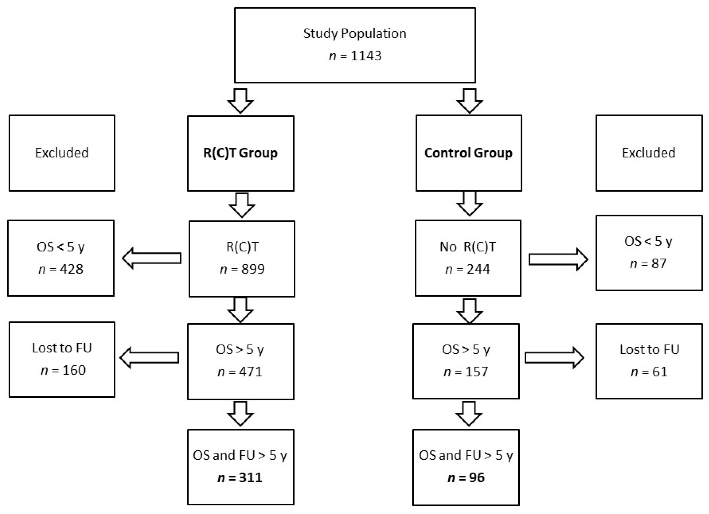
2. Patients and Methods
3. Statistics
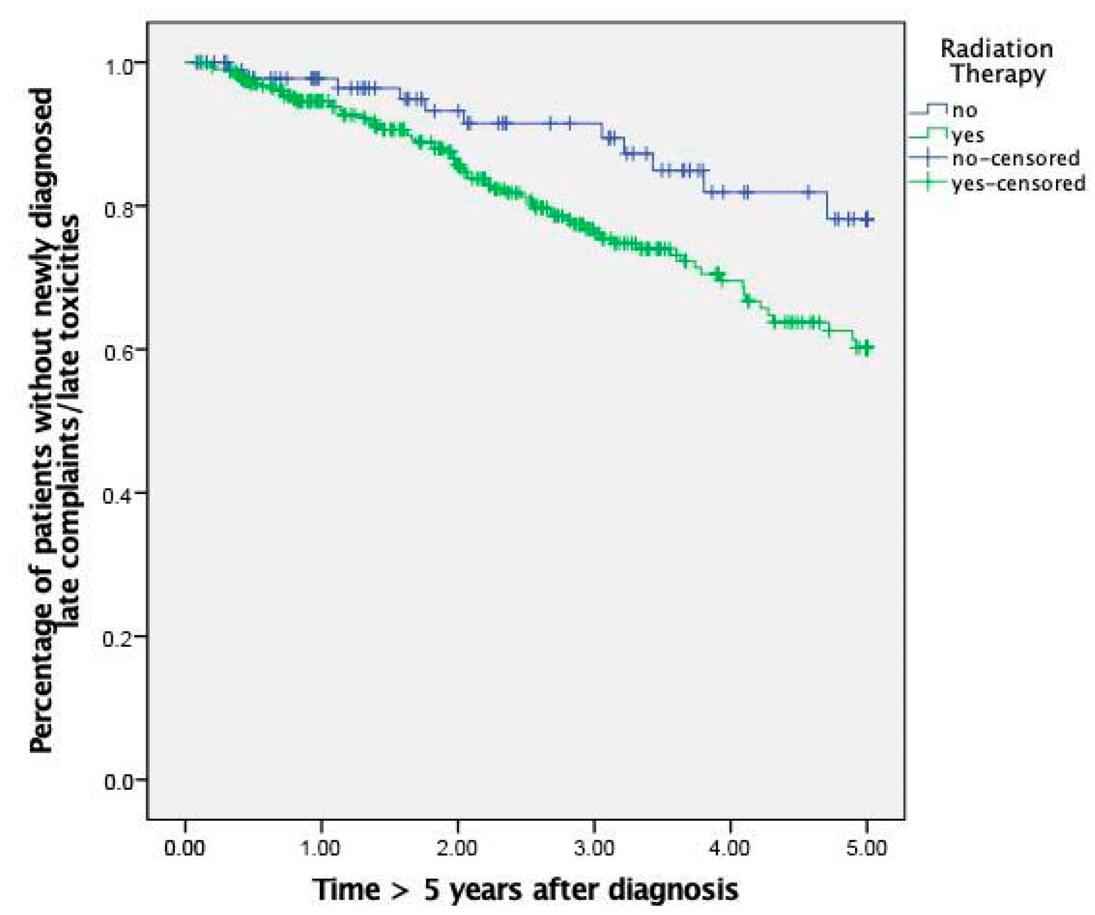
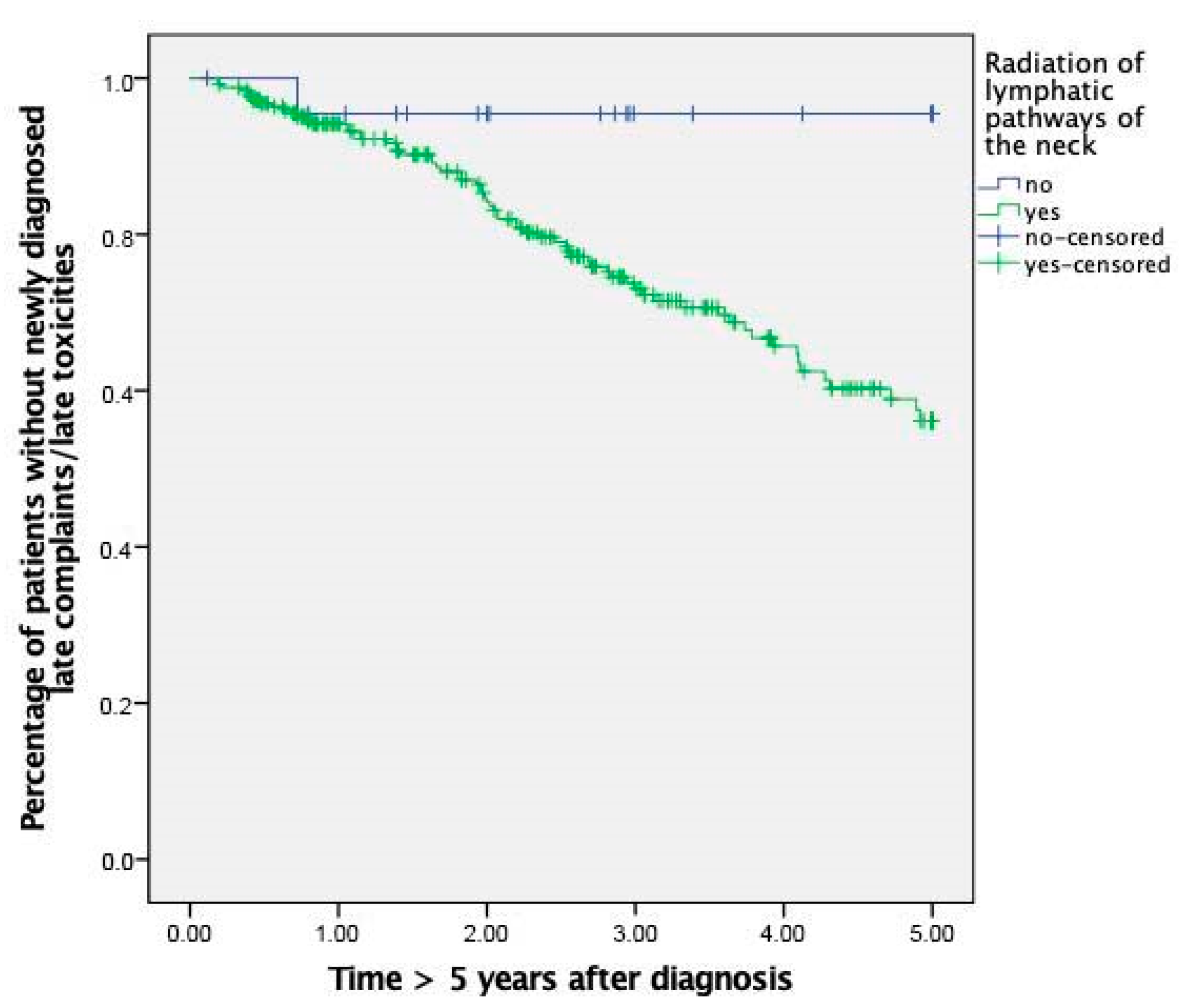
4. Results
4.1. Patient Characteristics
4.2. Late Radiation Induced Toxicities
5. Discussion
6. Conclusions
Author Contributions
Funding
Institutional Review Board Statement
Informed Consent Statement
Data Availability Statement
Acknowledgments
Conflicts of Interest
References
- Dorr, W. Radiation effect in normal tissue—Principles of damage and protection. Nuklearmedizin 2010, 49 (Suppl. 1), S53–S58. [Google Scholar]
- LENT SOMA scales for all anatomic sites. Int. J. Radiat. Oncol. Biol. Phys. 1995, 31, 1049–1091. [CrossRef]
- Hoeller, U.; Tribius, S.; Kuhlmey, A.; Grader, K.; Fehlauer, F.; Alberti, W. Increasing the rate of late toxicity by changing the score? A comparison of RTOG/EORTC and LENT/SOMA scores. Int. J. Radiat. Oncol. Biol. Phys. 2003, 55, 1013–1018. [Google Scholar] [CrossRef]
- Barnett, G.C.; West, C.M.; Dunning, A.M.; Elliott, R.M.; Coles, C.E.; Pharoah, P.D.; Burnet, N.G. Normal tissue reactions to radiotherapy: Towards tailoring treatment dose by genotype. Nat. Rev. Cancer 2009, 9, 134–142. [Google Scholar] [CrossRef] [PubMed] [Green Version]
- Machtay, M.; Moughan, J.; Trotti, A.; Garden, A.S.; Weber, R.S.; Cooper, J.S.; Forastiere, A.; Ang, K.K. Factors associated with severe late toxicity after concurrent chemoradiation for locally advanced head and neck cancer: An RTOG analysis. J. Clin. Oncol. Off. J. Am. Soc. Clin. Oncol. 2008, 26, 3582–3589. [Google Scholar] [CrossRef] [PubMed]
- Travis, E.L. Organizational response of normal tissues to irradiation. Semin. Radiat. Oncol. 2001, 11, 184–196. [Google Scholar] [CrossRef] [PubMed]
- Bentzen, S.M. Preventing or reducing late side effects of radiation therapy: Radiobiology meets molecular pathology. Nat. Rev. Cancer 2006, 6, 702–713. [Google Scholar] [CrossRef]
- Straub, J.M.; New, J.; Hamilton, C.D.; Lominska, C.; Shnayder, Y.; Thomas, S.M. Radiation-induced fibrosis: Mechanisms and implications for therapy. J. Cancer Res. Clin. Oncol. 2015, 141, 1985–1994. [Google Scholar] [CrossRef] [Green Version]
- Bourhis, J.; Lapeyre, M.; Tortochaux, J.; Rives, M.; Aghili, M.; Bourdin, S.; Lesaunier, F.; Benassi, T.; Lemanski, C.; Geoffrois, L.; et al. Phase III randomized trial of very accelerated radiation therapy compared with conventional radiation therapy in squamous cell head and neck cancer: A GORTEC trial. J. Clin. Oncol. Off. J. Am. Soc. Clin. Oncol. 2006, 24, 2873–2878. [Google Scholar] [CrossRef]
- Johansson, S.; Svensson, H.; Denekamp, J. Dose response and latency for radiation-induced fibrosis, edema, and neuropathy in breast cancer patients. Int. J. Radiat. Oncol. Biol. Phys. 2002, 52, 1207–1219. [Google Scholar] [CrossRef]
- Machtay, M.; Moughan, J.; Farach, A.; Martin-O’Meara, E.; Galvin, J.; Garden, A.S.; Weber, R.S.; Cooper, J.S.; Forastiere, A.; Ang, K.K. Hypopharyngeal dose is associated with severe late toxicity in locally advanced head-and-neck cancer: An RTOG analysis. Int. J. Radiat. Oncol. Biol. Phys. 2012, 84, 983–989. [Google Scholar] [CrossRef] [Green Version]
- Givens, D.J.; Karnell, L.H.; Gupta, A.K.; Clamon, G.H.; Pagedar, N.A.; Chang, K.E.; Van Daele, D.J.; Funk, G.F. Adverse events associated with concurrent chemoradiation therapy in patients with head and neck cancer. Arch. Otolaryngol. Head Neck Surg. 2009, 135, 1209–1217. [Google Scholar] [CrossRef] [PubMed] [Green Version]
- Eisbruch, A.; Kim, H.M.; Terrell, J.E.; Marsh, L.H.; Dawson, L.A.; Ship, J.A. Xerostomia and its predictors following parotid-sparing irradiation of head-and-neck cancer. Int. J. Radiat. Oncol. Biol. Phys. 2001, 50, 695–704. [Google Scholar] [CrossRef]
- Eisbruch, A.; Schwartz, M.; Rasch, C.; Vineberg, K.; Damen, E.; Van As, C.J.; Marsh, R.; Pameijer, F.A.; Balm, A.J. Dysphagia and aspiration after chemoradiotherapy for head-and-neck cancer: Which anatomic structures are affected and can they be spared by IMRT? Int. J. Radiat. Oncol. Biol. Phys. 2004, 60, 1425–1439. [Google Scholar] [CrossRef] [PubMed]
- Eisbruch, A.; Ship, J.A.; Dawson, L.A.; Kim, H.M.; Bradford, C.R.; Terrell, J.E.; Chepeha, D.B.; Teknos, T.N.; Hogikyan, N.D.; Anzai, Y.; et al. Salivary gland sparing and improved target irradiation by conformal and intensity modulated irradiation of head and neck cancer. World J. Surg. 2003, 27, 832–837. [Google Scholar] [CrossRef] [PubMed] [Green Version]
- Meyer, F.; Fortin, A.; Wang, C.S.; Liu, G.; Bairati, I. Predictors of severe acute and late toxicities in patients with localized head-and-neck cancer treated with radiation therapy. Int. J. Radiat. Oncol. Biol. Phys. 2012, 82, 1454–1462. [Google Scholar] [CrossRef] [PubMed]
- Alam, A.; Mukhopadhyay, N.D.; Ning, Y.; Reshko, L.B.; Cardnell, R.J.; Alam, O.; Rabender, C.S.; Yakovlev, V.A.; Walker, L.; Anscher, M.S.; et al. A Preliminary Study on Racial Differences in HMOX1, NFE2L2, and TGFβ1 Gene Polymorphisms and Radiation-Induced Late Normal Tissue Toxicity. Int. J. Radiat. Oncol. Biol. Phys. 2015, 93, 436–443. [Google Scholar] [CrossRef] [PubMed] [Green Version]
- Wolff, K.-D.; Beck, J.; Bikowski, K.; Böhme, P.; Budach, W.; Burkhardt, A.; Danker, H.; Eberhardt, W.; Engers, K.; Fietkau, R.; et al. AWMF-Leitlinie—Diagnostik und Therapie des Mundhöhlenkarzinoms. Leitlinienprogramm Onkologie (2.0). 2012. Available online: https://www.awmf.org/uploads/tx_szleitlinien/007-100OLl_S3-Diagnostik-Therapie-Mundhoehlenkarzinom_2021-03.pdf (accessed on 4 June 2019).
- Bundesamt für Umwelt, Naturschutz und Nukleare Sicherheit: Strahlenschutzverordnung. Available online: https://www.bmu.de/gesetz/richtlinie-zur-strahlenschutzverordnung/ (accessed on 30 May 2020).
- Greco, A.; Gallo, A.; De Virgilio, A.; Marinelli, C.; Macri, G.F.; Fusconi, M.; Pagliuca, G.; de Vincentiis, M. Carotid stenosis after adjuvant cervical radiotherapy in patients with head and neck cancers: A prospective controlled study. Clin. Otolaryngol. 2012, 37, 376–381. [Google Scholar] [CrossRef]
- Gujral, D.M.; Chahal, N.; Senior, R.; Harrington, K.J.; Nutting, C.M. Radiation-induced carotid artery atherosclerosis. Radiother. Oncol. J. Eur. Soc. Ther. Radiol. Oncol. 2014, 110, 31–38. [Google Scholar] [CrossRef] [PubMed]
- Simonetto, C.; Mayinger, M.; Ahmed, T.; Borm, K.; Kundrát, P.; Pigorsch, S.; Kaiser, J.C.; Combs, S.E. Longitudinal atherosclerotic changes after radio(chemo)therapy of hypopharyngeal carcinoma. Radiat. Oncol. 2020, 15, 102. [Google Scholar] [CrossRef] [PubMed]
- Hoy, M.; Domer, A.; Plowman, E.K.; Loch, R.; Belafsky, P. Causes of dysphagia in a tertiary-care swallowing center. Ann. Otol Rhinol. Laryngol. 2013, 122, 335–338. [Google Scholar] [CrossRef]
- Deantonio, L.; Masini, L.; Brambilla, M.; Pia, F.; Krengli, M. Dysphagia after definitive radiotherapy for head and neck cancer. Correlation of dose-volume parameters of the pharyngeal constrictor muscles. Strahlenther. Onkol. 2013, 189, 230–236. [Google Scholar] [CrossRef] [PubMed]
- Duprez, F.; Madani, I.; De Potter, B.; Boterberg, T.; De Neve, W. Systematic review of dose—Volume correlates for structures related to late swallowing disturbances after radiotherapy for head and neck cancer. Dysphagia 2013, 28, 337–349. [Google Scholar] [CrossRef] [PubMed]
- Tsai, C.J.; Hofstede, T.M.; Sturgis, E.M.; Garden, A.S.; Lindberg, M.E.; Wei, Q.; Tucker, S.L.; Dong, L. Osteoradionecrosis and radiation dose to the mandible in patients with oropharyngeal cancer. Int. J. Radiat. Oncol. Biol. Phys. 2013, 85, 415–420. [Google Scholar] [CrossRef] [PubMed]
- Adelstein, D.; Gillison, M.L.; Pfister, D.G.; Spencer, S.; Adkins, D.; Brizel, D.M.; Burtness, B.; Busse, P.M.; Caudell, J.J.; Cmelak, A.J.; et al. NCCN Guidelines Insights: Head and Neck Cancers, Version 2. 2017. J. Natl. Compr. Canc. Netw. 2017, 15, 761–770. [Google Scholar] [CrossRef] [PubMed]
- Spellberg, B.; Harrington, D.; Black, S.; Sue, D.; Stringer, W.; Witt, M. Capturing the diagnosis: An internal medicine education program to improve documentation. Am. J. Med. 2013, 126, 739–743.e731. [Google Scholar] [CrossRef]
- Hess, D.R. Retrospective studies and chart reviews. Respir. Care 2004, 49, 1171–1174. [Google Scholar]
- Tofthagen, C. Threats to validity in retrospective studies. J. Adv. Pract. Oncol. 2012, 3, 181–183. [Google Scholar]
- Neugut, A.I.; Robinson, E.; Lee, W.C.; Murray, T.; Karwoski, K.; Kutcher, G.J. Lung cancer after radiation therapy for breast cancer. Cancer 1993, 71, 3054–3057. [Google Scholar] [CrossRef]
- Zablotska, L.B.; Neugut, A.I. Lung carcinoma after radiation therapy in women treated with lumpectomy or mastectomy for primary breast carcinoma. Cancer 2003, 97, 1404–1411. [Google Scholar] [CrossRef]
- Oeffinger, K.C.; Baxi, S.S.; Novetsky Friedman, D.; Moskowitz, C.S. Solid tumor second primary neoplasms: Who is at risk, what can we do? Semin. Oncol. 2013, 40, 676–689. [Google Scholar] [CrossRef] [Green Version]
- De Gonzalez, A.B.; Curtis, R.E.; Kry, S.F.; Gilbert, E.; Lamart, S.; Berg, C.D.; Stovall, M.; Ron, E. Proportion of second cancers attributable to radiotherapy treatment in adults: A cohort study in the US SEER cancer registries. Lancet Oncol. 2011, 12, 353–360. [Google Scholar] [CrossRef] [Green Version]
- Bernier, J.; Domenge, C.; Ozsahin, M.; Matuszewska, K.; Lefèbvre, J.L.; Greiner, R.H.; Giralt, J.; Maingon, P.; Rolland, F.; Bolla, M.; et al. Postoperative irradiation with or without concomitant chemotherapy for locally advanced head and neck cancer. N. Engl. J. Med. 2004, 350, 1945–1952. [Google Scholar] [CrossRef] [PubMed] [Green Version]
- Ringash, J. Facing head and neck cancer deaths head on: Lessons for survival. Cancer 2014, 120, 1446–1449. [Google Scholar] [CrossRef]
- Aahlin, E.K.; Tranø, G.; Johns, N.; Horn, A.; Søreide, J.A.; Fearon, K.C.; Revhaug, A.; Lassen, K. Health-Related Quality of Life, Cachexia and Overall Survival After Major Upper Abdominal Surgery: A Prospective Cohort Study. Scand. J. Surg. 2017, 106, 40–46. [Google Scholar] [CrossRef] [PubMed] [Green Version]
- Ediebah, D.E.; Coens, C.; Zikos, E.; Quinten, C.; Ringash, J.; King, M.T.; Schmucker von Koch, J.; Gotay, C.; Greimel, E.; Flechtner, H.; et al. Does change in health-related quality of life score predict survival? Analysis of EORTC 08975 lung cancer trial. Br. J. Cancer 2014, 110, 2427–2433. [Google Scholar] [CrossRef] [PubMed] [Green Version]
- Oskam, I.M.; Verdonck-de Leeuw, I.M.; Aaronson, N.K.; Kuik, D.J.; de Bree, R.; Doornaert, P.; Langendijk, J.A.; Leemans, C.R. Quality of life as predictor of survival: A prospective study on patients treated with combined surgery and radiotherapy for advanced oral and oropharyngeal cancer. Radiother. Oncol. J. Eur. Soc. Ther. Radiol. Oncol. 2010, 97, 258–262. [Google Scholar] [CrossRef]


| Category | Feature | RT | Control | ||
|---|---|---|---|---|---|
| n | % | n | % | ||
| Gender | Male | 239 | 76.8 | 77 | 80.2 |
| Female | 72 | 23.2 | 19 | 19.8 | |
| Total | 311 | 100 | 96 | 100 | |
| Age at | Median (range) | 57.7 (27.8–88.8) | 61.3 (39.0–90.3) | ||
| Diagnosis (years) | Mean ± SD | 57.6 ± 9.8 | 62.1 ± 10.1 | ||
| Localization of Primary Tumor | Oral cavity | 38 | 12.2 | 25 | 26 |
| Oropharynx | 146 | 46.9 | 18 | 18.8 | |
| Hypopharynx | 52 | 16.7 | 1 | 1 | |
| Larynx | 63 | 20.3 | 52 | 54.2 | |
| ≥2 synchronic tumors | 9 | 2.9 | 0 | 0 | |
| Cervical CUP | 3 | 1 | 0 | 0 | |
| Total | 311 | 100 | 96 | 100 | |
| T-Stadium | 1 | 83 | 26.7 | 76 | 79.1 |
| 2 | 97 | 31.2 | 14 | 14.6 | |
| 3 | 68 | 21.9 | 3 | 3.1 | |
| 4 | 60 | 19.2 | 1 | 1 | |
| n/a | 3 | 1 | 2 | 2.1 | |
| Total | 311 | 100 | 96 | 100 | |
| N-Stadium | 0 | 105 | 33.8 | 73 | 76 |
| 1 | 56 | 18 | 3 | 3.1 | |
| 2a | 18 | 5.8 | 3 | 3.1 | |
| 2b | 81 | 26 | 4 | 4.2 | |
| 2c | 35 | 11.3 | 1 | 1 | |
| 3 | 8 | 2.6 | 0 | 0 | |
| n/a | 8 | 2.6 | 12 | 12.5 | |
| Total | 311 | 100 | 96 | 100 | |
| M-Stadium | 0 | 246 | 79.1 | 66 | 68.8 |
| 1 | 2 | 0.6 | 0 | 0 | |
| X | 51 | 16.4 | 28 | 29.2 | |
| n/a | 12 | 3.9 | 2 | 2.1 | |
| Total | 311 | 100 | 96 | 100 | |
| Grading | 1 | 17 | 5.5 | 9 | 9.4 |
| 2 | 141 | 45.3 | 66 | 68.8 | |
| 3 | 144 | 46.3 | 20 | 20.8 | |
| 4 | 2 | 0.6 | 0 | 0 | |
| n/a | 7 | 2.3 | 1 | 1 | |
| Total | 311 | 100 | 96 | 100 | |
| UICC-Stadium | I | 23 | 7.4 | 71 | 74 |
| II | 40 | 12.9 | 11 | 11.5 | |
| III | 79 | 25.4 | 4 | 4.2 | |
| IVA | 155 | 49.8 | 8 | 8.3 | |
| IVB | 11 | 3.5 | 0 | 0 | |
| IVC | 2 | 0.6 | 0 | 0 | |
| n/a | 1 | 0.3 | 2 | 2.1 | |
| Total | 311 | 100 | 96 | 100 | |
| Therapy | No Radiation | 0 | 0 | 96 | 100 |
| Definitive RT | 15 | 4.8 | |||
| Definitive RCT | 58 | 18.6 | |||
| Adj. RT | 77 | 24.8 | |||
| Adj. RCT | 161 | 51.8 | |||
| Total | 311 | 100 | |||
| Risk factors | Yes | 194 | 62.4 | 60 | 62.5 |
| Tobacco | No | 54 | 17.4 | 3 | 3.1 |
| n/a | 63 | 20.3 | 33 | 34.4 | |
| Total | 311 | 100 | 96 | 100 | |
| Yes | 134 | 43.1 | 31 | 33.3 | |
| Alcohol | No | 114 | 36.7 | 32 | 32.3 |
| n/a | 63 | 20.3 | 33 | 34.4 | |
| Total | 311 | 100 | 96 | 100 | |
| Follow-Up (years) | Median (range) | 8 (5.1–25.1) | 8.1 (5.1–14.6) | ||
| Mean ± SD | 8.5 ± 2.8 | 8.1 ± 2.3 | |||
| Radiation (total dose of RT (Gy) | Number of Pat. | 250 * | |||
| Median Gy (range) | 64 (34.0–78.0) | ||||
| Mean Gy ± SD | 63.8 ± 5.8 | ||||
| Variation | RT | Control | |||
|---|---|---|---|---|---|
| n | % | n | % | ||
| OS | 5-y OS | 311 | 34.6 | 96 | 39.3 |
| 10-y OS | 70 | 7.8 | 19 | 7.8 | |
| Late Complaints | Total | 86 | 27.7 | 13 | 13.5 |
| FU for 10 years | 72 | 23.2 | 11 | 11.4 | |
| Complaints Diagnosed during entire FU | Stenosis of Carotid Artery | 21 | 24.4 | 1 | 7.7 |
| Changes of Carotid Artery | 11 | 12.8 | 3 | 23.1 | |
| Metachronic Carcinoma | 15 | 17.4 | 7 | 53.8 | |
| Stenosis of prox. Esophagus | 6 | 7 | 0 | 0 | |
| Osteoradionecrosis | 11 | 12.8 | 0 | 0 | |
| Problems with Tracheo-esophageal Fistula | 11 | 12.8 | 0 | 0 | |
| Dysphagia | 9 | 10.5 | 2 | 15.4 | |
| Others | 2 | 2.3 | 0 | 0 | |
| Total | 86 | 100 | 13 | 100 | |
| Complaints Diagnosed with max. 10-y FU | Stenosis of Carotid Artery | 16 | 22.2 | 1 | 9.1 |
| Changes of Carotid Artery | 10 | 13.9 | 3 | 27.3 | |
| Metachronic Carcinoma | 8 | 11.1 | 5 | 45.5 | |
| Stenosis of prox. Esophagus | 6 | 8.3 | 0 | 0 | |
| Osteoradionecrosis | 11 | 15.3 | 0 | 0 | |
| Problems with Tracheo-esophageal Fistula | 10 | 13.9 | 0 | 0 | |
| Dysphagia | 9 | 12.5 | 2 | 18.2 | |
| Others | 2 | 2.8 | 0 | 0 | |
| Total | 72 | 100 | 11 | 100 | |
| Time to new Complaint (years) | Number of Patients | 86 | 13 | ||
| Median (range) | 7.2 (5.2–20.8) | 8.1 (5.3–11.1) | |||
| Mean ± SD | 8.2 ± 2.9 | 7.9 ± 1.9 | |||
| Time to new Complaint with FU max. 10 years | Number of Patients | 72 | 11 | ||
| Median (range) | 7.0 (5.2–9.9) | 7.0 (5.3–9.7) | |||
| Mean ± SD | 7.1 ± 1.3 | 7.3 ± 1.4 | |||
Publisher’s Note: MDPI stays neutral with regard to jurisdictional claims in published maps and institutional affiliations. |
© 2021 by the authors. Licensee MDPI, Basel, Switzerland. This article is an open access article distributed under the terms and conditions of the Creative Commons Attribution (CC BY) license (https://creativecommons.org/licenses/by/4.0/).
Share and Cite
Buchberger, A.M.S.; Strzelczyk, E.A.; Wollenberg, B.; Combs, S.E.; Pickhard, A.; Pigorsch, S.U. Report on Late Toxicity in Head-and-Neck Tumor Patients with Long Term Survival after Radiochemotherapy. Cancers 2021, 13, 4292. https://doi.org/10.3390/cancers13174292
Buchberger AMS, Strzelczyk EA, Wollenberg B, Combs SE, Pickhard A, Pigorsch SU. Report on Late Toxicity in Head-and-Neck Tumor Patients with Long Term Survival after Radiochemotherapy. Cancers. 2021; 13(17):4292. https://doi.org/10.3390/cancers13174292
Chicago/Turabian StyleBuchberger, Anna Maria Stefanie, Elmar Anton Strzelczyk, Barbara Wollenberg, Stephanie Elisabeth Combs, Anja Pickhard, and Steffi Ulrike Pigorsch. 2021. "Report on Late Toxicity in Head-and-Neck Tumor Patients with Long Term Survival after Radiochemotherapy" Cancers 13, no. 17: 4292. https://doi.org/10.3390/cancers13174292