

Integration of Riboflavin-Modified Carbon Fiber Mesh Electrode Systems in a 3D-Printed Catheter Hub

Abstract
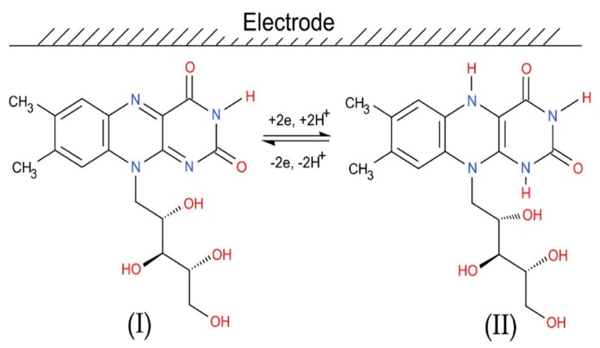
:1. Introduction
2. Materials and Methods
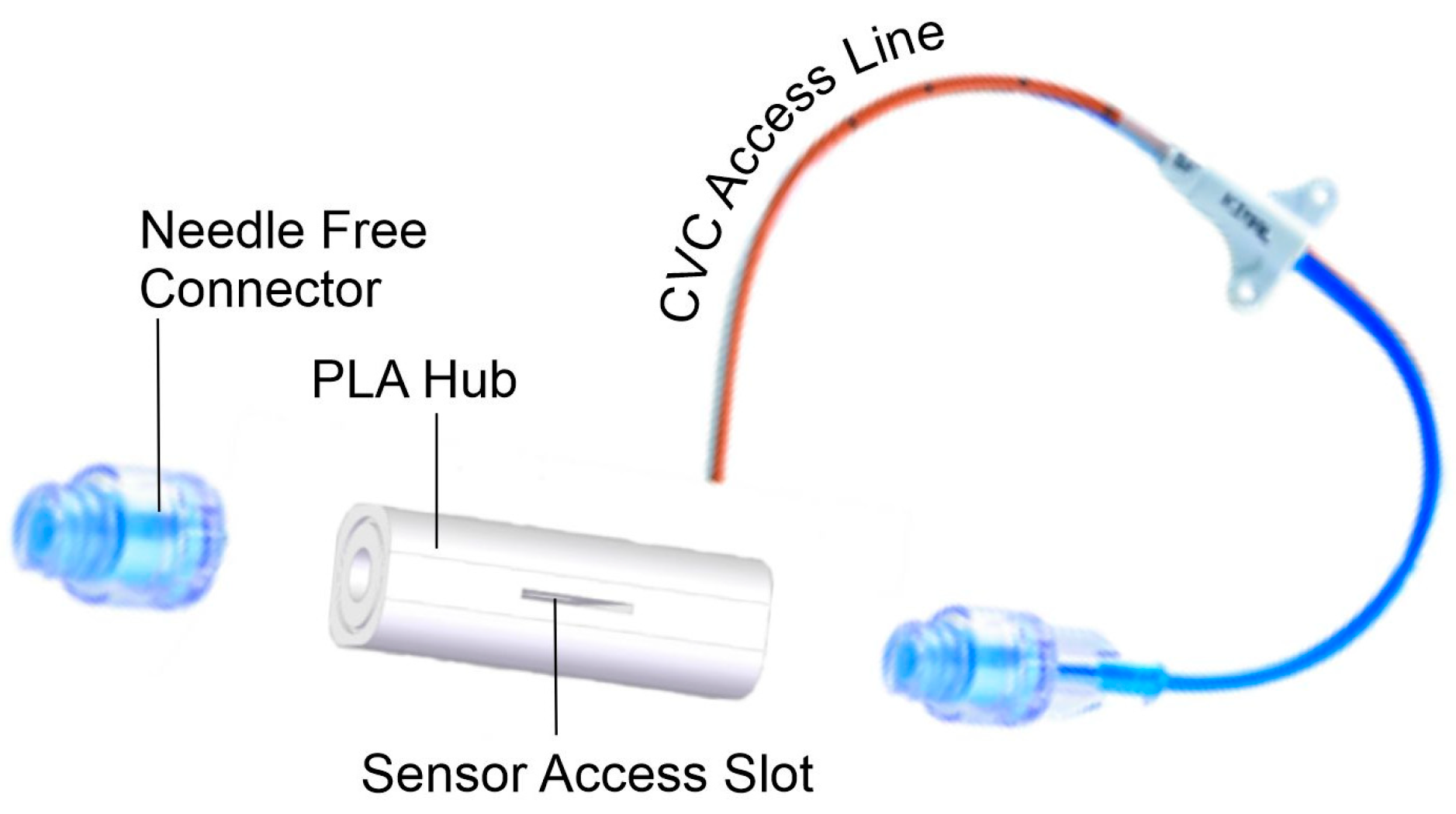
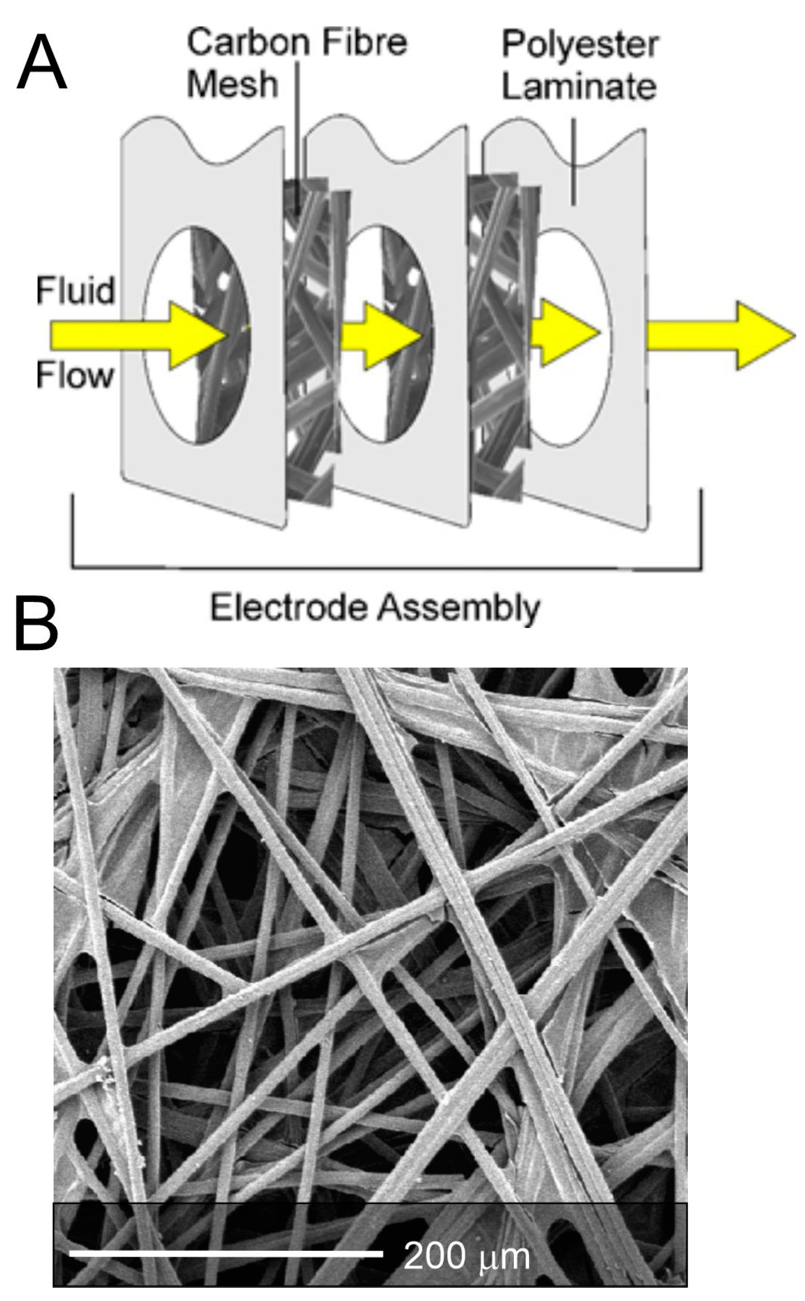
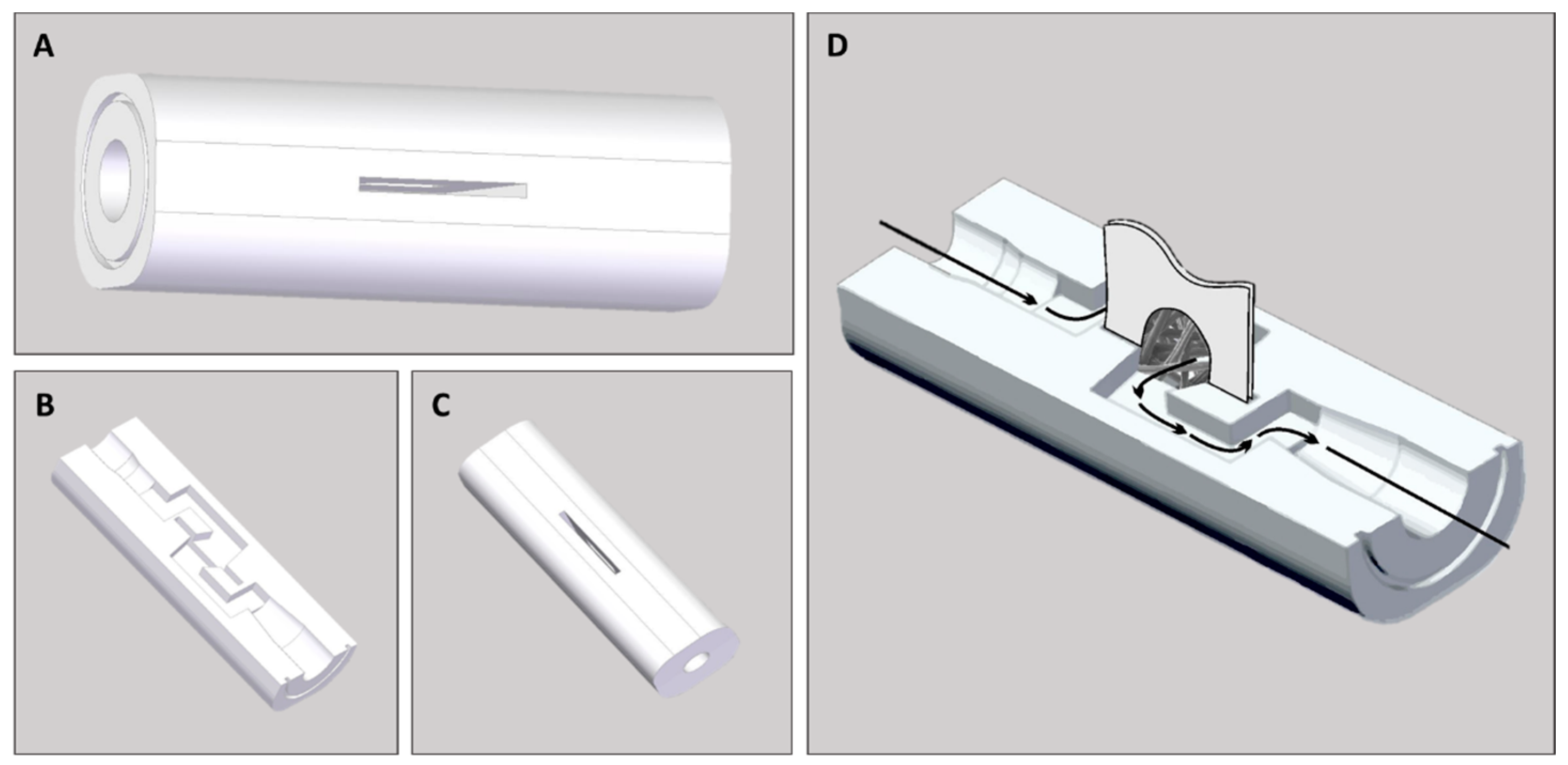
Electrode—Printed Hub Design
3. Results
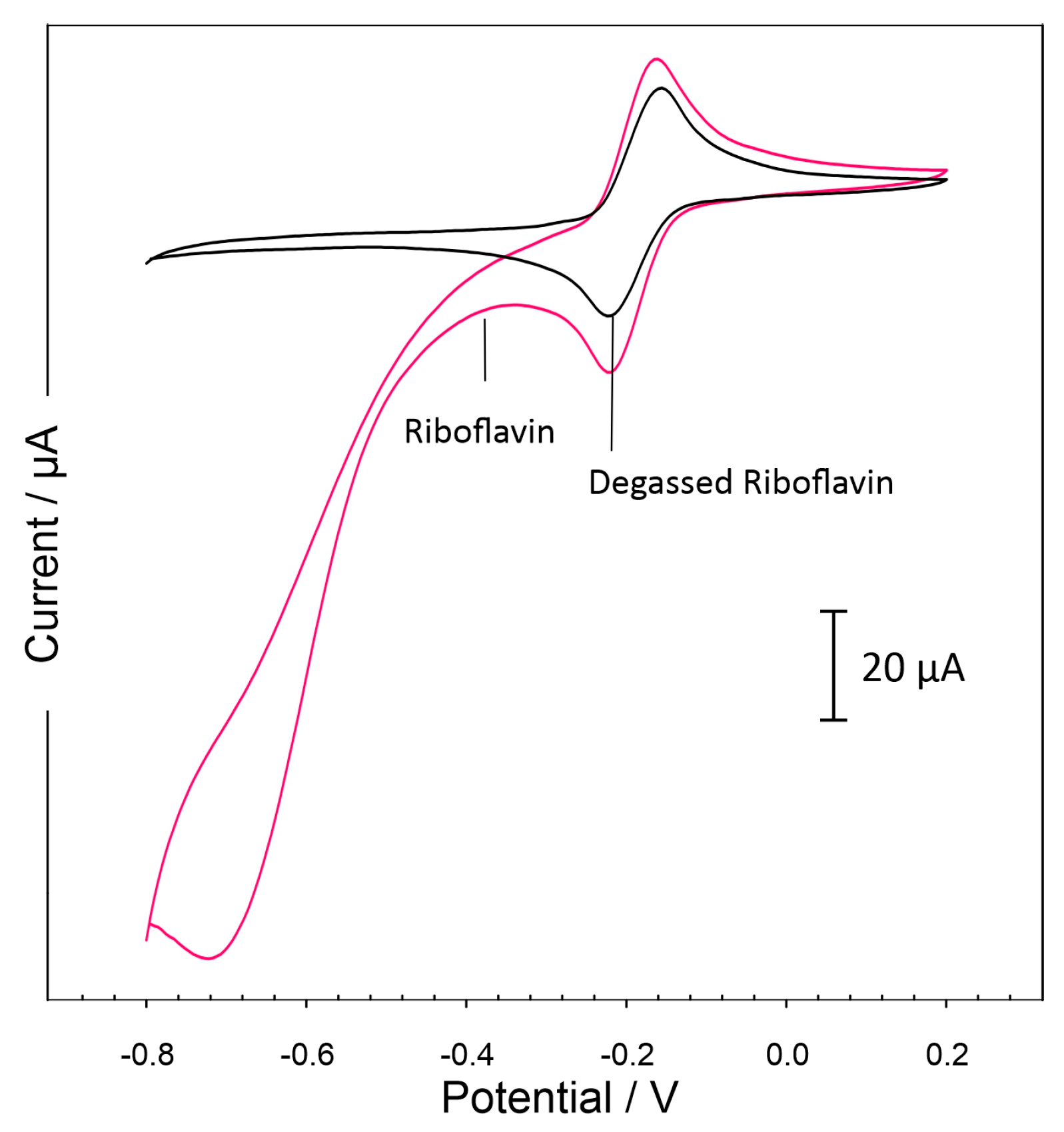
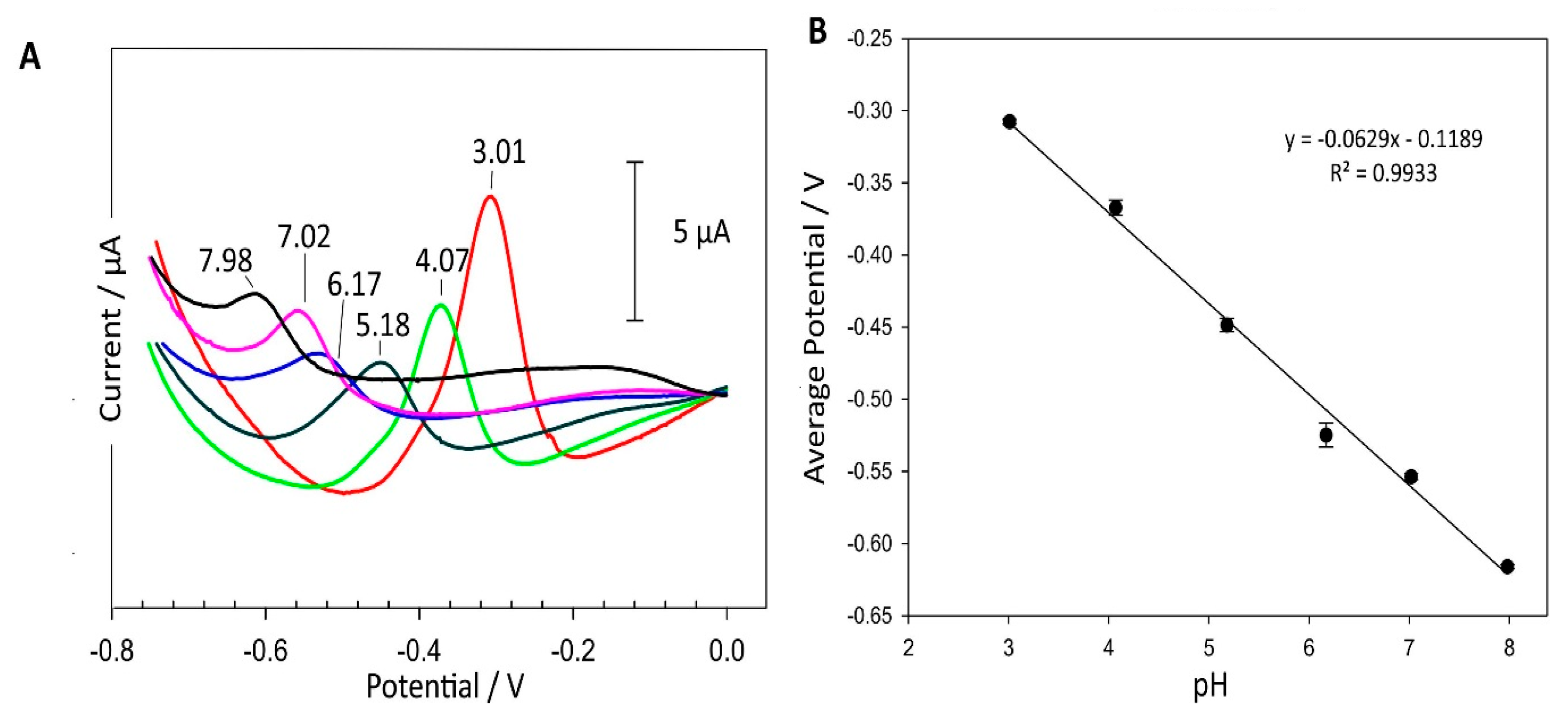
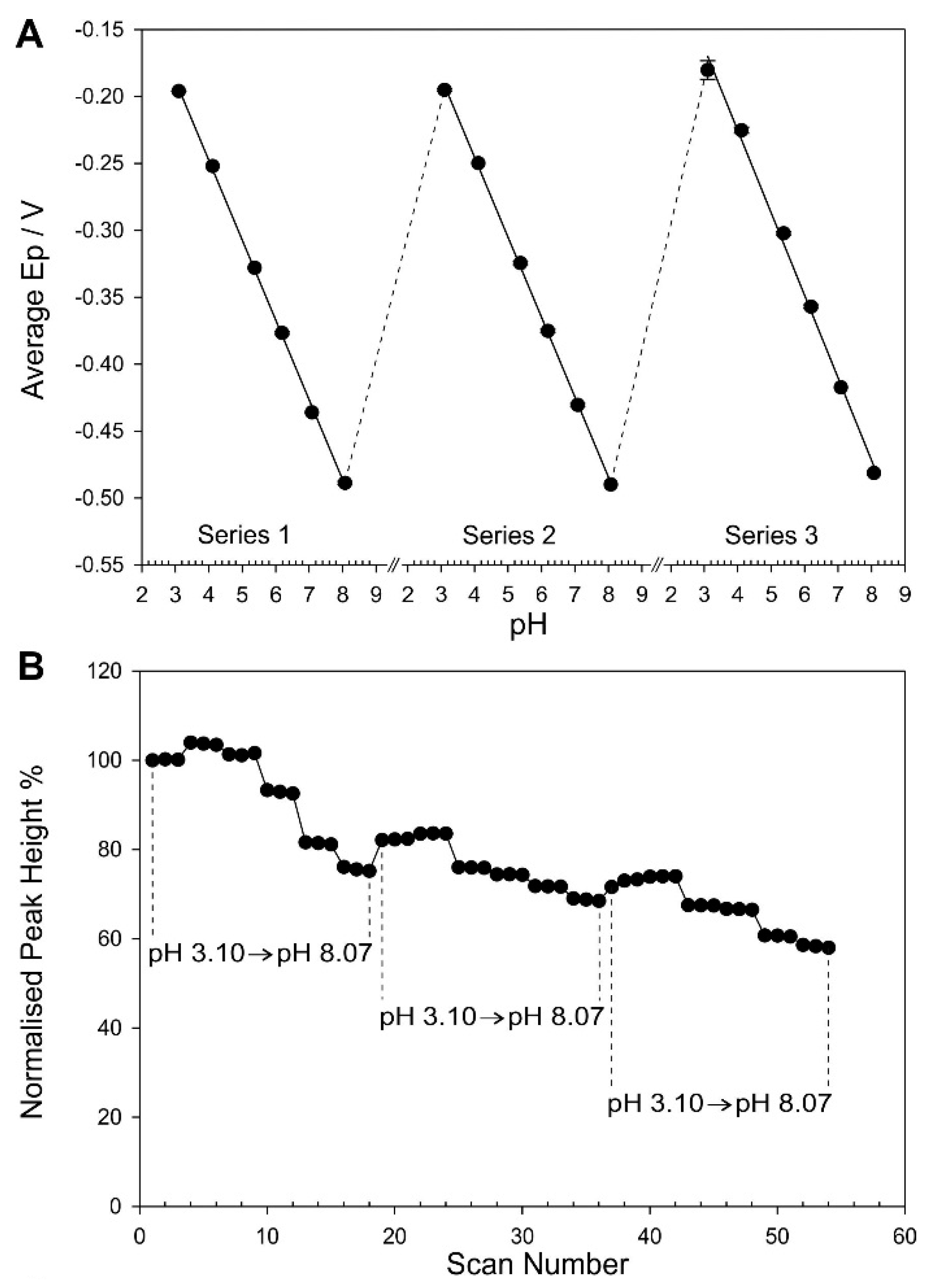
3.1. Preliminary Characterization
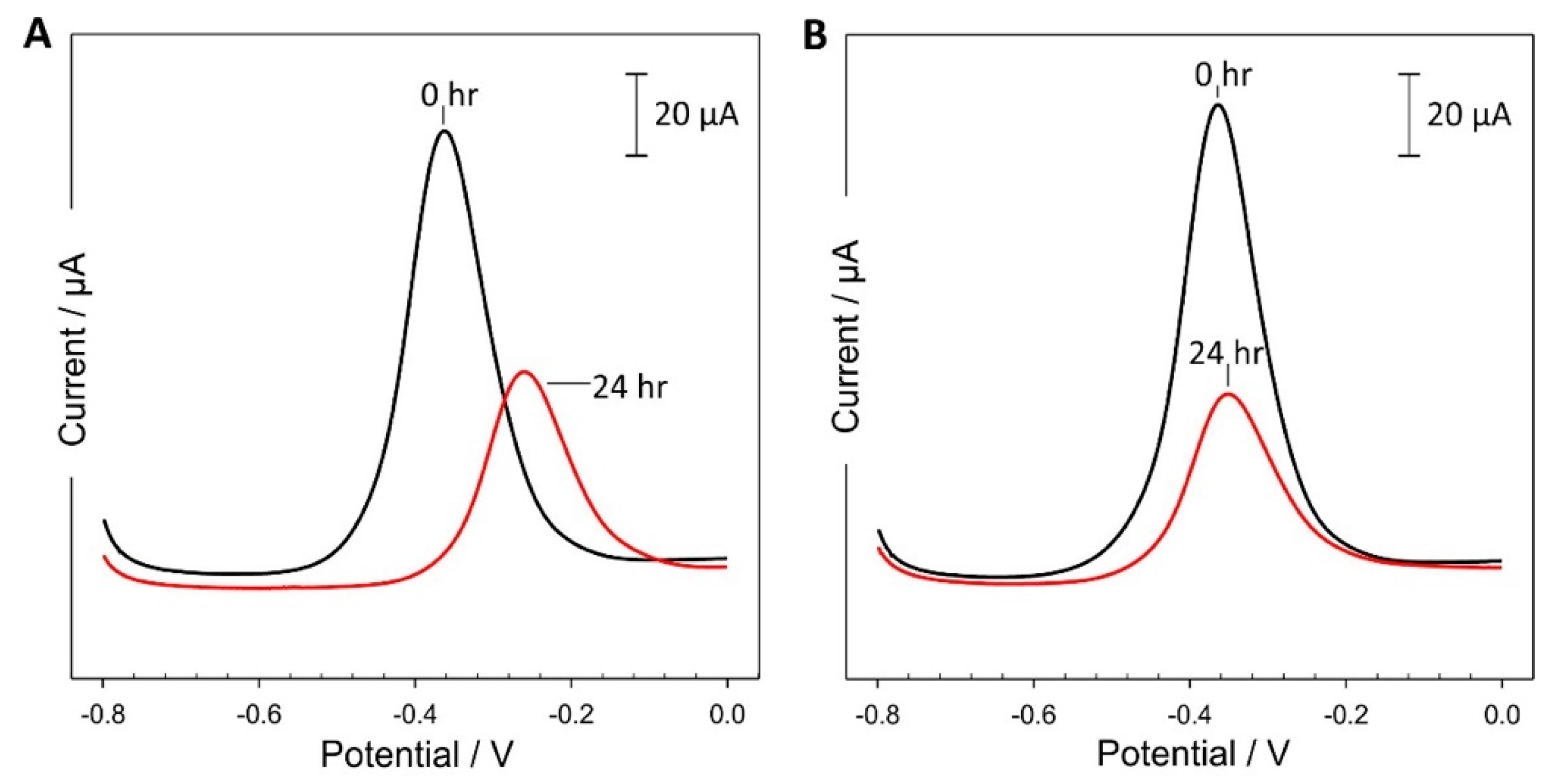
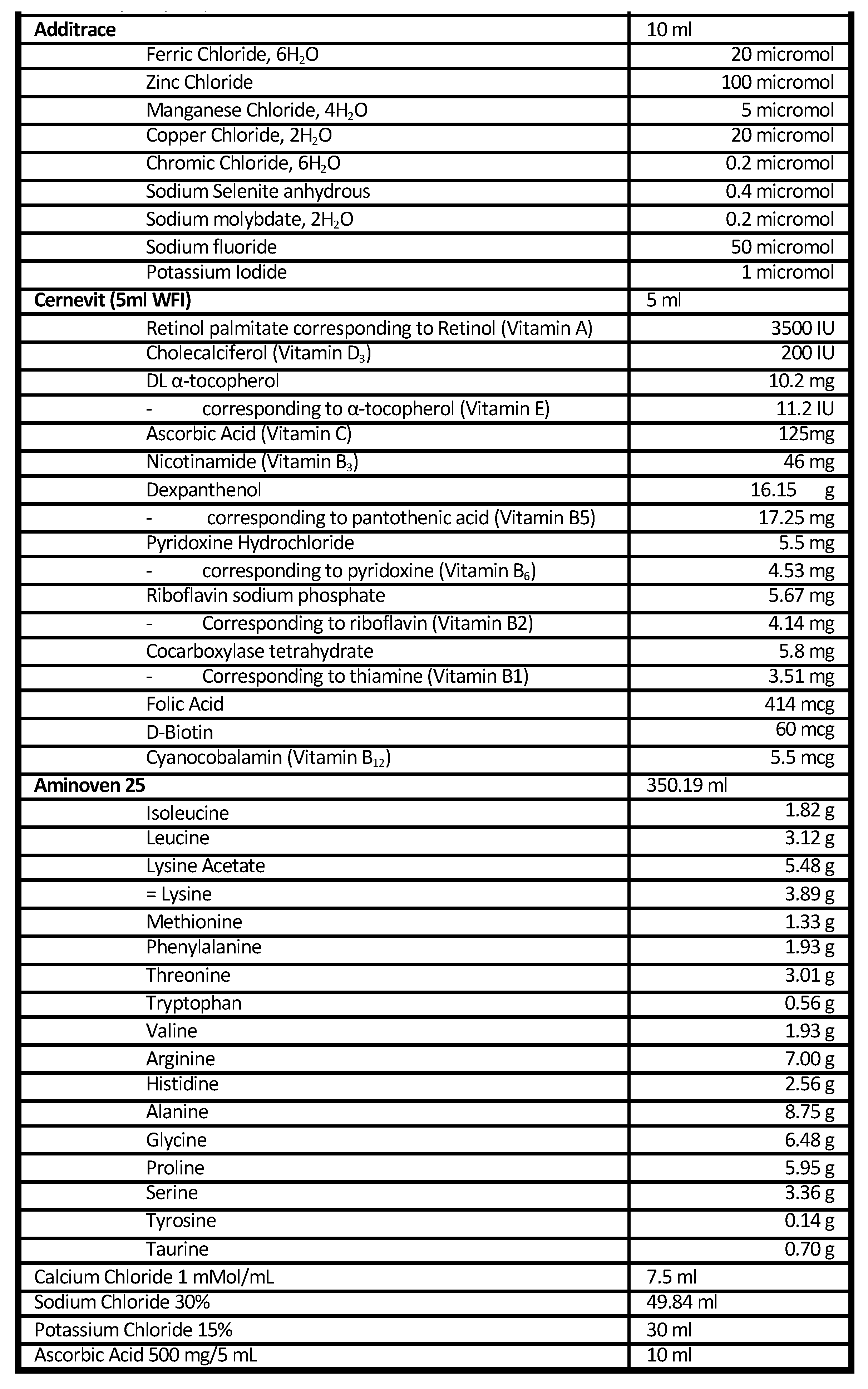
3.2. Evaluation of the Printed Catheter Hub with Authentic Infusate
3.3. Critical Assessment of the Technology and Practice Implications
4. Conclusions
Author Contributions
Funding
Data Availability Statement
Conflicts of Interest
References
- The Joint Commission. Preventing Central Line–Associated Bloodstream Infections: A Global Challenge, a Global Perspective; Joint Commission Resources: Oak Brook, IL, USA, May 2012; Available online: https://www.jointcommission.org/-/media/tjc/documents/resources/hai/clabsi_monographpdf.pdf (accessed on 28 December 2023).
- Lim, S.; Gangoli, G.; Adams, E.; Hyde, R.; Broder, M.S.; Chang, E.; Reddy, S.R.; Tarbox, M.H.; Bentley, T.; Ovington, L.; et al. Increased Clinical and Economic Burden Associated With Peripheral Intravenous Catheter–Related Complications: Analysis of a US Hospital Discharge Database. INQUIRY J. Health Care Organ. Provis. Financ. 2019, 56, 0046958019875562. [Google Scholar] [CrossRef] [PubMed]
- Zhang, L.; Cao, S.; Marsh, N.; Ray-Barruel, G.; Flynn, J.; Larsen, E.; Rickard, C.M. Infection risks associated with peripheral vascular catheters. J. Infect. Prev. 2016, 17, 207–213. [Google Scholar] [CrossRef] [PubMed]
- Gahlot, R.; Nigam, C.; Kumar, V.; Yadav, G.; Anupurba, S. Catheter-related bloodstream infections. Int. J. Crit. Illn. Inj. Sci. 2014, 4, 161. [Google Scholar] [CrossRef] [PubMed]
- Norris, L.B.; Kablaoui, F.; Brilhart, M.K.; Bookstaver, P.B. Systematic review of antimicrobial lock therapy for prevention of central-line-associated bloodstream infections in adult and pediatric cancer patients. Int. J. Antimicrob. Agents 2017, 50, 308–317. [Google Scholar] [CrossRef] [PubMed]
- Marschall, J.; Mermel, L.A.; Fakih, M.; Hadaway, L.; Kallen, A.; O’Grady, N.P.; Pettis, A.M.; Rupp, M.E.; Sandora, T.; Maragakis, L.L.; et al. Strategies to Prevent Central Line–Associated Bloodstream Infections in Acute Care Hospitals: 2014 Update. Infect. Control. Hosp. Epidemiol. 2014, 35, 753–771. [Google Scholar] [CrossRef] [PubMed]
- Lutwick, L.; Al-Maani, A.S.; Mehtar, S.; Memish, Z.; Rosenthal, V.D.; Dramowski, A.; Lui, G.; Osman, T.; Bulabula, A.; Bearmani, G. Managing and preventing vascular catheter infections: A position paper of the international society for infectious diseases. Int. J. Infect. Dis. 2019, 84, 22–29. [Google Scholar] [CrossRef]
- Centers for Disease Control and Prevention. CDC National and State Healthcare Progress Report. 2019. Available online: https://www.cdc.gov/hai/data/portal/progress-report.html (accessed on 6 September 2021).
- Valencia, C.; Hammami, N.; Agodi, A.; Lepape, A.; Herrejon, E.P.; Blot, S.; Vincent, J.-L.; Lambert, M.-L. Poor adherence to guidelines for preventing central line-associated bloodstream infections (CLABSI): Results of a worldwide survey. Antimicrob. Resist. Infect. Control. 2016, 5, 49. [Google Scholar] [CrossRef]
- Hadaway, L.; Richardson, D. Needleless connectors: A primer on terminology. J. Infus. Nurs. 2010, 33, 22–31. [Google Scholar] [CrossRef]
- Curran, E. Needleless connectors: The vascular access catheter’s microbial gatekeeper. J. Infect. Prev. 2016, 17, 234–240. [Google Scholar] [CrossRef]
- Shah, H.; Bosch, W.; Thompson, K.M.; Hellinger, W.C. Intravascular Catheter-Related Bloodstream Infection. Neurohospitalist 2013, 3, 144–151. [Google Scholar] [CrossRef]
- Holroyd, J.L.; Vasilopoulos, T.; Rice, M.J.; Rand, K.H.; Fahy, B.G. Incidence of central venous catheter hub contamination. J. Crit. Care. 2017, 39, 162–168. [Google Scholar] [CrossRef] [PubMed]
- Casimero, C.; Ruddock, T.; Hegarty, C.; Barber, R.; Devine, A.; Davis, J. Minimising Blood Stream Infection: Developing New Materials for Intravascular Catheters. Medicines 2020, 7, 49. [Google Scholar] [CrossRef] [PubMed]
- Loveday, H.P.; Wilson, J.A.; Pratt, R.J.; Golsorkhi, M.; Tingle, A.; Bak, A.; Browne, J.; Prieto, J.; Wilcox, M. Epic3: National evidence-based guidelines for preventing healthcare-associated infections in NHS hospitals in England. J. Hosp. Infect. 2014, 86, S1–S70. [Google Scholar] [CrossRef] [PubMed]
- Haddadin, Y.; Regunath, H. Central Line Associated Blood Stream Infections (CLABSI); StatPearls Publishing: Tampa, FL, USA, 2018. Available online: http://www.ncbi.nlm.nih.gov/pubmed/28613641 (accessed on 13 November 2018).
- O’grady, N.P.; Alexander, M.; Burns, L.A.; Dellinger, E.P.; Garland, J.; Heard, S.O.; Lipsett, P.A.; Masur, H.; Mermel, L.A.; Pearson, M.L.; et al. Guidelines for the Prevention of Intravascular Catheter-Related Infections. Clin. Infect. Dis. 2011, 52, 162–193. [Google Scholar] [CrossRef] [PubMed]
- The Joint Commission. CVC Maintenance Bundles. CLABSI Toolkit—Prev Cent Assoc Bloodstream Infect Useful Tools, An Int Perspective. 2013, pp. 1–7. Available online: https://www.jointcommission.org/assets/1/6/CLABSI_Toolkit_Tool_3-22_CVC_Maintenance_Bundles.pdf (accessed on 28 December 2023).
- Xia, M.Y.; Agca, B.N.; Yoshida, T.; Choi, J.; Amjad, U.; Bose, K.; Keren, N.; Zukerman, S.; Cima, M.J.; Graybiel, A.M. Scalable, flexible carbon fiber electrode thread arrays for three-dimensional probing of neurochemical activity in deep brain structures of rodents. Biosen. Bioelectron. 2023, 241, 115625. [Google Scholar] [CrossRef] [PubMed]
- Yang, C.; Zhao, Y.L.; Dong, X.X.; Lu, S. Bimetallic cobalt-nickel supported on carbon fiber for electrochemical simultaneous determination of dopamine and hydroquinone. Electrochem. Commun. 2023, 156, 107597. [Google Scholar] [CrossRef]
- Li, J.; Yang, J.J.; Ren, H.; Wang, X.H.; Xu, Y.C.; Guo, Y.; Xiao, D. Facile fabrication of Fe-Fe3C nanoparticles decorated with carbon nanotubes for sensitive dopamine detection. J. Electroanal. Chem. 2023, 948, 117793. [Google Scholar] [CrossRef]
- Chen, F.F.; Lv, C.K.; Xing, Y.K.; Luo, L.; Wang, J.Y.; Cheng, Y.L.; Xie, X.Y. Electrospinning carbon fibers based molecularly imprinted polymer self-supporting electrochemical sensor for sensitive detection of glycoprotein. Sens. Act. B. 2023, 396, 134552. [Google Scholar] [CrossRef]
- Zhang, Z.H.; Fan, Y.; Wang, X.Y.; Tu, H.Y.; Jiang, J.Z.; Zhang, C.Y.; Zhao, X.H.; Ma, J.J.; Wang, M.Y.; Xu, R.B. Efficient simultaneous determination of baicalein and luteolin based on a carbon fiber paper electrode modified with CuO/ZnO-CCNT ternary nanocomposite. J. Appl. Electrochem. 2023, 1954, 4. [Google Scholar] [CrossRef]
- Bukharinova, M.A.; Khamzina, E.I.; Stozhko, N.Y.; Tarasov, A.V. Highly sensitive voltammetric determination of Allura Red (E129) food colourant on a planar carbon fiber sensor modified with shungite. Anal. Chim. Acta. 2023, 1272, 341481. [Google Scholar] [CrossRef]
- Su, J.Y.; Jin, G.P.; Chen, T.; Liu, X.D.; Chen, C.N.; Tian, J.J. The characterization and application of prussian blue at graphene coated carbon fibers in a separated adsorption and electrically switched ion exchange desorption processes of cesium. Electrochim. Acta. 2017, 230, 399–406. [Google Scholar] [CrossRef]
- Radzevi, A.; Niaura, G.; Ignatjev, I.; Rakickas, T.; Celieit, R.; Pauliukaite, R. Electropolymerisation of the natural monomer riboflavin and its characterisation. Electrochim. Acta 2016, 222, 1818–1830. [Google Scholar]
- Casimero, C.; McConville, A.; Fearon, J.J.; Lawrence, C.L.; Taylor, C.M.; Smith, R.B.; Davis, J. Sensor systems for bacterial reactors: A new flavin-phenol composite film for the in situ voltammetric measurement of pH. Anal. Chim. Acta 2018, 1027, 1–8. [Google Scholar] [CrossRef] [PubMed]
- Barber, R.; Cameron, S.; Devine, A.; McCombe, A.; Pourshahidi, L.K.; Cundell, J.C.; Souradeep, R.; Mathur, A.; Casimero, C.; Papakonstantinou, P.; et al. Laser induced graphene sensors for assessing pH: Application to wound management. Electrochem. Commun. 2021, 123, 106914. [Google Scholar] [CrossRef]
- Raad, I.; Hanna, H.; Maki, D. Intravascular catheter-related infections: Advances in diagnosis, prevention, and management. Lancet Infect Dis. 2007, 7, 645–657. [Google Scholar] [CrossRef] [PubMed]
- Gominet, M.; Compain, F.; Beloin, C.; Lebeaux, D. Central venous Catheters and Biofilms: Where Do We Stand in 2017? Apmis 2017, 125, 365–375. [Google Scholar] [CrossRef]
- Beghetto, M.G.; Victorino, J.; Teixeira, L.; de Azevedo, M.J. Parenteral nutrition as a risk factor for central venous catheter-related infection. J. Parenter. Enter Nutr. 2005, 29, 367–373. [Google Scholar] [CrossRef]
- Dibb, M.J.; Abraham, A.; Chadwick, P.R.; Shaffer, J.L.; Teubner, A.; Carlson, G.L.; Lal, S. Central venous catheter salvage in home parenteral nutrition catheter-related bloodstream infections: Long-term safety and efficacy data. J. Parenter Enter Nutr. 2016, 40, 699–704. [Google Scholar] [CrossRef]
- Duesing, L.A.; Fawley, J.A.; Wagner, A.J. Central Venous Access in the Pediatric Population with Emphasis on Complications and Prevention Strategies. Nutr. Clin. Pract. 2016, 31, 490–501. [Google Scholar] [CrossRef]
- Santarpia, L.; Buonomo, A.; Pagano, M.C.; Alfonsi, L.; Foggia, M.; Mottola, M.; Marinosci, G.Z.; Contaldo, F.; Pasanisi, F. Central venous catheter related bloodstream infections in adult patients on home parenteral nutrition: Prevalence, predictive factors, therapeutic outcome. Clin. Nutr. 2016, 35, 1394–1398. [Google Scholar] [CrossRef]
- Leite, A.M.D.O.; Miguel, M.A.L.; Peixoto, R.S.; Rosado, A.S.; Silva, J.T.; Paschoalin, V.M.F. Microbiological, technological and therapeutic properties of kefir: A natural probiotic beverage. Braz. J. Microbiol. 2013, 44, 341–349. [Google Scholar] [CrossRef] [PubMed]
- Plessas, S.; Nouska, C.; Mantzourani, I.; Kourkoutas, Y.; Alexopoulos, A.; Bezirtzoglou, E. Microbiological exploration of different types of Kefir grains. Fermentation 2017, 3, 1. [Google Scholar] [CrossRef]
- Rosa, D.D.; Dias, M.M.S.S.; Grześkowiak, Ł.M.; Reis, S.A.; Conceição, L.L.; Peluzio, M. Milk kefir: Nutritional, microbiological and health benefits. Nutr. Res. Rev. 2017, 30, 82–96. [Google Scholar] [CrossRef] [PubMed]
- Piermaria, J.A.; Pinotti, A.; Garcia, M.A.; Abraham, A.G. Films based on kefiran, an exopolysaccharide obtained from kefir grain: Development and characterization. Food Hydrocoll. 2009, 23, 684–690. [Google Scholar] [CrossRef]
- Witthuhn, R.C.; Schoeman, T.; Britz, T.J. Characterisation of the microbial population at different stages of Kefir production and Kefir grain mass cultivation. Int. Dairy J. 2005, 15, 383–389. [Google Scholar] [CrossRef]
- Prado, M.R.; Blandón, L.M.; Vandenberghe, L.P.; Rodrigues, C.; Castro, G.R.; Thomaz-Soccol, V.; Soccol, C.R. Milk kefir: Composition, microbial cultures, biological activities, and related products. Front Microbiol. 2015, 6, 1177. [Google Scholar] [CrossRef]
- Fiorda, F.A.; de Melo Pereira, G.V.; Thomaz-Soccol, V.; Rakshit, S.K.; Pagnoncelli, M.G.B.; de Souza Vandenberghe, L.P.; Soccol, C.R. Microbiological, biochemical, and functional aspects of sugary kefir fermentation—A review. Food Microbiol. 2017, 66, 86–95. [Google Scholar] [CrossRef]
- Li, H.; Wu, G.; Weng, Z.; Sun, H.; Nistala, R.; Zhang, Y. Microneedle-Based Potentiometric Sensing System for Continuous Monitoring of Multiple Electrolytes in Skin Interstitial Fluids. ACS Sens. 2021, 6, 2181–2190. [Google Scholar] [CrossRef]
- Zheng, Y.; Omar, R.; Zhang, R.; Tang, N.; Khatib, M.; Xu, Q.; Milyutin, Y.; Saliba, W.; Broza, Y.Y.; Wu, W.; et al. A wearable microneedle-based extended gate transistor for real-time detection of sodium in interstitial fluids. Adv. Mater. 2022, 34, 2108607. [Google Scholar] [CrossRef]
- Sannoh, S.; Clones, B.; Munoz, J.; Montecalvo, M.; Parvez, B. A multimodal approach to central venous catheter hub care can decrease catheter-related bloodstream infection. Am. J. Infect. Control. 2010, 38, 424–429. [Google Scholar] [CrossRef]
- Young, E.M.; Commiskey, M.L.; Wilson, S.J. Translating evidence into practice to prevent central venous catheter- associated bloodstream infections: A systems-based intervention. Am. J. Infect. Control 2006, 34, 503–506. [Google Scholar] [CrossRef] [PubMed]
- Jeong, I.S.; Park, S.M.; Lee, J.M.; Song, J.Y.; Lee, S.J. Effect of central line bundle on central line-associated bloodstream infections in intensive care units. Am. J. Infect. Control 2013, 41, 710–716. [Google Scholar] [CrossRef] [PubMed]
- Hadaway, L. Intermittent Intravenous Administration Sets: Survey of Current Practices. J. Assoc. Vasc. Access. 2007, 12, 143–147. [Google Scholar] [CrossRef]
- Ista, E.; van der Hoven, B.; Kornelisse, R.F.; van der Starre, C.; Vos, M.C.; Boersma, E.; Helder, O.K. Effectiveness of insertion and maintenance bundles to prevent central-line-associated bloodstream infections in critically ill patients of all ages: A systematic review and meta-analysis. Lancet Infect. Dis. 2016, 16, 724–734. [Google Scholar] [CrossRef]
- Moureau, N.L.; Flynn, J. Disinfection of Needleless Connector Hubs: Clinical Evidence Systematic Review. Nurs. Res. Pract. 2015, 2015, 1–20. [Google Scholar] [CrossRef]
- Hadaway, L. Needleless connectors: Improving practice, reducing risks. JAVA-J. Assoc. Vasc. Access 2011, 16, 20–24. [Google Scholar] [CrossRef]


| With Kefir/h | pH Probe | Average Ep (N = 3) | Calculated pH | % Error |
|---|---|---|---|---|
| 0 h | 5.65 | −0.363 | 5.89 | 4.29 |
| 4 h | 4.29 | −0.259 | 4.10 | −4.33 |
| 23 h | 3.78 | −0.220 | 3.42 | −9.42 |
| 24 h | 3.74 | −0.230 | 3.60 | −3.67 |
Disclaimer/Publisher’s Note: The statements, opinions and data contained in all publications are solely those of the individual author(s) and contributor(s) and not of MDPI and/or the editor(s). MDPI and/or the editor(s) disclaim responsibility for any injury to people or property resulting from any ideas, methods, instructions or products referred to in the content. |
© 2023 by the authors. Licensee MDPI, Basel, Switzerland. This article is an open access article distributed under the terms and conditions of the Creative Commons Attribution (CC BY) license (https://creativecommons.org/licenses/by/4.0/).
Share and Cite
Casimero, C.; Smith, R.B.; Davis, J. Integration of Riboflavin-Modified Carbon Fiber Mesh Electrode Systems in a 3D-Printed Catheter Hub. Micromachines 2024, 15, 79. https://doi.org/10.3390/mi15010079
Casimero C, Smith RB, Davis J. Integration of Riboflavin-Modified Carbon Fiber Mesh Electrode Systems in a 3D-Printed Catheter Hub. Micromachines. 2024; 15(1):79. https://doi.org/10.3390/mi15010079
Chicago/Turabian StyleCasimero, Charnete, Robert B. Smith, and James Davis. 2024. "Integration of Riboflavin-Modified Carbon Fiber Mesh Electrode Systems in a 3D-Printed Catheter Hub" Micromachines 15, no. 1: 79. https://doi.org/10.3390/mi15010079