

Microwave Imaging and Sensing Techniques for Breast Cancer Detection

Biomedical Device Innovation Center, Shenzhen Technology University, Shenzhen 518118, China
Micromachines 2023, 14(7), 1462; https://doi.org/10.3390/mi14071462
Submission received: 8 June 2023
/
Revised: 14 July 2023
/
Accepted: 17 July 2023
/
Published: 21 July 2023
(This article belongs to the Special Issue Applications of Modern Artificial Intelligence and Antenna Technology: Design, Materials, Processing Techniques, Signal Processing and Sensing Methods)
Abstract
:Medical imaging techniques, including X-ray mammography, ultrasound, and magnetic resonance imaging, play a crucial role in the timely identification and monitoring of breast cancer. However, these conventional imaging modalities have their limitations, and there is a need for a more accurate and sensitive alternative. Microwave imaging has emerged as a promising technique for breast cancer detection due to its non-ionizing, non-invasive, and cost-effective nature. Recent advancements in microwave imaging and sensing techniques have opened up new possibilities for the early diagnosis and treatment of breast cancer. By combining microwave sensing with machine learning techniques, microwave imaging approaches can rapidly and affordably identify and classify breast tumors. This manuscript provides a comprehensive overview of the latest developments in microwave imaging and sensing techniques for the early detection of breast cancer. It discusses the principles and applications of microwave imaging and highlights its advantages over conventional imaging modalities. The manuscript also delves into integrating machine learning algorithms to enhance the accuracy and efficiency of microwave imaging in breast cancer detection.
1. Introduction
Breast cancer is the leading cause of female cancer-related death worldwide [1]. Approximately 13% of females are at risk of breast cancer in the United States [2]. Early detection is crucial for successful treatment, but the current imaging techniques have limitations. Mammography, the most widely used tool for breast cancer screening, cannot be utilized for pregnant women or those with dense breasts [3]. Ultrasound imaging, on the other hand, produces low-quality images that may not accurately identify cancerous cells [4]. Magnetic resonance imaging (MRI) is more effective in detecting breast cancer, but it is not only expensive but also uncomfortable for patients [5]. Given these limitations, researchers have been exploring alternative methods for breast cancer diagnosis, and microwave breast imaging (MBI) has shown promise.
MBI is non-invasive, meaning it does not require invasive procedures and does not utilize ionizing radiation, making it safe for patients. Additionally, it is cost-effective, making it a viable option for widespread use [6]. As shown in Figure 1, MBI works by analyzing and differentiating the changes in backscattered signals caused by the differences in dielectric properties between cancerous and normal cells [7]. This enables the identification of malignant cells, which is crucial for an accurate diagnosis. Although holographic microwave imaging (HMI) algorithms have shown potential for breast cancer detection, further development, and validation are necessary before these techniques can be implemented in clinical trials [8,9,10,11,12,13]. Some MBI systems have already reached the clinical trials stage, as shown in Figure 2 [14,15]. However, these systems have limitations, such as low-resolution images, coupling noises between antennas, and system complexity.
In the MBI system, the antenna plays a vital role as it transmits microwave signals into the breast and measures the backscattered signals (see Figure 3) [16]. To make MBI portable and affordable, custom sensors are required. Ultra-wideband (UWB) patch antennas are commonly used in MBI applications. However, there is room for improvement in their radiation capacity, gain, and bandwidth [17]. Recently, there have been advancements in the development of custom antennas using metamaterial (MTM), metasurface (MTS), and artificial magnetic conductor (AMC) technologies [16,17,18,19,20]. These technologies present opportunities for the sensitivity and accuracy of MBI systems. They have improved the overall performance of MBI systems and significantly increased detection accuracy [17,21,22]. However, despite these advancements, there are still limitations to these antennas, and further research and development are needed to overcome these limitations and fully harness the potential of custom antennas for MBI applications.
Researchers globally have made significant progress in advancing the development of MBI technologies. These technologies can potentially revolutionize breast cancer diagnosis by providing a non-invasive, radiation-free, and cost-effective alternative to traditional imaging techniques such as mammography. However, despite the progress made, further research and development are needed to establish connections between research labs, industrial partners, and patients who can benefit from these technologies. Collaboration between these stakeholders is essential for translating research findings into practical clinical applications. One of the critical technical challenges that researchers must address is enhancing imaging algorithms to interpret microwave signals obtained during the imaging process accurately. These algorithms play a crucial role in reconstructing high-resolution images of breast tissue and detecting abnormalities. Improving the accuracy of these algorithms will enable healthcare professionals to make more reliable and precise diagnoses. In addition to algorithm development, researchers must also focus on developing high-sensitivity acquisition hardware. The hardware used in MBI systems must be capable of capturing weak microwave signals emitted by breast tissue. By enhancing the sensitivity of the hardware, researchers can improve the signal-to-noise ratio and enhance the overall performance of MBI systems. Overcoming these technical challenges is crucial for improving the performance of MBI systems and increasing their reliability and effectiveness in detecting breast abnormalities. By addressing these challenges, researchers can pave the way for the widespread adoption of MBI technologies in breast cancer diagnosis.
This manuscript presents state-of-the-art microwave imaging and sensing methods for early breast cancer diagnosis. The manuscript also discusses the dielectric properties of breast tissues and how they can be utilized for microwave imaging. Furthermore, the manuscript reviews recently developed MBI techniques, including deep learning-based approaches. These approaches leverage advanced machine learning algorithms to improve the accuracy and sensitivity of MBI systems. The paper is structured as follows. Section 1 introduces the background of this study. Section 2 provides an overview of breast imaging techniques, and Section 3 discusses the dielectric properties of breast tissues. Section 4 reviews recently developed microwave breast imaging techniques, including deep learning-based approaches. Section 5 presents microwave sensing techniques for breast cancer detection, while Section 6 addresses the challenges and prospects of microwave imaging and sensing techniques, and Section 7 concludes this study.
2. Breast Imaging Techniques
Conventional medical imaging modalities, such as X-ray mammography, ultrasound, MRI, CT, and positron emission tomography (PET), play a pivotal role in detecting breast cancer. X-ray mammography is commonly employed as the first line of defense due to its simple operation, high resolution, and high repeatability. However, it may not be a suitable option for all women, particularly those with dense breast tissue or who are pregnant [3]. Ultrasound imaging is a safe alternative and more convenient for high-risk patients. Still, it may not reveal breast lesions in adipose tissue and has a relatively low detection rate for malignant tumors [4]. MRI is a sensitive imaging technique that is particularly effective in detecting tumors in dense breast tissue but has limitations due to its cost, magnetic field exposure, and the noise generated during the procedure [5]. Breast CT examination provides a detailed view of breast tissue but is limited by its high radiation exposure and cost [23]. PET imaging is a powerful method for detecting breast cancer. Still, it has limitations due to its unsuitability for early stage tumor detection, the potential false positives in young patients, and the need for simultaneous use with CT imaging and its associated radiation exposure and expense [24].
MBI has been proposed as a complementary modality for early breast cancer diagnosis to overcome some limitations of conventional medical imaging modalities. Researchers aim to integrate this innovative imaging technique into routine clinical practice, providing patients with an accurate and reliable tool for detecting breast abnormalities at an early stage. Despite the significant progress in advancing MBI technologies, further research and development are necessary to establish connections between research labs, industrial partners, and patients who can benefit from these technologies.
3. Dielectric Properties of Breast Tissues
Microwave imaging (MWI) leverages electromagnetic (EM) waves ranging from 300 MHz to 30 GHz, penetrating the breast tissue. As these waves traverse the tissue, they interact with its dielectric properties, including permittivity (, the capacity to store electrical energy) and conductivity (σ, the ability to conduct electrical energy). Normal and malignant tumor tissues exhibit distinct dielectric properties, causing EM waves to scatter and reflect in each tissue type at varying degrees. This scattering and reflection serve as the foundation for the microwave imaging of breast tumors.
As shown in Figure 4, the dielectric properties of breast tissue change with working frequencies, and it is a nonlinear dependency [25]. The Debye and Cole–Cole models are commonly used to simulate biological tissues. The Debye model can be defined as follows [26]:
where denotes the permittivity, and its value strongly corresponds to the water content of the tissue, represents the static permittivity, and denotes the relaxation time.
The Cole–Cole model can represent the complex dielectric constant of biological tissues [27].
where denotes the complex dielectric, means the static frequency constant, represents infinite frequency constant, means the angular frequency, and denotes the time constant. The exponent parameter (0 < < 1) represents different spectral shapes. When , the Cole–Cole model becomes the Debye model. When , the relaxation time is increased.
The following empirical model can represent the relationship between the dielectric parameters and the moisture content model [28].
where is the frequency, , and .
The dielectric properties of breast tissue exhibit a nonlinear dependence on working frequencies, which is commonly simulated using Debye or Cole–Cole models. These models consider the tissue’s water content and relaxation time. Experimental studies have shown that normal and malignant breast tissues have different electromagnetic responses due to differences in water and salt content [29,30]. Researchers have extensively studied the dielectric properties of biological tissues under the irradiation of electromagnetic waves with different frequencies, with findings indicating a significant difference in the dielectric properties of normal and tumor tissues [31,32]. Chaudhary et al. [33] discovered a substantial difference in the dielectric properties of normal and tumor tissues over the frequency range of 3–100 MHz. Joines et al. [34] replicated a similar study and confirmed Chaudhary’s findings. Gabriel et al. [29,30] reported their research findings on the characterization of biological tissues over the frequency range of 0.01 GHz to 20 GHz.
Furthermore, tissue at the infiltrating edge of the tumor has been found to have increased dielectric properties. Surowiec et al. [35] observed that the tissue at the infiltrating edge of the tumor had increased dielectric properties. Lazebnik et al. [31,32] studied the characterization of the dielectric properties of normal, malignant, and benign breast tissues over the frequency range of 0.5 GHz to 20 GHz. Halter et al. [36] conducted a similar measurement study on in vivo tissue, investigating the dielectric properties of breast tissues with and without tumor presence. Results showed negligible effects on the dielectric properties of tissues between excision and measurement.
Abas et al. [37] reported that the dielectric properties of tissues change significantly in the first few seconds after tissue excision. The authors investigated these effects based on ex vivo breast tissue measurement results. It is important to note that tissue dielectric properties can change significantly in the first few seconds after tissue excision due to temperature and water content changes.
Researchers have also developed breast phantoms based on measured dielectric properties of human breast tissue in the frequency range of 0.5–50 GHz, which have been tested using microwave imaging systems. Martellosio et al. [38] investigated the dielectric properties of breast tissues for the frequency range from 0.5 to 50 GHz. They employed Cole–Cole models for analyzing normal and tumorous tissues based on experimental measurements of 222 tissue samples from 53 patients aged 28 to 85. More recently, Meo et al. [39] developed three breast phantoms according to the dielectric properties of human breast ex vivo tissues in the frequency range of 0.5–50 GHz. The developed breast phantoms were tested using a microwave imaging system.
4. Microwave Breast Imaging Techniques
Microwave breast imaging (MBI) techniques can be classified into passive, hybrid, and active methods. Each method has its advantages and limitations. Passive methods rely solely on the radiation emitted by the body to create an image of the breast tissue. This approach does not require any external radiation sources. Passive methods are non-invasive but may have lower sensitivity. Hybrid microwave imaging combines microwave imaging with other imaging modalities such as ultrasound, MRI, or optical imaging. This combination of imaging techniques provides clinicians with more comprehensive and detailed information for accurate breast cancer diagnosis. Active methods utilize an external source of microwave radiation to probe the breast tissue. Active methods are more sensitive but need additional external radiation sources. Active MBI techniques such as microwave tomography (MT) and radar-based techniques have shown promise in accurately diagnosing breast cancer lesions. Radar-based methods use microwave radar to scan breast tissue in a non-invasive manner.
4.1. Microwave Tomography
Microwave tomography (MT) is an advanced imaging technique that uses microwave frequencies to create detailed cross-sectional images of the breast. It takes advantage of the different electrical properties of different types of tissues to accurately detect and visualize abnormalities, particularly breast cancer. Various methods are commonly employed in MT to process the collected data and solve inverse problems. Gradient-based approaches (such as conjugate gradient least squares and Landweber) and global techniques (such as genetic algorithm and particle swarm optimization) are utilized [40,41,42,43]. Several image reconstruction algorithms have been developed and applied in MT for detecting breast cancer, enhancing the accuracy and reliability of the imaging technique [44,45,46,47,48].
Meaney et al. [49] developed the first 2D MT clinical prototype (see Figure 5) for breast imaging, consisting of a cylindrical array of 16 monopole antennas that function as transceivers, operating at frequencies ranging from 300 to 1000 MHz. However, due to its complexity, this prototype is time-consuming and requires heavy computational work. To overcome these limitations, the same research group developed a 3D prototype for breast cancer detection using the 3D finite element modeling (FEM) method [50]. This new system provided high-resolution images of breast tissue with sub-centimeter image resolution in less than 2 min, ensuring a quick and efficient process for patients. Clinical results from this prototype showcased its capability to detect centimeter-sized tumors. In further research efforts, magnetic nanoparticles have been incorporated into MT to enhance tumor detection specificity, sensitivity, and accuracy [51,52,53]. This innovation holds promise for improving the overall performance of MT in breast cancer diagnosis.
Another significant advancement was made by Jeon et al. [54], who developed a clinical trial prototype MT system operating within the 3 to 6 GHz range and utilizing a fast-forward solver algorithm. This system was tested on women aged 40 to 68 and demonstrated its ability to detect breast tumors as small as 25 mm. Despite these advancements, there are still limitations to MT. It has relatively low spatial resolution and limited penetration depth compared to other imaging modalities, which can impede the detection of small tumors and restrict its widespread use in everyday clinical settings. Further research is needed to fully evaluate the clinical utility of MT and its integration into routine breast cancer diagnosis and management.
Table 1 compares the recently developed MBI systems for breast cancer detection. MT utilizes changes in the dielectric properties of breast tissues to differentiate between healthy and cancerous tissues. Various MBI algorithms, such as confocal microwave imaging (CMI), tissue-sensing adaptive radar (TSAR), microwave imaging via space-time (MIST), multi-static adaptive (MSA) imaging, and holographic microwave imaging (HMI), are used to create a visual representation of the breast’s internal structures based on the collected microwave signals. This form of image processing involves solving an inverse problem, where the information about the tissue properties is estimated or reconstructed from the measured signals. The reconstructed image can provide insights into the presence and location of abnormal tissue, aiding in the detection of breast cancer. The advantages of MBI include non-invasively reducing patient discomfort and avoiding the risks associated with invasive procedures, providing additional information about the breast tissues compared to traditional methods such as mammography, and not involving ionizing radiation, making it a safer alternative for repeated screenings. However, MBI techniques are still in the research and development stage, with limited clinical availability. The accuracy and sensitivity of MBI methods need further improvement to match or exceed existing those of methods such as mammography. The complexity of image reconstruction algorithms and hardware requirements may hinder their widespread adoption and affordability. Future research and the development of MBI methods are expected to address these limitations for breast cancer detection. Improvements in image resolution, data acquisition speed, and reconstruction algorithms may enable it to become a viable alternative or complementary technique to existing methods. Large-scale clinical trials and studies are needed to validate its performance and ensure its effectiveness in real-world scenarios.
4.2. Radar-Based Microwave Breast Imaging Approaches
In 1997, Bridges et al. [119] developed the first radar-based MBI system utilizing UWB microwave signals and an antenna array placed at different locations surrounding the breast to illuminate breast tissues and collect backscattered signals to identify tumors based on the dielectric property difference between normal and tumor tissues.
Radar-based MBI techniques include CMI, TSAR, MIST, MSA, HMI, and time domain data-adaptive (TDDA) imaging. These techniques use various methods to measure and analyze electromagnetic signals to create high-resolution images of target objects. CMI uses a highly focused microwave beam to perform subsurface imaging to reconstruct high-resolution images of target objects. TSAR measures EM signals that penetrate target tissue and analyze and image biological tissues. MIST uses measured radar signals from different locations and times to produce a 3D breast image. MSA uses multiple transmit-receive radar pairs placed strategically for breast screening, while TDDA uses advanced algorithms that analyze the radar signal’s time domain characteristics to perform subsurface target imaging.
Various beamforming algorithms have been applied for breast cancer detection, which are classified into eight categories in Table 2: image reconstruction algorithms, year of study, operating frequency, number of antennas employed, radar-based MI techniques, 2D or 3D image, and a tumor size that can be detected. Table 2 compares radar-based MBI techniques such as CMI, MIST, and TSAR. These methods work by sending low-power microwave signals into breast tissue and detecting the reflected signals. The scattered signals are collected using an array of antennas and processed to generate a 2D or 3D breast image. Radar-based MBI methods play a crucial role in improving the accuracy and reliability of MBI systems for detecting breast abnormalities and offer several advantages over traditional breast imaging modalities such as X-ray mammography. Radar-based MBI methods are safe for frequent examinations. Additionally, microwave signals can penetrate dense breast tissue more effectively, making them suitable for women with higher breast density. Each radar-based MBI technique has advantages and limitations regarding imaging quality and resolution for different breast abnormalities. It is important to note that radar-based MBI methods are still under active research and development. While these techniques show promise in early studies, further validation and refinement are necessary before they can be widely adopted as standard breast cancer detection tools.
Various breast cancer detection systems have been developed, utilizing different technologies and techniques. For example, researchers from the University of Bristol developed a multi-static adaptive system (MAS) consisting of 16 UWB aperture-coupled stacked-patch antennas placed on a hemisphere section to better conform to the shape of the breast. Frenchay Hospital Breast Cancer Center researchers conducted a clinical trial using a radar system consisting of 31 antennas, which was later upgraded to a system with 60 antennas to address issues related to slight patient movements during scans and variations in blood flow and temperature [211].
Hagness et al. [56] developed the contrast-enhanced microwave imaging (CMI) method for breast tumor detection, which could detect small tumors with a diameter of 2 mm using a 2D FDTD solver. However, the FDTD algorithm required significant computational resources and long simulation times. Karamfard et al. [175] proposed time domain synthetic aperture radar (TSAR) imaging for detecting breast cancer, which could detect lesions with a diameter greater than 4 mm. However, the hardware system for TSAR was expensive and caused significant reflections from the skin, presenting a challenge in obtaining clear imaging results.
Bond et al. [63] developed a MIST system consisting of 16 UWB horn antennas for breast cancer imaging, which was later upgraded to improve detection accuracy and could detect small tumors with a diameter as small as 4 mm. These systems demonstrate the potential of microwave-based breast cancer detection and highlight the continuous efforts of researchers to improve the accuracy and effectiveness of these systems for the early detection of breast abnormalities.
Researchers have extensively investigated near- and far-field HMI techniques for detecting biological objects and breast tumors. Elsdon et al. [212,213,214] proposed near-field HMI for biological object detection, which was cost-effective compared to other radar-based imaging techniques. Wang et al. [215,216,217] focused their research on far-field HMI for various biomedical applications, including detecting breast tumors, which requires a shorter data acquisition time than near-field HMI does. In their recent work, the authors introduced a compressive sensing technique to HMI to overcome the challenge of producing high-resolution images with a lower sampling rate.
4.3. Deep Learning Approaches for Medical Breast Imaging Applications
In medical breast imaging, machine learning (ML) approaches, including deep learning (DL), convolutional neural networks (CNN), deep neural networks (DNN), and radial basis function neural networks, have been applied for various purposes, such as tumor detection [218], classification [219], and segmentation [220]. Although some of these approaches were validated using only synthetic data, they showed promising results and could be applied to real-world scenarios. For example, Chen et al. [221] utilized a CNN to learn the complex mapping from MR images to dielectric images, which was used as prior information for microwave images. Rekanos et al. [222] developed radial basis function neural networks to estimate the location and size of proliferated marrow inside bone tissue.
In MBI, ML algorithms have been utilized for tumor detection, classification, and segmentation. For example, Shah et al. [223] incorporated learning using a CNN in the second stage of the two-stage approach to improve the spatial resolution of microwave images. Yahya et al. [224] combined wavelet transforms and neural networks to diagnose early breast cancer with 100% success in tumor detection. Li et al. [225] applied a DNN to improve microwave imaging performance, and Khoshdel et al. [226] investigated the feasibility of using DL with U-net to enhance 2D and 3D breast images. The training dataset consisted of 3D contrast-source inversion (CSI) images. The network successfully removed artifacts in CSI reconstructions and improved tumor detectability. Although the network was trained using only synthetic data, it performed exceptionally well with synthetic and experimental data.
Xu et al. [227] developed an end-to-end DL-based method to reconstruct pressure density images directly from sinogram data. They created a TAT-Net to transfer the sinogram domain to the image domain. The TAT-Net reduced the root mean square error to 0.0143 and increased the structural similarity and peak signal-to-noise ratio to 0.988 and 38.64, respectively. These results demonstrate the potential of the TAT-Net in improving image reconstruction quality and facilitating fast quantitative reconstruction.
Tumor segmentation is a critical task in medical breast imaging, and various ML algorithms have been utilized for this purpose. Yang et al. [228] proposed an automatic segmentation model that combines U-Net and level set methods for medical images. Rana et al. [93] applied ML in radar-based imaging for breast lesion detection. Wang [11] proposed a modified AlexNet with transfer learning to automatically detect, classify, and quantify different high-resolution microwave imaging breast images. The proposed transfer learning network achieved 100% accuracy in identifying and classifying HMI images, demonstrating promising applications in microwave breast imaging.
Tumor classification is crucial in determining cancer type and guiding treatment decisions. Mojabi et al. [229] proposed a CNN with the U-Net architecture for classifying breast tissues using tomographic microwave and ultrasound images. Gerazov et al. [219] investigated the feasibility of using a DNN for breast tumor classification and achieved an accuracy of 93.44%. These ML approaches demonstrate great potential in improving breast cancer detection and diagnosis.
DL approaches have demonstrated promising results in improving tumor detection sensitivity and enhancing the quality of microwave images. By rapidly processing large volumes of data, DL-based techniques are valuable in medical imaging applications where efficiency is crucial. Moreover, these approaches can detect and classify tumors automatically, reducing the reliance on manual intervention and minimizing human error. However, it is important to acknowledge the limitations of DL-based microwave imaging approaches. Requiring a substantial amount of training data poses a challenge in medical imaging applications, as acquiring such large datasets can be difficult. Additionally, the interpretability of DL-based methods is a concern in medical contexts where understanding the reasoning behind a decision is crucial. The lack of explainability can hinder the widespread adoption of DL-based approaches in certain medical settings.
Furthermore, DL-based methods demand significant computational resources to train and deploy, making them potentially expensive and limiting their application in resource-constrained environments. These cost considerations can hinder their implementation in certain healthcare institutions or regions. Therefore, further research and validation are necessary to ensure the effectiveness and practicality of these approaches in real-world scenarios.
5. Microwave Breast Imaging Systems
A typical MBI system generally includes a microwave signal generator to generate microwave signals, microwave transmitters and microwave detectors, and a computer with an imaging program tool to analyze the measured microwave signals to reconstruct the target breast.
Microwave Antennas and Antenna Arrays for Breast Cancer Detection
Microwave antennas and antenna arrays are crucial in the MBI system. These antennas emit low-energy microwave signals that can penetrate breast tissue. As these signals interact with the tissue, they undergo scattering and are reflected to the antenna. These scattered signals contain valuable information about the internal structure of the breast tissue, which can be utilized for imaging. Multiple antennas are used in an antenna array configuration to enhance the spatial resolution and image quality of the MBI system. This approach has the potential to improve early detection rates and reduce patient discomfort that may be associated with traditional microwave imaging algorithms. However, the current performance of these antennas, in terms of their radiation capacity, gain, and bandwidth, requires improvement.
Monopole antennas offer a simple design among the different antennas used in the MBI system. They can be easily fabricated using printed circuit board (PCB) technology, making them cost-effective. Slot antennas, on the other hand, provide wideband performance and have a simple and low-cost design. UWB patch antennas are compact and low-profile, making them suitable for integration into wearable breast imaging systems. However, similar to other antenna types, their radiation capacity, gain, and bandwidth performance require improvement.
Regarding clinical trials and prototype testing, several groups have made significant progress in breast imaging. For example, the University of Bristol and Micrima Ltd. (Bristol, UK) conducted clinical tests on their MBI prototype called MARIA, involving 225 patients. They achieved a sensitivity rate of 76% [230]. Umbria Bioengineering Technologies (Rivotorto, Italy) conducted clinical trials on their prototype MammoWave, involving 58 patients, and achieved a sensitivity of 74% [231]. They also reported using support vector machine-based MBI for automatically identifying breast lesions, with an accuracy rate of 91%, a sensitivity rate of 84.4%, and a specificity rate of 97.2% [232]. Mitos Medical Technologies (Istanbul, Turkey) tested their MBI device called SAFE on 54 subjects, achieving a sensitivity rate of 63% [233]. Microwave Vision (MVG) Medical Imaging Department (Paris, France) clinically validated their MBI prototype called Wavelia on 24 patients, successfully distinguishing benign from malignant lesions with an accuracy rate of 88.5% [234].
Recently, researchers have started utilizing new technologies such as MTM [17], MTS [18], and AMC [19] in the development of microwave antennas. For instance, Hamza et al. [16] proposed an MTM microstrip patch antenna with AMC to enhance gain, achieving a gain of 10.61 dBi at 8.6 GHz. Mahmood et al. [21] designed UWB four-element multiple-input and -output (MIMO) wearable antennas to improve detection accuracy. With the advancement of the metasurface, MIMO, and deep learning technologies, new opportunities have emerged for researching and developing UWB MTS antennas to enhance detection accuracy and sensitivity further.
6. Challenges and Future Works
In the past few decades, researchers worldwide have extensively studied various breast tumor detection methods, including X-ray mammography, breast ultrasound, breast MRI, and microwave breast imaging. However, each technique faces some challenges. Mammography involves exposure to ionizing radiation, which may pose a risk, especially for women with a high genetic predisposition to breast cancer or those who require frequent screenings. Mammograms can be less effective in detecting tumors in women with dense breast tissue, reducing sensitivity. Compression of the breasts during mammography can cause discomfort and pain for some women. Possible ways to overcome these challenges include developing advanced imaging techniques such as digital breast tomosynthesis, which can provide clearer images and reduce false positives and negatives. The integration of artificial intelligence (AI) algorithms can aid radiologists in interpreting mammograms and lessen the chances of human error. Considering individual risk factors, genetic predispositions, and breast density, personalized screening strategies can improve detection accuracy and limit unnecessary screenings. Research on alternative breast imaging methods, such as ultrasound, MRI, or microwave breast imaging that do not involve ionizing radiation can help mitigate the risks associated with mammography.
The accuracy of breast ultrasound highly relies on the operator’s skills and experience, making it susceptible to inter-operator variability. Ultrasound may be less effective in detecting certain tumor types, particularly smaller lesions or those deep within the breast. Breast ultrasound can result in false-positive findings, leading to additional unnecessary biopsies or interventions. High-quality breast ultrasound machines may not be readily available in all healthcare settings, restricting access for some patients. Possible ways to overcome these challenges include establishing standardized breast ultrasound protocols and ensuring adequate operator training, which can improve consistency and accuracy. Developing AI algorithms that can automatically analyze ultrasound images and assist in detecting abnormalities can reduce operator dependence and enhance sensitivity. Advancements in ultrasound technology, such as shear wave elastography or contrast-enhanced ultrasound, can improve the sensitivity and specificity of breast ultrasound. Research on cost-effective ultrasound equipment and mobile ultrasound devices can improve accessibility and reduce the resource burden associated with breast ultrasound.
Breast MRI can produce false-positive results, leading to unnecessary biopsies or interventions. However, MRI scans can be expensive, time-consuming, often requiring around 30–45 min, and challenging for claustrophobic patients or those with mobility issues. Using contrast agents in breast MRI may pose risks, such as allergic reactions or kidney damage, particularly in patients with pre-existing conditions. Possible ways to overcome these challenges include the development of more affordable MRI machines, streamlined protocols, and reduced scan times to make breast MRI more cost-effective and accessible. Research on innovative MRI techniques, such as diffusion-weighted or dynamic contrast-enhanced MRI, can improve accuracy and reduce false positives. Making MRI scans more comfortable, such as by introducing wider bore machines or open MRI systems, can reduce anxiety and claustrophobia. Investigating safer and more tolerable contrast agents for breast MRI can minimize risks and enhance patient safety.
The electromagnetic properties of human tissues reveal the physiological system’s health status. Non-invasive detection and imaging methods have attracted significant attention. Microwave imaging has the potential to non-invasively produce electromagnetic properties of organs through external controllable excitation, dynamic real-time imaging, and electromagnetic attribute reconstruction. However, existing microwave imaging methods face limitations in terms of imaging speed and resolution, which affect their ability to support the 3D electromagnetic attribute modeling of multi-scale tissues and organs in virtual diagnosis. Researchers have focused on developing 3D MBI techniques using driving mechanisms, imaging, and reconstruction algorithms.
1. Driver: Overcoming the heterogeneity and viscosity of tissues and organs is challenging for imaging systems. Most microwave imaging systems use antenna operating frequencies below 10 GHz as scanning drivers. However, small-sized driving sources face limitations in scattering and diffraction, making them difficult to perform three-dimensional imaging directly. To overcome this, researchers need to develop new strategies for driving mechanisms that can effectively overcome the challenges posed by tissue heterogeneity and viscosity.
2. Imaging: Existing MBI devices use multiple-repeat scanning and sampling, resulting in a long scanning time. Two-dimensional microwave imaging techniques usually take several minutes to produce 2D images, and 3D microwave imaging techniques require an even longer time to create 3D images. Developing rapid scanning and imaging mechanisms is a challenging issue in microwave imaging techniques that needs to be solved. Improving imaging speed while maintaining high-resolution imaging is crucial for efficient and effective microwave imaging.
3. Reconstruction: Microwave propagation within organisms involves multiple parameter variables, and existing reconstruction methods may not apply to multi-scale, an-isotropic tissues or organs. Developing efficient, high-resolution, and sTable 3D microwave imaging algorithms is an ideal solution to achieve fast and high-quality 3D microwave breast images, but it is also challenging. Researchers need to focus on developing advanced reconstruction algorithms that can accurately reconstruct the electromagnetic attributes of multi-scale tissues and organs while handling the complexity of the propagation process.
In future research, improving the low signal-to-noise ratio of 3D microwave imaging is a crucial area of focus since it is challenging due to the multi-scale effect of human organs. Dealing with electromagnetic attribute reconstruction in noise interference and obtaining weak signals are also key challenges. Enhancing the time efficiency of 3D microwave imaging is another important scientific issue that requires attention, as long 3D imaging screening times are a significant issue that hinders its practical clinical application. Comparing and evaluating antennas using standardized metrics can help determine the most effective antenna designs and MBI algorithms. Developing wearable microwave imaging systems and antenna arrays can also improve microwave imaging systems’ spatial resolution and image quality. However, there are still challenges to be addressed before microwave imaging can become a widely adopted clinical tool for breast cancer screening and diagnosis. One challenge is the development of imaging systems that can accurately detect small and early stage tumors. Another challenge is the standardization of imaging protocols and the validation of imaging results through clinical trials and comparative studies with other imaging modalities. Researchers must continue to overcome these challenges and establish the reliability and effectiveness of microwave imaging in clinical practice. Overall, future research should focus on leveraging advancements in technology, imaging techniques, and AI algorithms to improve the accuracy, sensitivity, and accessibility of breast tumor detection methods. Collaboration between researchers, clinicians, and industry stakeholders is crucial to address the current challenges and achieve better breast cancer detection and diagnosis outcomes.
7. Conclusions
The successful clinical trials of breast cancer detection using microwave sensing and imaging techniques have shown promising results, suggesting that microwave breast imaging (MBI) has the potential to be an additional or alternative tool to the current standard, X-ray mammography, in detecting early-stage breast cancer. It is crucial to focus on developing microwave imaging algorithms and measurement systems to improve the effectiveness and accuracy of MBI. This paper comprehensively summarized different breast imaging approaches, covering the conventional methods and the dielectric properties of biological tissues, microwave breast imaging techniques, and microwave measurement systems. The advantages of microwave breast imaging techniques were extensively discussed, including the ability to provide safe, non-ionizing radiation imaging, the potential to detect small and soft tissue abnormalities, and the ability to classify malignant and benign breast tumors. However, implementing MBI techniques has challenges that must be addressed. These challenges include variability in breast tissue composition, accurate modeling of complex breast geometries, compensating for patient movement, and achieving optimal resolution. The paper explored possible solutions, including developing sophisticated computational algorithms for the variability in breast composition, using advanced imaging techniques to handle complex breast geometries, implementing motion compensation techniques, and improving sensor arrays to enhance resolution and image quality.
Furthermore, potential areas for future research and improvement in microwave breast imaging techniques were highlighted in this study, including the integration of multiple imaging modalities to enhance diagnostic accuracy, exploration of advanced machine learning techniques for data analysis, investigation of new sensor technologies, and development of personalized imaging protocols for patient care. Overall, the paper provides a comprehensive overview of the recent advancements in microwave imaging and sensing for breast cancer detection. It emphasizes the potential of MBI as a valuable tool in early-stage breast cancer detection. It explores various strategies to enhance the effectiveness and accuracy of microwave breast imaging techniques. Additionally, it identifies areas for further research and improvement to maximize the potential and clinical application of MBI.
Funding
This research was funded by the International Science and Technology Cooperation Project of the Shenzhen Science and Technology Commission (GJHZ20200731095804014).
Data Availability Statement
Not applicable.
Conflicts of Interest
The authors declare no conflict of interest.
References
- Siegel, R.; Miller, K.; Fuchs, H.; Jemal, A. Cancer statistics. CA Cancer J. Clin. 2022, 72, 7–33. [Google Scholar] [CrossRef] [PubMed]
- American Cancer Society. Breast Cancer Facts & Figures 2022–2024; American Cancer Society, Inc.: Atlanta, GA, USA, 2022. [Google Scholar]
- Sollip, K.; Seungjun, L. Recent advances in microwave imaging for breast cancer detection. Int. J. Biomed. Imaging 2016, 2016, 5054912. [Google Scholar]
- Lee, J.M.; Arao, R.F.; Sprague, B.L.; Kerlikowske, K.; Lehman, C.D.; Smith, R.A.; Henderson, L.M.; Rauscher, G.H.; Miglioretti, D.L. Performance of Screening Ultrasonography as an Adjunct to Screening Mammography in Women Across the Spectrum of Breast Cancer Risk. JAMA Intern. Med. 2019, 179, 658–667. [Google Scholar] [CrossRef]
- Leithner, D.; Moy, L.; Morris, E.A.; Marino, M.A.; Pinker, K. Abbreviated MRI of the breast: Does it provide value? J. Magn. Reson. Imaging 2019, 49, e85–e100. [Google Scholar] [CrossRef]
- Janjic, A.; Akduman, I.; Cayoren, M.; Bugdayci, O.; Aribal, M.E. Gradient-Boosting Algorithm for Microwave Breast Lesion Classification—SAFE Clinical Investigation. Diagnostics 2022, 12, 3151. [Google Scholar] [CrossRef] [PubMed]
- Hussein, M.; Awwad, F.; Jithin, D.; el Hasasna, H.; Athamneh, K.; Iratni, R. Breast cancer cells exhibits specific dielectric signature in vitro using the open-ended coaxial probe technique from 200MHz to 13.6GHz. Sci. Rep. 2019, 9, 4681. [Google Scholar] [CrossRef] [Green Version]
- Wang, L. Enhanced Holographic Microwave Imaging for MNP Target Tumor Detection. IEEE Access 2019, 7, 37167–37180. [Google Scholar] [CrossRef]
- Wang, L. Basic Principles and Potential Applications of Holographic Microwave Imaging; ASME Press: New York, NY, USA, 2016; ISBN 0791860434. [Google Scholar]
- Wang, L. Multi-frequency Holographic Microwave Imaging for Breast Lesion Detection. IEEE Access 2019, 7, 83984–83993. [Google Scholar] [CrossRef]
- Wang, L. Microwave Breast Image Classification Using Convolutional Neural Network. Micromachines 2022, 13, 2049. [Google Scholar] [CrossRef]
- Wang, L. Deep Learning Techniques to Diagnose Lung Cancer. Cancers 2022, 14, 5569. [Google Scholar] [CrossRef]
- Wang, L. Electromagnetic Induction Imaging: Theory And Biomedical Applications; ASME Press: New York, NY, USA, 2019; ISBN 9780791860465. [Google Scholar]
- Meaney, P.M.; Kaufman, P.A.; Muffly, L.S.; Click, M.; Poplack, S.P.; Wells, W.A.; Schwartz, G.N.; di Florio-Alexander, R.M.; Tosteson, T.D.; Li, Z.; et al. Microwave imaging for neoadjuvant chemotherapy monitoring: Initial clinical experience. Breast Cancer Res. 2013, 15, R35. [Google Scholar] [CrossRef]
- Sani, L.; Ghavami, N.; Vispa, A.; Paoli, M.; Raspa, G.; Ghavami, M.; Sacchetti, F.; Vannini, E.; Ercolani, S.; Saracini, A.; et al. Novel microwave apparatus for breast lesions detection: Preliminary clinical results. Biomed. Signal Process. Control 2019, 52, 257–263. [Google Scholar] [CrossRef]
- Hamza, M.N.; Abdulkarim, Y.I.; Saeed, S.R.; Altıntaş, O.; Mahmud, R.H.; Appasani, B.; Ravariu, C. Low-Cost Antenna-Array-Based Metamaterials for Non-Invasive Early-Stage Breast Tumor Detection in the Human Body. Biosensors 2022, 12, 828. [Google Scholar] [CrossRef]
- Shahmirzadi, N.V.; Tyagi, V.; Nguyen, J.; Kazemivala, R.; Nikolova, N.K.; Chen, C.H. Planar Array of UWB Active Slot Antennas for Microwave Imaging of the Breast. IEEE Trans. Antennas Propag. 2023, 71, 2946–2957. [Google Scholar] [CrossRef]
- Alhawari, A.; Almawgani, A.; Hindi, A.T.; Alghamdi, H.; Saeidi, T. Metamaterial-based wearable flexible elliptical UWB antenna for WBAN and breast imaging applications. AIP Adv. 2021, 11, 015128. [Google Scholar] [CrossRef]
- Zhang, Z.; Liu, T.; Cao, X.; Yang, H.; Jidi, L.; Gao, J. An integrated 2-bit metasurface array antenna with broadband low radar cross-section covering large incident angle space. IET Microw. Antennas Propag. 2022, 16, 367–377. [Google Scholar] [CrossRef]
- Zerrad, F.E.; Taouzari, M.; Makroum, E.M.; el Aoufi, J.; Islam, M.T.; Ozkaner, V.; Abdulkarim, Y.I.; Karaaslan, M. Multilayered metamaterials array antenna based on artificial magnetic conductor’s structure for the application diagnostic breast cancer detection with microwave imaging. Med. Eng. Phys. 2022, 99, 103737. [Google Scholar] [CrossRef]
- Mahmood, S.; Ishak, A.; Jalal, A.; Abbasi, Q. A bra monitoring system using a miniaturized wearable ultra-wideband MIMO antenna for breast cancer imaging. Electronics 2021, 10, 2563. [Google Scholar] [CrossRef]
- Kouhalvandi, L.; Matekovits, L.; Peter, I. Deep learning assisted automatic methodology for implanted MIMO antenna designs on large ground plane. Electronics 2022, 11, 47. [Google Scholar] [CrossRef]
- Yang, K.; Kwan, A.; Huang, S.; Boone, J. TH-C-332-03: Noise Power Properties of a Cone-Beam CT System for Breast Cancer Detection. Med. Phys. 2008, 35 Pt 26, 5317–5327. [Google Scholar] [CrossRef] [PubMed] [Green Version]
- Suzuki, M.; Doi, H.; Hosoya, T.; Watanabe, Y. In vivo molecular imaging by positron emission tomography (PET) and its application to drug design and diagnosis. Biophysics 2004, 44, 265–270. [Google Scholar] [CrossRef] [Green Version]
- Mustafa, S.; Abbosh, A.M.; Nguyen, P.T. Modeling human head tissues using fourth-order Debye model in convolution-based three-dimensional finite-difference time-domain. IEEE Trans. Antenna Propag. 2014, 62, 1354–1361. [Google Scholar] [CrossRef] [Green Version]
- Lazebnik, M.; Okoniewski, M.; Booske, J.H.; Hagness, S.C. Highly accurate Debye models for normal and malignant breast tissue dielectric properties at microwave frequencies. IEEE Microw. Wirel. Compon. 2007, 17, 822–824. [Google Scholar] [CrossRef]
- Kang, K.; Chu, X.; Dilmaghani, R.; Ghavami, M. Low-complexity Cole-Cole expression for modelling human biological tissues in (FD)2TD method. Electron. Lett. 2017, 43, 143–144. [Google Scholar] [CrossRef]
- Zastrow, E.; Davis, S.K.; Lazebnik, M.; Kelcz, F.; Veen, B.D.V.; Hagness, S.C. Development of anatomically realistic numerical breast phantoms with accurate dielectric properties for modeling microwave interactions with the human breast. IEEE Trans. Bio-Med Eng. 2008, 55, 2792–2800. [Google Scholar] [CrossRef] [Green Version]
- Gabriel, S.; Lau, R.W.; Gabriel, C. The dielectric properties of biological tissues. III. parametric models for the dielectric spectrum of tissues. Phys. Med. Biol. 1996, 41, 2271–2293. [Google Scholar] [CrossRef] [Green Version]
- Gabriel, G.C.; Gabriel, S. Compilation of the Dielectric Properties of Body Tissues at RF and Microwave Frequencies; Technical Report; Department of Physics, King’s College London: London, UK, 1996. [Google Scholar]
- Lazebnik, M.; McCartney, L.; Popovic, D.; Watkins, C.B.; Lindstrom, M.J.; Harter, J.; Sewall, S.; Magliocco, A.; Booske, J.H.; Okoniewski, M.; et al. A large-scale study of the ultrawideband microwave dielectric properties of normal breast tissue obtained from reduction surgeries. Phys. Med. Biol. 2007, 52, 2637–2656. [Google Scholar] [CrossRef] [Green Version]
- Lazebnik, M.; Popovic, D.; McCartney, L.; Watkins, C.B.; Lindstrom, M.J.; Harter, J.; Sewall, S.; Ogilvie, T.; Magliocco, A.; Breslin, T.M.; et al. A large-scale study of the ultrawideband microwave dielectric properties of normal, benign and malignant breast tissues obtained from cancer surgeries. Phys. Med. Biol. 2007, 52, 6093–6115. [Google Scholar] [CrossRef]
- Chaudhary, S.S.; Mishra, R.K.; Swarup, A.; Thomas, J.M. Dielectric properties of normal & malignant human breast tissues at radio wave & microwave frequencies. Indian J. Biochem. Biophys. 1984, 21, 76–79. [Google Scholar]
- Joines, W.T.; Yang, Z.; Li, C.; Jirtle, R.L. The measured electrical properties of normal and malignant human tissues from 50 to 900 mhz. Med. Phys. 1994, 21, 547–550. [Google Scholar] [CrossRef]
- Surowiec, A.J.; Stuchly, S.S. Dielectric properties of breast carcinoma and the surrounding tissues. IEEE Trans. Biomed. Eng. 1988, 35, 257–263. [Google Scholar] [CrossRef]
- Halter, R.J.; Zhou, T.; Meaney, P.M.; Hartov, A.; Paulsen, K.D. The correlation of in vivo and ex vivo tissue dielectric properties to validate electromagnetic breast imaging: Initial clinical experience. Physiol. Meas. 2009, 30, S121–S136. [Google Scholar] [CrossRef] [Green Version]
- Abas, S.; Camerin, H.; Sima, N.; Edward, S.; Tim, W. Study of the effects of changing physiological conditions on dielectric properties of breast tissues. ISRN Biomed. Imaging 2013, 2013, 894153. [Google Scholar]
- Martellosio, A.; Pasian, M.; Bozzi, M.; Perregrini, L.; Mazzanti, A.; Svelto, F.; Summers, P.E.; Renne, G.; Preda, L.; Bellomi, M. Dielectric properties characterization from 0.5 to 50 GHz of breast cancer tissues. IEEE Trans. Microw. Theory Tech. 2017, 65, 998–1011. [Google Scholar] [CrossRef]
- Meo, S.D.; Matrone, G.; Pasian, M. Experimental validation on tissue-mimicking phantoms of millimeter-wave imaging for breast cancer detection. Appl. Sci. 2021, 11, 432. [Google Scholar] [CrossRef]
- Grayaa, K. Microwave imaging of dielectric cylinders using level set method and conjugate gradient algorithm. Prog. Electromagn. Res. M 2012, 23, 195–205. [Google Scholar] [CrossRef] [Green Version]
- Salerno, E. Using a nonlinear Landweber algorithm to reconstruct 1D permittivity range profiles from coherent microwave backscattering data. Sensors 2005, 5, 493–506. [Google Scholar]
- Xiao, F.C.; Yabe, H. Microwave imaging of perfectly conducting cylinders from real data by micro genetic algorithm coupled with deterministic method. IEICE Trans. Electron. 1998, 81, 1784–1792. [Google Scholar]
- Noghanian, S.; Sabouni, A.; Pistorius, S. A numerical approach to microwave imaging based on genetic algorithm optimization. In Health Monitoring and Smart Nondestructive Evaluation of Structural and Biological Systems V, Proceedings of the Proceedings Volume 6177, San Diego, CA, USA, 26 February–2 March 2006; SPIE: Bellingham, WA, USA, 2006; p. 61771B. [Google Scholar] [CrossRef]
- Ozgun, O.; Kuzuoglu, M. Coordinate transformation aided finite element method for contour detection of breast tumors in microwave imaging. Commun. Numer. Methods Eng. 2018, 34, e3124.1–e3124.17. [Google Scholar] [CrossRef]
- Purkait, K.; Datta, A.N. An improved form of iterative reconstruction algorithm for first order and second order microwave image reconstruction. Indian J. Pure Appl. Phys. 1996, 34, 420–424. [Google Scholar]
- Donelli, M.; Craddock, I.J.; Gibbins, D.; Sarafianou, M. A three-dimensional time domain microwave imaging method for breast cancer detection based on an evolutionary algorithm. Prog. Electromagn. Res. M 2011, 18, 179–195. [Google Scholar] [CrossRef] [Green Version]
- Guo, B.; Wang, Y.; Li, J.; Stoica, P.; Wu, R. Microwave imaging via adaptive beamforming methods for breast cancer detection. J. Electromagn. Waves Appl. 2006, 20, 53–63. [Google Scholar] [CrossRef] [Green Version]
- Bindu, G.; Abraham, S.J.; Lonappan, A.; Thomas, V.; Aanandan, C.K.; Mathew, K.T. Active microwave imaging for breast cancer detection. Prog. Electromagn. Res. 2006, 58, 149–169. [Google Scholar] [CrossRef] [Green Version]
- Meaney, P.M.; Fanning, M.W.; Raynolds, T.; Fox, C.J.; Fang, Q.; Kogel, C.A.; Poplack, S.P.; Paulsen, K.D. Initial clinical experience with microwave breast imaging in women with normal mammography. Acad. Radiol. 2007, 14, 207–218. [Google Scholar] [CrossRef] [Green Version]
- Meaney, P.M.; Golnabi, A.H.; Epstein, N.R.; Geimer, S.D.; Fanning, M.W.; Weaver, J.B.; Paulsen, K.D. Integration of microwave tomography with magnetic resonance for improved breast imaging. Med. Phys. 2013, 40, 103101. [Google Scholar] [CrossRef] [PubMed] [Green Version]
- Bucci, O.M.; Bellizzi, G.; Borgia, A.; Costanzo, S.; Crocco, L.; Massa, G.D.; Scapaticci, R. Experimental framework for magnetic nanoparticles enhanced breast cancer microwave imaging. IEEE Access 2017, 5, 1. [Google Scholar] [CrossRef]
- Bevacqua, M.T.; Scapaticci, R. A compressive sensing approach for 3D breast cancer microwave imaging with magnetic nanoparticles as contrast agent. IEEE Trans. Med. Imaging 2016, 35, 665–673. [Google Scholar] [CrossRef]
- Li, C.; He, W.; Liu, J.; Liu, Q. Magnetic nanoparticle-enhanced microwave imaging: A review. Sensors 2019, 19, 225. [Google Scholar]
- Jeon, H.; Yu, C.; Oh, G.; Park, J. A prototype Microwave Tomography system for early breast cancer detection targeting the “killer” tumor size. IEEE Access 2018, 6, 70947–70955. [Google Scholar]
- Bocquet, B.; Van de Velde, J.; Mamouni, A.; Leroy, Y.; Giaux, G.; Delannoy, J.; Delvalee, D. Microwave radiometric imaging at 3 GHz for the exploration of breast tumors. IEEE Trans. Microw. Theory Tech. 1990, 38, 791–793. [Google Scholar] [CrossRef]
- Hagness, S.C.; Taflove, A.; Bridges, J.E. Two-dimensional FDTD analysis of a pulsed microwave confocal system for breast cancer detection: Fixed-focus and antenna-array sensors. IEEE Trans. Biomed. Eng. 1998, 45, 1470–1479. [Google Scholar] [CrossRef] [PubMed] [Green Version]
- Hagness, S.C.; Taflove, A.; Bridges, J.E. Three-dimensional FDTD analysis of a pulsed microwave confocal system for breast cancer detection: Design of an antenna-array element. IEEE Trans. Antennas Propag. 1999, 47, 783–791. [Google Scholar] [CrossRef] [Green Version]
- Souvorov, A.E.; Bulyshev, A.E.; Semenov, S.Y.; Svenson, R.H.; Tatsis, G.P. Two-dimensional computer analysis of a microwave flat antenna array for breast cancer tomography. IEEE Trans. Microw. Theory Tech. 2000, 48, 1413–1415. [Google Scholar] [CrossRef]
- Bulyshev, A.E.; Semenov, S.Y.; Souvorov, A.E.; Svenson, R.H.; Nazarov, A.G.; Sizov, Y.E.; Tatsis, G.P. Computational modeling of three-dimensional microwave tomography of breast cancer. IEEE Trans. Biomed. Eng. 2001, 48, 1053–1056. [Google Scholar] [CrossRef]
- Fear, E.C.; Li, X.; Hagness, S.C.; Stuchly, M.A. Confocal microwave imaging for breast cancer detection: Localization of tumors in three dimensions. IEEE Trans. Biomed. Eng. 2002, 49, 812–822. [Google Scholar] [CrossRef] [Green Version]
- Fear, E.; Stuchly, M. Confocal microwave imaging for breast tumor detection: A study of resolution and detection ability. In Proceedings of the 23rd Annual International Conference of the IEEE Engineering in Medicine and Biology Society, Istanbul, Turkey, 25–28 October 2001; Volume 3, pp. 2355–2358. [Google Scholar]
- Li, X.; Hagness, S.C.; Van Veen, B.D.; van der Weide, D. Experimental investigation of microwave imaging via space-time beamforming for breast cancer detection. In Proceedings of the IEEE MTT-S International Microwave Symposium Digest, Philadelphia, PA, USA, 8–13 June 2003; Volume 1, pp. 379–382. [Google Scholar]
- Bond, E.J.; Li, X.; Hagness, S.C.; Van Veen, B.D. Microwave imaging via space-time beamforming for early detection of breast cancer. IEEE Trans. Antennas Propag. 2003, 51, 1690–1705. [Google Scholar] [CrossRef] [Green Version]
- Li, X.; Bond, E.J.; Van Veen, B.D.; Hagness, S.C. An overview of ultra-wideband microwave imaging via space-time beamforming for early-stage breast-cancer detection. IEEE Antennas Propag. Mag. 2005, 47, 19–34. [Google Scholar]
- Stec, B.; Dobrowolski, A.; Susek, W. Multifrequency microwave thermograph for biomedical applications. IEEE Trans. Biomed. Eng. 2004, 51, 548–550. [Google Scholar] [CrossRef]
- Xie, Y.; Guo, B.; Xu, L.; Li, J.; Stoica, P. Multistatic adaptive microwave imaging for early breast cancer detection. IEEE Trans. Biomed. Eng. 2006, 53, 1647–1657. [Google Scholar] [CrossRef]
- Elsdon, M.; Leach, M.; Skobelev, S.; Smith, D. Microwave holographic imaging of breast cancer. In Proceedings of the 2007 International Symposium on Microwave, Antenna, Propagation and EMC Technologies for Wireless Communications, Hangzhou, China, 16–17 August 2007; pp. 966–969. [Google Scholar]
- Smith, D.; Yurduseven, O.; Livingstone, B. The use of indirect holographic techniques for microwave imaging. In Proceedings of the 2013 Conference on Microwave Techniques (COMITE), Pardubice, Czech Republic, 17–18 April 2013; pp. 16–21. [Google Scholar]
- Elsdon, M.; Leach, M.; Fdo, M.J.; Foti, S.; Smith, D. Early Stage Breast Cancer Detection using Indirect Microwave Holography. In Proceedings of the 2006 European Microwave Conference, Manchester, UK, 10–15 September 2006; pp. 1256–1259. [Google Scholar]
- O’Halloran, M.; Jones, E.; Glavin, M. Quasi-multistatic MIST beamforming for the early detection of breast cancer. IEEE Trans. Biomed. Eng. 2009, 57, 830–840. [Google Scholar] [CrossRef]
- Kwon, K.C.; Lim, Y.T.; Kim, C.H.; Kim, N.; Park, C.; Yoo, K.H.; Son, S.H.; Jeon, S.I. Microwave tomography analysis system for breast tumor detection. J. Med. Syst. 2012, 36, 1757–1767. [Google Scholar] [CrossRef]
- Son, S.H.; Simonov, N.; Kim, H.J.; Lee, J.M.; Jeon, S.I. Preclinical prototype development of a microwave tomography system for breast cancer detection. ETRI J. 2010, 32, 901–910. [Google Scholar] [CrossRef]
- Grzegorczyk, T.M.; Meaney, P.M.; Kaufman, P.A.; Paulsen, K.D. Fast 3-D tomographic microwave imaging for breast cancer detection. IEEE Trans. Med. Imaging 2012, 31, 1584–1592. [Google Scholar] [CrossRef] [Green Version]
- Meaney, P.M.; Fanning, M.W.; Li, D.; Poplack, S.P.; Paulsen, K.D. A clinical prototype for active microwave imaging of the breast. IEEE Trans. Microw. Theory Tech. 2000, 48, 1841–1853. [Google Scholar]
- Meaney, P.M.; Fanning, M.W.; di Florio-Alexander, R.M.; Kaufman, P.A.; Geimer, S.D.; Zhou, T.; Paulsen, K.D. Microwave tomography in the context of complex breast cancer imaging. In Proceedings of the 2010 Annual International Conference of the IEEE Engineering in Medicine and Biology, Buenos Aires, Argentina, 31 August–4 September 2010; pp. 3398–3401. [Google Scholar]
- Poplack, S.P.; Paulsen, K.D.; Hartov, A.; Meaney, P.M.; Pogue, B.W.; Tosteson, T.D.; Grove, M.R.; Soho, S.K.; Wells, W.A. Electromagnetic breast imaging: Average tissue property values in women with negative clinical findings. Radiology 2004, 2, 231. [Google Scholar] [CrossRef]
- Fear, E.; Sill, J. Preliminary investigations of tissue sensing adaptive radar for breast tumor detection. In Proceedings of the 25th Annual International Conference of the IEEE Engineering in Medicine and Biology Society (IEEE Cat. No. 03CH37439), Cancun, Mexico, 17–21 September 2003; Volume 4, pp. 3787–3790. [Google Scholar]
- Williams, T.C.; Fear, E.C.; Westwick, D.T. Tissue sensing adaptive radar for breast cancer detection-investigations of an improved skin-sensing method. IEEE Trans. Microw. Theory Tech. 2006, 54, 1308–1314. [Google Scholar] [CrossRef] [Green Version]
- Bourqui, J.; Sill, J.M.; Fear, E.C. A prototype system for measuring microwave frequency reflections from the breast. J. Biomed. Imaging 2012, 2012, 9. [Google Scholar] [CrossRef] [Green Version]
- Aguilar, S.M.; Al-Joumayly, M.A.; Burfeindt, M.J.; Behdad, N.; Hagness, S.C. Multiband miniaturized patch antennas for a compact, shielded microwave breast imaging array. IEEE Trans. Antennas Propag. 2013, 62, 1221–1231. [Google Scholar] [CrossRef] [Green Version]
- Wang, L.; Simpkin, R.; Al-Jumaily, A. Holographic microwave imaging array: Experimental investigation of breast tumour detection. In Proceedings of the 2013 IEEE InternationalWorkshop on Electromagnetics, Applications and Student Innovation Competition, Hong Kong, China, 1–3 August 2013; pp. 61–64. [Google Scholar]
- Wang, L.; Fatemi, M. Compressive Sensing Holographic Microwave Random Array Imaging of Dielectric Inclusion. IEEE Access 2018, 6, 56477–56487. [Google Scholar] [CrossRef]
- Bucci, O.M.; Crocco, L.; Scapaticci, R. On the optimal measurement configuration for magnetic nanoparticles-enhanced breast cancer microwave imaging. IEEE Trans. Biomed. Eng. 2015, 62, 407–414. [Google Scholar] [CrossRef] [PubMed]
- Medina, Y.; Augusto, M.; Paz, A.V. Microwave imaging for breast cancer detection: Experimental comparison of Confocal and Holography algorithms. In Proceedings of the 2016 IEEE ANDESCON, Arequipa, Peru, 19–21 October 2016; pp. 1–4. [Google Scholar]
- Klemm, M.; Craddock, I.; Leendertz, J.; Preece, A.; Benjamin, R. Experimental and clinical results of breast cancer detection using UWB microwave radar. In Proceedings of the 2008 IEEE Antennas and Propagation Society International Symposium, San Diego, CA, USA, 5–12 July 2008; pp. 1–4. [Google Scholar]
- Klemm, M.; Craddock, I.J.; Leendertz, J.A.; Preece, A.; Benjamin, R. Radar-based breast cancer detection using a hemispherical antenna array—Experimental results. IEEE Trans. Antennas Propag. 2009, 57, 1692–1704. [Google Scholar] [CrossRef] [Green Version]
- Henriksson, T.; Klemm, M.; Gibbins, D.; Leendertz, J.; Horseman, T.; Preece, A.; Benjamin, R.; Craddock, I. Clinical trials of a multistatic UWB radar for breast imaging. In Proceedings of the 2011 Loughborough Antennas & Propagation Conference, Loughborough, UK, 14–15 November 2011; pp. 1–4. [Google Scholar]
- Preece, A.W.; Craddock, I.; Shere, M.; Jones, L.; Winton, H.L. MARIA M4: Clinical evaluation of a prototype ultrawideband radar scanner for breast cancer detection. J. Med. Imaging 2016, 3, 033502. [Google Scholar] [CrossRef] [PubMed]
- Porter, E.; Duff, K.; Popovic, M.; Coates, M. Investigation of time-domain microwave radar with breast clinic patients. In Proceedings of the 2016 10th European Conference on Antennas and Propagation (EuCAP), Davos, Switzerland, 10–15 April 2016; pp. 1–3. [Google Scholar]
- Song, H.; Sasada, S.; Kadoya, T.; Okada, M.; Arihiro, K.; Xiao, X.; Kikkawa, T. Detectability of Breast Tumor by a Hand-held Impulse-Radar Detector: Performance Evaluation and Pilot Clinical Study. Sci. Rep. 2017, 7, 16353. [Google Scholar] [CrossRef] [Green Version]
- Yang, F.; Sun, L.; Hu, Z.; Wang, H.; Pan, D.; Wu, R.; Zhang, X.; Chen, Y.; Zhang, Q. A large-scale clinical trial of radar-based microwave breast imaging for Asian women: Phase I. In Proceedings of the 2017 IEEE International Symposium on Antennas and Propagation USNC/URSI National Radio Science Meeting, San Diego, CA, USA, 9–14 July 2017; pp. 781–783. [Google Scholar]
- Kuwahara, Y. Microwave Imaging for Early Breast Cancer Detection. In Breast Imaging; Malik, A.M., Ed.; IntechOpen: Rijeka, Croatia, 2017. [Google Scholar]
- Rana, S.P.; Dey, M.; Tiberi, G.; Sani, L.; Vispa, A.; Raspa, G.; Duranti, M.; Ghavami, M.; Dudley, S. Machine Learning Approaches for Automated Lesion Detection in Microwave Breast Imaging Clinical Data. Sci. Rep. 2019, 9, 10510. [Google Scholar] [CrossRef] [Green Version]
- Hammouch, N.; Ammor, H. A confocal microwave imaging implementation for breast cancer detection. Indones. J. Electr. Eng. Inform. 2019, 7, 263–270. [Google Scholar]
- Islam, M.; Mahmud, M.; Islam, M.T.; Kibria, S.; Samsuzzaman, M. A Low Cost and Portable Microwave Imaging System for Breast Tumor Detection Using UWB Directional Antenna array. Sci. Rep. 2019, 9, 15491. [Google Scholar] [CrossRef] [Green Version]
- Srinivasan, D.; Gopalakrishnan, M. Breast Cancer Detection Using Adaptable Textile Antenna Design. J. Med. Syst. 2019, 43, 177. [Google Scholar] [CrossRef]
- Soltani, M.; Rahpeima, R.; Kashkooli, F.M. Breast cancer diagnosis with a microwave thermoacoustic imaging technique—A numerical approach. Med. Biol. Eng. Comput. 2019, 57, 1497–1513. [Google Scholar] [CrossRef]
- Sheeba, I.R.; Jayanthy, T. Design and Analysis of a Flexible Softwear Antenna for Tumor Detection in Skin and Breast Model. Wirel. Pers. Commun. 2019, 107, 887–905. [Google Scholar] [CrossRef]
- Islam, M.T.; Samsuzzaman, M.; Kibria, S.; Misran, N.; Islam, M.T. Metasurface Loaded High Gain Antenna based Microwave Imaging using Iteratively Corrected Delay Multiply and Sum Algorithm. Sci. Rep. 2019, 9, 17317. [Google Scholar] [CrossRef] [Green Version]
- Felício, J.M.; Bioucas-Dias, J.M.; Costa, J.R.; Fernandes, C.A. Microwave Breast Imaging Using a Dry Setup. IEEE Trans. Comput. Imaging 2020, 6, 167–180. [Google Scholar] [CrossRef]
- Abdollahi, N.; Jeffrey, I.; LoVetri, J. Improved Tumor Detection via Quantitative Microwave Breast Imaging Using Eigenfunction-Based Prior. IEEE Trans. Comput. Imaging 2020, 6, 1194–1202. [Google Scholar] [CrossRef]
- Kumari, V.; Ahmed, A.; Kanumuri, T.; Shakher, C.; Sheoran, G. Early detection of cancerous tissues in human breast utilizing near field microwave holography. Int. J. Imaging Syst. Technol. 2020, 30, 391–400. [Google Scholar] [CrossRef]
- Ahmed, S.S.; Mahdi, J.F.; Kadhim, M.A. Design of Ultra-Wideband Microwave Antenna Array for Detection Breast Cancer Tumours. IOP Conf. Ser. Mater. Sci. Eng. 2020, 881, 012112. [Google Scholar]
- Rahpeima, R.; Soltani, M.; Kashkooli, F.M. Numerical Study of Microwave Induced Thermoacoustic Imaging for Initial Detection of Cancer of Breast on Anatomically Realistic Breast Phantom. Comput. Methods Programs Biomed. 2020, 196, 105606. [Google Scholar] [CrossRef]
- Miraoui, A.; Sidi, L.M.; Meriah, M. Detection and Localization of Breast Tumor in 2D Using Microwave Imaging. In Intelligent Computing; Arai, K., Kapoor, S., Bhatia, R., Eds.; Springer: Berlin/Heidelberg, Germany, 2020; pp. 132–141. [Google Scholar]
- Cosgun, S.; Bilgin, E.; Çayören, M. Microwave imaging of breast cancer with factorization method: SPIONs as contrast agent. Med. Phys. 2020, 47, 3113–3122. [Google Scholar] [CrossRef]
- Kaur, G.; Kaur, A. Breast tissue tumor detection using ‘S’ parameter analysis with an UWB stacked aperture coupled microstrip patch antenna having a ‘+’ shaped defected ground structure. Int. J. Microw. Wirel. Technol. 2020, 12, 635–651. [Google Scholar] [CrossRef]
- Kaur, A.; Kaur, A. Monostatic Radar Based Ultra-Wideband Microwave Imaging System Featuring a Miniature Fork Shaped Microstrip Patch Antenna with a Reduced DGS for Early Breast Tumor Detection. In Intelligent Communication, Control and Devices; Springer: Berlin/Heidelberg, Germany, 2020; pp. 113–122. [Google Scholar]
- Song, H.; Sasada, S.; Masumoto, N.; Kadoya, T.; Okada, M.; Arihiro, K.; Xiao, X.; Kikkawa, T. A Two-Stage Rotational Surface Clutter Suppression Method for Microwave Breast Imaging with Multistatic Impulse-Radar Detector. IEEE Trans. Instrum. Meas. 2020, 69, 9586–9598. [Google Scholar] [CrossRef]
- Vispa, A.; Sani, L.; Paoli, M.; Bigotti, A.; Raspa, G.; Ghavami, N.; Caschera, S.; Ghavami, M.; Duranti, M.; Tiberi, G. UWB Device for Breast Microwave Imaging: Phantom and Clinical Validations. Measurement 2019, 146, 582–589. [Google Scholar] [CrossRef]
- Norouzzadeh, E.; Chamaani, S.; Moll, J.; Kexel, C.; Nguyen, D.H.; Hübner, F.; Bazrafshan, B.; Vogl, T.J.; Krozer, V. Numerical and experimental analysis of a transmission-based breast imaging system: A study of application to patients. Int. J. Microw. Wirel. Technol. 2020, 12, 469–476. [Google Scholar] [CrossRef]
- Xiao, X.; Liu, Y.; Song, H.; Kikkawa, T. Optimal microwave breast imaging using quality metrics and simulated annealing algorithm. Int. J. RF Microw. Comput.-Aided Eng. 2020, 30, e22364. [Google Scholar] [CrossRef]
- Mehranpour, M.; Jarchi, S.; Ghorbani, A.; Keshtkar, A. A novel approach of high-resolution UWB microwave imaging system based on an improved 3D back-projection method for early-stage breast cancer detection applications. Int. J. Microw. Wirel. Technol. 2021, 13, 344–358. [Google Scholar] [CrossRef]
- Carr, K.L.; Cevasco, P.; Dunlea, P.; Shaeffer, J. Radiometric sensing: An adjuvant to mammography to determine breast biopsy. In Proceedings of the 2000 IEEE MTT-S International Microwave Symposium Digest (Cat. No. 00CH37017), Boston, MA, USA, 11–16 June 2000; Volume 2, pp. 929–932. [Google Scholar]
- Syed, A.; Sobahi, N.; Sheikh, M.; Mittra, R.; Rmili, H. Modified 16-Quasi Log Periodic Antenna Array for Microwave Imaging of Breast Cancer Detection. Appl. Sci. 2022, 12, 147. [Google Scholar] [CrossRef]
- Karam, S.A.S.; O’loughlin, D.; Oliveira, B.L.; O’halloran, M.; Asl, B.M. Weighted delay-and-sum beamformer for breast cancer detection using microwave imaging. Measurement 2021, 177, 109283. [Google Scholar] [CrossRef]
- Mehranpour, M.; Jarchi, S.; Keshtkar, A.; Ghorbani, A.; Araghi, A.; Khalily, M. Low-profile aperture stacked patch antenna for early-stage breast cancer detection applications. Int. J. RF Microw. Comput.-Aided Eng. 2021, 31, e22531. [Google Scholar] [CrossRef]
- Jamlos, M.A.; Othman, N.A.; Mustafa, W.A.; Jamlos, M.F.; Rohani, M.N.K.H. Zero-index metamaterial superstrates uwb antenna for microwave imaging detection. Comput. Mater. Contin. 2023, 75, 277–292. [Google Scholar]
- Bridges, J.E.; Meaney, P.M.; Wilson, B.J. Microwave imaging of tissue with ultrawideband pulses. IEEE Trans. Microw. Theory Tech. 1997, 45, 2267–2273. [Google Scholar]
- Fear, E.C.; Stuchly, M.A. Microwave system for breast tumour detection. IEEE Microw. Guid. Wave Lett. 1999, 9, 470–472. [Google Scholar] [CrossRef]
- Fear, E.; Stuchly, M. Microwave detection of breast cancer: A study of tumour response variations. In Proceedings of the 22nd Annual International Conference of the IEEE Engineering in Medicine and Biology Society, Chicago, IL, USA, 23–28 July 2000; Volume 1, pp. 74–77. [Google Scholar]
- Li, X.; Hagness, S.C. A confocal microwave imaging algorithm for breast cancer detection. IEEE Microw. Wirel. Compon. Lett. 2001, 11, 130–132. [Google Scholar]
- Fear, E.; Stuchly, M. Confocal microwave imaging for breast tumour detection: Comparison of immersion liquids. In Proceedings of the IEEE Antennas and Propagation Society International Symposium, Boston, MA, USA, 8–13 July 2001; Volume 1, pp. 250–253. [Google Scholar]
- Fear, E.; Low, A.; Sill, J.; Stuchly, M.A. Microwave system for breast tumour detection: Experimental concept evaluation. In Proceedings of the IEEE Antennas and Propagation Society International Symposium, San Antonio, TX, USA, 16–21 June 2002; Volume 1, pp. 819–822. [Google Scholar]
- Zhao, Y.; Shao, W.; Wang, G. UWB microwave imaging for early breast cancer detection: Effect of two synthetic antenna array configurations. In Proceedings of the 2004 IEEE International Conference on Systems, Man and Cybernetics, The Hague, The Netherlands, 10–13 October 2004; Volume 5, pp. 4468–4473. [Google Scholar]
- Kosmas, P.; Rappaport, C.M.; Bishop, E. Modeling with the FDTD method for microwave breast cancer detection. IEEE Trans. Microw. Theory Tech. 2004, 52, 1890–1897. [Google Scholar] [CrossRef]
- Nilavalan, R.; Leendertz, J.; Craddock, I.; Preece, A.; Benjamin, R. Numerical analysis of microwave detection of breast tumours using synthetic focussing techniques. In Proceedings of the IEEE Antennas and Propagation Society International Symposium, Monterey, CA, USA, 20–25 June 2004; Volume 3, pp. 2440–2443. [Google Scholar]
- Zhi, W.; Chin, F. Entropy-based time window for artifact removal in UWB imaging of breast cancer detection. IEEE Signal Process. Lett. 2006, 13, 585–588. [Google Scholar] [CrossRef]
- Fear, E.C.; Sill, J.; Stuchly, M.A. Experimental feasibility study of confocal microwave imaging for breast tumour detection. IEEE Trans. Microw. Theory Tech. 2003, 51, 887–892. [Google Scholar] [CrossRef]
- Zhang, Z.Q.; Liu, Q.H.; Xiao, C.; Ward, E.; Ybarra, G.; Joines, W.T. Microwave breast imaging: 3-D forward scattering simulation. IEEE Trans. Biomed. Eng. 2003, 50, 1180–1189. [Google Scholar] [CrossRef]
- Mirza, A.F.; See, C.H.; Danjuma, I.M.; Asif, R.; Abd-Alhameed, R.A.; Noras, J.M.; Clarke, R.W.; Excell, P.S. An Active Microwave Sensor for Near Field Imaging. IEEE Sens. J. 2017, 17, 2749–2757. [Google Scholar] [CrossRef]
- Davis, S.K.; Li, X.; Bond, E.J.; Hagness, S.C.; Van Veen, B.D. Frequency-domain penalized least-squares beamformer design for early detection of breast cancer via microwave imaging. In Proceedings of the IEEE Sensor Array and Multichannel Signal Processing Workshop Proceedings, Rosslyn, VA, USA, 6 August 2002; pp. 120–124. [Google Scholar]
- Li, X.; Davis, S.K.; Hagness, S.C.; Van der Weide, D.W.; Van Veen, B.D. Microwave imaging via space-time beamforming: Experimental investigation of tumour detection in multilayer breast phantoms. IEEE Trans. Microw. Theory Tech. 2004, 52, 1856–1865. [Google Scholar] [CrossRef] [Green Version]
- Sill, J.M.; Fear, E.C. Tissue sensing adaptive radar for breast cancer detection Experimental investigation of simple tumour models. IEEE Trans. Microw. Theory Tech. 2005, 53, 3312–3319. [Google Scholar] [CrossRef]
- Bond, E.J.; Van Veen, B.D.; Hagness, S.C. Multiple window based ultra-wideband microwave imaging for early-stage breast cancer detection. In Proceedings of the 2006 Fourth IEEE Workshop on Sensor Array and Multichannel Processing, Waltham, MA, USA, 12–14 July 2006; pp. 127–131. [Google Scholar]
- Zhuge, X.; Hajian, M.; Yarovoy, A.; Ligthart, L. Ultra-wideband imaging for detection of early-stage breast cancer. In Proceedings of the European Radar Conference (EuRAD), Munich, Germany, 10–12 October 2007; pp. 39–42. [Google Scholar]
- Wang, Y.; Abbosh, A.; Henin, B. Microwave breast imaging sensor using compact and directive antenna with fixed mainbeam direction. In Proceedings of the 2012 Cairo International IEEE Biomedical Engineering Conference (CIBEC), Giza, Egypt, 20–22 December 2012; pp. 187–190. [Google Scholar]
- Hossain, M.D.; Mohan, A.S.; Abedin, M.J. Beamspace time-reversal microwave imaging for breast cancer detection. IEEE Antennas Wirel. Propag. Lett. 2013, 12, 241–244. [Google Scholar] [CrossRef]
- Elahi, M.; Shahzad, A.; Glavin, M.; Jones, E.; O’halloran, M. Hybrid artifact removal for confocal microwave breast imaging. IEEE Antennas Wirel. Propag. Lett. 2014, 13, 149–152. [Google Scholar] [CrossRef]
- Conceição, R.C.; Byrne, D.; Ghavami, N.; Smith, P.P.; Craddock, I. Spectral filtering in phase delay beamforming for multistatic UWB breast cancer imaging. In Proceedings of the 2015 9th European Conference on Antennas and Propagation (EuCAP), Lisbon, Portugal, 12–17 April 2015; pp. 1–4. [Google Scholar]
- Elahi, M.; Glavin, M.; Jones, E.; ÓHalloran, M. Adaptive artifact removal for selective multistatic microwave breast imaging signals. Biomed. Signal Process. Control 2017, 34, 93–100. [Google Scholar] [CrossRef] [Green Version]
- Elahi, M.A. Confocal Microwave Imaging and Artifact Removal Algorithms for the Early Detection of Breast Cancer. Ph.D. Thesis, University of Galway, Galway, Ireland, 2018. [Google Scholar]
- Song, H.; Sasada, S.; Masumoto, N.; Kadoya, T.; Shiroma, N.; Orita, M.; Arihiro, K.; Okada, M.; Kikkawa, T. Detectability of Breast Tumors in Excised Breast Tissues of Total Mastectomy by IR-UWB-Radar-Based Breast Cancer Detector. IEEE Trans. Biomed. Eng. 2019, 66, 2296–2305. [Google Scholar] [CrossRef] [PubMed]
- Li, D.; Meaney, P.M.; Paulsen, K.D. Conformal microwave imaging for breast cancer detection. IEEE Trans. Microw. Theory Tech. 2003, 51, 1179–1186. [Google Scholar]
- Baang, S.; Lee, Y.U.; Park, C.Y.; Kim, J.D. Signal Modeling with Random Scatterers for Confocal Microwave Imaging. In Proceedings of the 27th Annual International Conference of the Engineering in Medicine and Biology Society (IEEE-EMBS), Shanghai, China, 17–18 January 2006; pp. 1622–1625. [Google Scholar]
- Shea, J.; Kosmas, P.; Van Veen, B.; Hagness, S. Contrast-enhanced microwave imaging of breast tumours: A computational study using 3D realistic numerical phantoms. Inverse Probl. 2010, 26, 074009. [Google Scholar] [CrossRef] [Green Version]
- Teo, J.; Chen, Y.; Soh, C.B.; Gunawan, E.; Low, K.S.; Putti, T.C.; Wang, S.C. Breast lesion classification using ultra-wideband early time breast lesion response. IEEE Trans. Antennas Propag. 2010, 58, 2604–2613. [Google Scholar] [CrossRef]
- Golnabi, A.H.; Meaney, P.M.; Epstein, N.R.; Paulsen, K.D. Microwave imaging for breast cancer detection: Advances in three-Dimensional image reconstruction. In Proceedings of the 2011 Annual International Conference of the IEEE Engineering in Medicine and Biology Society (EMBC), Boston, MA, USA, 30 August–3 September 2011; pp. 5730–5733. [Google Scholar]
- Nilavalan, R.; Gbedemah, A.; Craddock, I.; Li, X.; Hagness, S.C. Numerical investigation of breast tumour detection using multi-static radar. Electron. Lett. 2003, 39, 1787–1789. [Google Scholar] [CrossRef] [Green Version]
- Xiao, X.; Kubota, S.; Kikkawa, T. A method for quasi 3-dimensional imaging for early breast cancer detection by UWB. In Proceedings of the IEEE International Conference on Ultra-Wideband (ICUWB), Singapore, 24–26 September 2007; pp. 596–599. [Google Scholar]
- Xiao, X.; Kikkawa, T. Extraction of calibration waveform for confocal microwave imaging for early breast cancer detection. In Proceedings of the 2007 International Symposium on Microwave, Antenna, Propagation and EMC Technologies for Wireless Communications, Hangzhou, China, 16–17 August 2007; pp. 1287–1290. [Google Scholar]
- Klemm, M.; Craddock, I.; Leendertz, J.; Preece, A.; Benjamin, R. Breast cancer detection using symmetrical antenna array. In Proceedings of the Second European Conference on Antennas and Propagation (EuCAP), Edinburgh, UK, 11–16 November 2007; pp. 1–5. [Google Scholar]
- Klemm, M.; Craddock, I.; Leendertz, J.; Preece, A.; Benjamin, R. Improved delay-and-sum beamforming algorithm for breast cancer detection. Int. J. Antennas Propag. 2008, 2008, 761402. [Google Scholar] [CrossRef] [Green Version]
- Shahzad, A.; O’halloran, M.; Jones, E.; Glavin, M. Prefiltered beamforming for early-stage breast cancer detection. IEEE Antennas Wirel. Propag. Lett. 2013, 12, 500–503. [Google Scholar] [CrossRef]
- Bassi, M.; Caruso, M.; Khan, M.S.; Bevilacqua, A.; Capobianco, A.D.; Neviani, A. An integrated microwave imaging radar with planar antennas for breast cancer detection. IEEE Trans. Microw. Theory Tech. 2013, 61, 2108–2118. [Google Scholar] [CrossRef]
- Moll, J.; Kelly, T.N.; Byrne, D.; Sarafianou, M.; Krozer, V.; Craddock, I.J. Microwave radar imaging of heterogeneous breast tissue integrating a priori information. J. Biomed. Imaging 2014, 2014, 943549. [Google Scholar] [CrossRef] [Green Version]
- Ünal, İ.; Türetken, B.; Canbay, C. Spherical Conformal Bowtie Antenna for Ultra-Wide Band Microwave Imaging of Breast Cancer Tumour. Appl. Comput. Electromagn. Soc. J. 2014, 29, 124–133. [Google Scholar]
- Song, H.; Xiao, X.; Wang, Z.; Kikkawa, T. UWB microwave breast cancer detection with MRI-derived 3-D realistic numerical breast model. In Proceedings of the 2015 IEEE International Symposium on Antennas and Propagation & USNC/URSI National Radio Science Meeting, Vancouver, BC, Canada, 19–24 July 2015; pp. 544–545. [Google Scholar]
- Bahramiabarghouei, H.; Porter, E.; Santorelli, A.; Gosselin, B.; Popović, M.; Rusch, L.A. Flexible 16 antenna array for microwave breast cancer detection. In Proceedings of the 2015 9th European Conference on Antennas and Propagation (EuCAP), Lisbon, Portugal, 12–17 April 2015; pp. 1–2. [Google Scholar]
- Byrne, D.; Craddock, I.J. Time-domain wideband adaptive beamforming for radar breast imaging. IEEE Trans. Antennas Propag. 2015, 63, 1725–1735. [Google Scholar] [CrossRef]
- Khan, M.; Chatteqee, D. UWB microwave sensor array characterization for early detection of breast cancer. In Proceedings of the 2015 IEEE Applied Electromagnetics Conference (AEMC), Guwahati, India, 18–21 December 2015; pp. 1–2. [Google Scholar]
- O’Loughlin, D.; Krewer, F.; Glavin, M.; Jones, E.; O’Halloran, M. Estimating average dielectric properties for microwave breast imaging using focal quality metrics. In Proceedings of the 2016 10th European Conference on Antennas and Propagation (EuCAP), Davos, Switzerland, 10–15 April 2016; pp. 1–5. [Google Scholar]
- Kwon, S.; Lee, H.; Lee, S. Image enhancement with Gaussian filtering in time-domain microwave imaging system for breast cancer detection. Electron. Lett. 2016, 52, 342–344. [Google Scholar] [CrossRef]
- Li, Y.; Santorelli, A.; Coates, M. Comparison of microwave breast cancer detection results with breast phantom data and clinical trial data: Varying the number of antennas. In Proceedings of the 2016 10th European Conference on Antennas and Propagation (EuCAP), Davos, Switzerland, 10–15 April 2016; pp. 1–5. [Google Scholar]
- Sarafianou, M.; Craddock, I.; Henriksson, T.; Klemm, M.; Gibbins, D.; Preece, A.; Leendertz, J.; Benjamin, R. MUSIC processing for permittivity estimation in a Delay-and-Sum imaging system. In Proceedings of the 2013 7th European Conference on Antennas and Propagation (EuCAP), Gothenburg, Sweden, 8–12 April 2013; pp. 839–842. [Google Scholar]
- Liu, B.; Xiao, X.; Liu, X. Ultra-wideband microwave image reconstruction by robust capon beamforming algorithm for early breast cancer detection. In Proceedings of the 2011 International Conference on Control, Automation and Systems Engineering (CASE), Singapore, 30–31 July 2011; pp. 1–4. [Google Scholar]
- Byrne, D.; O’Halloran, M.; Glavin, M.; Jones, E. Data independent radar beamforming algorithms for breast cancer detection. Prog. Electromagn. Res. 2010, 107, 331–348. [Google Scholar] [CrossRef]
- Deprez, J.F.; Klemm, M.; Smith, P.P.; Craddock, I. Twin target correction for ultra-wideband radar imaging of breast tumours. In Proceedings of the 2010 IEEE International Symposium on Biomedical Imaging: From Nano to Macro, Rotterdam, The Netherlands, 14–17 April 2010; pp. 213–216. [Google Scholar]
- O’Halloran, M.; Glavin, M.; Jones, E. Improved Confocal Microwave Imaging of the breast using path-dependent signal weighting. In Proceedings of the 2011 URSI General Assembly and Scientific Symposium, Istanbul, Turkey, 13–20 August 2011; pp. 1–4. [Google Scholar]
- Padhi, S.; Mase, A.; Ito, N.; Zhang, D. Experimental performance of compact UWB antenna for breast cancer screening. In Proceedings of the 2012 International Conference on Electromagnetics in Advanced Applications (ICEAA), Cape Town, South Africa, 2–7 September 2012; pp. 490–493. [Google Scholar]
- Shaheen, A.M.A.; Quboa, K.M. Breast cancer detection using UWB-confocal imaging techniques with halved averaging algorithm. In Proceedings of the 2012 20th IEEE Telecommunications Forum (TELFOR), Belgrade, Serbia, 20–22 November 2012; pp. 635–638. [Google Scholar]
- Jalilvand, M.; Li, X.; Zwirello, L.; Zwick, T. Ultra wideband compact near-field imaging system for breast cancer detection. IET Microw. Antennas Propag. 2015, 9, 1009–1014. [Google Scholar] [CrossRef]
- Santorelli, A.; Porter, E.; Kang, E.; Piske, T.; Popović, M.; Schwartz, J.D. A time-domain microwave system for breast cancer detection using a flexible circuit board. IEEE Trans. Instrum. Meas. 2015, 64, 2986–2994. [Google Scholar] [CrossRef]
- Porter, E.; Bahrami, H.; Santorelli, A.; Gosselin, B.; Rusch, L.A.; Popović, M. AWearable Microwave Antenna Array for Time-Domain Breast Tumour Screening. IEEE Trans. Med. Imaging 2016, 35, 1501–1509. [Google Scholar] [CrossRef] [Green Version]
- Karamfard, S.S.; Asl, B.M. 2-stage Delay-Multiply-And-Sum beamforming for breast cancer detection using Microwave Imaging. In Proceedings of the 2017 Iranian Conference on Electrical Engineering (ICEE), Tehran, Iran, 2–4 May 2017; pp. 101–106. [Google Scholar]
- Ahadi, M.; Nourinia, J.; Ghobadi, C. Square monopole antenna application in localization of tumors in three dimensions by confocal microwave imaging for breast cancer detection: Experimental measurement. Wirel. Pers. Commun. Int. J. 2021, 116, 2391–2409. [Google Scholar] [CrossRef]
- Karam, S.A.S.; O’loughlin, D.; Asl, B.M. A novel sophisticated form of DMAS beamformer: Application to breast cancer detection. Biomed. Signal Process. Control. 2022, 74, 103516. [Google Scholar] [CrossRef]
- Xia, X.; Li, X.; Qin-Wei, L. A double constrained robust capon beamforming based imaging method for early breast cancer detection. Chin. Phys. B 2013, 22, 094101. [Google Scholar] [CrossRef]
- Xiao, X.; Li, Q.; Wang, L. Early breast cancer detection via UWB based on MRI-derived model. In Proceedings of the 2015 Asia-Pacific Microwave Conference (APMC), Nanjing, China, 6–9 December 2015; Volume 2, p. 1. [Google Scholar]
- Elahi, M.; Shahzad, A.; Glavin, M.; Jones, E.; O’Halloran, M. GPU accelerated confocal microwave imaging algorithms for breast cancer detection. In Proceedings of the 2015 9th European Conference on Antennas and Propagation (EuCAP), Lisbon, Portugal, 12–17 April 2015; pp. 1–2. [Google Scholar]
- Chen, Y.; Gunawan, E.; Kim, Y.; Low, K.S.; Soh, C.B.; Thi, L.L. UWB microwave breast cancer detection: Generalized models and performance prediction. In Proceedings of the 2006 28th Annual International Conference of the IEEE Engineering in Medicine and Biology Society (EMBS’06), New York, NY, USA, 30 August–3 September 2006; pp. 2630–2633. [Google Scholar]
- Kurrant, D.J.; Fear, E.C. An improved technique to predict the time-of-arrival of a tumour response in radar-based breast imaging. IEEE Trans. Biomed. Eng. 2009, 56, 1200–1208. [Google Scholar] [CrossRef]
- Shao, W.; Adams, R.S. UWB microwave imaging for early breast cancer detection: A novel confocal imaging algorithm. In Proceedings of the 2011 IEEE International Symposium on Antennas and Propagation (APSURSI), Spokane, WA, USA, 3–8 July 2011; pp. 707–709. [Google Scholar]
- Kosmas, P.; Rappaport, C.M. FDTD-based time reversal for microwave breast cancer detection-localization in three dimensions. IEEE Trans. Microw. Theory Tech. 2006, 54, 1921–1927. [Google Scholar] [CrossRef]
- Abedin, M.J.; Mohan, A.S. UWB imaging for early breast cancer detection using time reversal MUSIC algorithm. In Proceedings of the Asia-Pacific Microwave Conference (APMC), Bangkok, Thailand, 11–14 December 2007; pp. 1–4. [Google Scholar]
- Jin, Y.; Jiang, Y.; Moura, J.M. Time reversal beamforming for microwave breast cancer detection. In Proceedings of the IEEE International Conference on Image Processing (ICIP), San Antonio, TX, USA, 16 September–19 October 2007. [Google Scholar]
- Chen, Y.; Gunawan, E.; Low, K.S.; Wang, S.C.; Soh, C.B.; Putti, T.C. Time-reversal ultra-wideband breast imaging: Pulse design criteria considering multiple tumours with unknown tissue properties. IEEE Trans. Antennas Propag. 2008, 56, 3073–3077. [Google Scholar] [CrossRef]
- Klemm, M.; Craddock, I.; Preece, A.; Leendertz, J.; Benjamin, R. Evaluation of a hemi-spherical wideband antenna array for breast cancer imaging. Radio Sci. 2008, 43, 1–15. [Google Scholar] [CrossRef] [Green Version]
- Sajjadieh, M.; Foroozan, F.; Asif, A. Breast cancer detection using time reversal signal processing. In Proceedings of the IEEE 13th International Multitopic Conference (INMIC), Islamabad, Pakistan, 14–15 December 2009; pp. 1–5. [Google Scholar]
- Sajjadieh, M.H.; Asif, A. Unsupervised time reversal based microwave imaging for breast cancer detection. In Proceedings of the 2011 24th Canadian Conference on Electrical and Computer Engineering (CCECE), Niagara Falls, ON, Canada, 8–11 May 2011; pp. 001411–001415. [Google Scholar]
- Hossain, M.D.; Mohan, A.S. Breast cancer localization in three dimensions using time reversal DORT method. In Proceedings of the 2012 International Symposium on Antennas and Propagation (ISAP), Nagoys, Japan, 29 October–2 November 2012; pp. 471–474. [Google Scholar]
- Bah, M.H.; Hong, J.S.; Jamro, D.A. UWB patch antenna and breast mimicking phantom design and implementation for microwave breast cancer detection using TIM REVERSAL MUSIC. Microw. Opt. Technol. Lett. 2016, 58, 549–554. [Google Scholar] [CrossRef]
- Hossain, M.D.; Mohan, A.S. Cancer Detection in Highly Dense Breasts using Coherently Focused Time Reversal Microwave Imaging. IEEE Trans. Comput. Imaging 2017, 3, 928–939. [Google Scholar] [CrossRef]
- Yin, T.; Ali, F.H.; Reyes-Aldasoro, C.C. A robust and artifact resistant algorithm of ultra-wideband imaging system for breast cancer detection. IEEE Trans. Biomed. Eng. 2015, 62, 1514–1525. [Google Scholar] [CrossRef] [Green Version]
- Craddock, I.; Nilavalan, R.; Leendertz, J.; Preece, A.; Benjamin, R. Experimental investigation of real aperture synthetically organised radar for breast cancer detection. In Proceedings of the IEEE Antennas and Propagation Society International Symposium, Washington, DC, USA, 3–8 July 2005; Volume 1, pp. 179–182. [Google Scholar]
- Craddock, I.; Preece, A.; Leendertz, J.; Klemm, M.; Nilavalan, R.; Benjamin, R. Development of a hemispherical wideband antenna array for breast cancer imaging. In Proceedings of the 2006 First European Conference on Antennas and Propagation, Nice, France, 6–11 November 2006; pp. 1–5. [Google Scholar]
- Bialkowski, M.; Abbosh, A.; Wang, Y.; Ireland, D.; Bakar, A.A.; Mohammed, B. Microwave imaging systems employing cylindrical, hemispherical and planar arrays of ultra-wideband antennas. In Proceedings of the 2011 Asia-Pacific Microwave Conference Proceedings (APMC), Melbourne, VIC, Australia, 5–8 December 2011; pp. 191–194. [Google Scholar]
- Zeng, X.; Fhager, A.; Persson, M.; Linner, P.; Zirath, H. Accuracy evaluation of ultra-wideband time domain systems for microwave imaging. IEEE Trans. Antennas Propag. 2011, 59, 4279–4285. [Google Scholar] [CrossRef] [Green Version]
- Donelli, M.; Craddock, I.; Gibbins, D.; Sarafianou, M. Modelling complex electromagnetic sources formicrowave imaging systems with wave field synthesis technique. Electron. Lett. 2012, 48, 1478–1479. [Google Scholar] [CrossRef]
- Oral, E.; Sahakian, A. 3-D microwave imaging of breast tumours with matched-filtering. In Proceedings of the 26th Annual International Conference of the IEEE Engineering in Medicine and Biology Society (IEMBS’04), Francisco, CA, USA, 1–5 September 2004; Volume 1, pp. 1423–1426. [Google Scholar]
- Liu, W.; Jafari, H.; Hranilovic, S.; Deen, M. Time domain analysis of UWB breast cancer detection. In Proceedings of the 2006 23rd Biennial Symposium on Communications, Kigston, ON, Canada, 29 May–1 June 2006; pp. 336–339. [Google Scholar]
- Sarafianou, M.; Gibbins, D.; Craddock, I.; Klemm, M.; Leendertz, J.; Preece, A.; Benjamin, R. Breast surface reconstruction algorithm for a multi-static radar-based breast imaging system. In Proceedings of the 2010 Proceedings of the Fourth European Conference on Antennas and Propagation (EuCAP), Barcelona, Spain, 12–16 April 2010; pp. 1–5. [Google Scholar]
- Sarafianou, M.; Gibbins, D.; Craddock, I. A novel 3-D breast surface reconstruction algorithm for a multi-static radar-based breast imaging system. In Proceedings of the 2011 Loughborough Antennas and Propagation Conference (LAPC), Loughborough, UK, 14–15 November 2011; pp. 1–4. [Google Scholar]
- Davis, S.K.; Tandradinata, H.; Hagness, S.C.; Van Veen, B.D. Ultrawideband microwave breast cancer detection: A detection-theoretic approach using the generalized likelihood ratio test. IEEE Trans. Biomed. Eng. 2005, 52, 1237–1250. [Google Scholar] [CrossRef]
- Chen, Y.; Gunawan, E.; Low, K.S.; Wang, S.C.; Kim, Y.; Soh, C.B. Pulse design for time reversal method as applied to ultra-wideband microwave breast cancer detection: A two-dimensional analysis. IEEE Trans. Antennas Propag. 2007, 55, 194–204. [Google Scholar] [CrossRef]
- Samsuzzaman, M.; Islam, M.T.; Islam, M.T.; Shovon, A.A.S.; Faruque, M.R.I.; Misran, N. A 16-modified antipodal vivaldi antenna array for microwave-based breast tumor imaging applications. Microw. Opt. Technol. Lett. 2019, 61, 2110–2118. [Google Scholar] [CrossRef]
- Roy, G.M.; Kanaujia, B.K.; Dwari, S.; Kumar, S.; Song, H. Performance of ultra-wide band dcblna with suspended strip line radiator for human breast cancer diagnosis medical imaging application. IET Circuits Devices Syst. 2020, 14, 1228–1234. [Google Scholar]
- Mehranpour, M.; Jarchi, S.; Keshtkar, A.; Ghorbani, A.; Araghi, A.; Yurduseven, O.; Khalily, M. Robust Breast Cancer Imaging Based on a Hybrid Artifact Suppression Method for Early-Stage Tumor Detection. IEEE Access 2020, 8, 206790–206805. [Google Scholar] [CrossRef]
- Umezu, G.; Yamauchi, Y.; Kidera, S. Contrast Source Inversion Enhanced Confocal Imaging for Highly Heterogeneous Breast Media in Microwave Mammography. IEEE J. Electromagn. RF Microw. Med. Biol. 2022, 6, 494–500. [Google Scholar] [CrossRef]
- Song, H.; Sasada, S.; Okada, M.; Arihiro, K.; Xiao, X.; Ishikawa, T.; Kikkawa, T. Dot Product of Confocal Images for Portable Radar-Based Microwave Imaging. In Proceedings of the 2023 17th European Conference on Antennas and Propagation (EuCAP), Florence, Italy, 26–31 March 2023; pp. 1–4. [Google Scholar] [CrossRef]
- Epstein, N.R.; Meaney, P.M.; Paulsen, K.D. 3D parallel-detection microwave tomography for clinical breast imaging. Rev. Sci. Instrum. 2014, 85, 124704. [Google Scholar] [CrossRef] [Green Version]
- Elsdon, M.; Smith, D.; Leach, M.; Foti, S.J. Experimental investigation of breast tumor imaging using indirect microwave holography. Microw. Opt. Technol. Lett. 2006, 48, 480–482. [Google Scholar] [CrossRef]
- Smith, D.; Leach, M.; Elsdon, M.; Foti, S. Indirect holographic techniques for determining antenna radiation characteristics and imaging aperture fields. IEEE Antennas Propag. Mag. 2007, 49, 54–67. [Google Scholar] [CrossRef]
- Elsdon, M.; Yurduseven, O.; Smith, D. Early stage breast cancer detection using indirect microwave holography. Prog. Electromagn. Res. 2013, 143, 405–419. [Google Scholar] [CrossRef] [Green Version]
- Wang, L.; Simpkin, R.; Al-Jumaily, A.M. 3D Breast Cancer Imaging Using Holographic Microwave Interferometry. In IVCNZ 12 Proceedings of the 27th Conference on Image and Vision Computing, Dunedin, New Zealand, 26–28 November 2012; No. 1. pp. 180–185. [Google Scholar]
- Wang, L.; Simpkin, R.; Al-Jumaily, A.M. Holographic microwave imaging for medical applications. J. Biomed. Sci Eng. 2013, 6, 823–833. [Google Scholar] [CrossRef] [Green Version]
- Wang, L. Holographic microwave imaging for breast cancer detection based on compressive sensing. In Proceedings of the ASME 2017 International Mechanical Engineering Congress and Exposition, Tampa, FL, USA, 3–9 November 2017; American Society of Mechanical Engineers: New York, NY, USA, 2018. No. 4. pp. 1–5. [Google Scholar]
- Houssein, E.H.; Emam, M.M.; Ali, A.A.; Suganthan, P.N. Deep and machine learning techniques for medical imaging-based breast cancer: A comprehensive review. Expert Syst. Appl. 2021, 167, 114161. [Google Scholar] [CrossRef]
- Gerazov, B.; Conceicao, R.C. Deep learning for tumour classification in homogeneous breast tissue in medical microwave imaging. In Proceedings of the IEEE EUROCON 2017-17th International Conference on Smart Technologies, Ohrid, Macedonia, 6–8 July 2017; pp. 564–569. [Google Scholar]
- Srinitya, G.; Sharmila, D.; Logeswari, S.; Raja, S. Automated SAR image segmentation and classification using modified deep learning. Int. J. Pattern Recognit. Artif. Intell. 2023, 37, 2252027. [Google Scholar] [CrossRef]
- Chen, Y.; Ling, L.; Huang, Q. Classification of breast tumors in ultrasound using biclustering mining and neural network. In Proceedings of the 2016 9th International Congress on Image and Signal Processing, BioMedical Engineering and Informatics (CISP-BMEI), Datong, China, 15–17 October 2016; pp. 1788–1791. [Google Scholar]
- Rekanos, I.T. Neural-network-based inverse-scattering technique for online microwave medical imaging. IEEE Trans. Magn. 2002, 38, 1061–1064. [Google Scholar] [CrossRef]
- Shah, P.; Moghaddam, M. Super resolution for microwave imaging: A deep learning approach. In Proceedings of the 2017 IEEE International Symposium on Antennas and Propagation & USNC/URSI National Radio Science Meeting, San Diego, CA, USA, 9–14 July 2017; p. 17280864. [Google Scholar]
- Yahya, Y.N.; Jantan, A.; Abdul, R.; Setarka, L.B. Early Breast Cancer Detection Using Wavelet Transform and Neural Network. J. Telecommun. Electron. Comput. Eng. 2018, 10, 101–106. [Google Scholar]
- Li, Y.; Ding, Z.; Li, J.; Guo, Y.; Zhang, D.; Sun, J. Deep Neural Networks for Microwave Imaging Performance Improvement. Sensors 2016, 16, 419. [Google Scholar] [CrossRef] [PubMed] [Green Version]
- Khoshdel, V.; Mahloojifar, A.; Orooji, M.; Ayati, N. Feasibility of Deep Learning with U-Net Architecture for Enhancing 2D and 3D Breast Images. J. Imaging 2019, 5, 21. [Google Scholar]
- Xu, Q.; Zheng, Z.; Jiang, H. Deep learning for image reconstruction in thermoacoustic tomography. Chin. Phys. B 2021, 31, 024302. [Google Scholar] [CrossRef]
- Yang, Y.; Feng, C.; Wang, R. Automatic segmentation model combining u-net and level set method for medical images. Expert Syst. Appl. 2020, 153, 113419. [Google Scholar] [CrossRef]
- Mojabi, P.; Khoshdel, V.; Lovetri, J. Tissue-Type ClassificationWith Uncertainty Quantification of Microwave and Ultrasound Breast Imaging: A Deep Learning Approach. IEEE Access 2020, 8, 182092–182104. [Google Scholar] [CrossRef]
- Shere, M.; Lyburn, I.; Sidebottom, R.; Massey, H.; Gillett, C.; Jones, L. MARIA® M5: A multicentre clinical study to evaluate the ability of the Micrima radio-wave radar breast imaging system (MARIA®) to detect lesions in the symptomatic breast. Eur. J. Radiol. 2019, 116, 61–67. [Google Scholar] [CrossRef]
- Sani, L.; Vispa, A.; Loretoni, R.; Duranti, M.; Ghavami, N.; Sánchez-Bayuela, D.A.; Caschera, S.; Paoli, M.; Bigotti, A.; Badia, M.; et al. Breast lesion detection through MammoWave device: Empirical detection capability assessment of microwave images’ parameters. PLoS ONE 2021, 16, e0250005. [Google Scholar] [CrossRef]
- Rana, S.P.; Dey, M.; Loretoni, R.; Duranti, M.; Sani, L.; Vispa, A.; Ghavami, M.; Dudley, S.; Tiberi, G. Radial Basis Function for Breast Lesion Detection from MammoWave Clinical Data. Diagnostics 2021, 11, 1930. [Google Scholar] [CrossRef]
- Janjic, A.; Cayoren, M.; Akduman, I.; Yilmaz, T.; Onemli, E.; Bugdayci, O.; Aribal, M.E. SAFE: A Novel Microwave Imaging System Design for Breast Cancer Screening and Early Detection—Clinical Evaluation. Diagnostics 2021, 11, 533. [Google Scholar] [CrossRef]
- Fasoula, A.; Duchesne, L.; Gil Cano, J.D.; Moloney, B.M.; Elwahab, S.M.A.; Kerin, M.J. Automated Breast Lesion Detection and Characterization with the Wavelia Microwave Breast Imaging System: Methodological Proof-of-Concept on First-in-Human Patient Data. Appl. Sci. 2021, 11, 9998. [Google Scholar] [CrossRef]
Figure 1.
Microwave radar-based breast imaging systems [3] (a) at Bristol University and (b) from Micrima Ltd. Image from the article published under an open-access Creative Common CC BY license.
Figure 1.
Microwave radar-based breast imaging systems [3] (a) at Bristol University and (b) from Micrima Ltd. Image from the article published under an open-access Creative Common CC BY license.

Figure 2.
Comparison of clinical images [6]. (a) MRI image of the malignant lesion (blue circle) in a non-dense breast; (b) microwave image of the malignant lesion (high-contrast region) inside the scanning domain (blue circle) at the same location as reported via MRI; (c) ultrasound image of the benign lesion in the breast of unknown density (blue arrow); (d) microwave image of the benign lesion (high-contrast region), inside the scanning domain (blue circle), at the same location as reported via ultrasound. Images from articles published under an open-access Creative Common CC BY license.


Figure 3.
(a) An example of a breast imaging system consisting of several microstrip patch antennas; (b) an example of a microstrip patch antenna [16]—images from articles published under an open-access Creative Common CC BY license.
Figure 3.
(a) An example of a breast imaging system consisting of several microstrip patch antennas; (b) an example of a microstrip patch antenna [16]—images from articles published under an open-access Creative Common CC BY license.

Figure 4.
Dielectric property data for normal and malignant breast tissue [3]. Image from articles published under an open-access Creative Common CC BY license.
Figure 4.
Dielectric property data for normal and malignant breast tissue [3]. Image from articles published under an open-access Creative Common CC BY license.

Figure 5.
Microwave tomography at Dartmouth College [49]. Image from articles published under an open-access Creative Common CC BY license.
Figure 5.
Microwave tomography at Dartmouth College [49]. Image from articles published under an open-access Creative Common CC BY license.


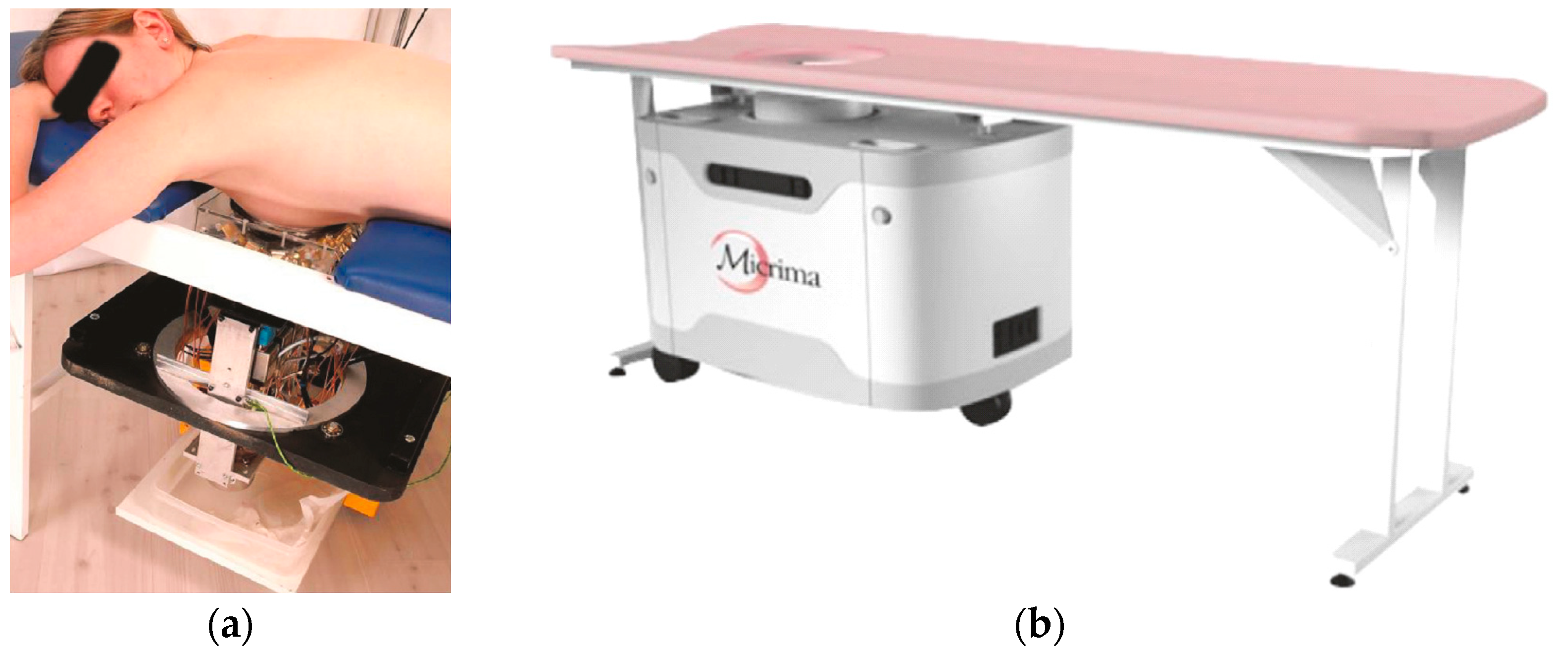
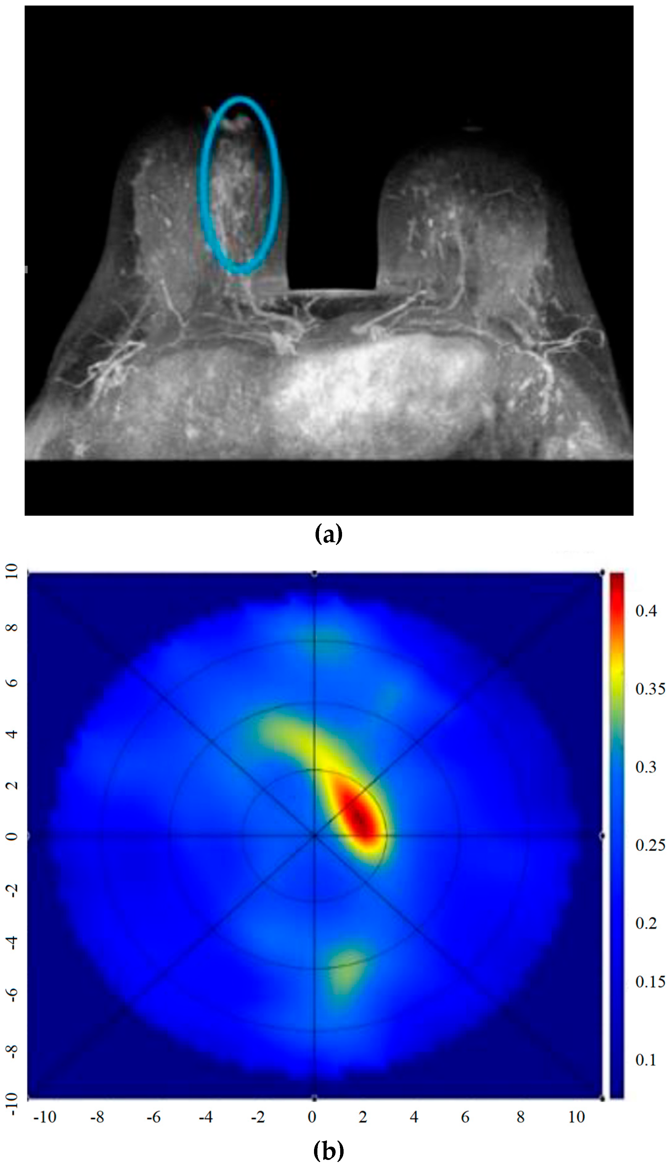
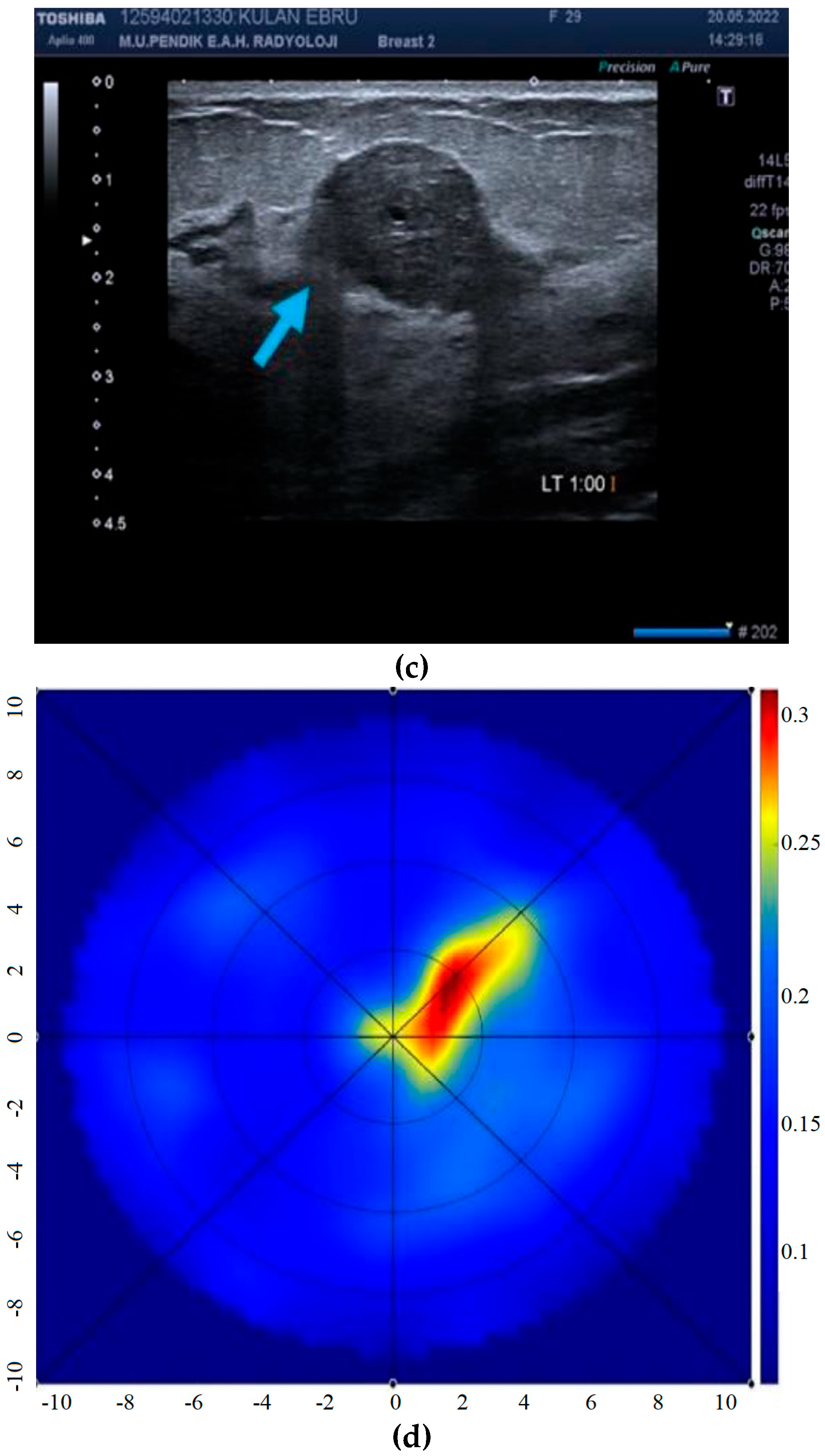
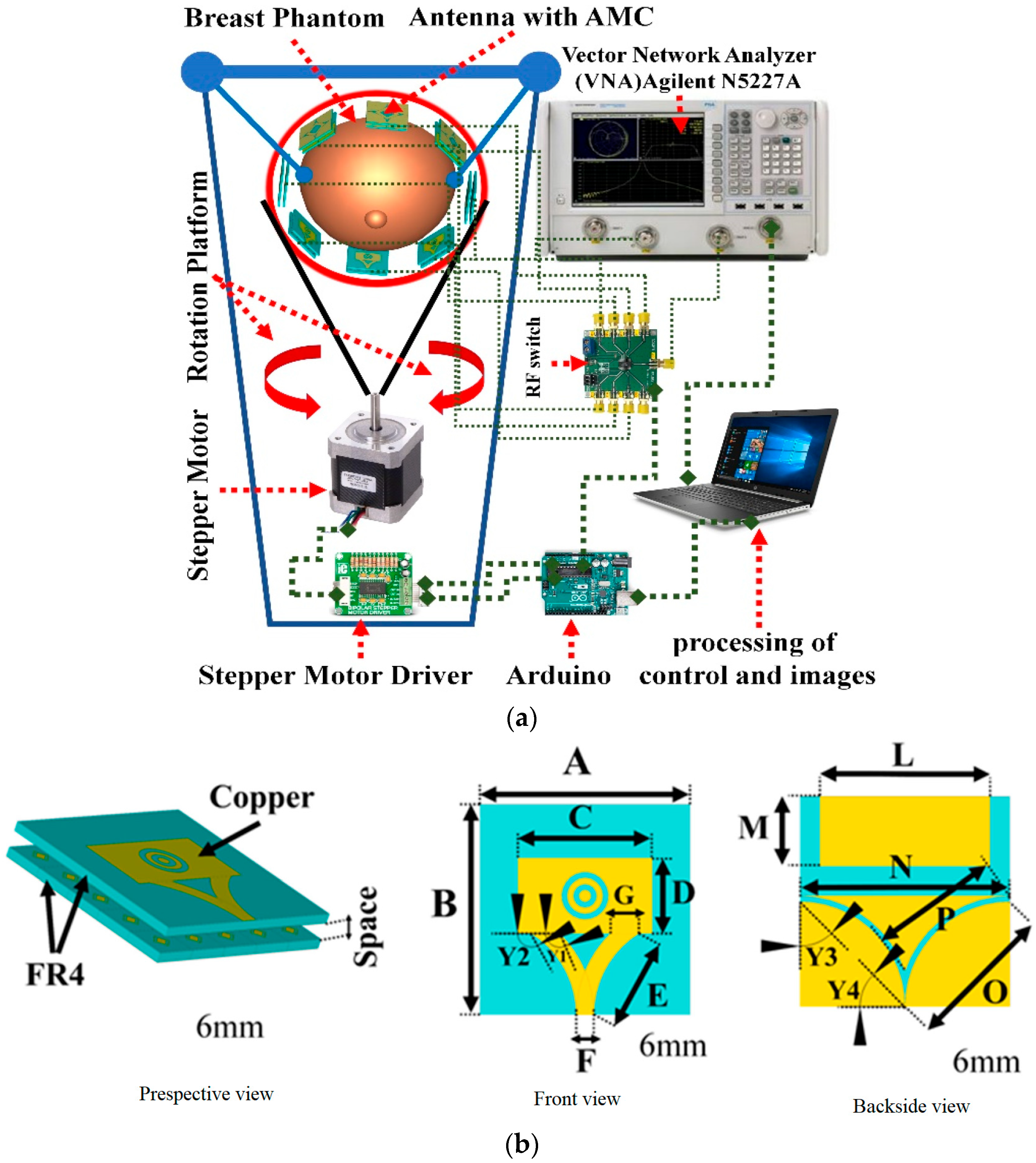
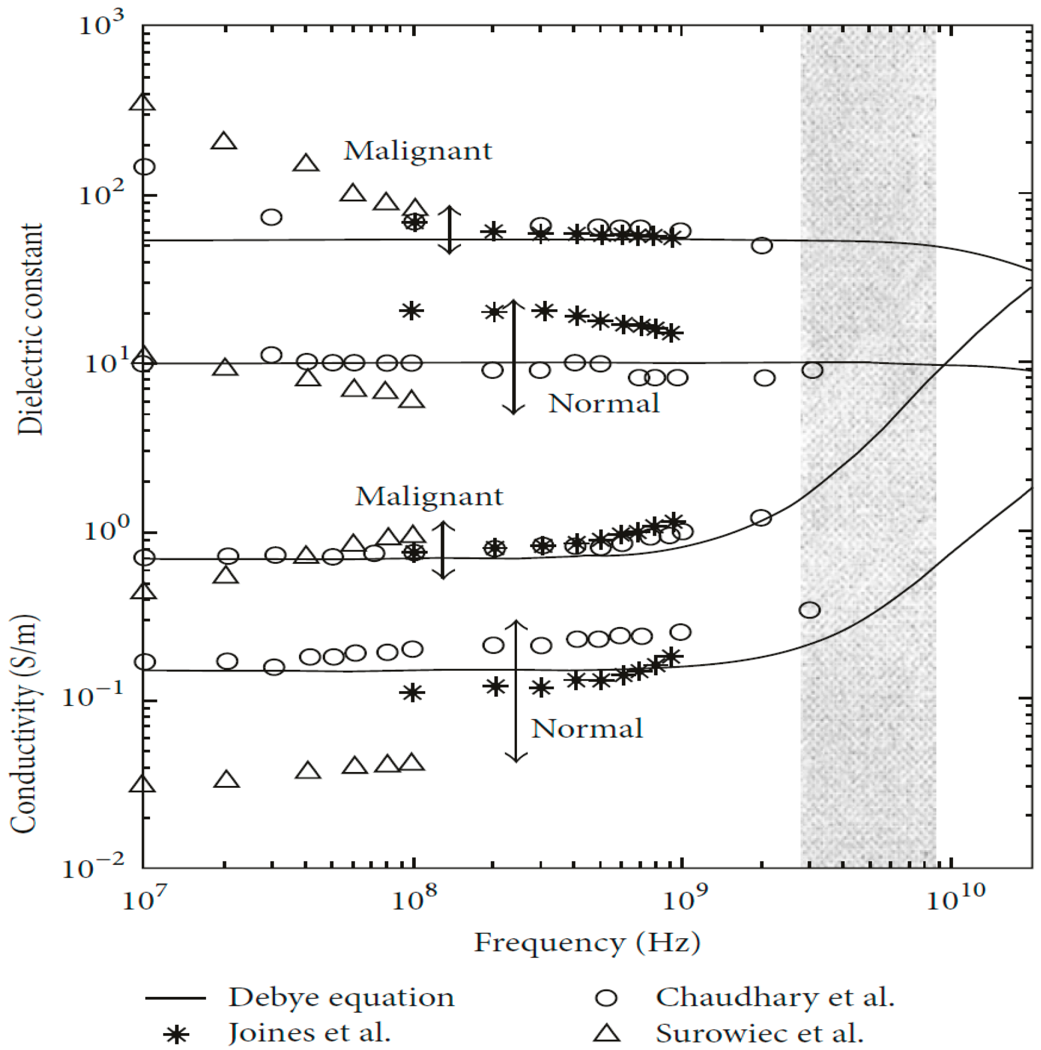
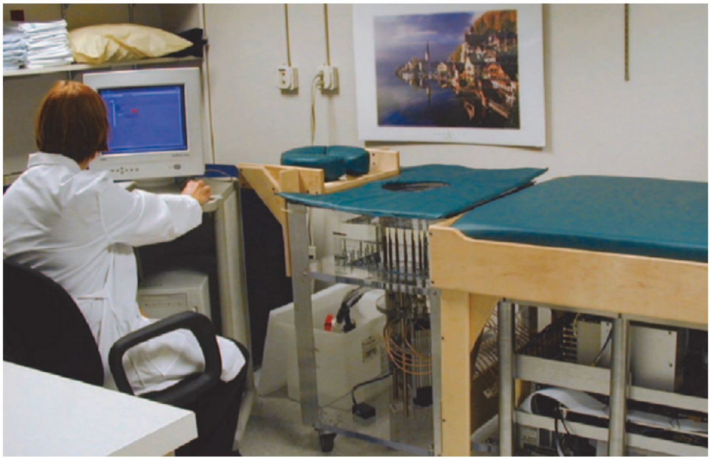
Table 1.
MBI systems for breast cancer detection.
| Year | Authors | Frequency (GHz) | Method | Test Method | Image | Antenna | Clinical Trials | Research Findings |
|---|---|---|---|---|---|---|---|---|
| 1990 | Bocquet et al. [55] | 2.5–3.5 | Radiometry | Human | 2D | Multi-probe radiometer | 97 patients | The malignant lesions had a radiometric ratio greater than 65% |
| 1998 | Hagness et al. [56] | N/A | Confocal microwave imaging (CMI) | Simulation | 2D | Monopole antenna | No | Could detect tumors (2 mm in diameter) |
| 1999 | Hagness et al. [57] | N/A | CMI | Simulation | 3D | UWB bowtie antenna | No | Could detect early stage breast cancer |
| 2000–2001 | Bulyshev et al. [58,59] | 2, 3.5, and 5 | MWT | Simulation | 2D and 3D | Helmholtz equation | No | Could detect tumors with a size > 1 cm |
| 2000–2001 | Fear et al. [60,61] | N/A | CMI | Simulation | 2D and 3D | bowtie antenna | No | Could detect and localize breast tumors in 3D |
| 2003 | Li et al. [62] | 1–11 | Microwave imaging via space-time (MIST) | Phantom | N/A | UWB antennas | No | Could detect very small tumors |
| 2003–2005 | Bond et al. [63,64] | N/A | MIST | N/A | N/A | Horn antenna | No | Could detect very small tumors |
| 2004 | Dobrowolski et al. [65] | 1.5–4.4 | Radiometry | Simulation and phantom | 2D | Three-band radiometer with mini hypodermic probes | No | Could detect and measure spatial temperature distribution inside a human body |
| 2006 | Xie et al. [66] | N/A | Multi-static adaptive (MSA) imaging | Simulation | 3D | No | The system showed better resolution and noise rejection capabilities than those of existing methods | |
| 2006–2013 | Elsdon et al. [67,68,69] | 9.4 | Holographic microwave imaging (HMI) | Phantom | 2D | Transmitting and receiving antenna | No | Could produce real-time images |
| 2010 | O’Halloran et al. [70] | MIST | Phantom | 2D | horn antenna | No | Could detect tumors (5 mm in diameter) | |
| 2010–2012 | Son et al. [71,72] | 0.5–3 | Microwave tomography (MT) | Phantom | 2D and 3D | monopole | No | Could detect tumors (5 mm in diameter) |
| 2000–2014 | Meaney et al. [73,74,75,76] | 1300 MHz | MT | Phantom | 2D, 3D | Monopole antenna | 500+ patients | Early stage breast cancer detection |
| 2003–2012 | Fear et al. [77,78,79] | 0.05–15 | Tissue sensing adaptive radar (TSAR) | Phantom | 2D, 3D | single antenna | 8 patients | Could detect and localize tumors with sizes of >4 mm in diameter |
| 2013 | Aguilar et al. [80] | 1.36–3.03 | CMI | Simulation & phantom | 3D | slot-loaded patch antenna | No | Miniaturized antennas are suitable candidates as array elements for multi-band microwave breast imaging systems |
| 2013–2018 | Wang et al. [81,82] | 12.6 | HMI | Simulation & phantom | 2D | 16-element sensor array | No | Could detect small tumors (<5 mm in diameter) |
| 2015 | Bucci et al. [83] | 2 | Magnetic nanoparticle enhanced MWI | Simulation and phantom | 2D | Using 50 antennas on each of the eight measurement circles present in the design | No | Allowed the measurement of the differential field arising in an MNP-enhanced way |
| 2016 | Medina et al. [84] | 2–15 | HMI | Phantom | 2D | Vivaldi antenna | No | Could detect tumors (>15 mm in diameter) |
| 2008–2016 | Klemm et al. [85,86,87,88] | 4–10 | MSA | Phantom | 2D, 3D | 16 stacked patch antennas | 86 patients | Could detect tumors 4 to 6 mm in diameter |
| 2016 | Porter et al. [89] | 2–4 | Active | Human | Multistatic radar with the 16 sensors | 3 patients | Many sources of variability were identified | |
| 2017 | Song et al. [90] | 3.1–10.6 | Active | Human | 3D | 4 × 4 dome antennas | 5 patients | Could detect tumor |
| 2017 | Yang et al. [91] | 4–8.5 | Active | Phantom | 2D | UWB horn antenna | 11 patients | Sensitive to increased tissue |
| 2017 | Kuwahara et al. [92] | 1–3 | MIST | Simulation | 3D | stacked patch antennas | 2 patients | Data correlation between the measured and calculated data is larger than 0.99 |
| 2019 | Rana et al. [93] | 1–9 | Radar-based | Simulation | 2D | N/A | clinical trials: normal and malignant tissues | Higher specificity |
| 2019 | Sani et al. [15] | 1–9 | Active | Simulation | 2D | one transmitting antenna and one receiving antenna | clinical trials: normal and malignant tissues | The apparatus is safe |
| 2019 | Hammouch et al. [94] | 3.1–14 | CMI | Simulation | 2D | Microstrip patch antenna | No | Could detect breast tumor |
| 2019 | Islam et al. [95] | 2.80–7.00 | Radar-based | Simulation and phantom | 2D | side-slotted Vivaldi antenna | No | Could detect tumors inside the breast |
| 2019 | Srinivasan et al. [96] | 2.45 | Dielectric substrate | Simulation | 2D & 3D | textile wearable antenna | No | Could detect tumors |
| 2019 | Soltani et al. [97] | 2.45 | Microwave-induced thermoacoustic imaging | Simulation | 3D | rectangular waveguide | No | Could detect small tumors (5 mm in diameter) |
| 2019 | Sheeba et al. [98] | 2.4 | Active | Simulation and phantom | 2D | Flexible soft-wear hexagonal patch antenna | No | tumor as 20.3 A/m2 and 19 A/m2 and gain as 7.20 and 7.25 dB |
| 2019 | Islam et al. [99] | 2.7–11.2 | Active | Simulation and phantom | 2D and 3D | Metasurface Loaded High Gain Antenna | No | Could detect multiple abnormalities inside the breast |
| 2019 | Wang [10] | 1–4 | Multi-frequency HMI | Simulation | 2D and 3D | waveguide | No | Could detect small breast lesions with higher accuracy compared to single-frequency HMI |
| 2020 | Felício et al. [100] | 2–5 GHz | Radar-based | Simulation and phantom | 2D, 3D | antipodal Vivaldi antenna | No | Could detect tumors with different positions |
| 2020 | Abdollahi et al. [101] | 0.8, 0.85, 0.9, and 0.95 GHz | Active | Simulation | 2D | Perfect electric conductor (PEC) chamber and 2D transverse magnetic (TM) transceivers | No | The highest AUC of over 0.99 |
| 2020 | Kumari et al. [102] | 8.5 | Near-field HMI | Phantom | 2D | Vivaldi antenna | No | Minimum size of 4 mm and a maximum depth of 25 mm in the phantom |
| 2020 | Ahmed et al. [103] | 6.1–12 | Radar-based | Simulation and phantom | 3D | 18 Peano patch antenna | No | Could improve the image resolution |
| 2020 | Rahpeima et al. [104] | 2.45 | MITAI | Simulation | 3D | No | Could detect tumors with a radius of 0.25 cm | |
| 2020 | Miraoui et al. [105] | 4 | Radar-based ANN | Simulation | 2D | Bowtie antennas | No | ANN presented more precision in tumors detection |
| 2020 | Cosgun et al. [106] | 1.9–2.02 | SPION-enhanced MWI | Simulation & phantom | 2D | dipole antennas | No | Could detect smaller tumors |
| 2020 | Kaur et al. [107] | 4.9–10.9 | Synthetic aperture radar | Phantom | 2D | microstrip antenna | No | The phantom at 5.7 GHz was 0.271 W/kg, and that at 6.5 GHz was 1.115 W/kg for 1 g of body tissue. |
| 2020 | Kaur et al. [108] | 3.71–11.48 | Radar-based | Phantom | 3D | Fork-shaped microstrip patch antenna | No | More reflections, less specific absorption rate and more conduction current |
| 2020 | Song et al. [109] | 3.5–15 | Radar-based | Phantom | 3D | 16-element dome antenna array | 1 patient | Improved image quality |
| 2020 | Vispa et al. [110] | 1–9 | Radar-based | Phantom | horn antenna and microstrip monopole antenna | 51 breasts | Microwave images of non-healthy breasts had a mean MAX/AVG of approximately 7% greater than those of healthy breasts | |
| 2020 | Norouzzadeh et al. [111] | 1–9 | Radar-based system | Simulation | 2D | UWB bowtie antennas | 2 patients | Cancerous tissues have inhomogeneous dielectric properties |
| 2020 | Xiao et al. [112] | 6 | MWI | Simulation and phantom | 2D, 3D | Patch antenna | No | Could accurately find the permittivity |
| 2020 | Mehranpour et al. [113] | 1.3–6.8 | Radar-based system | Phantom | 2D | UWB bowtie antenna | No | Could detect tumors with a radius of 7 mm |
| 2020 | Carr et al. [114] | Radiometry | Human | ONCOSCAN system | 138 patients | 41% higher than mammography | ||
| 2021 | Syed et al. [115] | 2–5 | Distributed antenna system (DAS) | Simulation | 2D | Quasi log periodic | No | Two different locations of tumor in the breast phantom were detected |
| 2021 | Mahmood et al. [21] | 4.8–30 | Near-field microwave imaging | Simulation and phantom | 2D | Dual-polarized, multiple input–multiple output wearable antenna | No | A maximum radiation efficiency and directive gain of 96% and 5.72 dBi were obtained |
| 2021 | Karam et al. [116] | 1.5–4.5 | Weighted-DAS | phantom | 2D | 24 antennas arranged into three rings around the breast | BRIGID dataset consists of 7 realistic physical breast models | The contrast is improved as well, with an absolutely negligible computational load compared to that of DAS |
| 2021 | Mehranpour et al. [117] | 2.5–15 | N/A | Simulation and phantom | Low-profile aperture-stacked patch (LP-ASP) antenna | No | Fidelity factor is obtained more at than 90% and 78% in the E-plane and H-plane, respectively, which are satisfactory in the wide-range angles of 90 from the boresight of the designed antenna | |
| 2022 | Wang [11] | 1–12 | HMI and Alexnet | Simulation | 2D, 3D | Waveguide antenna | No | Could detect and classify tumors |
| 2023 | Jamlos et al. [118] | 3.1–10.6 | N/A | Simulation and phantom | 2D | Zero-index metamaterial superstrates UWB antenna | No | MTM antenna consists of 10 × 5 MTM unit cells that recorded a gain of 5.66 dB, having a return loss (S11) of −19.734 dB at a 2.70 GHz frequency |
Table 2.
Radar-based MBI methods for breast cancer detection.
| Image Algorithms | Year | Frequency (GHz) | Antenna Number | MBI Method | Image | Tumor Size (mm) | Ref |
|---|---|---|---|---|---|---|---|
| Skin subtraction algorithm (SSA) | 1999 | 4 & 6 | 9 | FDTD-CMI | 3D | 4 | [120] |
| SSA | 2000 | 3.6 | 30 | FDTD-CMI | 2D | 6 | [121] |
| SSA | 2001 | 6 | 17 | FDTD-CMI | 2D | 2 | [122] |
| SSA | 2001 | 4 | 10 | FDTD-CMI | 3D | 6 | [123] |
| SSA | 2002 | 4 | 16 | FDTD-CMI | 2D | 3 | [124] |
| SSA | 2003 | 6 | 17 | FDTD-MIST | 2D | 2 | [63] |
| SSA | 2004 | 3.2 | 24 | FDTD-CMI | 2D | 3 | [125] |
| SSA | 2004 | 8 | 24 | FDTD | 3D | 4 | [126] |
| SSA | 2004 | 6.5 | 16 | FDTD-CMI | 2D | 2 | [127] |
| SSA | 2006 | N/A | 13 | FDTD | 2D | 2 | [128] |
| Inverse scattering algorithm (ISA) | 2002 | 4 | NA | FDTD-CMI | 3D | 6 | [60] |
| ISA | 2003 | 10 | 2 | FDTD-CMI | 2/3D | 12 | [129] |
| ISA | 2003 | 0.8 | 32 | MOM | 2D | 14 | [130] |
| ISA | 2017 | 6 | 2 | 3D | 4 | [131] | |
| Artifact removal algorithms (AR) | 2002 | 6 | 49 | FDTD-MIST | 2D | 2 | [132] |
| AR | 2004 | 6 | 49 | FDTD-MIST | 3D | 4 | [133] |
| AR | 2005 | 4 | 15 | FDTD-TSAR | 3D | 10 | [134] |
| AR | 2006 | NA | 17 | FDTD-MIST | 2D | 2 | [135] |
| AR | 2007 | 5.5 | 10 | FDTD-MIST | 3D | 2 | [136] |
| AR | 2010 | 7.5 | 14 | FDTD-MIST | 2D | 5 | [70] |
| AR | 2012 | 6.85 | 12 | FDTD-CMI | 3D | 2.5 | [137] |
| AR | 2013 | 4.5 | 21 | FDTD | 2D | 5 | [138] |
| AR | 2014 | 6 | 50 | FDTD-CMI | 3D | 8 | [139] |
| AR | 2015 | 5 | 60 | FDTD | 3D | NA | [140] |
| AR | 2017 | 6 | 25 | FDTD | 3D | 15 | [141] |
| Multistatic Artifact Removal (MAR) | 2018 | 0.05–15 | 30 × 6 | FDTD-CMI | 3D | 15 | [142] |
| AR | 2019 | 0.5–20 | N/A | CMI | 2D | 18 × 9, 12 × 8 | [143] |
| Gauss-Newton iterative algorithm (GNIA) | 2003 | 0.9 | 16 | FEM-CMI | 2D | 19 | [144] |
| GNIA | 2006 | NA | 5 | FDTD-CMI | 2D | 6 | [145] |
| GNIA | 2010 | 3 | 40 | FDTD | 3D | 10 & 20 | [146] |
| GNIA | 2010 | 6 | 5 | FDTD | 2D | 5 | [147] |
| GNIA | 2011 | 1.3 | 16 | FDTD | 3D | 30 | [148] |
| Delay and sum logarithm (DAS) | 2003 | NA | 17 | FDTD | 2D | 2 | [149] |
| DAS and AR | 2005 | 6 | M | FDTD-MIST | 2D | 2 | [64] |
| DAS | 2006 | 5 | 72 | FDTD-CMI | 2D | 4 | [66] |
| DAS | 2007 | 5 | 6 | FDTD-CMI | 2D | 6 | [150] |
| DAS | 2007 | 5 | 6 | FDTD-CMI | 2D | 10 | [151] |
| DAS | 2007 | 6 | 16 | FDTD | 3D | 4 & 10 | [152] |
| DAS | 2008 | 6 | 16 | FDTD | 3D | 7 & 10 | [153] |
| DAS | 2010 | 6 | 31 | FDTD | 3D | 7 | [153] |
| DAS | 2013 | 6 | 16 | FDTD | 3D | 3 | [154] |
| DAS | 2013 | 2 | 16 | FDTD | 3D | 10 | [155] |
| DAS | 2014 | 2 | 31 | FDTD | 3D | 4 | [156] |
| DAS | 2014 | 4.5 | 7 | FEM | 3D | 2 | [157] |
| DAS | 2015 | NA | 16 | FDTD-CMI | 3D | 4 | [158] |
| DAS | 2015 | 3 | 16 | FDTD | 3D | N/A | [159] |
| DAS | 2015 | 3.2 | 31 | FDTD | 3D | 10 | [160] |
| DAS | 2015 | 7.3 | 9 | 3D | NA | [161] | |
| DAS | 2016 | 6 | 20 | FDTD-CMI | 3D | 3 & 5 | [162] |
| DAS | 2016 | 9 | 1 | FDTD-CMI | 2D | 15 | [84] |
| DAS | 2016 | 4.7 | 16 | 3D | 3 | [163] | |
| DAS | 2016 | 1 | 16 | 3D | NA | [164] | |
| Weighted DAS | 2021 | 0.5–10 | 24 | CMI | 2D | BRIGID dataset of the National University of Ireland Galway | [116] |
| DAS and robust capon beamforming (RCB) | 2009 | 6 | 16 | FDTD | 3D | 4 & 6 | [86] |
| DAS and Multiple Signal Classification | 2013 | 2 | 31 | FDTD | 2D | 20 | [165] |
| Robust capon beamforming | 2011 | 5 | 7 | FDTD | 2D | 6 | [166] |
| Delay-multiply-and-sum (DMAS) | 2010 | 7.5 | 53 | FDTD-CMI | 2D | 5, 10 & 15 | [167] |
| DMAS | 2010 | 3.5 | 31 | FDTD-CMI | 3D | 7 | [168] |
| DMAS | 2011 | 7.5 | 20 | FDTD-CMI | 2D | 2.5 & 5 | [169] |
| DMAS | 2012 | 5 | 36 | CMI | 2D | 2 | [170] |
| DMAS | 2012 | 5 | 12 | FDTD-CMI | 2D | 2 | [171] |
| DMAS | 2015 | 2.5 | 16 | FDTD-CMI | 3D | 10 | [172] |
| DMAS | 2015 | 3 | 16 | 3D | 10 | [173] | |
| DMAS | 2016 | 3 | 16 | 3D | NA | [174] | |
| DMAS | 2017 | 5 | 73 | FDTD | 2D | 14 | [175] |
| DMAS | 2021 | N/A | 16 | FDTD-CMI | 2D | 5 | [176] |
| Delay-multiply-and-sum-degree-4 (DMAS-D4) | 2022 | N/A | N/A | CMI | 2D, 3D | N/A | [177] |
| Double-constrained robust Capon beamforming (DCRCB) | 2013 | 5 | 9 | FDTD | 3D | 2 | [178] |
| Double constrained robust Capon beamforming (DCRCB) | 2015 | NA | 8 | FDTDI-CM | 3D | 4 | [179] |
| Modified delay-and-sum (MDAS) | 2015 | NA | 16 | FDTD-CMI | 3D | NA | [180] |
| Time-of-arrival algorithm (TOA) | 2006 | 6 | 9 | FDTD | 2D | 4 | [181] |
| TOA | 2009 | 4 | 128 | FDTD | 3D | Irregular shaped | [182] |
| TOA | 2011 | 9.75 | 22 | FDTD-CMI | 3D | 3 | [183] |
| Time Reversal (TR) | 2006 | NA | 22 | FDTD | 3D | 3 | [184] |
| TR | 2007 | 6 | 13 | FDTD-CMI | 2D | 10 | [185] |
| TR | 2007 | 9 | 2 | FDTD | 2D | 2 | [186] |
| TR | 2008 | 8 | 7 | FDTD | 2D | 4 & 6 | [187] |
| TR | 2008 | 5 | 16 | FDTD | 2D | 8 | [188] |
| TR | 2009 | NA | 24 | FDTD | 2D | Irregular shaped | [189] |
| TR | 2011 | NA | NA | FDTD | 2D | 2 | [190] |
| TR | 2012 | 5 | 29 | FDTD | 3D | 7 | [191] |
| Time-Reversal Multiple Signal Classification (TR-MUSIC) | 2016 | 4 | 4 | 3D | NA | [192] | |
| TR-MUSIC | 2017 | 3 | 115 | FDTD | 3D | 10 | [193] |
| Robust and artifact-resistant (RAR) | 2015 | 6.85 | 24 | FDTD | 3D | 10 | [194] |
| Inverse Fast Fourier Transform (IFFT) | 2005 | 7 | M | FDTD | 2D | 4 | [195] |
| IFFT | 2006 | 3 | 16 | FDTD | 2D | 10 | [196] |
| IFFT | 2011 | 4 | 12 | FDTD-CMI | 2D | 4 | [197] |
| IFFT | 2011 | 4.5 | 16 | FDTD | 2D | 5 | [198] |
| IFFT | 2012 | 3.5 | 16 | FDTD-CMI | 3D | NA | [199] |
| Matched-filtering algorithm (MFA) | 2004 | 6 | 2 | FEM-CMI | 2D | 5 | [200] |
| MFA | 2006 | 6 | 2 | FDTD | 2D | 10 | [201] |
| MFA | 2010 | 5 | 8 | FDTD | 3D | N/A | [202] |
| MFA | 2011 | N/A | 24 | FDTD | 2D | 12 | [203] |
| Generalized likelihood ratio test (GLRT) | 2005 | 6 | 24 | FDTD-CMI | 3D | 4 | [204] |
| TR and GLRT | 2007 | 6 | 7 | FDTD-CMI | 2D | 6 | [205] |
| MERIT open-source software | 2019 | 2.5–11 | 16 | Radar-Based Imaging | 2D | 1.8 | [206] |
| Differential cascode balun low noise amplifier (DCBLNA) | 2020 | 1.5–15.7 | N/A | UWB | N/A | highest figure of merit of 3.2 | [207] |
| Hybrid Artifact Suppression (HAS) | 2020 | 2.2–13.5 | 37 | CMI | 2D,3D | 5 | [208] |
| Contrast source inversion (CSI) | 2022 | 1.38–3.47 | 10 | FDTD-CMI | 2D | 2 | [209] |
| two-stage rotational clutter suppression (TSR) | 2023 | 3.1–10.6 | 16 | CMI | 2D,3D | 10 | [210] |
Disclaimer/Publisher’s Note: The statements, opinions and data contained in all publications are solely those of the individual author(s) and contributor(s) and not of MDPI and/or the editor(s). MDPI and/or the editor(s) disclaim responsibility for any injury to people or property resulting from any ideas, methods, instructions or products referred to in the content. |
© 2023 by the author. Licensee MDPI, Basel, Switzerland. This article is an open access article distributed under the terms and conditions of the Creative Commons Attribution (CC BY) license (https://creativecommons.org/licenses/by/4.0/).
Share and Cite
MDPI and ACS Style
Wang, L. Microwave Imaging and Sensing Techniques for Breast Cancer Detection. Micromachines 2023, 14, 1462. https://doi.org/10.3390/mi14071462
AMA Style
Wang L. Microwave Imaging and Sensing Techniques for Breast Cancer Detection. Micromachines. 2023; 14(7):1462. https://doi.org/10.3390/mi14071462
Chicago/Turabian StyleWang, Lulu. 2023. "Microwave Imaging and Sensing Techniques for Breast Cancer Detection" Micromachines 14, no. 7: 1462. https://doi.org/10.3390/mi14071462
Note that from the first issue of 2016, this journal uses article numbers instead of page numbers. See further details here.