
SiC Electrochemical Sensor Validation for Alzheimer Aβ42 Antigen Detection
 , , and
Abstract
:1. Introduction
- Electrical Current: Used in techniques such as Cyclic Voltammetry (CV), Square Wave Voltammetry (SWV), Differential Pulse Voltammetry (DPV), and Chronoamperometry (CA).
- Electrical Impedance: Used in Electrochemical Impedance Spectroscopy (EIS).
- Optical Luminescence: Used in Electrochemiluminescence (ECL).

| Method * | Electrode Material | Functionalizing Antibody | Aβ42 Solution Media | Aβ42 Detection Range ** | Aβ42 LOD ** | Refs. |
|---|---|---|---|---|---|---|
| CV | Gold electrode | Aβ1–42 | PBS | [24] | ||
| SWV | Carbon fiber microelectrode | mHJ2, mHJ7.4 | Mice CSF | [25] | ||
| CV | Gold electrode | 6E10 | aCSF | [26] | ||
| EIS | AAO Sensing electrode | 12F4 | BSA | [27] | ||
| CV | Screen printed carbon electrode | H31L21 | BSA | [10] | ||
| CV | Gold electrode | 6E10 12F4 | aCSF | [28] | ||
| CV, EIS | Gold Electrode | 6E10 | Nutrient Mixture F12 | - | [29] | |
| EIS | Carbon printed electrode | antimAβ | 0.02% (v/v) ammonia water at 200 mM concentration | [9] | ||
| CA | Screen printed carbon electrode | anti-Aβ | Human CSF, Serum and Plasma | [30] | ||
| CV, DPV | Screen printed gold electrode | Aβ1–28 | CSF | [12] | ||
| SWV, EIS | Gold electrode | DE2B4 | PBS | [31] | ||
| ECL | Glassy Carbon Electrode | anti-Aβ | PBS | [32] | ||
| SWV, CV, EIS | Glassy Carbon Electrode | anti-Aβ | PBS | [33] | ||
| CV | Screen printed carbon electrode | 12F4, 1E11 | Human Serum and Plasma | [34] | ||
| DPV, EIS | Gold Electrode | EPR9296 | PBS and Human Serum | [11] | ||
| LSV | Conductive silk fibroin-based immunoparticles | mOC31 | Serum | [35] | ||
| EIS, CV | ICE (Ti+Au) | anti-Aβ | Human Serum | [36] | ||
| EIS | ICE | anti-Aβ | Human Serum | [37] | ||
| DPV | Graphene-modified Screen-printed electrode | H31L21 | Spiked human and mice plasmas | [38] | ||
| EIS | Gold Electrode | 12F4 | PBS | [39] | ||
| CV | Gold Electrode | Aβ1–28 | PBS | [13] |
2. Experimental
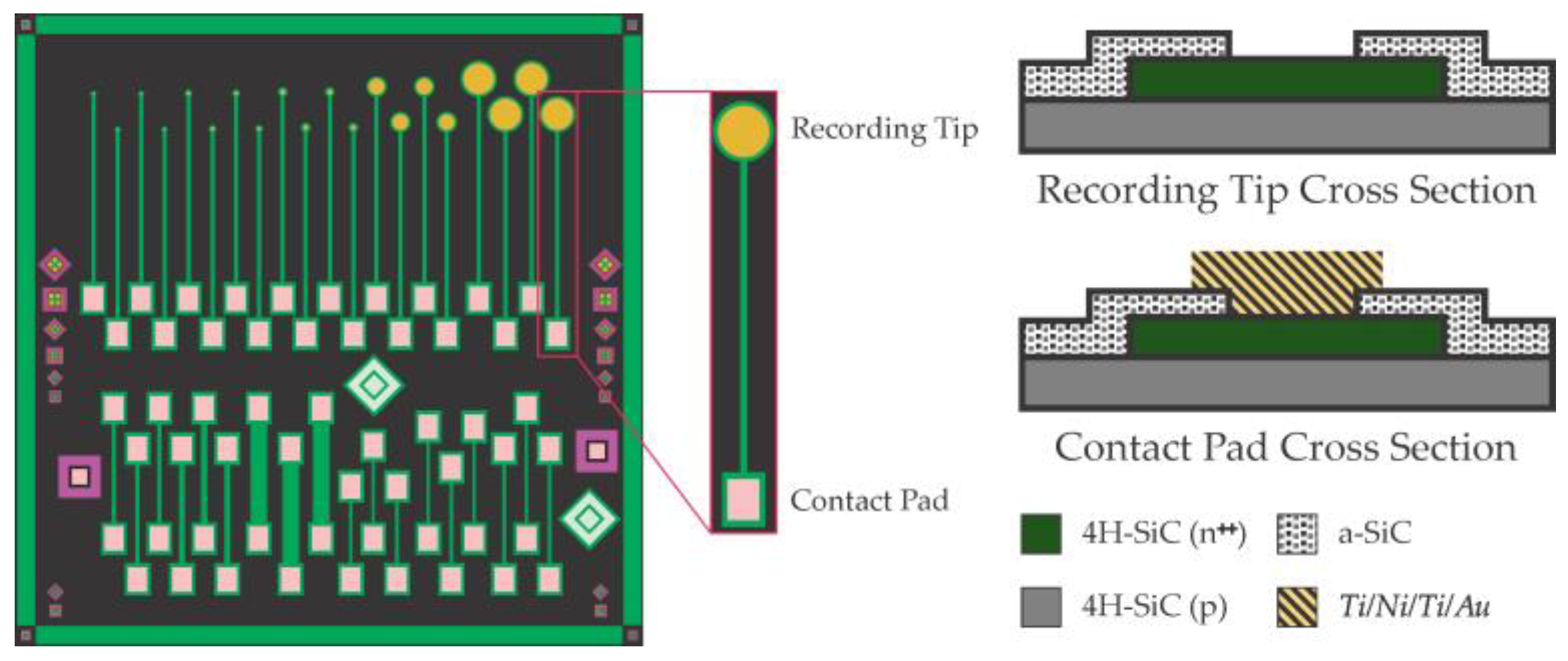
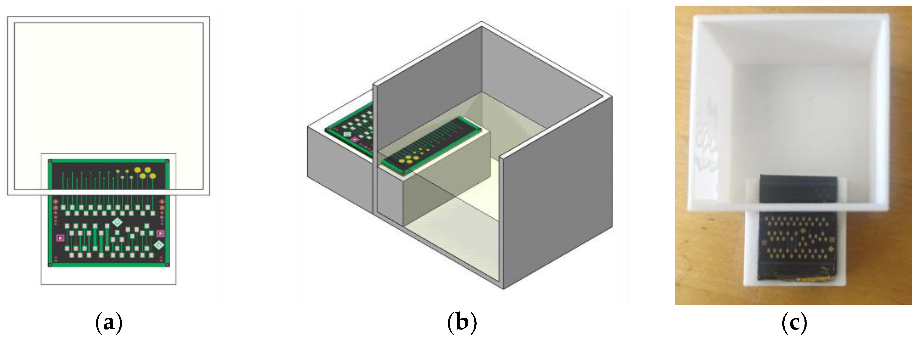
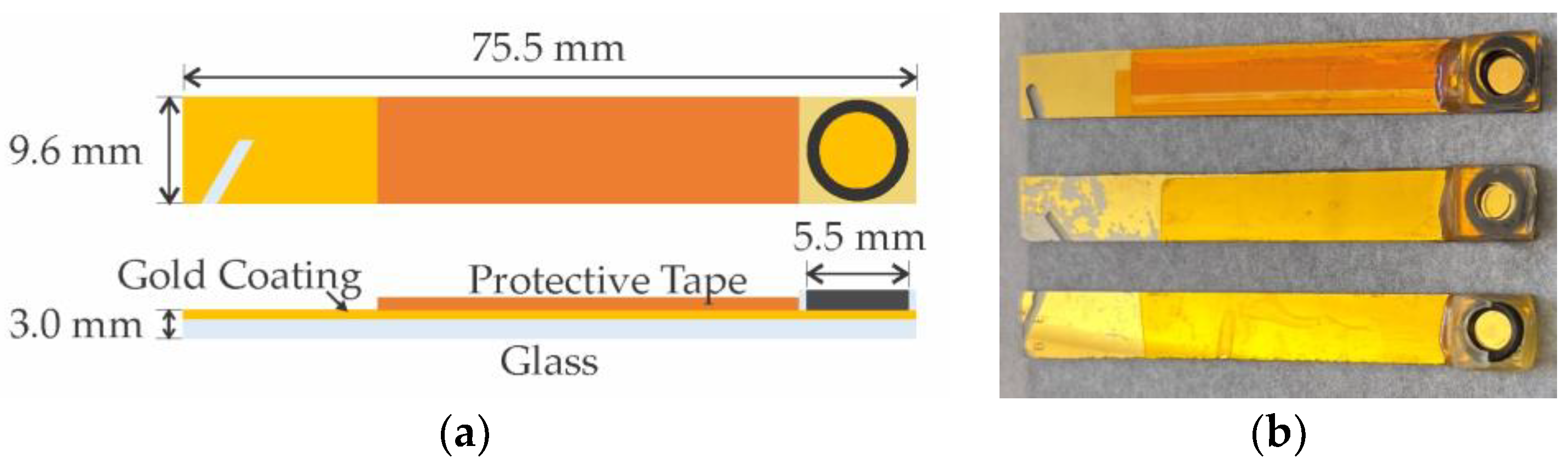
2.1. Apparatus and Electrodes
2.2. Reagents and Solutions
2.3. Antibody and Antigen Solutions
2.4. Methods
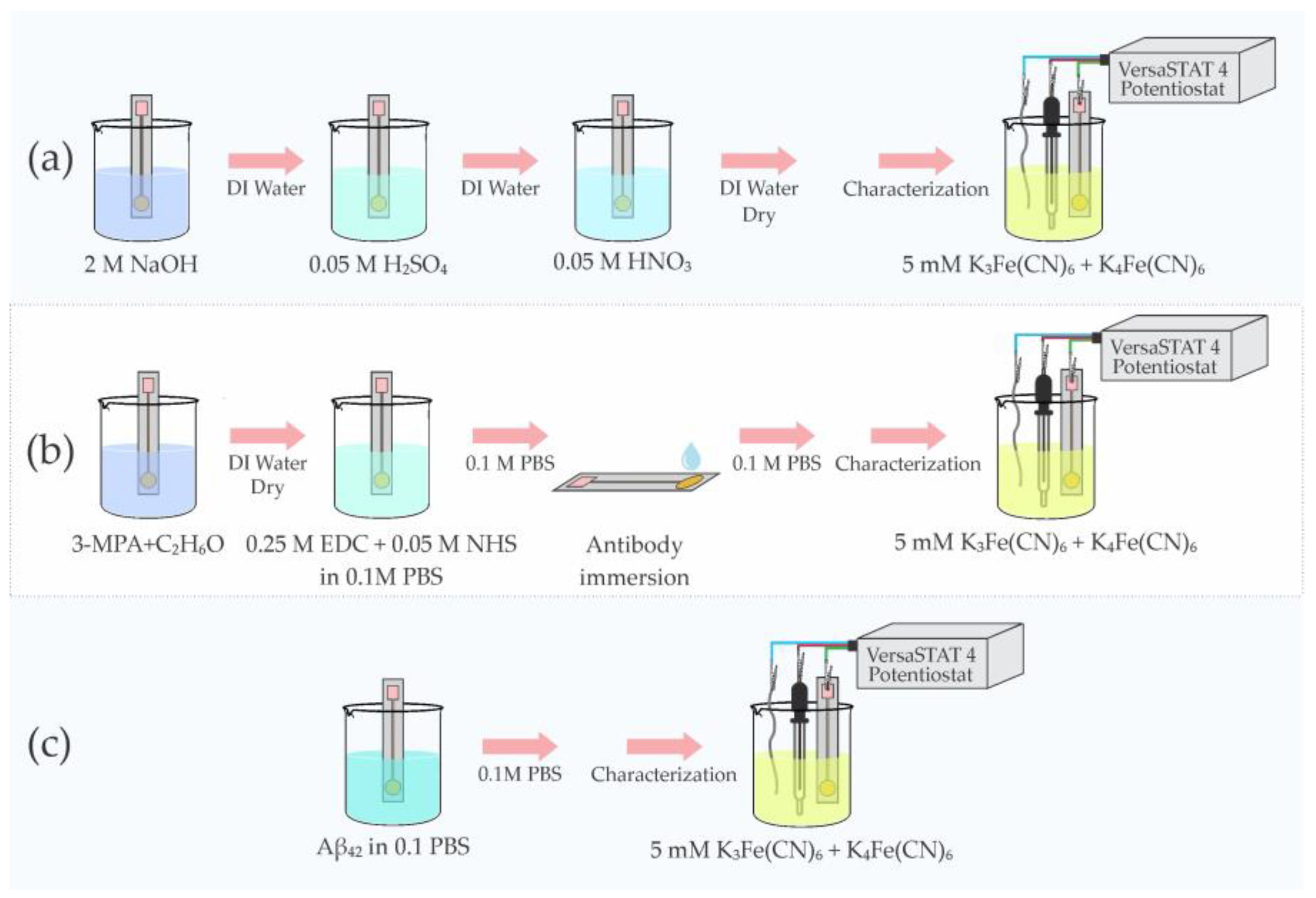
2.4.1. Cleaning
2.4.2. Electrochemical Cells
2.4.3. Procedure & Measurements
3. Results
4. Conclusions and Future Implications
Author Contributions
Funding
Data Availability Statement
Acknowledgments
Conflicts of Interest
References
- World Health Organization. Dementia. Available online: https://www.who.int/news-room/fact-sheets/detail/dementia (accessed on 28 January 2023).
- 2022 Alzheimer’s disease facts and figures. Alzheimer’s Dement. 2022, 18, 700–789. [CrossRef] [PubMed]
- Tolar, M.; Abushakra, S.; Sabbagh, M. The path forward in Alzheimer’s disease therapeutics: Reevaluating the amyloid cascade hypothesis. Alzheimer’s Dement. 2019, 16, 1553–1560. [Google Scholar] [CrossRef] [PubMed]
- Barage, S.H.; Sonawane, K.D. Amyloid cascade hypothesis: Pathogenesis and therapeutic strategies in Alzheimer’s disease. Neuropeptides 2015, 52, 1–18. [Google Scholar] [CrossRef] [PubMed]
- Castellani, R.J.; Plascencia-Villa, G.; Perry, G. The amyloid cascade and Alzheimer’s disease therapeutics: Theory versus observation. Lab. Investig. 2019, 99, 958–970. [Google Scholar] [CrossRef]
- Afzal, S.; Maqsood, M.; Khan, U.; Mehmood, I.; Nawaz, H.; Aadil, F.; Song, O.-Y.; Nam, Y. Alzheimer Disease Detection Techniques and Methods: A Review. Int. J. Interact. Multimed. Artif. Intell. 2021, 6, 26. [Google Scholar] [CrossRef]
- Zetterberg, H.; Bendlin, B.B. for Alzheimer’s disease—Preparing for a new era of disease-modifying therapies. Mol. Psychiatry 2020, 26, 296–308. [Google Scholar] [CrossRef]
- Humpel, C. Identifying and validating biomarkers for Alzheimer’s disease. Trends Biotechnol. 2011, 29, 26. [Google Scholar] [CrossRef] [Green Version]
- Lien, T.T.N.; Takamura, Y.; Tamiya, E.; Vestergaard, M.C. Modified screen printed electrode for development of a highly sensitive label-free impedimetric immunosensor to detect amyloid beta peptides. Anal. Chim. Acta 2015, 892, 69–76. [Google Scholar] [CrossRef]
- Rama, E.C.; González-García, M.B.; Costa-García, A. Competitive electrochemical immunosensor for amyloid-beta 1–42 detection based on gold nanostructurated Screen-Printed Carbon Electrodes. Sens. Actuators B Chem. 2014, 201, 567–571. [Google Scholar] [CrossRef]
- Dai, Y.; Molazemhosseini, A.; Liu, C.C. In Vitro Quantified Determination of β-Amyloid 42 Peptides, a Biomarker of Neuro-Degenerative Disorders, in PBS and Human Serum Using a Simple, Cost-Effective Thin Gold Film Biosensor. Biosensors 2017, 7, 29. [Google Scholar] [CrossRef] [Green Version]
- Li, S.-S.; Lin, C.-W.; Wei, K.-C.; Huang, C.-Y.; Hsu, P.-H.; Liu, H.-L.; Lu, Y.-J.; Lin, S.-C.; Yang, H.-W.; Ma, C.-C.M. Non-invasive screening for early Alzheimer’s disease diagnosis by a sensitively immunomagnetic biosensor. Sci. Rep. 2016, 6, 25155. [Google Scholar] [CrossRef] [Green Version]
- Hsu, C.-H.; Gupta, A.K.; Purwidyantri, A.; Prabowo, B.A.; Chen, C.-H.; Chuang, C.-C.; Tian, Y.-C.; Lu, Y.-J.; Lai, C.-S. Sensing Alzheimer’s Disease Utilizing Au Electrode by Controlling Nanorestructuring. Chemosensors 2022, 10, 94. [Google Scholar] [CrossRef]
- Sigma Aldrich. Antibody Generation. Available online: https://www.sigmaaldrich.com/CO/es/technical-documents/technical-article/protein-biology/elisa/generation-of-antibodies (accessed on 24 May 2023).
- Veerabhadrappa, B.; Delaby, C.; Hirtz, C.; Vialaret, J.; Alcolea, D.; Lleó, A.; Fortea, J.; Santosh, M.S.; Choubey, S.; Lehmann, S. Detection of amyloid beta peptides in body fluids for the diagnosis of alzheimer’s disease: Where do we stand? Crit. Rev. Clin. Lab. Sci. 2019, 57, 99–113. [Google Scholar] [CrossRef] [PubMed]
- Jiang, M.; Wang, X.Y.; Wang, X.B. Advances in Detection Methods of β-Amyloid Protein. Chin. J. Anal. Chem. 2018, 46, 1339–1349. [Google Scholar] [CrossRef]
- Campuzano, S.; Pedrero, M.; Gamella, M.; Serafín, V.; Yáñez-Sedeño, P.; Pingarrón, J.M. Beyond Sensitive and Selective Electrochemical Biosensors: Towards Continuous, Real-Time, Antibiofouling and Calibration-Free Devices. Sensors 2020, 20, 3376. [Google Scholar] [CrossRef]
- Sharma, S.; Byrne, H.; O’Kennedy, R.J. Antibodies and antibody-derived analytical biosensors. Essays Biochem. 2016, 60, 9. [Google Scholar] [CrossRef]
- Yi, Y.; Weinberg, G.; Prenzel, M.; Greiner, M.; Heumann, S.; Becker, S.; Schlögl, R. Electrochemical corrosion of a glassy carbon electrode. Catal. Today 2017, 295, 32–40. [Google Scholar] [CrossRef]
- Saddow, S.E. Recent advances in SiC biomedical devices: Healthcare applications. Silicon Carbide Technol. Adv. Hum. Healthc. Appl. 2022, 1–48. [Google Scholar] [CrossRef]
- De Napoli, M. SiC detectors: A review on the use of silicon carbide as radiation detection material. Front. Phys. 2022, 10, 769. [Google Scholar] [CrossRef]
- Oliveros, A.; Guiseppi-Elie, A.; Saddow, S.E. Silicon carbide: A versatile material for biosensor applications. Biomed. Microdevices 2013, 15, 353–368. [Google Scholar] [CrossRef]
- Saddow, S.E.; Agarwal, A. Advances in Silicon Carbide Processing and Applications, 1st ed.; Artech House Publishers: Norwood, MA, USA, 2004; Available online: https://www.researchgate.net/publication/272944831_Advances_in_Silicon_Carbide_Processing_and_Applications (accessed on 25 May 2023).
- Islam, K.; Jang, Y.C.; Chand, R.; Jha, S.K.; Lee, H.H.; Kim, Y.S. Microfluidic biosensor for β-amyloid(1–42) detection using cyclic voltammetry. J. Nanosci. Nanotechnol. 2011, 11, 5657–5662. [Google Scholar] [CrossRef]
- Prabhulkar, S.; Piatyszek, R.; Cirrito, J.R.; Wu, Z.-Z.; Li, C.-Z. Microbiosensor for Alzheimer’s Disease Diagnostics: Detection of Amyloid Beta Biomarkers. J. Neurochem. 2012, 122, 374–381. [Google Scholar] [CrossRef] [Green Version]
- Liu, L.; Zhao, F.; Ma, F.; Zhang, L.; Yang, S.; Xia, N. Electrochemical detection of β-amyloid peptides on electrode covered with N-terminus-specific antibody based on electrocatalytic O2 reduction by Aβ(1–16)-heme-modified gold nanoparticles. Biosens. Bioelectron. 2013, 49, 231–235. [Google Scholar] [CrossRef] [PubMed]
- Wu, C.-C.; Ku, B.-C.; Ko, C.-H.; Chiu, C.-C.; Wang, G.-J.; Yang, Y.-H.; Wu, S.-J. Electrochemical impedance spectroscopy analysis of A-beta (1–42) peptide using a nanostructured biochip. Electrochim. Acta 2014, 134, 249–257. [Google Scholar] [CrossRef]
- Liu, L.; He, Q.; Zhao, F.; Xia, N.; Liu, H.; Li, S.; Liu, R.; Zhang, H. Competitive electrochemical immunoassay for detection of β-amyloid (1–42) and total β-amyloid peptides using p-aminophenol redox cycling. Biosens. Bioelectron. 2014, 51, 208–212. [Google Scholar] [CrossRef] [PubMed]
- Rushworth, J.V.; Ahmed, A.; Griffiths, H.H.; Pollock, N.M.; Hooper, N.M.; Millner, P.A. A label-free electrical impedimetric biosensor for the specific detection of Alzheimer’s amyloid-beta oligomers. Biosens. Bioelectron. 2014, 56, 83–90. [Google Scholar] [CrossRef]
- De la Escosura-Muñiz, A.; Plichta, Z.; Horák, D.; Merkoçi, A. Alzheimer′s disease biomarkers detection in human samples by efficient capturing through porous magnetic microspheres and labelling with electrocatalytic gold nanoparticles. Biosens. Bioelectron. 2015, 67, 162–169. [Google Scholar] [CrossRef] [Green Version]
- Carneiro, P.; Loureiro, J.; Delerue-Matos, C.; Morais, S.; Pereira, M.D.C. Alzheimer’s disease: Development of a sensitive label-free electrochemical immunosensor for detection of amyloid beta peptide. Sens. Actuators B Chem. 2017, 239, 157–165. [Google Scholar] [CrossRef] [Green Version]
- Wang, J.X.; Zhuo, Y.; Zhou, Y.; Wang, H.J.; Yuan, R.; Chai, Y.Q. Ceria Doped Zinc Oxide Nanoflowers Enhanced Luminol-Based Electrochemiluminescence Immunosensor for Amyloid-β Detection. ACS Appl. Mater. Interfaces 2016, 8, 12968–12975. [Google Scholar] [CrossRef]
- Han, J.; Zhang, M.; Chen, G.; Zhang, Y.; Wei, Q.; Zhuo, Y.; Xie, G.; Yuan, R.; Chen, S. Ferrocene covalently confined in porous MOF as signal tag for highly sensitive electrochemical immunoassay of amyloid-β. J. Mater. Chem. B 2017, 5, 8330–8336. [Google Scholar] [CrossRef]
- Diba, F.S.; Kim, S.; Lee, H.J. Electrochemical immunoassay for amyloid-beta 1–42 peptide in biological fluids interfacing with a gold nanoparticle modified carbon surface. Catal. Today 2017, 295, 41–47. [Google Scholar] [CrossRef]
- Liu, T.-C.; Lee, Y.-C.; Ko, C.-Y.; Liu, R.-S.; Ke, C.-C.; Lo, Y.-C.; Hong, P.-S.; Chu, C.-Y.; Chang, C.-W.; Wu, P.-W.; et al. Highly sensitive/selective 3D nanostructured immunoparticle-based interface on a multichannel sensor array for detecting amyloid-beta in Alzheimer’s disease. Theranostics 2018, 8, 4210–4225. [Google Scholar] [CrossRef] [PubMed]
- Le, H.T.N.; Park, J.; Chinnadayyala, S.R.; Cho, S. Sensitive electrochemical detection of amyloid beta peptide in human serum using an interdigitated chain-shaped electrode. Biosens. Bioelectron. 2019, 144, 111694. [Google Scholar] [CrossRef]
- Le, H.T.N.; Park, J.; Cho, S. A Probeless Capacitive Biosensor for Direct Detection of Amyloid Beta 1–42 in Human Serum Based on an Interdigitated Chain-Shaped Electrode. Micromachines 2020, 11, 791. [Google Scholar] [CrossRef] [PubMed]
- Sethi, J.; Van Bulck, M.; Suhail, A.; Safarzadeh, M.; Perez-Castillo, A.; Pan, G. A label-free biosensor based on graphene and reduced graphene oxide dual-layer for electrochemical determination of beta-amyloid biomarkers. Mikrochim. Acta 2020, 187, 288. [Google Scholar] [CrossRef] [Green Version]
- Wang, B.-Y.; Gu, B.-C.; Wang, G.-J.; Yang, Y.-H.; Wu, C.-C. Detection of Amyloid-β(1–42) Aggregation With a Nanostructured Electrochemical Sandwich Immunoassay Biosensor. Front. Bioeng. Biotechnol. 2022, 10, 853947. [Google Scholar] [CrossRef]
- Bernardin, E.K.; Frewin, C.L.; Everly, R.; Hassan, J.U.; Saddow, S.E. Demonstration of a Robust All-Silicon-Carbide Intracortical Neural Interface. Micromachines 2018, 9, 412. [Google Scholar] [CrossRef] [Green Version]
- Yaghoubi, H.; Li, Z.; Jun, D.; Saer, R.; Slota, J.E.; Beerbom, M.; Schlaf, R.; Madden, J.D.; Beatty, J.T.; Takshi, A. The role of gold-adsorbed photosynthetic reaction centers and redox mediators in the charge transfer and photocurrent generation in a bio-photoelectrochemical cell. J. Phys. Chem. C 2012, 116, 24868–24877. [Google Scholar] [CrossRef]
- Bernardin, E.; Frewin, C.L.; Dey, A.; Everly, R.; Hassan, J.U.; Janzén, E.; Pandrazio, J.; Saddow, S.E. Development of an all-SiC neuronal interface device. MRS Adv. 2016, 1, 3679–3684. [Google Scholar] [CrossRef]
- Saddow, S.E. Silicon Carbide Technology for Advanced Human Healthcare Applications. Micromachines 2022, 13, 346. [Google Scholar] [CrossRef]
- Willner, I.; Riklin, A. Electrical Communication between Electrodes and NAD(P)+-Dependent Enzymes Using Pyrroloquinolinequinone-Enzyme Electrodes in a Self-Assembled Monolayer Configuration: Design of a New Class of Amperometric Biosensors. Anal. Chem. 1994, 66, 1535–1539. [Google Scholar] [CrossRef]
- Campuzano, S.; Gálvez, R.; Pedrero, M.; de Villena, F.J.M.; Pingarrón, J. Preparation, characterization and application of alkanethiol self-assembled monolayers modified with tetrathiafulvalene and glucose oxidase at a gold disk electrode. J. Electroanal. Chem. 2002, 526, 92–100. [Google Scholar] [CrossRef]
- Schneider, M.; Fritzsche, N.; Puciul-Malinowska, A.; Baliś, A.; Mostafa, A.; Bald, I.; Zapotoczny, S.; Taubert, A. Surface Etching of 3D Printed Poly(lactic acid) with NaOH: A Systematic Approach. Polymers 2020, 12, 1711. [Google Scholar] [CrossRef] [PubMed]
- Bard, A.J.; Faulkner, L.R. Electrochemical Methods: Fundamentals and Applications, 2nd ed.; Wiley: Hoboken, NJ, USA, 2001; Available online: https://www.wiley.com/en-us/Electrochemical+Methods:+Fundamentals+and+Applications,+2nd+Edition-p-9780471043720 (accessed on 27 May 2023).


Disclaimer/Publisher’s Note: The statements, opinions and data contained in all publications are solely those of the individual author(s) and contributor(s) and not of MDPI and/or the editor(s). MDPI and/or the editor(s) disclaim responsibility for any injury to people or property resulting from any ideas, methods, instructions or products referred to in the content. |
© 2023 by the authors. Licensee MDPI, Basel, Switzerland. This article is an open access article distributed under the terms and conditions of the Creative Commons Attribution (CC BY) license (https://creativecommons.org/licenses/by/4.0/).
Share and Cite
Montero-Arevalo, B.; Seufert, B.I.; Hossain, M.S.; Bernardin, E.; Takshi, A.; Saddow, S.E.; Schettini, N. SiC Electrochemical Sensor Validation for Alzheimer Aβ42 Antigen Detection. Micromachines 2023, 14, 1262. https://doi.org/10.3390/mi14061262
Montero-Arevalo B, Seufert BI, Hossain MS, Bernardin E, Takshi A, Saddow SE, Schettini N. SiC Electrochemical Sensor Validation for Alzheimer Aβ42 Antigen Detection. Micromachines. 2023; 14(6):1262. https://doi.org/10.3390/mi14061262
Chicago/Turabian StyleMontero-Arevalo, Brayan, Bianca I. Seufert, Mohammad S. Hossain, Evans Bernardin, Arash Takshi, Stephen E. Saddow, and Norelli Schettini. 2023. "SiC Electrochemical Sensor Validation for Alzheimer Aβ42 Antigen Detection" Micromachines 14, no. 6: 1262. https://doi.org/10.3390/mi14061262