Article

Fluorescence Properties of ZnOQDs-GO-g-C3N4 Nanocomposites

1
College of Clinical Medicine, Jiamusi University, Jiamusi 154007, China
2
College of Pharmacy, Jiamusi University, Jiamusi 154007, China
3
School of Mechanical Engineering, Jiamusi University, Jiamusi 154007, China
4
College of Basic Medicine, Jiamusi University, Jiamusi 154007, China
*
Authors to whom correspondence should be addressed.
These authors contributed equally to this work.
Micromachines 2023, 14(4), 711; https://doi.org/10.3390/mi14040711
Submission received: 4 March 2023 / Revised: 17 March 2023 / Accepted: 20 March 2023 / Published: 23 March 2023

Abstract

:
In this paper, the fluorescence properties of ZnOQD-GO-g-C3N4 composite materials (ZCGQDs) were studied. Firstly, the addition of a silane coupling agent (APTES) in the synthesis process was explored, and it was found that the addition of 0.04 g·mL−1 APTES had the largest relative fluorescence intensity and the highest quenching efficiency. The selectivity of ZCGQDs for metal ions was also investigated, and it was found that ZCGQDs showed good selectivity for Cu2+. ZCGQDs were optimally mixed with Cu2+ for 15 min. ZCGQDs also had good anti-interference capability toward Cu2+. There was a linear relationship between the concentration of Cu2+ and the fluorescence intensity of ZCGQDs in the range of 1~100 µM. The regression equation was found to be F0/F = 0.9687 + 0.12343C. The detection limit of Cu2+ was about 1.74 μM. The quenching mechanism was also analyzed.

1. Introduction

ZnO is an eco-friendly oxide semiconductor, and compared with traditional CdSe or CdTe quantum dots, it is an inexpensive luminescent material, which makes it more attractive in practical applications [1,2,3]. Over the past few years, a variety of physical and chemical synthesis techniques have been developed to synthesize ZnO QDS. Various chemical methods have been developed to synthesize ZnO nanocrystals, and many research groups have used the sol–gel method to improve the process of obtaining ZnO quantum dots with different sizes and fluorescence characteristics [4]. ZnO quantum dots have been widely used in the photoelectric field [5,6,7], biomedical field [8] energy electrochemical field [9,10,11,12], fluorescence imaging [13], gas sensing [14] selective detection [15], and other fields.
Graphene oxide (GO) is a carbon-based nanomaterial widely used for adsorption and catalytic degradation due to its high specific surface area and tunable structure [16]. It is also a promising nanomaterial for a wide range of applications, including but not limited to energy storage [17,18], electrical sensors [19,20], and antimicrobial activity [21,22]. GO exhibits excellent performance in photocatalytic activity due to its high electron mobility, good electrical conductivity and chemical stability. GO can be involved in the synthesis of various composite materials, such as nanoparticles, polymers, and novel metal–organic frame-derived materials, which can be used in separation [23], catalysis [24], electrochemical devices [25], and coatings [26].
In recent decades, a type of non-metallic semiconductor graphitic carbon nitride (g-C3N4) has attracted great attention among researchers worldwide because it has good photocatalytic activity under visible light irradiation and shows great potential in various environmental remediation applications. As a visible light-responsive non-metallic organic semiconductor, it has been widely used in sewage treatment [27], CO2 reduction [28,29], hydrogen production from water photolysis [30,31] energy conversion [32,33], and other fields.
Yang [34] et al. established a highly efficient and stable Al3+ fluorescent probe using the fluorescent “on” mode of zinc sulfide crystal composite zinc oxide quantum dots (ZnS/ZnOQDs). The as-synthesized ZnS/ZnOQDs exhibited fluorescence enhancement for Al, confirming the availability of fluorescence sensing probes.
Copper is a toxic heavy metal element. Cu2+ is involved in many reaction activities in the human body and plays an important role in various physiological processes such as the formation of red blood cells and the normal growth and maintenance of brain tissue, kidney, heart, and other organs of the body [35]. However, the content of Cu2+ is also a double-edged sword, and the excess or deficiency of Cu2+ can lead to the disorder of cellular homeostasis and damage the central nervous system. The abnormal accumulation of Cu2+ will lead to the appearance of some human diseases, including Alzheimer’s disease, Parkinson’s disease, Menkes disease, and Wilson’s disease. If Cu2+ is deficient in the human body, gastrointestinal diseases and abnormal bone growth will occur [36].
Copper accumulation in the body will produce certain harm [37]. In addition, heavy metal ions affect the activities of proteins and enzymes in the human body and accumulate in some organs, resulting in chronic poisoning [38]. Heavy metal pollution seriously endangers human health, which has raised great concern in the world. Therefore, it is of great significance and urgency to develop a rapid and sensitive detection method for heavy metal ions for environmental and public safety.
The traditional detection methods of heavy metal ions include electrochemical analysis [39,40], atomic absorption spectroscopy [41], atomic emission spectroscopy, inductively coupled plasma mass spectrometry [42], and surface-enhanced Raman spectroscopy [43,44], but these methods have the problems of cumbersome detection steps, high instrument costs, difficulty in real-time detection, and low sensitivity. Their application in the field of heavy metal ion detection is greatly restricted [45]. Compared with traditional detection methods, fluorescence analysis methods have the advantages of high resolution, good sensitivity and rapid response, and have strong potential for application. In most cases, the fluorescence intensity of fluorescent probes is related to metal ions, so it is relatively easy to qualitatively and quantitatively analyze heavy metal ions by measuring the fluorescence intensity [46]. Therefore, it is of great significance to develop fluorescent probes that can selectively detect important metal ions such as copper.
In this paper, the selectivity of ZnOQD-GO-g-C3N4 composite materials (ZCGQDs) for metal ions and the mixing time, anti-interference ability, linear relationship, and fluorescence quenching mechanism of ZCGQDs composite materials and the target metal ion Cu2+ were analyzed, which provides a new idea for the research of zinc oxide quantum dots.

2. Experimental

2.1. Experimental Reagents

The inorganic compounds containing Al3+, Ba2+, Ca2+, Cd2+, Co2+, Cr3+, Cu2+, Fe2+, Fe3+, Hg2+, Mn2+, Mg2+, Ni2+, Pb2+. AlCl3, BaCl2, CaCl2, CdCl2·25H2O, CoCl2·6H2O, CuCl2·2H2O, CrCl3·6H2O, FeCl2·4H2O, Fe(NO3)3 9H2O, HgCl2, Mn(NO3)2·4H2O, Mg(NO3)2·6H2O, NiCl2·6H2O, and Pb(NO3)2 were purchased from Sinopharm Chemical Reagent Co., Ltd. (Huai’an, China).

2.2. Metal Ion Detection

2.2.1. Selective Detection of Metal Ions Based on ZCGQDs

  • The inorganic compounds containing Al3+, Ba2+, Ca2+, Cd2+, Co2+, Cr3+, Cu2+, Fe2+, Fe3+, Hg2+, Mn2+, Mg2+, Ni2+, and Pb2+ were used as metal ion sources, dissolved in deionized water to prepare 50 mM solutions of different metal ions;
  • A pipette was used to accurately pipette 0.005 mL of different metal ion solutions of the same concentration configured in step a;
  • Then, 4.995 mL of the prepared ZCGQDs composite sample was taken;
  • Various metal ions were added to the ZCGQDs composite sample to obtain a 50 μM colloidal solution of different metal ions and left for 20 min;
  • A quartz cuvette was used, with a diameter of 1 cm and transparent on all sides, and the different metal ion colloid solutions in step d were added;
  • The mixed colloidal solutions were scanned to obtain their fluorescence spectra and intensity values. To reduce systematic errors, each mixed colloidal solution was scanned three times, and the average was calculated as the final intensity result.
The fluorescence intensities of different mixed colloidal solutions were compared to determine the selectivity of the fluorescent probes for metal ions.

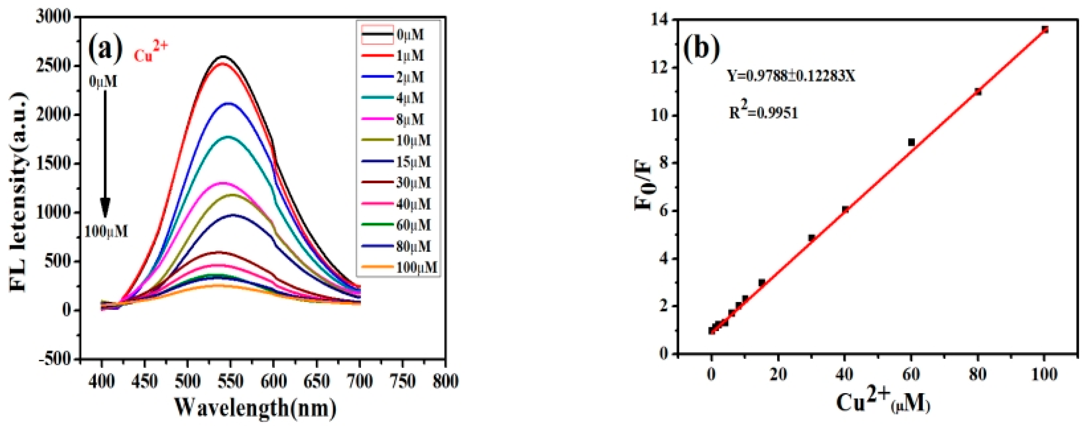
2.2.2. Determination of the Linear Relationship between ZCGQDs and Target Metal Ion Cu2+

In order to explore the selectivity of the prepared ZCGQDs complexes to Cu2+, the relationship between Cu2+ concentration and the change in the fluorescence intensity of the ZCGQDs complexes was investigated.
Experimental process:
  • Cu2+ was derived from the CuCl2·2H2O inorganic compound, which was dissolved in deionized water, and several solutions with different concentrations of Cu2+ were prepared using the stepwise dilution method;
  • A pipette was used to accurately pipette 0.005 mL of Cu2+ solutions at the different concentrations configured in step a;
  • Then, 4.995 mL samples of the ZCGQDs composites were taken;
  • Different concentrations of Cu2+ solution were added to the ZCGQDs composite sample, mixed and shaken well to obtain a colloidal solution, which was left for 15 min;
  • A quartz cuvette was used, with a diameter of 1 cm and transparent on all sides, and the colloidal solutions with different concentrations of Cu2+ in step d were added;
  • The mixed colloidal solutions were scanned to obtain their fluorescence spectra and intensity values. In order to reduce systematic errors, each mixed colloidal solution was scanned three times, and the average intensity value was used as the representative.

2.3. Experimental Optimization

The addition of metal ions can cause the fluorescence intensity of ZCGQDs complexes to weaken, resulting in a fluorescence quenching phenomenon. Among these metal ions, the fluorescence intensity of the solution changed most significantly after the addition of Cu2+.Using this principle, the ZCGQDs complexes were used as fluorescent probes to detect Cu2+. However, it should be noted that there were differences in experimental settings, and different quenching efficiencies were obtained. We optimized the experiment in two aspects: (a) the addition of different concentrations of APTES in the preparation of ZCGQDs complexes; (b) the mixing of ZCGQDs complexes and Cu2+ at different times.

3. Results and Discussion

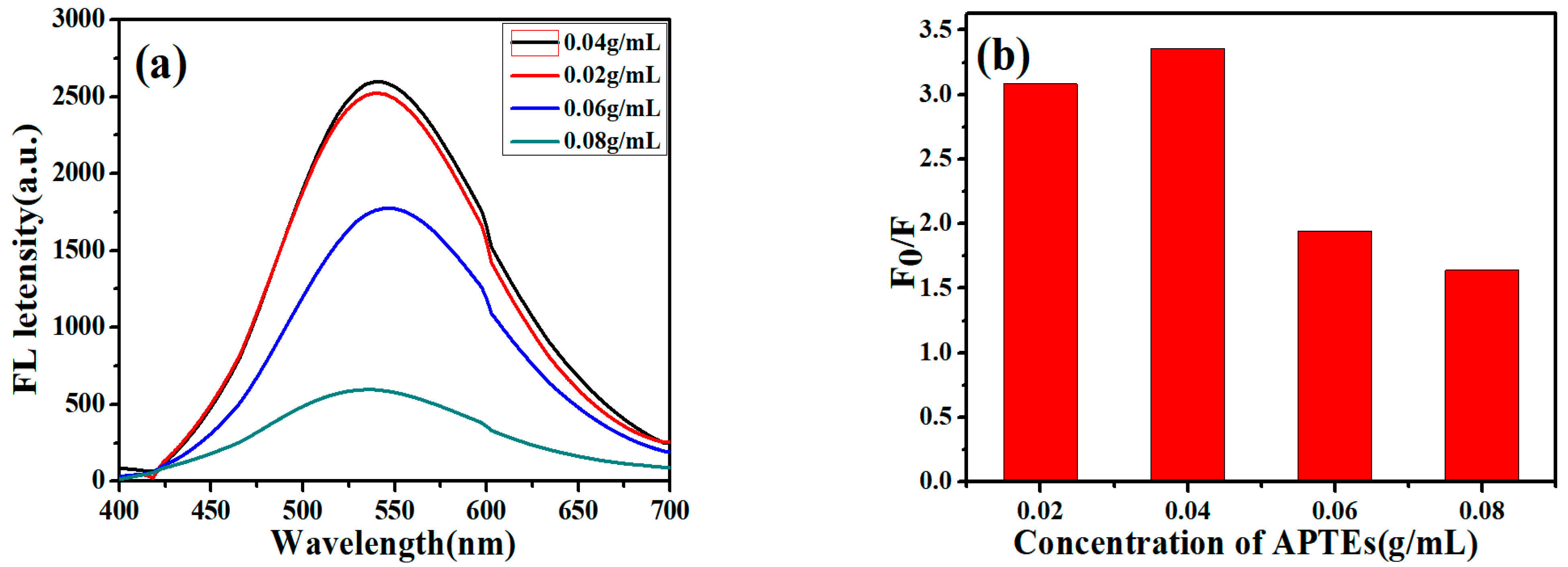
3.1. Effect of APTES Concentration Used to Synthesize ZCGQDs

In order to explore the effect of the addition of different concentrations of APTES on the quenching efficiency during the preparation of synthetic ZCGQDs complexes, the experiments were conducted as follows: (a) During the preparation of the ZCGQDs composites, 0.02 g·mL−1, 0.04 g·mL−1, 0.06 g·mL−1, and 0.08 g·mL−1 of a silane coupling agent (APTES) were added; (b) the ZCGQDs complexes were scanned with a fluorescence spectrophotometer to obtain their fluorescence spectra and fluorescence peak intensity values; (c)The fluorescence intensity changes of the solutions before and after the addition of Cu2+ were measured, and the fluorescence intensity values were labeled as F0 and F, respectively; the corresponding fluorescence quenching rates were represented by F0/F. To reduce systematic errors, the above procedure was repeated three times and the average fluorescence intensity values were calculated.
From Figure 1a, it can be concluded that with the increase in the APTES concentration, the fluorescence intensity of the ZCGQDs complexes increased. However, when the APTES concentration increased to a certain extent, the fluorescence intensity decreased instead of increasing. The reason may be that the luminescence of ZnOQDs in the visible light range is due to the deep-level emission caused by lattice defects. This is mainly caused by two kinds of defects: (1) Starting from the lower part of the conduction band, the electrons gradually leap toward the OZn and Oi deep body defects; (2) starting from the deep body defect VO, the electrons gradually leap to the upper end of the valence band. Therefore, there should be a certain concentration range and an optimum value. As shown in Figure 1a, which illustrates the change in the APTES concentration and the fluorescence intensity of the ZCGQDs complexes, the wavelength blue shift occurred when the APTES concentration exceeded 0.02 g·mL−1, which indicates that the appropriate increase in the APTES concentration was beneficial to the ZCGQDs complexes. The surface quickly formed a coating, which in turn reduced the agglomeration of the ZCGQDs complexes. By adding 0.04 g·mL−1 APTES, the fluorescence intensity was the highest.
Figure 1b shows the relative fluorescence intensity values after the addition of equal amounts of Cu2+ (20 μM) to the complexes formed by different concentrations of APTES and ZCGQDs. With the increase in the APTES concentration, the agglomeration degree of the ZCGQDs complexes weakened, the relative fluorescence intensity increased, and the quenching efficiency increased. However, when the APTES concentration increased to a certain extent, the relative fluorescence intensity did not increase but decreased. The excessive increase in the APTES concentration caused the coating layer of the ZCGQDs complexes to be too thick, thus destroying the fluorescence emission of the quantum dots caused by surface defects, decreasing the relative fluorescence intensity, and reducing the quenching efficiency. Notably, 0.04 g·mL−1 APTES had the highest relative fluorescence intensity and the highest quenching efficiency. Therefore, the concentration of 0.04 g·mL−1 APTES was selected to synthesize the ZCGQDs complexes for subsequent experimental analysis.

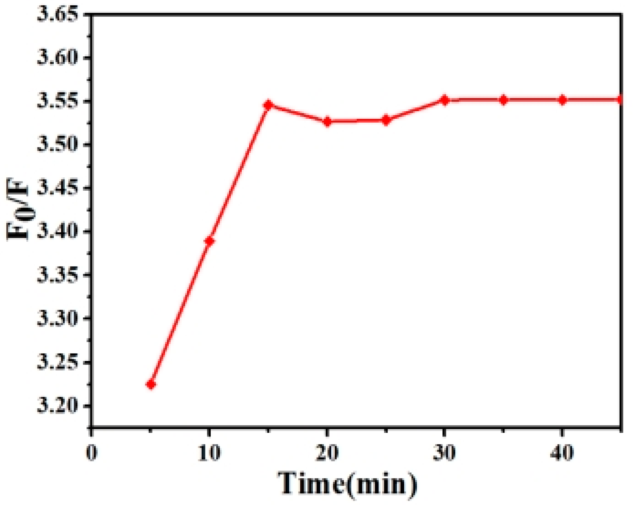
3.2. Effect of Mixing Time of ZCGQDs and Cu2+

As shown in Figure 2, nine time points were selected from 5 to 45 min to determine the extent of the fluorescence quenching of Cu2+ and ZCGQDs complexes at different mixing times [47,48]. In the range of 5–15 min, the quenching efficiency enhanced with increasing time and remained basically stable after 15 min. The highest efficiency was reached at 15 min. In order to improve the fluorescence properties of the ZCGQDs complexes in subsequent experiments, a fixed mixing time of 15 min was selected.

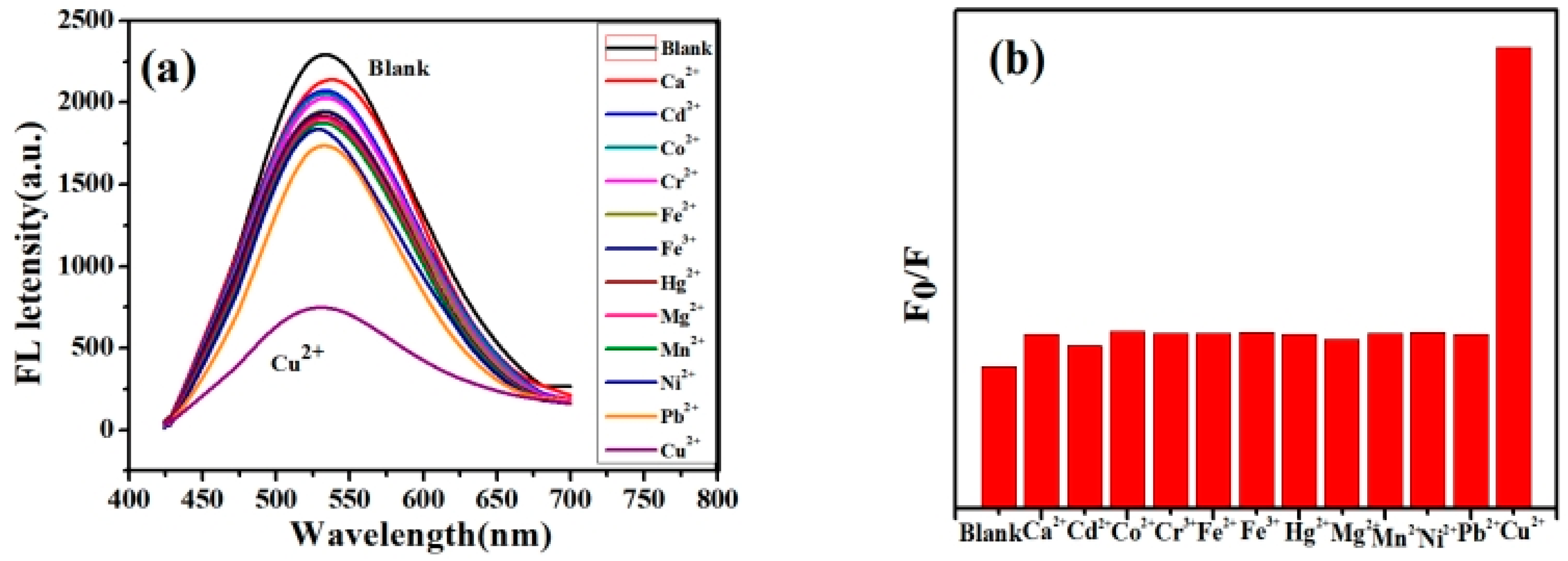
3.3. Selectivity of ZCGQDs to Metal Ions

Equal concentrations of various metal ions were added to the ZCGQDs composite sample to obtain a 50 μM colloidal solution of different metal ions and allowed to stand for 15 min [49]. The changes in the fluorescence intensity of the ZCGQDs complexes before and after the addition of Cu2+ were measured. The fluorescence intensity values were marked as F0 and F, and the corresponding fluorescence quenching rate was expressed as F0/F. In order to reduce the systematic error, the above process was repeated three times, and the fluorescence average intensity values were calculated.
Figure 3a,b show the effects of different metal ions on the fluorescence intensity of the ZCGQDs complexes under the same concentration conditions. It was found that Cu2+ resulted in a marked fluorescence quenching phenomenon on the ZCGQDs complexes, and the as-prepared ZCGQDs complexes exhibited good selectivity toward Cu2+.

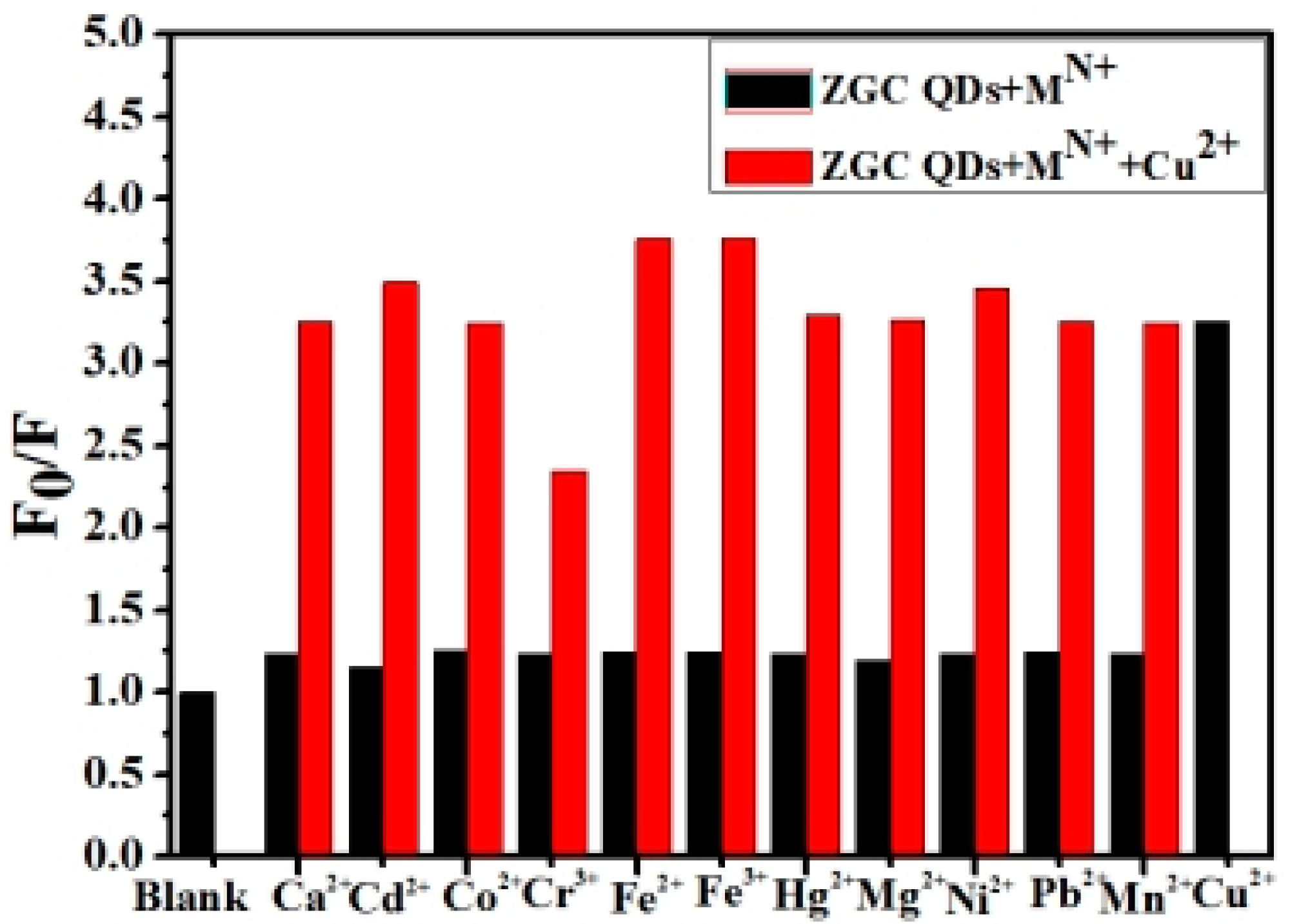
3.4. Anti-Interference Performance of ZCGQDs on Cu2+

Different metal ions of equal concentration were prepared and added to ZCGQDs composite samples, and the mixtures were shaken to obtain the colloidal solutions of the different metal ions. Then, Cu2+ was added to the solutions and shaken well, and the mixtures were left to stand for 15 min to test their anti-interference performance [47]. The changes in the fluorescence intensity of the ZCGQDs complexes before and after the addition of Cu2+ were measured. The fluorescence intensity values were marked as F0 and F, and the relative fluorescence intensity (marked as F0/F) was used to represent the corresponding fluorescence quenching rate. In order to reduce the systematic error, the above process was repeated three times, and the average fluorescence intensity values were calculated. As shown in Figure 4, Cu2+ still exhibited strong fluorescence quenching under the same concentration of different metals, and the fluorescence quenching efficiency did not significantly change compared with Cu2+ without interference, which indicates that the prepared ZCGQDs composites had good anti-interference capability toward Cu2+.

3.5. Linear Relationship between ZCGQDs and Cu2+ Concentration

The stepwise dilution method was used to obtain the Cu2+ solutions of different concentrations, which were added to the ZCGQDs composite samples, mixed, and shaken to obtain the colloidal solutions of copper ions, and the solutions were allowed to stand for 10 min [48,49]. The changes in the fluorescence intensity of the ZCGQDs complexes before and after the addition of Cu2+ were measured. The fluorescence intensity values were marked as F0 and F, and the relative fluorescence intensity (marked as F0/F) was used to represent the corresponding fluorescence quenching rate.
From Figure 5a, it can be concluded that the fluorescence intensity of the ZCGQDs complexes decreased sequentially with the increase in Cu2+ concentration. This shows a linear relationship between the concentration of Cu2+ and the fluorescence intensity of ZCGQDs complexes. As seen in Figure 5b, the linear regression equation for the concentration of Cu2+ between 1 and 100 μM is F0/F = 0.9788 + 0.12283C (R2 = 0.9951), where C represents the concentration of Cu2+ in the solution and R2 represents the correlation constant. Using this equation, the limit of detection for Cu2+ was obtained, which was about 1.74 μM.

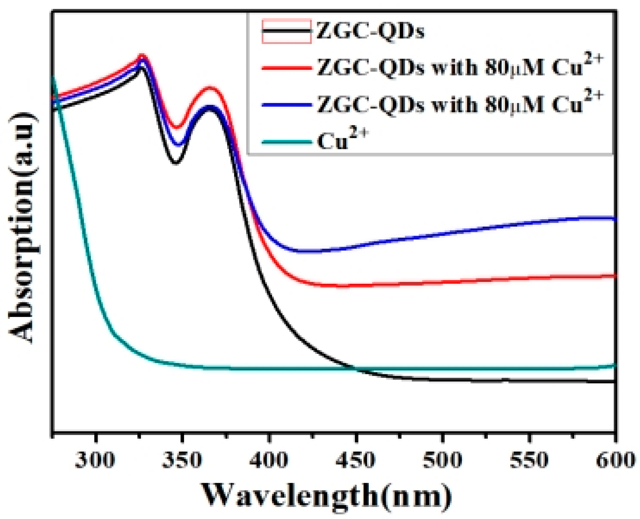
3.6. Analysis of Fluorescence Quenching Mechanism of ZCGQDs

Copper ions had a marked fluorescence quenching effect on the ZCGQDs complexes, and there was a strong linear relationship between the Cu2+ concentration and the relative fluorescence intensity of the ZCGQDs complexes.
Figure 6a,b shows the particle dispersion maps before and after the addition of Cu2+ to the ZCGQDs composite solution. A comparison was made using transmission electron microscopy and aggregation phenomenon was observed in the ZCGQDs composite, with a consequent weakening of the fluorescence intensity. Thus, the aggregation phenomenon in the ZCGQDs composite induced fluorescence quenching (AIQ) of the ZCGQDs complex, which was caused by Cu2+.
The addition of metal ions can lead to fluorescence quenching of quantum dots. There are two mechanisms for this phenomenon. One is dynamic quenching, that is, after the quenching agent is added, it can carry out frequent energy transfer between the fluorescent material and reduce the fluorescence lifetime of the material. The other is static quenching, in which the two are mixed together to form a corresponding non-fluorescent complex that shows exactly the specific absorption peak when exposed to UV light.
As shown in Figure 7, after the different concentrations of Cu2+ were added to the ZCGQDs solutions, their ultraviolet absorption spectra were plotted, and no obvious difference was observed. This indicates that the sol system formed a new non-fluorescent complex, and the reason for the fluorescence quenching of ZCGQDs complexes by Cu2+ was the dynamic quenching caused by energy transfer.

4. Conclusions

In this paper, the fluorescence performance of ZCGQDs was analyzed, and it was found that the addition of metal ions would cause the fluorescence intensity of ZCGQDs complexes to weaken, resulting in fluorescence quenching. The fluorescence quenching phenomenon produced by Cu2+ was the most obvious. Using this principle, the ZCGQDs complexes were used as fluorescent probes to detect Cu2+. The mixing time of 15 min makes the most obvious fluorescence burst of Cu2+ on ZCGQDs with good anti-interference performance. There was a linear relationship between ZCGQDs and Cu2+ in the range of 1–100 µM, with F0/F = 0.9687 + 0.12343C, so the detection limit of Cu2+ was obtained, which was about 1.74 µm. In addition, the characterization of the materials using transmission electron microscopy and UV spectroscopy proved that Cu2+ induced the fluorescence quenching of ZCGQDs complexes, which involved the induced aggregation quenching and dynamic quenching.

Author Contributions

T.L., L.W. and Y.L.: writing—original draft preparation, writing—review and editing, methodology, formal analysis; R.J., Y.T. and Y.H.: writing—original draft preparation, formal analysis, visualization, project administration; C.S. and S.L.: data analysis; Y.L.: resources. All authors have read and agreed to the published version of the manuscript.

Funding

This research was funded by [the Department of Scientific Research Project in Heilongjiang Province] grant number [LH2022B022], and the APC was funded by [Tianze Liu]. [Jiamusi University Horizontal Research Project] grant number [210428001; 021040301], and the APC was funded by [Shuang Liu]. [the Innovation and Entrepreneurship Project for College Students in Heilongjiang Province] grant number [S202210222018], and the APC was funded by [Tianze Liu].

Data Availability Statement

The data is unavailable due to privacy or ethical restrictions.

Conflicts of Interest

The authors declare no conflict of interest.

References

  1. Wei, J.R.; Chen, H.Y.; Zhang, W.; Pan, J.X.L.; Dang, F.Q.; Zhang, Z.Q.; Zhang, J. Ratiometric fluorescence for sensitive and selective detection of mitoxantrone using a MIP@rQDs@SiO2 fluorescence probe. Sens. Actuators B Chem. 2017, 244, 31–37. [Google Scholar] [CrossRef]
  2. Qian, J.; Wang, K.; Wang, C.; Ren, C.; Liu, Q.; Hao, N.; Wang, K. Ratiometric fluorescence nanosensor for selective and visual detection of cadmium ions using quencher displacement-induced fluorescence recovery of CdTe quantum dots-based hybrid probe. Sens. Actuators B Chem. 2017, 241, 1153–1160. [Google Scholar] [CrossRef]
  3. Gui, W.; Wang, H.; Liu, Y.; Ma, Q. Ratiometric fluorescent sensor with molecularly imprinted mesoporous microspheres for malachite green detection. Sens. Actuators B Chem. 2018, 266, 685–691. [Google Scholar] [CrossRef]
  4. Yadav, A.; Upadhyay, Y.; Bera, R.K.; Sahoo, S.K. Vitamin B6 cofactors guided highly selective fluorescent turn-on sensing of histamine using beta-cyclodextrin stabilized ZnO quantum dots. Food Chem. 2020, 320, 126611. [Google Scholar] [CrossRef]
  5. Wu, W.J.; Zhao, Q.; Zhou, R.; Linag, Y.C.; Zhao, W.B.; Shan, C.X. Ratiometric fluorescence sensor based on europium-grafted ZnO quantum dots for visual and colorimetric detection of tetracycline. Spectrochim. Acta Part A Mol. Biomol. Spectrosc. 2021, 259, 119901. [Google Scholar] [CrossRef]
  6. Omata, T.; Tani, Y.; Kobayashi, S.; Takahashi, K.; Miyanaga, A.; Maeda, Y.; Otsuka-Yao-Matsuo, S. Ultraviolet electroluminescence from colloidal ZnO quantum dots in an all-inorganic multilayer light-emitting device. Appl. Phys. Lett. 2012, 100, 061104. [Google Scholar] [CrossRef]
  7. Moyen, E.; Kim, J.H.; Kim, J.; Jinag, J. ZnO nanoparticles for quantum-dot-based light-emitting diodes. ACS Appl. Nano Mater. 2020, 3, 5203–5211. [Google Scholar] [CrossRef]
  8. Wang, J.; Lee, J.S.; Kim, D.; Zhu, L. Exploration of zinc oxide nanoparticles as a multitarget and multifunctional anticancer nanomedicine. ACS Appl. Mater. Interfaces 2017, 9, 39971–39984. [Google Scholar] [CrossRef]
  9. Tu, Z.; Yang, G.; Song, H.; Wang, C. Amorphous ZnO quantum dot/mesoporous carbon bubble composites for a high-performance lithium-ion battery anode. ACS Appl. Mater. Interfaces 2017, 9, 439–446. [Google Scholar] [CrossRef]
  10. Fernando, J.F.S.; Zhang, C.; Firestein, K.L.; Nerkar, J.Y.; Golberg, D.V. ZnO quantum dots anchored in multilayered and flexible amorphous carbon sheets for high performance and stable lithium ion batteries. J. Mater. Chem. A 2019, 7, 8460–8471. [Google Scholar] [CrossRef]
  11. Tan, Q.; Kong, X.; Guan, X.; Wang, C.; Xu, B. Crystallization of zinc oxide quantum dots on graphene sheets as an anode material for lithium ion batteries. Crystengcomm 2020, 22, 320–329. [Google Scholar] [CrossRef]
  12. Ma, X.; Li, Z.; Chen, D.; Li, Z.; Yan, L.; Li, S.; Liang, C.; Ling, M.; Peng, X. Nitrogen-doped porous carbon sponge-confined ZnO quantum dots for metal collector-free lithium ion battery. J. Electroanal. Chem. 2019, 848, 113275. [Google Scholar] [CrossRef]
  13. Jia, Z.; Misra, R.D.K. Tunable ZnO quantum dots for bioimaging: Synthesis and photoluminescence. Mater. Technol. 2013, 28, 221–227. [Google Scholar] [CrossRef]
  14. Wu, T.; Wang, Z.; Tian, M.; Miao, H.; Zhang, H.; Sun, J. UVexcitation NOz gas sensor sensitized by ZnO quantum dotsatroomtemperature. Sens. Actuators B Chem. 2018, 259, 526–531. [Google Scholar] [CrossRef]
  15. Zhao, D.; Song, H.; Hao, L.; Liu, X.; Zhang, L.; Lv, Y. Luminescent ZnO quantum dots for sensitive and selective detection of dopamine. Talanta 2013, 107, 133–139. [Google Scholar] [CrossRef]
  16. Jiříčková, A.; Jankovský, O.; Sofer, Z.; Sedmidubský, D. Synthesis and Applications of Graphene Oxide. Materials 2022, 15, 920. [Google Scholar] [CrossRef] [PubMed]
  17. Tiwari, S.; Purabgola, A.; Kandasubramanian, B. Functionalised graphene as flexible electrodes for polymer photovoltaics. J. Alloy. Compd. 2020, 825, 153954. [Google Scholar] [CrossRef]
  18. Jafari, E.A.; Moradi, M.; Hajati, S.; Kiani, M.A.; Espinos, J.P. Electrophoretic deposition of mixed copper oxide/GO as cathode and N-doped GO as anode for electrochemical energy storage. Electrochim. Acta 2018, 268, 392–402. [Google Scholar] [CrossRef]
  19. Sangeetha, M.; Madhan, D. Ultra sensitive molybdenum disulfide (MoS2)/graphene based hybrid sensor for the detection of NO2 and formaldehyde gases by fiber optic clad modified method. Opt. Laser Technol. 2020, 127, 106193. [Google Scholar] [CrossRef]
  20. Zeng, S.; Pan, Q.; Huang, Z.; Gu, C.; Wang, T.; Xu, J.; Yan, Z.; Zhao, F.; Li, P.; Tu, Y.; et al. Ultrafast response of self-powered humidity sensor of flexible graphene oxide film. Mater. Des. 2023, 226, 111683. [Google Scholar] [CrossRef]
  21. Zheng, J.; Li, J.; Zhang, L.; Chen, X.; Yu, Y.; Huang, H. Post-graphene 2D materials-based antimicrobial agents: Focus on fabrication strategies and biosafety assessments. J. Mater. Sci. 2020, 55, 7226–7246. [Google Scholar] [CrossRef]
  22. Govindaraju, S.; Samal, M.; Yun, K. Superior antibacterial activity of GlcN-AuNP-GO by ultraviolet irradiation. Mater. Sci. Eng. C 2016, 69, 366–372. [Google Scholar] [CrossRef]
  23. Gong, J.Q.; He, C.; Zhang, J.L.; Wang, L.Z. GO-P25@SA gel beads with excellent separation performance for photocatalytic degradation of rhodamine B. Res. Chem. Intermed. 2021, 47, 2331–2338. [Google Scholar] [CrossRef]
  24. Zhou, C.; He, X.H.; Hou, Y.H.; Zhang, L.; Shi, F.W.; Hu, J.L. Alginate/r-GO/SiO2 aerogels with double catalytic activity sites and high mechanical performance. Mater. Lett. 2021, 288, 129394. [Google Scholar] [CrossRef]
  25. Hosseini, M.G.; Shahryari, E.; Sefidi, P.Y. Polyaniline grafted chitosan/GO-CNT/Fe3O4 nanocomposite as a superior electrode material for supercapacitor application. J. Appl. Polymer Sci. 2021, 138, e50976. [Google Scholar] [CrossRef]
  26. Shen, L.; Zhao, W.J.; Wang, K.; Xu, J.G. GO-Ti3C2 two-dimensional heterojunction nanomaterial for anticorrosion enhancement of epoxy zinc-rich coatings. J. Hazard. Mater. 2021, 417, 126048. [Google Scholar] [CrossRef]
  27. Huo, X.; Yi, H.; Fu, Y.; An, Z.; Qin, L.; Liu, X.; Li, B.; Liu, S.; Li, L.; Zhang, M.; et al. Porous graphitic carbon nitride nanomaterials for water treatment. Environ. Sci. Nano 2021, 8, 1835–1862. [Google Scholar] [CrossRef]
  28. Shi, H.; Long, S.; Hou, J.; Ye, L.; Sun, Y.; Ni, W.; Song, C.; Li, K.; Gurzadyan, G.G.; Guo, X. Defects promote ultrafast charge separation in graphitic carbon nitride for enhanced visible-light-driven CO2 reduction activity. Chem. Eur. J. 2019, 25, 5028–5035. [Google Scholar] [CrossRef]
  29. Akple, M.S.; Ishigaki, T.; Madhusudan, P. Bio-inspired honeycomb-like graphitic carbon nitride for enhanced visible light photocatalytic CO2 reduction activity. Environ. Sci. Pollut. Res. 2020, 27, 22604–22618. [Google Scholar] [CrossRef] [PubMed]
  30. Sharma, R.; Almáši, M.; Nehra, S.P.; Rao, V.S.; Panchal, P.; Paul, D.R.; Jain, I.P.; Sharma, A. Photocatalytic hydrogen production using graphitic carbon nitride (GCN): A precise review. Renew. Sustain. Energy Rev. 2022, 168, 112776. [Google Scholar] [CrossRef]
  31. Li, L.; Yu, H.; Xu, J.; Zhao, S.; Liu, Z.; Li, Y. Rare earth element, Sm, modified graphite phase carbon nitride heterostructure for photocatalytic hydrogen production. New J. Chem. 2019, 43, 1716–1724. [Google Scholar] [CrossRef]
  32. Wang, Y.; Liu, L.; Ma, T.; Zhang, Y.; Huang, H. 2D graphitic carbon nitride for energy conversion and storage. Adv. Funct. Mater. 2021, 31, 2102540. [Google Scholar] [CrossRef]
  33. Niu, W.; Yang, Y. Graphitic carbon nitride for electrochemical energy conversion and storage. ACS Energy Lett. 2018, 3, 2796–2815. [Google Scholar] [CrossRef]
  34. Yang, Y.; Zou, T.; Zhao, R.; Kong, Y.; Su, L.; Ma, D.; Xiao, X.; Wang, Y. Fluorescence ‘turn-on’ probe for Al3+ detection in water based on ZnS/ZnO quantum dots with excellent selectivity and stability. Nanotechnology 2021, 31, 2102540. [Google Scholar] [CrossRef]
  35. Zhan, M.; Jia, H.; Fan, J.; Yu, H.; Amador, E.; Chen, W. Two D-pi-A schiff-base-functionalized silica gel adsorbents for preconcentration of copper ions in foods and water for detection. Anal. Chem. 2019, 91, 6103–6110. [Google Scholar] [CrossRef] [PubMed]
  36. Karakus, E. A rhodamine based fluorescent chemodosimeter for the selective and sensitive detection of copper(II)ions in aqueous media and living cells. J. Mol. Struct. 2021, 1224. [Google Scholar] [CrossRef]
  37. Falina, S.; Syamsul, M.; Rhaffor, N.A.; Hamid, S.S.; Zain, K.A.M.; Manaf, A.A.; Kawarada, H. Ten years progress of electrical detection of heavy metal ions(HMIs)using various field-effect transistor (FET) nanosensors: A review. Biosensors 2021, 11, 478. [Google Scholar] [CrossRef]
  38. Guo, W.; Zhang, C.; Ma, T.; Liu, X.; Chen, Z.; Li, S.; Deng, Y. Advances in aptamer screening and aptasensors’detection of heavymetal ions. J. Nanobiotechnol. 2021, 19, 166. [Google Scholar] [CrossRef]
  39. Chen, S.-H.; Li, Y.-X.; Li, P.-H.; Xiao, X.-Y.; Jiang, M.; Li, S.-S.; Zhou, W.-Y.; Yang, M.; Huang, X.-J.; Liu, W.-Q. Electrochemical spectral methods for trace detection of heavy metals: A review. TrAC Trends Anal. Chem. 2018, 106, 139–150. [Google Scholar] [CrossRef]
  40. Zhu, X.; Chen, L.; Lin, Z.; Qiu, B.; Chen, G. A highly sensitive and selective “signal-on”electrochemiluminescent biosensor for mercury. Chem. Commun. 2010, 46, 3149–3151. [Google Scholar] [CrossRef] [PubMed]
  41. Zhu, Y.; Jiang, X.; Wang, H.; Wang, S.; Wang, H.; Sun, B.; Su, Y.; He, Y. A poly adenine-mediated assembly strategy for designing surface-enhanced resonance raman scattering substrates in controllable manners. Anal. Chem. 2015, 87, 6631–6638. [Google Scholar] [CrossRef]
  42. Roy, A.; NIandi, M.; Roy, P. Dual chemosensors for metal ions: A comprehensive review. TrAC Trends Anal. Chem. 2021, 138, 116204. [Google Scholar] [CrossRef]
  43. Peng, J.; Li, J.; Xu, W.; Wang, L.; Su, D.; Teoh, C.L.; Chang, Y.-T. Silica nanoparticle-enhanced fluorescent sensor array for heavy metal ions detection in colloid solution. Anal. Chem. 2018, 90, 1628–1634. [Google Scholar] [CrossRef]
  44. Yang, X.; Li, Z.; Liu, N.; Song, W.; Sun, Q.; Xie, Y. Fabrication of hydrophilic luminescent zinc oxide quantum dots for selective detection of copper ions and efficient inhibition of harmful fungi. Arab. J. Chem. 2022, 15, 104266. [Google Scholar] [CrossRef]
  45. Ng, S.M.; Chin, S.F. Interface study on zinc oxide quantum dots using fluorometric and regression analysis in view of optical sensing. Anal. Lett. 2013, 46, 1278–1288. [Google Scholar] [CrossRef]
  46. Moussa, H.; Merlin, C.; Dezanet, C.; Balan, L.; Medjahdi, G.; Ben-Attia, M.; Schneider, R. Trace amounts of Cu2+ ions influence ROS production and cytotoxicity of ZnO quantum dots. J. Hazard. Mater. 2016, 304, 532–542. [Google Scholar] [CrossRef]
  47. Zou, T.; Xing, X.; Yang, Y.; Wang, Z.; Wang, Z.; Zhao, R.; Zhang, X.; Wang, Y. Water-soluble ZnO quantum dots modified by (3-aminopropyl) triethoxysilane: The promising fluorescent probe for the selective detection of Cu2+ ion in drinking water. J. Alloys Compd. 2020, 825, 153904. [Google Scholar] [CrossRef]
  48. Khan, M.M.R.; Mitra, T.; Sahoo, D. Metal oxide QD based ultrasensitive microsphere fluorescent sensor for copper, chromium and iron ions in water. RSC Adv. 2020, 10, 9512–9524. [Google Scholar] [CrossRef] [Green Version]
  49. Ng, S.M.; Wong, D.S.N.; Phung, J.H.C.; Chua, H.S. Integrated miniature fluorescent probe to leverage the sensing potential of ZnO quantum dots for the detection of copper (II) ions. Talanta 2013, 116, 514–519. [Google Scholar] [CrossRef]
Figure 1. (a) Fluorescence emission spectra of ZCGQDs; (b) relative fluorescence intensity with the addition of 20 µM Cu2+ to ZCGQDs.
Micromachines 14 00711 g001
Figure 2. The plot of relative fluorescence intensity versus time.
Micromachines 14 00711 g002
Figure 3. (a) Fluorescence spectra of the different heavy metal ions added; (b) relative fluorescence intensity of the different heavy metal ions added.
Micromachines 14 00711 g003
Figure 4. Relative fluorescence intensity of ZCGQDs in the coexistence of Cu2+ with other metal ions.
Micromachines 14 00711 g004
Figure 5. (a) Fluorescence spectra of different concentrations of Cu2+; (b) fitted curve of the linear relationship.
Micromachines 14 00711 g005
Figure 6. (a) HRTEM image of ZCGQDs; (b) HRTEM image of ZCGQDs after Cu2+ was added.
Micromachines 14 00711 g006
Figure 7. UV–vis absorption spectra of mixed colloids.
Micromachines 14 00711 g007
Disclaimer/Publisher’s Note: The statements, opinions and data contained in all publications are solely those of the individual author(s) and contributor(s) and not of MDPI and/or the editor(s). MDPI and/or the editor(s) disclaim responsibility for any injury to people or property resulting from any ideas, methods, instructions or products referred to in the content.

