
MEMS Enabled Miniature Two-Photon Microscopy for Biomedical Imaging

Abstract
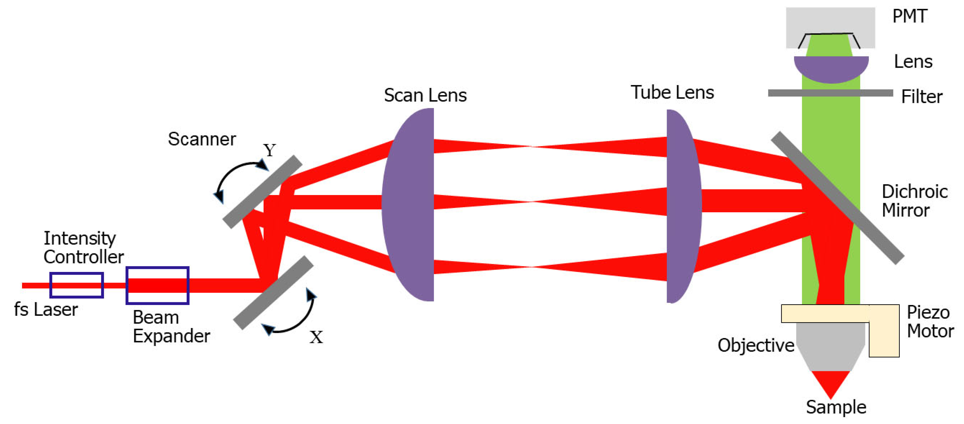
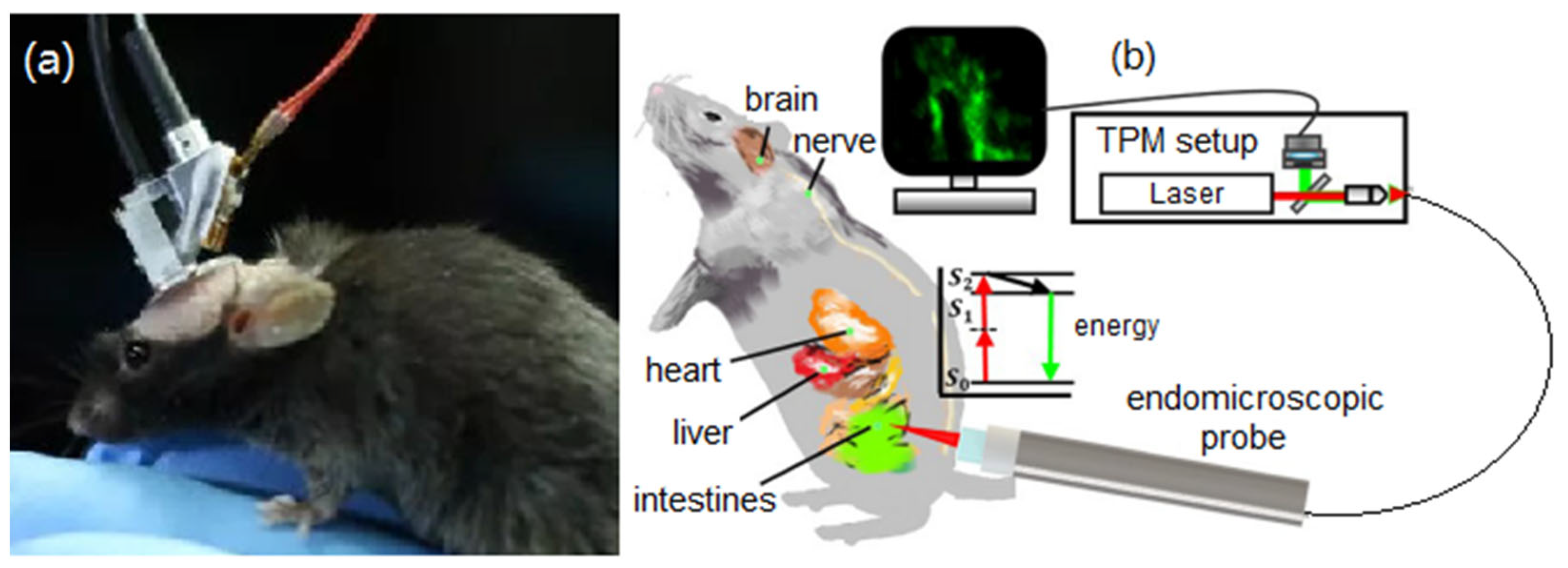
:1. Introduction
2. Miniature Two-Photon Microscopy
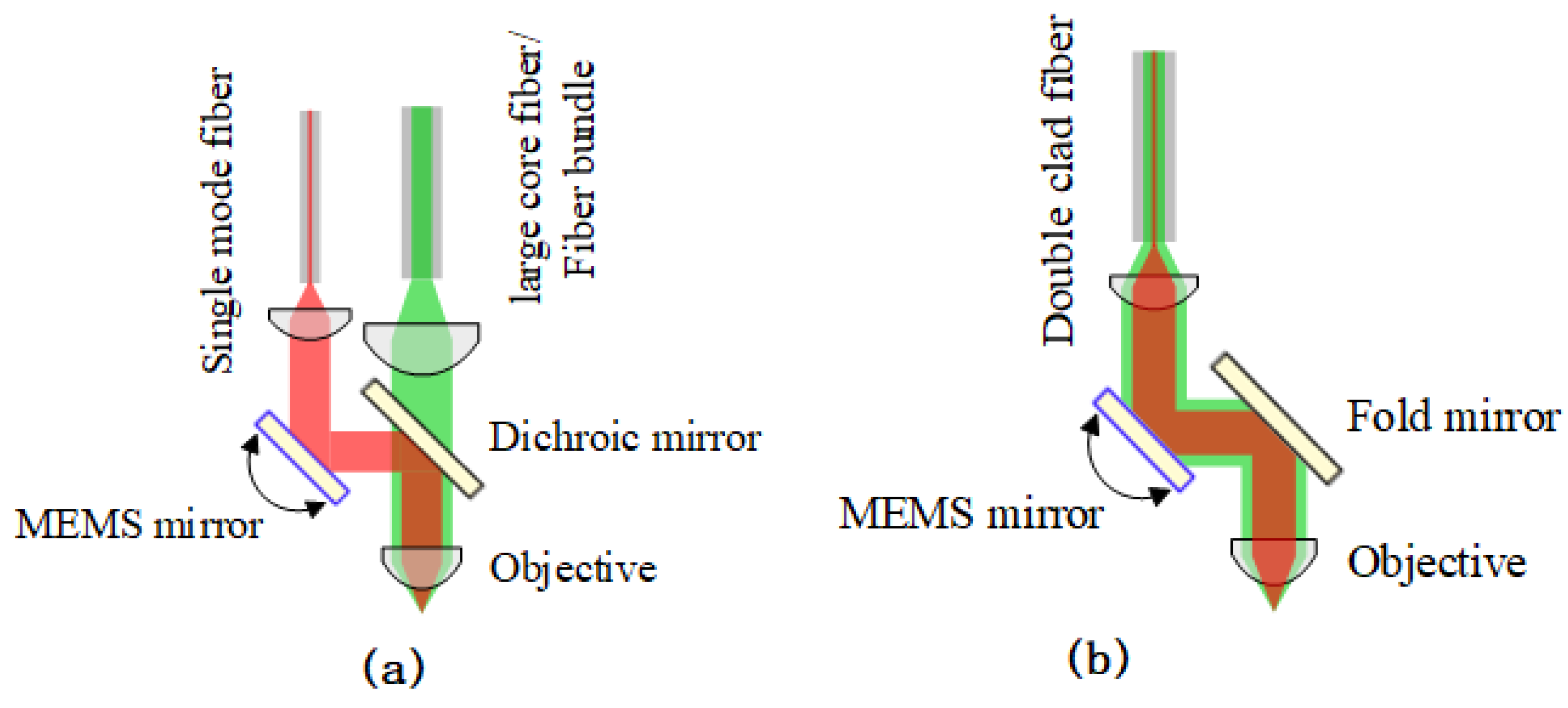
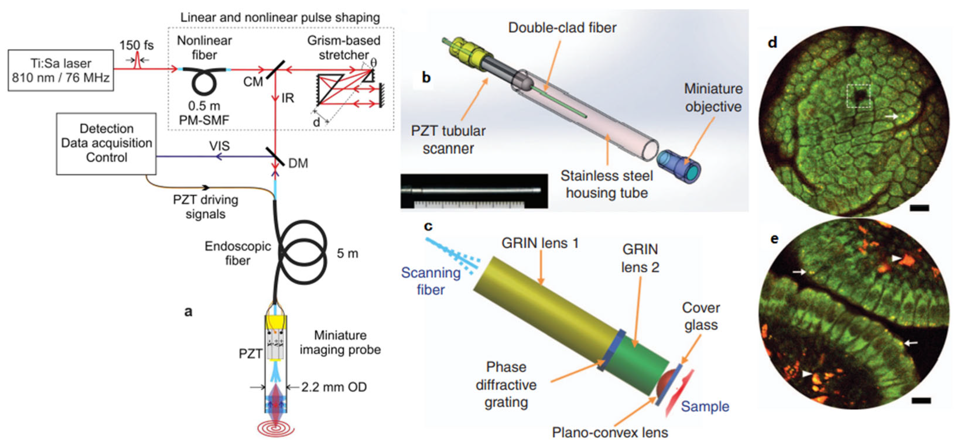
2.1. Fiber Optics

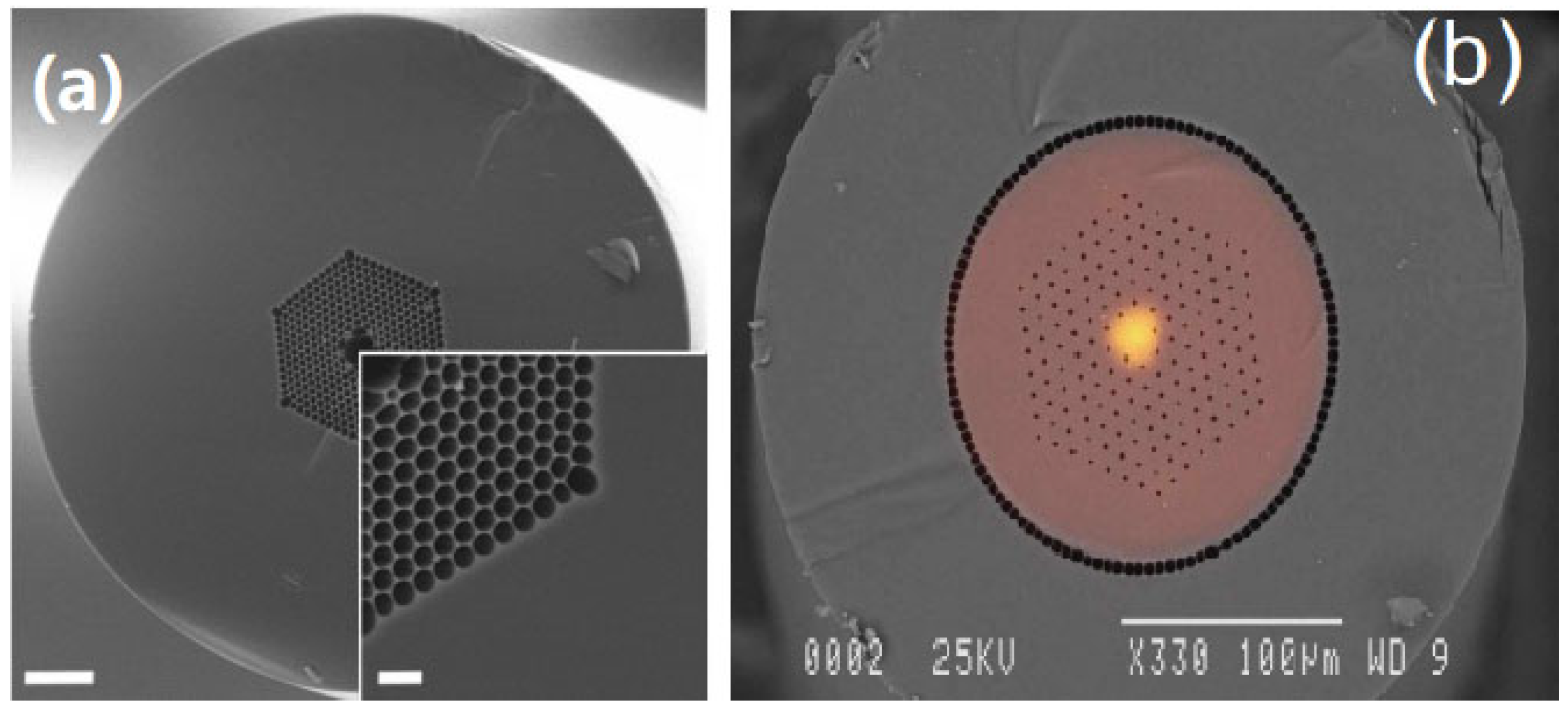
2.2. Objective Lenses
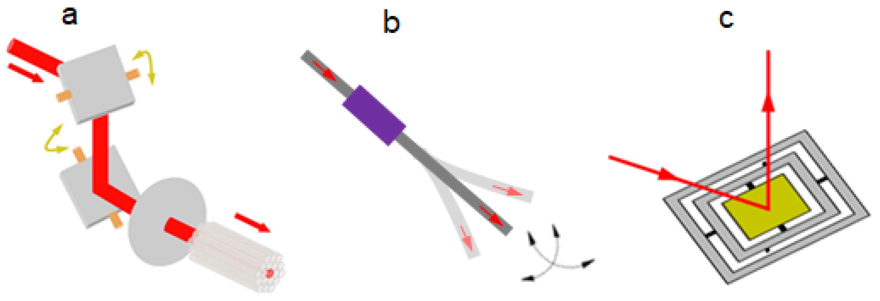
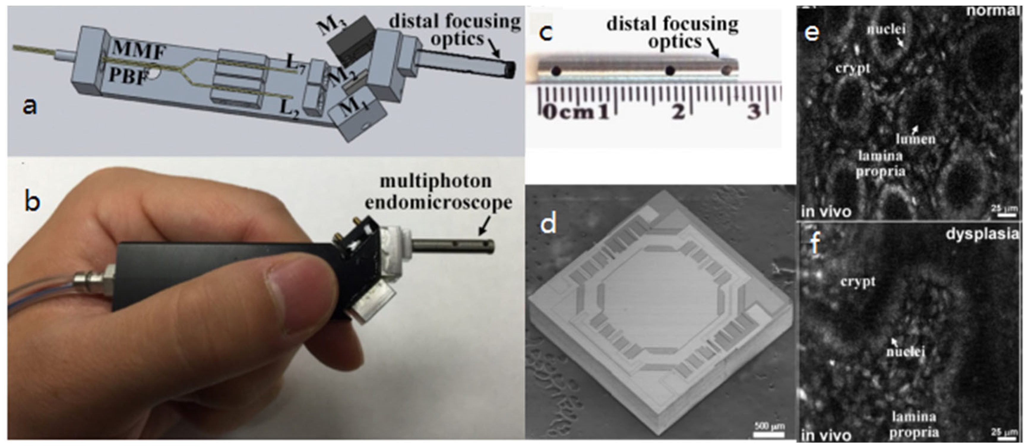
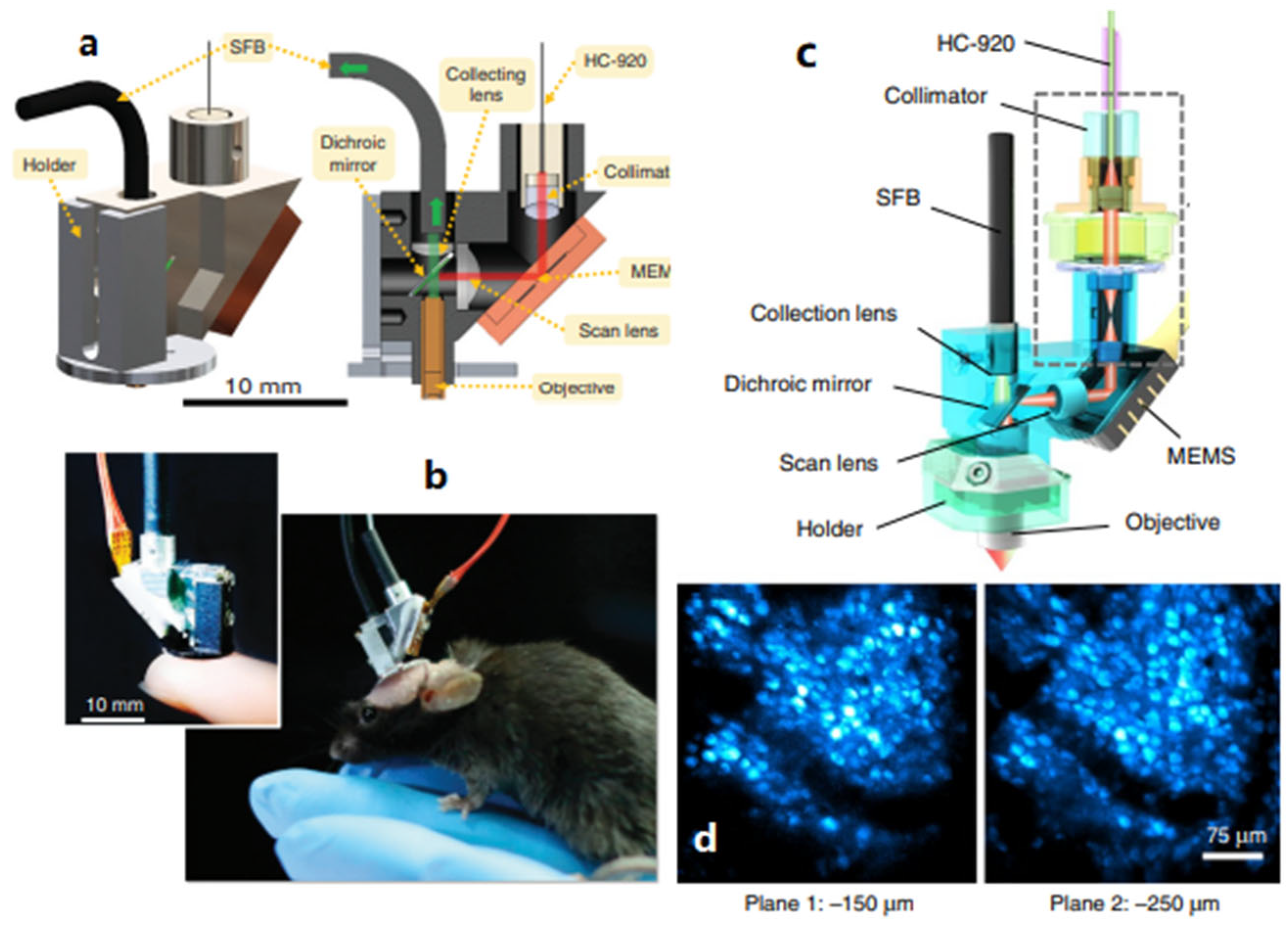
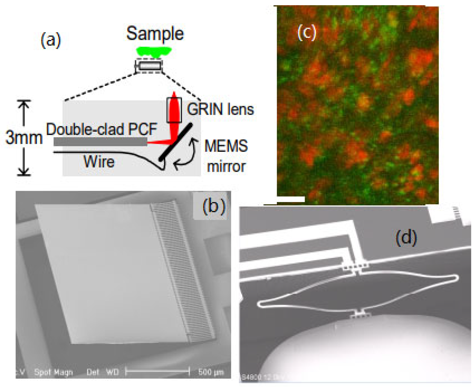
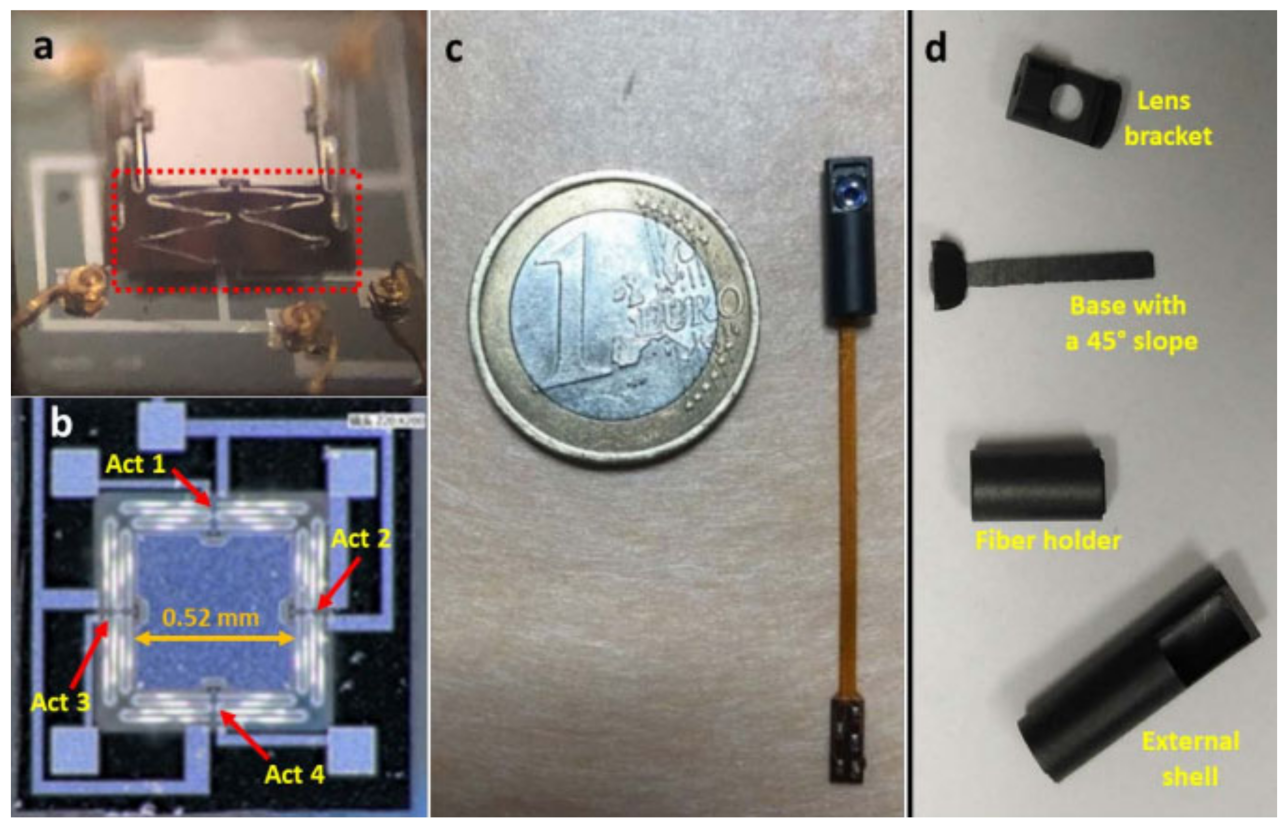
2.3. Transverse Scan Mechanisms
3. MEMS-Based TPM
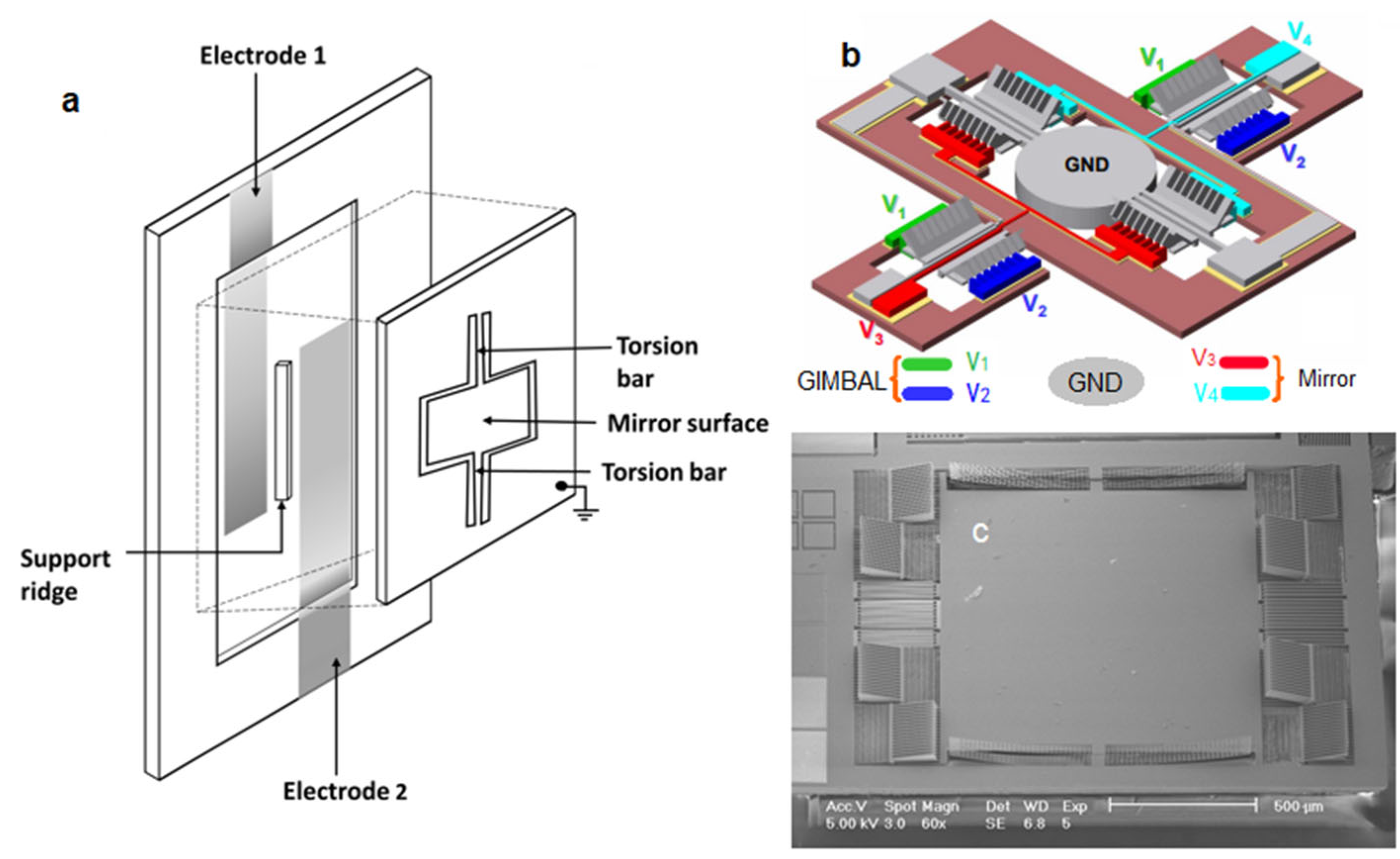
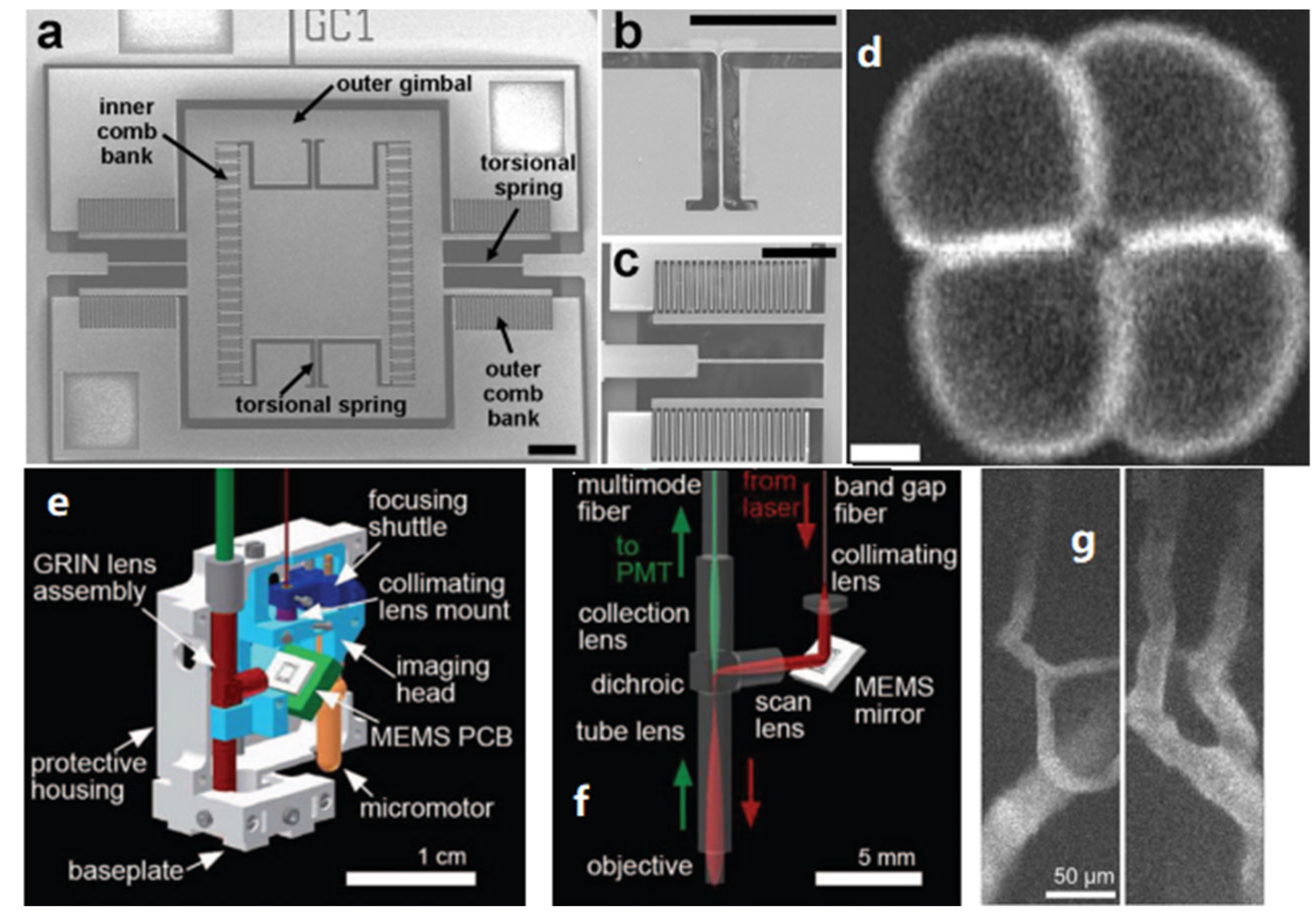
3.1. Electrostatic MEMS-Based TPM
3.2. Electromagnetic MEMS-Based TPM
3.3. Electrothermal MEMS-Based TPM
4. Z-Scan TPM
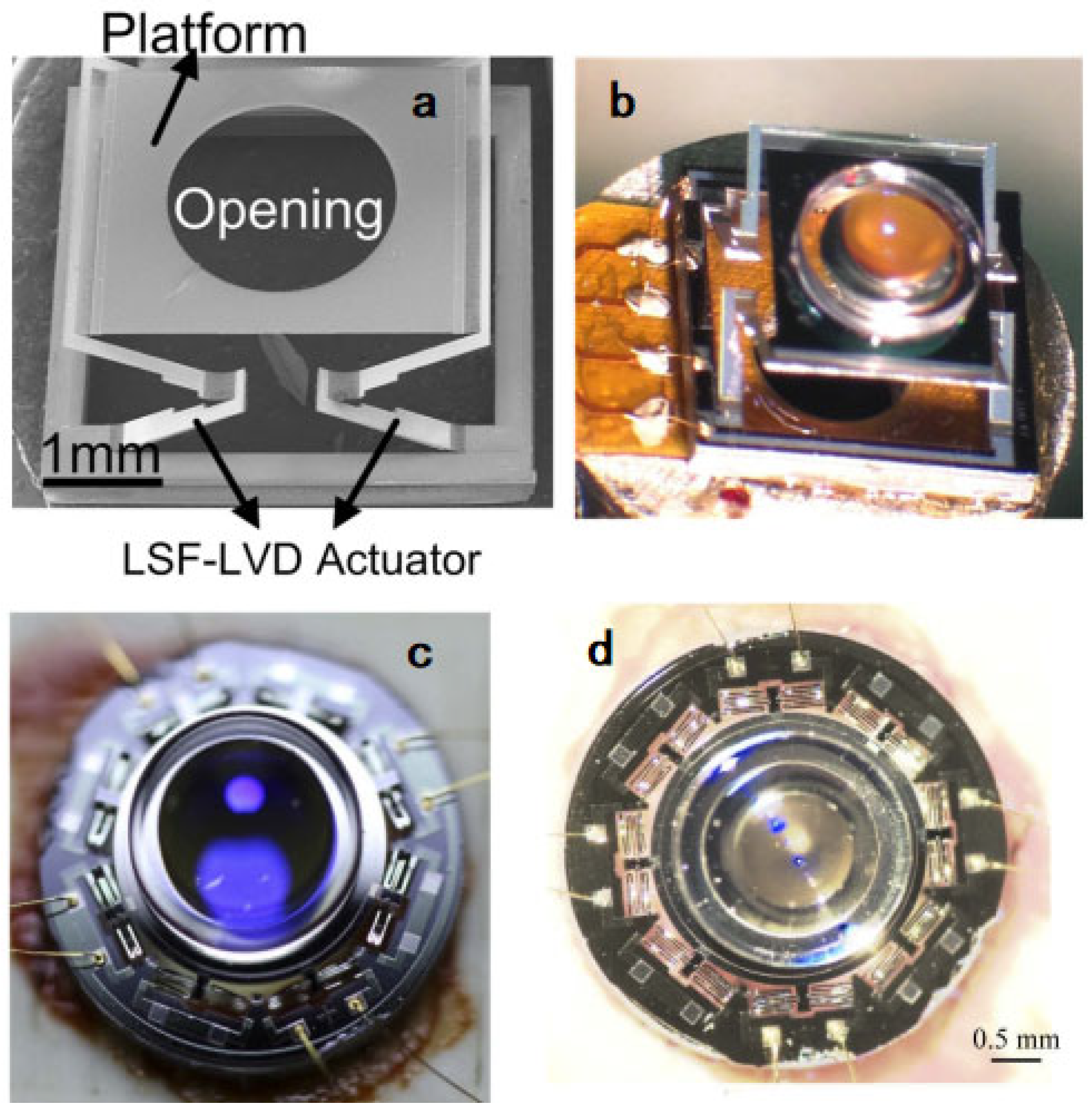
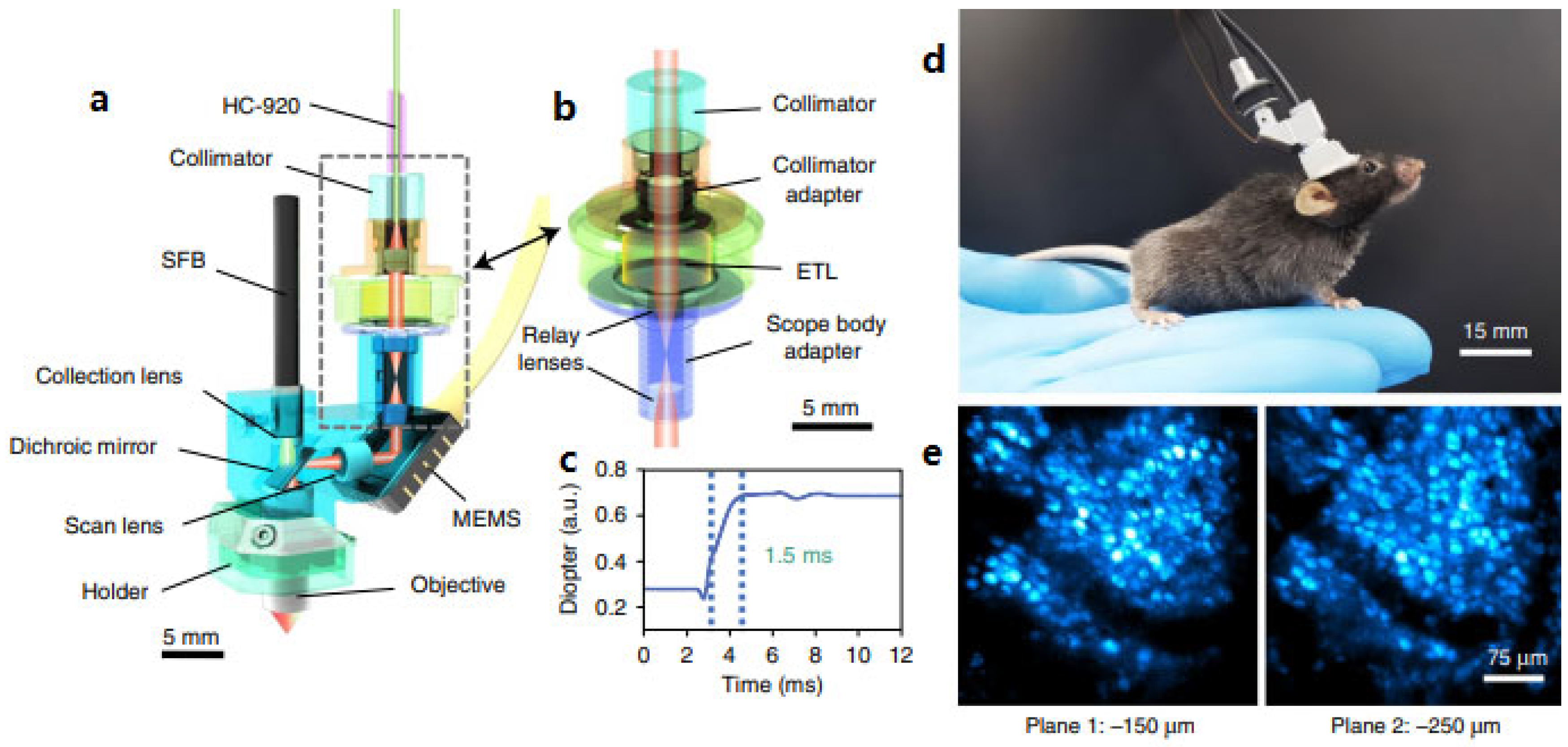
4.1. MEMS Actuated Tunable Microlens Scanner
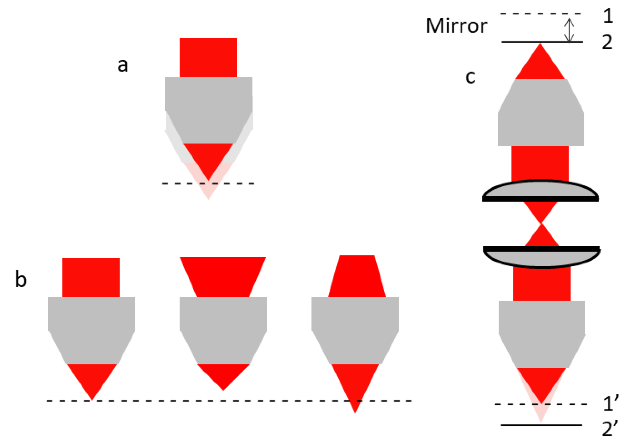
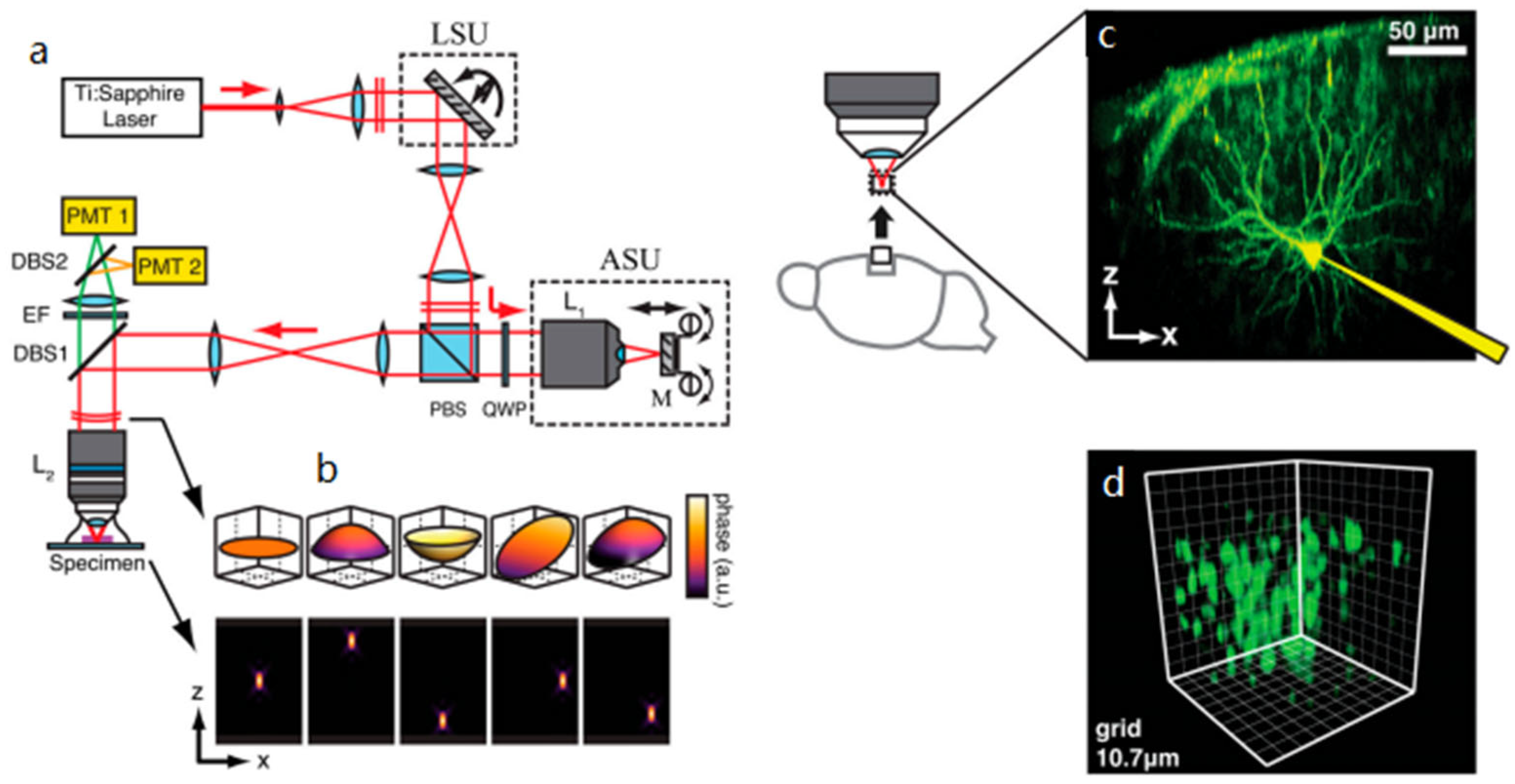
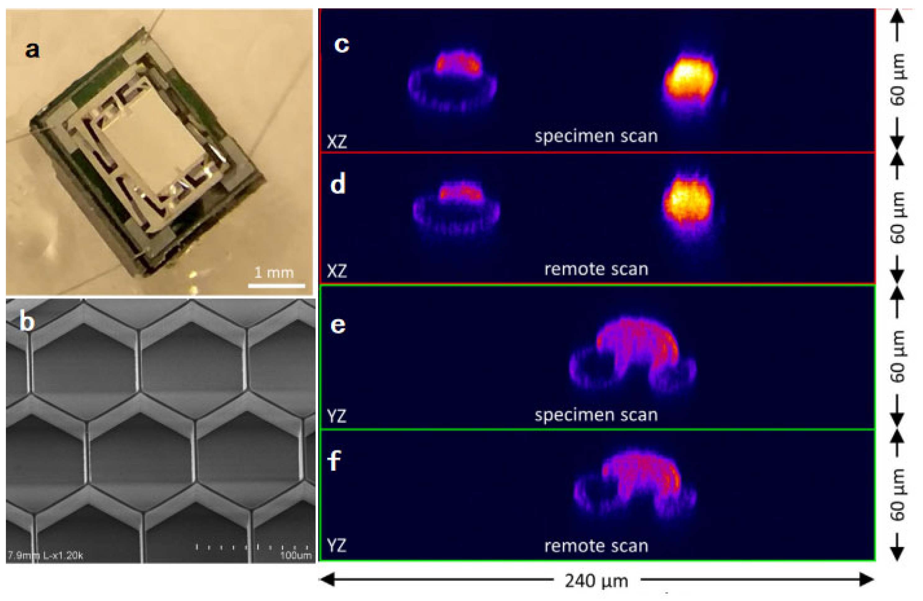
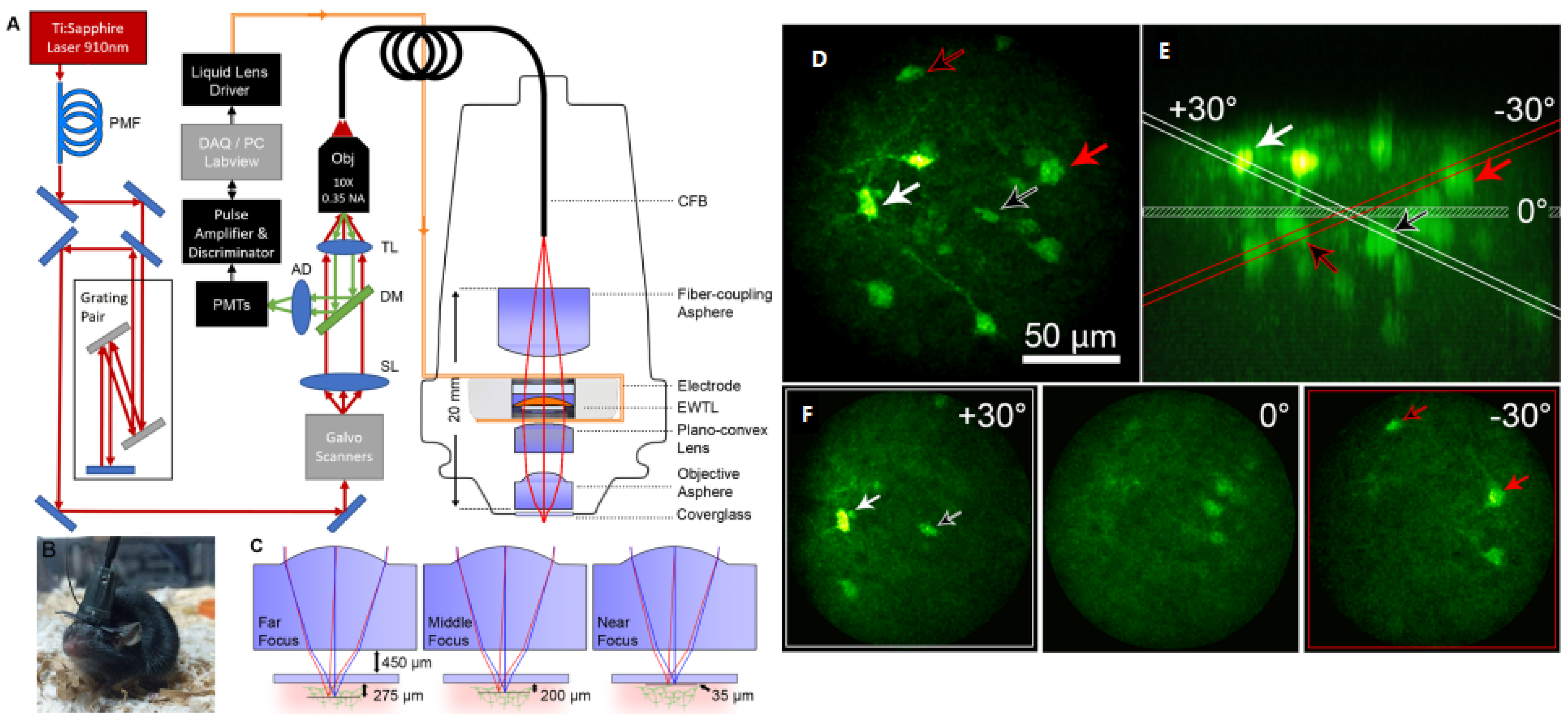
4.2. Remote Focusing
4.3. Liquid Lens
4.4. Deformable MEMS
5. Discussion and Summary
Author Contributions
Funding
Informed Consent Statement
Data Availability Statement
Conflicts of Interest
References
- Denk, W.; Strickler, J.H.; Webb, W.W. Two-photon laser scanning fluorescence microscopy. Science 1990, 248, 73–76. [Google Scholar] [CrossRef] [PubMed] [Green Version]
- Maria, G.M. Über elementarakte mit zwei quantensprüngen. Ann. Phys. 1931, 401, 273–294. [Google Scholar]
- Zipfel, W.R.; Williams, R.M.; Webb, W.W. Nonlinear magic: Multiphoton microscopy in the biosciences. Nat. Biotechnol. 2003, 21, 1369–1377. [Google Scholar] [CrossRef] [PubMed]
- Lefort, C. A review of biomedical multiphoton microscopy and its laser sources. J. Phys. D Appl. Phys. 2017, 50, 423001. [Google Scholar] [CrossRef] [Green Version]
- Tsai, P.S.; Kleinfeld, D. In vivo two-photon laser scanning microscopy with concurrent plasma-mediated ablation principles and hardware realization. In Vivo Optical Imaging of Brain Function, 2nd ed.; CRC Press/Taylor & Francis: Boca Raton, FL, USA, 2009; pp. 64–66. [Google Scholar]
- Rosenegger, D.G.; Tran, C.H.; LeDue, J.; Zhou, N.; Gordon, G.R. A high performance, cost-effective, open-source microscope for scanning two-photon microscopy that is modular and readily adaptable. PLoS ONE 2014, 9, e110475. [Google Scholar] [CrossRef] [Green Version]
- Young, M.D.; Field, J.J.; Sheetz, K.E.; Bartels, R.A.; Squier, J. A pragmatic guide to multiphoton microscope design. Adv. Opt. Photonics 2015, 7, 276–378. [Google Scholar] [CrossRef] [Green Version]
- Kim, D.Y.; Hwang, K.; Ahn, J.; Seo, Y.H.; Kim, J.B.; Lee, S.; Yoon, J.H.; Kong, E.; Jeong, Y.; Jon, S.; et al. Lissajous scanning two-photon endomicroscope for in vivo tissue imaging. Sci. Rep. 2019, 9, 3560–3567. [Google Scholar] [CrossRef] [Green Version]
- Lyons, M.R.; West, A.E. Mechanisms of specificity in neuronal activity-regulated gene transcription. Prog. Neurobiol. 2011, 94, 259–295. [Google Scholar] [CrossRef] [Green Version]
- Lin, M.Z.; Schnitzer, M.J. Genetically encoded indicators of neuronal activity. Nat. Neurosci. 2016, 19, 1142. [Google Scholar] [CrossRef] [Green Version]
- Grienberger, C.; Konnerth, A. Imaging calcium in neurons. Neuron 2012, 73, 862–885. [Google Scholar] [CrossRef] [Green Version]
- Svoboda, K.; Yasuda, R. Principles of two-photon excitation microscopy and its applications to neuroscience. Neuron 2006, 50, 823–839. [Google Scholar] [CrossRef] [Green Version]
- Mank, M.; Santos, A.F.; Direnberger, S.; Flogel, T.D.; Hofer, S.B.; Stein, V.; Hendel, T.; Reiff, D.F.; Levelt, C.; Borst, A.; et al. A genetically encoded calcium indicator for chronic in vivo two-photon imaging. Nat. Methods 2008, 5, 805. [Google Scholar] [CrossRef]
- Kobat, D.; Horton, N.G.; Xu, C. In vivo two-photon microscopy to 1.6-mm depth in mouse cortex. J. Biomed. Opt. 2011, 16, 106014. [Google Scholar] [CrossRef] [Green Version]
- Dombeck, D.A.; Khabbaz, A.N.; Collman, F.; Adelman, T.L.; Tank, D.W. Imaging large-scale neural activity with cellular resolution in awake, mobile mice. Neuron 2007, 56, 43–57. [Google Scholar] [CrossRef] [Green Version]
- Helmchen, F. Two-Photon Functional Imaging of Neuronal Activity; CRC Press: Boca Raton, FL, USA, 2009. [Google Scholar]
- Svoboda, K.; Tank, D.W.; Denk, W. Direct measurement of coupling between dendritic spines and shafts. Science 1996, 272, 716–719. [Google Scholar] [CrossRef]
- Ma, Z.G.; Du, X.L.; Wang, F.F.; Ding, R.; Li, Y.Y.; Liu, A.L.; Wei, L.P.; Hou, S.W.; Chen, F.; Hu, Q.; et al. Cortical plasticity induced by anodal transcranial pulsed current stimulation investigated by combining two-photon imaging and electrophysiological recording. Front. Cell. Neurosci. 2019, 13, 00400. [Google Scholar] [CrossRef]
- Kelly, P.; Hudry, E.; Hou, S.S.; Bacskai, B.J. In vivo two photon imaging of astrocytic structure and function in Alzheimer’s disease. Front. Aging Neurosci. 2018, 10, 219. [Google Scholar] [CrossRef]
- Ladewig, T.; Kloppenburg, P.; Lalley, P.M.; Zipfel, W.R.; Webb, W.W.; Keller, B.U. Spatial profiles of store-dependent calcium release in motoneurones of the nucleus hypoglossus from newborn mouse. J. Physiol. 2003, 547, 775–787. [Google Scholar] [CrossRef]
- Chen, S.C.; Choi, H.J.; So, P.T.C.; Culpepper, M.L. Thermomechanical actuator-based three-axis optical scanner for high-speed two-photon endomicroscope imaging. J. Microelectromech. Syst. 2014, 23, 570–578. [Google Scholar] [CrossRef] [Green Version]
- Helmchen, F.; Fee, S.D.; Tank, D.W.; Denk, W. A miniature head-mounted two-photon microscope: High-resolution brain imaging in freely moving animals. Neuron 2001, 31, 903–912. [Google Scholar] [CrossRef] [Green Version]
- Piyawattanametha, W.; Cocker, E.D.; Burns, L.D.; Barretto, R.P.; Jung, J.C.; Ra, H.; Solgaard, O.; Schnitzer, M.J. In vivo brain imaging using a portable 2.9 g two-photon microscope based on a microelectromechanical systems scanning mirror. Opt. Lett. 2009, 34, 2309–2311. [Google Scholar] [CrossRef] [PubMed] [Green Version]
- Tang, S.; Jung, W.; Mccormic, D.; Xie, T.; Su, J.; Ahn, Y.C.; Tromberg, B.J.; Chen, Z. Design and implementation of fiber-based multiphoton endoscopy with microelectromechanical systems scanning. J. Biomed. Opt. 2009, 14, 034005. [Google Scholar] [CrossRef] [PubMed] [Green Version]
- Duan, X.; Li, H.; Qiu, Z.; Joshi, B.P.; Pant, A.; Smith, A.; Kurabayashi, K.; Oldham, K.R.; Wang, T.D. MEMS-based multiphoton endomicroscope for repetitive imaging of mouse colon. Biomed. Opt. Express 2015, 6, 3074–3083. [Google Scholar] [CrossRef] [PubMed] [Green Version]
- Hoy, C.L.; Durr, N.J.; Chen, P.; Piyawattanametha, W.; Yakar, A.B. Miniaturized probe for femtosecond laser microsurgery and two-photon imaging. Opt. Express 2008, 16, 9996–10005. [Google Scholar] [CrossRef] [PubMed] [Green Version]
- Zong, W.J.; Wu, R.L.; Li, M.L.; Hu, Y.H.; Li, Y.J.; Li, J.H.; Rong, H.; Wu, H.T.; Xu, Y.Y.; Lu, Y.; et al. Fast high-resolution miniature two-photon microscopy for brain imaging in freely behaving mice. Nat. Methods 2017, 14, 713–719. [Google Scholar] [CrossRef]
- Fu, L.; Jain, A.; Xie, H.K.; Cranfield, C.; Gu, M. Nonlinear optical endoscopy based on a double-clad photonic crystal fiber and a MEMS mirror. Opt. Express 2006, 14, 1027–1032. [Google Scholar] [CrossRef]
- Myaing, M.T.; Ye, J.Y.; Norris, T.B.; Baker, J.R.; Wadsworth, W.J.; Bouwmans, G.; Knight, J.C.; Russell, P.J. Enhanced two-photon biosensing with double-clad photonic crystal fibers. Opt. Lett. 2003, 28, 1224–1226. [Google Scholar] [CrossRef]
- Fu, L.; Gan, X.; Min, G. Nonlinear optical microscopybased on double-clad photonic crystal fibers. Opt. Express 2005, 13, 5528–5534. [Google Scholar] [CrossRef]
- Jung, J.C.; Mehta, A.D.; Aksay, E.; Stepnoski, R.; Schnitzer, M.J. In vivo mammalian brain imaging using one-and two-photon fluorescence microendoscopy. J. Neurophysiol. 2004, 5, 3121–3133. [Google Scholar] [CrossRef]
- Liang, W.; Hall, G.; Messerschmidt, B.; Li, M.J.; Li, X. Nonlinear optical endomicroscopy for label-free functional histology in vivo. Light Sci. Appl. 2017, 6, e17082. [Google Scholar] [CrossRef] [Green Version]
- Jung, W.Y.; Tang, S.; Mccormic, D.T.; Xie, T.; Chen, Z. Miniaturized probe based on a microelectromechanical system mirror for multiphoton microscopy. Opt. Lett. 2008, 33, 1324–1326. [Google Scholar] [CrossRef] [Green Version]
- Helmchen, F.; Denk, W. Deep tissue two-photon microscopy. Nat. Methods 2005, 2, 932–940. [Google Scholar] [CrossRef]
- Zong, W.J.; Wu, R.L.; Chen, S.Y.; Wu, J.J.; Wang, H.B.; Zhao, Z.; Chen, G.Q.; Tu, R.; Wu, D.L.; Hu, Y.H.; et al. Miniature two-photon microscopy for enlarged field-of-view, multi-plane and long-term brain imaging. Nat. Methods 2021, 18, 46–49. [Google Scholar] [CrossRef]
- Flusberg, B.A.; Cocker, E.D.; Piyawattanametha, W.; Jung, J.C.; Cheung, E.L.; Schnitzer, M.J. Fiber-optic fluorescence imaging. Nat. Methods 2005, 2, 941. [Google Scholar] [CrossRef]
- Engelbrecht, C.J.; Johnston, R.S.; Seibel, E.J.; Helmchen, F. Ultra-compact fiber-optic two-photon microscope for functional fluorescence imaging in vivo. Opt. Express 2008, 16, 5556–5564. [Google Scholar] [CrossRef] [Green Version]
- Chung, H.H.; Kuo, W.C.; Cheng, Y.H.; Yu, C.H.; Chia, S.H.; Lin, C.Y.; Chen, J.S.; Tsai, H.J.; Fedotov, A.B.; Ivanov, A.; et al. Blu-ray disk lens as the objective of a miniaturized two-photon fluorescence microscope. Opt. Express 2013, 21, 31604–31614. [Google Scholar] [CrossRef] [Green Version]
- Wu, Y.C.; Xi, J.F.; Cobb, J.M.; Li, X.D. Scanning fiber-optic nonlinear endomicroscopy with miniature aspherical compound lens and multimode fiber collector. Opt. Lett. 2009, 34, 953–955. [Google Scholar] [CrossRef] [Green Version]
- Lee, C.M.; Engelbrecht, C.J.; Soper, T.D.; Helmchen, F.; Seibel, E.J. Scanning fiber endoscopy with highly flexible, 1 mm catheterscopes for wide-field, full-color imaging. J. Biophotonics 2010, 3, 385–407. [Google Scholar] [CrossRef] [Green Version]
- Meinert, T.; Weber, N.; Zappe, H.; Seifert, A. Varifocal MOEMS fiber scanner for confocal endomicroscopy. Opt. Express 2014, 22, 31529–31544. [Google Scholar] [CrossRef]
- Do, D.; Yoo, H.; Gweon, D.G. Fiber-optic raster scanning two-photon endomicroscope using a tubular piezoelectric actuator. J. Biomed. Opt. 2014, 19, 066010. [Google Scholar] [CrossRef] [Green Version]
- Bao, H.C.; Allen, J.; Pattie, R.; Vance, R.; Gu, M. Fast handheld two-photon fluorescence microendoscope with a 475 µm × 475 µm field of view for in vivo imaging. Opt. Lett. 2008, 33, 1333–1335. [Google Scholar] [CrossRef]
- Ducourthial, G.; Leclerc, P.; Mansuryan, T.; Fabert, M.; Brevier, J.; Habert, R.; Braud, F.; Batrin, R.; Vever-Bizet, C.; Bourg-Heckly, G.; et al. Development of a real-time flexible multiphoton microendoscope for label-free imaging in a live animal. Sci. Rep. 2015, 5, 18303–18311. [Google Scholar] [CrossRef] [PubMed] [Green Version]
- Urey, H.; Holmstrm, S.T.; Baran, U. MEMS laser scanners: A review. J. Microelectromech. Syst. 2014, 23, 259–275. [Google Scholar]
- Aksyuk, V.A.; Pardo, F.; Carr, D.; Greywall, D.; Chan, H.B.; Simon, M.E.; Gasparyan, A.; Shea, H. Beam-steering micromirrors for large optical cross-connects. J. Lightwave Technol. 2003, 21, 634. [Google Scholar] [CrossRef]
- Zhang, X.Y.; Koppal, S.J.; Zhang, R.; Zhou, L.; Butler, E.; Xie, H.K. Wide-angle structured light with a scanning MEMS mirror in liquid. Opt. Express 2016, 24, 3479–3487. [Google Scholar] [CrossRef]
- Kasturi, A.; Milanovic, V.; Atwood, B.H.; Yang, J. UAV-borne lidar with MEMS mirror-based scanning capability. In Laser Radar Technology and Applications XXI; International Society for Optics and Photonics: St. Bellingham, WA, USA, 2016; p. 9832. [Google Scholar]
- Pengwang, E.; Rabenorosoa, K.; Rakotondrabe, M.; Andreff, N. Scanning micromirror platform based on MEMS technology for medical application. Micromachines 2016, 7, 24. [Google Scholar] [CrossRef]
- Ra, H.; Piyawattanametha, W.; Taguchi, Y.; Lee, D.; Mandella, M.J.; Solgaard, O. Two-dimensional MEMS scanner for dual-axes confocal microscopy. J. Microelectromech. Syst. 2007, 16, 969–976. [Google Scholar] [CrossRef]
- Fu, L.; Jain, A.; Xie, H.K.; Cranfield, C.; Gu, M. Integration of a Double-clad Photonic Crystal Fiber, a GRIN Lens and a MEMS Mirror for Nonlinear Optical Endoscopy. In Biomedical Topical Meeting; Optica Publishing Group: Fort Lauderdale, FL, USA, 2006. [Google Scholar]
- Petersen, K.E. Silicon torsional scanning mirror. IBM J. Res. Dev. 1980, 24, 631–637. [Google Scholar] [CrossRef]
- Lee, B. Introduction to ±12 degree orthogonal digital micromirror devices (dmds). Tex. Instrum. Inc. 2018, 3, 1–12. [Google Scholar]
- Hornbeck, L.J. The DMD TM projection display chip: A MEMS-based technology. MRS Bull. 2001, 26, 325–327. [Google Scholar] [CrossRef]
- Tang, W.C.; Nguyen, T.C.H.; Judy, M.W.; Howe, R.T. Electrostatic-comb drive of lateral polysilicon resonators. Sens. Actuators A Phys. 1990, 21, 328–331. [Google Scholar] [CrossRef]
- Selvakumar, A.; Najafi, K.; Juan, W.H.; Pang, S. Vertical comb array microactuators. In Proceedings of the 1995 IEEE Micro Electro Mechanical Systems Conference, Amsterdam, The Netherlands, 29 January–2 February 1995; IEEE: Piscataway, NJ, USA, 1995; pp. 43–48. [Google Scholar]
- Aguirre, A.D.; Herz, P.R.; Chen, Y.; Fujimoto, J.G.; Piyawattanametha, W.; Fan, L.; Wu, M.C. Two-axis MEMS scanning catheter for ultrahigh resolution three-dimensional and en face imaging. Opt. Express 2007, 15, 2445–2453. [Google Scholar] [CrossRef] [Green Version]
- Xie, H.K.; Pan, Y.T.; Fedder, G.K. A CMOS-MEMS mirror with curled-hinge comb drives. J. Microelectromech. Syst. 2003, 12, 450–457. [Google Scholar]
- Piyawattanametha, W.; Patterson, P.R.; Hah, D.; Toshiyoshi, H.; Wu, M.C. Surface-and bulk-micromachined two-dimensional scanner driven by angular vertical comb actuators. J. Microelectromech. Syst. 2005, 14, 1329–1338. [Google Scholar] [CrossRef] [Green Version]
- Jung, D.; Sandner, T.; Kallweit, D.; Schenk, H. Vertical comb drive microscanners for beam steering, linear scanning, and laser projection applications. In MOEMS and Miniaturized Systems XI; SPIE: San Francisco, CA, USA, 2012; Volume 8252, p. 82520U. [Google Scholar]
- Jung, I.W.; López, D.; Qiu, Z.; Piyawattanametha, W. 2-D MEMS Scanner for Handheld Multispectral Dual-Axis Confocal Microscopes. J. Microelectromech. Syst. 2018, 27, 605–612. [Google Scholar] [CrossRef]
- Arslan, A.; Brown, D.; Davis, W.O.; Holmstrom, S.; Gokce, S.K.; Urey, H. Comb-actuated resonant torsional microscanner with mechanical amplification. J. Microelectromech. Syst. 2010, 19, 936–943. [Google Scholar] [CrossRef]
- Shahid, W.; Qiu, Z.; Duan, X.; Li, H.; Wang, T.D.; Oldham, K.R. Modeling and simulation of a parametrically resonant micromirror with duty-cycled excitation. J. Microelectromech. Syst. 2014, 23, 1440–1453. [Google Scholar] [CrossRef] [Green Version]
- Piyawattanametha, W.; Barretto, R.P.J.; Ko, T.H. Fast-scanning two-photon fluorescence imaging based on a microelectromechanical systems two-dimensional scanning mirror. Opt. Lett. 2006, 31, 2018–2020. [Google Scholar] [CrossRef]
- Milanovic, V.; Matus, G.A.; McCormick, D.T. Gimbal-less monolithic silicon actuators for tip-tilt-piston micromirror applications. IEEE J. Sel. Top. Quantum Electron. 2004, 10, 462–471. [Google Scholar] [CrossRef] [Green Version]
- Miller, R.A.; Tai, Y.C. Micromachined electromagnetic scanning mirrors. Opt. Eng. 1997, 36, 1399–1407. [Google Scholar] [CrossRef] [Green Version]
- Yalcinkaya, A.D.; Urey, H.; Brown, D.; Montague, T.; Sprague, R. Two-axis electromagnetic microscanner for high resolution displays. J. Microelectromech. Syst. 2006, 15, 786–794. [Google Scholar] [CrossRef]
- Kim, K.H.; Park, B.H.; Maguluri, G.N.; Lee, T.W.; Rogomentich, F.J.; Bancu, M.G. Two-axis magnetically-driven MEMS scanning catheter for endoscopic high-speed optical coherence tomography. Opt. Express 2007, 15, 18130–18140. [Google Scholar] [CrossRef]
- Watanabe, Y.; Abe, Y.; Iwamatsu, S.; Kobayashi, S.; Takahashi, Y. Electromagnetically driven 2-axis optical beam steering MEMS mirror and its dependence of actuation on magnetic field. IEEJ Trans. Sens. Micromach. 2010, 130, 107–112. [Google Scholar] [CrossRef]
- Miyajima, H.; Asaoka, N.; Isokawa, T.; Ogata, M.; Aoki, Y.; Fujimori, O.; Katashiro, M.; Matsumoto, K. A MEMS electromagnetic optical scanner for a commercial confocal laser scanning microscope. J. Microelectromech. Syst. 2003, 12, 243–251. [Google Scholar] [CrossRef]
- Ye, L.C.; Zhang, G.F.; You, Z. Large-aperture kHz operating frequency Ti-alloy based optical micro scanning mirror for LiDAR application. Micromachines 2017, 8, 120. [Google Scholar] [CrossRef]
- Kang, S.Y.; Park, J.H.; Ji, C.H. Design optimization of a 6.4 mm-diameter electromagnetic 2D scanning micromirror. Opt. Express 2020, 28, 31272–31286. [Google Scholar] [CrossRef]
- Wang, Y.; Gokdel, Y.D.; Triesault, N.; Wang, L.; Huang, Y.; Zhang, X. Magnetic-actuated stainless steel scanner for two-photon hyperspectral fluorescence microscope. J. Microelectromech. Syst. 2014, 23, 1208–1218. [Google Scholar] [CrossRef]
- Geisberger, A.; Sarkar, N. Techniques in MEMS microthermal actuators and their applications. In MEMS/NEMS; Springer: Berlin/Heidelberg, Germany, 2006; pp. 1191–1251. [Google Scholar]
- Jain, A.; Kopa, A.; Pan, Y.; Feeder, G.K.; Xie, H. A two-axis electrothermal micromirror for endoscopic optical cohenrence tomography. IEEE J. Sel. Top. Quantum Electron. 2004, 10, 636–642. [Google Scholar] [CrossRef]
- Morrison, J.; Imboden, M.; Little, T.D.C.; Bishop, D.J. Electrothermally actuated tip-tilt-piston micromirror with integrated varifocal capability. Opt. Express 2015, 23, 235441–235452. [Google Scholar] [CrossRef]
- Mehidine, H.; Li, M.; Lendresse, J.F.; Bouvet, F.; Xie, H.K.; Haidar, D.A. A customized two photon fluorescencce imaging probe based on 2D scanning MEMS mirror including electrothermal two-level-ladder dual S-shaped actuators. Micromachines 2020, 11, 704. [Google Scholar] [CrossRef]
- Buser, R.A.; Rooij, N.F.D.; Tischhauser, H.; Dommann, A.; Staufert, G. Bianxial scaning mirror activeated by bimorph structures for medical applications. Sens. Actuators A Phys. 1992, 31, 29–34. [Google Scholar] [CrossRef]
- Singh, J.; Teo, J.H.S.; Xu, Y.; Premachandran, C.S.; Sheppard, C.J.R. A two axes scanning SOI MEMS micromirror for endoscopic bioimaging. J. Micromech. Microeng. 2007, 18, 025001. [Google Scholar] [CrossRef]
- Wu, L.; Xie, H.K. A large vertical displacement electrothermal bimorph microactuator with very small lateral shift. Sens. Actuators A Phys. 2008, 145–146, 371–379. [Google Scholar] [CrossRef]
- Jia, K.P.; Pal, S.; Xie, H.K. An electrothermal tip-tilt-pistion micromirror based on folded dual s-shaped bimorphs. J. Microelectromech. Syst. 2009, 18, 1004–1015. [Google Scholar]
- Li, M.Y.; Chen, Q.; Liu, Y.B.; Ding, Y.T.; Xie, H.K. Modelling and experimental verification of step response overshoot removal in electrthermally-actuated MEMS mirrors. Micromachines 2017, 8, 289. [Google Scholar] [CrossRef] [Green Version]
- Zhang, X.Y.; Zhou, L.; Xie, H.K. A fast, large-stroke electrothermal MEMS mirror based on Cu/W bimorph. Micromachines 2015, 6, 1876–1889. [Google Scholar] [CrossRef] [Green Version]
- Zhou, L.; Zhang, X.Y.; Xie, H.K. An electrothermal Cu/W bimorph tip-tilt-piston MEMS mirror with high reliability. Micromachines 2019, 10, 323. [Google Scholar] [CrossRef] [Green Version]
- Shain, W.J.; Vickers, N.A.; Goldberg, B.B.; Bifano, T.; Mertz, J. Extended depth-of-field microscopy with a high-speed deformable mirror. Opt. Lett. 2017, 42, 995–998. [Google Scholar] [CrossRef]
- Sherman, L.; Ye, J.Y.; Albert, O.; Norris, T.B. Adaptive correction of depth-induced aberrations in multiphoton scanning microscopy using a deformable mirror. J. Microsc. 2002, 206, 65–71. [Google Scholar] [CrossRef]
- Booth, M.J. Adaptive optical microscopy: The ongoing quest for a perfect image. Light Sci. Appl. 2014, 3, e165. [Google Scholar] [CrossRef] [Green Version]
- Ozbay, B.N.; Losacco, J.T.; Cormack, R.; Weir, R.; Bright, V.M.; Gopinath, J.T.; Restrepo, D.; Gibson, E.A. Miniaturized fiber-coupled confocal fluorescence microscope with an electrowetting variable focus lens using no moving parts. Opt. Lett. 2015, 40, 2553–2556. [Google Scholar] [CrossRef] [PubMed] [Green Version]
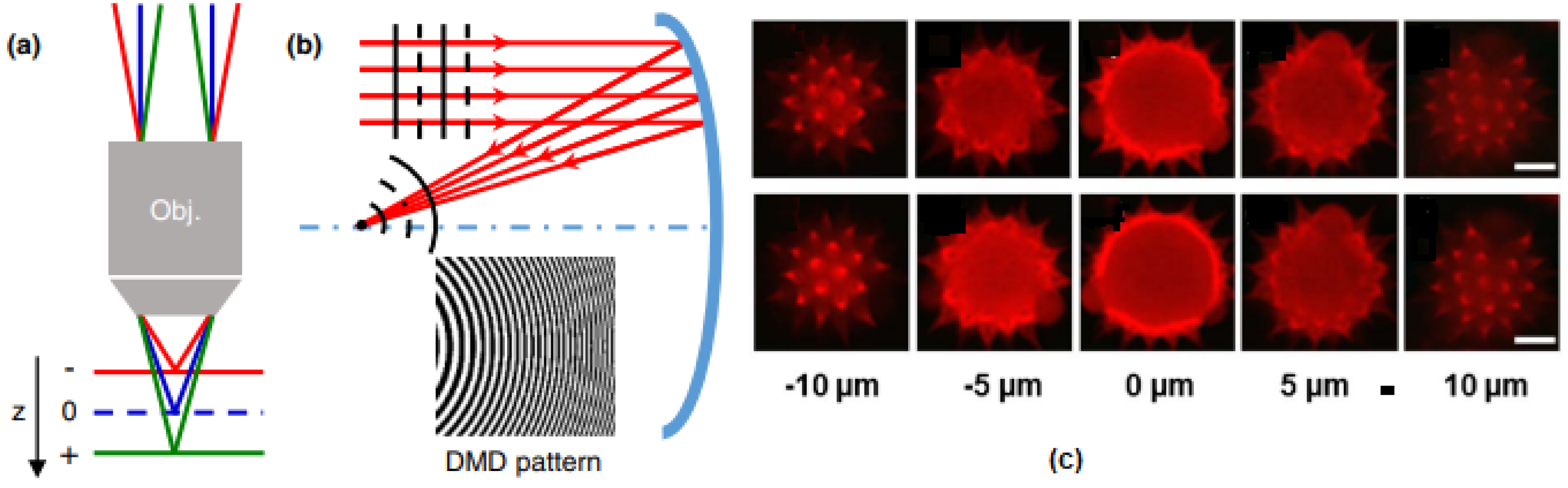
- Cheng, J.Y.; Gu, C.L.; Zhang, D.P.; Wang, D.; Chen, S.C. Ultrafast axial scanning for two-photon microscopy via a digital micromirror device and binary holography. Opt. Lett. 2016, 41, 1451–1454. [Google Scholar] [CrossRef] [PubMed]
- Blum, M.; Büeler, M.; Grätzel, C.; Aschwanden, M. Compact optical design solutions using focus tunable lenses. In Optical Design and Engineering IV; SPIE: Maseille, France, 2011; Volume 8167, p. 81670W. [Google Scholar]
- Ozbay, B.N.; Futia, G.L.; Ma, M. Three dimensional two-photon brain imaging in freely moving mice using a miniature fiber coupled microscope with active axial-scanning. Sci. Rep. 2018, 8, 1–14. [Google Scholar] [CrossRef] [PubMed] [Green Version]
- Archer-Zhang, C.C.; Foster, W.B.; Downey, R.D.; Arrasmith, C.L.; Dickensheets, D.L. Dynamic performance of microelectromechanical systems deformable mirrors for use in an active/adaptive two-photon microscope. J. Biomed. Opt. 2016, 21, 121507. [Google Scholar] [CrossRef] [Green Version]
- Ji, N.; Freeman, J.; Smith, S.L. Technologies for imaging neural activity in large volumes. Nat. Neurosci. 2016, 19, 1154. [Google Scholar] [CrossRef] [Green Version]
- Duan, X.Y.; Li, H.J.; Li, X.; Oldham, K.R.; Wang, T.D. Axial beam scanning in multiphoton microscopy with MEMS-based actuator. Opt. Express 2017, 25, 2195–2205. [Google Scholar] [CrossRef] [Green Version]
- Li, H.J.; Duan, X.Y.; Li, G.; Oldham, K.R.; Wang, T.D. An electrostatic MEMS translational scanner with large out-of-plane stroke for remote axial-scanning in multi-photon microscopy. Micromachines 2017, 8, 159. [Google Scholar] [CrossRef] [Green Version]
- Kwon, S.; Lee, L.P. Micromachined transmissive scanning confoncal microscope. Opt. Lett. 2004, 29, 706–708. [Google Scholar] [CrossRef]
- Michael, A.; Kwok, C.Y. Piezoelectric micro-lens actuator. Sens. Actuators A Phys. 2015, 236, 116–129. [Google Scholar] [CrossRef]
- Wu, L.; Xie, H.K. A millimeter-tunalbe-range microlens for endoscopic biomedical imaging applications. IEEE J. Quantum Elect. 2010, 46, 1237–1244. [Google Scholar] [CrossRef]
- Liu, L.; Wang, E.; Zhang, X.Y.; Liang, W.X.; Li, X.D.; Xie, H.K. MEMS-based 3D confocal scanning mircoendoscope using MEMS scanner for both lateral and axial scan. Sens. Actuators A Phys. 2014, 215, 89–95. [Google Scholar] [CrossRef] [PubMed] [Green Version]
- Zhou, L.; Yu, X.M.; Feng, P.; Li, J.H.; Xie, H.K. A MEMS lens scanner based on serpentine electrothermal bimorph actuator for large axial tuning. Opt. Express 2020, 28, 23439–23453. [Google Scholar] [CrossRef] [PubMed]
- Edward, E.J.; Christopher, C.W.; Michael, M.K.; Delphine, D.; Martin, J.B.; Rimas, J.; Ole, P.; Tony, W. Aberration-free three-dimensional multiphoton imaging of neuronal activity at kHz rates. Proc. Natl. Acad. Sci. USA 2012, 109, 2919–2924. [Google Scholar]
- Birla, M.; Zou, J.Y.; Afkhami, Z.; Duan, X.Y.; Li, H.J.; Wang, T.D.; Oldham, K.R. Multi-photon 3D imaging with an electrothermal actuator with low thermal and inertial mass. Sens. Actuators A Phys. 2021, 329, 112791. [Google Scholar] [CrossRef]
- Grewe, B.F.; Voige, F.F.; Hoff, M.V.; Helmchen, F. Fast two-layer two-photon imaging of neuronal cell populations using an electrically tunable lens. Biomed. Opt. Express 2011, 2, 2035–2046. [Google Scholar] [CrossRef] [Green Version]
- Ersumo, N.T.; Yalcin, C.; Antipa, N.; Pegard, P.; Waller, L.; Lopez, D.; Muller, R. A micromirror array with annular partitioning for high-speed random-access axial focusing. Light Sci. Appl. 2020, 9, 183–197. [Google Scholar] [CrossRef]
- Yalcin, C.; Ersaro, N.T.; Ghanbari, M.M.; Bocchetti, G.; Alamouti, S.F.; Antipa, N.; Lopez, D.; Pegards, N.C.; Waller, L.; Muller, R. A MEMS-based optical scanning system for precise, high-speed neural interfacing. IEEE J. Solid-St. Circ. 2022, 57, 3442–3452. [Google Scholar] [CrossRef]


| TPM Probe | MEMS Mirror | |||||||||
|---|---|---|---|---|---|---|---|---|---|---|
| Size (mm3) | FOV (µm × µm) | Frame Rate (Hz) | Resolution (µm) | Working Distance (µm) | Mirror Size (mm2) | Chip Size (mm2) | Optical Angle (°) | Drive Voltage (V) | Resonance Frequency (kHz) | |
| 2006 [64] | - | 250 × 90 | - | ~1 | 35~47 | 0.75 × 0.75 | 3.2 × 3.0 | 16 | 45~58 | inner 3.52 outer 1.02 |
| 2008 [26] | 10 × 15 × 40 | Φ: 310 µm | 10 | lateral 1.6 axial 16.4 | 210 | 0.5 × 0.5 | - | inner ±10 outer ±10.5 | 80 | 1.54, 2.73 |
| 2008 [33] | Φ: 10 long: 140 | 128 × 128 pixels | 10 | - | - | Φ: 2 mm | - | 14 | - | 1.26, 0.784 |
| 2009 [24] | Φ: 10 mm long: 140 | 720 × 720 pixels | 0.25 | 2 | 210 | Φ: 2 mm | 3.3 × 2.6 | 20 | 90 | 1.26, 0.784 |
| 2009 [23] | 20 × 19 × 11 | 295 × 100 | 15 | lateral 1.3 axial 10.3 | 280 | 1 × 1 | - | inner ±5 outer ±4.3 | 45 | 1.08, 0.56 |
| 2021 [35] | 16 × 9 × 30 | 420 × 420 | 10 | lateral 1.1 axial 12.2 | 1 mm | - | - | ±4.5 | - | - |
| 2017 [27] | 1 cm3 | 130 × 130 | 40 | lateral 0.6 axial 3.4 | 170 | Φ: 0.8 mm | 9 × 9 | ±10 | 10 | 6 |
| 2015 [25] | Φ: 3.4 mm long: 26 mm | 300 × 300 | 5 | lateral 2 axial 9.0 | 60 | Φ: 1.8 mm | 3 × 3 | ±4.5 | 40 | 2.91, 0.805 |
Disclaimer/Publisher’s Note: The statements, opinions and data contained in all publications are solely those of the individual author(s) and contributor(s) and not of MDPI and/or the editor(s). MDPI and/or the editor(s) disclaim responsibility for any injury to people or property resulting from any ideas, methods, instructions or products referred to in the content. |
© 2023 by the authors. Licensee MDPI, Basel, Switzerland. This article is an open access article distributed under the terms and conditions of the Creative Commons Attribution (CC BY) license (https://creativecommons.org/licenses/by/4.0/).
Share and Cite
Yu, X.; Zhou, L.; Qi, T.; Zhao, H.; Xie, H. MEMS Enabled Miniature Two-Photon Microscopy for Biomedical Imaging. Micromachines 2023, 14, 470. https://doi.org/10.3390/mi14020470
Yu X, Zhou L, Qi T, Zhao H, Xie H. MEMS Enabled Miniature Two-Photon Microscopy for Biomedical Imaging. Micromachines. 2023; 14(2):470. https://doi.org/10.3390/mi14020470
Chicago/Turabian StyleYu, Xiaomin, Liang Zhou, Tingxiang Qi, Hui Zhao, and Huikai Xie. 2023. "MEMS Enabled Miniature Two-Photon Microscopy for Biomedical Imaging" Micromachines 14, no. 2: 470. https://doi.org/10.3390/mi14020470