

Molecularly Imprinted Polymer-Coated Inorganic Nanoparticles: Fabrication and Biomedical Applications

Abstract
:1. Introduction
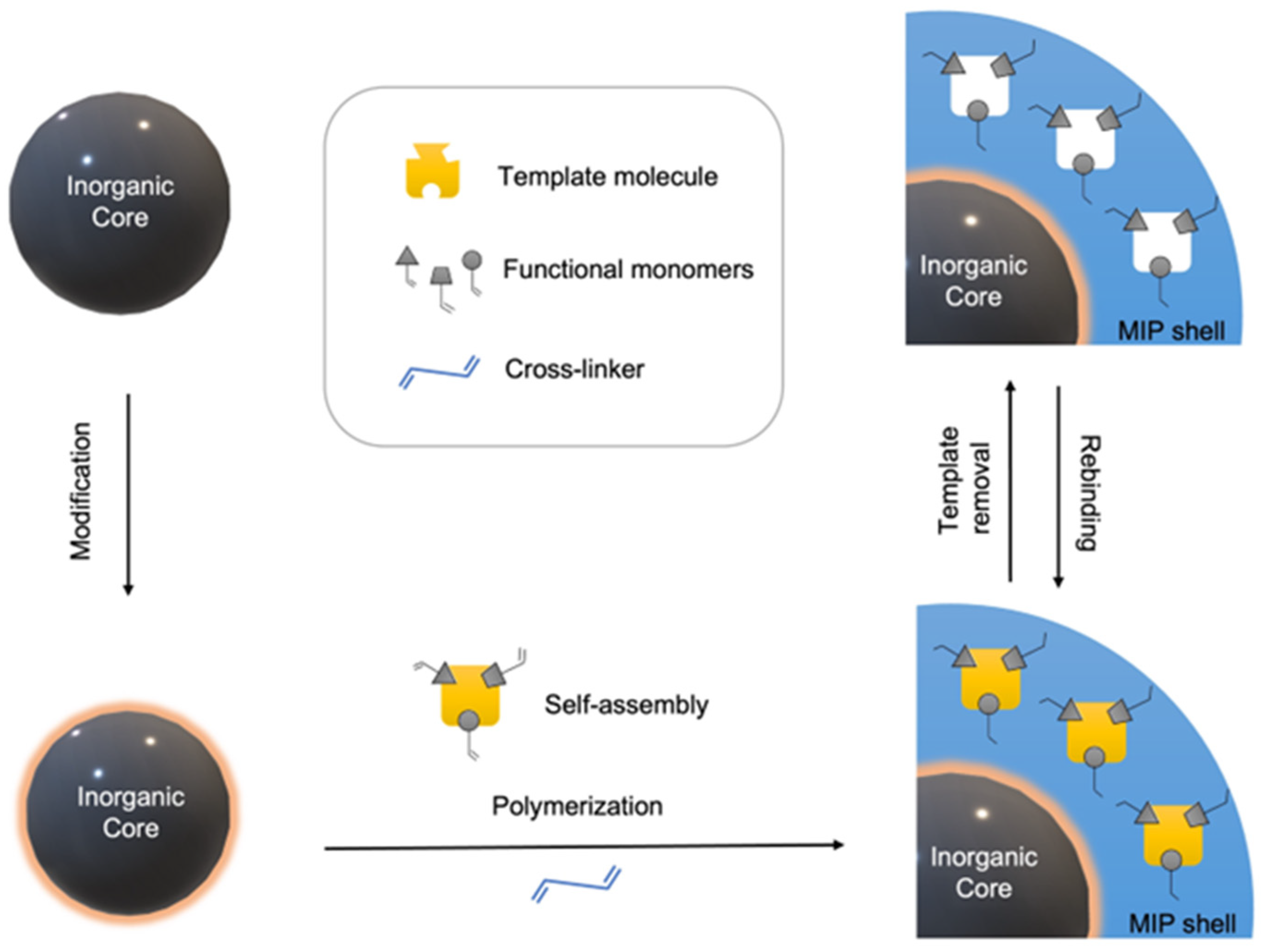
2. MIP Synthesis
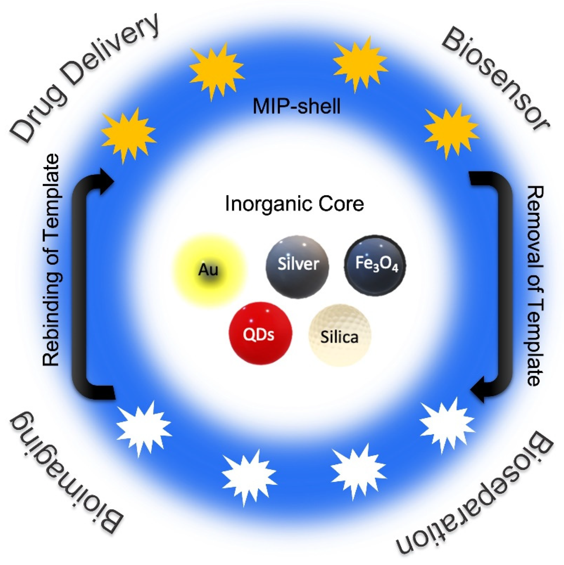
3. Fabrication of Core-Shell Nanoparticles
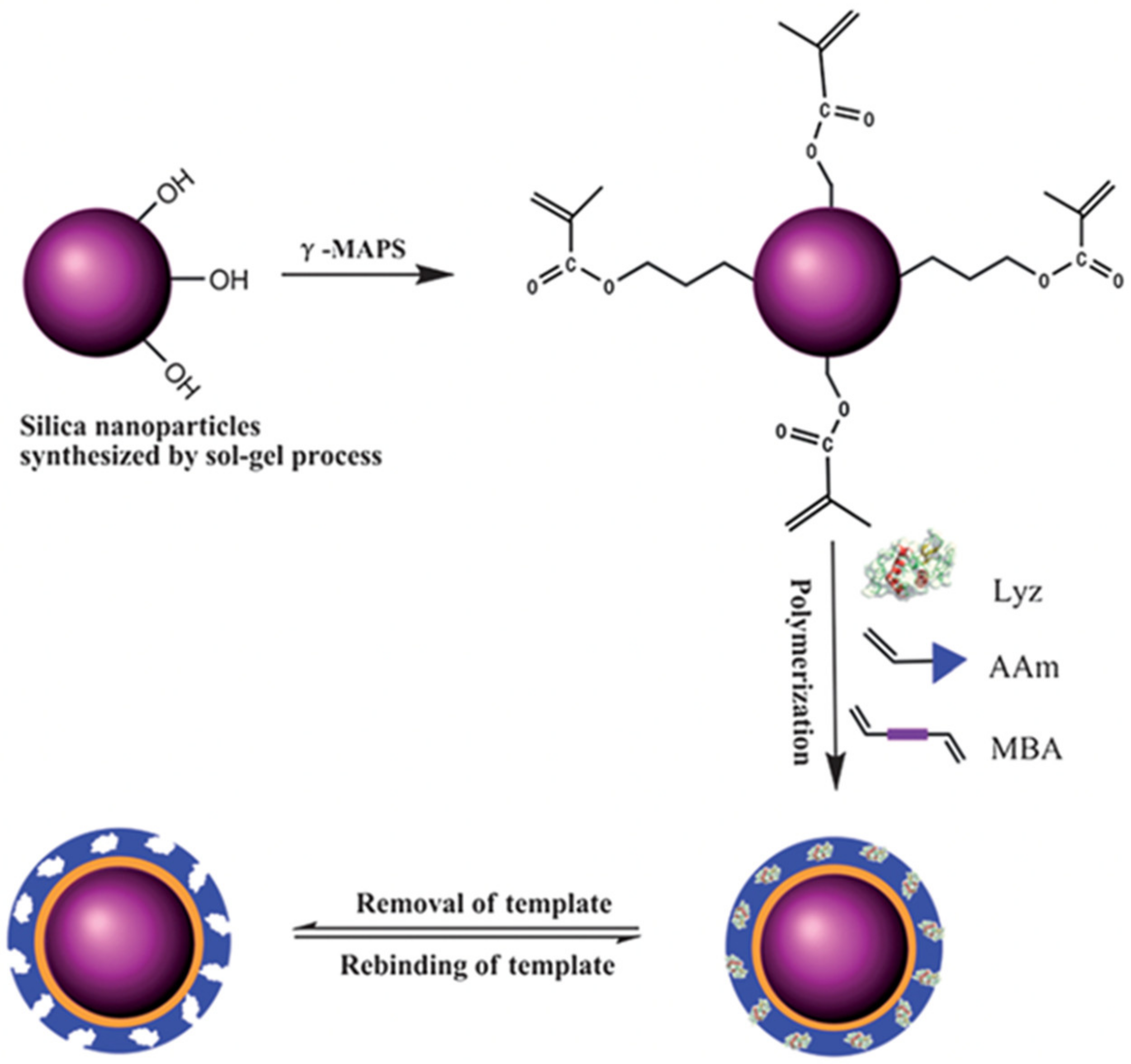
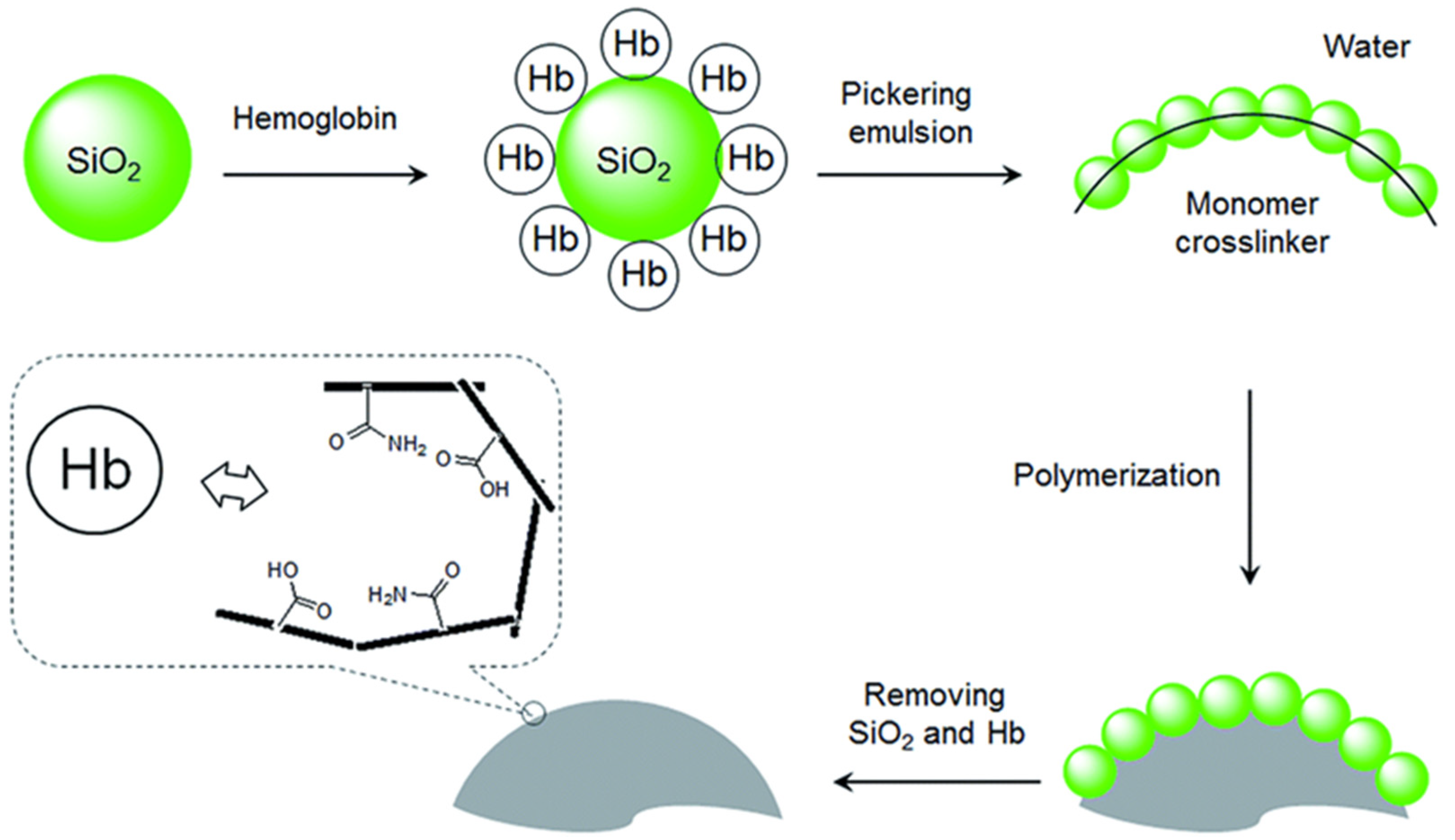
3.1. Silica (SiO2) NPs
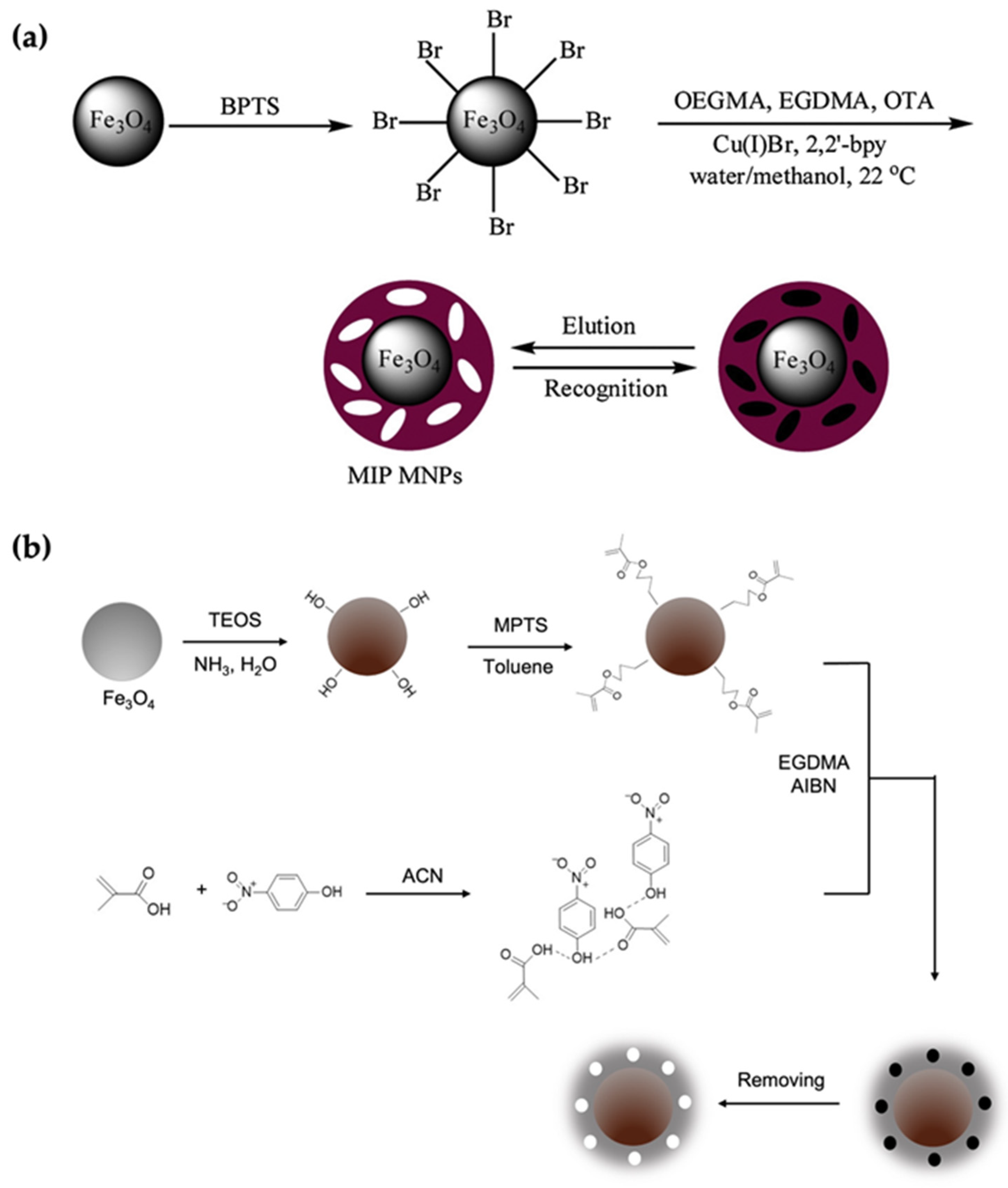
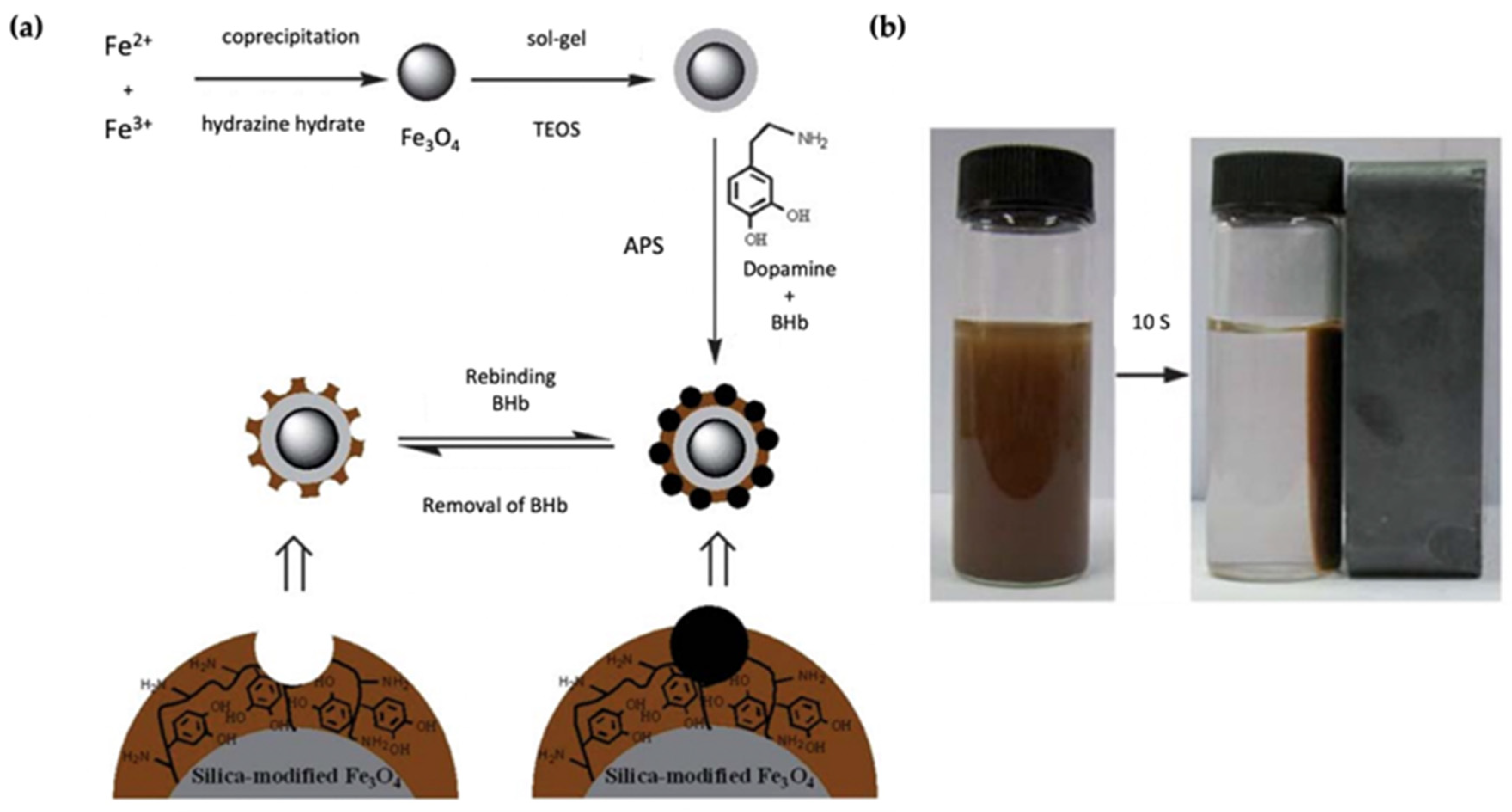
3.2. Magnetic NPs
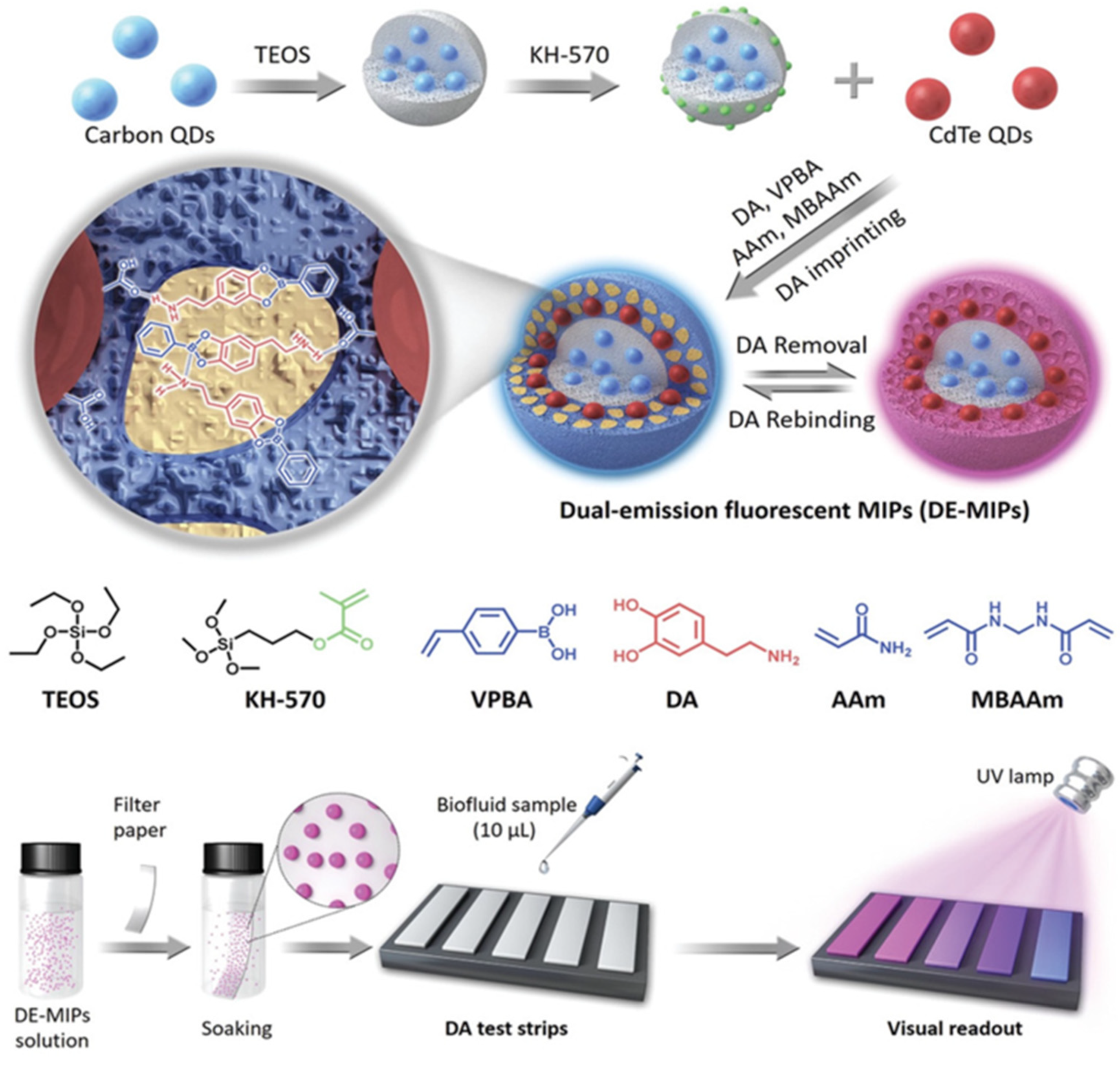
3.3. Quantum Dots (QDs)
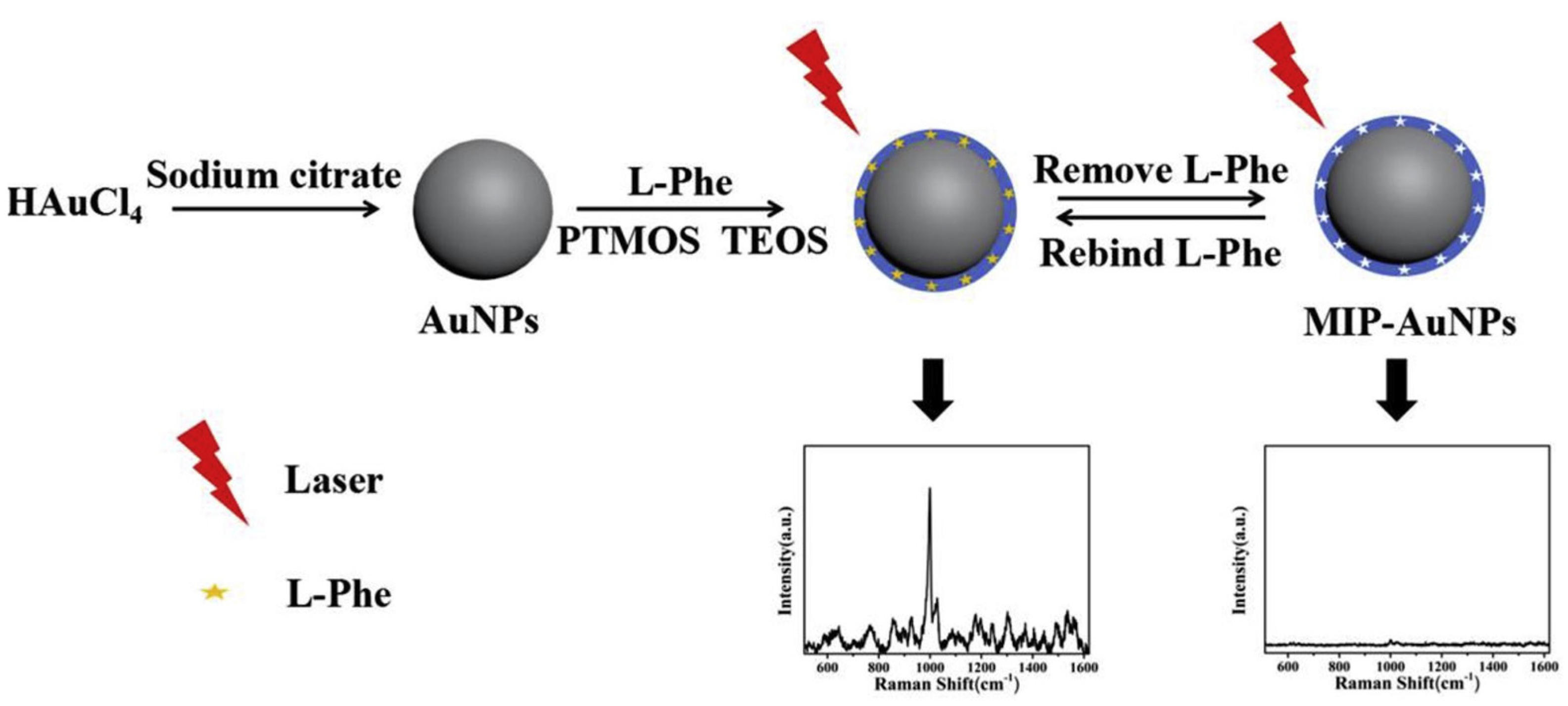
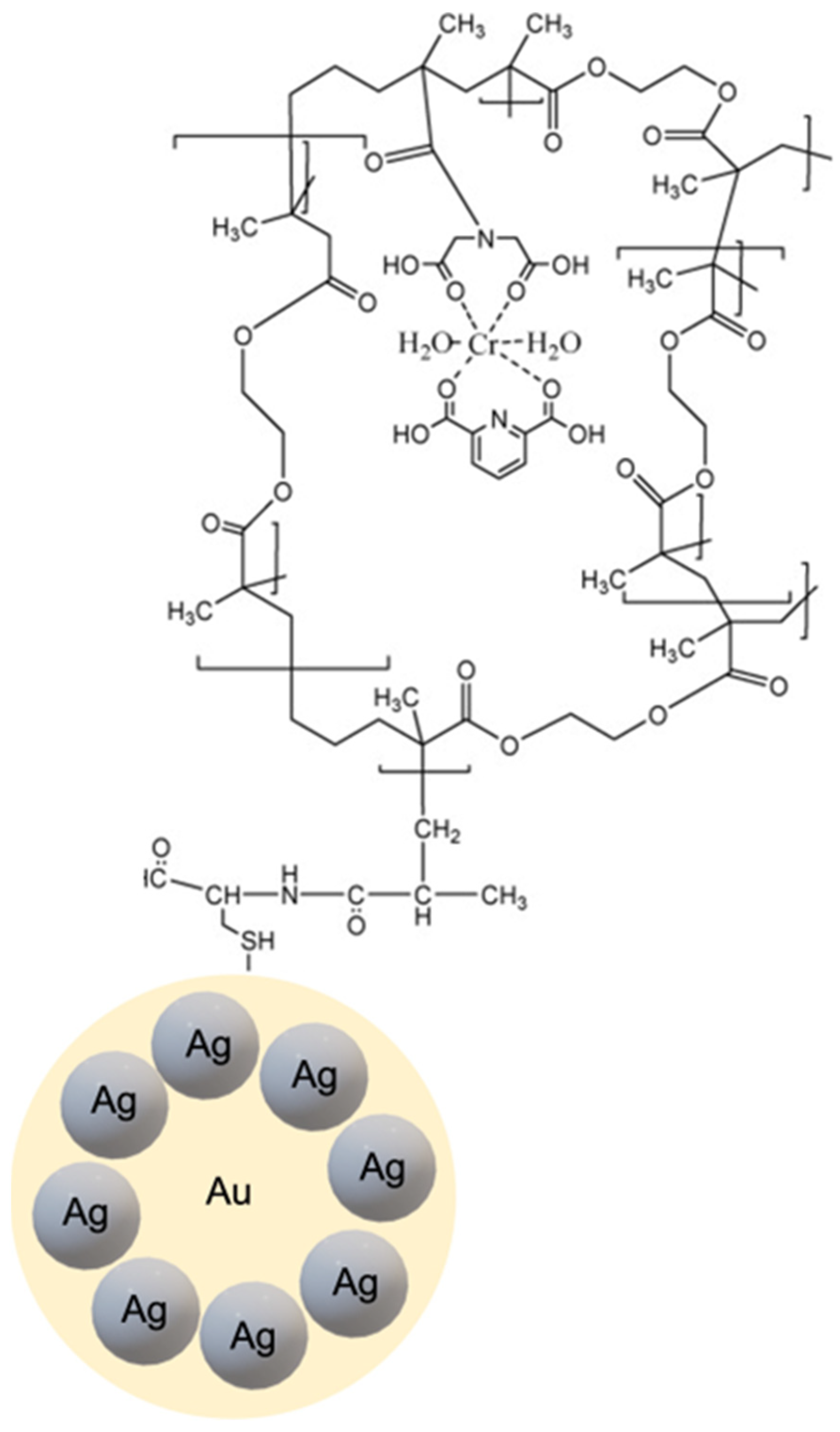
3.4. Gold (Au) and Silver (Ag) NPs
4. Biomedical Applications of Core-Shell MIPs
4.1. Biosensors
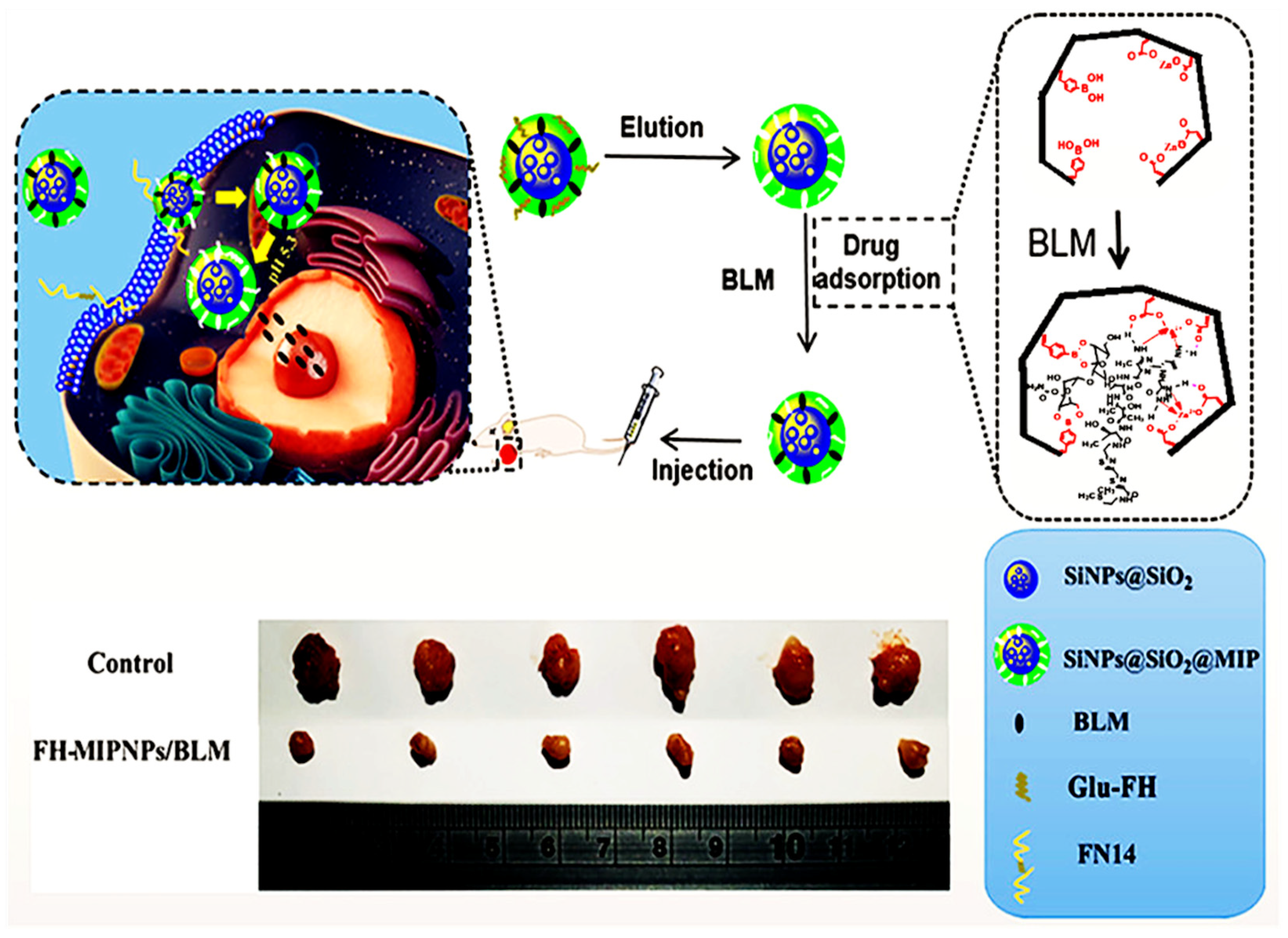
4.2. Drug Delivery
4.3. Bioimaging
4.4. Bioseparation
5. Conclusions and Future Perspectives
Author Contributions
Funding
Institutional Review Board Statement
Informed Consent Statement
Data Availability Statement
Conflicts of Interest
References
- Kemp, E.; Palomäki, T.; Ruuth, I.A.; Boeva, Z.A.; Nurminen, T.A.; Vänskä, R.T.; Zschaechner, L.K.; Pérez, A.G.; Hakala, T.A.; Wardale, M.; et al. Influence of Enzyme Immobilization and Skin-Sensor Interface on Non-Invasive Glucose Determination from Interstitial Fluid Obtained by Magnetohydrodynamic Extraction. Biosens. Bioelectron. 2022, 206, 114123. [Google Scholar] [CrossRef] [PubMed]
- Makaraviciute, A.; Ramanaviciene, A. Site-Directed Antibody Immobilization Techniques for Immunosensors. Biosens. Bioelectron. 2013, 50, 460–471. [Google Scholar] [CrossRef] [PubMed]
- Numnuam, A.; Kanatharana, P.; Mattiasson, B.; Asawatreratanakul, P.; Wongkittisuksa, B.; Limsakul, C.; Thavarungkul, P. Capacitive Biosensor for Quantification of Trace Amounts of DNA. Biosens. Bioelectron. 2009, 24, 2559–2565. [Google Scholar] [CrossRef] [PubMed]
- Haupt, K.; Medina Rangel, P.X.; Bui, B.T.S. Molecularly Imprinted Polymers: Antibody Mimics for Bioimaging and Therapy. Chem. Rev. 2020, 120, 9554–9582. [Google Scholar] [CrossRef]
- Haupt, K. Peer Reviewed: Molecularly Imprinted Polymers: The Next Generation. Anal. Chem. 2003, 75, 376A–383A. [Google Scholar] [CrossRef]
- Haupt, K.; Mosbach, K. Molecularly Imprinted Polymers and Their Use in Biomimetic Sensors. Chem. Rev. 2000, 100, 2495–2504. [Google Scholar] [CrossRef]
- Dinu, A.; Apetrei, C. A Review of Sensors and Biosensors Modified with Conducting Polymers and Molecularly Imprinted Polymers Used in Electrochemical Detection of Amino Acids: Phenylalanine, Tyrosine, and Tryptophan. Int. J. Mol. Sci. 2022, 23, 1218. [Google Scholar] [CrossRef]
- Che Lah, N.F.; Ahmad, A.L.; Mohd Amri, M.H.; Chin, J.Y. Configuration of Molecularly Imprinted Polymers for Specific Uptake of Pharmaceutical in Aqueous Media through Radical Polymerization Method. J. Polym. Res. 2022, 29, 41. [Google Scholar] [CrossRef]
- Saylan, Y.; Akgönüllü, S.; Çimen, D.; Derazshamshir, A.; Bereli, N.; Yılmaz, F.; Denizli, A. Development of Surface Plasmon Resonance Sensors Based on Molecularly Imprinted Nanofilms for Sensitive and Selective Detection of Pesticides. Sens. Actuators B Chem. 2017, 241, 446–454. [Google Scholar] [CrossRef]
- Huang, Y.-C.; Lin, C.-C.; Liu, C.-Y. Preparation and Evaluation of Molecularly Imprinted Polymers Based on 9-Ethyladenine for the Recognition of Nucleotide Bases in Capillary Electrochromatography. Electrophoresis 2004, 25, 554–561. [Google Scholar] [CrossRef]
- Amorim, M.S.; Sales, M.G.F.; Frasco, M.F. Recent Advances in Virus Imprinted Polymers. Biosens. Bioelectron. X 2022, 10, 100131. [Google Scholar] [CrossRef]
- Lieberzeit, P.A.; Bajwa, S.; Mustafa, G.; Wangchareansak, T.; Dickert, F.L. From Metal Ions to Biospecies: Template-Assisted Synthesis as a Strategy to Generate Artificial Receptor Materials. Adv. Mater. Lett. 2011, 2, 319–321. [Google Scholar] [CrossRef]
- Zhang, H.; Ye, L.; Mosbach, K. Non-Covalent Molecular Imprinting with Emphasis on Its Application in Separation and Drug Development. J. Mol. Recognit. 2006, 19, 248–259. [Google Scholar] [CrossRef] [PubMed]
- Cheong, W.J.; Yang, S.H.; Ali, F. Molecular Imprinted Polymers for Separation Science: A Review of Reviews: Other Techniques. J. Sep. Sci. 2013, 36, 609–628. [Google Scholar] [CrossRef]
- Saad, E.M.; El Gohary, N.A.; Abdel-Halim, M.; Handoussa, H.; Mohamed El Nashar, R.; Mizaikoff, B. Molecularly Imprinted Polymers for Selective Extraction of Rosmarinic Acid from Rosmarinus officinalis L. Food Chem. 2021, 335, 127644. [Google Scholar] [CrossRef]
- Wulff, G.; Liu, J. Design of Biomimetic Catalysts by Molecular Imprinting in Synthetic Polymers: The Role of Transition State Stabilization. Acc. Chem. Res. 2012, 45, 239–247. [Google Scholar] [CrossRef] [PubMed]
- Mirata, F.; Resmini, M. Molecularly Imprinted Polymers for Catalysis and Synthesis. In Molecularly Imprinted Polymers in Biotechnology; Mattiasson, B., Ye, L., Eds.; Advances in Biochemical Engineering/Biotechnology; Springer International Publishing: Cham, Switzerland, 2015; Volume 150, pp. 107–129. ISBN 978-3-319-20728-5. [Google Scholar]
- Leibl, N.; Haupt, K.; Gonzato, C.; Duma, L. Molecularly Imprinted Polymers for Chemical Sensing: A Tutorial Review. Chemosensors 2021, 9, 123. [Google Scholar] [CrossRef]
- Ramanavicius, S.; Jagminas, A.; Ramanavicius, A. Advances in Molecularly Imprinted Polymers Based Affinity Sensors (Review). Polymers 2021, 13, 974. [Google Scholar] [CrossRef]
- Saylan, Y.; Akgönüllü, S.; Yavuz, H.; Ünal, S.; Denizli, A. Molecularly Imprinted Polymer Based Sensors for Medical Applications. Sensors 2019, 19, 1279. [Google Scholar] [CrossRef] [PubMed]
- Jahangiri-Manesh, A.; Mousazadeh, M.; Nikkhah, M.; Abbasian, S.; Moshaii, A.; Masroor, M.J.; Norouzi, P. Molecularly Imprinted Polymer-Based Chemiresistive Sensor for Detection of Nonanal as a Cancer Related Biomarker. Microchem. J. 2022, 173, 106988. [Google Scholar] [CrossRef]
- Fernández-Puig, S.; Lazo-Fraga, A.R.; Korgel, B.A.; Oza, G.; Dutt, A.; Vallejo-Becerra, V.; Valdés-González, A.C.; Chávez-Ramírez, A.U. Molecularly Imprinted Polymer-Silica Nanocomposite Based Potentiometric Sensor for Early Prostate Cancer Detection. Mater. Lett. 2022, 309, 131324. [Google Scholar] [CrossRef]
- He, S.; Zhang, L.; Bai, S.; Yang, H.; Cui, Z.; Zhang, X.; Li, Y. Advances of Molecularly Imprinted Polymers (MIP) and the Application in Drug Delivery. Eur. Polym. J. 2021, 143, 110179. [Google Scholar] [CrossRef]
- Han, S.; Song, Y.; Liu, S.; Zhao, L.; Sun, R. Dual Responsive Molecularly Imprinted Polymers Based on UiO-66-DOX for Selective Targeting Tumor Cells and Controlled Drug Release. Eur. Polym. J. 2022, 171, 111219. [Google Scholar] [CrossRef]
- Ali, Z.; Sajid, M.; Manzoor, S.; Ahmad, M.M.; Khan, M.I.; Elboughdiri, N.; Kashif, M.; Shanableh, A.; Rajhi, W.; Mersni, W.; et al. Biodegradable Magnetic Molecularly Imprinted Anticancer Drug Carrier for the Targeted Delivery of Docetaxel. ACS Omega 2022, 7, 28516–28524. [Google Scholar] [CrossRef] [PubMed]
- Zhang, Z.; Ma, P.; Ahmed, R.; Wang, J.; Akin, D.; Soto, F.; Liu, B.; Li, P.; Demirci, U. Advanced Point-of-Care Testing Technologies for Human Acute Respiratory Virus Detection. Adv. Mater. 2022, 34, 2103646. [Google Scholar] [CrossRef]
- Hu, X.; Zhang, P.; Wang, D.; Jiang, J.; Chen, X.; Liu, Y.; Zhang, Z.; Tang, B.Z.; Li, P. AIEgens Enabled Ultrasensitive Point-of-Care Test for Multiple Targets of Food Safety: Aflatoxin B1 and Cyclopiazonic Acid as an Example. Biosens. Bioelectron. 2021, 182, 113188. [Google Scholar] [CrossRef]
- Saylan, Y.; Denizli, A. Molecularly Imprinted Polymer-Based Microfluidic Systems for Point-of-Care Applications. Micromachines 2019, 10, 766. [Google Scholar] [CrossRef]
- González, G.P.; Hernando, P.F.; Alegría, J.S.D. A Morphological Study of Molecularly Imprinted Polymers Using the Scanning Electron Microscope. Anal. Chim. Acta 2006, 557, 179–183. [Google Scholar] [CrossRef]
- Shen, X.S.; Wang, G.Z.; Hong, X.; Zhu, W. Nanospheres of Silver Nanoparticles: Agglomeration, Surface Morphology Control and Application as SERS Substrates. Phys. Chem. Chem. Phys. 2009, 11, 7450. [Google Scholar] [CrossRef]
- Lu, H.; Tian, H.; Wang, C.; Xu, S. Designing and Controlling the Morphology of Spherical Molecularly Imprinted Polymers. Mater. Adv. 2020, 1, 2182–2201. [Google Scholar] [CrossRef]
- Sagawa, T.; Oishi, M.; Yataka, Y.; Sato, R.; Iijima, K.; Hashizume, M. Control of the Molecular Permeability of Polysaccharide Composite Films Utilizing a Molecular Imprinting Approach. Polym. J. 2022, 54, 571–579. [Google Scholar] [CrossRef]
- Chen, M.; Lu, J.; Gao, J.; Yu, C.; Xing, W.; Dai, J.; Meng, M.; Yan, Y.; Wu, Y. Design of Self-Cleaning Molecularly Imprinted Membrane with Antibacterial Ability for High-Selectively Separation of Ribavirin. J. Membr. Sci. 2022, 642, 119994. [Google Scholar] [CrossRef]
- Cao, Y.; Liu, G.; Qin, X.; Li, H.; Liu, C. Preparation and Application of 2-Chlorophenol Molecularly Imprinted Photonic Crystal Hydrogel Sensor. J. Macromol. Sci. Part A 2021, 58, 336–343. [Google Scholar] [CrossRef]
- Takimoto, K.; Takano, E.; Kitayama, Y.; Takeuchi, T. Synthesis of Monodispersed Submillimeter-Sized Molecularly Imprinted Particles Selective for Human Serum Albumin Using Inverse Suspension Polymerization in Water-in-Oil Emulsion Prepared Using Microfluidics. Langmuir 2015, 31, 4981–4987. [Google Scholar] [CrossRef]
- Aravind, A.; Mathew, B. Nano Layered Ion Imprinted Polymer Based Electrochemical Sensor and Sorbent for Mn (II) Ions from Real Samples. J. Macromol. Sci. Part A 2020, 57, 256–265. [Google Scholar] [CrossRef]
- Pu, H.; Xu, L. Molecularly Imprinted Nanoparticles Synthesized by Electrochemically Mediated Atom Transfer Radical Precipitation Polymerization. Macro Chem. Phys. 2022, 223, 2100478. [Google Scholar] [CrossRef]
- Canfarotta, F.; Cecchini, A.; Piletsky, S. Chapter 1. Nano-Sized Molecularly Imprinted Polymers as Artificial Antibodies. In Polymer Chemistry Series; Kutner, W., Sharma, P.S., Eds.; Royal Society of Chemistry: Cambridge, UK, 2018; pp. 1–27. ISBN 978-1-78262-647-3. [Google Scholar]
- Ansell, R.J.; Mosbach, K. Magnetic Molecularly Imprinted Polymer Beads for Drug Radioligand Binding Assay. Analyst 1998, 123, 1611–1616. [Google Scholar] [CrossRef]
- Mustafa, G.; Lieberzeit, P.A. Molecularly Imprinted Polymer–Ag 2 S Nanoparticle Composites for Sensing Volatile Organics. RSC Adv. 2014, 4, 12723–12728. [Google Scholar] [CrossRef]
- Chiozzi, V.; Rossi, F. Inorganic–Organic Core/Shell Nanoparticles: Progress and Applications. Nanoscale Adv. 2020, 2, 5090–5105. [Google Scholar] [CrossRef]
- Wulff, G.; Sarhan, A. The Use of Polymers with Enzyme-Analogous Structures for the Resolution of Racemates. Angew. Chem. Int. Ed. 1972, 11, 341–344. [Google Scholar]
- Wulff, G. Molecular Imprinting in Cross-Linked Materials with the Aid of Molecular Templates—A Way towards Artificial Antibodies. Angew. Chem. Int. Ed. Engl. 1995, 34, 1812–1832. [Google Scholar] [CrossRef]
- Arshady, R.; Mosbach, K. Synthesis of Substrate Selective Polymers by Host-Guest Polymerization. Makromol. Chem. 1981, 182, 687–692. [Google Scholar] [CrossRef]
- Sellergren, B.; Lepistoe, M.; Mosbach, K. Highly Enantioselective and Substrate-Selective Polymers Obtained by Molecular Imprinting Utilizing Noncovalent Interactions. NMR and Chromatographic Studies on the Nature of Recognition. J. Am. Chem. Soc. 1988, 110, 5853–5860. [Google Scholar] [CrossRef]
- Yoshimatsu, K.; Reimhult, K.; Krozer, A.; Mosbach, K.; Sode, K.; Ye, L. Uniform Molecularly Imprinted Microspheres and Nanoparticles Prepared by Precipitation Polymerization: The Control of Particle Size Suitable for Different Analytical Applications. Anal. Chim. Acta 2007, 584, 112–121. [Google Scholar] [CrossRef]
- Ye, L.; Cormack, P.A.G.; Mosbach, K. Molecularly Imprinted Monodisperse Microspheres for Competitive Radioassay. Anal. Commun. 1999, 36, 35–38. [Google Scholar] [CrossRef]
- Ruela, A.L.M.; de Figueiredo, E.C.; Carvalho, F.C.; de Araújo, M.B.; Pereira, G.R. Adsorption and Release of Nicotine from Imprinted Particles Synthesised by Precipitation Polymerisation: Optimising Transdermal Formulations. Eur. Polym. J. 2018, 100, 67–76. [Google Scholar] [CrossRef]
- Pardeshi, S.; Singh, S.K. Precipitation Polymerization: A Versatile Tool for Preparing Molecularly Imprinted Polymer Beads for Chromatography Applications. RSC Adv. 2016, 6, 23525–23536. [Google Scholar] [CrossRef]
- Vaihinger, D.; Landfester, K.; Kräuter, I.; Brunner, H.; Tovar, G.E.M. Molecularly Imprinted Polymer Nanospheres as Synthetic Affinity Receptors Obtained by Miniemulsion Polymerisation. Macromol. Chem. Phys. 2002, 203, 1965–1973. [Google Scholar] [CrossRef]
- Mehmood, A.; Ghafar, H.; Yaqoob, S.; Gohar, U.F.; Ahmad, B. Mesoporous Silica Nanoparticles: A Review. J. Dev. Drugs 2017, 06, 1000174. [Google Scholar] [CrossRef]
- Lin, Z.; Xia, Z.; Zheng, J.; Zheng, D.; Zhang, L.; Yang, H.; Chen, G. Synthesis of Uniformly Sized Molecularly Imprinted Polymer-Coated Silica Nanoparticles for Selective Recognition and Enrichment of Lysozyme. J. Mater. Chem. 2012, 22, 17914. [Google Scholar] [CrossRef]
- Gao, R.; Zhang, J.; He, X.; Chen, L.; Zhang, Y. Selective Extraction of Sulfonamides from Food by Use of Silica-Coated Molecularly Imprinted Polymer Nanospheres. Anal. Bioanal Chem. 2010, 398, 451–461. [Google Scholar] [CrossRef] [PubMed]
- Chang, L.; Li, Y.; Chu, J.; Qi, J.; Li, X. Preparation of Core-Shell Molecularly Imprinted Polymer via the Combination of Reversible Addition-Fragmentation Chain Transfer Polymerization and Click Reaction. Anal. Chim. Acta 2010, 680, 65–71. [Google Scholar] [CrossRef]
- Zhou, T.; Zhang, K.; Kamra, T.; Bülow, L.; Ye, L. Preparation of Protein Imprinted Polymer Beads by Pickering Emulsion Polymerization. J. Mater. Chem. B 2015, 3, 1254–1260. [Google Scholar] [CrossRef] [PubMed]
- Dadfar, S.M.; Roemhild, K.; Drude, N.I.; von Stillfried, S.; Knüchel, R.; Kiessling, F.; Lammers, T. Iron Oxide Nanoparticles: Diagnostic, Therapeutic and Theranostic Applications. Adv. Drug Deliv. Rev. 2019, 138, 302–325. [Google Scholar] [CrossRef] [PubMed]
- Oz, Y.; Arslan, M.; Gevrek, T.N.; Sanyal, R.; Sanyal, A. Modular Fabrication of Polymer Brush Coated Magnetic Nanoparticles: Engineering the Interface for Targeted Cellular Imaging. ACS Appl. Mater. Interfaces 2016, 8, 19813–19826. [Google Scholar] [CrossRef]
- Oz, Y.; Abdouni, Y.; Yilmaz, G.; Becer, C.R.; Sanyal, A. Magnetic Glyconanoparticles for Selective Lectin Separation and Purification. Polym. Chem. 2019, 10, 3351–3361. [Google Scholar] [CrossRef]
- Turan, E.; Şahin, F. Molecularly Imprinted Biocompatible Magnetic Nanoparticles for Specific Recognition of Ochratoxin A. Sens. Actuators B Chem. 2016, 227, 668–676. [Google Scholar] [CrossRef]
- Mehdinia, A.; Baradaran Kayyal, T.; Jabbari, A.; Aziz-Zanjani, M.O.; Ziaei, E. Magnetic Molecularly Imprinted Nanoparticles Based on Grafting Polymerization for Selective Detection of 4-Nitrophenol in Aqueous Samples. J. Chromatogr. A 2013, 1283, 82–88. [Google Scholar] [CrossRef]
- Ramimoghadam, D.; Bagheri, S.; Hamid, S.B.A. Progress in Electrochemical Synthesis of Magnetic Iron Oxide Nanoparticles. J. Magn. Magn. Mater. 2014, 368, 207–229. [Google Scholar] [CrossRef]
- Liu, Y.; Huang, Y.; Liu, J.; Wang, W.; Liu, G.; Zhao, R. Superparamagnetic Surface Molecularly Imprinted Nanoparticles for Water-Soluble Pefloxacin Mesylate Prepared via Surface Initiated Atom Transfer Radical Polymerization and Its Application in Egg Sample Analysis. J. Chromatogr. A 2012, 1246, 15–21. [Google Scholar] [CrossRef]
- Zora, A.; Triberis, G.P.; Simserides, C. Near-Field Optical Properties of Quantum Dots, Applications and Perspectives. Recent Pat. Nanotechnol. 2011, 5, 188–224. [Google Scholar] [CrossRef] [PubMed]
- Abbasi, E.; Kafshdooz, T.; Bakhtiary, M.; Nikzamir, N.; Nikzamir, N.; Nikzamir, M.; Mohammadian, M.; Akbarzadeh, A. Biomedical and Biological Applications of Quantum Dots. Artif. Cells Nanomed. Biotechnol. 2016, 44, 885–891. [Google Scholar] [CrossRef] [PubMed]
- Díaz-Álvarez, M.; Martín-Esteban, A. Molecularly Imprinted Polymer-Quantum Dot Materials in Optical Sensors: An Overview of Their Synthesis and Applications. Biosensors 2021, 11, 79. [Google Scholar] [CrossRef] [PubMed]
- Lu, X.; Wei, F.; Xu, G.; Wu, Y.; Yang, J.; Hu, Q. Surface Molecular Imprinting on Silica-Coated CdTe Quantum Dots for Selective and Sensitive Fluorescence Detection of p-Aminophenol in Water. J. Fluoresc. 2017, 27, 181–189. [Google Scholar] [CrossRef]
- Zhou, L.; Gao, C.; Hu, X.; Xu, W. One-Pot Large-Scale Synthesis of Robust Ultrafine Silica-Hybridized CdTe Quantum Dots. ACS Appl. Mater. Interfaces 2010, 2, 1211–1219. [Google Scholar] [CrossRef]
- Ren, X.; Chen, L. Quantum Dots Coated with Molecularly Imprinted Polymer as Fluorescence Probe for Detection of Cyphenothrin. Biosens. Bioelectron. 2015, 64, 182–188. [Google Scholar] [CrossRef]
- Ren, X.; Chen, L. Preparation of Molecularly Imprinted Polymer Coated Quantum Dots to Detect Nicosulfuron in Water Samples. Anal. Bioanal Chem. 2015, 407, 8087–8095. [Google Scholar] [CrossRef]
- Chao, M.-R.; Hu, C.-W.; Chen, J.-L. Comparative Syntheses of Tetracycline-Imprinted Polymeric Silicate and Acrylate on CdTe Quantum Dots as Fluorescent Sensors. Biosens. Bioelectron. 2014, 61, 471–477. [Google Scholar] [CrossRef] [PubMed]
- Yeh, Y.-C.; Creran, B.; Rotello, V.M. Gold Nanoparticles: Preparation, Properties, and Applications in Bionanotechnology. Nanoscale 2012, 4, 1871–1880. [Google Scholar] [CrossRef]
- Yang, G.; Zhao, F. Electrochemical Sensor for Dimetridazole Based on Novel Gold Nanoparticles@molecularly Imprinted Polymer. Sens. Actuators B Chem. 2015, 220, 1017–1022. [Google Scholar] [CrossRef]
- Zhou, J.; Sheth, S.; Zhou, H.; Song, Q. Highly Selective Detection of L-Phenylalanine by Molecularly Imprinted Polymers Coated Au Nanoparticles via Surface-Enhanced Raman Scattering. Talanta 2020, 211, 120745. [Google Scholar] [CrossRef] [PubMed]
- Gültekin, A.; Diltemiz, S.E.; Ersöz, A.; Sarıözlü, N.Y.; Denizli, A.; Say, R. Gold–Silver Nanoclusters Having Dipicolinic Acid Imprinted Nanoshell for Bacillus Cereus Spores Recognition. Talanta 2009, 78, 1332–1338. [Google Scholar] [CrossRef] [PubMed]
- Hu, R.; Tang, R.; Xu, J.; Lu, F. Chemical Nanosensors Based on Molecularly-Imprinted Polymers Doped with Silver Nanoparticles for the Rapid Detection of Caffeine in Wastewater. Anal. Chim. Acta 2018, 1034, 176–183. [Google Scholar] [CrossRef] [PubMed]
- Sun, B.; Ni, X.; Cao, Y.; Cao, G. Electrochemical Sensor Based on Magnetic Molecularly Imprinted Nanoparticles Modified Magnetic Electrode for Determination of Hb. Biosens. Bioelectron. 2017, 91, 354–358. [Google Scholar] [CrossRef]
- Rajpal, S.; Bhakta, S.; Mishra, P. Biomarker Imprinted Magnetic Core–Shell Nanoparticles for Rapid, Culture Free Detection of Pathogenic Bacteria. J. Mater. Chem. B 2021, 9, 2436–2446. [Google Scholar] [CrossRef]
- Balayan, S.; Chauhan, N.; Kumar, P.; Chandra, R.; Jain, U. Fabrication of a Sensing Platform for Identification of Tumor Necrosis Factor-Alpha: A Biomarker for Neonatal Sepsis. 3 Biotech 2022, 12, 37. [Google Scholar] [CrossRef]
- Shane, A.L.; Sánchez, P.J.; Stoll, B.J. Neonatal Sepsis. Lancet 2017, 390, 1770–1780. [Google Scholar] [CrossRef]
- Hosseini Ghalehno, M.; Mirzaei, M.; Torkzadeh-Mahani, M. Electrochemical Aptasensor for Tumor Necrosis Factor α Using Aptamer–Antibody Sandwich Structure and Cobalt Hexacyanoferrate for Signal Amplification. J. Iran. Chem. Soc. 2019, 16, 1783–1791. [Google Scholar] [CrossRef]
- Ben Halima, H.; Bellagambi, F.G.; Alcacer, A.; Pfeiffer, N.; Heuberger, A.; Hangouët, M.; Zine, N.; Bausells, J.; Elaissari, A.; Errachid, A. A Silicon Nitride ISFET Based Immunosensor for Tumor Necrosis Factor-Alpha Detection in Saliva. A Promising Tool for Heart Failure Monitoring. Anal. Chim. Acta 2021, 1161, 338468. [Google Scholar] [CrossRef]
- Mehrzad-Samarin, M.; Faridbod, F.; Dezfuli, A.S.; Ganjali, M.R. A Novel Metronidazole Fluorescent Nanosensor Based on Graphene Quantum Dots Embedded Silica Molecularly Imprinted Polymer. Biosens. Bioelectron. 2017, 92, 618–623. [Google Scholar] [CrossRef]
- Wang, J.; Dai, J.; Xu, Y.; Dai, X.; Zhang, Y.; Shi, W.; Sellergren, B.; Pan, G. Molecularly Imprinted Fluorescent Test Strip for Direct, Rapid, and Visual Dopamine Detection in Tiny Amount of Biofluid. Small 2019, 15, 1803913. [Google Scholar] [CrossRef] [PubMed] [Green Version]
- Franco, R.; Reyes-Resina, I.; Navarro, G. Dopamine in Health and Disease: Much More Than a Neurotransmitter. Biomedicines 2021, 9, 109. [Google Scholar] [CrossRef] [PubMed]
- Yu, D.; Zeng, Y.; Qi, Y.; Zhou, T.; Shi, G. A Novel Electrochemical Sensor for Determination of Dopamine Based on AuNPs@SiO2 Core-Shell Imprinted Composite. Biosens. Bioelectron. 2012, 38, 270–277. [Google Scholar] [CrossRef] [PubMed]
- Feng, J.; Li, X.; Cheng, H.; Huang, W.; Kong, H.; Li, Y.; Li, L. A Boronate-Modified Molecularly Imprinted Polymer Labeled with a SERS-Tag for Use in an Antibody-Free Immunoassay for the Carcinoembryonic Antigen. Microchim. Acta 2019, 186, 774. [Google Scholar] [CrossRef] [PubMed]
- Das, S.S.; Bharadwaj, P.; Bilal, M.; Barani, M.; Rahdar, A.; Taboada, P.; Bungau, S.; Kyzas, G.Z. Stimuli-Responsive Polymeric Nanocarriers for Drug Delivery, Imaging, and Theragnosis. Polymers 2020, 12, 1397. [Google Scholar] [CrossRef] [PubMed]
- Chambre, L.; Degirmenci, A.; Sanyal, R.; Sanyal, A. Multi-Functional Nanogels as Theranostic Platforms: Exploiting Reversible and Nonreversible Linkages for Targeting, Imaging, and Drug Delivery. Bioconjugate Chem. 2018, 29, 1885–1896. [Google Scholar] [CrossRef]
- Feyzioğlu Demir, E.; Akgöl, S. Synthesis and Characterization of Double Molecular Imprinted Nanoparticles and Investigation to Adsorption of Respiratory Drugs. Polym.-Plast. Technol. Mater. 2022, 61, 384–389. [Google Scholar] [CrossRef]
- Jia, C.; Zhang, M.; Zhang, Y.; Ma, Z.-B.; Xiao, N.-N.; He, X.-W.; Li, W.-Y.; Zhang, Y.-K. Preparation of Dual-Template Epitope Imprinted Polymers for Targeted Fluorescence Imaging and Targeted Drug Delivery to Pancreatic Cancer BxPC-3 Cells. ACS Appl. Mater. Interfaces 2019, 11, 32431–32440. [Google Scholar] [CrossRef]
- Zavareh, S.; Mahdi, M.; Erfanian, S.; Hashemi-Moghaddam, H. Synthesis of Polydopamine as a New and Biocompatible Coating of Magnetic Nanoparticles for Delivery of Doxorubicin in Mouse Breast Adenocarcinoma. Cancer Chemother. Pharmacol. 2016, 78, 1073–1084. [Google Scholar] [CrossRef]
- Hashemi-Moghaddam, H.; Kazemi-Bagsangani, S.; Jamili, M.; Zavareh, S. Evaluation of Magnetic Nanoparticles Coated by 5-Fluorouracil Imprinted Polymer for Controlled Drug Delivery in Mouse Breast Cancer Model. Int. J. Pharm. 2016, 497, 228–238. [Google Scholar] [CrossRef]
- Nerantzaki, M.; Michel, A.; Petit, L.; Garnier, M.; Ménager, C.; Griffete, N. Biotinylated Magnetic Molecularly Imprinted Polymer Nanoparticles for Cancer Cell Targeting and Controlled Drug Delivery. Chem. Commun. 2022, 58, 5642–5645. [Google Scholar] [CrossRef] [PubMed]
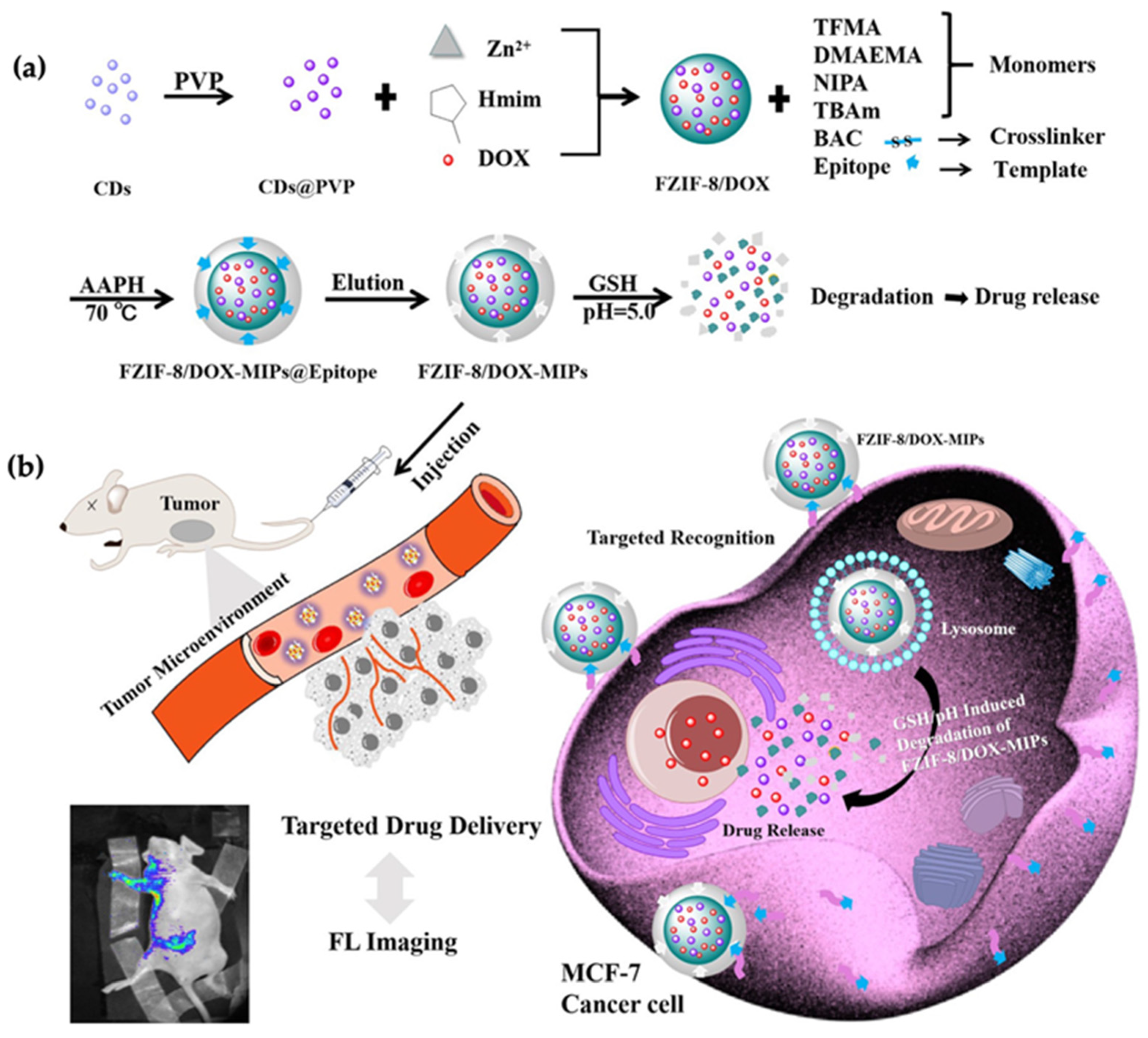
- Qin, Y.-T.; Feng, Y.-S.; Ma, Y.-J.; He, X.-W.; Li, W.-Y.; Zhang, Y.-K. Tumor-Sensitive Biodegradable Nanoparticles of Molecularly Imprinted Polymer-Stabilized Fluorescent Zeolitic Imidazolate Framework-8 for Targeted Imaging and Drug Delivery. ACS Appl. Mater. Interfaces 2020, 12, 24585–24598. [Google Scholar] [CrossRef] [PubMed]
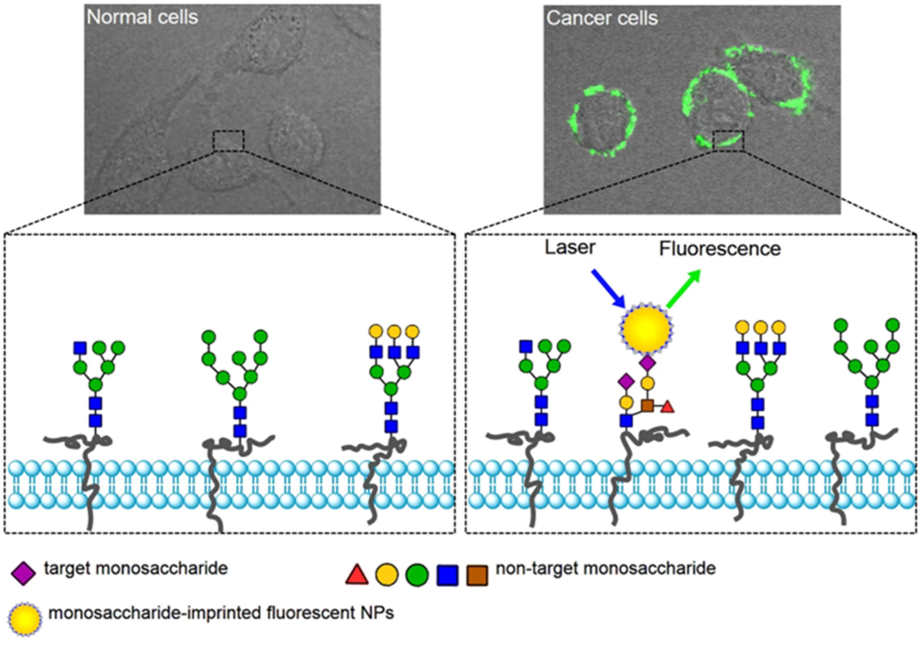
- Wang, S.; Yin, D.; Wang, W.; Shen, X.; Zhu, J.-J.; Chen, H.-Y.; Liu, Z. Targeting and Imaging of Cancer Cells via Monosaccharide-Imprinted Fluorescent Nanoparticles. Sci. Rep. 2016, 6, 22757. [Google Scholar] [CrossRef] [PubMed]
- Yin, D.; Wang, S.; He, Y.; Liu, J.; Zhou, M.; Ouyang, J.; Liu, B.; Chen, H.-Y.; Liu, Z. Surface-Enhanced Raman Scattering Imaging of Cancer Cells and Tissues via Sialic Acid-Imprinted Nanotags. Chem. Commun. 2015, 51, 17696–17699. [Google Scholar] [CrossRef] [PubMed]
- Kunath, S.; Panagiotopoulou, M.; Maximilien, J.; Marchyk, N.; Sänger, J.; Haupt, K. Cell and Tissue Imaging with Molecularly Imprinted Polymers as Plastic Antibody Mimics. Adv. Healthc. Mater. 2015, 4, 1322–1326. [Google Scholar] [CrossRef] [PubMed]
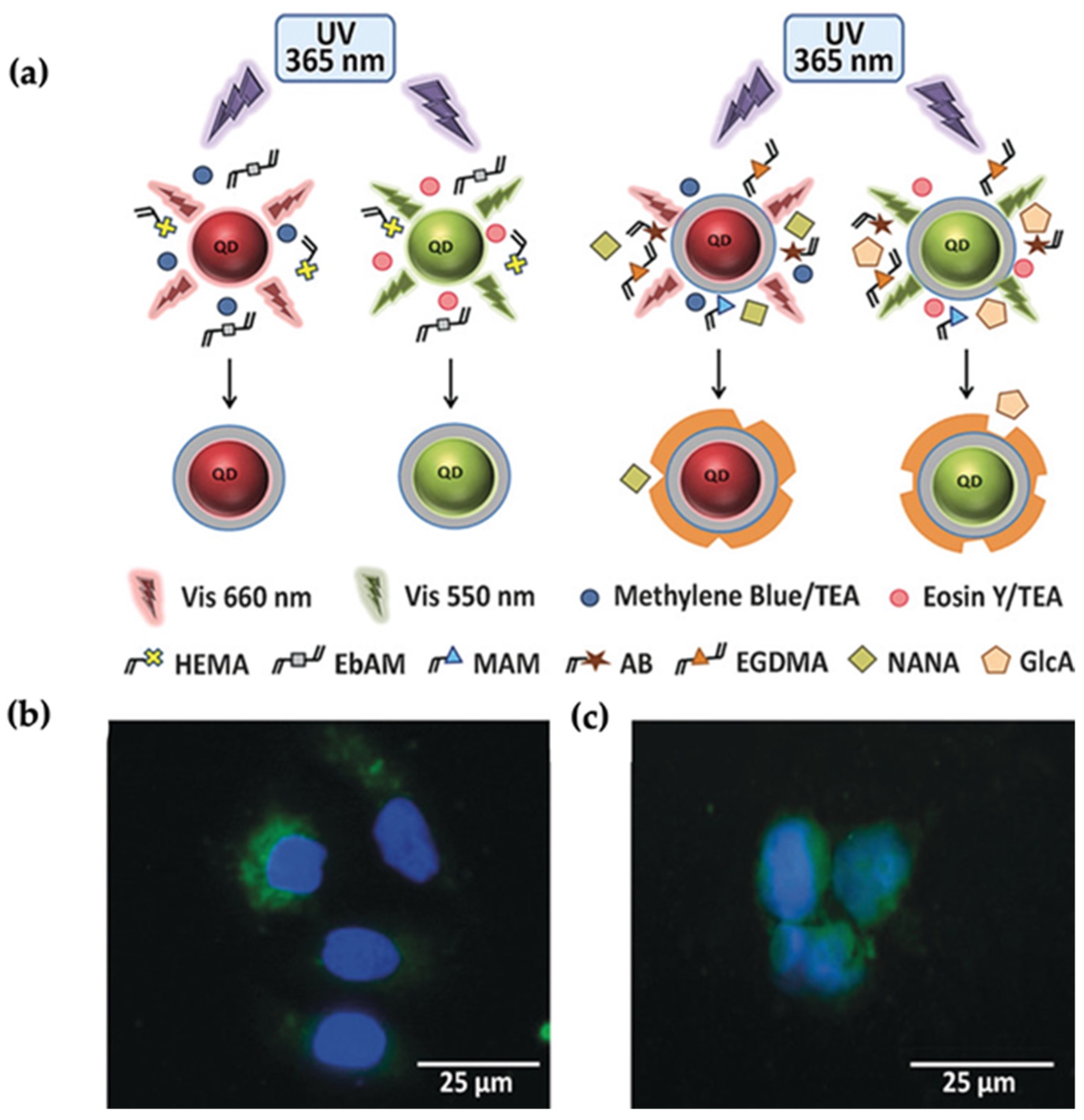
- Panagiotopoulou, M.; Salinas, Y.; Beyazit, S.; Kunath, S.; Duma, L.; Prost, E.; Mayes, A.G.; Resmini, M.; Tse Sum Bui, B.; Haupt, K. Molecularly Imprinted Polymer Coated Quantum Dots for Multiplexed Cell Targeting and Imaging. Angew. Chem. Int. Ed. 2016, 55, 8244–8248. [Google Scholar] [CrossRef]
- Bagheri, H.F.; Arvand, M.; Habibi, M.F. An Ultra-Sensitive Tailor-Made Sensor for Specific Adsorption and Separation of Rutin Based on Imprinted Cavities on Magnetic Sensing Platform. Microchem. J. 2022, 181, 107712. [Google Scholar] [CrossRef]
- Lie, K.R.; Samuel, A.O.; Hasanah, A.N. Molecularly Imprinted Mesoporous Silica: Potential of the Materials, Synthesis and Application in the Active Compound Separation from Natural Product. Chem. Pap. 2022, 76, 2595–2613. [Google Scholar] [CrossRef]
- Akbulut Söylemez, M.; Kemaloğulları, B.Ö. Surface Modification of Magnetic Nanoparticles via Admicellar Polymerization for Selective Removal of Tetracycline from Real Water Samples. New J. Chem. 2021, 45, 6415–6423. [Google Scholar] [CrossRef]
- Jia, X.; Xu, M.; Wang, Y.; Ran, D.; Yang, S.; Zhang, M. Polydopamine-Based Molecular Imprinting on Silica-Modified Magnetic Nanoparticles for Recognition and Separation of Bovine Hemoglobin. Analyst 2013, 138, 651–658. [Google Scholar] [CrossRef]
- Xia, Z.; Lin, Z.; Xiao, Y.; Wang, L.; Zheng, J.; Yang, H.; Chen, G. Facile Synthesis of Polydopamine-Coated Molecularly Imprinted Silica Nanoparticles for Protein Recognition and Separation. Biosens. Bioelectron. 2013, 47, 120–126. [Google Scholar] [CrossRef]
- Zengin, A.; Yildirim, E.; Tamer, U.; Caykara, T. Molecularly Imprinted Superparamagnetic Iron Oxide Nanoparticles for Rapid Enrichment and Separation of Cholesterol. Analyst 2013, 138, 7238. [Google Scholar] [CrossRef] [PubMed]
- Polyakova, I.; Borovikova, L.; Osipenko, A.; Vlasova, E.; Volchek, B.; Pisarev, O. Surface Molecularly Imprinted Organic-Inorganic Polymers Having Affinity Sites for Cholesterol. React. Funct. Polym. 2016, 109, 88–98. [Google Scholar] [CrossRef]
- Vahidifar, M.; Es’haghi, Z.; Oghaz, N.M.; Mohammadi, A.A.; Kazemi, M.S. Multi-Template Molecularly Imprinted Polymer Hybrid Nanoparticles for Selective Analysis of Nonsteroidal Anti-Inflammatory Drugs and Analgesics in Biological and Pharmaceutical Samples. Environ. Sci. Pollut. Res. 2022, 29, 47416–47435. [Google Scholar] [CrossRef] [PubMed]


| Properties | Bulk MIPs | Core-Shell MIP NPs |
|---|---|---|
| Synthesis method | Easy | Complex |
| Morphology | Irregular | Uniform |
| Surface area | Smaller | Larger |
| Adsorption capacity | Lower | Higher |
| Binding sites distribution | Heterogeneous | Homogeneous |
| Template leakage | More | Less |
| Yield | Lower | Higher |
| Target | Core Material | Linear Range | LOD | Ref. |
|---|---|---|---|---|
| Hemoglobin | Fe3O4@SiO2 | 0.005–0.1 mg/mL | 0.0010 mg/mL | [76] |
| Pyocyanin | Fe3O4@SiO2 | - | - | [77] |
| TNF-α | Fe3O4@SiO2 | 0.01 pM–100 nM | 0.01 pM | [78] |
| Metronidazole | GQDs@SiO2 | 0.2–15 μM | 0.15 μM | [82] |
| Dopamine | CdTe QDs@SiO2 | 0–1.2 × 10⁻⁶ M | (100–50) × 10−9 M | [83] |
| Dopamine | Au@SiO2 | 4 × 10⁻⁸–5 × 10⁻⁵ M | 2 × 10⁻⁸ M | [85] |
| Carcinoembryonic Antigen | Au | - | 0.1 ng/mL | [86] |
Publisher’s Note: MDPI stays neutral with regard to jurisdictional claims in published maps and institutional affiliations. |
© 2022 by the authors. Licensee MDPI, Basel, Switzerland. This article is an open access article distributed under the terms and conditions of the Creative Commons Attribution (CC BY) license (https://creativecommons.org/licenses/by/4.0/).
Share and Cite
Orbay, S.; Kocaturk, O.; Sanyal, R.; Sanyal, A. Molecularly Imprinted Polymer-Coated Inorganic Nanoparticles: Fabrication and Biomedical Applications. Micromachines 2022, 13, 1464. https://doi.org/10.3390/mi13091464
Orbay S, Kocaturk O, Sanyal R, Sanyal A. Molecularly Imprinted Polymer-Coated Inorganic Nanoparticles: Fabrication and Biomedical Applications. Micromachines. 2022; 13(9):1464. https://doi.org/10.3390/mi13091464
Chicago/Turabian StyleOrbay, Sinem, Ozgur Kocaturk, Rana Sanyal, and Amitav Sanyal. 2022. "Molecularly Imprinted Polymer-Coated Inorganic Nanoparticles: Fabrication and Biomedical Applications" Micromachines 13, no. 9: 1464. https://doi.org/10.3390/mi13091464