


Highly Durable Antimicrobial Tantalum Nitride/Copper Coatings on Stainless Steel Deposited by Pulsed Magnetron Sputtering
 ,
,  and
and 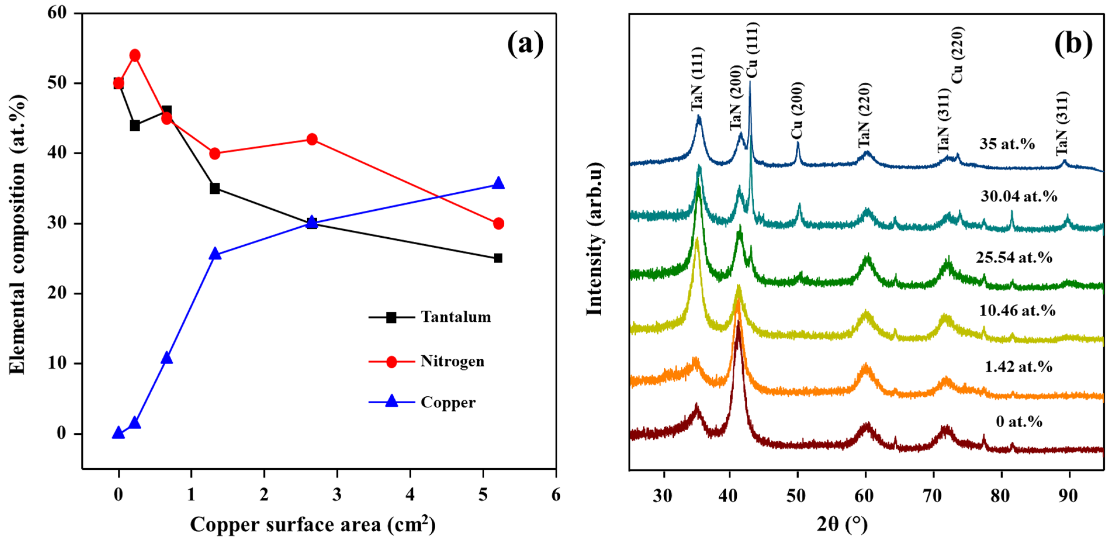
Abstract
:1. Introduction
2. Materials and Methods
2.1. Deposition of TaN/Cu Nanocomposite on Stainless Steel
2.2. Physiochemical Characterization of TaN/Cu Coatings
2.3. Tribological Characterization of TaN/Cu Coatings
2.4. Evaluation of Antibacterial Activity of TaN/Cu Coatings
3. Results
3.1. Elemental Composition Analysis of TaN/Cu Nanocomposite Coatings
3.2. Crystalline Structure of TaN/Cu Nanocomposite Coatings
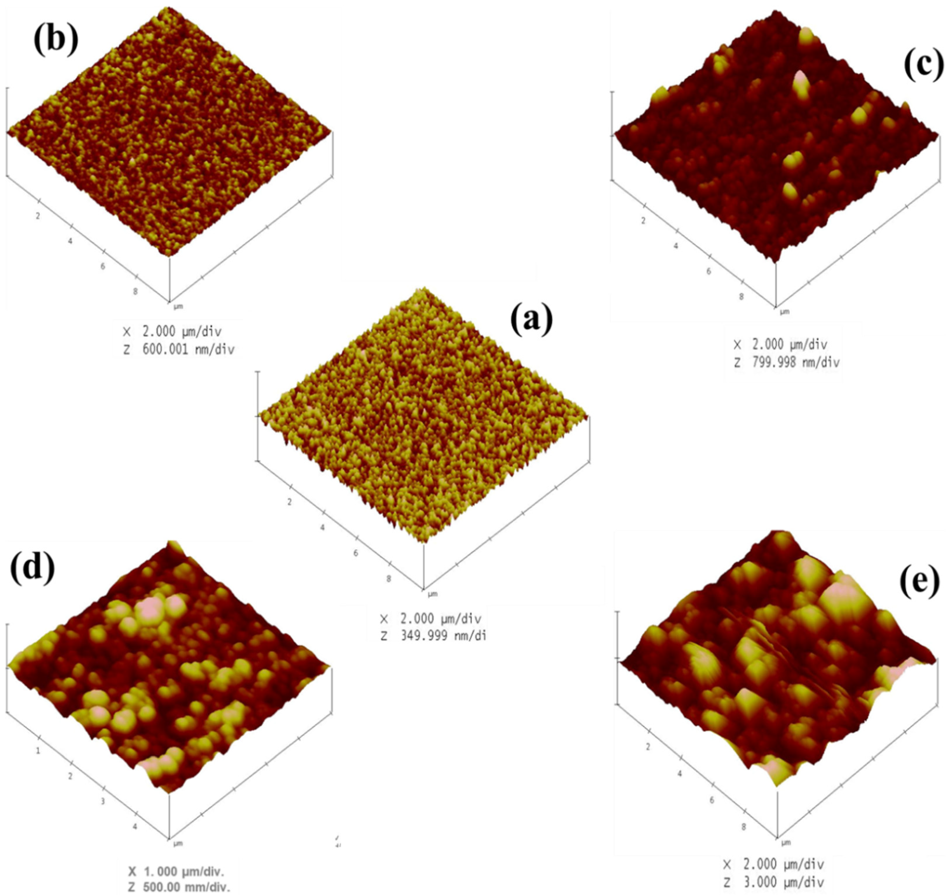
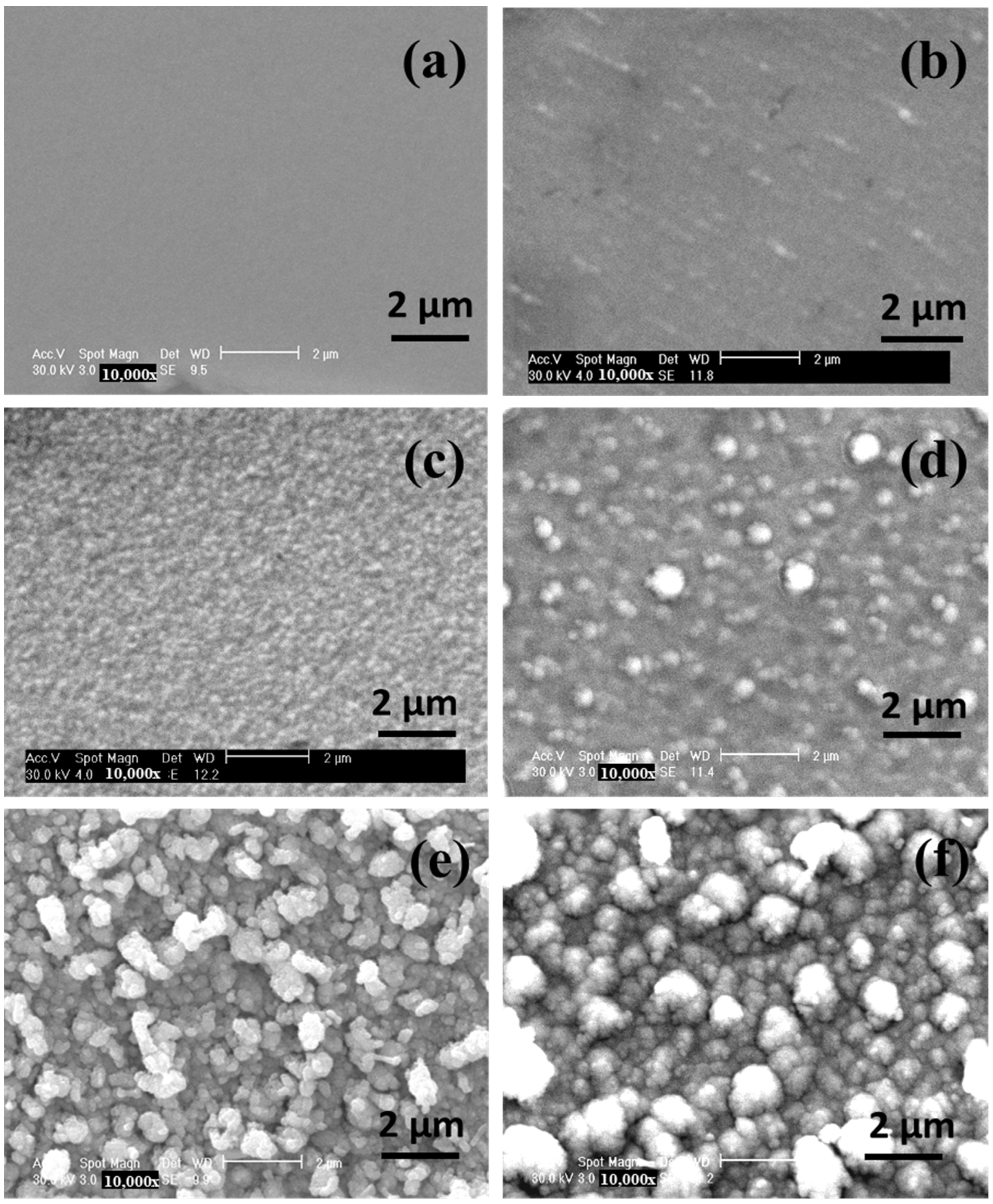
3.3. Morphology of TaN/Cu Nanocomposite Coatings
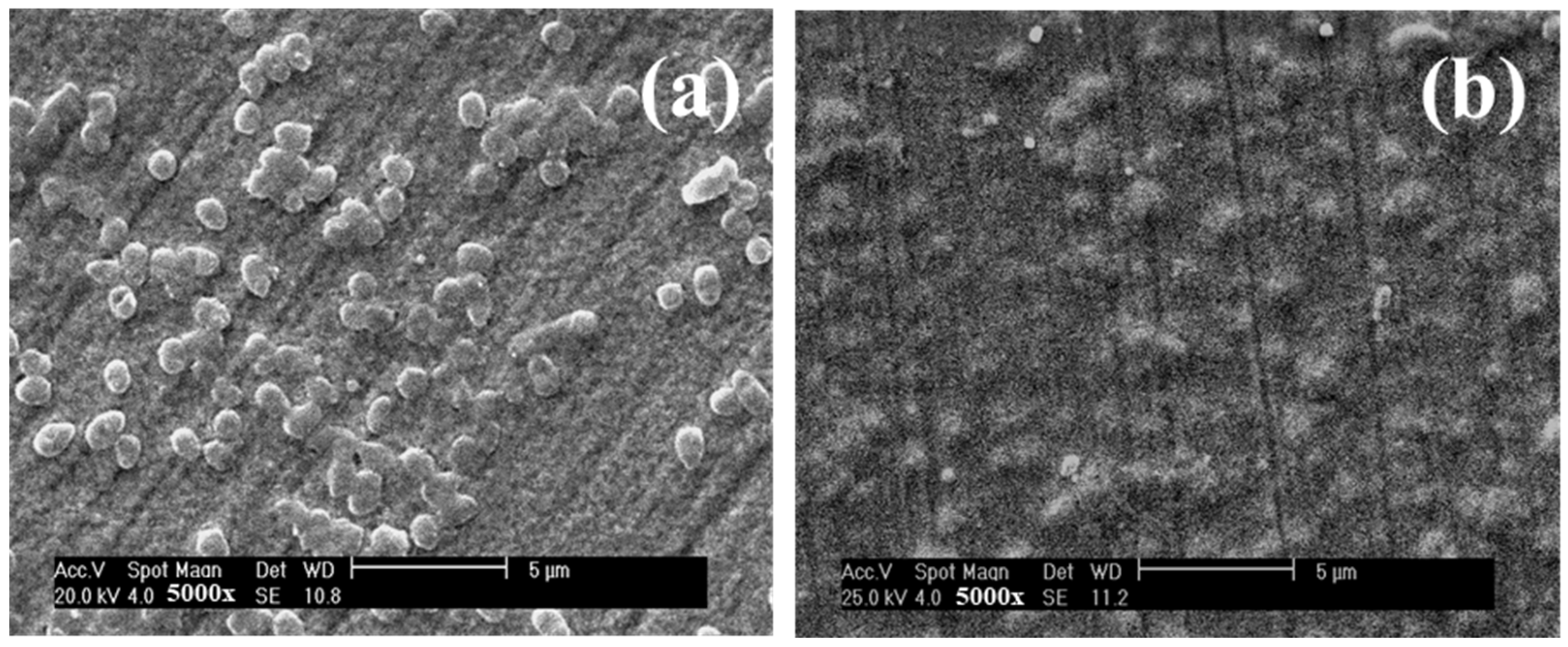
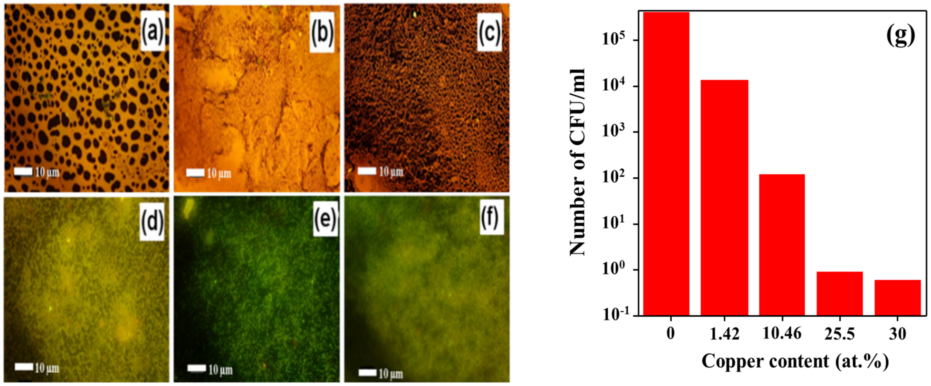
3.4. Antibacterial Properties of TaN/Cu Nanocomposite Coatings
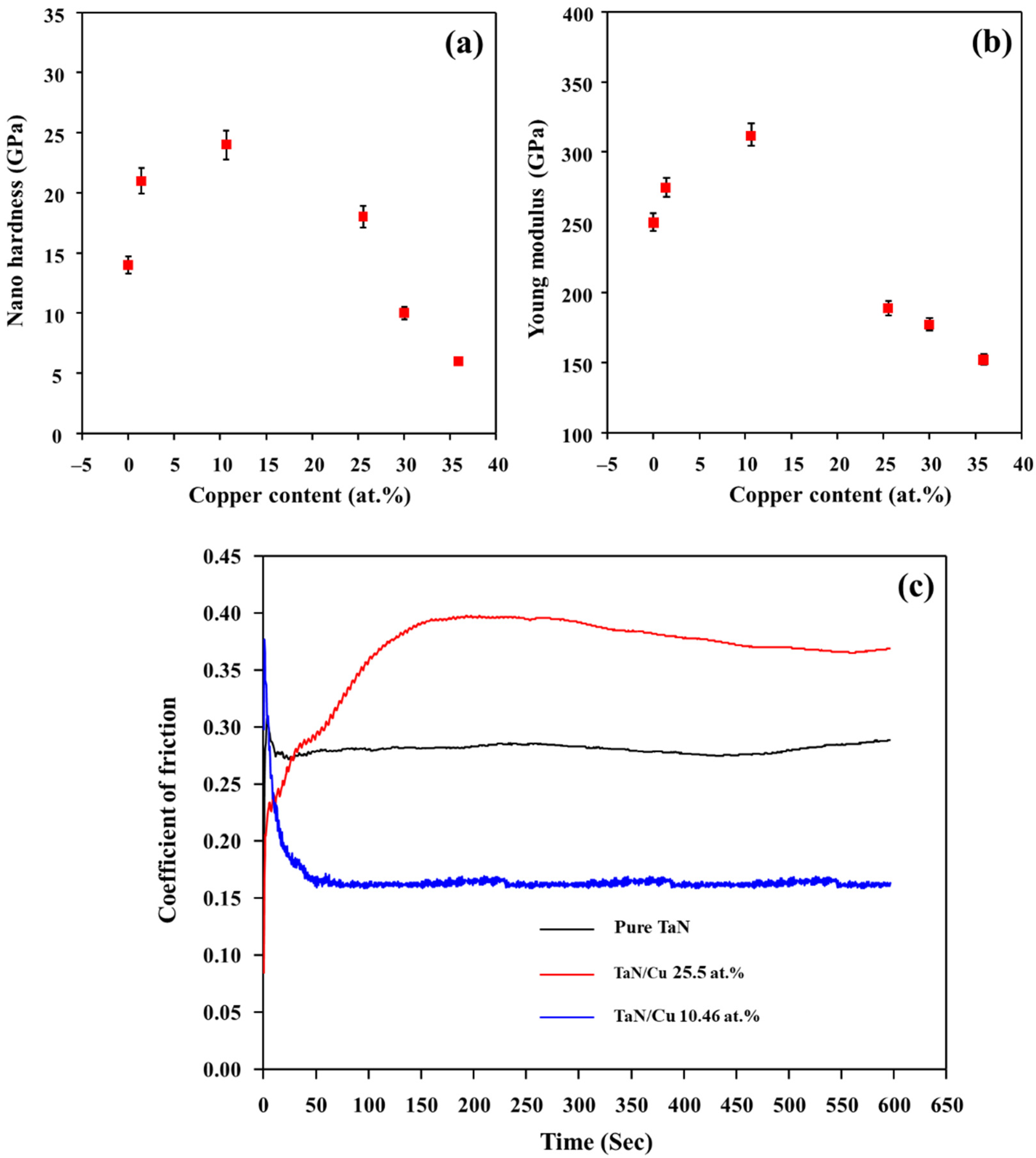
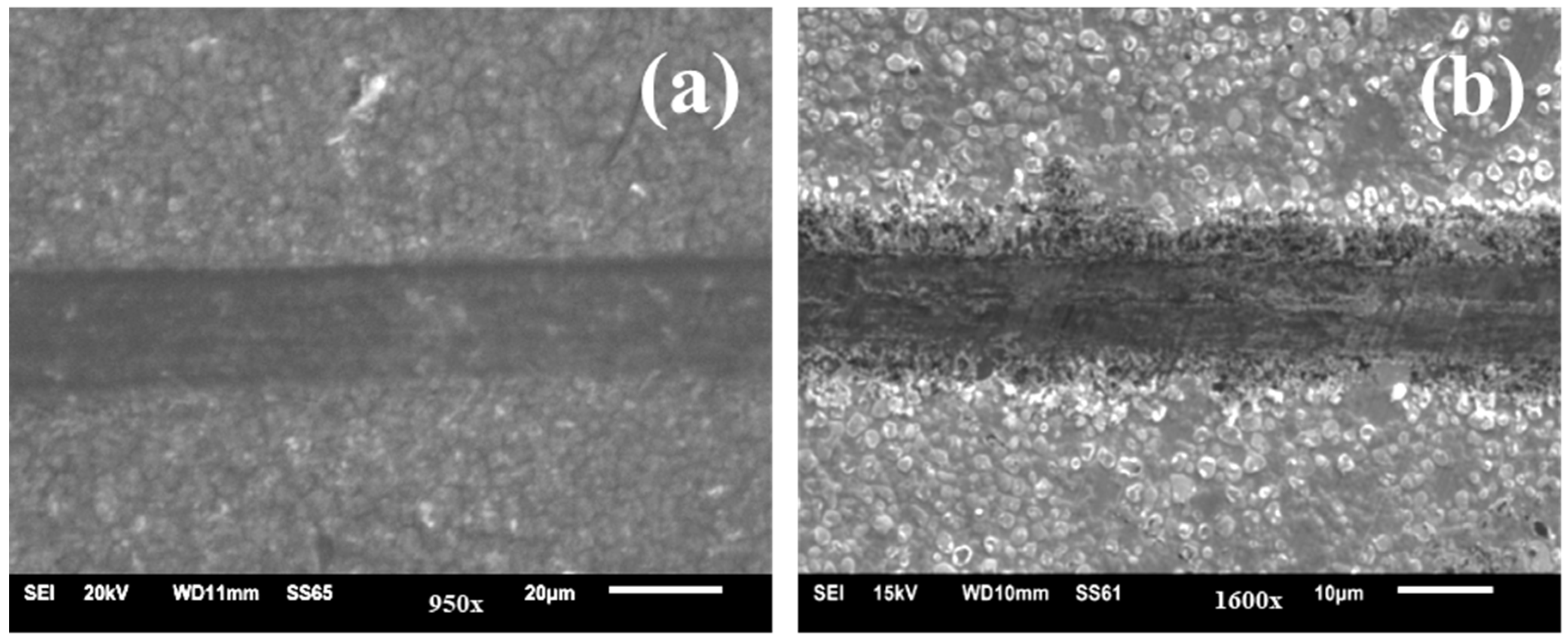
3.5. Tribological Properties of TaN/Cu Nanocomposite Coatings
4. Conclusions
Author Contributions
Funding
Data Availability Statement
Conflicts of Interest
References
- Hyde, F.W.; Alberg, M.; Smith, K. Comparison of Fluorinated Polymers against Stainless Steel, Glass and Polypropylene in Microbial Biofilm Adherence and Removal. J. Ind. Microbiol. Biotechnol. 1997, 19, 142–149. [Google Scholar] [CrossRef] [PubMed]
- Constantin, R.; Miremad, B. Performance of Hard Coatings, Made by Balanced and Unbalanced Magnetron Sputtering, for Decorative Applications. Surf. Coat. Technol. 1999, 120–121, 728–733. [Google Scholar] [CrossRef]
- Leone, M.; Garnier, F.; Avidan, M.; Martin, C. Catheter-Associated Urinary Tract Infections in Intensive Care Units. Microbes Infect. 2004, 6, 1026–1032. [Google Scholar] [CrossRef] [PubMed]
- Birkett, M.; Dover, L.; Lukose, C.C.; Zia, A.W.; Tambuwala, M.M.; Serrano-Aroca, Á. Recent Advances in Metal-Based Antimicrobial Coatings for High-Touch Surfaces. Int. J. Mol. Sci. 2022, 23, 1162. [Google Scholar] [CrossRef] [PubMed]
- Clark, N.M. Emerging Infections in Intensive Care Units. Semin. Respir. Crit. Care Med. 2003, 24, 061–068. [Google Scholar] [CrossRef] [PubMed]
- Squier, C.; Yu, V.L.; Stout, J.E. Waterborne Nosocomial Infections. Curr. Infect. Dis. Rep. 2000, 2, 490–496. [Google Scholar] [CrossRef]
- Mitra, D.; Li, M.; Kang, E.T.; Neoh, K.G. Transparent Copper-Based Antibacterial Coatings with Enhanced Efficacy against Pseudomonas Aeruginosa. ACS Appl. Mater. Interfaces 2019, 11, 73–83. [Google Scholar] [CrossRef] [PubMed]
- Tan, S.; Zhang, X.; Zhang, Y.; Zhen, R.; Zhu, X.; Tian, Z.; Wang, Z. Microstructure and Properties of Cr-Cu-N Coatings with Ultra-Low Copper Content. Surf. Coat. Technol. 2015, 275, 270–275. [Google Scholar] [CrossRef]
- Gao, H.; Li, Y.; Li, C.; Ma, F.; Song, Z.; Xu, K. Tuning the Electronic Properties in TaNx/Ag Nanocomposite Thin Films. RSC Adv. 2016, 6, 30998–31004. [Google Scholar] [CrossRef]
- Lv, C.F.; Zhang, G.F.; Cao, B.S.; He, Y.Y.; Hou, X.D.; Song, Z.X. Structure and Mechanical Properties of A-C/(AlCrWTaTiNb)CxNy Composite Films. Surf. Eng. 2016, 32, 541–546. [Google Scholar] [CrossRef]
- Bharadishettar, N.; Bhat, K.U.; Panemangalore, D.B. Coating Technologies for Copper Based Antimicrobial Active Surfaces: A Perspective Review. Metals 2021, 11, 711. [Google Scholar] [CrossRef]
- Wan, Y.Z.; Xiong, G.Y.; Liang, H.; Raman, S.; He, F.; Huang, Y. Modification of Medical Metals by Ion Implantation of Copper. Appl. Surf. Sci. 2007, 253, 9426–9429. [Google Scholar] [CrossRef]
- Dan, Z.G.; Ni, H.W.; Xu, B.F.; Xiong, J.; Xiong, P.Y. Microstructure and Antibacterial Properties of AISI 420 Stainless Steel Implanted by Copper Ions. Thin Solid Films 2005, 492, 93–100. [Google Scholar] [CrossRef]
- Kelly, P.J.; Li, H.; Whitehead, K.A.; Verran, J.; Arnell, R.D.; Iordanova, I. A Study of the Antimicrobial and Tribological Properties of TiN/Ag Nanocomposite Coatings. Surf. Coat. Technol. 2009, 204, 1137–1140. [Google Scholar] [CrossRef]
- Tian, X.B.; Wang, Z.M.; Yang, S.Q.; Luo, Z.J.; Fu, R.K.Y.; Chu, P.K. Antibacterial Copper-Containing Titanium Nitride Films Produced by Dual Magnetron Sputtering. Surf. Coat. Technol. 2007, 201, 8606–8609. [Google Scholar] [CrossRef]
- Elangovan, T.; George, R.P.; Kuppusami, P.; Mangalaraj, D.; Bera, S.; Mohandas, E.; Kim, D.E. Development of a CrN/Cu Nanocomposite Coating on Titanium-Modified Stainless Steel for Antibacterial Activity against Pseudomonas Aeruginosa. Biofouling 2012, 28, 779–787. [Google Scholar] [CrossRef]
- Liu, L.T.; Chin, A.W.H.; Yu, P.; Poon, L.L.M.; Huang, M.X. Anti-Pathogen Stainless Steel Combating COVID-19. Chem. Eng. J. 2021, 433, 133783. [Google Scholar] [CrossRef]
- Hsieh, J.H.; Cheng, M.K.; Chang, Y.K.; Li, C.; Chang, C.L.; Liu, P.C. Effects of Annealing on Antiwear and Antibacteria Behaviors of TaN–Cu Nanocomposite Thin Films. J. Vac. Sci. Technol. A Vacuum Surf. Films 2008, 26, 1093–1097. [Google Scholar] [CrossRef]
- Aryasomayajula, A.; Valleti, K.; Aryasomayajula, S.; Bhat, D.G. Pulsed DC Magnetron Sputtered Tantalum Nitride Hard Coatings for Tribological Applications. Surf. Coat. Technol. 2006, 201, 4401–4405. [Google Scholar] [CrossRef]
- Echavarría, A.M.; Bejarano, G.G.; Meza, J.M. Características Mecánicas y Tribológicas de Recubrimientos Nanocompuestos Dúplex de TaN (Ag-Cu): Su Respuesta al Tratamiento Térmico. Rev. Chil. Ing. 2017, 25, 662–673. [Google Scholar] [CrossRef] [Green Version]
- Elangovan, T.; Kuppusami, P.; Thirumurugesan, R.; Sudha, C.; Mohandas, E.; Mangalaraj, D. A Study on the Influence of Copper Content in CrN/Cu Nanocomposite Thin Films Prepared by Pulsed Dc Magnetron Sputtering. J. Nanosci. Nanotechnol. 2009, 9, 5436–5440. [Google Scholar] [CrossRef] [PubMed]
- Sandu, C.S.; Sanjinés, R.; Benkahoul, M.; Parlinska-Wojtan, M.; Karimi, A.; Lévy, F. Influence of Ge Addition on the Morphology and Properties of TiN Thin Films Deposited by Magnetron Sputtering. Thin Solid Films 2005, 496, 336–341. [Google Scholar] [CrossRef]
- Elangovan, T.; Murugeshan, S.; Mangalaraj, D.; Kuppusami, P.; Khan, S.; Sudha, C.; Ganesan, V.; Divakar, R.; Mohandas, E. Synthesis and High Temperature XRD Studies of Tantalum Nitride Thin Films Prepared by Reactive Pulsed Dc Magnetron Sputtering. J. Alloys Compd. 2011, 509, 6400–6407. [Google Scholar] [CrossRef]
- Korotaev, A.D.; Ditenberg, I.A.; Berezovskaya, V.R.; Denisov, K.I.; Pinzhin, Y.P.; Borisov, D.P. Condensed-State Physics: The Influence of the Ion-Plasma Synthesis Regimes on the Features of Structural-Phase State of Multicomponent Nanocomposite Al–Cr–Si–Ti–Cu–N Coatings. Russ. Phys. J. 2015, 57, 1301–1307. [Google Scholar] [CrossRef]
- Nocentini, S. Comet Assay Analysis of Repair of DNA Strand Breaks in Normal and Deficient Human Cells Exposed to Radiations and Chemicals. Evidence for a Repair Pathway Specificity of DNA Ligation. Radiat. Res. 1995, 144, 170–180. [Google Scholar] [CrossRef]
- Yang, Y.H.; Chen, D.J.; Wu, F.B. Microstructure, Hardness, and Wear Resistance of Sputtering TaN Coating by Controlling RF Input Power. Surf. Coat. Technol. 2016, 303, 32–40. [Google Scholar] [CrossRef]
- Liu, K.Y.; Lee, J.W.; Wu, F.B. Fabrication and tribological behavior of sputtering TaN coatings. Surf. Coat. Technol. 2014, 259, 123–128. [Google Scholar] [CrossRef]
- Bernoulli, D.; Müller, U.; Schwarzenberger, M.; Hauert, R.; Spolenak, R. Magnetron sputter deposited tantalum and tantalum nitride thin films: An analysis of phase, hardness and composition. Thin Solid Films 2013, 548, 157–161. [Google Scholar] [CrossRef]
- Zeman, P.; Mayrhofer, P.H.; Mitterer, C.; Musil, J. Structure and Properties of Hard and Superhard Zr-Cu-N Nanocomposite Coatings. Mater. Sci. Eng. A 2000, 289, 189–197. [Google Scholar] [CrossRef]
- Musil, J.; Vlček, J. Magnetron Sputtering of Hard Nanocomposite Coatings and Their Properties. Surf. Coat. Technol. 2001, 142, 557–566. [Google Scholar] [CrossRef]
- Musil, J.; Blažek, J.; Fajfrlík, K.; Čerstvý, R. Flexible Antibacterial Al–Cu–N Films. Surf. Coat. Technol. 2015, 264, 114–120. [Google Scholar] [CrossRef]
- Mortazavi, V.; Nosonovsky, M. Friction-Induced Pattern Formation and Turing Systems. Langmuir 2011, 27, 4772–4779. [Google Scholar] [CrossRef] [PubMed]
- Zhang, X.; Luster, B.; Church, A.; Muratore, C.; Voevodin, A.A.; Kohli, P.; Aouadi, S.; Talapatra, S. Carbon Nanotube-MoS2 Composites as Solid Lubricants. ACS Appl. Mater. Interfaces 2009, 1, 735–739. [Google Scholar] [CrossRef] [PubMed]
- Kim, B. Microstructure and Tribological Behavior of CrN-Cu Nanocoatings Deposited by PVD Systems. In The SAE Technical Papers, Proceedings of the SAE 2016 World Congress and Exhibition, 12 April 2016; SAE International: Warrendale, PA, USA, 2016. [Google Scholar]
- Yu, L.H.; Huang, T.; Xu, J.H. Microstructure, Mechanical and Tribological Properties of TaCN Composite Films. Surf. Eng. 2016, 33, 1–6. [Google Scholar] [CrossRef]
- Yan, Y.; Lin, J.; Liu, T.; Liu, B.; Wang, B.; Qiao, L.; Tu, J.; Cao, J.; Qi, J. Corrosion Behavior of Stainless Steel-Tungsten Carbide Joints Brazed with AgCuX (X = In, Ti) Alloys. Corros. Sci. 2022, 200, 110231. [Google Scholar] [CrossRef]


Publisher’s Note: MDPI stays neutral with regard to jurisdictional claims in published maps and institutional affiliations. |
© 2022 by the authors. Licensee MDPI, Basel, Switzerland. This article is an open access article distributed under the terms and conditions of the Creative Commons Attribution (CC BY) license (https://creativecommons.org/licenses/by/4.0/).
Share and Cite
Elangovan, T.; Balasankar, A.; Arokiyaraj, S.; Rajagopalan, R.; George, R.P.; Oh, T.H.; Kuppusami, P.; Ramasundaram, S. Highly Durable Antimicrobial Tantalum Nitride/Copper Coatings on Stainless Steel Deposited by Pulsed Magnetron Sputtering. Micromachines 2022, 13, 1411. https://doi.org/10.3390/mi13091411
Elangovan T, Balasankar A, Arokiyaraj S, Rajagopalan R, George RP, Oh TH, Kuppusami P, Ramasundaram S. Highly Durable Antimicrobial Tantalum Nitride/Copper Coatings on Stainless Steel Deposited by Pulsed Magnetron Sputtering. Micromachines. 2022; 13(9):1411. https://doi.org/10.3390/mi13091411
Chicago/Turabian StyleElangovan, Thangavel, Athinarayanan Balasankar, Selvaraj Arokiyaraj, Ramaseshan Rajagopalan, Rani P. George, Tae Hwan Oh, Parasuraman Kuppusami, and Subramaniyan Ramasundaram. 2022. "Highly Durable Antimicrobial Tantalum Nitride/Copper Coatings on Stainless Steel Deposited by Pulsed Magnetron Sputtering" Micromachines 13, no. 9: 1411. https://doi.org/10.3390/mi13091411