
Revisiting Defect-Induced Light Field Enhancement in Optical Thin Films


Abstract
:1. Introduction
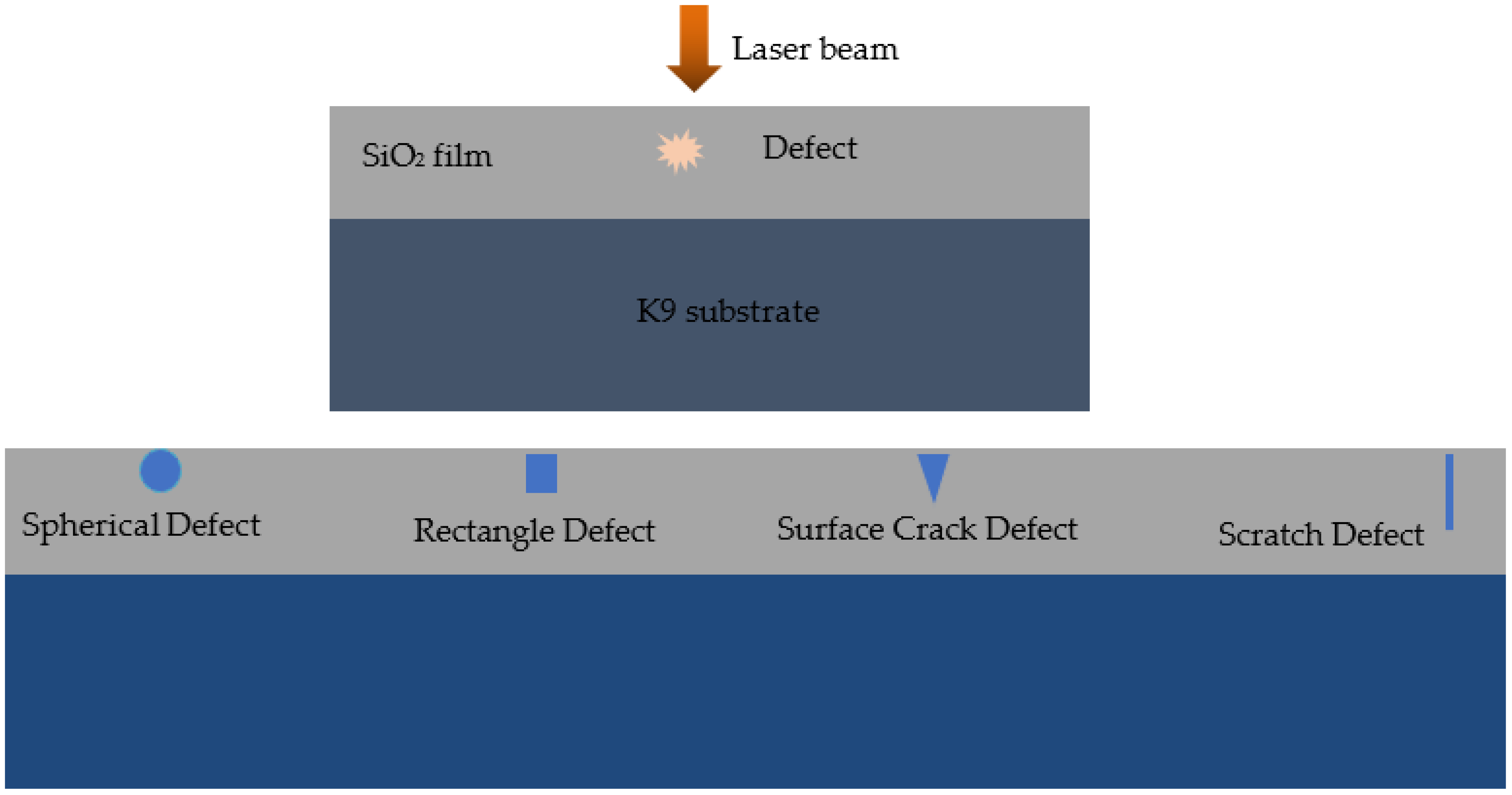
2. Theory and Model
3. Results and Discussions
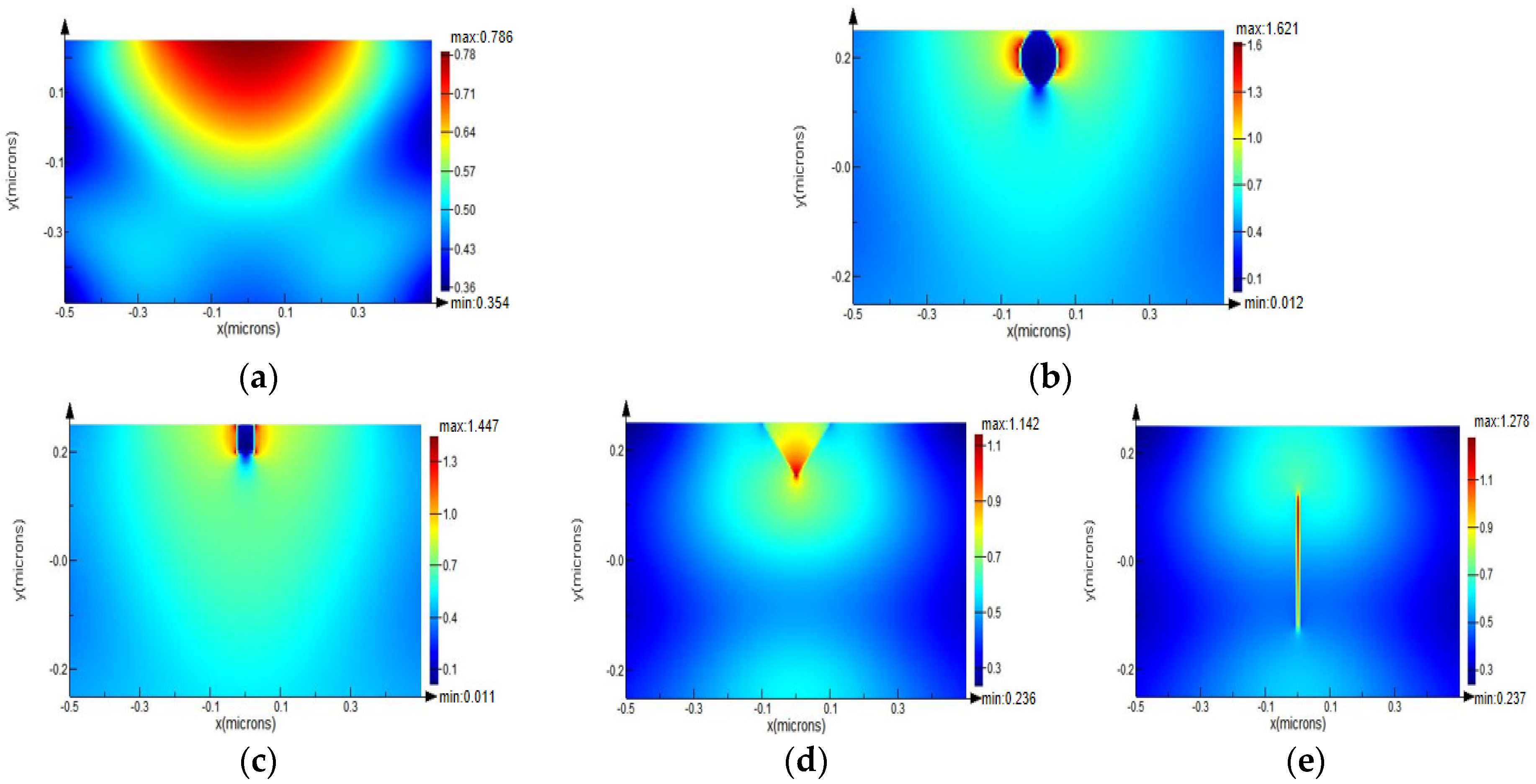
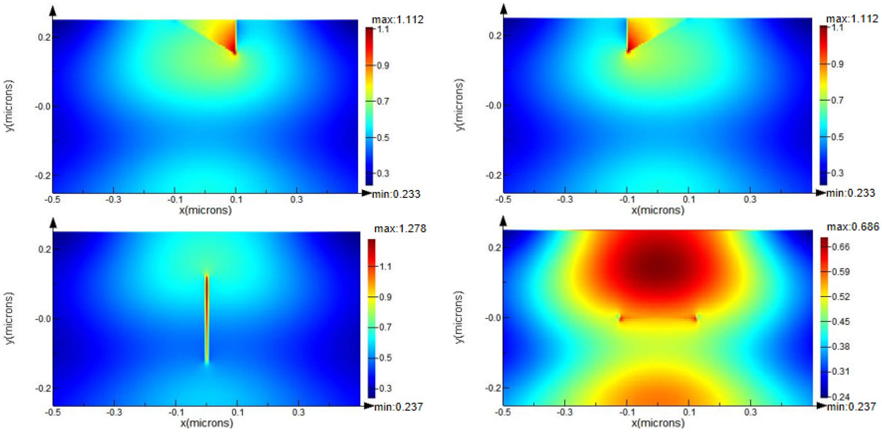
3.1. Light Field Enhancement Induced by Defect with Different Geometries
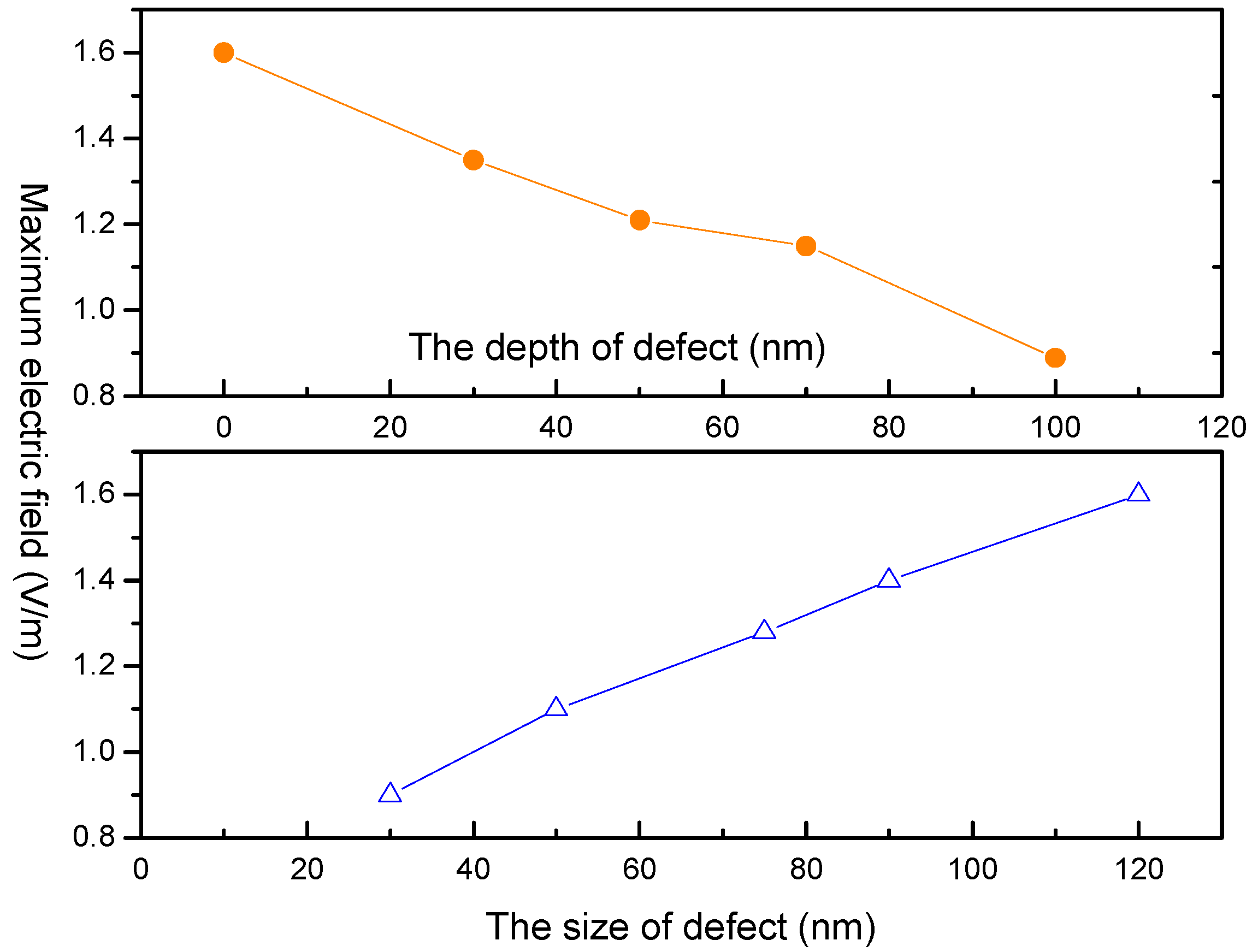
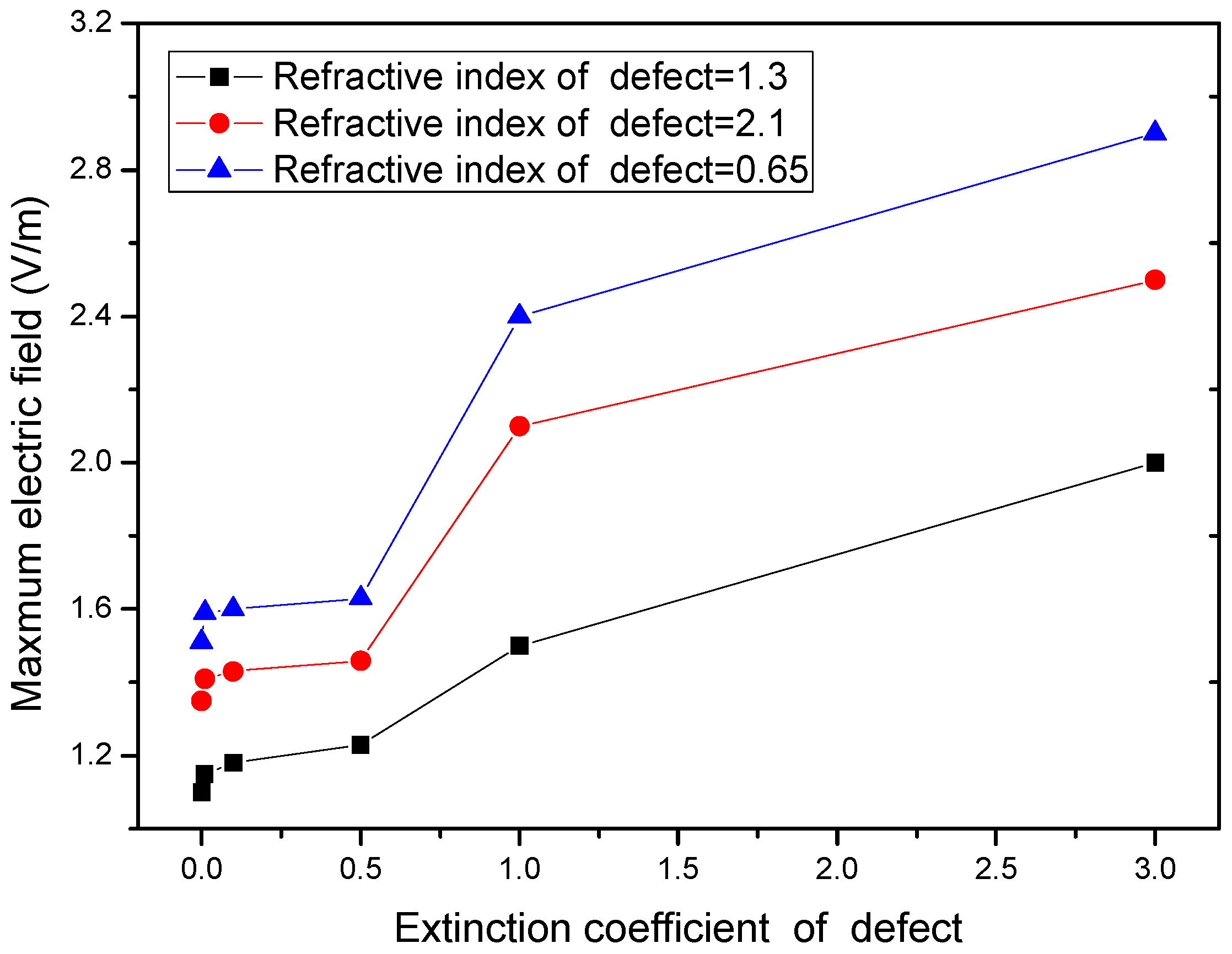
3.2. Light Field Enhancement Induced by Defect with Different Characteristics
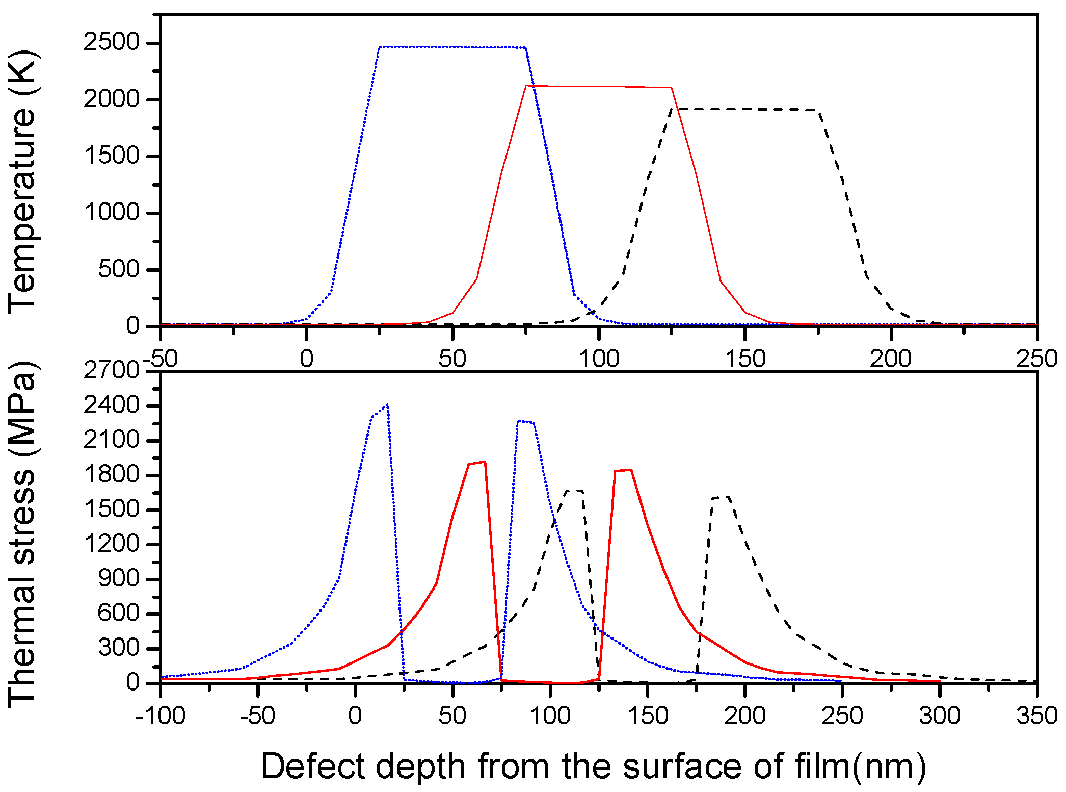
3.3. Thermal-Mechanical Effect Due to Light Field Intensification
4. Conclusions
Author Contributions
Funding
Data Availability Statement
Conflicts of Interest
References
- Liu, Z.; Zheng, Y.; Pan, F.; Lin, Q.; Ma, P.; Wang, J. Investigation of laser induced damage threshold measurement with single-shot on thin films. Appl. Sur. Sci. 2016, 382, 294–301. [Google Scholar] [CrossRef]
- Demos, S.G.; Staggs, M.; Minoshima, K.; Fujimoto, J. Characterization of laser induced damage sites in optical components. Opt. Express 2002, 10, 1444–1450. [Google Scholar] [CrossRef] [PubMed]
- Du, Y.; Liu, S.; He, H.; Jin, Y.; Kong, F.; Guan, H. Laser-induced damage properties of antireflective porous glasses. Opt. Commun. 2012, 285, 5512–5518. [Google Scholar] [CrossRef]
- Jiao, H.; Cheng, X.; Lu, J.; Bao, G.; Zhang, J.; Ma, B.; Liu, H.; Wang, Z. Study for improvement of laser induced damage of 1064 nm AR coatings in nanosecond pulse. J. Opt. Soc. Korea 2013, 17, 1–4. [Google Scholar] [CrossRef] [Green Version]
- Dijon, J.; Poiroux, T.; Desrumaux, C. Nano absorbing centers: A key point in the laser damage of thin films. Proc. SPIE 1997, 2966, 315–325. [Google Scholar]
- Cheng, X.; Wang, Z. Defect-related properties of optical coatings. Adv. Opt. Technol. 2014, 3, 65–90. [Google Scholar] [CrossRef]
- Cheng, X.; Shen, Z.; Jiao, H.; Zhang, J.; Ma, B.; Ding, T.; Lu, J.; Wang, X.; Wang, Z. Laser damage study of nodules in electron-beam evaporated HfO2/SiO2 high reflectors. Appl. Opt. 2011, 50, C357–C363. [Google Scholar] [CrossRef]
- Zhang, L.X.; Zhu, X.B.; Li, F.Y.; Zhang, R.Z. Laser-induced thermal damage influenced by surface defects of materials. Acta Opt. Sin. 2016, 36, 0914001. [Google Scholar] [CrossRef]
- Zhu, Z.; Chenga, X.; Huanga, L.; Liu, Z. Light field intensification induced by nano-inclusions in optical thin-films. Appl. Surf. Sci. 2012, 258, 5126–5130. [Google Scholar] [CrossRef]
- Stolz, C.J.; Feigenbaum, E. Impact of high refractive coating material on the nodular-induced electric field enhancement for near infrared multilayer mirrors. Appl. Opt. 2020, 59, A20–A25. [Google Scholar] [CrossRef]
- Stolz, C.J.; Hafeman, S.; Pistor, T.V. Light intensification modeling of coating inclusions irradiated at 351 and 1053 nm. Appl. Opt. 2008, 47, C162–C166. [Google Scholar] [CrossRef]
- Zhang, J.; Jiao, H.; Ma, B.; Wang, Z.; Cheng, X. Laser-induced damage of nodular defects in dielectric multilayer coatings. Opt. Eng. 2018, 57, 121909. [Google Scholar] [CrossRef]
- Cheng, J.; Chen, M.; Liao, W. Fabrication of spherical mitigation pit on KH2PO4 crystal by micro-milling and modeling of its induced light intensification. Opt. Express 2013, 21, 16799–16813. [Google Scholar] [CrossRef] [PubMed]
- Boling, N.L.; Crisp, M.D.; Dubé, G. Laser Induced Surface Damage. Appl. Opt. 1973, 12, 650–660. [Google Scholar] [CrossRef] [PubMed]
- Mutilin, S.V.; Khasanov, T. The refractive index of homogeneous SiO2 thin films. Opt. Spectrosc. 2008, 105, 461–465. [Google Scholar] [CrossRef]
- Crisp, M.D.; Boling, N.L.; Dube, G. Importance of Fresnel reflections in laser surface damage of transparent dielectrics. Appl. Phys. Lett. 1972, 21, 364–366. [Google Scholar] [CrossRef]
- Cheng, X.; Tuniyazi, A.; Wei, Z.; Zhang, J.; Ding, T.; Jiao, H.; Ma, B.; Li, H.; Li, T.; Wang, Z. Physical insight toward electric field enhancement at nodular defects in optical coatings. Opt. Express 2015, 23, 8609–8617. [Google Scholar] [CrossRef] [PubMed]
- Stolz, C.J.; Feit, M.D.; Pistor, T.V. Laser intensification by spherical inclusions embedded within multilayer coatings. Appl. Opt. 2006, 45, 1594–1601. [Google Scholar] [CrossRef]
- Ling, X.; Shao, J.; Fan, Z. Thermal-mechanical modeling of nodular defect embedded within multilayer coatings. J. Vac. Sci. Technol. A 2009, 27, 183–186. [Google Scholar] [CrossRef]
- Cheng, X.; Zhang, J.; Ding, T.; Wei, Z.; Li, H.; Wang, Z. The effect of an electric field on the thermomechanical damage of nodular defects in dielectric multilayer coatings irradiated by nanosecond laser pulses. Light: Sci. Appl. 2013, 2, e80. [Google Scholar] [CrossRef] [Green Version]
- Duchateau, G.; Dyan, A. Coupling statistics and heat transfer to study laser-induced crystal damage by nanosecond pulses. Opt. Express 2007, 15, 4557–4576. [Google Scholar] [CrossRef] [PubMed]
- Li, B.; Hou, C.X.; Tian, C.X. Layer by layer exposure of subsurface defects and laser-induced damage mechanism of fused silica. Appl. Surf. Sci. 2020, 508, 145186. [Google Scholar] [CrossRef]


Publisher’s Note: MDPI stays neutral with regard to jurisdictional claims in published maps and institutional affiliations. |
© 2022 by the authors. Licensee MDPI, Basel, Switzerland. This article is an open access article distributed under the terms and conditions of the Creative Commons Attribution (CC BY) license (https://creativecommons.org/licenses/by/4.0/).
Share and Cite
Ling, X.; Chen, X.; Liu, X. Revisiting Defect-Induced Light Field Enhancement in Optical Thin Films. Micromachines 2022, 13, 911. https://doi.org/10.3390/mi13060911
Ling X, Chen X, Liu X. Revisiting Defect-Induced Light Field Enhancement in Optical Thin Films. Micromachines. 2022; 13(6):911. https://doi.org/10.3390/mi13060911
Chicago/Turabian StyleLing, Xiulan, Xin Chen, and Xiaofeng Liu. 2022. "Revisiting Defect-Induced Light Field Enhancement in Optical Thin Films" Micromachines 13, no. 6: 911. https://doi.org/10.3390/mi13060911