Article

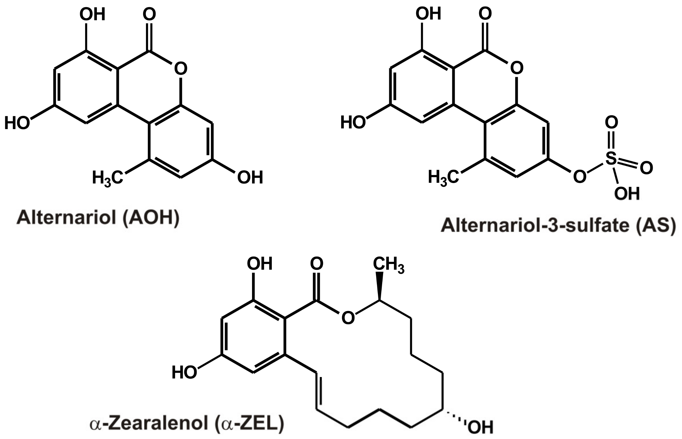
Probing the Interactions of 31 Mycotoxins with Xanthine Oxidase: Alternariol, Alternariol-3-Sulfate, and α-Zearalenol Are Allosteric Inhibitors of the Enzyme

1
Department of Pharmacology, Faculty of Pharmacy, University of Pécs, Rókus u. 2, H-7624 Pécs, Hungary
2
Department of Pharmaceutics and Central Clinical Pharmacy, Faculty of Pharmacy, University of Pécs, H-7624 Pécs, Hungary
3
Unit of Pharmacoinformatics, Department of Pharmacology and Pharmacotherapy, Medical School, University of Pécs, Szigeti út 12, H-7624 Pécs, Hungary
4
Food Biotechnology Research Group, János Szentágothai Research Centre, University of Pécs, Ifjúság útja 20, H-7624 Pécs, Hungary
*
Author to whom correspondence should be addressed.
Toxins 2023, 15(4), 250; https://doi.org/10.3390/toxins15040250
Submission received: 24 February 2023 / Revised: 22 March 2023 / Accepted: 27 March 2023 / Published: 29 March 2023
(This article belongs to the Special Issue Toxicity Mechanisms and Management Strategies of Mycotoxin)

Abstract

:
Mycotoxins are frequent toxic contaminants in foods and beverages, causing a significant health threat. Interactions of mycotoxins with biotransformation enzymes (e.g., cytochrome P450 enzymes, sulfotransferases, and uridine 5′-diphospho-glucuronosyltransferases) may be important due to their possible detoxification or toxic activation during enzymatic processes. Furthermore, mycotoxin-induced enzyme inhibition may affect the biotransformation of other molecules. A recent study described the strong inhibitory effects of alternariol and alternariol-9-methylether on the xanthine oxidase (XO) enzyme. Therefore, we aimed to test the impacts of 31 mycotoxins (including the masked/modified derivatives of alternariol and alternariol-9-methylether) on XO-catalyzed uric acid formation. Besides the in vitro enzyme incubation assays, mycotoxin depletion experiments and modeling studies were performed. Among the mycotoxins tested, alternariol, alternariol-3-sulfate, and α-zearalenol showed moderate inhibitory actions on the enzyme, representing more than tenfold weaker impacts compared with the positive control inhibitor allopurinol. In mycotoxin depletion assays, XO did not affect the concentrations of alternariol, alternariol-3-sulfate, and α-zearalenol in the incubates; thus, these compounds are inhibitors but not substrates of the enzyme. Experimental data and modeling studies suggest the reversible, allosteric inhibition of XO by these three mycotoxins. Our results help the better understanding of the toxicokinetic interactions of mycotoxins.
Key Contribution: The interactions of 31 mycotoxins were examined with the xanthine oxidase enzyme. Among the compounds tested, alternariol, alternariol-3-sulfate, and α-zearalenol are moderate, allosteric inhibitors of the enzyme.

1. Introduction

Mycotoxins are secondary metabolites of molds. Mycotoxin contamination in foods and beverages causes significant health threats worldwide [1]. Their typically high thermal stability and frequent occurrence in the food chain make the removal of mycotoxins very challenging [2,3]. Biotransformation enzymes (such as cytochrome P450 enzymes, sulfotransferases, and uridine 5′-diphospho-glucuronosyltransferases) play important roles in the toxic activation or detoxification of mycotoxins; in addition, the mycotoxin-induced inhibition of these enzymes may affect the biotransformation of other molecules [4].
The Alternaria mycotoxins alternariol (AOH) and alternariol-9-methylether (AME), as well as their masked (glucoside) and modified (sulfate) derivatives, typically contaminate tomato products (Table 1) [5,6,7]. The sulfate metabolites, such as alternariol sulfate (AS) and alternariol monomethyl ether sulfate (AMS), are also produced by sulfotransferases in mammals [8,9].
Aflatoxins, sterigmatocystin (STC), and cyclopiazonic acid (CPA) are usually produced by Aspergillus molds (Table 1) [10,11,12,13]. Aflatoxin B1 (AFB1) is the most common and most toxic aflatoxin derivative, whereas aflatoxin M1 (AFM1) frequently contaminates milk [11]. STC can also be considered an intermediate product in aflatoxin biosynthesis [12]. Citrinin (CIT), ochratoxins, and patulin (PAT) are typical products of Aspergillus and Penicillium fungi (Table 1) [1,14,15]. Furthermore, dihydrocitrinone (DHC) is the main metabolite of CIT in the urine of mammals [16]. Among ochratoxins, ochratoxin A (OTA) is the most common contaminant; however, ochratoxin B (OTB) and ochratoxin C (OTC) also appear in the food chain (e.g., in some wines) [17,18,19].
Fusarium strains are responsible for the production of several mycotoxins, including beauvericin (BEA), deoxynivalenol (DON or vomitoxin), fumonisin B1 (FB1), T-2 toxin (T2), and zearalenone (ZEN) (Table 1) [20]. During the biotransformation of ZEN in mammals, reduced and conjugated metabolites are formed. Reduced derivatives are zearalenols (ZEL), zearalanone (ZAN), and zearalanols (ZAL); some of these metabolites (e.g., α-ZEL and α-ZAL) exert much stronger xenoestrogenic effects than the parent mycotoxin [21,22]. In mammals, uridine 5′-diphospho-glucuronosyltransferase and sulfotransferase enzymes can produce glucuronide (e.g., zearalenone-14-glucuronide) and sulfate (e.g., zearalenone-14-sulfate (Z14S)) conjugates, respectively [21]. In addition, masked/modified derivatives also appear in cereals (e.g., wheat, maize, and barley), such as Z14S or zearalenone-14-glucoside (Z14Glz) [21].
Xanthine oxidoreductase is a 300 kDa homodimer protein containing molybdenum cofactor, flavin adenine dinucleotide (FAD) site, and Fe2S2 sites [23]. The enzyme is important in purine catabolism because it catalyzes the transformation of hypoxanthine to xanthine and then xanthine to uric acid [24]. Xanthine oxidoreductase designation means two interconvertible forms of the same enzyme, including xanthine dehydrogenase and xanthine oxidase (XO). Xanthine dehydrogenase can be reversibly or irreversibly converted to XO [25]. Typically, xanthine dehydrogenase is intracellularly presented, while XO is dominant in the extracellular water space [23]. High uric acid levels cause hyperuricemia which may result in the development of gout, cardiovascular diseases, and metabolic syndrome [26]. In addition, XO generates superoxide anion radicals and hydrogen peroxide, which may also be involved in the unpleasant impacts of high uric acid formation [27]. Allopurinol, a potent inhibitor of XO, is commonly used to treat hyperuricemia and gout [28]. Furthermore, XO is also an important enzyme in the biotransformation of certain drugs, such as 6-mercaptopurine (used in the treatment of cancer and autoimmune diseases) [29].
Previous studies demonstrated that aflatoxins and ochratoxins could cause the development of gout in certain animals [30]. AFB1-contaminated diet (for 14 days and 21 days) increased XO activity and uric acid levels in the serum of fish [31]. In rats, chronic exposure to ZEN significantly elevated XO activity in the liver and kidneys [32]. The upregulation of xanthine oxidoreductase was observed in the liver and the jejunum of chickens after three weeks of DON-contaminated feeding [33]. In contrast, fumonisins (FB1 + FB2) alone and in combination with DON (15 days of exposure) decreased the expression of xanthine dehydrogenase in the jejunum of broiler chickens [34]. These results suggest that certain mycotoxins may be able to increase the expression and/or the activity of xanthine oxidoreductase.
Typically, less information is available regarding the inhibitory effects of mycotoxins on XO. In a recent study, Fan et al. described that AOH (IC50 = 0.2 μM) and AME (IC50 = 0.5 μM) are strong inhibitors of XO, where these mycotoxins showed approximately tenfold higher inhibitory potency compared with allopurinol [35]. Considering these results and the relatively low acute toxicity of AOH and AME, Fan et al. suggested that AOH may be a potential lead compound in the development of new potent XO inhibitors [35]. Urolithins (colon metabolites of ellagitannins) are considered barely toxic compounds [36]. They have very similar chemical structure to Alternaria mycotoxins. In the study of Fan et al. [35], urolithins examined were much weaker inhibitors of XO than AOH or AME. In another paper, considerably weaker inhibitory actions of AOH (IC50 = 15.5 μM) and AME (IC50 = 60.5 μM) were described regarding the XO enzyme [37]. In addition, the consumption of wheat grain contaminated with Alternaria spp. did not affect XO activity in broiler chickens [38].
Importantly, XO is a major component in bovine milk [39], which is commonly contaminated with certain mycotoxins (e.g., AFM1, OTA, ZEN, and α-ZEL) [40]. Thus, mycotoxin–XO interactions can also be important from this point of view.
In this work, the interactions of 31 mycotoxins with the XO enzyme were examined by applying in vitro enzyme assays and modeling studies. We planned to test the effects of AOH, AME, and their masked/modified derivatives (AS, AG, AMS, and AMG) on XO-catalyzed uric acid formation to confirm the previously reported data and to find potentially stronger inhibitors among AOH derivatives. In addition, we also investigated the impacts of 25 other mycotoxins, including AFB1, AFB2, AFG1, AFG2, AFM1, STC, CPA, CIT, DHC, OTA, OTB, OTC, PAT, BEA, DON, FB1, T2, ZEN, α-ZEL, β-ZEL, ZAN, α-ZAL, β-ZAL, Z14S, and Z14Glz (Table 1). The potential inhibitory actions of mycotoxins on XO, the reversibility of their inhibitory effects, and the XO-induced mycotoxin depletion were assessed. Experimental results and molecular modeling studies suggest the moderate, allosteric inhibition of XO by AOH, AS, and α-ZEL (Figure 1).

2. Results and Discussion

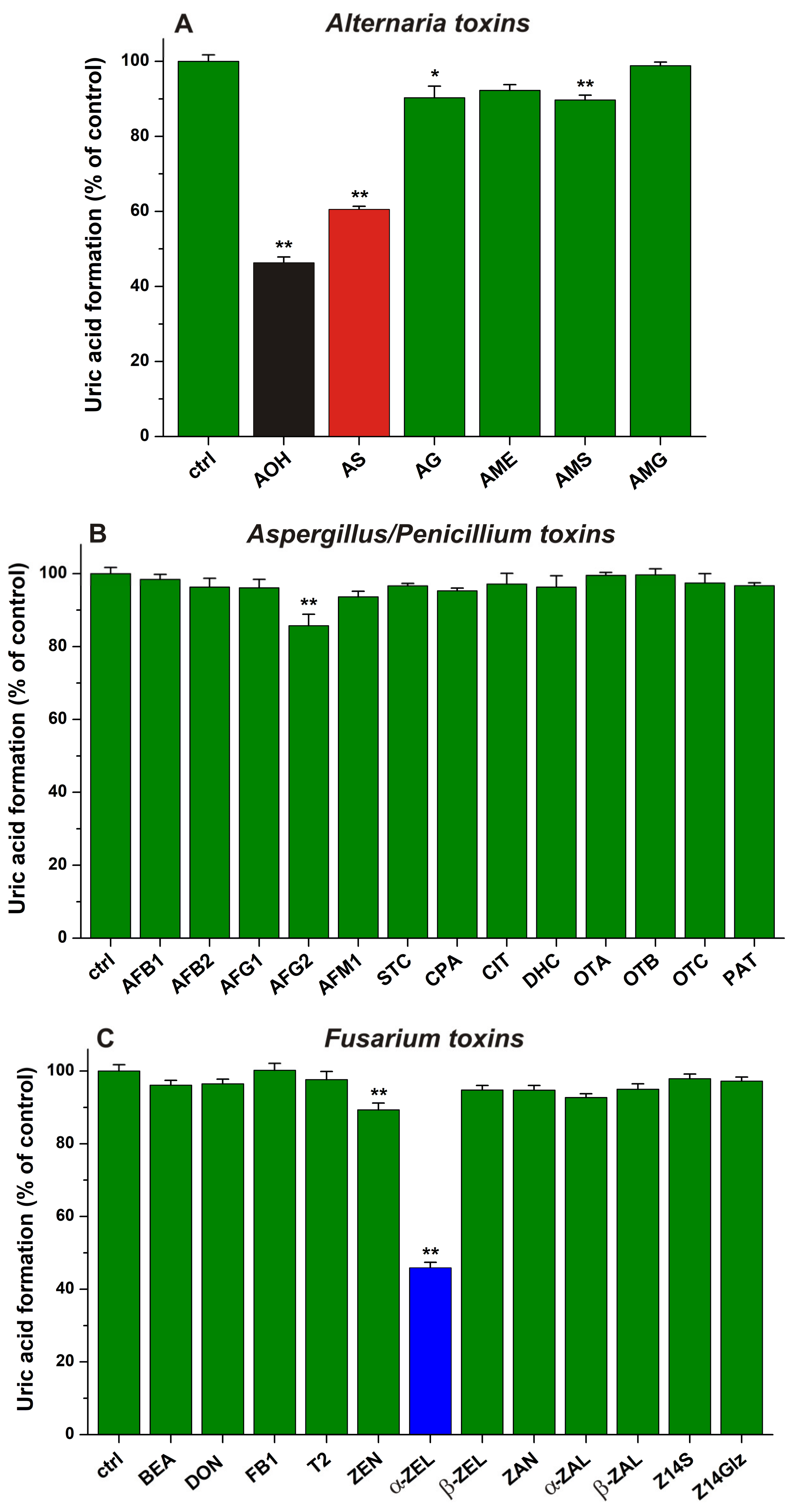
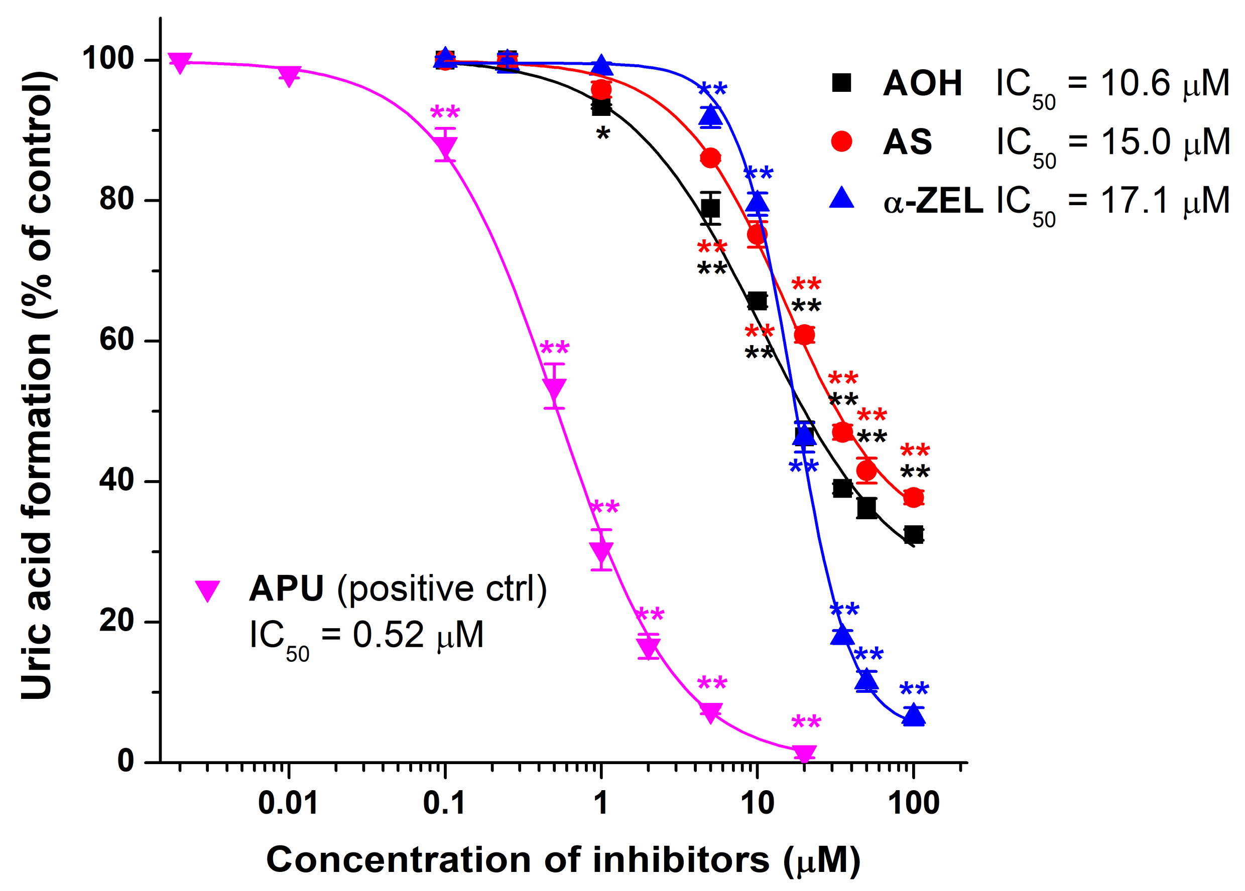
2.1. Inhibitory Effects of Mycotoxins on Xanthine Oxidase Enzyme

The impacts of 20 μM mycotoxin concentrations were tested to evaluate the potential inhibitory effects of mycotoxins on XO-catalyzed xanthine oxidation. Results are demonstrated in Figure 2, where Alternaria, Aspergillus/Penicillium, and Fusarium toxins are presented in three separate panels.
AOH, AS, AG, and AMS induced a statistically significant decrease in metabolite formation, while AME and AMG caused only slight or no effects, respectively (Figure 2A). Among the Alternaria mycotoxins tested, AOH and AS showed the strongest inhibitory actions (resulting in a 54% and 40% decrease in uric acid formation, respectively). AG and AMS induced approximately 10% inhibition; therefore, these mycotoxins can be considered weak inhibitors of XO. Interestingly, our results suggest negligible inhibition of the enzyme by AME, while in a previous report, AME and AOH showed similarly strong inhibitory effects [35]. Furthermore, AS caused a slightly weaker impact vs. AOH (Figure 2A). This result is in accordance with our previous observation that quercetin-3′-sulfate was also a similarly strong inhibitor of XO than the parent flavonoid quercetin [41].
In Figure 2B, the impacts of Aspergillus/Penicillium mycotoxins are presented: AFB1, AFB2, AFG1, AFM1, STC, CPA, CIT, DHC, OTA, OTB, OTC, and PAT did not affect the XO-catalyzed metabolite formation. AFG2 showed a statistically significant but weak inhibitory action (14%) on XO (Figure 2B).
Among the Fusarium mycotoxins, BEA, DON, FB1, T2, β-ZEL, ZAN, α-ZAL, β-ZAL, Z14S, and Z14Glz did not influence the XO-catalyzed uric acid formation (Figure 2C). However, ZEN induced a small decrease (10%), while α-ZEL caused a marked (54%) decrease in metabolite production. To the best of our knowledge, the inhibitory actions of ZEN and α-ZEL on the XO enzyme have not been previously reported. Nevertheless, in a recent in vivo study, chronic exposure to ZEN significantly elevated XO activity in the liver and kidneys of rats [32]. These observations suggest that ZEN and/or its metabolites may affect the expression of XO, which mechanism may overwrite the direct inhibitory actions noticed for ZEN and α-ZEL in the current study.
The concentration-dependent inhibitory effects of AOH, AS, and α-ZEL on XO were also evaluated compared to the positive control allopurinol. Even 1 μM of AOH induced statistically significant (p < 0.05) inhibition, while each compound caused a significant (p < 0.01) decrease in uric acid formation at 5 μM concentration (Figure 3). At their lower levels (1, 5, and 10 μM), both AOH and AS showed stronger inhibitory effects than α-ZEL; however, at 20 μM, AOH and α-ZEL produced similar impacts. In addition, at their highest concentrations tested (35, 50, and 100 μM), α-ZEL caused much stronger inhibition than AOH and AS (Figure 3). In the presence of 100 μM of mycotoxins, AOH, AS, and α-ZEL induced a 68%, 62%, and 93% decrease in metabolite formation, respectively.
Based on sigmoidal fitting, the IC50 values of AOH, AS, and α-ZEL were 10.6 μM, 15.0 μM, and 17.1 μM, respectively. Nevertheless, it is important to note that α-ZEL can produce close to complete inhibition at high concentrations; while for AOH and AS, we observed the lower plateau of the sigmoid curve around 35% metabolite formation. In the same assay, allopurinol showed much stronger inhibition on XO-catalyzed xanthine oxidation; its IC50 value was 0.5 μM. These data demonstrated that AOH, AS, and α-ZEL are approximately 20- to 35-fold weaker inhibitors of XO compared with the positive control allopurinol. Therefore, our results do not support the previous study of Fan et al. [35], which suggested that AOH and AME are highly potent inhibitors of XO. This is surprising because, in our earlier studies with flavonoids and XO [41,42], we determined similar IC50 values to other research groups. However, in regard to their inhibitory actions on XO enzyme, another study suggested higher IC50 values of AOH (IC50 = 15.5 μM) and AME (IC50 = 60.5 μM) [37]. These data are in accordance with our findings, showing a similar IC50 value of AOH determined in the current study and explaining why we did not notice relevant inhibitory effect of AME (20 μM). Nevertheless, the consumption of wheat grain contaminated with Alternaria spp. did not affect XO activity in broiler chickens [38], suggesting the minor in vivo relevance of the moderate inhibitory actions of Alternaria mycotoxins on XO.
In the following experiment, we examined the reversibility of mycotoxin-induced XO inhibition. Therefore, the XO enzyme was preincubated with AOH, AS, or α-ZEL (each 50 μM) for 10 min, then we started the reaction with the addition of the substrate (final concentrations: 5, 10, or 25 μM). Similar to the previous experiments, the reaction was stopped after 5 min incubation. In a concentration-dependent fashion, the higher levels of the substrate significantly increased the XO-catalyzed uric acid formation (Figure 4). These results demonstrate that AOH, AS, and α-ZEL are reversible inhibitors of XO.
AOH, ZEN, and their derivatives appear in the circulation (and likely in tissues), typically at nanomolar concentrations [8,21]. However, the therapeutic plasma concentrations of allopurinol (competitive inhibitor and false substrate of XO) and its metabolite oxipurinol (pseudo-irreversible inhibitor of XO) are approximately 40 μM together [43]. In addition, allopurinol and oxipurinol are highly potent inhibitors of XO. Considering the significantly higher IC50 values of AOH, AS, and α-ZEL (IC50 = 10.6–17.1 μM) compared with allopurinol (IC50 = 0.5 μM) as well as the much lower concentrations of these mycotoxins in the body, it is very unlikely that AOH, AS, or α-ZEL can induce a clinically relevant decrease in uric acid levels. Based on these data, it is also reasonable to hypothesize that AOH, AS, and α-ZEL are not able to disrupt the XO-mediated biotransformation of 6-mercaptopurine or other drugs (e.g., azathioprine). Considering the previous observations in animal studies [30,31,32,33,34], some mycotoxins may be able to induce the increased expression of XO, which likely have more in vivo relevance than the inhibitory effects noticed in the current study.

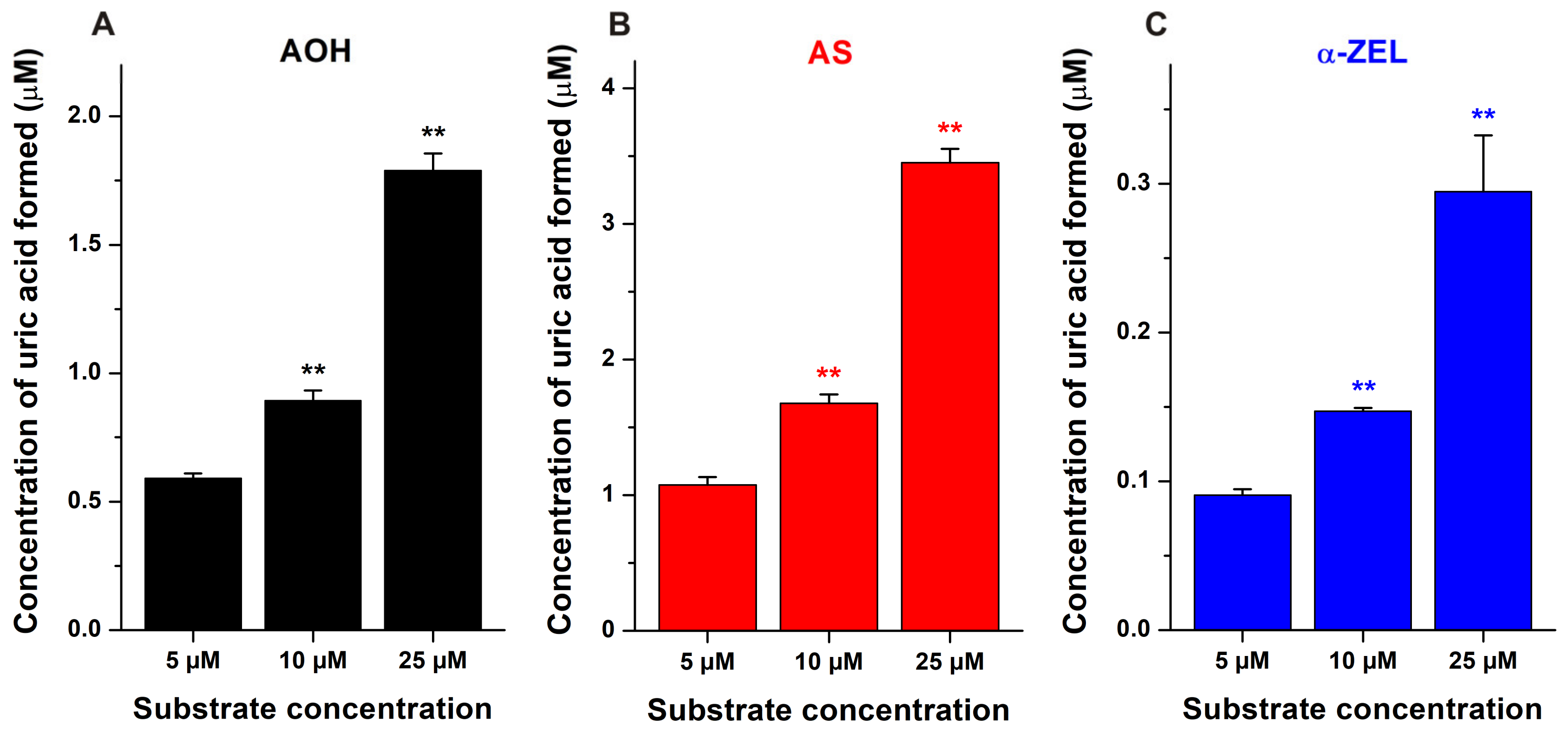
2.2. Mycotoxin Depletion Assays

AOH, AS, and α-ZEL showed significant inhibitory effects on XO; therefore, we examined whether these mycotoxins are simply inhibitors or if they are also substrates of the enzyme. AOH, AS, and α-ZEL were incubated for 0 min, 30 min, and 60 min in the presence of the same amount of XO (0.0012 U/mL) used in the enzyme inhibition studies. Nevertheless, we did not see any changes in the concentrations of these mycotoxins (Table 2), suggesting that XO is not involved in the biotransformation of AOH, AS, and α-ZEL.
XO is a major constituent of bovine milk [39]. The simultaneous presence of XO and certain mycotoxins (e.g., AFM1, OTA, ZEN, and α-ZEL) [40] in milk also makes important mycotoxin–XO interactions, including the potential XO-mediated biotransformation. Our data suggest that XO is not involved in the metabolism of AOH, AS, and α-ZEL. Nevertheless, α-ZEL commonly contaminates bovine milk. Therefore, it is reasonable to hypothesize that α-ZEL appears in milk partly in XO-bound form, which may affect its release and absorption from the gastrointestinal tract.

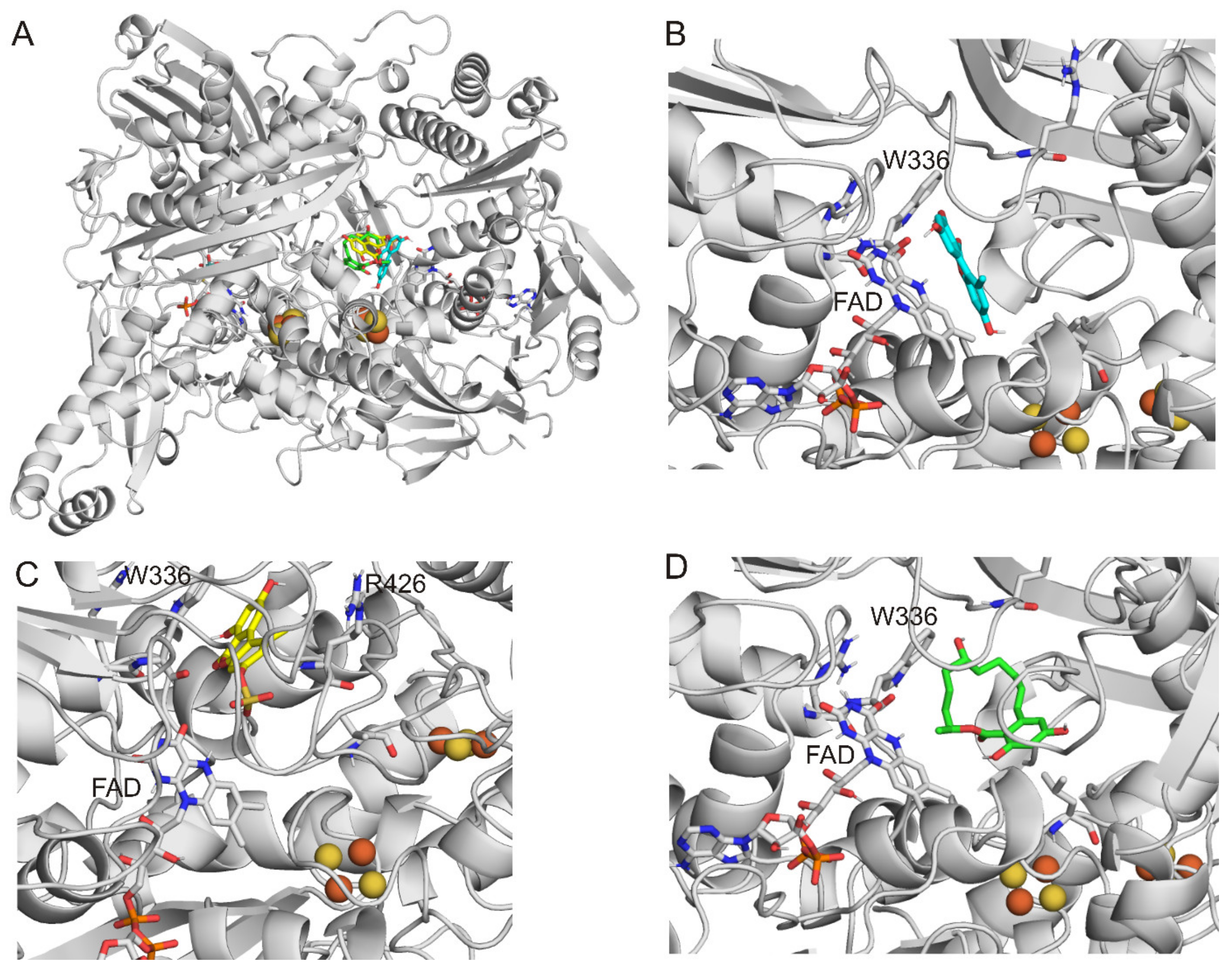
2.3. Modeling Studies

During the 300 blind docking runs in total, none of the ligands bound to the binding pocket of xanthine [44]. Nevertheless, in the best ranks for each ligand tested, another binding pocket was found (Figure 5A), which was originally described by Kuwabara et al. [45]. AOH (8th-rank binding mode; Figure 5B), AS (1st-rank binding mode; Figure 5C), and α-ZEL (2nd-rank binding mode; Figure 5D) interact with W336, which amino acid is located in an alternative binding pocket far from the xanthine binding site. Furthermore, modeling studies suggest the interaction of AOH and AS with K433. Among the three mycotoxins examined, AS has the most favorable ΔGbinding value, followed by α-ZEL and AOH. In this alternative binding pocket, AS forms hydrophilic interactions with W336, R426, K433, FAD, S1225, K1228, and S1234, while α-ZEL showed hydrophobic interactions with L147, A338, I1229, and A1231.
Notably, in our present investigations, FAD and Fe2S2 were included in XO, and their proper partial charge distributions were calculated. In the recent study of Fan et al. [35], another binding pocket of AOH was also suggested during the blind docking calculations; nevertheless, this earlier evaluation was performed with the exclusion of FAD and Fe2S2.
Considering the results of incubation assays, mycotoxin depletion experiments, and modeling studies, AOH, AS, and α-ZEL seem to be allosteric inhibitors of the XO enzyme. It is also supported by the previous report of Fan et al. [35], where the non-competitive inhibitory mechanisms of AOH and AME were described.

3. Conclusions

In summary, the effects of 31 mycotoxins were examined on XO-catalyzed uric acid formation. Based on the in vitro enzyme incubation assays, mycotoxin depletion experiments, and molecular modeling studies, AOH, AS, and α-ZEL proved to be moderate allosteric inhibitors of XO. Our results also demonstrated that AOH, AS, and α-ZEL are inhibitors but not substrates of the enzyme. The above-listed observations make the suitability of AOH as a leading compound in the development of new XO inhibitors questionable, even if the structural modification results in the decreased toxicity of a derivative. Considering the typically nanomolar concentrations of mycotoxins in the circulation, it is reasonable to hypothesize that AOH, AS, and α-ZEL are not able to produce a clinically relevant decrease in uric acid levels and cannot interfere with the pharmacokinetics of drugs biotransformed by XO (e.g., 6-mercaptopurine). Because α-ZEL is a frequent contaminant in bovine milk, it is reasonable to hypothesize that this mycotoxin partly appears in milk in XO-bound form. Our results promote the deeper understanding of mycotoxin-XO interactions.

4. Materials and Methods

4.1. Reagents

Alternariol-9-methylether (AME), aflatoxin B1 (AFB1), aflatoxin B2 (AFB2), aflatoxin G1 (AFG1), aflatoxin G2 (AFG2), sterigmatocystin (STC), cyclopiazonic acid (CPA), citrinin (CIT), ochratoxin A (OTA), patulin (PAT), deoxynivalenol (DON), fumonisin B1 (FB1), T-2 toxin (T2), zearalenone (ZEN), α-zearalenol (α-ZEL), β-zearalenol (β-ZEL), zearalanone (ZAN), α-zearalanol (α-ZAL), β-zearalanol (β-ZAL), xanthine oxidase (XO; from bovine milk), xanthine, uric acid, and allopurinol were from Merck (Darmstadt, Germany). Alternariol (AOH), beauvericin (BEA), ochratoxin B (OTB), and ochratoxin C (OTC) were purchased from Cfm Oskar Tropitzsch GmbH (Marktredwitz, Germany). Alternariol-3-sulfate (AS), alternariol-3-glucoside (AG), alternariol-9-methylether-3-sulfate (AMS), alternariol-9-methylether-3-glucoside (AMG), zearalenone-14-sulfate (Z14S), and zearalenone-14-glucoside (Z14Glz) were obtained from ASCA GmbH (Berlin, Germany). Aflatoxin M1 (AFM1) and dihydrocitrinone (DHC) were from Apollo Scientific (Cheshire, UK) and AnalytiCon Discovery (Potsdam, Germany), respectively. Stock solutions of xanthine (1 mM, in DMSO) and uric acid (2 mM, dissolved in 0.01 M sodium hydroxide) were prepared and stored at −20 °C. Stock solutions of mycotoxins (5 mM or 10 mM) were prepared in ethanol or in dimethyl sulfoxide (DMSO).

4.2. Xanthine Oxidase Assay

The in vitro XO assay was carried out as was previously reported [41,42]. Xanthine (5 μM) was incubated with XO (0.0012 U/mL) enzyme in the absence and presence of increasing concentrations of mycotoxins (0.00, 0.10, 0.25, 1.0, 5.0, 10, 20, 35, 50, and 100 μM) in a thermomixer (5 min, 700 rpm, 37 °C). Incubates were prepared in sodium phosphate buffer (0.05 M, pH 7.5) with 500 μL final volumes. Solvent controls were tested in each experiment. As a positive control inhibitor, the impacts of allopurinol (0.000, 0.002, 0.010, 0.10, 0.50, 1.0, 2.0, 5.0, and 20 μM) were also examined. Incubations were started with pipetting XO solution to the samples, after which the enzyme reaction was stopped with 30 μL of 6 M HClO4. Samples were vortexed, then 97 μL of 1 M potassium hydroxide solution was added. After cooling (to 3 °C) and centrifugation (5 min, 12,000 rpm, 3 °C), concentrations of xanthine and uric acid was directly quantified from the supernatant by HPLC-UV (see details in Section 4.4).
The same experimental design was applied with the following modifications to test the reversibility of the mycotoxin-induced inhibition of XO. In these experiments, incubates contained mycotoxin (50 μM), the enzyme (0.0012 U/mL), and increasing concentrations of xanthine (5, 10, or 25 μM). XO was preincubated (10 min, 700 rpm, 37 °C) with the mycotoxins; thereafter, the incubation (5 min, 700 rpm, 37 °C) was started with the addition of xanthine. Other experimental details remained unchanged.
After the concentrations of uric acid (curic acid) and xanthine (cxanthine) were quantified in the samples, we calculated the rate of metabolite formation (R).
R (%) = 100 × curic acid / (curic acid + cxanthine),
Then the R values of control samples were used as the bases of comparison (100%) when the inhibitory actions of mycotoxins were examined:
Uric acid formation (%) = 100 × Rinhibitor / Rcontrol,
where Rinhibitor and Rcontrol are the metabolite formation rates in the presence and absence of the inhibitor, respectively. IC50 values were determined with sigmoidal (Hill1) fitting employing the OriginPro 8 program using these data (OriginLab Corporation, Northampton, MA, USA).

4.3. Mycotoxin Depletion Assays

In order to test the potential XO-catalyzed biotransformation of AOH, AS, and α-ZEL, these mycotoxins (each 5 μM) were incubated with XO enzyme (0.0012 U/mL) for 0 min, 30 min, and 60 min. Incubations were performed in the absence of xanthine. Other experimental details were the same as described in Section 4.2. Mycotoxin levels in the supernatants were quantified with HPLC-FLD (see details in Section 4.4).

4.4. HPLC Analyses

HPLC measurements were carried out using an integrated HPLC system (Jasco, Tokyo, Japan) containing a binary pump (PU-4180), an autosampler (AS-4050), a UV detector (UV-470), and a fluorescence detector (FP-920). Chromatograms were evaluated with ChromNAV2 software (Jasco).
The quantitative analyses of xanthine and uric acid were performed applying the following HPLC-UV method [41]. A Security Guard (C18, 4.0 × 3.0 mm; Phenomenex, Torrance, CA, USA) precolumn and a Kinetex EVO C18 (250 × 4.6 mm, 5 μm; Phenomenex) analytical column were used (isocratic elution; room temperature; flow rate: 1 mL/min; injected sample volume: 20 μL; detection: 275 nm). The mobile phase contained sodium phosphate buffer (10 mM, pH 4.55) and methanol (98:2 v/v%).
AOH was analyzed employing the following HPLC-FLD method [46]. A Security Guard (C18, 4.0 × 3.0 mm; Phenomenex) precolumn and a Kinetex EVO C18 (250 × 4.6 mm, 5 μm; Phenomenex) analytical column were used (isocratic elution; room temperature; flow rate: 1 mL/min; injected sample volume: 20 μL; detection: λex = 335 nm, λem = 455 nm). The mobile phase contained acetonitrile and 1 mM phosphoric acid (35:65 v/v%). The same method was applied for the quantification of AS, except for the eluent used, which contained acetonitrile and 1 mM phosphoric acid (52:48 v/v%) [47].
The quantitative analysis of α-ZEL was performed by applying the following HPLC-FLD method [48]. A Security Guard (C18, 4.0 × 3.0 mm; Phenomenex) precolumn and a Kinetex EVO C18 (250 × 4.6 mm, 5 μm; Phenomenex) analytical column were used (isocratic elution; room temperature; flow rate: 1 mL/min; injected sample volume: 20 μL; detection: λex = 274 nm, λem = 440 nm). The mobile phase contained water, acetonitrile, and methanol (46:46:8 v/v%).

4.5. Modeling Studies

The structures of AOH, AS, and α-ZEL were built in Maestro (Schrödinger, Maestro Schrödinger Release 2020-4). The energy minimization of the ligands was carried out with OpenBabel [49], using a steepest descent and a conjugate gradient algorithm. Gasteiger–Marsilli partial charges [50] were assigned to the ligand atoms in AutoDock Tools [51]. Flexibility was allowed on the ligands at all active torsions.
Atomic coordinates of XO were obtained from the Protein Data Bank (PDB) with PDB code 3eub [44], similar to our earlier study [41]. The amino acids of the target molecule were equipped with polar hydrogen atoms and Gasteiger–Marsilli partial charges in AutoDock Tools. The geometry and partial charges of the non-amino acid molecules, as the flavine-adenine dinucleotide (FAD), molibdopteroate, and the Fe2S2 inorganic cluster were calculated by MOPAC [52] with a PM7 parametrization [53], and a gradient norm of 0.001. The reduced form of FAD was used, according to Kuwabara et al. [45].
Ligands were docked to XO using AutoDock 4.2.6 [51]. The number of grid points was set to 126 × 126 × 126 at a 0.850 A grid spacing. A blind docking [54,55] investigation was carried out, where the docking box covered the whole surface of the target molecule. The Lamarckian genetic algorithm was used for the global search. A hundred docking runs were executed for each ligand, and the resulting ligand conformations were ranked by their free energy [56]. The lower rank means the higher calculated free energy (ΔGbinding). The docked ligand conformations demonstrated were employed for subsequent evaluations [57].

4.6. Statistical Analyses

The mean and standard error of the mean (± SEM) values are demonstrated in the figures and tables. Statistical evaluations (p < 0.05 and p < 0.01) were executed by applying one-way ANOVA and Tukey’s post hoc test with SPSS Statistics software (IBM, Armonk, NY, USA).

Author Contributions

Conceptualization, R.G.V. and M.P.; Formal analysis, O.B. and B.Z.Z.; Funding acquisition, C.H. and M.P.; Investigation, O.B., Á.D. and B.Z.Z.; Methodology, C.H. and M.P.; Supervision, C.H., R.G.V. and M.P.; Validation, C.H. and M.P.; Writing—original draft, O.B., C.H. and M.P. All authors have read and agreed to the published version of the manuscript.

Funding

The work of M.P. is supported by the Hungarian National Research, Development and Innovation Office (FK138184) and by the János Bolyai Research Scholarship of the Hungarian Academy of Sciences. This project has also been supported by the Medical School, University of Pécs (PTE ÁOK KA-2022-26), and by the European Union, co-financed by the European Social Fund (project name and code: Comprehensive Development for Implementing Smart Specialization Strategies at the University of Pécs, EFOP-3.6.1-16-2016-00004).

Institutional Review Board Statement

Not applicable.

Informed Consent Statement

Not applicable.

Data Availability Statement

Data will be made available upon request.

Acknowledgments

The authors thank Katalin Fábián (Department of Pharmacology, Faculty of Pharmacy, University of Pécs) for her excellent assistance in the experimental work. We acknowledge the support from the Governmental Information Technology Development Agency, Hungary. We also acknowledge that the results of this research have been achieved using the DECI resource Archer2 based in the UK at the National Supercomputing Service with support from the PRACE aisbl.

Conflicts of Interest

The authors declare no conflict of interest.

References

  1. da Rocha, M.E.B.; Freire, F.C.O.; Maia, F.E.F.; Guedes, M.I.F.; Rondina, D. Mycotoxins and their effects on human and animal health. Food Control 2014, 36, 59–165. [Google Scholar]
  2. Kabak, B. The fate of mycotoxins during thermal food processing. J. Sci. Food Agric. 2009, 89, 549–554. [Google Scholar] [CrossRef]
  3. Milani, J.; Maleki, G. Effects of processing on mycotoxin stability in cereals. J. Sci. Food Agric. 2014, 94, 2372–2375. [Google Scholar] [CrossRef]
  4. Lootens, O.; Vermeulen, A.; Croubels, S.; De Saeger, S.; Van Bocxlaer, J.; De Boevre, M. Possible Mechanisms of the Interplay between Drugs and Mycotoxins—Is There a Possible Impact? Toxins 2022, 14, 873. [Google Scholar] [CrossRef]
  5. Walravens, J.; Mikula, H.; Rychlik, M.; Asam, S.; Devos, T.; Njumbe Ediage, E.; Diana Di Mavungu, J.; Jacxsens, L.; Van Landschoot, A.; Vanhaecke, L.; et al. Validated UPLC-MS/MS Methods to Quantitate Free and Conjugated Alternaria Toxins in Commercially Available Tomato Products and Fruit and Vegetable Juices in Belgium. J. Agric. Food Chem. 2016, 64, 5101–5109. [Google Scholar] [CrossRef] [Green Version]
  6. Puntscher, H.; Cobankovic, I.; Marko, D.; Warth, B. Quantitation of Free and Modified Alternaria Mycotoxins in European Food Products by LC-MS/MS. Food Control 2019, 102, 157–165. [Google Scholar] [CrossRef]
  7. Qin, Q.; Fan, Y.; Jia, Q.; Duan, S.; Liu, F.; Jia, B.; Wang, G.; Guo, W.; Wang, C. The Potential of Alternaria Toxins Production by A. alternata in Processing Tomatoes. Toxins 2022, 14, 827. [Google Scholar] [CrossRef] [PubMed]
  8. EFSA Panel on Contaminants in the Food Chain (CONTAM). Scientific Opinion on the Risks for Animal and Public Health Related to the Presence of Alternaria Toxins in Feed and Food. EFSA J. 2011, 9, 2407. [Google Scholar] [CrossRef]
  9. Aichinger, G.; Del Favero, G.; Warth, B.; Marko, D. Alternaria toxins—Still emerging? Compr. Rev. Food Sci. Food Saf. 2021, 20, 4390–4406. [Google Scholar] [CrossRef]
  10. Rushing, B.R.; Selim, M.I. Aflatoxin B1: A review on metabolism, toxicity, occurrence in food, occupational exposure, and detoxification methods. Food Chem. Toxicol. 2019, 124, 81–100. [Google Scholar] [CrossRef]
  11. Klingelhöfer, D.; Zhu, Y.; Braun, M.; Bendels, M.H.K.; Brüggmann, D.; Groneberg, D.A. Aflatoxin—Publication analysis of a global health threat. Food Control 2018, 89, 280–290. [Google Scholar] [CrossRef]
  12. Nieto, C.H.D.; Granero, A.M.; Zon, M.A.; Fernández, H. Sterigmatocystin: A mycotoxin to be seriously considered. Food Chem. Toxicol. 2018, 118, 460–470. [Google Scholar] [CrossRef]
  13. Ostry, V.; Toman, J.; Grosse, Y.; Malir, F. Cyclopiazonic acid: 50th anniversary of its discovery. World Mycotoxin J. 2018, 11, 135–148. [Google Scholar] [CrossRef] [Green Version]
  14. European Food Safety Authority (EFSA). Scientific opinion on the risks for public and animal health related to the presence of citrinin in food and feed. EFSA J. 2012, 10, 2605. [Google Scholar]
  15. Vidal, A.; Ouhibi, S.; Ghali, R.; Hedhili, A.; De Saeger, S.; De Boevre, M. The mycotoxin patulin: An updated short review on occurrence, toxicity and analytical challenges. Food Chem. Toxicol. 2019, 129, 249–256. [Google Scholar] [CrossRef]
  16. Degen, G.H.; Ali, N.; Gundert-Remy, U. Preliminary data on citrinin kinetics in humans and their use to estimate citrinin exposure based on biomarkers. Toxicol. Lett. 2018, 282, 43–48. [Google Scholar] [CrossRef]
  17. European Food Safety Authority (EFSA). Risk assessment of ochratoxin A in food. EFSA J. 2020, 18, 6113. [Google Scholar]
  18. Di Stefano, V.; Avellone, G.; Pitonzo, R.; Capocchiano, V.G.; Mazza, A.; Cicero, N.; Dugo, G. Natural co-occurrence of ochratoxin A, ochratoxin B and aflatoxins in Sicilian red wines. Food Addit. Contam. A 2015, 32, 1343–1351. [Google Scholar] [CrossRef] [PubMed]
  19. Remiro, R.; Gonzalez-Penas, E.; Lizarraga, E.; Lopez de Cerain, A. Quantification of ochratoxin A and five analogs in Navarra red wines. Food Control 2012, 27, 139–145. [Google Scholar] [CrossRef]
  20. Bryła, M.; Pierzgalski, A.; Zapaśnik, A.; Uwineza, P.A.; Ksieniewicz-Woźniak, E.; Modrzewska, M.; Waśkiewicz, A. Recent Research on Fusarium Mycotoxins in Maize—A Review. Foods 2022, 11, 3465. [Google Scholar] [CrossRef]
  21. EFSA Panel on Contaminants in the Food Chain (CONTAM). Risks for animal health related to the presence of zearalenone and its modified forms in feed. EFSA J. 2017, 15, e04851. [Google Scholar]
  22. Rai, A.; Das, M.; Tripathi, A. Occurrence and toxicity of a fusarium mycotoxin, zearalenone. Crit. Rev. Food Sci. Nutr. 2020, 60, 2710–2729. [Google Scholar] [CrossRef]
  23. Harrison, R. Structure and function of xanthine oxidoreductase: Where are we now? Free Radic. Biol. Med. 2002, 33, 774–797. [Google Scholar] [CrossRef] [PubMed]
  24. Glantzounis, G.K.; Tsimoyiannis, E.C.; Kappas, A.M.; Galaris, D.A. Uric acid and oxidative stress. Curr. Pharm. Des. 2005, 11, 4145–4151. [Google Scholar] [CrossRef] [PubMed]
  25. Schmidt, H.M.; Kelley, E.E.; Straub, A.C. The impact of xanthine oxidase (XO) on hemolytic diseases. Redox. Biol. 2019, 21, 101072. [Google Scholar] [CrossRef] [PubMed]
  26. Zhang, S.; Wang, Y.; Cheng, J.; Huangfu, N.; Zhao, R.; Xu, Z.; Zhang, F.; Zheng, W.; Zhang, D. Hyperuricemia and Cardiovascular Disease. Curr. Pharm. Des. 2019, 25, 700–709. [Google Scholar] [CrossRef]
  27. Galbusera, C.; Orth, P.; Fedida, D.; Spector, T. Superoxide radical production by allopurinol and xanthine oxidase. Biochem. Pharmacol. 2006, 71, 1747–1752. [Google Scholar] [CrossRef]
  28. Day, R.O.; Graham, G.G.; Hicks, M.; McLachlan, A.J.; Stocker, S.L.; Williams, K.M. Clinical Pharmacokinetics and Pharmacodynamics of Allopurinol and Oxypurinol. Clin. Pharmacokinet. 2007, 46, 623–644. [Google Scholar] [CrossRef]
  29. Leong, R.W.; Gearry, R.B.; Sparrow, M.P. Thiopurine hepatotoxicity in inflammatory bowel disease: The role for adding allopurinol. Expert Opin. Drug Saf. 2008, 7, 607–616. [Google Scholar] [CrossRef] [PubMed]
  30. Costantini, A.V. The fungal etiology of gout and hyperuricemia: The antifungal mode of action of colchicine. Biomed. Rev. 1992, 1, 47–52. [Google Scholar] [CrossRef]
  31. Baldissera, M.D.; Souza, C.F.; Doleski, P.H.; Zeppenfeld, C.C.; Descovi, S.; Da Silva, A.S.; Baldisserotto, B. Xanthine oxidase activity exerts pro-oxidative and pro-inflammatory effects in serum of silver catfish fed with a diet contaminated with aflatoxin B1. J. Fish Dis. 2018, 41, 1153–1158. [Google Scholar] [CrossRef] [PubMed]
  32. Owumi, S.E.; Najophe, S.E.; Idowu, T.B.; Nwozo, S.O. Protective mechanisms of gallic acid on hepatorenal dysfunction of zearalenone treated rat. Biologia 2021, 76, 3123–3135. [Google Scholar] [CrossRef]
  33. Osselaere, A.; Santos, R.; Hautekiet, V.; De Backer, P.; Chiers, K.; Ducatelle, R.; Croubels, S. Deoxynivalenol impairs hepatic and intestinal gene expression of selected oxidative stress, tight junction and inflammation proteins in broiler chickens, but addition of an adsorbing agent shifts the effects to the distal parts of the small intestine. PLoS ONE 2013, 8, e69014. [Google Scholar] [CrossRef] [Green Version]
  34. Antonissen, G.; Van Immerseel, F.; Pasmans, F.; Ducatelle, R.; Janssens, G.P.J.; De Baere, S.; Mountzouris, K.C.; Su, S.; Wong, E.A.; De Meulenaer, B.; et al. Mycotoxins Deoxynivalenol and Fumonisins Alter the Extrinsic Component of Intestinal Barrier in Broiler Chickens. J. Agric. Food Chem. 2015, 63, 10846–10855. [Google Scholar] [CrossRef] [PubMed]
  35. Fan, J.; Sun, S.; Lv, C.; Li, Z.; Guo, M.; Yin, Y.; Wang, H.; Wang, W. Discovery of mycotoxin alternariol as a potential lead compound targeting xanthine oxidase. Chem. Biol. Interact. 2022, 360, 109948. [Google Scholar] [CrossRef]
  36. Al-Harbi, S.A.; Abdulrahman, A.O.; Zamzami, M.A.; Khan, M.I. Urolithins: The Gut Based Polyphenol Metabolites of Ellagitannins in Cancer Prevention, a Review. Front. Nutr. 2021, 8, 647582. [Google Scholar] [CrossRef] [PubMed]
  37. Gu, W. Bioactive metabolites from Alternaria brassicicola ML-P08, an endophytic fungus residing in Malus halliana. World J. Microbiol. Biotechnol. 2009, 25, 1677–1683. [Google Scholar] [CrossRef]
  38. Puvaca, N.; Tanaskovic, S.; Bursic, V.; Petrovic, A.; Merkuri, J.; Kika, T.S.; Marinkovic, D.; Vukovic, G.; Cara, M. Optical Characterization of Alternaria spp. Contaminated Wheat Grain and Its Influence in Early Broilers Nutrition on Oxidative Stress. Sustainability 2021, 13, 4005. [Google Scholar] [CrossRef]
  39. Harrison, R. Milk xanthine oxidase: Properties and physiological roles. Int. Dairy J. 2006, 16, 546–554. [Google Scholar] [CrossRef]
  40. Weidenbörner, M. Natural Mycotoxin Contamination in Humans and Animals; Springer International Publishing: Cham, Switzerland, 2015. [Google Scholar]
  41. Mohos, V.; Pánovics, A.; Fliszár-Nyúl, E.; Schilli, G.; Hetényi, C.; Mladěnka, P.; Needs, P.W.; Kroon, P.A.; Pethő, G.; Poór, M. Inhibitory Effects of Quercetin and Its Human and Microbial Metabolites on Xanthine Oxidase Enzyme. Int. J. Mol. Sci. 2019, 20, 2681. [Google Scholar] [CrossRef] [Green Version]
  42. Mohos, V.; Fliszár-Nyúl, E.; Poór, M. Inhibition of Xanthine Oxidase-Catalyzed Xanthine and 6-Mercaptopurine Oxidation by Flavonoid Aglycones and Some of Their Conjugates. Int. J. Mol. Sci. 2020, 21, 3256. [Google Scholar] [CrossRef]
  43. Turnheim, K.; Krivanek, P.; Oberbauer, R. Pharmacokinetics and pharmacodynamics of allopurinol in elderly and young subjects. Br. J. Clin. Pharmacol. 1999, 48, 501–509. [Google Scholar] [CrossRef] [Green Version]
  44. Pauff, J.M.; Cao, H.; Hille, R. Substrate Orientation and Catalysis at the Molybdenum Site in Xanthine Oxidase. J. Biol. Chem. 2009, 284, 8760–8767. [Google Scholar] [CrossRef] [Green Version]
  45. Kuwabara, Y.; Nishino, T.; Okamoto, K.; Matsumura, T.; Eger, B.T.; Pai, E.F.; Nishino, T. Unique Amino Acids Cluster for Switching from the Dehydrogenase to Oxidase Form of Xanthine Oxidoreductase. Proc. Natl. Acad. Sci. USA 2003, 100, 8170–8175. [Google Scholar] [CrossRef] [PubMed] [Green Version]
  46. Fliszár-Nyúl, E.; Lemli, B.; Kunsági-Máté, S.; Szente, L.; Poór, M. Interactions of Mycotoxin Alternariol with Cyclodextrins and Its Removal from Aqueous Solution by Beta-Cyclodextrin Bead Polymer. Biomolecules 2019, 9, 428. [Google Scholar] [CrossRef] [PubMed] [Green Version]
  47. Lemli, B.; Vilmányi, P.; Fliszár-Nyúl, E.; Zsidó, B.Z.; Hetényi, C.; Szente, L.; Poór, M. Testing Serum Albumins and Cyclodextrins as Potential Binders of the Mycotoxin Metabolites Alternariol-3-Sulfate, Alternariol-9-Monomethylether and Alternariol-9-Monomethylether-3-Sulfate. Int. J. Mol. Sci. 2022, 23, 14353. [Google Scholar] [CrossRef] [PubMed]
  48. Poór, M.; Faisal, Z.; Zand, A.; Bencsik, T.; Lemli, B.; Kunsági-Máté, S.; Szente, L. Removal of Zearalenone and Zearalenols from Aqueous Solutions Using Insoluble Beta-Cyclodextrin Bead Polymer. Toxins 2018, 10, 216. [Google Scholar] [CrossRef] [Green Version]
  49. O’Boyle, N.M.; Banck, M.; James, C.A.; Morley, C.; Vandermeersch, T.; Hutchison, G.R. Open Babel. J. Cheminform. 2011, 3, 33. [Google Scholar] [CrossRef] [Green Version]
  50. Gasteiger, J.; Marsili, M. Iterative Partial Equalization of Orbital Electronegativity-a Rapid Access to Atomic Charges. Tetrahedron 1980, 36, 3219–3228. [Google Scholar] [CrossRef]
  51. Morris, G.M.; Goodsell, D.S.; Halliday, R.S.; Huey, R.; Hart, W.E.; Belew, R.K.; Olson, A.J. Automated Docking Using a Lamarckian Genetic Algorithm and Empirical Binding Free Energy Function. J. Comput. Chem. 1998, 19, 1639–1662. [Google Scholar] [CrossRef]
  52. Stewart, J.J.P. MOPAC; Stewart Computational Chemistry: Colorado Springs, CO, USA, 2016. [Google Scholar]
  53. Stewart, J.J.P. Optimization of Parameters for Semiempirical Methods VI: More Modifications to the NDDO Approximations and Re-Optimization of Parameters. J. Mol. Model. 2013, 19, 1–32. [Google Scholar] [CrossRef] [PubMed] [Green Version]
  54. Hetényi, C.; Van Der Spoel, D. Blind Docking of Drug-Sized Compounds to Proteins with up to a Thousand Residues. FEBS Lett. 2006, 580, 1447–1450. [Google Scholar] [CrossRef] [Green Version]
  55. Hetényi, C.; Van Der Spoel, D. Toward Prediction of Functional Protein Pockets Using Blind Docking and Pocket Search Algorithms. Protein Sci. 2011, 20, 880–893. [Google Scholar] [CrossRef] [PubMed] [Green Version]
  56. Zsidó, B.Z.; Börzsei, R.; Szél, V.; Hetényi, C. Determination of Ligand Binding Modes in Hydrated Viral Ion Channels to Foster Drug Design and Repositioning. J. Chem. Inf. Model. 2021, 61, 4011–4022. [Google Scholar] [CrossRef] [PubMed]
  57. Zsidó, B.Z.; Balog, M.; Erős, N.; Poór, M.; Mohos, V.; Fliszár-Nyúl, E.; Hetényi, C.; Nagane, M.; Hideg, K.; Kálai, T.; et al. Synthesis of Spin-Labelled Bergamottin: A Potent CYP3A4 Inhibitor with Antiproliferative Activity. Int. J. Mol. Sci. 2020, 21, 508. [Google Scholar] [CrossRef] [Green Version]
Figure 1. Chemical structures of mycotoxins alternariol, alternariol sulfate, and α-zearalenol.
Toxins 15 00250 g001
Figure 2. Effects of Alternaria (A), Aspergillus/Penicillium (B), and Fusarium (C) mycotoxins (each 20 μM) on XO-catalyzed uric acid formation (substrate concentration: 5 μM; incubation: 5 min; n = 3; * p < 0.05 and ** p < 0.01). We started the reaction with the addition of XO. The highest solvent concentrations applied (0.4% ethanol or 0.4% DMSO) did not affect the XO-catalyzed uric acid formation (101.1 ± 1.2% in the presence of 0.4% ethanol; 99.2 ± 1.9% in the presence of 0.4% DMSO).
Toxins 15 00250 g002
Figure 3. Concentration-dependent inhibitory actions of AOH, AS, α-ZEL, and allopurinol (APU, positive control) on XO-catalyzed xanthine oxidation (substrate concentration: 5 μM; incubation: 5 min; n = 3; * p < 0.05 and ** p < 0.01). We started the reaction with the addition of XO. The 50 μM and 100 μM AOH and AS samples contained 0.5% (uric acid formation = 99.0 ± 1.1%) and 1.0% (uric acid formation = 91.5 ± 2.1%) DMSO, respectively. The 50 μM and 100 μM α-ZEL samples contained 1.0% (uric acid formation = 96.8 ± 1.8%) and 2.0% (uric acid formation = 95.8 ± 1.3%) ethanol, respectively. Since the highest levels of ethanol (2.0%) and DMSO (1.0%) applied caused some inhibitory effects on the enzyme, the metabolite formation in the presence of 100 μM of AOH, AS, and α-ZEL was calculated compared to the corresponding solvent controls. The highest concentration APU sample (20 μM) tested contained 0.4% DMSO, which did not affect metabolite formation (99.2 ± 1.9%).
Toxins 15 00250 g003
Figure 4. XO-catalyzed uric acid formation in the presence of 50 μM AOH (A), 50 μM AS (B) or 50 μM α-ZEL (C), with increasing concentrations of xanthine (5–25 μM; incubation: 5 min; n = 3). The enzyme was preincubated (10 min, 700 rpm, and 37 °C) with the mycotoxins, after which we started the reaction with the addition of the substrate (xanthine). Metabolite formation in the presence of 10 μM and 25 μM xanthine was compared to the product formation determined with 5 μM substrate concentration (** p < 0.01).
Toxins 15 00250 g004
Figure 5. Binding modes of AOH (teal sticks), AS (yellow sticks), and α-ZEL (green sticks) in the alternative binding pocket of XO enzyme (A). The top 8th-ranked binding mode of AOH (B), the 1st-ranked binding mode of AS (C), and the 2nd-ranked binding mode of α-ZEL (D). XO is represented with a grey cartoon, FAD and molibdopteroate are demonstrated with grey sticks, and the Fe2S2 cluster is shown with spheres.
Toxins 15 00250 g005
Table 1. Most important properties of mycotoxins and mycotoxin metabolites that were examined.
MycotoxinAbbreviationOccurrence FungiToxic EffectsReference
Alternariol
Alternariol-9-methylether
Alternariol-3-sulfate
Alternariol-3-glucoside
AME-3-sulfate
AME-3-glucoside
AOH
AME
AS
AG
AMS
AMG
Tomatoes, grapes, and corresponding productsAlternaria sp.Endocrine disruptor and carcinogenicity[5,6,7,8,9]
Aflatoxin B1
Aflatoxin B2
Aflatoxin G1
Aflatoxin G2
Aflatoxin M1
AFB1
AFB2
AFG1
AFG2
AFM1
Cereals, nuts, figs, vegetables, meat, milk, and dairy productsAspergillus sp.Hepatotoxicity[10,11]
SterigmatocystinSTCGrains, coffee beans, cheese, spices, and soybeansAspergillus sp.Hepatotoxicity and
nephrotoxicity
[12]
Cyclopiazonic acidCPAOilseeds, cereals, meat, and milkAspergillus sp.
Penicillium sp.
Gastrointestinal toxicity and
neurotoxicity
[13]
Citrinin
Dihydrocitrinone
CIT
DHC
Grains, rice, fruits, and spicesAspergillus sp.
Penicillium sp.
Monascus sp.
Nephrotoxicity[14,16]
Ochratoxin A
Ochratoxin B
Ochratoxin C
OTA
OTB
OTC
Cereals, fruits, meat, dairy products, and beveragesAspergillus sp.
Penicillium sp.
Nephrotoxicity[17,18,19]
PatulinPATApple, pear, and corresponding productsAspergillus sp.
Penicillium sp.
Gastrointestinal toxicity and
immunotoxicity
[15]
BeauvericinBEACereals and corresponding productsFusarium sp.Low toxicity[20]
DeoxynivalenolDONCereals and corresponding productsFusarium sp.Gastrointestinal toxicity[20]
Fumonisin B1FB1Cereals and corresponding productsFusarium sp.Neural tube defects[20]
T-2 toxinT2Cereals and corresponding productsFusarium sp.Gastrointestinal toxicity and
toxic aleukia
[20]
Zearalenone
α-Zearalenol
β-Zearalenol
Zearalanone
α-Zearalanol
β-Zearalanol
Zearalenone-14-sulfate
Zearalenone-14-glucoside
ZEN
α-ZEL
β-ZEL
ZAN
α-ZAL
β-ZAL
Z14S
Z14Glz
Cereals and corresponding productsFusarium sp.Xenoestrogen,
endocrine disruptor
[20,21,22]
Table 2. AOH, AS, and α-ZEL levels (% ± SEM) in samples after 0 min, 30 min, and 60 min incubation with XO (0.0012 U/mL; initial mycotoxin concentration: 5 μM; n = 3). We started the reaction with the addition of XO.
Incubation
Time (min)
AOH
Concentration (%)
AS
Concentration (%)
α-ZEL
Concentration (%)
0100.0 ± 1.0100.0 ± 0.1100.0 ± 0.2
60102.0 ± 0.9100.3 ± 1.498.9 ± 0.9
12096.4 ± 1.099.9 ± 1.0100.0 ± 0.8
Disclaimer/Publisher’s Note: The statements, opinions and data contained in all publications are solely those of the individual author(s) and contributor(s) and not of MDPI and/or the editor(s). MDPI and/or the editor(s) disclaim responsibility for any injury to people or property resulting from any ideas, methods, instructions or products referred to in the content.

