Article

Histone 2-Hydroxyisobutyryltransferase Encoded by Afngg1 Is Involved in Pathogenicity and Aflatoxin Biosynthesis in Aspergillus flavus

1
College of Biological Engineering, Henan University of Technology, Zhengzhou 450001, China
2
Henan Provincial Key Laboratory of Biological Processing and Nutritional Function of Wheat, Zhengzhou 450001, China
*
Author to whom correspondence should be addressed.
These authors contributed equally to this work.
Submission received: 23 November 2022 / Revised: 7 December 2022 / Accepted: 16 December 2022 / Published: 21 December 2022
(This article belongs to the Special Issue Research on Pathogenic Fungi and Mycotoxins in China (Volume II))

Abstract

:
Aflatoxin, a carcinogenic secondary metabolite produced by Aspergillus flavus, is a significant threat to human health and agricultural production. Histone 2-hydroxyisobutyrylation is a novel post-translational modification that regulates various biological processes, including secondary metabolism. In this study, we identified the novel histone 2-hydroxyisobutyryltransferase Afngg1 in A. flavus, and explored its role in cell growth, development and aflatoxin biosynthesis. Afngg1 gene deletion markedly decreased lysine 2-hydroxyisobutyrylation modification of histones H4K5 and H4K8 compared with the control strain. Additionally, Afngg1 deletion inhibited mycelial growth of A. flavus, and the number of conidia and hydrophobicity were significantly decreased. Notably, aflatoxin B1 biosynthesis and sclerotia production were completely inhibited in the ΔAfngg1 strain. Furthermore, the pathogenicity of the ΔAfngg1 strain infecting peanut and corn grains was also diminished, including reduced spore production and aflatoxin biosynthesis compared with A. flavus control and Afngg1 complementation strains. Transcriptome analysis showed that, compared with control strains, differentially expressed genes in ΔAfngg1 were mainly involved in chromatin remodelling, cell development, secondary metabolism and oxidative stress. These results suggest that Afngg1 is involved in histone 2-hydroxyisobutyrylation and chromatin modification, and thus affects cell development and aflatoxin biosynthesis in A. flavus. Our results lay a foundation for in-depth research on the 2-hydroxyisobutyrylation modification in A. flavus, and may provide a novel target for aflatoxin contamination prevention.
Key Contribution: The 2-hydroxyisobutyryltransferase Afngg1 affects 2-hydroxyisobutyrylation modification of histones H4K5 and H4K8 and blocks aflatoxin biosynthesis in Aspergillus flavus.

1. Introduction

Aspergillus flavus, one of the most abundant and widely distributed fungi on earth, is often found in pre- or post-harvest crops, and it causes serious economic losses [1,2]. It can produce teratogenic and carcinogenic aflatoxins that pose a serious safety hazard to both humans and animals [3]. It is estimated that aflatoxin contamination costs the US corn industry nearly USD 1.68 billion annually [4]. Aflatoxin causes 28% of hepatocellular carcinoma worldwide [5]. Therefore, an in-depth understanding of aflatoxin biosynthetic pathways and their regulatory mechanisms is urgently required to develop effective strategies to control aflatoxin contamination.
Histone post-translational modifications (HPTMs), including acetylation, phosphorylation, ubiquitination and 2-hydroxyisobutyrylation, play a crucial role in many cellular processes in eukaryotes, including secondary metabolite biosynthesis and chromatin regulation [6]. Among them, 2-hydroxyisobutyrylation is a recently discovered post-translational modification that plays an important regulatory role in gene transcription, metabolism and enzyme activity [7]. Previous studies have shown that lysine de-2-hydroxyisobutyrylase CobB can regulate the catalytic activity of enolase by decreasing modification at the K343 site, thereby regulating glycolysis and cell growth in bacteria [8]. In Ustilaginoidea virens, mutation of the 2-hydroxyisobutyrylation modification site of mitogen-activated protein kinase UvSlt2 decreased UvSlt2 enzyme activity and significantly reduced the development and virulence of plant pathogenic fungi [9]. Significantly, 2-hydroxyisobutyrylation modification is involved in the pathogenicity of Botrytis cinerea, Candida albicans, Fusarium oxysporum and Toxoplasma gondii [7,10,11,12]. Our previous studies have shown that 2-hydroxyisobutyrylation modification regulates mycelial growth and development of A. flavus, as well as aflatoxin biosynthesis [13]. These results indicate that lysine 2-hydroxyisobutyrylation modification plays a key role in A. flavus, but the enzymes involved in histone 2-hydroxyisobutyrylation modification have not been reported in A. flavus. Herein, we identified histone 2-hydroxyisobutyryltransferase Afngg1 in A. flavus, and explored its underlying gene regulatory mechanisms using RNA sequencing (RNA-seq)-based transcriptional analysis.
In this study, Afngg1, a homolog of Candida albicans histone acetyltransferase Ngg1, was found to be associated with histone 2-hydroxyisobutyrylation modification. Additionally, the functions of Afngg1 in cell development and aflatoxin B1 (AFB1) production were profiled, and the underlying regulatory mechanisms were investigated through transcriptome analysis. The findings suggest that Afngg1 is a potential target for the effective control of aflatoxin contamination.

2. Results

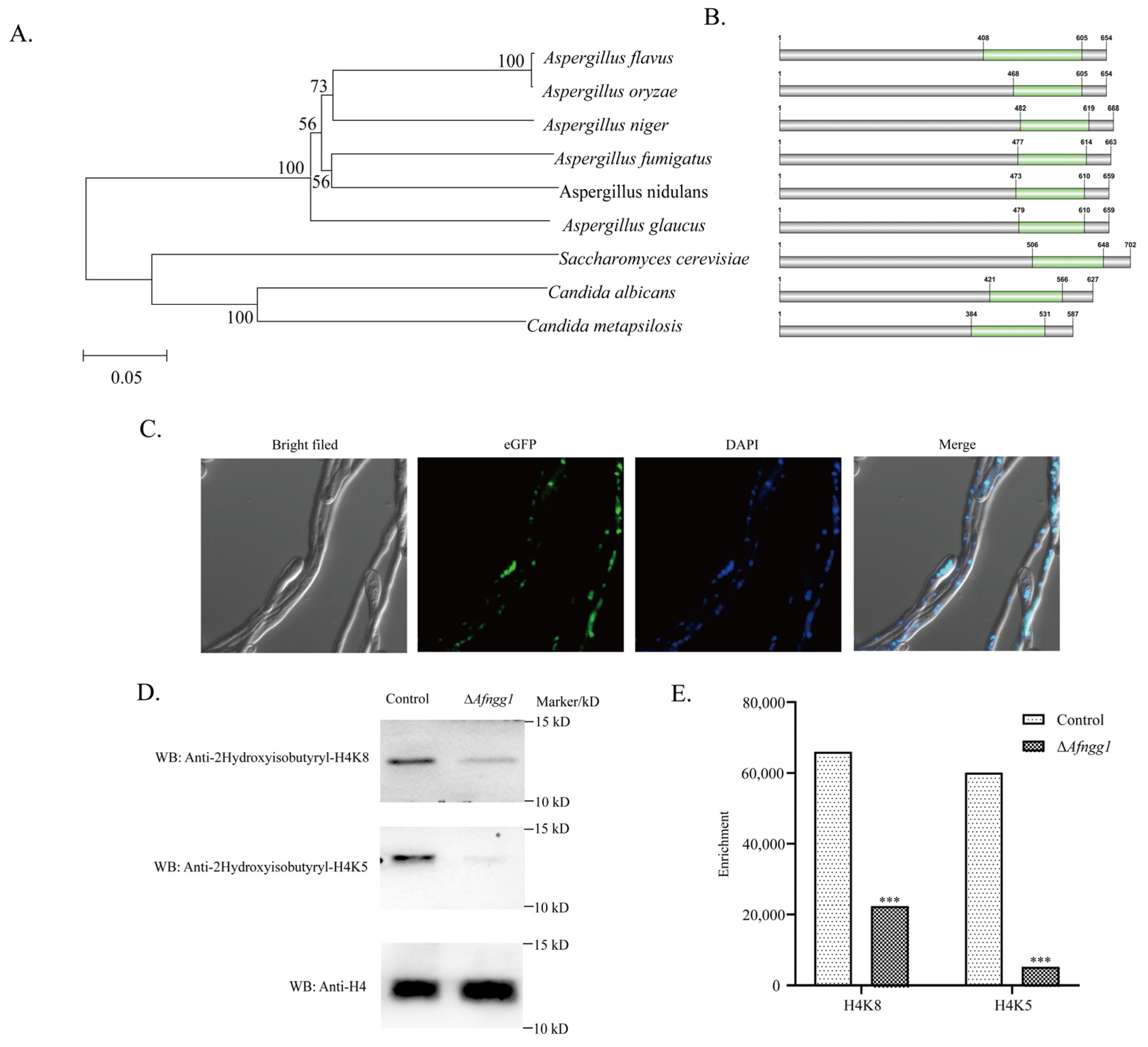
2.1. Characterisation of A. Flavus Histone 2-Hydroxyisobutyryltransferase Afngg1

In order to characterise homologs of Afngg1 in A. flavus, we obtained homologous sequences by BLAST searching the NCBI database, and constructed phylogenetic trees to analyse their evolutionary relationships. The results revealed that Afngg1 in A. flavus is closely related to homologs in other fungi, and most similar to homolog in A. oryzae RIB40 (Figure 1A). The analysis also indicated that it was homologous to Candida albicans histone acetyltransferase Ngg1. The domains in Afngg1 were further analysed via the NCBI database and visualised using DOG2.0. A conserved Ada3 (histone acetyltransferase subunit 3) domain was found in homologs of Afngg1 proteins in nine species (Figure 1B). Additionally, Afngg1 was localised in the nucleus of A. flavus (Figure 1C).
Given the similarity to Ngg1 in C. albicans, we speculated that Afngg1 might also be involved in histone modification. To test this hypothesis, the effects of Afngg1 on the level of acetylation of histone H3 and 2-hydroxyisobutyrylation modification of histone H4 were investigated by collecting mycelia of A. flavus control and ΔAfngg1 strains, and proteins were extracted for Western blotting detection using corresponding antibodies. The results indicated that the acetylation modification level of H3K14 in strain ∆Afngg1 was decreased compared with the A. flavus control strain, and notably, acetylation modification of H3K9 was completely absent (Figure S1). Unexpectedly, our results also showed that 2-hydroxyisobutyrylation modification levels of H4K8 and H4K5 were significantly lower than in the A. flavus control strain (Figure 1D). These results suggest that Afngg1 contributed to lysine acetylation and 2-hydroxyisobutyrylation as a histone acetyltransferase.

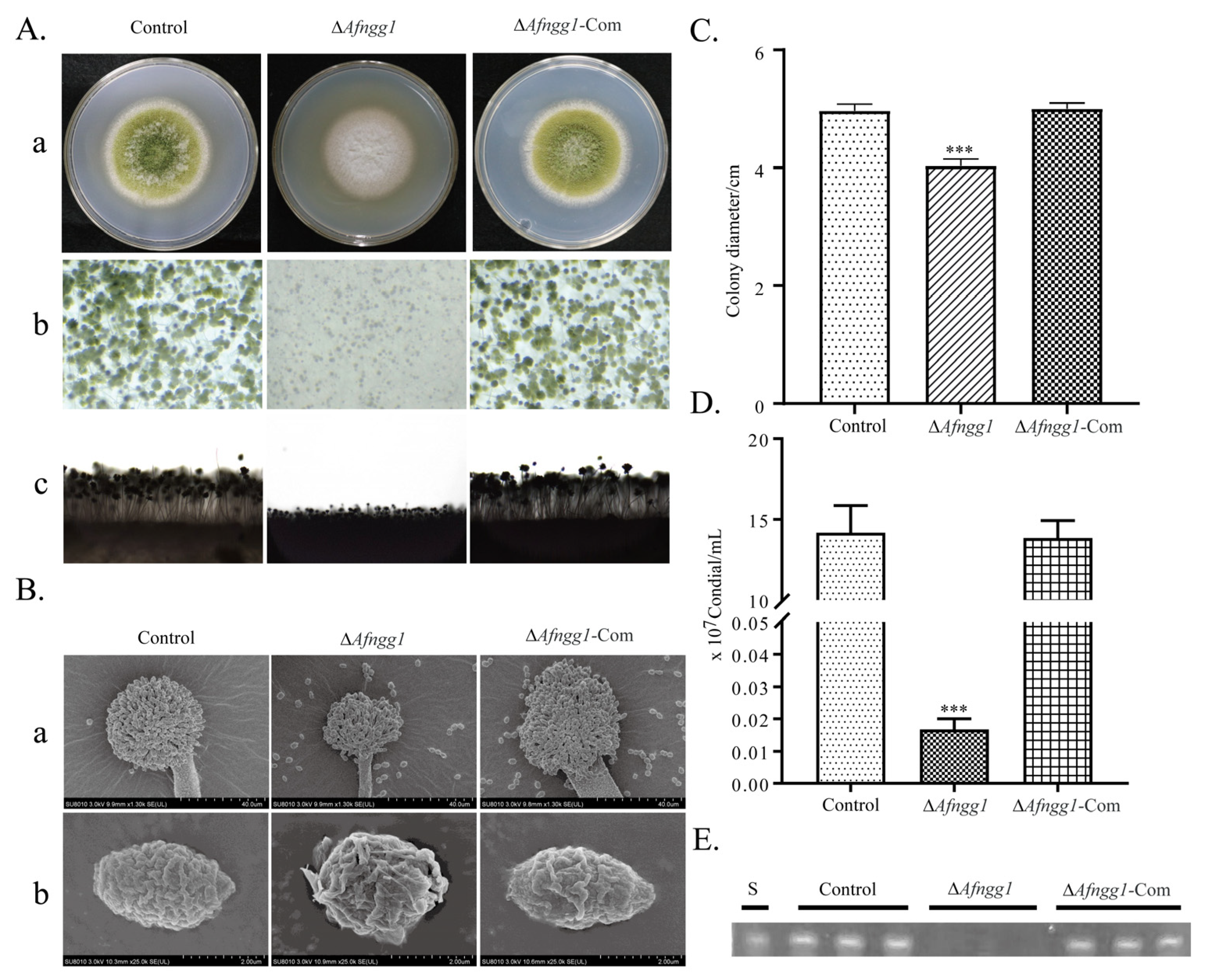
2.2. Afngg1 Is Involved in A. flavus Growth, Conidia Formation and Aflatoxin Biosynthesis

To further explore the potential role of Afngg1 in A. flavus, we constructed ΔAfngg1 and ΔAfngg1-Com strains (Figure S2), and the effects of Afngg1 on A. flavus growth, conidia formation and biosynthesis of aflatoxin were evaluated. Unlike the A. flavus control strain, mycelia of the ∆Afngg1 strain were white and fluffy (Figure 2Aa). Stereoscopic microscope observation showed that, compared with the A. flavus control strain, conidia heads of the ∆Afngg1 strain were smaller and fewer in number (Figure 2Ab). Microscopy observation revealed that deletion of Afngg1 reduced the length of conidiophores and conidial number (Figure 2Ac). SEM results showed that conidial heads of the A. flavus control strain were fully formed, while conidial heads of the Afngg1 strain were significantly smaller and deformed (Figure 2B). Additionally, compared with the A. flavus control strain, the mycelia growth rate was decreased after Afngg1 deletion, and conidia production was decreased by 99.88%, consistent with microscopy observation (Figure 2C,D). Qualitative thin-layer chromatography (TLC) analysis showed that AFB1 was no longer produced by the A. flavus strain lacking Afngg1 (Figure 2E).

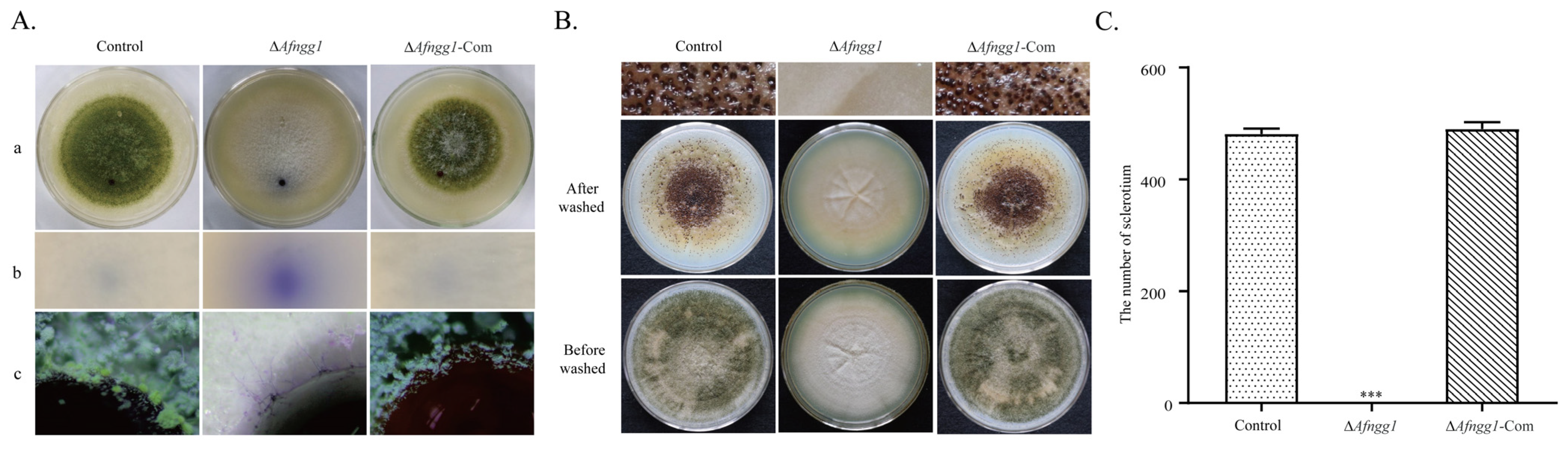
2.3. Effects of Afngg1 on Hydrophobicity and Sclerotia Formation of A. flavus Colonies

We studied the effects of Afngg1 on the surface hydrophobicity of A. flavus cells. The results revealed spherical water droplets on A. flavus control strain colonies, but these were dispersed and absorbed on ΔAfngg1 strain colonies (Figure 3(Aa,Ab)). In order to further probe the hydrophobicity differences, the droplet state of mycelia around droplets on colonies and Bromophenol Blue droplets were observed by stereoscopic microscope. The results showed that mycelia and spores around the A. flavus control strain were attached to the outer side of the droplet, while Bromophenol Blue solution of the ∆Afngg1 strain was diffused throughout mycelia (Figure 3(Ac)). Additionally, the results showed that compared with the A. flavus control strain, the ∆Afngg1 mutant did not produce sclerotia at all on PDA medium (Figure 3B,C). These results indicate that Afngg1 is essential for sclerotia formation in A. flavus.

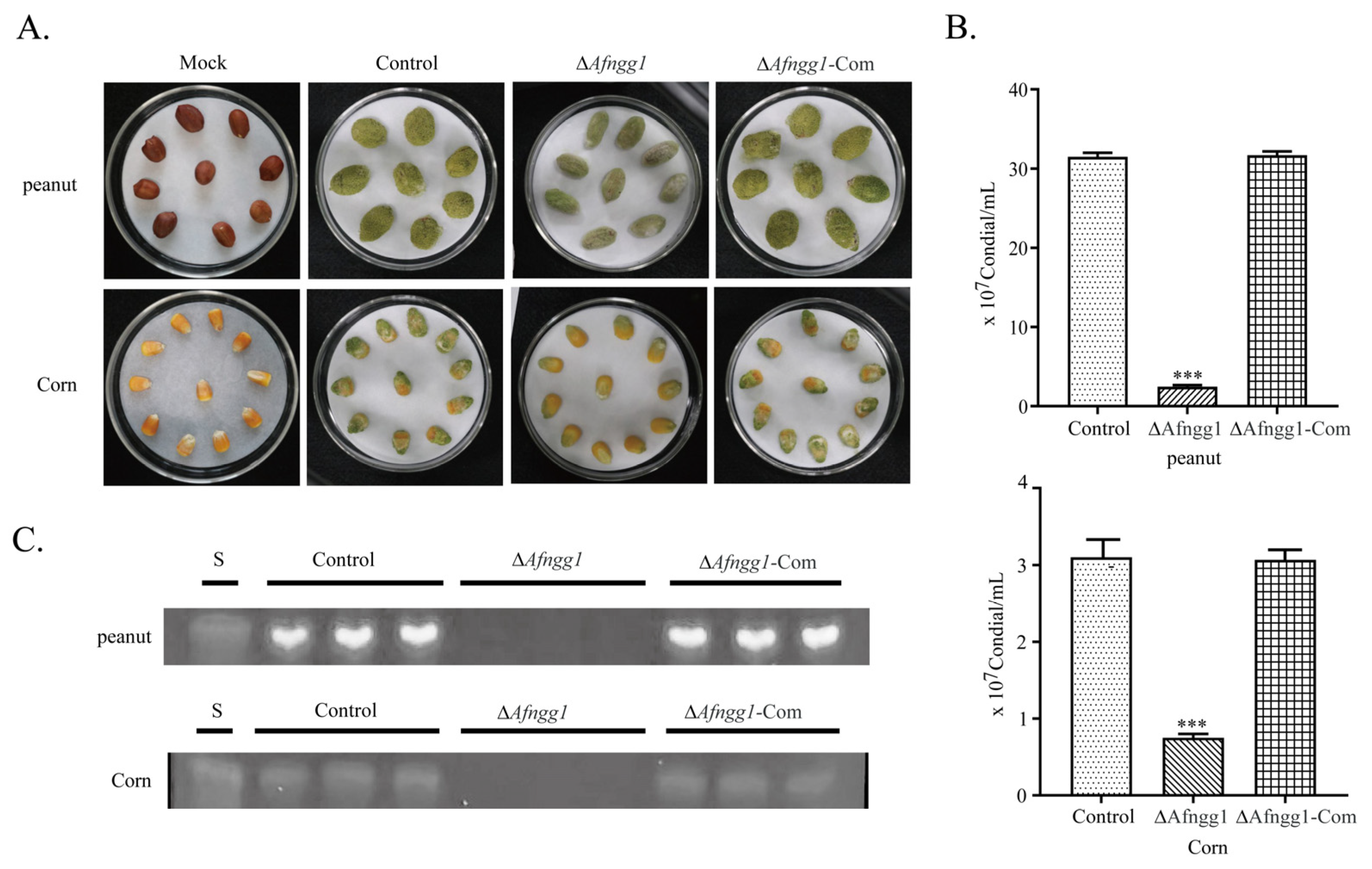
2.4. Afngg1 Influences the Infection Ability of A. flavus on Peanut and Corn

In order to study the pathogenicity of Afngg1 deletion in A. flavus, maize and corn grains were inoculated with spore suspensions. The results showed that the ∆Afngg1 mutant had a decreased ability to colonise peanut and corn grains, and conidia production was decreased by 97.39% and 91.94%, respectively (Figure 4A,B). We also explored the yield of AFB1 on peanut and corn, and AFB1 was not detected on either peanut or corn grains infected with the ∆Afngg1 mutant (Figure 4C). These results indicate that Afngg1 plays a crucial role in conidia formation of A. flavus, and in the biosynthesis of aflatoxin during A. flavus colonisation on peanut and corn grains.

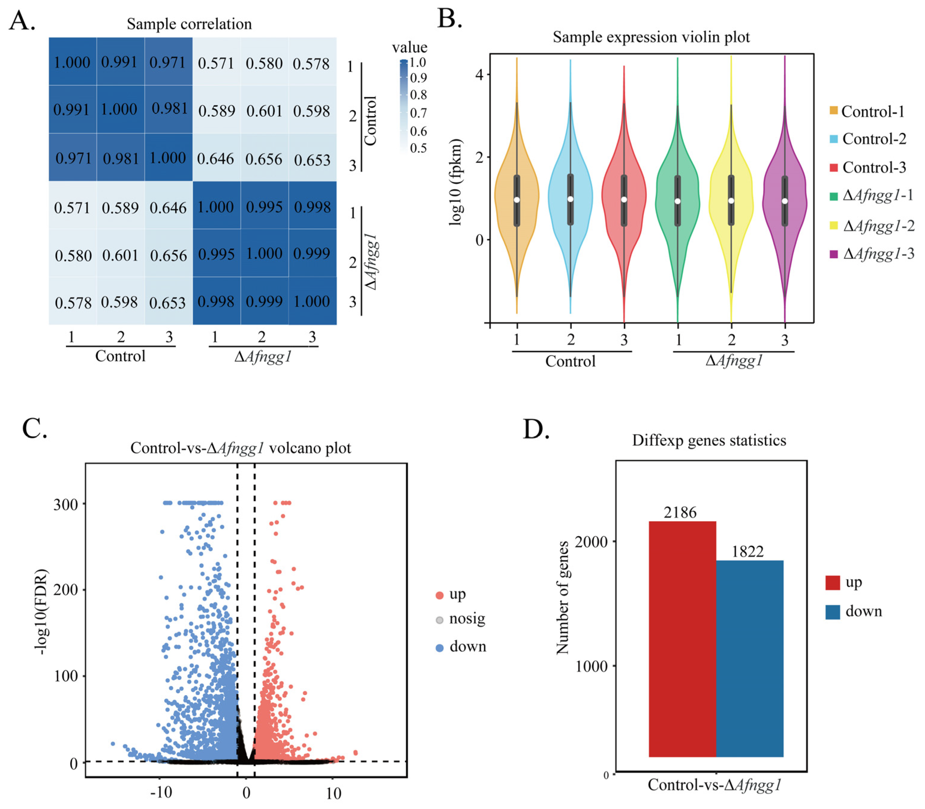
2.5. Transcriptome Overview

The Pearson correlation coefficient between all pairs of samples was >0.97, suggesting that samples were adequate for subsequent analysis (Figure 5A). Based on the FPKM (Fragments Per Kilobase per Million) value of each gene, gene expression levels were normalised and differentially expressed genes (DEGs) were compared (Figure 5B). The volcano map directly shows the expression ratio and significance of genes (Figure 5C). A total of 4008 significant DEGs were identified, among which 2186 (54.54%) were upregulated and 1822 (45.45%) were downregulated (Figure 5D).
The biological functions of DEGs were characterised by GO functional enrichment analysis. The results showed that most DEGs were linked to oxidation–reduction metabolic processes (Figure 6A). In terms of cellular components, DEGs were mainly associated with the plasma membrane, component of membrane and cell periphery subcategories (Figure 6B). The main molecular functions of DEGs were oxidoreductase activity, binding and catalytic activity (Figure 6C). KEGG pathway enrichment analysis results showed that DEGs were mainly involved in metabolic pathways, biosynthesis of secondary metabolites and aflatoxin biosynthesis (Figure 6D).

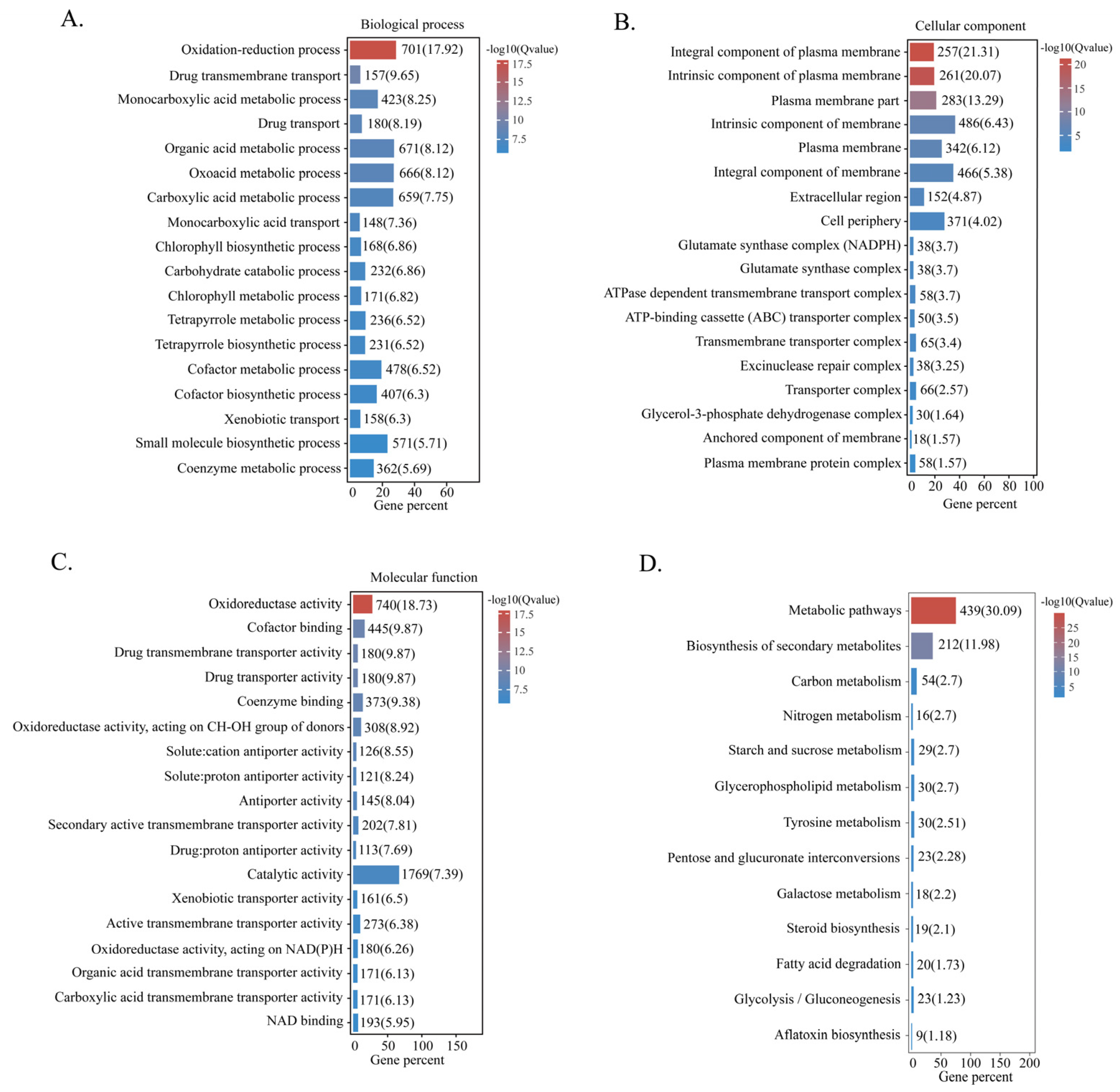
2.6. Categorisation of DEGs

To further elucidate the regulatory effects of Afngg1 on A. flavus cell development and aflatoxin biosynthesis, representative DEGs were divided into four groups: chromatin remodelling, cell development, secondary metabolism and oxidative stress (Table 1).
Afngg1 deletion altered the expression of genes involved in chromatin remodelling, including histone deacetylases (clr3, RPD3 and epl1) and chromatin remodelling complex subunits (mit1). Deletion of Afngg1 inhibited the expression of conidia-related genes wetA, abaA, brlA, stuA, vosA, fluG and con-6, and conidia hydrophobicity-related genes rodA and dewA. Additionally, ags1, ags2, MP65, chiB1, dcw1, ecm33, erg4, erg7 and erg5 genes related to cell wall and membrane formation were downregulated.
Secondary metabolism was also affected. The results showed that AFB1 biosynthetic pathway genes aflC, aflH, aflX, aflJ, aflW, aflQ, aflT and norA were downregulated after afngg1 deletion. Genes associated with the synthesis of other secondary metabolites, including conidia pigments (pksP and pks1), polyketones (fluP and aurA), ustiloxin B (ustYa, ustO, ustC, ustP, ustQ, ustD and ustF2) and gliotoxin (gliK, gliA and gliC) were also downregulated. Furthermore, afngg1 deletion inhibited the expression of catalase-related genes cta1, ctl-2 and cat.

3. Discussion

As an important post-translational modification, 2-hydroxyisobutyrylation plays a vital role in protein synthesis, transcriptional regulation and metabolism [14]. In this study, we performed a functional analysis of histone 2-hydroxyisobutyryltransferase Afngg1 in A. flavus. Deletion of Afngg1 reduced the level of histone 2-hydroxyisobutyrylation modification, indicating that Afngg1 functions as a histone 2-hydroxyisobutyrylyltransferase. Additionally, Afngg1 plays a crucial role in the growth and development of A. flavus and the biosynthesis of aflatoxin, and its potential regulatory mechanisms were explored through transcriptome analysis.

3.1. Afnng1 Correlates with the Level of Histone 2-Hydroxyisobutyrylation Modification

Previous reports have shown that Ngg1 is required for histone H3 acetylation of Candida albicans histone H3, and loss of Ngg1 greatly reduced the level of acetylation modification of histone H3 in Ngg1-A and Ngg1-B strains compared with wild-type strains [15]. However, its specific lysine acetylation sites were not characterised. In the present study, we identified homologs of C. albicans acetyltransferase NGG1 in A. flavus, and identified K9ac and K14ac as the key sites for alteration of histone H3 acetylation. Notably, we also found that Afngg1 is associated with 2-hydroxyisobutyrylation of histone H4, and deletion of Afngg1 significantly reduced lysine 2-hydroxyisobutyrylation levels at the K5 and K8 sites of histone H4.

3.2. Afnng1 Is Involved in Altering Chromatin Remodelling

Histone modification plays a crucial role in altering chromatin structure, among which histone acetylation and 2-hydroxyisobutyrylation modification are important contributors to chromatin remodelling [16,17]. RPD3 is a conserved histone deacetylase that regulates various cellular processes. In Fusarium graminearum, deletion of FgRpd3 led to increased acetylation of histone H4 [18]. Additionally, the crl3 gene encoding histone deacetylase affects the growth of Penicillium brasilianum, and the production of many secondary metabolites [16]. As a chromatin remodelling factor, Mit1 is mainly involved in regulating transcription of heterochromatin regions, and it promotes the deacetylation of histone H3 by clr3 [19]. We found that following deletion of Afngg1, expression levels of clr3, RPD3, epl1 and mit1 genes related to chromatin remodelling were altered. Additionally, previous studies have shown that cobB, de-2-hydroxyisobutyrylation enzyme, is involved in regulating the growth and metabolism of prokaryotes [8]. In human cells, deletion of the histone 2-hydroxyisobutyltransferase EP300 decreases the level of 2-hydroxyisobutyrylation modification at specific regulatory sites, and thus reduces the activity of cell metabolism-related enzymes [17]. Our current work showed that histone acetylation modification and 2-hydroxyisobutyrylation levels of the ΔAfngg1 strain were reduced. These results suggest that Afngg1 may alter chromatin conformation, altering the accessibility of genes, thus affecting cell development and secondary metabolism of A. flavus.

3.3. Afngg1 Affects Conidial Development and Sclerotium Formation

Recent studies have shown that fungal growth, development and production of secondary metabolites are regulated by post-translational modifications such as 2-hydroxyisobutyrylation [13]. Expression of A. flavus conidiogenesis genes is regulated by a cascade involving brlA, abaA and wetA [20]. BrlA and abaA are key transcription factors, of which brlA initiates conidia formation, and abaA contributes to conidia development and also affects sclerotia formation and AFB1 production [21,22]. StuA affects the expression of downstream genes by influencing brlA and abaA, and regulates sporulation [23]. VosA, a member of the velvet protein family, co-regulates spore-specific genes with wetA, and is critical for spore maturation and dormancy [24]. Loss of VosA resulted in increased sensitivity to oxidative stress and decreased trehalose content in A. flavus conidia [25]. FluG plays a balancing role in asexual and sexual development, and deletion of fluG can delay and reduce the formation of A. flavus conidia [26]; fluG can also interact with velB or laeA to control the production of conidia and sclerotia [27]. In the present study, the genes wetA, abaA, brlA, stuA, vosA, fluG and con-6 related to conidia were downregulated following deletion of Afngg1, mutant strain colonies were white, conidia production was decreased significantly and the strain no longer produced sclerotia. These results suggest that Afngg1 may regulate developmental balance, oxidative stress and conidia trehalose synthesis, and thereby affect the formation of conidia. Additionally, our experiment found that the ΔAfngg1 strain no longer produced sclerotia, consistent with the results of fluG deletion [27]. Transcriptomic analysis also showed downregulation of genes encoding hydrophobic proteins, namely rodA and dewA, which was confirmed by hydrophobic assay, and is reminiscent of the deletion of laeA in A. flavus [28].

3.4. Afngg1 Is Required in the Formation of the Cell Wall

The fungal cell wall is a complex structure composed of chitin, dextran and other polymers that play an important role in fungal growth and survival [29]. Ags1 and Ags2 are involved in glucan synthesis, and growth was inhibited after Ags1 deletion [30]. DCW1 is necessary for incorporation of cell wall glycoproteins into the cell wall, and deletion of the dcw1 gene in yeast led to cell wall abnormalities [31]. MP65 encodes β-glucan mannoprotein, which is associated with cell wall integrity and biofilm formation [32]. The GPI-anchored protein Ecm33 is required for cell wall composition, and is involved in fungal cell wall integrity and multiple stress tolerance [33]. One study found that the absence of ecm33 and MP65 increased sensitivity to cell wall degraders and altered morphology [32,33]. Herein, transcriptomic results showed that expression levels of the cell wall-related genes ags1, ags2, MP65, chiB1, dcw1 and ecm33 in the ΔAfngg1 strain were downregulated. Additionally, ergosterol is an important component of cell membranes, and we found that expression levels of genes associated with ergosterol biosynthesis such as erg4, erg7 and erg5 were significantly downregulated after deletion of Afngg1. These results suggest that Afngg1 may impede the synthesis of cell wall components such as glucan, reduce cell wall tolerance and cause cell wall defects.

3.5. Afngg1 Plays a Key Role in Secondary Metabolism

It has been reported that chromatin remodelling is closely related to the biosynthesis of aflatoxin [34]. Aflatoxin biosynthesis includes complex enzymatic reactions that require multiple enzymes, such as fatty acid synthases, polyketone synthases, oxidoreductases, cytochrome P450 monooxygenases, esterases, and others [35]. AflC (pksA) encodes a polyketone synthase that is involved in the conversion of acetate to norsolorinic acid, and AFB1 production can be significantly increased by upregulating aflC, aflR and aflK [36,37]. Both AflX and AflQ encode oxidoreductases, and AflQ is involved in the formation of AFB1 precursor hydroxyl-methylsterigmatocystin, and it plays a role in the later stages of the biosynthetic pathway [38,39]. Previous studies revealed a strong linear relationship between aflQ expression and aflatoxin biosynthesis [40]. AflW encodes a single monooxygenase that catalyses the conversion of hydroxyversicolorone to versiconal hemiacetal acetate [41]. Additionally, aflS encodes an esterase (AflJ) that interacts with nearby aflR genes to regulate the biosynthesis of aflatoxin [42]. Following aflS deletion, expression levels of aflatoxin biosynthetic pathway genes including aflC, aflD, aflM and aflP were reduced 5–20-fold, and biosynthesis of aflatoxin intermediates was blocked [43]. In the present study, deletion of Afngg1 downregulated genes encoding enzymes related to the aflatoxin biosynthetic pathway, such as aflC, aflX, aflJ, aflQ, aflT and norA, indicating that Afngg1 can inhibit the biosynthesis of aflatoxin by regulating the expression of multiple genes in the gene cluster via chromatin modification. Our study also found that the ΔAfngg1 strain no longer produced AFB1, consistent with knockout of histone acetyltransferase AflGcnE [6].
Other secondary metabolic pathways are also affected. Ustiloxin B is a secondary metabolite of Ustilaginoidea virens, and 18 genes form a gene cluster for the synthesis of ustiloxin B. Deletion of a relevant gene led to a complete or substantial loss of ustiloxin B production in A. flavus [44]. Biosynthesis of gliotoxin is carried out by a synthetic gene cluster consisting of 12 genes in A. fumigatus [45]. Loss of gliA greatly reduced the biosynthesis of glioxin [46], and deletion of gliK significantly hindered efflux of glioxins, and increased sensitivity to exogenous glioxins [47]. Herein, we found that expression levels of ustYa, ustO, ustC, ustP, ustQ, ustD, ustF2, gliK, gliA and gliC, which are responsible for the biosynthesis of ustiloxin B and gliatoxin, were downregulated. In addition, A. flavus produces a variety of polyketone-derived secondary metabolites, which are the most abundant of fungal secondary metabolites [48]. PksP was found to be associated with the biosynthesis and virulence of A. fumigatus conidia pigments [49]. The Pks1 gene encodes a polyketone synthase involved in melanin biosynthesis [50]. Deletion of polyketone synthase gene Flup resulted in decreased filament growth rate and conidial production in A. parasitica [51]. The present study found that expression levels of polyketone synthase-related genes fluP, pksP, aurA and Pks1 were downregulated. These results suggest that Afngg1 may play an important regulatory role in mycotoxin efflux and self-protection.

3.6. Afngg1 Alters Oxidative Stress Homeostasis

Oxidative stress is often associated with the synthesis of fungal secondary metabolites. Biosynthesis of aflatoxin has been reported to be inhibited by increased reactive oxygen species (ROS) levels and decreased activity of antioxidant enzymes [52]. Cells produce a variety of antioxidant enzymes to remove excessive ROS to protect cells from oxidative stress [53]. Previous reports have shown that deletion of cat1 significantly increased intracellular ROS levels, which in turn increased oxidative stress levels and reduced aflatoxin biosynthesis and virulence [52]. In our current work, expression levels of catalase-related genes cat1, ctl-2 and cat were downregulated, suggesting that deletion of Afngg1 might prevent cells from clearing ROS in a timely manner, thereby diminishing their defences against oxidative stress, and inhibiting the biosynthesis of aflatoxin.
In conclusion, we identified histone 2-hydroxyisobutyryltransferase Afngg1 in A. flavus, and explored its potential roles in cell development and aflatoxin biosynthesis. The results indicated that deletion of Afngg1 reduced the degree of lysine 2-hydroxyisobutyrylation modification of histone H4, inhibited cell growth, conidia formation, sclerotia production and AFB1 production in A. flavus, and decreased hydrophobicity. RNA-seq was subsequently used to investigate the underlying regulatory mechanisms, and analysis of DEGs showed that genes associated with chromatin remodelling, cell development, oxygen stress and biosynthesis of aflatoxin, ustiloxin B and gliatoxin, were downregulated. Therefore, these results suggest that histone 2-hydroxyisobutyryltransferase Afngg1 affects chromosomal modification, alters gene expression and regulates the growth, development and aflatoxin biosynthesis of A. flavus. The findings enhance our understanding of the role of histone 2-hydroxyisobutyrylation in aflatoxin biosynthesis and fungal pathogenicity regulation, and provide potential targets for exploring new control strategies for A. flavus.

4. Materials and Methods

4.1. Strains and Culture Conditions

A. flavus CA14 (kusA, pyrG+) served as a control strain, and the Afngg1 knockout strain (ΔAfngg1), Afngg1 complementation strain (ΔAfngg1-Com) and Afngg1-enhanced green fluorescence protein (eGFP) strain were derived in this study. Potato dextrose agar (PDA) was used to evaluate growth, conidia formation, cell hydrophobicity and aflatoxin B1 production, with appropriate amounts of uridine (10 mmol/L) added to the medium as needed. For sclerotia production, spore suspensions were maintained on PDA medium at 37 °C for 7 days in the dark. Each experiment was repeated three times.

4.2. Sequence Resources and Phylogenetic Tree Analysis

We used the protein sequence of Afngg1 to perform a BLAST search to identify homologous sequences in the National Center for Biotechnology Information (NCBI) database. Afngg1 domains were predicted by DOG 1.0 software (Ren and Wen, Hefei, China). Afngg1 protein sequences were aligned by ClustalW using MEGA software (7.0 version, iGEM, Temple University, Philadelphia, PA, USA), and a neighbour-joining phylogenetic tree was constructed [54].

4.3. Construction of ΔAfngg1, ΔAfngg1-Com and Afngg1-eGFP Strains

The ΔAfngg1 strains were constructed using homologous recombination with PCR primers listed in Table S1. PCR amplification was performed using P505 DNA polymerase (Vazyme, Nanjing, China). For construction of the ΔAfngg1 strain, primers ΔAfngg1-1F, ΔAfngg1-1R, ΔAfngg1-2F and ΔAfngg1-2R were used to amplify the 1.1 kb upstream and 1.5 kb downstream regions of Afngg1 fragments in the A. flavus genome, respectively. Meanwhile, primers pyrG-F and pyrG-R were used to amplify the pyrG selection marker. Fusion PCR was performed using primers ΔAfngg1-1F and ΔAfngg1-2R to generate a fragment containing the upstream and downstream segments, and the pyrG selection marker [55]. The fusion fragment was purified and transformed into the A. flavus control strain according to previous methods [34], and transformants were selected for verification.
Construction of the Afngg1-Com strain was performed according to previous methods [56]. Primers ΔAfngg1-Com-F and ΔAfngg1-Com-R were used to amplify a fragment including the promoter, coding sequence and terminator from A. flavus control strain genomic DNA, and this was ligated to the pPTRI plasmid following HindIII and SmaI restriction enzyme digestion. The resulting plasmid was transferred into A. flavus ΔAfngg1 protoplasts, and the ΔAfngg1-Com strain was confirmed by PCR.
For construction of the Afngg1-eGFP strain, linker, eGFP and TglaA were sequentially connected next to the Afngg1 coding sequence without a stop codon, using primers Afngg1-eGFP-2F and Afngg1-eGFP-2R. The fragment was ligated to the pPTRI plasmid according to previous methods [52]. The confirmed plasmid was transferred into ΔAfngg1 protoplasts, and transformants were selected for verification.

4.4. Subcellular Localisation

To detect subcellular localisation of Afngg1, A. flavus spore suspensions (106) were inoculated into Dextrose Peptone Yeast (DPY) liquid medium and cultured at 30 °C for 24 h. After collection, mycelia were washed with phosphate-buffered saline (PBS), stained with 4,6-diamidino-2-phenylindole for 10 min according to a previous method [57], and observed with an FV1000 laser confocal microscope (Olympus, Beijing, China).

4.5. Protein Extraction and Western Blotting Analysis

To explore the effect of Afngg1 deletion on the acetylation level of histone H3 and the 2-hydroxyisobutyrylation level of histone H4 in A. flavus, A. flavus control and ΔAfngg1 strains were cultured on PDA medium at 30 °C for 72 h. Mycelia were frozen with liquid nitrogen and ground to a powder. An appropriate amount of each protein sample was mixed with sample buffer and protein lysis solution to make a final concentration of 1–2 mg/mL, and heated at 95 °C for 10 min prior to sodium dodecyl sulphate polyacrylamide gel electrophoresis (SDS-PAGE). Proteins were transferred to a PVDF membrane, and this was blocked with 5% skimmed milk powder after transfer. For analysis of lysine acetylation of histone H3, anti-H3K9ac, anti-H3K14ac and anti-H3 were used as primary antibodies. For analysis of lysine 2-hydroxyisobutyrylation, anti-H4K5hib (PTM-854; 1:10,000 dilution; PTM-Bio, Hangzhoou, China) and anti-H4K8hib (PTM-805; 1:10,000 dilution; PTM-Bio, Hangzhoou, China) were used as primary antibodies. Goat anti-mouse IgGI (1:10,000 dilution; Thermo, Shanghai, China) was used as secondary antibody, and chemiluminescent horseradish peroxidase (HRP) substrate (Millipore) was added and incubated for 2 min for signal detection. Meanwhile, protein gray scale analysis was performed using Image-Pro Plus software (4.5 version, Media Cybernetics, Silver Spring, USA) according to a previous method [6].

4.6. Morphological and Physiological Analyses

To investigate the effect of Afngg1 on the growth of A. flavus, 2 μL spore suspensions (106 spores/mL) of A. flavus control, ΔAfngg1 and ΔAfngg1-Com strains were separately inoculated in the centre of PDA plates and cultured at 30 °C for 5 days; then, the colony diameter was measured and conidial heads were counted under a stereoscopic microscope (Nikon, Shanghai, China). Next, 6 mL sterile water was added to each Petri dish to harvest the mycelium, and the number of conidia was calculated using a hemocytometer. To assess the hydrophobicity of A. flavus mycelia, 20 μL sterile water and 20 μL 3% Bromophenol Blue solution were added to the edges of colonies of A. flavus control, ΔAfngg1 and ΔAfngg1-Com strains, cells were cultured at 30 °C for 2 days, and analysed by a stereoscopic microscope. Quantitative analysis of sclerotia production was performed at 37 °C for 7 days. After 7 days, 75% ethanol was sprayed to wash the medium and expose the sclerotia, and five holes were made along the diameter to count the number of sclerotia. Additionally, scanning electron microscopy (SEM) was used to photograph the conidia microstructure of A. flavus control, ΔAfngg1 and ΔAfngg1-Com strains as described previously [58].

4.7. Determination of AFB1 Production

AFB1 production analysis was carried out according to a previously described method [59]. Spore suspensions (106 spores/mL) of A. flavus control, ΔAfngg1 and ΔAfngg1-Com strains were cultured in 50 mL Yeast Extract Sucrose (YES) liquid medium at 30 °C in the dark for 5 days. An equal volume of chloroform was added to extract aflatoxin from the 50 mL medium. After the chloroform was completely volatilised, the residue was dissolved in 1 mL methanol and then analysed by TLC. The AFB1 yield of A. flavus strains was evaluated using an AFB1 standard sample (0.1 mg/mL).

4.8. Peanut and Corn Infection Assay

A peanut and corn infection experiment was performed as described previously [60]. Peanut and corn seeds were inoculated with 106/mL spore suspensions of A. flavus control, ΔAfngg1 strain and ΔAfngg1-Com strain and cultured at 28 °C in the dark for 6 days, with water replenished each day. Sterile water was added to collect the spore suspension and the number of conidia was calculated using a hemocytometer according to a previous method [34]. Finally, chloroform was added to extract aflatoxin as described above.

4.9. Transcriptome Analysis

Total RNA was extracted using Trizol reagent (Thermo Fisher, Shanghai, China) following the manufacturer’s instructions. RNA quality and integrity analyses, cDNA library construction, and RNA-Seq were performed by Guangzhou Gene Denovo Biotechnology (Guangzhou, China). The Pearson correlation coefficient was used to analyse the accuracy and reliability of the three biological duplicate samples. DESeq was used to analyse differences in FPKM value, and differences were considered statistically when log2(fold change) >1 and p-value < 0.05 criteria were met. Genome Ontology (GO) functional analysis and Kyoto Encyclopedia of Genes and Genomes (KEGG) pathway analysis were carried out using FungiFun (https://sbi.hki-jena.de/FungiFun/FungiFun.cgi) (accessed on 15 August 2022) and the KEGG Automatic Annotation Sever (http://www.kegg.jp/ or http://www.genome.jp/kegg/) (accessed on 15 August 2022).

Supplementary Materials

The following supporting information can be downloaded at: https://www.mdpi.com/article/10.3390/toxins15010007/s1, Figure S1: Determination of acetylation modification levels of histone H3K9ac and H3K14ac in A. flavus control and ΔAfngg1 strains; Figure S2: Construction and verification of ΔAfngg1 strain, complement and localisation strains; Table S1: PCR primers were used in this study.

Author Contributions

Methodology, L.L.; validation, J.W.; data curation, S.W.; writing—original draft preparation, L.L. and J.W.; writing—review and editing, S.Z. and Y.L.; project administration, Y.H. All authors have read and agreed to the published version of the manuscript.

Funding

This work was sponsored by grants from the Natural Science Foundation of China (31972176), Natural Science Foundation of Henan Province (222300420037) and the Innovative Funds Plan of Henan University of Technology (2020ZKCJ01).

Institutional Review Board Statement

Not applicable.

Informed Consent Statement

Not applicable.

Data Availability Statement

The raw transcriptome read data are available in the SRA database under accession number SUB12287216.

Conflicts of Interest

The authors declare no conflict of interest.

References

  1. Mahmoud, M.A.; El-Samawaty, A.M.; Yassin, M.A.; Abd El-Aziz, A.R. Genetic diversity analysis of Aspergillus flavus isolates from plants and air by ISSR markers. Genet. Mol. Res. 2016, 15, 1–9. [Google Scholar] [CrossRef] [PubMed]
  2. Taniwaki, M.H.; Pitt, J.I.; Magan, N. Aspergillus species and mycotoxins: Occurrence and importance in major food commodities. Curr. Opin. Food Sci. 2018, 23, 38–43. [Google Scholar] [CrossRef] [Green Version]
  3. Tumukunde, E.; Ma, G.; Li, D.; Yuan, J.; Qin, L.; Wang, S. Current research and prevention of aflatoxins in China. World Mycotoxin J. 2020, 13, 121–138. [Google Scholar] [CrossRef]
  4. Mitchell, N.J.; Bowers, E.; Hurburgh, C.; Wu, F. Potential economic losses to the US corn industry from aflatoxin contamination. Food Addit. Contam. Part A Chem. Anal. Control. Expo. Risk Assess. 2016, 33, 540–550. [Google Scholar] [CrossRef] [Green Version]
  5. Wu, F. Perspective: Time to face the fungal threat. Nature 2014, 516, 7. [Google Scholar] [CrossRef]
  6. Lan, H.; Sun, R.; Fan, K.; Yang, K.; Zhang, F.; Nie, X.Y.; Wang, X.; Zhuang, Z.; Wang, S. The Aspergillus flavus Histone Acetyltransferase AflGcnE Regulates Morphogenesis, Aflatoxin Biosynthesis, and Pathogenicity. Front. Microbiol. 2016, 7, 1324. [Google Scholar] [CrossRef] [Green Version]
  7. Qian, H.; Wang, L.; Ma, X.; Yi, X.; Wang, B.; Liang, W. Proteome-Wide Analysis of Lysine 2-Hydroxyisobutyrylated Proteins in Fusarium oxysporum. Front. Microbiol. 2021, 12, 623735. [Google Scholar] [CrossRef]
  8. Dong, H.; Zhao, Y.; Bi, C.; Han, Y.; Zhang, J.; Bai, X.; Zhai, G.; Zhang, H.; Tian, S.; Hu, D.; et al. TmcA functions as a lysine 2-hydroxyisobutyryltransferase to regulate transcription. Nat. Chem. Biol. 2022, 18, 142–151. [Google Scholar] [CrossRef]
  9. Chen, X.; Li, P.; Liu, H.; Chen, X.; Huang, J.; Luo, C.; Li, G.; Hsiang, T.; Collinge, D.B.; Zheng, L. A novel transcription factor UvCGBP1 regulates development and virulence of rice false smut fungus Ustilaginoidea virens. Virulence 2021, 12, 1563–1579. [Google Scholar] [CrossRef]
  10. Xu, Y.; Li, X.; Liang, W.; Liu, M. Proteome-Wide Analysis of Lysine 2-Hydroxyisobutyrylation in the Phytopathogenic Fungus Botrytis cinerea. Front. Microbiol. 2020, 11, 585614. [Google Scholar] [CrossRef]
  11. Zheng, H.; Song, N.; Zhou, X.; Mei, H.; Li, D.; Li, X.; Liu, W. Proteome-Wide Analysis of Lysine 2-Hydroxyisobutyrylation in Candida albicans. mSystems 2021, 6, e01129-20. [Google Scholar] [CrossRef] [PubMed]
  12. Yin, D.; Jiang, N.; Zhang, Y.; Wang, D.; Sang, X.; Feng, Y.; Chen, R.; Wang, X.; Yang, N.; Chen, Q. Global Lysine Crotonylation and 2-Hydroxyisobutyrylation in Phenotypically Different Toxoplasma gondii Parasites. Mol. Cell Proteom. 2019, 18, 2207–2224. [Google Scholar] [CrossRef] [PubMed] [Green Version]
  13. Lv, Y.; Wang, J.; Yang, H.; Li, N.; Farzaneh, M.; Wei, S.; Zhai, H.; Zhang, S.; Hu, Y. Lysine 2-hydroxyisobutyrylation orchestrates cell development and aflatoxin biosynthesis in Aspergillus flavus. Environ. Microbiol. 2022, 24, 4356–4368. [Google Scholar] [CrossRef] [PubMed]
  14. Zhang, K.; Cao, H.; Ma, Y.; Si, H.; Zang, J.; Bai, H.; Yu, L.; Pang, X.; Zhou, F.; Xing, J.; et al. Global analysis of lysine 2-hydroxyisobutyrylation during Fusarium graminearum infection in maize. Front. Plant Sci. 2022, 13, 1000039. [Google Scholar] [CrossRef] [PubMed]
  15. Li, D.-D.; Fuchs, B.B.; Wang, Y.; Huang, X.-W.; Hu, D.-D.; Sun, Y.; Chai, D.; Jiang, Y.-Y.; Mylonakis, E. Histone acetyltransferase encoded by NGG1 is required for morphological conversion and virulence of Candida albicans. Future Microbiol. 2017, 12, 1497–1510. [Google Scholar] [CrossRef]
  16. Akiyama, D.Y.; Rocha, M.C.; Costa, J.H.; Teles, C.B.; da Silva Zuccoli, G.; Malavazi, I.; Fill, T.P. The Penicillium brasilianum Histone Deacetylase Clr3 Regulates Secondary Metabolite Production and Tolerance to Oxidative Stress. J. Fungi 2022, 8, 514. [Google Scholar] [CrossRef] [PubMed]
  17. Huang, H.; Tang, S.; Ji, M.; Tang, Z.; Shimada, M.; Liu, X.; Qi, S.; Locasale, J.W.; Roeder, R.G.; Zhao, Y.; et al. p300-Mediated Lysine 2-Hydroxyisobutyrylation Regulates Glycolysis. Mol. Cell 2018, 70, 663–678.e6. [Google Scholar] [CrossRef]
  18. Lin, C.; Cao, X.; Qu, Z.; Zhang, S.; Naqvi, N.I.; Deng, Y.Z. The Histone Deacetylases MoRpd3 and MoHst4 Regulate Growth, Conidiation, and Pathogenicity in the Rice Blast Fungus Magnaporthe oryzae. mSphere 2021, 6, e0011821. [Google Scholar] [CrossRef]
  19. Creamer, K.M.; Job, G.; Shanker, S.; Neale, G.A.; Lin, Y.-C.; Bartholomew, B.; Partridge, J.F. The Mi-2 Homolog Mit1 Actively Positions Nucleosomes within Heterochromatin to Suppress Transcription. Mol. Cell Biol. 2014, 34, 2046–2061. [Google Scholar] [CrossRef] [Green Version]
  20. Zhou, D.; Xie, M.; Bai, N.; Yang, L.; Zhang, K.-Q.; Yang, J. The Autophagy-Related Gene Aolatg4 Regulates Hyphal Growth, Sporulation, Autophagosome Formation, and Pathogenicity in Arthrobotrys oligospora. Front. Microbiol. 2020, 11, 592524. [Google Scholar] [CrossRef]
  21. Cho, H.-J.; Son, S.-H.; Chen, W.; Son, Y.-E.; Lee, I.; Yu, J.-H.; Park, H.-S. Regulation of Conidiogenesis in Aspergillus flavus. Cells 2022, 11, 2796. [Google Scholar] [CrossRef] [PubMed]
  22. Mead, M.E.; Borowsky, A.T.; Joehnk, B.; Steenwyk, J.L.; Shen, X.-X.; Sil, A.; Rokas, A. Recurrent Loss of abaA, a Master Regulator of Asexual Development in Filamentous Fungi, Correlates with Changes in Genomic and Morphological Traits. Genome Biol. Evol. 2020, 12, 1119–1130. [Google Scholar] [CrossRef] [PubMed]
  23. Miller, K.Y.; Wu, J.; Miller, B.L. StuA is required for cell pattern formation in Aspergillus. Genes Dev. 1992, 6, 1770–1782. [Google Scholar] [CrossRef] [PubMed] [Green Version]
  24. Wu, M.-Y.; Mead, M.E.; Lee, M.-K.; Neuhaus, G.F.; Adpressa, D.A.; Martien, J.I.; Son, Y.-E.; Moon, H.; Amador-Noguez, D.; Han, K.-H.; et al. Transcriptomic, Protein-DNA Interaction, and Metabolomic Studies of VosA, VelB, and WetA in Aspergillus nidulans Asexual Spores. mBio 2021, 12, e03128-20. [Google Scholar] [CrossRef] [PubMed]
  25. Eom, T.-J.; Moon, H.; Yu, J.-H.; Park, H.-S. Characterization of the velvet regulators in Aspergillus flavus. J. Microbiol. 2018, 56, 893–901. [Google Scholar] [CrossRef] [PubMed]
  26. Chang, P.-K.; Scharfenstein, L.L.; Mack, B.; Ehrlich, K.C. Deletion of the Aspergillus flavus Orthologue of A. nidulans fluG Reduces Conidiation and Promotes Production of Sclerotia but Does Not Abolish Aflatoxin Biosynthesis. Appl. Environ. Microbiol. 2021, 78, 7557–7563. [Google Scholar] [CrossRef] [PubMed] [Green Version]
  27. Chang, P.-K.; Scharfenstein, L.L.; Li, P.; Ehrlich, K.C. Aspergillus flavus VelB acts distinctly from VeA in conidiation and may coordinate with FluG to modulate sclerotial production. Fungal Genet. Biol. 2013, 58–59, 71–79. [Google Scholar] [CrossRef] [PubMed]
  28. Chang, P.-K.; Scharfenstein, L.L.; Ehrlich, K.C.; Wei, Q.; Bhatnagar, D.; Ingber, B.F. Effects of laeA deletion on Aspergillus flavus conidial development and hydrophobicity may contribute to loss of aflatoxin production. Fungal Biol. 2012, 116, 298–307. [Google Scholar] [CrossRef]
  29. Adams, D.J. Fungal cell wall chitinases and glucanases. Microbiology 2004, 150, 2029–2035. [Google Scholar] [CrossRef]
  30. Bernard, M.; Latgé, J.-P. Aspergillus fumigatus cell wall: Composition and biosynthesis. Med. Mycol. 2001, 39, 9–17. [Google Scholar] [CrossRef]
  31. Pan, S.; Tang, L.; Pan, X.; Qi, L.; Yang, J. A member of the glycoside hydrolase family 76 is involved in growth, conidiation, and virulence in rice blast fungus. Physiol. Mol. Plant Pathol. 2021, 113, 101587. [Google Scholar] [CrossRef]
  32. Sandini, S.; Stringaro, A.; Arancia, S.; Colone, M.; Mondello, F.; Murtas, S.; Girolamo, A.; Mastrangelo, N.; De Bernardis, F. The MP65 gene is required for cell wall integrity, adherence to epithelial cells and biofilm formation in Candida albicans. BMC Microbiol. 2011, 11, 106. [Google Scholar] [CrossRef] [PubMed] [Green Version]
  33. Chen, Y.; Zhu, J.; Ying, S.-H.; Feng, M.-G. The GPI-anchored protein Ecm33 is vital for conidiation, cell wall integrity, and multi-stress tolerance of two filamentous entomopathogens but not for virulence. Appl. Microbiol. Biotechnol. 2014, 98, 5517–5529. [Google Scholar] [CrossRef] [PubMed]
  34. Lv, Y.; Yang, H.; Wang, J.; Wei, S.; Zhai, H.; Zhang, S.; Hu, Y. Afper1 contributes to cell development and aflatoxin biosynthesis in Aspergillus flavus. Int. J. Food Microbiol. 2022, 377, 109828. [Google Scholar] [CrossRef]
  35. Li, Q.Z.; He, Z.M. Advances in research of the structural gene characteristics of the aflatoxin biosynthetic gene cluster. J. Plant Sci. Phytopathol. 2015, 2, 068–082. [Google Scholar] [CrossRef] [Green Version]
  36. Crawford, J.M.; Korman, T.P.; Labonte, J.W.; Vagstad, A.L.; Hill, E.A.; Kamari-Bidkorpeh, O.; Tsai, S.-C.; Townsend, C.A. Structural basis for biosynthetic programming of fungal aromatic polyketide cyclization. Nature 2009, 461, 1139–1143. [Google Scholar] [CrossRef] [Green Version]
  37. Li, Y.; He, Y.; Li, X.; Fasoyin, O.E.; Hu, Y.; Liu, Y.; Yuan, J.; Zhuang, Z.; Wang, S. Histone Methyltransferase aflrmtA gene is involved in the morphogenesis, mycotoxin biosynthesis, and pathogenicity of Aspergillus flavus. Toxicon 2017, 127, 112–121. [Google Scholar] [CrossRef]
  38. Cary, J.W.; Ehrlich, K.C.; Bland, J.M.; Montalbano, B.G. The Aflatoxin Biosynthesis Cluster Gene, aflX, Encodes an Oxidoreductase Involved in Conversion of Versicolorin A to Demethylsterigmatocystin. Appl. Environ. Microbiol. 2006, 72, 1096–1101. [Google Scholar] [CrossRef] [Green Version]
  39. Ren, X.; Branà, M.T.; Haidukowski, M.; Gallo, A.; Zhang, Q.; Logrieco, A.F.; Li, P.; Zhao, S.; Altomare, C. Potential of Trichoderma spp. for Biocontrol of Aflatoxin-Producing Aspergillus flavus. Toxins 2022, 14, 86. [Google Scholar] [CrossRef]
  40. Jamali, M.; Karimipour, M.; Shams-Ghahfarokhi, M.; Amani, A.; Razzaghi-Abyaneh, M. Expression of aflatoxin genes aflO (omtB) and aflQ (ordA) differentiates levels of aflatoxin production by Aspergillus flavus strains from soils of pistachio orchards. Res. Microbiol. 2013, 164, 293–299. [Google Scholar] [CrossRef]
  41. Yabe, K.; Chihaya, N.; Hamamatsu, S.; Sakuno, E.; Hamasaki, T.; Nakajima, H.; Bennett, J.W. Enzymatic Conversion of Averufin to Hydroxyversicolorone and Elucidation of a Novel Metabolic Grid Involved in Aflatoxin Biosynthesis. Appl. Environ. Microbiol. 2003, 69, 66–73. [Google Scholar] [CrossRef] [PubMed] [Green Version]
  42. Chang, P.-K. The Aspergillus parasiticus protein AFLJ interacts with the aflatoxin pathway-specific regulator AFLR. Mol. Genet. Genom. 2003, 268, 711–719. [Google Scholar] [CrossRef] [PubMed]
  43. Yu, J.; Chang, P.K.; Ehrlich, K.C.; Cary, J.W.; Bhatnagar, D.; Cleveland, T.E.; Payne, G.A.; Linz, J.E.; Woloshuk, C.P.; Bennett, J.W. Clustered Pathway Genes in Aflatoxin Biosynthesis. Appl. Environ. Microbiol. 2004, 70, 1253–1262. [Google Scholar] [CrossRef] [Green Version]
  44. Umemura, M.; Nagano, N.; Koike, H.; Kawano, J.; Ishii, T.; Miyamura, Y.; Kikuchi, M.; Tamano, K.; Yu, J.; Shin-Ya, K.; et al. Characterization of the biosynthetic gene cluster for the ribosomally synthesized cyclic peptide ustiloxin B in Aspergillus flavus. Fungal Genet. Biol. 2014, 68, 23–30. [Google Scholar] [CrossRef] [PubMed] [Green Version]
  45. Kwon-Chung, K.J.; Sugui, J.A. What do we know about the role of gliotoxin in the pathobiology of Aspergillus fumigatus? Med. Mycol. 2009, 47, S97–S103. [Google Scholar] [CrossRef] [PubMed] [Green Version]
  46. Wang, D.-N.; Toyotome, T.; Muraosa, Y.; Watanabe, A.; Wuren, T.; Bunsupa, S.; Aoyagi, K.; Yamazaki, M.; Takino, M.; Kamei, K. GliA in Aspergillus fumigatus is required for its tolerance to gliotoxin and affects the amount of extracellular and intracellular gliotoxin. Med. Mycol. 2014, 52, 506–518. [Google Scholar] [CrossRef] [Green Version]
  47. Gallagher, L.; Owens, R.A.; Dolan, S.K.; O’Keeffe, G.; Schrettl, M.; Kavanagh, K.; Jones, G.W.; Doyle, S. The Aspergillus fumigatus Protein GliK Protects against Oxidative Stress and Is Essential for Gliotoxin Biosynthesis. Eukaryot. Cell 2012, 11, 1226–1238. [Google Scholar] [CrossRef] [Green Version]
  48. Keller, N.P.; Turner, G.; Bennett, J.W. Fungal secondary metabolism—From biochemistry to genomics. Nature reviews. Nat. Rev. Microbiol. 2005, 3, 937–947. [Google Scholar] [CrossRef]
  49. Langfelder, K.; Jahn, B.; Gehringer, H.; Schmidt, A.; Wanner, G.; Brakhage, A.A. Identification of a polyketide synthase gene (pksP) of Aspergillus fumigatus involved in conidial pigment biosynthesis and virulence. Med. Microbiol. Immunol. 1998, 187, 79–89. [Google Scholar] [CrossRef]
  50. Takano, Y.; Kubo, Y.; Shimizu, K.; Mise, K.; Okuno, T.; Furusawa, I. Structural analysis of PKS1, a polyketide synthase gene involved in melanin biosynthesis in Colletotrichum lagenarium. Mol. Gen. Genet. 1995, 249, 162–167. [Google Scholar] [CrossRef]
  51. Zhou, R.; Rasooly, R.; Linz, J.E. Isolation and analysis of fluP, a gene associated with hyphal growth and sporulation in Aspergillus parasiticus. Mol. Gen. Genet. 2000, 264, 514–520. [Google Scholar] [CrossRef] [PubMed]
  52. Zhu, Z.; Yang, M.; Bai, Y.; Ge, F.; Wang, S. Antioxidant-related catalaseCTA1regulates development, aflatoxin biosynthesis, and virulence in pathogenic fungus Aspergillus flavus. Environ. Microbiol. 2020, 22, 2792–2810. [Google Scholar] [CrossRef] [PubMed]
  53. Domènech, A.; Ayté, J.; Antunes, F.; Hidalgo, E. Using in vivo oxidation status of one- and two-component redox relays to determine H2O2 levels linked to signaling and toxicity. BMC Biol. 2018, 16, 61. [Google Scholar] [CrossRef] [PubMed]
  54. Kumar, S.; Stecher, G.; Tamura, K. MEGA7: Molecular Evolutionary Genetics Analysis Version 7.0 for Bigger Datasets. Mol. Biol. Evol. 2016, 33, 1870–1874. [Google Scholar] [CrossRef] [Green Version]
  55. Szewczyk, E.; Nayak, T.; Oakley, C.E.; Edgerton, H.; Xiong, Y.; Taheri-Talesh, N.; Osmani, S.A.; Oakley, B.R. Fusion PCR and gene targeting in Aspergillus nidulans. Nat. Protoc. 2006, 1, 3111–3120. [Google Scholar] [CrossRef] [PubMed]
  56. Ren, S.; Yang, M.; Yue, Y.; Ge, F.; Li, Y.; Guo, X.; Zhang, J.; Zhang, F.; Nie, X.; Wang, S. Lysine Succinylation Contributes to Aflatoxin Production and Pathogenicity in Aspergillus flavus. Mol. Cell Proteom. 2018, 17, 457–471. [Google Scholar] [CrossRef] [Green Version]
  57. Mengjuan, Z.; Guanglan, L.; Xiaohua, P.; Weitao, S.; Can, T.; Xuan, C.; Yanling, Y.; Zhenhong, Z. The PHD transcription factor Cti6 is involved in the fungal colonization and aflatoxin B1 biological synthesis of Aspergillus flavus. IMA Fungus 2021, 12, 12. [Google Scholar] [CrossRef]
  58. Zhang, W.; Lv, Y.; Lv, A.; Wei, S.; Zhang, S.; Li, C.; Hu, Y. Sub3 inhibits Aspergillus flavus growth by disrupting mitochondrial energy metabolism, and has potential biocontrol during peanut storage. J. Sci. Food Agric. 2021, 101, 486–496. [Google Scholar] [CrossRef]
  59. Hu, Y.; Yang, G.; Zhang, D.; Liu, Y.; Li, Y.; Lin, G.; Guo, Z.; Wang, S.; Zhuang, Z. The PHD Transcription Factor Rum1 Regulates Morphogenesis and Aflatoxin Biosynthesis in Aspergillus flavus. Toxins 2018, 10, 301. [Google Scholar] [CrossRef] [Green Version]
  60. Chang, P.-K.; Zhang, Q.; Scharfenstein, L.; Mack, B.; Yoshimi, A.; Miyazawa, K.; Abe, K. Aspergillus flavus GPI-anchored protein-encoding ecm33 has a role in growth, development, aflatoxin biosynthesis, and maize infection. Appl. Microbiol. Biotechnol. 2018, 102, 5209–5220. [Google Scholar] [CrossRef]
Figure 1. Bioinformatics analysis, subcellular localisation, and modification sites identification of Afngg1. (A) Phylogenetic tree based on sequence alignment. The numbers on the branches indicate bootstrap support value. (B) The domains of Afngg1 from the above nine species were further visualised by DOG2.0. The green area represented the Ada3 domain. (C) Subcellular localization of Afngg1. (D, E) Level of 2-hydroxyisobutyrylation modification of histones H4K5 and H4K8 in A. flavus control and ΔAfngg1 strains. *** represents p < 0.001.
Toxins 15 00007 g001
Figure 2. Effects of Afngg1 on the growth, conidia and aflatoxin biosynthesis of A. flavus. (A) The colony (a), stereoscopic microscope (b) and conidiophores (c) analysis. A. flavus control strain, ΔAfngg1 and Afngg1-Com strains were cultivated on PDA medium at 37 °C for 5 days. (B) SEM analysis of conidia (a) and conidial heads (b). (C) Determination of colony diameter cultured on PDA medium. (D) Determination of the number of conidia from different strains. (E) TLC analysis of AFB1 production. *** represents p < 0.001.
Toxins 15 00007 g002
Figure 3. Analysis of hydrophobicity and sclerotium yield. (A) Determination of hydrophobicity of A. flavus control strain, ΔAfngg1 and Afngg1-Com strains. Colony morphology (a), microscope (b) and stereoscopic microscopic (c) analysis after Bromophenol blue treatment. (B) Colony morphology of different strains after 7 days of culture on PDA medium. The plate was sprayed with 70% alcohol to expose the sclerotium. (C) Determination of the number of sclerotium in (B). *** represent p < 0.001.
Toxins 15 00007 g003
Figure 4. Effect of Afngg1 on the ability of A. flavus to infect crops. (A) Colonisation of peanut and corn grains by A. flavus control, ΔAfngg1 and ΔAfngg1-Com strains. (B) The number of conidia number was determined from the infected peanut and corn grains; (C) TLC analysis of AFB1 production from infected peanut and corn grains. *** represents p < 0.001.
Toxins 15 00007 g004
Figure 5. Sample correlation and gene expression analysis. (A) Person correlation coefficient. (B) Violin plot. (C) Volcano map of DEGs. (D) Total number of DEGs.
Toxins 15 00007 g005
Figure 6. Biological process (A), cellular component (B), molecular function (C) and KEGG pathway (D) analysis of DEGs.
Toxins 15 00007 g006
Table 1. Classification of representative DEGs in the ΔAfngg1 strain vs. control strain.
Gene CategoryNameLog2(fc)p-ValueDescription
Chromatin remodelling
AFLA_025120clr31.0892.82 × 10−21histone deacetylase
MSTRG.3596RPD31.151.26 × 10−6histone deacetylase RPD3
AFLA_055490epl11.515.53 × 10−44histone acetyltransferase complex component
AFLA_052300mit11.497.93 × 10−28chromatin remodelling complex subunit (Chd3)
Cell development
AFLA_052030wetA−2.321.96 × 10−120developmental regulatory protein WetA
AFLA_029620abaA−3.171.99 × 10−171transcription factor abaA
AFLA_082850brlA−5.270C2H2 type conidiation transcription factor
AFLA_046990stuA−1.035.33 × 10−46APSES transcription factor StuA
AFLA_026900vosA−2.281.98 × 10−75developmental regulator VosA
AFLA_039530fluG−3.171.28 × 10−13FluG family protein
AFLA_044800con-6−8.107.93 × 10−60conidiation protein Con-6
AFLA_098380rodA−7.582.79 × 10−67RodA/RolA7
AFLA_060780dewA−6.818.80 × 10−9hydrophobin family protein
AFLA_023460ags1−2.806.03 × 10−201alpha-1,3-glucan synthase Ags1
AFLA_134100ags2−2.284.34 × 10−119alpha-1,3-glucan synthase Ags2
AFLA_052780MP65−3.871.26 × 10−141cell wall glucanase (Scw4)
AFLA_104680chiB1−1.641.57 × 10−22class V chitinase ChiB1
AFLA_024280dcw1−5.490cell wall glycosyl hydrolase Dfg5
AFLA_113120ecm33−1.026.90 × 10−76GPI-anchored cell wall organization protein
AFLA_138060erg4−2.951.32 × 10−108c-24(28) sterol reductase
AFLA_001030erg7−9.211.07 × 10−73lanosterol synthase
AFLA_028640erg5−5.792.08 × 10−143cytochrome P450 sterol C-22 desaturase
Secondary metabolism
AFLA_139410aflC−1.003.76 × 10−32aflC/pksA/pksL1/polyketide synthase
AFLA_139330aflH−1.462.3 × 10−3aflH/short chain alcohol dehydrogenase
AFLA_139160aflX−2.371.05 × 10−2aflX/ordB/monooxygenase/oxidase
AFLA_139320aflJ−2.821.28 × 10−2aflJ/estA/esterase
AFLA_139170aflW−3.826.16 × 10−6aflW/moxY/monooxygenase
AFLA_002920aflQ−4.822.29 × 10−46flavonoid 3-hydroxylase
AFLA_024090aflT1.578.06 × 10−24efflux pump antibiotic resistance protein
AFLA_139310norA−8.347 × 10⁻⁴aflE/norA/aad/adh-2/ NOR reductase
AFLA_094990ustYa−7.922.80 × 10−21Ustiloxin B biosynthesis protein Ya
AFLA_094940ustO−2.982.18 × 10−108Ustiloxin B biosynthesis protein O
AFLA_094960ustC−4.141.27 × 10−48cytochrome P450
AFLA_095010ustP−4.591.18 × 10−84Ustiloxin B biosynthesis protein P
AFLA_095060ustQ−4.751.78 × 10−35tyrosinase
AFLA_095040ustD−6.189.10 × 10−2NRPS-like enzyme
AFLA_095050ustF2−7.295.02 × 10−20dimethylaniline monooxygenase
AFLA_064420gliK−3.263.49 × 10−12gliotoxin biosynthesis protein GliK
AFLA_118990gliA−4.911.47 × 10−127efflux pump antibiotic resistance protein
AFLA_064540gliC−6.691.62 × 10−55cytochrome P450 oxidoreductase
AFLA_114820fluP−1.441.78 × 10−50polyketide synthase
AFLA_006170pksP−6.540polyketide synthetase PksP
AFLA_010000aurA−2.232.02 × 10−84polyketide synthase
AFLA_054090pks1−2.227.5 × 10−3polyketide synthase
Oxidative stress
AFLA_034380cta1−7.680catalase
AFLA_096210ctl-2−6.966.43 × 10−272catalase
AFLA_100250cat−1.576.97 × 10−16catalase
Means with p-value < 0.05 are considered to have significant difference.
Disclaimer/Publisher’s Note: The statements, opinions and data contained in all publications are solely those of the individual author(s) and contributor(s) and not of MDPI and/or the editor(s). MDPI and/or the editor(s) disclaim responsibility for any injury to people or property resulting from any ideas, methods, instructions or products referred to in the content.

