Article

GTPase Rac Regulates Conidiation, AFB1 Production and Stress Response in Pathogenic Fungus Aspergillus flavus

Key Laboratory of Pathogenic Fungi and Mycotoxins of Fujian Province, Key Laboratory of Biopesticide and Chemical Biology of Education Ministry, School of Life Sciences, Fujian Agriculture and Forestry University, Fuzhou 350002, China
*
Authors to whom correspondence should be addressed.
These authors contributed equally to this work.
Toxins 2022, 14(9), 581; https://doi.org/10.3390/toxins14090581
Submission received: 1 August 2022 / Revised: 14 August 2022 / Accepted: 18 August 2022 / Published: 24 August 2022
(This article belongs to the Special Issue Research on Pathogenic Fungi and Mycotoxins in China (Volume II))

Abstract

:
As a member of the Rho family, Rac plays important roles in many species, including proliferation, differentiation, apoptosis, DNA damage responses, metabolism, angiogenesis, and immunosuppression. In this study, by constructing Rac-deleted mutants in Aspergillus flavus, it was found that the deletion of Rac gene led to the decline of growth and development, conidia production, AFB1 toxin synthesis, and seed infection ability of A. flavus. The deletion of Rac gene also caused the disappearance of A. flavus sclerotium, indicating that Rac is required for sclerotium formation in A. flavus. The sensitivity of Rac-deficient strains responding to cell wall stress and osmotic pressure stress increased when compared to A. flavus WT. The Western blot result showed that mitogen-activated serine/threonine-protein kinase Slt2 and mitogen-activated protein kinase Hog1 proteins were no longer phosphorylated in Rac-deficient strains of A. flavus, showing that Rac may be used as a molecular switch to control the Slt2-MAPK cascade pathway and regulate the osmotic Hog-MAPK cascade pathway in A. flavus in response to external stress. Altogether, these results indicated that Rac was involved in regulating the growth and development, conidia formation and AFB1 synthesis, and response to cell wall stress and osmotic pressure stress in A. flavus.
Key Contribution: In this study, the function of Rac in A. flavus was studied for the first time, and the possible role of Rac in the filamentous fungus A. flavus was discussed, which expanded a new idea for the study of Rac GTPase.

1. Introduction

Aspergillus flavus, as a common saprophytic filamentous fungus, is widely distributed all over the world. It is not only a plant pathogenic fungus, but also a conditional human and animal pathogenic fungus. Many secondary metabolites are produced during the growth and development of A. flavus, among which aflatoxins are produced from polyketones [1]. A. flavus produces aflatoxins by infecting cereal crops, including corn, peanut, and wheat [2], which causes a variety of harm to humans and animals, such as hepatotoxicity [3], carcinogenicity [4] and immunotoxicity [5]. Aflatoxin-contaminated cereal crops pose serious health risks in developing countries [6], as well as significant economic losses to the United States and other developed countries [7]. Therefore, the study of A. flavus is of more practical significance under the current situation of food security and shortage in many countries.
Rho family proteins belong to one of the important members of the Ras superfamily, including the Rho, Rac, and Cdc subfamilies. Members of this family have the inherent activity of hydrolyzing GTPases, so they are accustomed to being called Rho GTPases. As a molecular switch, Rho GTPases almost participate in the basic cellular process. The GTP binding state is activated, and the GDP binding state is inactivated [8]. Rho GTPase can regulate actin cytoskeleton [9], cell cycle progression [10], and signal network of gene expression, thus regulating normal cell proliferation, differentiation and apoptosis, and is closely related to tumor occurrence and metastasis.
As one of the most studied proteins in the Rho family, Rac plays an extremely important role in many species. As early as 1955, Rac was proved to regulate the production of reactive oxygen in macrophages in response to the attack of pathogens [11]. In Drosophila melanogaster, Rac not only plays an important role in neuron development [12] but also participates in axon growth and myoblast fusion [13]. Rac seems to activate LIM kinase to inhibit axon growth via the effector kinase PAK, but may also activate a PAK-independent pathway that promotes axon growth [12,14]. In fungi, such as Aspergillus nidulans, Rac-deficient mutants showed slow growth and development, but there is no obvious difference in mycelium morphogenesis. However, the conidia had various defects, such as the appearance of vesicles without conidia, shortening of stalk and vesicle branches, irregular shape of vesicles, and reduction in the number of conidia and vesicles [15]. The RacA gene was found to play an important role in the growth and pathogenesis of Aspergillus fumigatus. The absence of RacA in A. fumigatus reduced the production of reactive oxygen but had little effect on virulence [16]. In dimorphic plant pathogenic fungus Ustilago maydis, it was found that Rac1 deletion resulted in the loss of virulence, affecting cell morphology and interfering with mycelium growth [17]. Unlike the other Rho GTPases, Rac orthologs were not found in unicellular yeasts such as Saccharomyces cerevisiae and Schizosaccharomyces pombe, suggesting that the Rac gene might have evolved with the increase in cell complexity [18]. However, the function of Rac in A. flavus has not been explored until now. Therefore, this paper focused on the Rac biofunctions in A. flavus, and the main results were shown as follows.

2. Result

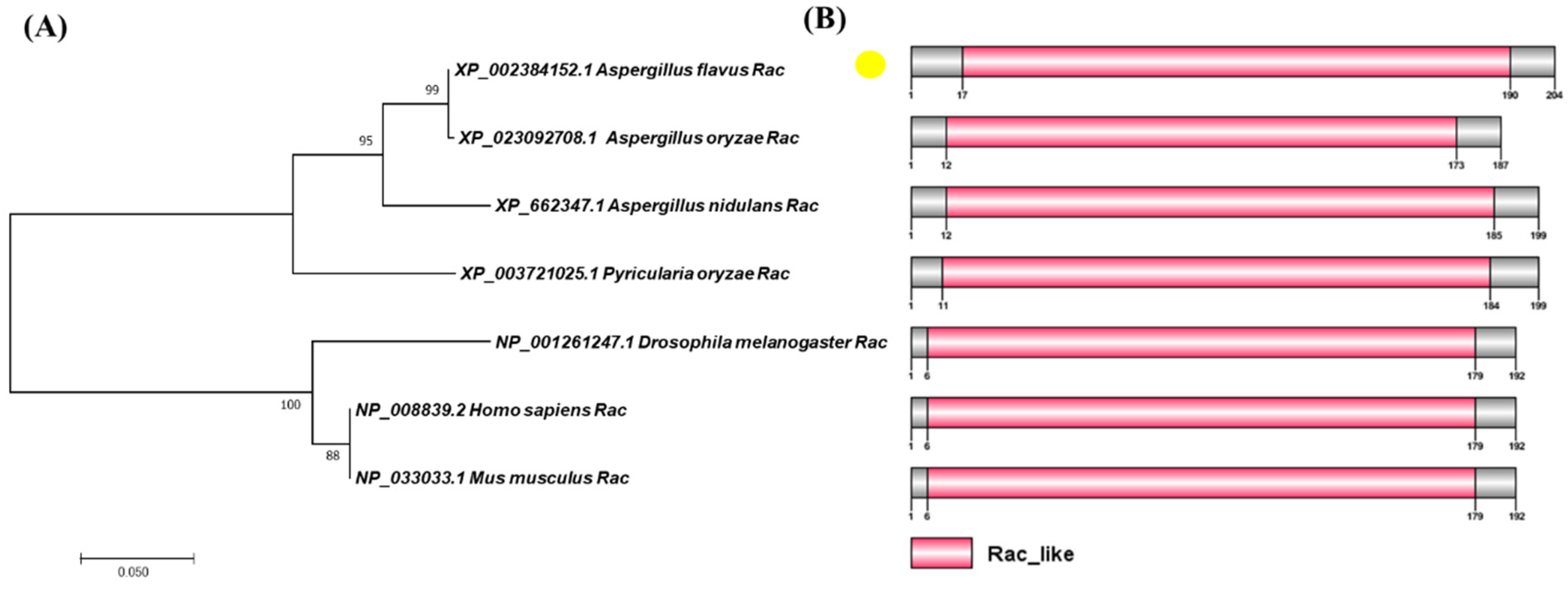
2.1. Biological Analysis of Rac

Blast comparison was carried out with A. nidulans Rac protein (XP_662347.1) in NCBI to identify homologous protein AflRac (XP_002384152.1) in A. flavus. Then, AflRac was compared with Rac proteins of other four fungi to study whether Rac proteins from fungi are conserved. Since Rac protein also plays an important role in the development of animal nerves, we compared Rac homologous proteins in two vertebrates. We used MEGA7 to map the phylogenetic tree based on the protein sequences blasted in NCBI. The result showed that Rac protein sequences are highly homologous in fungi and vertebrates (Figure 1A), indicating that the Rac protein is highly conserved in the process of evolution. We used DOG2.0 software (Jian Ren, Hefei, China) to draw the domain diagram of the proteins mentioned above and found that all these proteins have a Rac-like domain (Figure 1B), indicating that the Rac protein domain is also highly conserved in these species.

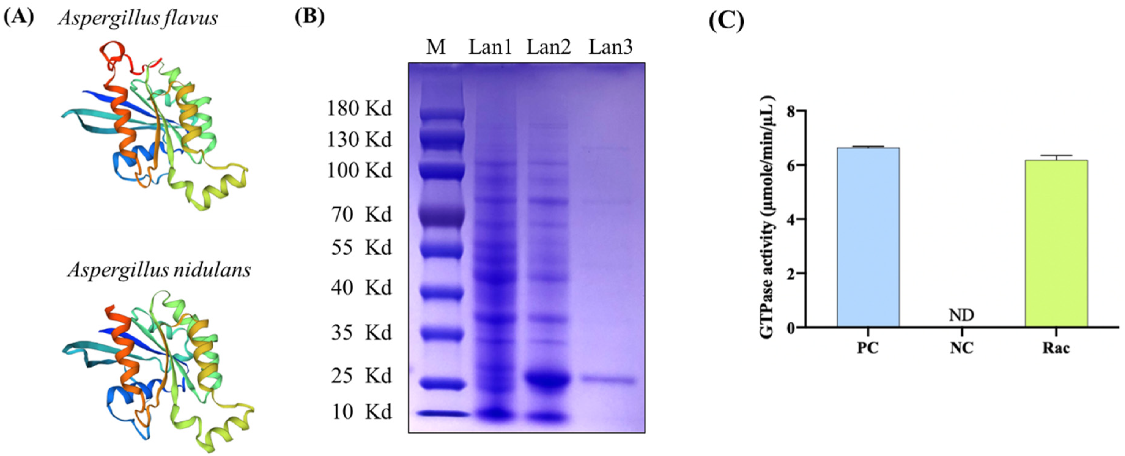
2.2. Rac from A. flavus Is a GTPase

After systematic prediction, sequence alignment showed that A. flavus Rac (XP_002384152.1) had high homology with A. nidulans Rac (XP_662347.1, 90.69% identification) and they two showed highly conserved three-dimensional protein structures (Figure 2A). In order to confirm whether AflRac has GTPase activity, we cloned the coding sequence of AflRac using the cDNA of A. flavus wild type (WT) as template and ligated it into the pET28a vector. After sequencing and twice transformation, the Rac protein expression strain was successfully constructed. After induction by IPTG, it was purified by a Ni2+-NTA column (Figure 2B). GTP enzyme activity was detected in vitro, and it was found that the AflRac protein had GTP enzyme activity (Figure 2C). The above results proved that AflRac has GTPase activity in A. flavus. Thus, in this paper, we directly define the protein XP_002384152.1 in A. flavus as Rac.

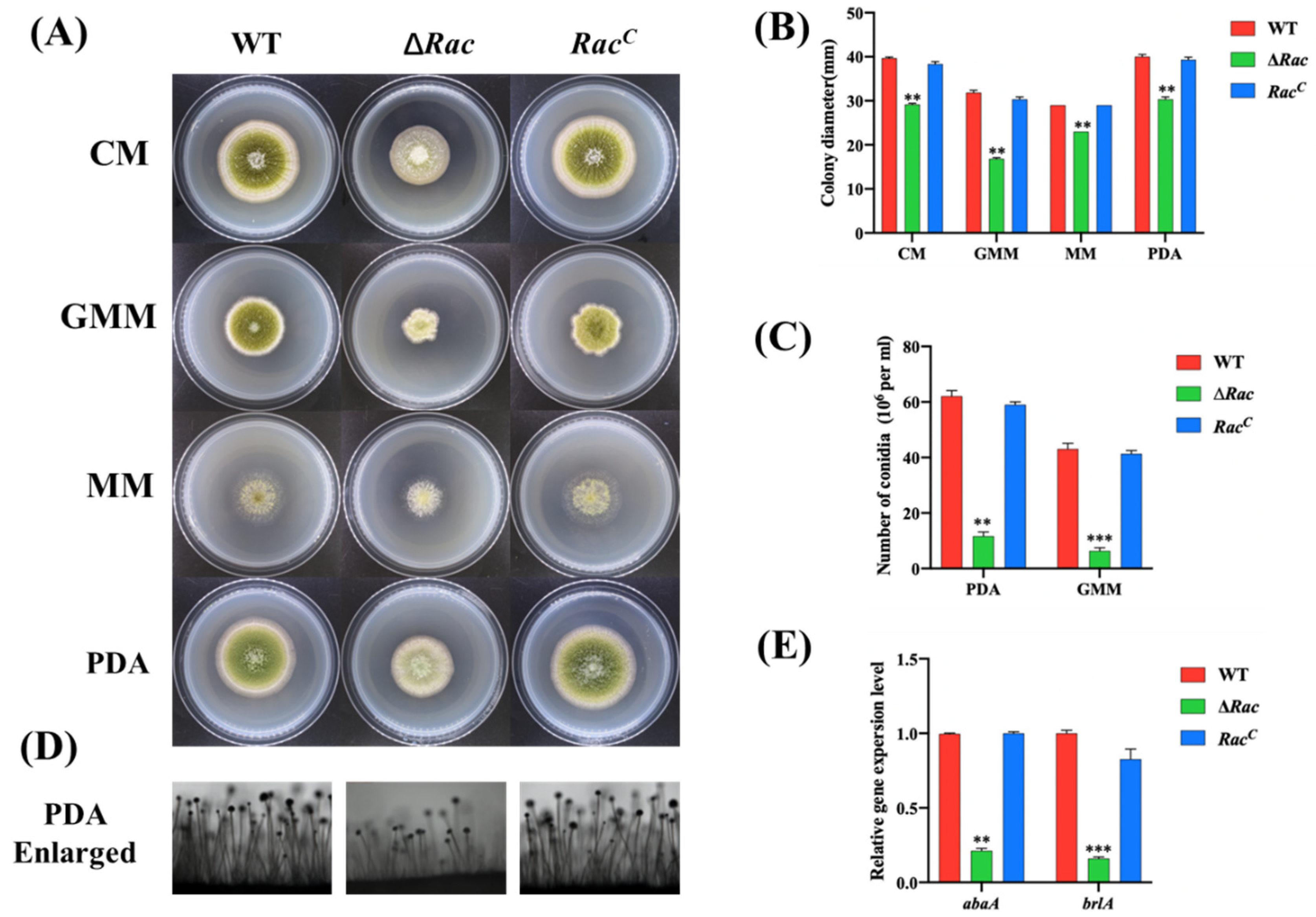
2.3. Rac Is Involved in Vegetative Growth and Conidiation

To study the function of Rac in A. flavus, we constructed Rac-deleted mutant strains. Using the principle of gene recombination, we replaced Rac in A. flavus wild type with AfupyrG to construct Rac deletion mutants (∆Rac) (Figure 3A). At the same time, we constructed Rac complementary strains (RacC). Then, PCR (Figure 3B), RT-PCR (Figure 3C), qPCR (Figure 3D), and Southern blot (Figure 3E) results verified that ∆Rac and RacC were successfully constructed. The above-constructed strains were diluted to the same multiple and inoculated in two kinds of rich media (CM and PDA) and two kinds of basic media (GMM and MM) for culture (Figure 4A). By observing the colony growth, it was found that the diameter of ∆Rac in all media was significantly smaller than that of WT and RacC (Figure 4B), which indicates that Rac participates in the vegetative growth process of A. flavus. During the observation, we also found that the color of ∆Rac was obviously white, as the green color on the strains was conidia of A. flavus. Therefore, a rich medium (PDA) and a basic medium (GMM) were selected to culture all the above strains, and the conidia were eluted to count the number with a blood cell counter under an optical microscope. The results showed that the conidia production of ∆Rac was significantly lower than that of WT and RacC (Figure 4C). The decrease in conidia made us wonder whether it was related to the decrease in conidiophore. Therefore, we observed the conidiophore and found that the number of conidiophore of ∆Rac was significantly decreased than that of WT and RacC (Figure 4D), indicating that Rac was very important for the formation of conidiophores of A. flavus. We also verified the key genes related to conidia formation in A. flavus by qPCR and found that the expression levels of genes abaA and brlA were significantly decreased in ∆Rac (Figure 4E), which further indicated that Rac regulates the expression of genes related to conidiation, thus affecting the conidia formation of A. flavus.

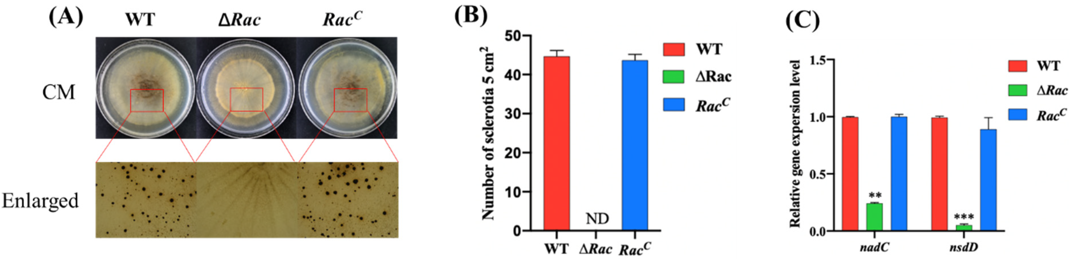
2.4. Rac Is Required for Sclerotial Formation in A. flavus

Sclerotium is another form of A. flavus reproduction, so the effect of Rac on the sclerotium formation of A. flavus deserves our study. We cultured the strains in CM medium for 7 days to observe the sclerotium formation, and the results showed that the ∆Rac did not produce sclerotium at all, but WT and RacC of A. flavus produced normal sclerotium (Figure 5A,B). qPCR result showed that the expression of nadC and nsdD, the key genes of sclerotium formation, was decreased markedly in ∆Rac when compared to WT and RacC (Figure 5C), which indicated that Rac was involved in sclerotium formation in A. flavus. This implies that Rac may act as a molecular switch to regulate the expression of genes related to the sclerotium formation pathway. When Rac was deleted, the sclerotium formation process was blocked and sclerotium disappeared due to the lack of control switch in A. flavus. All the above results showed that Rac is involved in the development and reproduction of A. flavus.

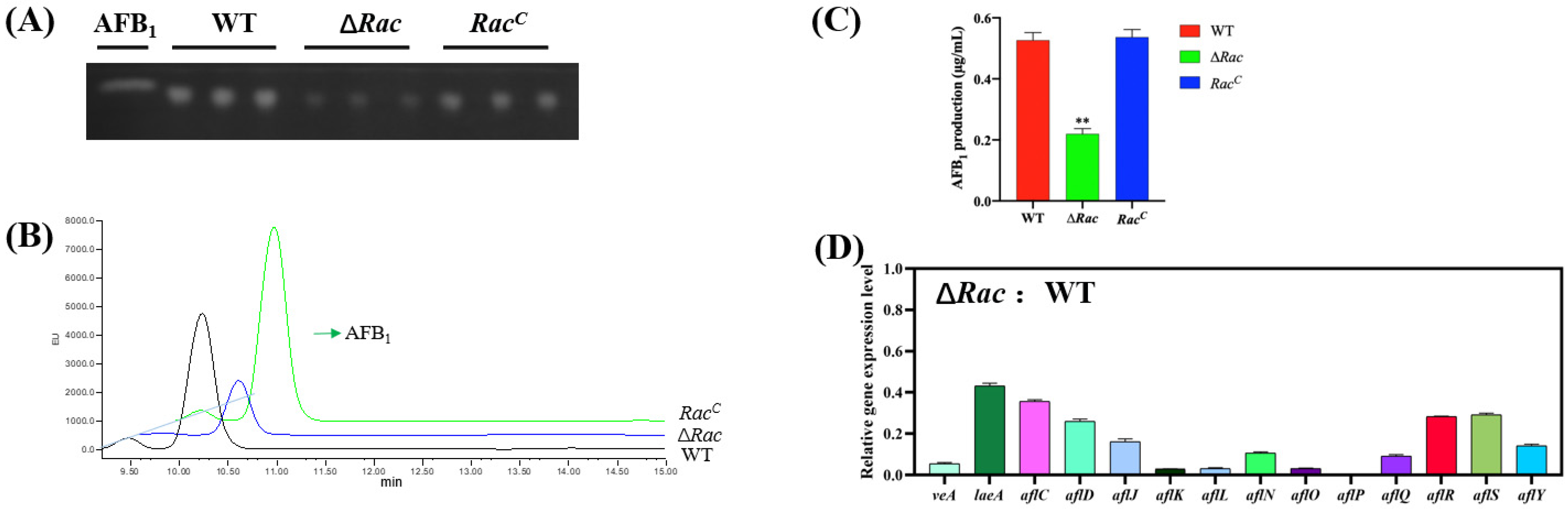
2.5. Effect of Rac on Aflatoxin Biosynthesis in A. flavus

AFB1 is the most harmful secondary metabolite produced by A. flavus. To study the role of Rac in AFB1 production, all strains were cultured in liquid medium, and TLC and HPLC were used to detect the aflatoxin production. The result showed that the ability of Rac mutant to produce toxin AFB1 was impaired significantly compared to WT and RacC (Figure 6A–C). To explore the reason for the significant decrease of AFB1 in ∆Rac, we amplified secondary metabolism-related genes and regulatory genes in the aflatoxin formation gene cluster by qPCR. It was found that the expression of secondary metabolism regulatory genes veA and laeA was decreased, and the expression of genes in the toxin formation gene cluster (aflR, aflC, aflD, aflJ, aflK, aflL, aflN, aflO, aflP, aflQ, aflR, aflS, aflY) was also downregulated (Figure 6D). It could be speculated that Rac might regulate the expression of genes related to toxin synthesis, thereby affecting the toxin synthesis ability in A. flavus.

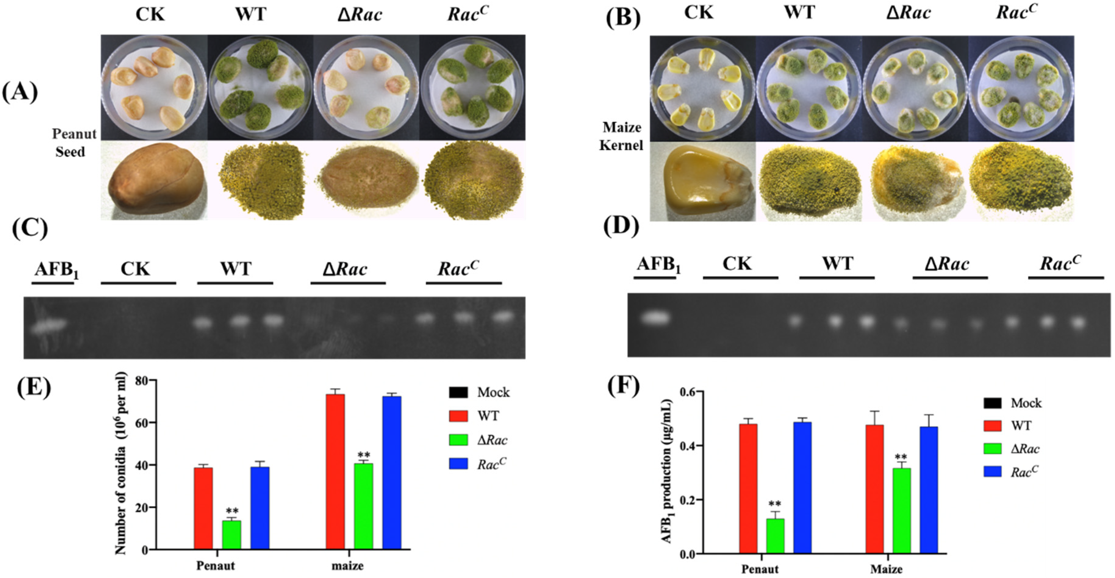
2.6. Role of Rac in Seed Infection

In order to explore the ability of ∆Rac to infect seeds and produce aflatoxin in the process of infecting the host, we conducted an in vitro infection experiment. The same amount of conidia was inoculated to corn and peanut seeds and cultured for 7 days, and it was found that the conidia produced by ∆Rac in the process of infecting seeds decreased significantly when compared to WT and RacC (Figure 7A,B,E), and the toxins produced by ∆Rac in the infecting process were significantly reduced when compared to WT and RacC (Figure 7C,D,F). All the above results showed that Rac also played an important role in the process of A. flavus infecting seeds.

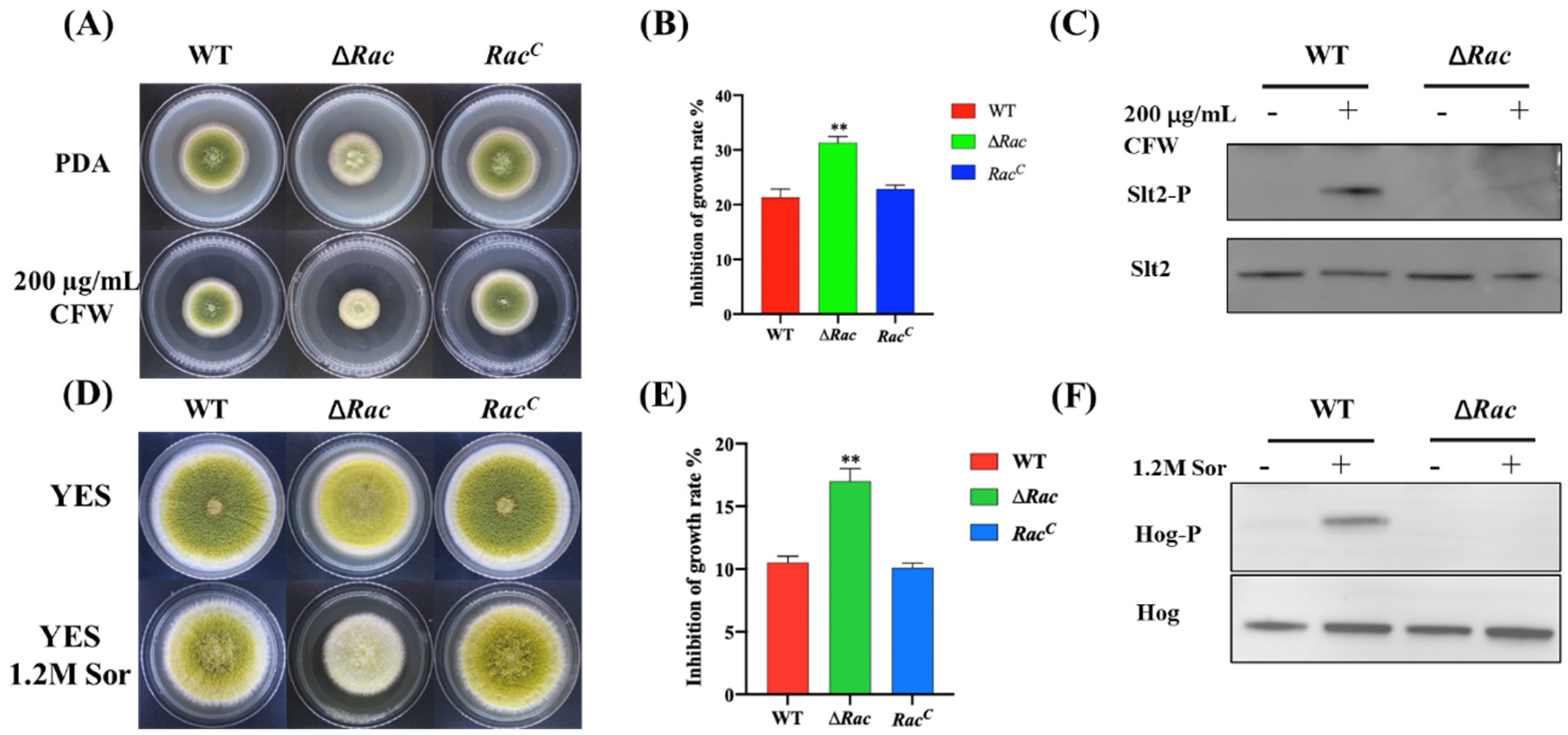
2.7. Rac Contributes to Cell Wall and Osmotic Stress Response

It was found that when 1.2 M calcofluor white (CFW) was added to the culture medium (Figure 8A), the inhibition rate of cell wall stress to ∆Rac was higher than that to WT and RacC (Figure 8B), indicating that the sensitivity of ∆Rac to respond to cell wall stress increased. Western blot results indicated that Slt2 could not be phosphorylated after Rac deletion (Figure 8C), showing that Rac may be used as the switch for the Slt2-MAPK cascade in A. flavus to control the pathway. When 1.2 M sorbitol was added into the culture medium (Figure 8D), the results showed that the inhibition rate of ∆Rac was also increased when compared to WT and RacC (Figure 8E), indicating the sensitivity of ∆Rac responding to osmotic stress was increased. Western blot results found that Hog could not be phosphorylated after Rac was deleted (Figure 8F), which indicated that Rac could be used as the switch of the osmotic glycerol pathway in A. flavus to regulate the Hog-MAPK cascade to cope with osmotic stress.

3. Discussion

In plants, Rac orthologs are thought to be involved in the regulation of pollen tube growth, which shares several features with filamentous growth [19]. As the first Rac homologous gene studied in fungi, the deletion of YlRAC1 resulted in the morphological changes of dimorphic yeast Yarrowia lipolytica cells, suggesting that the function of YlRAC1 may be related to some aspects of cell growth polarization [20]. In filamentous fungi, it was reported that the absence of RacA in Aspergillus niger leads to hyper-branched reproductive tubes and hyphae, which are short in length but wide in hyphal diameter. The frequent branching leads to tighter colonies on solid media, and the diameter of colonies becomes smaller due to the slow elongation at the tip [21]. In this study, we found that in A. flavus, when Rac was absent, the elongation rate of the apical hypha was low, the colony diameter was obviously reduced, and the strain morphology and development defects were serious, which was very similar to the phenotype of A. niger. Therefore, Rac in A. flavus may affect the hypha extension by acting on cell polarization and finally, affect the colony diameter.
Both A. nidulans and A. fumigatus, which are currently known to be sequenced, have been found to contain a plethora of clustered genes specifically for the production of secondary metabolites [22]. In A. nidulans, aflR and stcU were not expressed when laeA was deleted. However, when aflR is deleted, it does not affect the transcription level of laeA. When aflR is overexpressed, the expression of laeA decreases. This indicates that laeA can regulate the key gene aflR in the production of aflatoxins [23]. In A. parasiticus and A. flavus, veA was necessary for the transcription of the key genes of toxin production aflR and aflJ [24,25]. In this study, we found that the expression level of veA and laeA in ∆Rac was decreased compared with WT. The expression of the AFB1 synthesis gene cluster was decreased, and the AFB1 production ability of ∆Rac also declined. Therefore, we suspect that Rac may regulate the key genes aflR and aflS to control the other toxin production gene cluster to participate in toxin synthesis. It can also regulate the expression level of laeA and veA, which are involved in the switch toward secondary metabolism, thus regulating aflatoxin synthesis.
It is known in previous reports that not only aflatoxin production is regulated by veA, but also veA is a necessary gene for sclerotium formation in A. flavus [26]. In this study, it was found that ∆Rac did not produce sclerotium. The qPCR results also showed that compared with WT, the expression of veA of ∆Rac was extremely low, which was consistent with the previously reported results. It is not difficult to speculate that Rac may control the expression of veA in A. flavus, thus affecting sclerotium formation.
Compared with A. fumigatus, the virulence of RacA-deleted mutant is unchanged [16], but in U.maydis, the deletion of Rac1 leads to the loss of virulence [17]. In Claviceps purpurea, it was found that after Rac was deleted, its pathogenicity was completely lost [27]. The Rac-deleted mutant cannot penetrate the plant surface, which due to the serious cytoskeleton defects of the mutant, thus it cannot produce the mechanical force required for penetration. Unlike the absence of RacA in A. fumigatus [16], we found that the infectivity of A. flavus Rac-deleted mutant was greatly reduced compared with that of WT. Therefore, we suggested that when Rac is absent, the mutant strains have serious cytoskeleton defects, and the mechanical force required for penetration in A. flavus is low, which leads to the decline of infection ability.
It has been reported that under high osmotic pressure, Rac-induced activation of the PAK2 subtype leads to its phosphorylation and translocation to the intercellular junction, where it locally promotes the phosphorylation of MLC [28], which is a cascade reaction of cells to external osmotic pressure. According to our research results, the absence of Rac made Slt2 and Hog no longer phosphorylated, and the sensitivity of the strain to cope with cell wall stress and osmotic stress increased. It could be concluded that Rac is indeed involved in the pathway of A. flavus responding to external cell wall stress and osmotic stress. Combining with the reports [29], we speculated that Rac may play its molecular switch function as a member of the Rho family, and control the phosphorylation process of Slt2 and Hog, thus opening the communication path of the strain against environmental stress.

4. Materials and Methods

4.1. Strains and Media

All strains used in the experiment are listed in Table 1. A. flavus CA14 PTS (∆ku70pyrG, uracil auxotrophic) was used as the parent strain for transformation. The strains in this study were cultured on yeast extract sucrose (YES), minimal medium (MM), potato dextrose agar (PDA), glucose minimal medium (GMM), and complete medium (CM) for conidia culture and mycelial growth [30]. The sclerotia-inducing Wickerham (WKM) medium was used for sclerotia formation. YES liquid medium was used for AFB1 production. Each experiment was repeated at least 3 times.

4.2. Domain and Phylogenetic Tree Analysis

The sequence of A. flavus Rac (XP_002384152.1) was downloaded from NCBI and compared to the gene sequences of human, mouse, and other fungi. The phylogenetic tree was established with MEGA 7.0 software [31]. The protein domain was predicted by SMART [32] (http://smart.embl-heidelberg.de/) software and plotted by DOG2.0 software [33].

4.3. Expression of Recombinant Protein and Detection of GTPase Activity

The Rho GTPase Rac gene was cloned by PCR using special primers which include restriction sites of EcoR I and Hind III. The pET28a vector and Rac gene fragment were digested by EcoR I and Hind III, and then ligated by T4 ligase. Positive plasmid pET28a-Rac was extracted and transformed into an expression strain of E. coli Rosetta, grown in liquid LB medium, and further incubated at 37 °C for 4–5 h until the OD value reached 0.5. Isopropyl-β-D-thiogalactopyranoside (IPTG, 0.4 mM) was added into the medium for inducing target protein expression. After 4 h induction, cells were collected and then sonicated on ice. Cell lysates were centrifuged at 13,000 r/min until no significant precipitation. The supernatant was loaded into a Ni2+-NTA column for purification [34]. GTPase activities were measured using the ATPase/GTPase Activity Assay Kit (MAK-113, Sigma-Aldrich, St. Louis, MO, USA) according to the manufacturer’s instructions.

4.4. Construction of Mutant Strains

The Rac gene deletion mutants (∆Rac) were constructed by homologous recombination [35]. The primers used in this study were listed in Table 2. PCR amplification was used to generate a gene deletion cassette, and the PCR product was transformed into A. flavus CA14 protoplast. Positive transformants were screened and verified by diagnostic PCR [36] and Southern blot. Then, the two-step method was used to construct the complementary strains (RacC) [37], and positive transformants were verified by PCR verification.

4.5. Mycelial Growth, Conidiation and Sclerotia Analysis

The phenotypes of WT, ∆Rac, and RacC strains were observed and analyzed in different media. The mycelium growth and development, conidia formation, and sclerotium formation were analyzed according to the methods described previously [38].

4.6. Aflatoxins Analysis

In order to produce aflatoxin, 10 μL conidia suspensions of WT, ∆Rac, and RacC strains (1 × 10⁷ conidia/mL) were inoculated into 7.5 mL YES liquid medium, respectively, and cultured at 29 °C in the dark for 7 days. Thin-layer chromatography (TLC) was used to detect aflatoxin products [39]. All the culture toxin medium was transferred to a 50 mL centrifuge tube, and an equal volume of chloroform was added and mixed well. After shaking and extraction for 30 min, the lower layer of chloroform was taken and volatilized. The mycelium was filtered and dried to obtain the dry weight of mycelium. Chloroform lytic toxin was added according to the proportion of dry weight of mycelium. TLC developing agent (volume ratio, chloroform: acetone = 9:1) was poured into the chromatography cylinder, and then 10 µL of each sample was added to the TLC plate. After completion, the samples were air-dried and placed under the UV gel imaging system for toxin detection. In order to quantitatively analyze the yield of aflatoxin, the aflatoxin extract was further analyzed by high-performance liquid chromatography (HPLC). Aflatoxin was detected by fluorescence detector (Water, Milford, MA, USA) with excitation wavelength and emission wavelength of 365 nm and 455 nm, respectively. Each experiment was repeated 3 times [29].

4.7. Seeds Infections

The ability of WT, ∆Rac, and RacC strains to infect plant seeds was tested by previous methods [40]. Seeds were inoculated with conidia suspension and cultured at 29 °C; then, 700 μL of sterile water was added to keep the culture filter paper in a moist state. After 6 days of culture, the infected seeds were collected and placed into a 50 mL centrifuge tube, and then 15 mL sterile water and 7.5 mL dichloromethane were added. In order to make the conidia fully suspend in the liquid, the tube was shaken violently for 5 min. The number of conidia was counted, and aflatoxin was extracted according to the previous method [41]. Each experiment was repeated at least 3 times.

4.8. Cell Wall Stress and Osmotic Pressure Stress

Two hundred µg/mL CFW was added to PDA medium to construct cell wall stress medium, and 1.2 mol/mL sorbitol was added to YES medium to construct osmotic stress medium. The strains with the same dilution ratio were cultured at 37 °C for 3 days [42].

4.9. Western Blot Analysis

The conidia (6 × 10⁵) of WT, ∆Rac and RacC strains were inoculated into YES liquid medium, respectively, and cultured for 48 h. Whole-cell extraction and Western blot were carried out according to our previous publication [43]. Anti-Hog1 (Santa Cruz Biotechnology Company, Dallas, TX, USA) and anti-AflSlt2 antibodies (prepared by our laboratory) were used. Enhanced chemiluminescence (ECL) substrate was used for Western blot analysis, and chemiluminescence was determined by G-box Chemi XT4 (Syngene, Hong Kong, China).

4.10. Quantitative RT-PCR Analysis

The mycelia of WT, ∆Rac, and RacC strains were harvested on YES medium at 37 °C. Total RNA of mycelium was extracted by an RNA extraction kit (Tianmo Bio, Beijing, China), and reverse transcription cDNA was obtained by gene synthesis SuperMix (Transgen Biotech, Beijing, China). On the ThermoFisher Scientific real-time PCR system, SYBR Green qPCR Mix (Guangzhou Dongsheng Biotechnology, Guangzhou, China) was used for qRT-PCR. The actin gene of A. flavus was used as the reference gene, and the relative expression of the target gene was calculated by the 2-∆∆Ct method [44]. The qRT-PCR primers were listed in Table 3. All qRT-PCR tests were performed in triplicate for each sample, and each experiment was repeated at least 3 times.

4.11. Statistical Analysis

GraphPad Prism8 [45] (https://www.graphpad.com) was used for statistical analysis. T-test was used for comparison between the two groups, and ANOVA was used for comparison among multiple groups.

Author Contributions

Conceptualization, L.Q. and L.Y.; methodology, J.Z.; software, W.Z.; validation, M.S.; writing—original draft preparation, L.Y.; writing—review and editing, S.W. and J.Y.; project administration, J.Y.; funding acquisition, J.Y. All authors have read and agreed to the published version of the manuscript.

Funding

This research was funded by [National Natural Science Foundation of China] grant number [No. 31600118].

Institutional Review Board Statement

Not applicable.

Informed Consent Statement

Not applicable.

Data Availability Statement

Not applicable.

Conflicts of Interest

The authors declare that they have no known competing financial interests or personal relationships that could have appeared to influence the work reported in this paper.

References

  1. Kumar, P.; Mahato, D.K.; Kamle, M.; Mohanta, T.K.; Kang, S.G. Aflatoxins: A Global Concern for Food Safety, Human Health and Their Management. Front. Microbiol. 2016, 7, 2170. [Google Scholar] [CrossRef] [PubMed] [Green Version]
  2. Amaike, S.; Keller, N.P. Aspergillus flavus. Annu. Rev. Phytopathol. 2011, 49, 107–133. [Google Scholar] [CrossRef] [PubMed]
  3. Kensler, T.W.; Roebuck, B.D.; Wogan, G.N.; Groopman, J.D. Aflatoxin: A 50-year odyssey of mechanistic and translational toxicology. Toxicol. Sci. 2011, 120 (Suppl. S1), S28–S48. [Google Scholar] [CrossRef] [PubMed] [Green Version]
  4. Roze, L.V.; Hong, S.Y.; Linz, J.E. Aflatoxin biosynthesis: Current frontiers. Annu. Rev. Food Sci. Technol. 2013, 4, 293–311. [Google Scholar] [CrossRef]
  5. Arenas-Huertero, F.; Zaragoza-Ojeda, M.; Sánchez-Alarcón, J.; Milić, M.; Šegvić Klarić, M.; Montiel-González, J.M.; Valencia-Quintana, R. Involvement of Ahr Pathway in Toxicity of Aflatoxins and Other Mycotoxins. Front. Microbiol. 2019, 10, 2347. [Google Scholar] [CrossRef] [Green Version]
  6. Strosnider, H.; Azziz-Baumgartner, E.; Banziger, M.; Bhat, R.V.; Breiman, R.; Brune, M.N.; DeCock, K.; Dilley, A.; Groopman, J.; Hell, K.; et al. Workgroup report: Public health strategies for reducing aflatoxin exposure in developing countries. Environ. Health Perspect. 2006, 114, 1898–1903. [Google Scholar] [CrossRef]
  7. Robens, J.; Cardwell, K. The Costs of Mycotoxin Management to the USA: Management of Aflatoxins in the United States. Toxin Rev. 2003, 22, 139–152. [Google Scholar] [CrossRef]
  8. Vigil, D.; Cherfils, J.; Rossman, K.L.; Der, C.J. Ras superfamily GEFs and GAPs: Validated and tractable targets for cancer therapy? Nat. Rev. Cancer 2010, 10, 842–857. [Google Scholar] [CrossRef] [Green Version]
  9. Heasman, S.J.; Ridley, A.J. Mammalian Rho GTPases: New insights into their functions from in vivo studies. Nat. Rev. Mol. Cell Biol. 2008, 9, 690–701. [Google Scholar] [CrossRef]
  10. Mittnacht, S.; Paterson, H.; Olson, M.F.; Marshall, C.J. Ras signalling is required for inactivation of the tumour suppressor pRb cell-cycle control protein. Curr. Biol. 1997, 7, 219–221. [Google Scholar] [CrossRef] [Green Version]
  11. Bokoch, G.M. Regulation of the phagocyte respiratory burst by small GTP-binding proteins. Trends Cell Biol. 1995, 5, 109–113. [Google Scholar] [CrossRef]
  12. De Curtis, I. Functions of Rac GTPases during neuronal development. Dev. Neurosci. 2008, 30, 47–58. [Google Scholar] [CrossRef] [PubMed]
  13. Luo, L.; Liao, Y.J.; Jan, L.Y.; Jan, Y.N. Distinct morphogenetic functions of similar small GTPases: Drosophila Drac1 is involved in axonal outgrowth and myoblast fusion. Genes Dev. 1994, 8, 1787–1802. [Google Scholar] [CrossRef] [PubMed] [Green Version]
  14. Ng, J.; Luo, L. Rho GTPases regulate axon growth through convergent and divergent signaling pathways. Neuron 2004, 44, 779–793. [Google Scholar] [CrossRef] [PubMed] [Green Version]
  15. Virag, A.; Lee, M.P.; Si, H.; Harris, S.D. Regulation of hyphal morphogenesis by cdc42 and rac1 homologues in Aspergillus nidulans. Mol. Microbiol. 2007, 66, 1579–1596. [Google Scholar] [CrossRef] [PubMed] [Green Version]
  16. Li, H.; Barker, B.M.; Grahl, N.; Puttikamonkul, S.; Bell, J.D.; Craven, K.D.; Cramer, R.A., Jr. The small GTPase RacA mediates intracellular reactive oxygen species production, polarized growth, and virulence in the human fungal pathogen Aspergillus fumigatus. Eukaryot. Cell 2011, 10, 174–186. [Google Scholar] [CrossRef] [Green Version]
  17. Mahlert, M.; Leveleki, L.; Hlubek, A.; Sandrock, B.; Bölker, M. Rac1 and Cdc42 regulate hyphal growth and cytokinesis in the dimorphic fungus Ustilago maydis. Mol. Microbiol. 2006, 59, 567–578. [Google Scholar] [CrossRef]
  18. Boyce, K.J.; Hynes, M.J.; Andrianopoulos, A. Control of morphogenesis and actin localization by the Penicillium marneffei RAC homolog. J. Cell Sci. 2003, 116, 1249–1260. [Google Scholar] [CrossRef] [Green Version]
  19. Kost, B.; Lemichez, E.; Spielhofer, P.; Hong, Y.; Tolias, K.; Carpenter, C.; Chua, N.H. Rac homologues and compartmentalized phosphatidylinositol 4, 5-bisphosphate act in a common pathway to regulate polar pollen tube growth. J. Cell Biol. 1999, 145, 317–330. [Google Scholar] [CrossRef] [Green Version]
  20. Hurtado, C.A.; Beckerich, J.M.; Gaillardin, C.; Rachubinski, R.A. A rac homolog is required for induction of hyphal growth in the dimorphic yeast Yarrowia lipolytica. J. Bacteriol. 2000, 182, 2376–2386. [Google Scholar] [CrossRef] [Green Version]
  21. Kwon, M.J.; Nitsche, B.M.; Arentshorst, M.; Jørgensen, T.R.; Ram, A.F.; Meyer, V. The transcriptomic signature of RacA activation and inactivation provides new insights into the morphogenetic network of Aspergillus niger. PLoS ONE 2013, 8, e68946. [Google Scholar] [CrossRef] [PubMed] [Green Version]
  22. Kale, S.P.; Milde, L.; Trapp, M.K.; Frisvad, J.C.; Keller, N.P.; Bok, J.W. Requirement of LaeA for secondary metabolism and sclerotial production in Aspergillus flavus. Fungal Genet. Biol. 2008, 45, 1422–1429. [Google Scholar] [CrossRef] [PubMed] [Green Version]
  23. Bok, J.W.; Keller, N.P. LaeA, a regulator of secondary metabolism in Aspergillus spp. Eukaryot. Cell 2004, 3, 527–535. [Google Scholar] [CrossRef] [PubMed] [Green Version]
  24. Calvo, A.M. The VeA regulatory system and its role in morphological and chemical development in fungi. Fungal Genet. Biol. 2008, 45, 1053–1061. [Google Scholar] [CrossRef]
  25. Du, W.; Obrian, G.R.; Payne, G.A. Function and regulation of aflJ in the accumulation of aflatoxin early pathway intermediate in Aspergillus flavus. Food Addit. Contam. 2007, 24, 1043–1050. [Google Scholar] [CrossRef]
  26. Duran, R.M.; Cary, J.W.; Calvo, A.M. Production of cyclopiazonic acid, aflatrem, and aflatoxin by Aspergillus flavus is regulated by veA, a gene necessary for sclerotial formation. Appl. Microbiol. Biotechnol. 2007, 73, 1158–1168. [Google Scholar] [CrossRef]
  27. Rolke, Y.; Tudzynski, P. The small GTPase Rac and the p21-activated kinase Cla4 in Claviceps purpurea: Interaction and impact on polarity, development and pathogenicity. Mol. Microbiol. 2008, 68, 405–423. [Google Scholar] [CrossRef]
  28. Di Ciano-Oliveira, C.; Thirone, A.C.; Szászi, K.; Kapus, A. Osmotic stress and the cytoskeleton: The R(h)ole of Rho GTPases. Acta Physiol. 2006, 187, 257–272. [Google Scholar] [CrossRef]
  29. Qin, L.; Li, D.; Zhao, J.; Yang, G.; Wang, Y.; Yang, K.; Tumukunde, E.; Wang, S.; Yuan, J. The membrane mucin Msb2 regulates aflatoxin biosynthesis and pathogenicity in fungus Aspergillus flavus. Microb. Biotechnol. 2021, 14, 628–642. [Google Scholar] [CrossRef]
  30. Lan, H.; Wu, L.; Fan, K.; Sun, R.; Yang, G.; Zhang, F.; Yang, K.; Lin, X.; Chen, Y.; Tian, J.; et al. Set3 Is Required for Asexual Development, Aflatoxin Biosynthesis, and Fungal Virulence in Aspergillus flavus. Front. Microbiol. 2019, 10, 530. [Google Scholar] [CrossRef]
  31. Wang, Y.; Wang, S.; Nie, X.; Yang, K.; Xu, P.; Wang, X.; Liu, M.; Yang, Y.; Chen, Z.; Wang, S. Molecular and structural basis of nucleoside diphosphate kinase-mediated regulation of spore and sclerotia development in the fungus Aspergillus flavus. J. Biol. Chem. 2019, 294, 12415–12431. [Google Scholar] [CrossRef]
  32. Available online: http://smart.embl-heidelberg.de/ (accessed on 20 August 2022).
  33. Ren, J.; Wen, L.; Gao, X.; Jin, C.; Xue, Y.; Yao, X. DOG 1. 0: Illustrator of protein domain structures. Cell Res. 2009, 19, 271–273. [Google Scholar] [CrossRef]
  34. Lan, H.; Wu, L.; Sun, R.; Keller, N.P.; Yang, K.; Ye, L.; He, S.; Zhang, F.; Wang, S. The HosA Histone Deacetylase Regulates Aflatoxin Biosynthesis Through Direct Regulation of Aflatoxin Cluster Genes. Mol. Plant Microbe Interact. 2019, 32, 1210–1228. [Google Scholar] [CrossRef]
  35. Lan, H.; Sun, R.; Fan, K.; Yang, K.; Zhang, F.; Nie, X.Y.; Wang, X.; Zhuang, Z.; Wang, S. The Aspergillus flavus Histone Acetyltransferase AflGcnE Regulates Morphogenesis, Aflatoxin Biosynthesis, and Pathogenicity. Front. Microbiol. 2016, 7, 1324. [Google Scholar] [CrossRef] [Green Version]
  36. Yang, K.; Liu, Y.; Wang, S.; Wu, L.; Xie, R.; Lan, H.; Fasoyin, O.E.; Wang, Y.; Wang, S. Cyclase-Associated Protein Cap with Multiple Domains Contributes to Mycotoxin Biosynthesis and Fungal Virulence in Aspergillus flavus. J. Agric. Food Chem. 2019, 67, 4200–4213. [Google Scholar] [CrossRef]
  37. Jia, K.; Yan, L.; Jia, Y.; Xu, S.; Yan, Z.; Wang, S. aflN Is Involved in the Biosynthesis of Aflatoxin and Conidiation in Aspergillus flavus. Toxins 2021, 13, 831. [Google Scholar] [CrossRef]
  38. Tan, C.; Deng, J.L.; Zhang, F.; Zhu, Z.; Yan, L.J.; Zhang, M.J.; Yuan, J.; Wang, S.H. CWI pathway participated in vegetative growth and pathogenicity through a downstream effector AflRlm1 in Aspergillus flavus. iScience 2021, 24, 103159. [Google Scholar] [CrossRef]
  39. Yang, M.; Zhu, Z.; Bai, Y.; Zhuang, Z.; Ge, F.; Li, M.; Wang, S. A novel phosphoinositide kinase Fab1 regulates biosynthesis of pathogenic aflatoxin in Aspergillus flavus. Virulence 2021, 12, 96–113. [Google Scholar] [CrossRef]
  40. Yang, K.; Liang, L.; Ran, F.; Liu, Y.; Li, Z.; Lan, H.; Gao, P.; Zhuang, Z.; Zhang, F.; Nie, X.; et al. The DmtA methyltransferase contributes to Aspergillus flavus conidiation, sclerotial production, aflatoxin biosynthesis and virulence. Sci. Rep. 2016, 6, 23259. [Google Scholar] [CrossRef] [Green Version]
  41. Mengjuan, Z.; Guanglan, L.; Xiaohua, P.; Weitao, S.; Can, T.; Xuan, C.; Yanling, Y.; Zhenhong, Z. The PHD transcription factor Cti6 is involved in the fungal colonization and aflatoxin B1 biological synthesis of Aspergillus flavus. IMA Fungus 2021, 12, 12. [Google Scholar] [CrossRef]
  42. Yuan, J.; Chen, Z.; Guo, Z.; Li, D.; Zhang, F.; Shen, J.; Zhang, Y.; Wang, S.; Zhuang, Z. PbsB Regulates Morphogenesis, Aflatoxin B1 Biosynthesis, and Pathogenicity of Aspergillus flavus. Front. Cell Infect. Microbiol. 2018, 8, 162. [Google Scholar] [CrossRef] [Green Version]
  43. Zhu, Z.; Yang, M.; Bai, Y.; Ge, F.; Wang, S. Antioxidant-related catalase CTA1 regulates development, aflatoxin biosynthesis, and virulence in pathogenic fungus Aspergillus flavus. Environ. Microbiol. 2020, 22, 2792–2810. [Google Scholar] [CrossRef]
  44. Livak, K.J.; Schmittgen, T.D. Analysis of relative gene expression data using real-time quantitative PCR and the 2(-Delta Delta C(T)) Method. Methods 2001, 25, 402–408. [Google Scholar] [CrossRef]
  45. Available online: https://www.graphpad.com (accessed on 20 August 2022).
Figure 1. Bioinformatics analysis of Rac protein. (A) The phylogenetic relationship of Rac in selected eukaryotes was analyzed and visualized by MEGA7.0 software (Mega Limited, Auckland, New Zealand). (B) The domain of Rac protein was identified and visualized by SMART/(http://smart.embl-heidelberg.de) and DOG2.0 software (Jian Ren, Hefei, China).
Toxins 14 00581 g001
Figure 2. Expression and GTPase activity assay. (A) A. flavus Rac (XP_002384152.1) sequence is similar to A. nidulans Rac (XP_662347.1; 90.69% identify), and they had highly conserved three-dimensional protein structure. (B) SDS-PAGE assay of the expressed recombinant protein and purification result. Line 1: the expressed recombinant plasmid pET28a-Rac without IPTG, line 2: the expressed recombinant plasmid pET28a-Rac with IPTG induction, line 3: the purified recombinant protein. (C) GTPase activities of Rac protein. PC = positive control, NC = negative control.
Toxins 14 00581 g002
Figure 3. Generation of deletion and complementation strains. (A) The deletion mutant was constructed by homologous recombination. (B) The construction was verified by routine PCR with gDNA as template. (C) RT-PCR was used to verify the construction with cDNA as template. (D) The expression level of Rac gene was verified by qPCR. (E) Southern blot proved that ∆Rac strain and strain RacC were successfully constructed. The length of the WT fragment cut out by XhoI was 2904 bp, ∆Rac strain was 2326 bp, and RacC was 3278 bp.
Toxins 14 00581 g003
Figure 4. Rac was involved in vegetative growth and conidiation in A. flavus. (A) WT, ∆Rac and RacC were cultured in two kinds of rich media (CM, PDA) and two kinds of basic media (GMM, MM) at 37 °C for 3 days. (B) GraphPad Prism8 was used to analyze the growth diameter of colonies. (C) Spores number of these strains on the rich medium (PDA) and basic medium (GMM). (D) Amplification of conidiophore cultured on PDA medium. (E) qPCR verified the expression level of genes abaA and brlA in these strains. ** mean p < 0.05, *** means p < 0.0001.
Toxins 14 00581 g004
Figure 5. Rac is required for sclerotial formation in A. flavus. (A) The strain was cultured in CM medium at 37 °C for 7 days and then observed. (B) Number of sclerotia of these strains. (C) qPCR verified the expression level of genes nsdC and nsdD in these strains. ** means p < 0.05, *** means p < 0.0001.
Toxins 14 00581 g005
Figure 6. Effect of Rac on aflatoxin biosynthesis in A. flavus. (A) TLC was used to observe AFB1 production of the strains. (B) Determination of AFB1 production by HPLC. (C) According to the HPLC data, the amount of AFB1 produced in the strains was calculated, and the histogram was drawn and analyzed by GraphPad Prism8. ** means p < 0.05. (D)The expression level of toxin synthesis-related and regulatory genes in ∆Rac was calculated, and the histogram was drawn and analyzed by GraphPad Prism8.
Toxins 14 00581 g006
Figure 7. Role of Rac in seed infection. (A) Peanut was infected with uniformly diluted 1 × 10⁶ conidia solution and cultured at 29 °C for 7 days. (B) The corn was infected with the uniformly diluted 1 × 10⁶ conidia solution and cultured at 29 °C for 7 days. (C) Detection of aflatoxin production by infected peanut by TLC. (D) Detection of aflatoxin production by infected corn by TLC. (E) Spore number from infected peanut and corn. (F) Amount of toxin produced by infected peanut and corn. ** means p < 0.05.
Toxins 14 00581 g007
Figure 8. Rac contributes to cell wall stress and osmotic stress. (A) CFW was added to PDA culture medium, and strains with the same dilution ratio were cultured at 37 °C for 3 days. (B) The inhibition rate was calculated by the ratio of the absolute value of the growth diameter of the stressed strain to that of the unstressed strain. (C) Western blot assay of Slt2 and Slt2-P proteins in WT and ∆Rac were studied by Slt2- and Slt2-P-specific antibodies. (D) Sorbitol was added into PDA culture medium and cultured at 37 °C for 3 days. (E) The inhibition rate was calculated in these strains. (F) Western blot of Hog and Hog-P proteins in WT and ∆Rac by using Hog and Hog-P-specific antibodies. ** means p < 0.05.
Toxins 14 00581 g008
Table 1. Strains used in this study.
StrainsGenotype DescripionSource
A. flavus CA14Δku70, ΔpyrGPurchased from FGSC (Manhattan, NY, USA)
Wild-type (WT)Δku70, ΔpyrG::AfpyrGThis study
ΔAflRacΔku70, ΔpyrG::AfpyrG, ΔAflRacThis study
ΔAflRacCΔku70, ΔAflmsb2:: Aflmsb2, pyrGThis study
E. coil DH5α Takara
E. coil Rosetta Takara
Table 2. Primers used in this study.
PrimerSequence (5′-3′)Characteristics
Rac-p1GGTTTCCTCAACGGTGTTFor amplifying
Rac-p3GGGTGAAGAGCATTGTTTGAGGC
TCTTTCAGAATCTGCGATAT
5′UTR of ΔRac
Rac-p6GCATCAGTGCCTCCTCTCAGA
CAATTTTCTCCCGACTATAA
For amplifying
3′ UTR of ΔRac
Rac-p8CATCATTCCTAATGTGCTT
Rac-p2GTTGGGAAAGAGGTGTCGFor fusion PCR
Rac-p7GTCTCAGTGCGTGTTGCTof ΔRac
pyrG-FGCCTCAAACAATGCTCTTCACCCFor amplifying
pyrG-RGTCTGAGAGGAGGCACTGATGCA. fumigatus pyrG
Rac-p9CGGCTAATAGACGACCAATFor validating ORF
Rac-p10AGACGCTCTTCAGATTACG
Rac-C-p1GATTGTTCCCTTATCATTGFor amplifying
Rac-C-p2CGAACAAGGTGTATAGTCTORF of RacC
Rac-C-p3GATTGTTCCCTTATCATTG
Rac-C-p4GGGTGAAGAGCATTGTTTGAG
GCCTACAGAATCAGACATTTGCTCTTC
For amplifying
5′UTR of RacC
Rac-C-p5GCATCAGTGCCTCCTCTCAGA
CATCGATTCTTATAATTTTCTCCCGA
For amplifying
3′UTR of RacC
Rac-C-p6CGAACAAGGTGTATAGTCT
Rac-C-O1CCTGCCTTGTGGTATTTCFor fusion PCR
Rac-C-O2ATGCTTTGCTGACGCTATof RacC
Rac-S-O1ACCAGCCATTCAGTGTTCFor Southern blot
Rac-S-O2AATTGCAGTGACAAGAGATG
pyrG-907-FATGACGGCGATGTAGGGAFor screening ΔRac mutant
pyrG-919-RCGACATCCTCACCGATTTCA
Rac-G-O1AATGGGTCGCGGATCCCTGGAAGTTC
TGTTCCAGGGGCCCATGGCGACCGGT

For amplifying
Rac-G-O2GGTGGTGGTGGTGGTGCTCGAGCTACAGA
ATCAGACATTTGCTC
complete Rac gene
Table 3. qPCR primers used in this study.
PrimerSequence (5′-3′)Characteristics
abaA-qRT-FTCTTCGGTTGATGGATGATTTCFor amplifying abaA
abaA-qRT-RCCGTTGGGAGGCTGGGT
brlA-qRT-FGCCTCCAGCGTCAACCTTCFor amplifying brlA
brlA-qRT-RTCTCTTCAAATGCTCTTGCCTC
nsdC-qRT-FGCCAGACTTGCCAATCACFor amplifying nsdC
nsdC-qRT-RCATCCACCTTGCCCTTTA
nsdD-qRT-FGGACTTGCGGGTCGTGCTAFor amplifying nsdD
nsdD -qRT-RAGAACGCTGGGTCTGGTGC
veA -qRT-FTATCATTCCGTGGCTCAATFor amplifying veA
veA -qRT-RGAGAGGTACTGCTGGATG
laeA -qRT-FTTGTTGGGGTTGACCTTGCTFor amplifying laeA
laeA -qRT-RGCCATCCCATCACACTTCCA
aflC-qRT-FTTACGCTGCGATCAGTTCCTCFor amplifying aflC
aflC-qRT-RCGACTCGCATTACAGCATCTAAC
aflD-qRT-FGTGGTGGTTGCCAATGCGFor amplifying aflD
aflD-qRT-RCTGAAACAGTAGGACGGGAGC
aflJ-qRT-FCGGCGTATGAGGAGAATGFor amplifying aflJ
aflJ-qRT-RCTTCATCAACCTGGCATCA
aflK-qRT-FGAGCGACAGGAGTAACCGTAAGFor amplifying aflK
aflK-qRT-RCCGATTCCAGACACCATTAGCA
aflL-qRT-FGGCTGCGGAACTGTATTGFor amplifying aflL
aflL-qRT-RTGTGGAGTGCTGGAAGAG
aflN-qRT-FTTCATTCCTGAGCGATGGFor amplifying aflN
aflN-qRT-RCGTATGCTGGCGTAATATC
aflO-qRT-FGATTGGGATGTGGTCATGCGATTFor amplifying aflO
aflO-qRT-RGCCTGGGTCCGAAGAATGC
aflP-qRT-FACGAAGCCACTGGTAGAGGAGATGFor amplifying aflP
aflP-qRT-RGTGAATGACGGCAGGCAGGT
aflQ-qRT-FGTCGCATATGCCCCGGTCGGFor amplifying aflQ
aflQ-qRT-RGGCAACCAGTCGGGTTCCGG
aflR-qRT-FAAAGCACCCTGTCTTCCCTAACFor amplifying aflR
aflR-qRT-RGAAGAGGTGGGTCAGTGTTTGTAG
aflS-qRT-FAAGCTAAGGCCGAGTCTGGFor amplifying aflS
aflS-qRT-RCAGGTTGTGTTGCTGTTGATAG
aflY-qRT-FAGGCAGACTTTCTAACACTFor amplifying aflY
aflY-qRT-RCCTTCAGTTCCACACCAA
Rac -qRT-p9GGTGACGGTGCTGTTGGA
Rac -qRT-p10CGGGTCGTGGATTGAGAA
actin-qRT-FACGGTGTCGTCACAAACTGGFor amplifying actin
actin -qRT-RCGGTTGGACTTAGGGTTGATAG
Publisher’s Note: MDPI stays neutral with regard to jurisdictional claims in published maps and institutional affiliations.

