Cerebral Complications of Snakebite Envenoming: Case Studies

 ,
,
Abstract
:1. Introduction
2. Envenoming by Snakes That Mainly Have Hemotoxic Venoms
2.1. Bothrops
2.2. Deinagkistrodon
2.3. Trimeresurus
3. Envenoming by Snakes That Mainly Have Neurotoxic Venoms
3.1. Bungarus
3.2. Naja
4. Envenoming by Snakes That Have Both Hemotoxic and Neurotoxic Venoms
4.1. Daboia
4.2. Cerastes
4.3. Crotalus
4.4. Homoroselaps
5. Envenoming by Rear-Fanged Snakes
5.1. Oxybelis
5.2. Leptodeira
5.3. Other Studies of Colubridae Family
6. Brain Complications
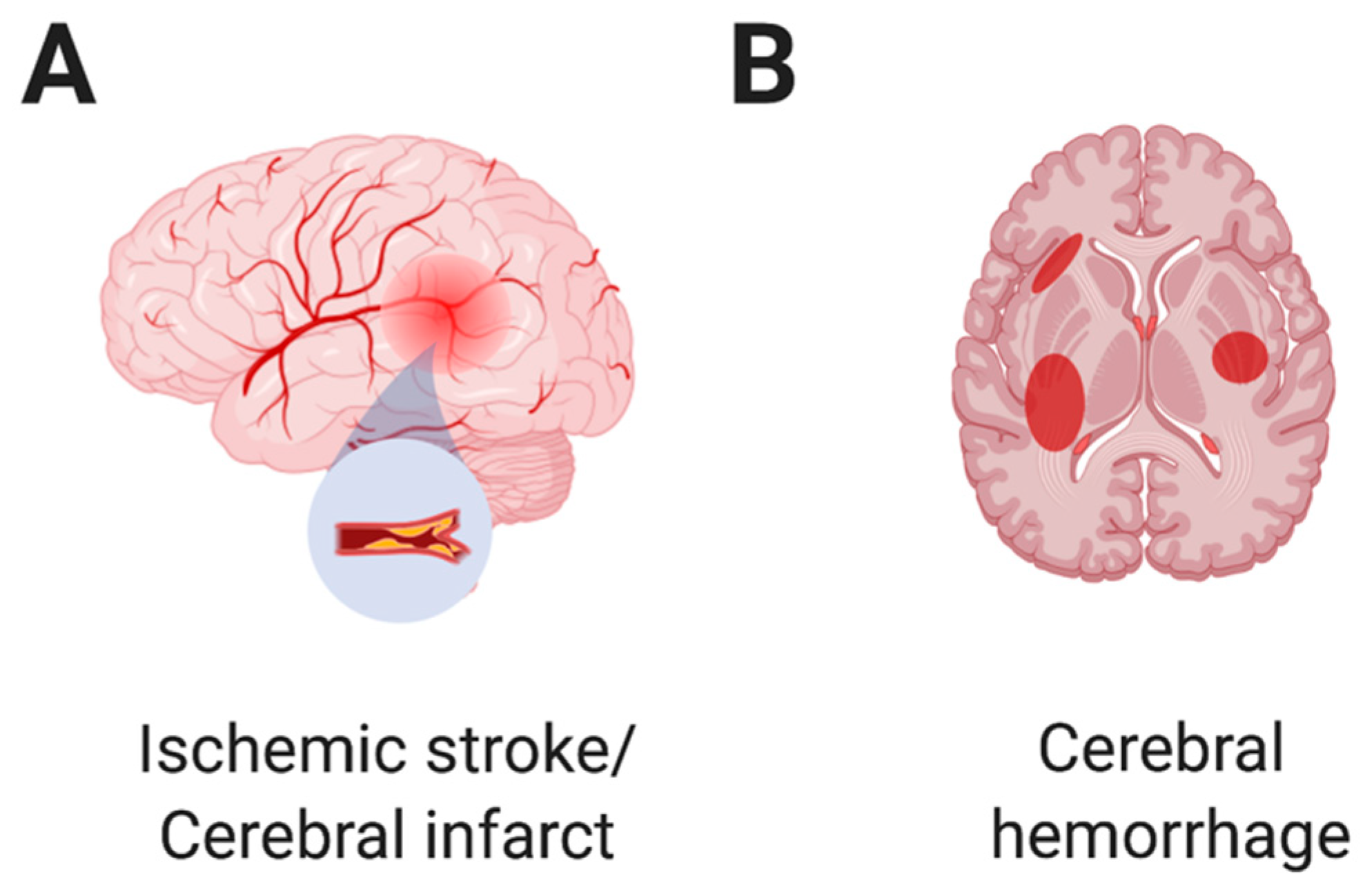
6.1. Cerebral Infarction and Ischemic Stroke
6.2. Brain Hemorrhage
6.3. Acute Demyelinating Encephalomyelitis (ADEM)
6.4. Acute Hemorrhagic Leukoencephalitis (AHL)
6.5. Posterior Reversible Encephalopathy Syndrome (PRES)
6.6. Early Morning Neuroparalytic Syndrome (EMNS)
6.7. Other Complications by Snakebite Envenoming
7. Treatment
8. Discussion and Conclusions
Author Contributions
Funding
Institutional Review Board Statement
Informed Consent Statement
Data Availability Statement
Conflicts of Interest
References
- Russell, F.E. When a snake strikes. Emerg. Med. 1990, 22, 21–43. [Google Scholar]
- Gold, B.S.; Barish, R.A.; Dart, R.C. North American snake envenomation: Diagnosis, treatment, and management. Emerg. Med. Clin. N. Am. 2004, 22, 423–443. [Google Scholar] [CrossRef]
- Kasturiratne, A.; Wickremasinghe, A.R.; de Silva, N.; Gunawardena, N.K.; Pathmeswaran, A.; Premaratna, R.; Savioli, L.; Lalloo, D.G.; de Silva, H.J. The global burden of snakebite: A literature analysis and modelling based on regional estimates of envenoming and deaths. PLoS Med. 2008, 5, e218. [Google Scholar] [CrossRef] [Green Version]
- Tu, A.T. Venoms: Chemistry and Molecular Biology; John. Wiley & Sons, Inc.: New York, NY, USA, 1977; p. 560. [Google Scholar]
- Park, K.H.; Shin, H.; Kang, H.; Kim, C.; Choi, H.J.; Yoo, K.; Oh, J.; Lim, T.H. Effectiveness of repeated antivenom therapy for snakebite-related systemic complications. J. Int. Med. Res. 2019, 47, 4808–4814. [Google Scholar] [CrossRef] [Green Version]
- Tasoulis, T.; Isbister, G.K. A Review and Database of Snake Venom Proteomes. Toxins 2017, 9, 290. [Google Scholar] [CrossRef] [Green Version]
- Markland, F.S., Jr.; Swenson, S. Snake venom metalloproteinases. Toxicon 2013, 62, 3–18. [Google Scholar] [CrossRef]
- Stocker, K.; Barlow, G.H. The coagulant enzyme from Bothrops atrox venom (batroxobin). Methods Enzym. 1976, 45, 214–223. [Google Scholar] [CrossRef]
- Serrano, S.M. The long road of research on snake venom serine proteinases. Toxicon 2013, 62, 19–26. [Google Scholar] [CrossRef]
- Silveira, G.G.; Machado, C.R.; Tuyama, M.; Lima, M.A. Intracranial Bleeding Following Bothrops sp. Snakebite. Neurologist 2016, 21, 11–12. [Google Scholar] [CrossRef]
- Malbranque, S.; Piercecchi-Marti, M.D.; Thomas, L.; Barbey, C.; Courcier, D.; Bucher, B.; Ridarch, A.; Smadja, D.; Warrell, D.A. Fatal diffuse thrombotic microangiopathy after a bite by the "Fer-de-Lance" pit viper (Bothrops lanceolatus) of Martinique. Am. J. Trop. Med. Hyg. 2008, 78, 856–861. [Google Scholar] [CrossRef] [Green Version]
- Namal Rathnayaka, R.M.; Kularatne, S.A.; Kumarasinghe, K.D.; Ranaweera, J.; Nishanthi Ranathunga, P.E. Ischemic brain infarcts and intracranial haemorrhages following Russell’s viper (Daboia russelii) bite in Sri Lanka. Toxicon 2017, 125, 70–73. [Google Scholar] [CrossRef] [PubMed]
- Rebahi, H.; Nejmi, H.; Abouelhassan, T.; Hasni, K.; Samkaoui, M.A. Severe envenomation by Cerastes cerastes viper: An unusual mechanism of acute ischemic stroke. J. Stroke Cerebrovasc. Dis. 2014, 23, 169–172. [Google Scholar] [CrossRef] [PubMed]
- Kitchens, C.; Eskin, T. Fatality in a case of envenomation by Crotalus adamanteus initially successfully treated with polyvalent ovine antivenom followed by recurrence of defibrinogenation syndrome. J. Med. Toxicol. 2008, 4, 180–183. [Google Scholar] [CrossRef] [PubMed] [Green Version]
- Waiddyanatha, S.; Silva, A.; Siribaddana, S.; Isbister, G.K. Long-term Effects of Snake Envenoming. Toxins 2019, 11, 193. [Google Scholar] [CrossRef] [PubMed] [Green Version]
- Gutierrez, J.M.; Rucavado, A.; Escalante, T.; Herrera, C.; Fernandez, J.; Lomonte, B.; Fox, J.W. Unresolved issues in the understanding of the pathogenesis of local tissue damage induced by snake venoms. Toxicon 2018, 148, 123–131. [Google Scholar] [CrossRef]
- Chen, Y.C.; Wang, T.Y.; Huang, Y.K.; Chang, K.C.; Chen, M.H.; Liu, C.C.; Liu, K.L.; Yang, Y.H.; Yen, D.H.; Fan, J.S. Effects of Sodium Silicate Complex against Hemorrhagic Activities Induced by Protobothrops mucrosquamatus Venom. Toxins 2021, 13, 59. [Google Scholar] [CrossRef]
- Zeng, X.; Hu, J.; Liang, X.; Wu, Y.; Yan, M.; Zhu, M.; Fu, Y. Acute cerebral infarction following a Trimeresurus stejnegeri snakebite: A case report. Medicine 2019, 98, e15684. [Google Scholar] [CrossRef]
- Silva de Oliveira, S.; Freitas-de-Sousa, L.A.; Alves, E.C.; de Lima Ferreira, L.C.; da Silva, I.M.; de Lacerda, M.V.G.; Fan, H.W.; Moura-da-Silva, A.M.; Monteiro, W.M. Fatal stroke after Bothrops snakebite in the Amazonas state, Brazil: A case report. Toxicon 2017, 138, 102–106. [Google Scholar] [CrossRef]
- Sinha, S.; Naik, B.B.; Ghanekar, J. Wall eyed bilateral internuclear ophthalmoplegia (WEBINO) syndrome as a false localising sign in intracranial haemorrhage due to snake bite. BMJ Case Rep. 2021, 14, e244830. [Google Scholar] [CrossRef]
- Xu, A.; Shan, R.; Huang, D.; Zhou, J.; Keenoo, A.; Qin, J. Case report: Acute demyelinating encephalomyelitis following viper bite. Medicine 2016, 95, e5310. [Google Scholar] [CrossRef]
- Gewin, V. Handling snakes for science. Nature 2021, 600, 352. [Google Scholar] [CrossRef]
- Magalhaes, S.F.V.; Peixoto, H.M.; Moura, N.; Monteiro, W.M.; de Oliveira, M.R.F. Snakebite envenomation in the Brazilian Amazon: A descriptive study. Trans. R. Soc. Trop. Med. Hyg. 2019, 113, 143–151. [Google Scholar] [CrossRef] [PubMed]
- Huang, J.; Song, W.; Hua, H.; Yin, X.; Huang, F.; Alolga, R.N. Antithrombotic and anticoagulant effects of a novel protein isolated from the venom of the Deinagkistrodon acutus snake. Biomed. Pharmacother. 2021, 138, 111527. [Google Scholar] [CrossRef] [PubMed]
- Chiang, L.C.; Tsai, W.J.; Liu, P.Y.; Ho, C.H.; Su, H.Y.; Lai, C.S.; Lai, K.L.; Lin, W.L.; Lee, C.H.; Yang, Y.Y.; et al. Envenomation by Trimeresurus stejnegeri stejnegeri: Clinical manifestations, treatment and associated factors for wound necrosis. J. Venom. Anim. Toxins Incl. Trop. Dis. 2020, 26, e20200043. [Google Scholar] [CrossRef]
- Laothong, C.; Sitprija, V. Decreased parasympathetic activities in Malayan krait (Bungarus candidus) envenoming. Toxicon 2001, 39, 1353–1357. [Google Scholar] [CrossRef]
- Hojer, J.; Tran Hung, H.; Warrell, D. Life-threatening hyponatremia after krait bite envenoming—A new syndrome. Clin. Toxicol. 2010, 48, 956–957. [Google Scholar] [CrossRef]
- Samanta, S.K.; Mahapatra, N.C.; Fariduddin, K.; Mazumdar, D.B.; Mandal, K. Cortical blindness and paraplegia following hypoxic ischemic encephalopathy as a complication of common krait bite. Nepal. J. Ophthalmol. 2011, 3, 206–209. [Google Scholar] [CrossRef] [Green Version]
- Kaushik, J.S.; Chakrabarty, B.; Gulati, S.; Patel, H.; Lodha, R.; Pai, G.; Kumar, A. Unusual late neurological complication in a child after an Indian krait bite. Pediatr. Neurol. 2014, 51, 130–132. [Google Scholar] [CrossRef]
- Anadure, R.K.; Narayanan, C.S.; Hande, V.; Singhal, A.; Varadaraj, G. Two Cases of Early Morning Neuroparalytic Syndrome (EMNS) in the Tropics—Masquerading as Brain Death. J. Assoc. Physicians India 2018, 66, 92–95. [Google Scholar]
- ALfaifi, M.S.; ALOtaibi, A.E.; AlQahtani, S.A.; ALShahrani, O.A.; ALSharani, K.M.; ALbshabshi, A.O.; ALZahrani, H.M.; ALAli, H.E. Cobra snakebite mimicking brain death treated with a novel combination of polyvalent snake antivenom and anticholinesterase. Am. J. Emerg. Med. 2020, 38, 2490.e5–2490.e7. [Google Scholar] [CrossRef]
- Ratanabanangkoon, K. A Quest for a Universal Plasma-Derived Antivenom Against All Elapid Neurotoxic Snake Venoms. Front. Immunol. 2021, 12, 668328. [Google Scholar] [CrossRef] [PubMed]
- Hermann, P.M.; Watson, S.N.; Wildering, W.C. Phospholipase A2—Nexus of aging, oxidative stress, neuronal excitability, and functional decline of the aging nervous system? Insights from a snail model system of neuronal aging and age-associated memory impairment. Front. Genet. 2014, 5, 419. [Google Scholar] [CrossRef] [PubMed]
- Chang, K.C.; Huang, Y.K.; Chen, Y.W.; Chen, M.H.; Tu, A.T.; Chen, Y.C. Venom Ophthalmia and Ocular Complications Caused by Snake Venom. Toxins 2020, 12, 576. [Google Scholar] [CrossRef] [PubMed]
- Wüster, W.; Chirio, L.; Trape, J.F.; Ineich, I.; Jackson, K.; Greenbaum, E.; Barron, C.; Kusamba, C.; Nagy, Z.T.; Storey, R.; et al. Integration of nuclear and mitochondrial gene sequences and morphology reveals unexpected diversity in the forest cobra (Naja melanoleuca) species complex in Central and West Africa (Serpentes: Elapidae). Zootaxa 2018, 4455, 68–98. [Google Scholar] [CrossRef] [PubMed]
- Narang, S.K.; Paleti, S.; Azeez Asad, M.A.; Samina, T. Acute ischemic infarct in the middle cerebral artery territory following a Russell’s viper bite. Neurol. India 2009, 57, 479–480. [Google Scholar] [CrossRef] [Green Version]
- Gouda, S.; Pandit, V.; Seshadri, S.; Valsalan, R.; Vikas, M. Posterior circulation ischemic stroke following Russell’s viper envenomation. Ann. Indian Acad. Neurol. 2011, 14, 301–303. [Google Scholar] [CrossRef]
- Pothukuchi, V.K.; Kumar, A.; Teja, C.; Verma, A. A Rare Case Series of Ischemic Stroke Following Russell’s Viper Snake Bite in India. Acta. Med. Indones 2017, 49, 343–346. [Google Scholar]
- Ittyachen, A.M.; Jose, M.B. Thalamic infarction following a Russell’s viper bite. Southeast. Asian J. Trop. Med. Public Health 2012, 43, 1201–1204. [Google Scholar]
- Das, S.K.; Khaskil, S.; Mukhopadhyay, S.; Chakrabarti, S. A patient of Russell’s viper envenomation presenting with cortical venous thrombosis: An extremely uncommon presentation. J. Postgrad. Med. 2013, 59, 235–236. [Google Scholar] [CrossRef]
- Lahiri, D.; Sawale, V.M.; Dubey, S.; Roy, B.K.; Das, S.K. Status epilepticus and bilateral middle cerebral artery infarction: A rare presentation after viper bite. Ann. Afr. Med. 2019, 18, 111–114. [Google Scholar] [CrossRef]
- Tripathy, S.; Routray, P.K.; Mohapatra, A.K.; Mohapatra, M.; Dash, S.C. Acute demyelinating encephalomyelitis after anti-venom therapy in Russell’s viper bite. J. Med. Toxicol. 2010, 6, 318–321. [Google Scholar] [CrossRef] [PubMed] [Green Version]
- Prabhakar, A.T.; Kamanahalli, R.; Sivadasan, A.; Joseph, E.; Viggeswarpu, S. Non-fatal acute haemorrhagic leukoencephalitis following snake bite: A case report. Trop. Doct. 2016, 46, 57–59. [Google Scholar] [CrossRef] [PubMed]
- Ibrahim, A.M.; ElSefi, T.T.; Ghanem, M.; Fayed, A.M.; Shaban, N.A. A Horned Viper Bite Victim with PRES. Case Rep. Neurol. Med. 2017, 2017, 1835796. [Google Scholar] [CrossRef] [PubMed] [Green Version]
- Heise, C.W.; Cunningham, C.; Ruha, A.M.; O’Connor, A.D. One Bite, Two Patients: Disparate Clinical Courses Following Simultaneous Crotalus oreganus abyssus Envenomation. Wilderness Environ. Med. 2020, 31, 354–357. [Google Scholar] [CrossRef] [PubMed]
- Tincu, R.C.; Ghiorghiu, Z.; Tomescu, D.; Macovei, R.A. The Compartment Syndrome Associated with Deep Vein Thrombosis due to Rattlesnake Bite: A Case Report. Balk. Med. J. 2017, 34, 367–370. [Google Scholar] [CrossRef]
- Bosak, A.R.; Ruha, A.M.; Graeme, K.A. A case of neurotoxicity following envenomation by the Sidewinder rattlesnake, Crotalus cerastes. J. Med. Toxicol. 2014, 10, 229–231. [Google Scholar] [CrossRef] [Green Version]
- Tilbury, C.R.; Peacock, F.; Harvey, J. Envenomation by the spotted harlequin snake, Homoroselaps lacteus (Linnaeus) 1754 (Serpentes: Atractaspidinae). Toxicon 2021, 198, 151–155. [Google Scholar] [CrossRef]
- Chauhan, V.; Thakur, S. The North-South divide in snake bite envenomation in India. J. Emerg. Trauma Shock 2016, 9, 151–154. [Google Scholar] [CrossRef]
- Suraweera, W.; Warrell, D.; Whitaker, R.; Menon, G.; Rodrigues, R.; Fu, S.H.; Begum, R.; Sati, P.; Piyasena, K.; Bhatia, M.; et al. Trends in snakebite deaths in India from 2000 to 2019 in a nationally representative mortality study. Elife 2020, 9, e54076. [Google Scholar] [CrossRef]
- Lin, J.H.; Lo, C.M.; Chuang, S.H.; Chiang, C.H.; Wang, S.D.; Lin, T.Y.; Liao, J.W.; Hung, D.Z. Collocation of avian and mammal antibodies to develop a rapid and sensitive diagnostic tool for Russell’s Vipers Snakebite. PLoS Negl. Trop. Dis. 2020, 14, e0008701. [Google Scholar] [CrossRef]
- Silva, A.; Kuruppu, S.; Othman, I.; Goode, R.J.; Hodgson, W.C.; Isbister, G.K. Neurotoxicity in Sri Lankan Russell’s Viper (Daboia russelii) Envenoming is Primarily due to U1-viperitoxin-Dr1a, a Pre-Synaptic Neurotoxin. Neurotox. Res. 2017, 31, 11–19. [Google Scholar] [CrossRef] [PubMed]
- Fatah, C.; Samah, S.; Fatima, L.D. Antiplatelet and anticoagulant activities of two phospholipase A2s purified from Cerastes cerastes venom: Structure-function relationship. J. Biochem. Mol. Toxicol. 2018, 32, e22219. [Google Scholar] [CrossRef] [PubMed]
- Keating, G.M. Crotalidae polyvalent immune Fab: In patients with North American crotaline envenomation. BioDrugs 2011, 25, 69–76. [Google Scholar] [CrossRef]
- Boyer, L.V.; Seifert, S.A.; Clark, R.F.; McNally, J.T.; Williams, S.R.; Nordt, S.P.; Walter, F.G.; Dart, R.C. Recurrent and persistent coagulopathy following pit viper envenomation. Arch. Intern. Med. 1999, 159, 706–710. [Google Scholar] [CrossRef] [PubMed] [Green Version]
- Portillo, F.; Stanley, E.L.; Branch, W.R.; Conradie, W.; Rodel, M.O.; Penner, J.; Barej, M.F.; Kusamba, C.; Muninga, W.M.; Aristote, M.M.; et al. Evolutionary history of burrowing asps (Lamprophiidae: Atractaspidinae) with emphasis on fang evolution and prey selection. PLoS ONE 2019, 14, e0214889. [Google Scholar] [CrossRef] [Green Version]
- Silva, K.V.; Said, R.D.C.; Assy, J.; Duarte, M.R.; Torrez, P.P.Q.; Franca, F.O.S. A case of envenomation caused by Oxybelis fulgidus (Serpentes, Colubridae) in Brazilian Amazon. Rev. Soc. Bras. Med. Trop. 2019, 52, e20180423. [Google Scholar] [CrossRef]
- Angarita-Sierra, T.; Montanez-Mendez, A.; Toro-Sanchez, T.; Rodriguez-Vargas, A. A case of envenomation by the false fer-de-lance snake Leptodeira annulata (Linnaeus, 1758) in the department of La Guajira, Colombia. Biomedica 2020, 40, 20–26. [Google Scholar] [CrossRef] [Green Version]
- Lazaro, R.P. Complex Regional Pain Syndrome Following Snakebite: A Putatively Rare Complication of Envenomation and Review of the Literature. Int. Med. Case Rep. J. 2020, 13, 603–607. [Google Scholar] [CrossRef]
- Heyborne, W.H.; Mackessy, S.P. Venoms of New World Vinesnakes (Oxybelis aeneus and O. fulgidus). Toxicon 2021, 190, 22–30. [Google Scholar] [CrossRef]
- Heyborne, W.H.; Mackessy, S.P. Identification and characterization of a taxon-specific three-finger toxin from the venom of the Green Vinesnake (Oxybelis fulgidus; family Colubridae). Biochimie 2013, 95, 1923–1932. [Google Scholar] [CrossRef]
- Ipek, S.; Gungor, S.; Gullu, U.U.; Dalkiran, T.; Mercan, M.; Demiray, S.; Gurbuz, Y. Snakebites in Pediatric Patients in Kahramanmaras: Is Pro-brain Natriuretic Peptide a Prognostic Biomarker for Snakebites? Cureus 2022, 14, e21570. [Google Scholar] [CrossRef] [PubMed]
- House, L.M.; Lewin, M.R.; Naidu, R.K.; Beqaj, H. Complex regional pain syndrome following southern pacific rattlesnake (C. oreganus helleri) envenoming. Clin. Case Rep. 2021, 9, e05019. [Google Scholar] [CrossRef] [PubMed]
- Kamiguti, A.S.; Theakston, R.D.; Sherman, N.; Fox, J.W. Mass spectrophotometric evidence for P-III/P-IV metalloproteinases in the venom of the Boomslang (Dispholidus typus). Toxicon 2000, 38, 1613–1620. [Google Scholar] [CrossRef]
- Ching, A.T.; Paes Leme, A.F.; Zelanis, A.; Rocha, M.M.; Furtado Mde, F.; Silva, D.A.; Trugilho, M.R.; da Rocha, S.L.; Perales, J.; Ho, P.L.; et al. Venomics profiling of Thamnodynastes strigatus unveils matrix metalloproteinases and other novel proteins recruited to the toxin arsenal of rear-fanged snakes. J. Proteome Res. 2012, 11, 1152–1162. [Google Scholar] [CrossRef] [PubMed]
- Urra, F.A.; Miranda-Calle, A.B.; Araya-Maturana, R. Philodryas (Serpentes: Dipsadidae) Envenomation, a Neglected Issue in Chile. Toxins 2019, 11, 697. [Google Scholar] [CrossRef] [Green Version]
- Bastida, J.; Crampet, A.; Meneghel, M.; Morais, V. Preliminary Biochemical and Venomic Characterization of the Venom of Phalotris lemniscatus (Serpentes, Colubridae). Curr. Top. Med. Chem. 2019, 19, 1981–1989. [Google Scholar] [CrossRef]
- Peichoto, M.E.; Teibler, P.; Ruiz, R.; Leiva, L.; Acosta, O. Systemic pathological alterations caused by Philodryas patagoniensis colubrid snake venom in rats. Toxicon 2006, 48, 520–528. [Google Scholar] [CrossRef]
- Al-Sadawi, M.; Mohamadpour, M.; Zhyvotovska, A.; Ahmad, T.; Schechter, J.; Soliman, Y.; McFarlane, S.I. Cerebrovascular Accident and Snake Envenomation: A Scoping Study. Int. J. Clin. Res. Trials 2019, 4, 133. [Google Scholar] [CrossRef]
- Abumiya, T.; Fitridge, R.; Mazur, C.; Copeland, B.R.; Koziol, J.A.; Tschopp, J.F.; Pierschbacher, M.D.; del Zoppo, G.J. Integrin alpha(IIb)beta(3) inhibitor preserves microvascular patency in experimental acute focal cerebral ischemia. Stroke 2000, 31, 1402–1409, discussion 1409–1410. [Google Scholar] [CrossRef] [Green Version]
- Tian, H.; Liu, M.; Li, J.; Xu, R.; Long, C.; Li, H.; Mwangi, J.; Lu, Q.; Lai, R.; Shen, C. Snake C-Type Lectins Potentially Contribute to the Prey Immobilization in Protobothrops mucrosquamatus and Trimeresurus stejnegeri Venoms. Toxins 2020, 12, 105. [Google Scholar] [CrossRef] [Green Version]
- Clemetson, K.J. Snaclecs (snake C-type lectins) that inhibit or activate platelets by binding to receptors. Toxicon 2010, 56, 1236–1246. [Google Scholar] [CrossRef] [PubMed]
- Del Brutto, O.H.; Del Brutto, V.J. Neurological complications of venomous snake bites: A review. Acta Neurol. Scand. 2012, 125, 363–372. [Google Scholar] [CrossRef] [PubMed]
- Fry, B.G.; Wuster, W. Assembling an arsenal: Origin and evolution of the snake venom proteome inferred from phylogenetic analysis of toxin sequences. Mol. Biol. Evol. 2004, 21, 870–883. [Google Scholar] [CrossRef] [PubMed] [Green Version]
- Larreche, S.; Chippaux, J.P.; Chevillard, L.; Mathe, S.; Resiere, D.; Siguret, V.; Megarbane, B. Bleeding and Thrombosis: Insights into Pathophysiology of Bothrops Venom-Related Hemostasis Disorders. Int. J. Mol. Sci. 2021, 22, 9643. [Google Scholar] [CrossRef] [PubMed]
- Rodrigues, C.R.; Teixeira-Ferreira, A.; Vargas, F.F.R.; Guerra-Duarte, C.; Costal-Oliveira, F.; Stransky, S.; Lopes-de-Souza, L.; Dutra, A.A.A.; Yarleque, A.; Bonilla, C.; et al. Proteomic profile, biological activities and antigenic analysis of the venom from Bothriopsis bilineata smaragdina (“loro machaco”), a pitviper snake from Peru. J. Proteomics 2018, 187, 171–181. [Google Scholar] [CrossRef] [PubMed]
- Kalita, B.; Singh, S.; Patra, A.; Mukherjee, A.K. Quantitative proteomic analysis and antivenom study revealing that neurotoxic phospholipase A2 enzymes, the major toxin class of Russell’s viper venom from southern India, shows the least immuno-recognition and neutralization by commercial polyvalent antivenom. Int. J. Biol. Macromol. 2018, 118, 375–385. [Google Scholar] [CrossRef]
- Rodrigues, C.R.; Molina Molina, D.A.; de Souza, D.L.N.; Cardenas, J.; Costal-Oliveira, F.; Guerra-Duarte, C.; Chavez-Olortegui, C. Biological and proteomic characterization of the venom from Peruvian Andes rattlesnake Crotalus durissus. Toxicon 2022, 207, 31–42. [Google Scholar] [CrossRef]
- Kini, R.M. Serine proteases affecting blood coagulation and fibrinolysis from snake venoms. Pathophysiol. Haemost. Thromb. 2005, 34, 200–204. [Google Scholar] [CrossRef]
- Sajevic, T.; Leonardi, A.; Krizaj, I. Haemostatically active proteins in snake venoms. Toxicon 2011, 57, 627–645. [Google Scholar] [CrossRef]
- Warrell, D.A. Snake bite. Lancet 2010, 375, 77–88. [Google Scholar] [CrossRef]
- Paul, G.; Paul, B.S.; Puri, S. Snake bite and stroke: Our experience of two cases. Indian J. Crit. Care Med. 2014, 18, 257–258. [Google Scholar] [CrossRef] [PubMed]
- Alangode, A.; Reick, M.; Reick, M. Sodium oleate, arachidonate, and linoleate enhance fibrinogenolysis by Russell’s viper venom proteinases and inhibit FXIIIa; a role for phospholipase A2 in venom induced consumption coagulopathy. Toxicon 2020, 186, 83–93. [Google Scholar] [CrossRef] [PubMed]
- Thomas, L.; Tyburn, B.; Bucher, B.; Pecout, F.; Ketterle, J.; Rieux, D.; Smadja, D.; Garnier, D.; Plumelle, Y. Prevention of thromboses in human patients with Bothrops lanceolatus envenoming in Martinique: Failure of anticoagulants and efficacy of a monospecific antivenom. Research Group on Snake Bites in Martinique. Am. J. Trop. Med. Hyg. 1995, 52, 419–426. [Google Scholar] [CrossRef] [PubMed]
- Fox, J.W.; Serrano, S.M. Structural considerations of the snake venom metalloproteinases, key members of the M12 reprolysin family of metalloproteinases. Toxicon 2005, 45, 969–985. [Google Scholar] [CrossRef] [PubMed]
- Kouyoumdjian, J.A.; Polizelli, C. [Snake bites by Bothrops moojeni: Correlation of the clinical picture with the snake size]. Rev. Inst. Med. Trop Sao Paulo 1989, 31, 84–90. [Google Scholar] [CrossRef] [PubMed]
- Cole, J.; Evans, E.; Mwangi, M.; Mar, S. Acute Disseminated Encephalomyelitis in Children: An Updated Review Based on Current Diagnostic Criteria. Pediatr. Neurol. 2019, 100, 26–34. [Google Scholar] [CrossRef]
- Weatherall, D.J.; Ledingham, J.; Warrell, D.A. Injuries, Envenoming, Poisoning, and Allergic Reactions Caused by Animals; Oxford University Press: Oxford, UK, 1996. [Google Scholar]
- Grzonka, P.; Scholz, M.C.; De Marchis, G.M.; Tisljar, K.; Ruegg, S.; Marsch, S.; Fladt, J.; Sutter, R. Acute Hemorrhagic Leukoencephalitis: A Case and Systematic Review of the Literature. Front. Neurol. 2020, 11, 899. [Google Scholar] [CrossRef]
- Del Brutto, O.H. Reversible posterior leukoencephalopathy after venomous bites and stings. Neurotoxicology 2013, 39, 10. [Google Scholar] [CrossRef]
- Porcello Marrone, L.C.; Marrone, B.F.; Neto, F.K.; Costa, F.C.; Thome, G.G.; Aramburu, M.B.; Schilling, L.P.; Pascoal, T.A.; Gadonski, G.; Huf Marrone, A.C.; et al. Posterior reversible encephalopathy syndrome following a scorpion sting. J. Neuroimaging 2013, 23, 535–536. [Google Scholar] [CrossRef]
- Loh, H.H.; Tan, C.H. Acute renal failure and posterior reversible encephalopathy syndrome following multiple wasp stings: A case report. Med. J. Malaysia 2012, 67, 133–135. [Google Scholar]
- Haneef, M.; George, D.E.; Babu, A.S. Early morning neuroparalytic syndrome. Indian J. Pediatr. 2009, 76, 1072. [Google Scholar] [CrossRef] [PubMed] [Green Version]
- Bawaskar, H.S.; Bawaskar, P.H. Envenoming by the common krait (Bungarus caeruleus) and Asian cobra (Naja naja): Clinical manifestations and their management in a rural setting. Wilderness Environ. Med. 2004, 15, 257–266. [Google Scholar] [CrossRef]
- Damm, M.; Hempel, B.F.; Sussmuth, R.D. Old World Vipers-A Review about Snake Venom Proteomics of Viperinae and Their Variations. Toxins 2021, 13, 427. [Google Scholar] [CrossRef] [PubMed]
- You, K.E.; Koo, M.A.; Lee, D.H.; Kwon, B.J.; Lee, M.H.; Hyon, S.H.; Seomun, Y.; Kim, J.T.; Park, J.C. The effective control of a bleeding injury using a medical adhesive containing batroxobin. Biomed. Mater. 2014, 9, 025002. [Google Scholar] [CrossRef] [PubMed]
- Guan, J.; Song, S.; Wang, W.; Ji, X.; Meng, R. Cerebral venous sinus thrombosis due to external compression of internal jugular vein. J. Int. Med. Res. 2021, 49, 3000605211006609. [Google Scholar] [CrossRef] [PubMed]
- Song, S.Y.; Dornbos, D., 3rd; Lan, D.; Jiao, B.L.; Wan, S.L.; Guo, Y.B.; Ding, Y.C.; Yang, Q.; Ji, X.M.; Meng, R. High-Resolution Magnetic Resonance Black Blood Thrombus Imaging and Serum D-Dimer in the Confirmation of Acute Cortical Vein Thrombosis. Front. Neurol. 2021, 12, 680040. [Google Scholar] [CrossRef]
- Singh, S.; Chattopadhya, A.; Sud, A.; Wanchu, A.; Bambery, P. Acute paraplegia following viper bite. J. Assoc. Physicians India 2002, 50, 1427–1429. [Google Scholar]
- Chacur, M.; Gutierrez, J.M.; Milligan, E.D.; Wieseler-Frank, J.; Britto, L.R.; Maier, S.F.; Watkins, L.R.; Cury, Y. Snake venom components enhance pain upon subcutaneous injection: An initial examination of spinal cord mediators. Pain 2004, 111, 65–76. [Google Scholar] [CrossRef]
- Chacur, M.; Milligan, E.D.; Sloan, E.M.; Wieseler-Frank, J.; Barrientos, R.M.; Martin, D.; Poole, S.; Lomonte, B.; Gutierrez, J.M.; Maier, S.F.; et al. Snake venom phospholipase A2s (Asp49 and Lys49) induce mechanical allodynia upon peri-sciatic administration: Involvement of spinal cord glia, proinflammatory cytokines and nitric oxide. Pain 2004, 108, 180–191. [Google Scholar] [CrossRef]
- Harris, J.B.; Scott-Davey, T. Secreted phospholipases A2 of snake venoms: Effects on the peripheral neuromuscular system with comments on the role of phospholipases A2 in disorders of the CNS and their uses in industry. Toxins 2013, 5, 2533–2571. [Google Scholar] [CrossRef] [Green Version]
- Biswas, R.; Irodi, A.; Paul, A.; Ghimere, G.; Joshi, K.R.; Alurkar, V.M.; Shetty, K.J. Anti-venom-induced myelopathy in a semipoisonous snakebite. Int. J. Clin. Pract. 2004, 58, 645–646. [Google Scholar] [CrossRef] [PubMed]
- Aye, M.T.H.; Naing, T.; Myint, K.T. Unusual ocular manifestations following viper bite. BMJ Case Rep. 2018, 2018, bcr2018225040. [Google Scholar] [CrossRef] [PubMed]
- Olcaysu, O.O.; Cadirci, K.; Altun, A.; Durur Karakaya, A.; Bayramlar, H. Unilateral Optic Neuropathy and Acute Angle-Closure Glaucoma following Snake Envenomation. Case Rep. Ophthalmol. Med. 2015, 2015, 687829. [Google Scholar] [CrossRef] [PubMed]
- Kumar, J.R.; Basavarajappa, B.S.; Vishwanath, B.S.; Gowda, T.V. Biochemical and pharmacological characterization of three toxic phospholipase A2s from Daboia russelii snake venom. Comp. Biochem. Physiol. C Toxicol. Pharmacol. 2015, 168, 28–38. [Google Scholar] [CrossRef]
- Cherifi, F.; Namane, A.; Laraba-Djebari, F. Isolation, functional characterization and proteomic identification of CC2-PLA(2) from Cerastes cerastes venom: A basic platelet-aggregation-inhibiting factor. Protein J. 2014, 33, 61–74. [Google Scholar] [CrossRef]
- Fry, B.G. Snakebite: When the Human Touch Becomes a Bad Touch. Toxins 2018, 10, 170. [Google Scholar] [CrossRef] [Green Version]
- Bhattacharya, S.; Krishnamurthy, A.; Gopalakrishnan, M.; Kalra, S.; Kantroo, V.; Aggarwal, S.; Surana, V. Endocrine and Metabolic Manifestations of Snakebite Envenoming. Am. J. Trop. Med. Hyg. 2020, 103, 1388–1396. [Google Scholar] [CrossRef]
- Frank, H.A. Snakebite or frostbite: What are we doing? An evaluation of cryotherapy for envenomation. Calif. Med. 1971, 114, 25–27. [Google Scholar]
- Yang, Y.; Huang, Z.; Zhang, X. Efficacy and safety of clopidogrel and/or aspirin for ischemic stroke/transient ischemic attack: An overview of systematic reviews and meta-analysis. Medicine 2021, 100, e27804. [Google Scholar] [CrossRef]
- Ryu, W.S.; Schellingerhout, D.; Hong, K.S.; Jeong, S.W.; Kim, B.J.; Kim, J.T.; Lee, K.B.; Park, T.H.; Park, S.S.; Park, J.M.; et al. Relation of Pre-Stroke Aspirin Use With Cerebral Infarct Volume and Functional Outcomes. Ann. Neurol. 2021, 90, 763–776. [Google Scholar] [CrossRef]
- Gutierrez, J.M.; Escalante, T.; Rucavado, A.; Herrera, C. Hemorrhage Caused by Snake Venom Metalloproteinases: A Journey of Discovery and Understanding. Toxins 2016, 8, 93. [Google Scholar] [CrossRef] [PubMed] [Green Version]
- Bjarnason, J.B.; Tu, A.T. Hemorrhagic toxins from Western diamondback rattlesnake (Crotalus atrox) venom: Isolation and characterization of five toxins and the role of zinc in hemorrhagic toxin e. Biochemistry 1978, 17, 3395–3404. [Google Scholar] [CrossRef] [PubMed]
- Friederich, C.; Tu, A.T. Role of metals in snake venoms for hemorrhagic, esterase and proteolytic activities. Biochem. Pharmacol. 1971, 20, 1549–1556. [Google Scholar] [CrossRef]
- Ownby, C.L.; Tu, A.T.; Kainer, R.A. Effect of diethylenetriaminepentaacetic acid and procaine on hemorrhage induced by rattlesnake venom. J. Clin. Pharmacol. 1975, 15, 419–426. [Google Scholar] [CrossRef]
- Ownby, C.L. The Merck Manual of Diagnosis and Therapy, 13th ed.; Talbott, R.B.J.H., Ed.; Merck & Co., Incorp: Rahway, NJ, USA, 1972. [Google Scholar]
- Albulescu, L.O.; Hale, M.S.; Ainsworth, S.; Alsolaiss, J.; Crittenden, E.; Calvete, J.J.; Evans, C.; Wilkinson, M.C.; Harrison, R.A.; Kool, J.; et al. Preclinical validation of a repurposed metal chelator as an early-intervention therapeutic for hemotoxic snakebite. Sci. Transl. Med. 2020, 12, eaay8314. [Google Scholar] [CrossRef]
- Lewin, M.; Samuel, S.; Merkel, J.; Bickler, P. Varespladib (LY315920) Appears to Be a Potent, Broad-Spectrum, Inhibitor of Snake Venom Phospholipase A2 and a Possible Pre-Referral Treatment for Envenomation. Toxins 2016, 8, 248. [Google Scholar] [CrossRef] [Green Version]
- Salvador, G.H.M.; Gomes, A.A.S.; Bryan-Quiros, W.; Fernandez, J.; Lewin, M.R.; Gutierrez, J.M.; Lomonte, B.; Fontes, M.R.M. Structural basis for phospholipase A2-like toxin inhibition by the synthetic compound Varespladib (LY315920). Sci. Rep. 2019, 9, 17203. [Google Scholar] [CrossRef]
- Zinenko, O.; Tovstukha, I.; Korniyenko, Y. PLA2 Inhibitor Varespladib as an Alternative to the Antivenom Treatment for Bites from Nikolsky’s Viper Vipera berus nikolskii. Toxins 2020, 12, 356. [Google Scholar] [CrossRef]
- Gutierrez, J.M.; Lewin, M.R.; Williams, D.J.; Lomonte, B. Varespladib (LY315920) and Methyl Varespladib (LY333013) Abrogate or Delay Lethality Induced by Presynaptically Acting Neurotoxic Snake Venoms. Toxins 2020, 12, 131. [Google Scholar] [CrossRef] [Green Version]
- Gutierrez, J.M.; Albulescu, L.O.; Clare, R.H.; Casewell, N.R.; Abd El-Aziz, T.M.; Escalante, T.; Rucavado, A. The Search for Natural and Synthetic Inhibitors That Would Complement Antivenoms as Therapeutics for Snakebite Envenoming. Toxins 2021, 13, 451. [Google Scholar] [CrossRef]
- Abouyannis, M.; FitzGerald, R.; Ngama, M.; Mwangudzah, H.; Nyambura, Y.K.; Ngome, S.; Riako, D.; Babu, L.; Lewa, F.; Else, L.; et al. TRUE-1: Trial of Repurposed Unithiol for snakebite Envenoming phase 1 (safety, tolerability, pharmacokinetics and pharmacodynamics in healthy Kenyan adults). Wellcome Open Res. 2022, 7, 90. [Google Scholar] [CrossRef] [PubMed]
- Li, M.; Xie, Z.H.; Yu, A.Y.; He, D.P. Increased Efficacy of Antivenom Combined with Hyperbaric Oxygen on Deinagkistrodon acutus Envenomation in Adult Rats. Chin. Med. J. (Engl.) 2018, 131, 323–329. [Google Scholar] [CrossRef] [PubMed]
- Bekbossynova, A.; Zharylgap, A.; Filchakova, O. Venom-Derived Neurotoxins Targeting Nicotinic Acetylcholine Receptors. Molecules 2021, 26, 3373. [Google Scholar] [CrossRef]
- de la Rosa, G.; Olvera, F.; Archundia, I.G.; Lomonte, B.; Alagon, A.; Corzo, G. Horse immunization with short-chain consensus alpha-neurotoxin generates antibodies against broad spectrum of elapid venomous species. Nat. Commun. 2019, 10, 3642. [Google Scholar] [CrossRef] [PubMed] [Green Version]
- Montoni, F.; Andreotti, D.Z.; Eichler, R.; Santos, W.D.S.; Kisaki, C.Y.; Arcos, S.S.S.; Lima, I.F.; Soares, M.A.M.; Nishiyama-Jr, M.Y.; Nava-Rodrigues, D.; et al. The impact of rattlesnake venom on mice cerebellum proteomics points to synaptic inhibition and tissue damage. J. Proteomics 2020, 221, 103779. [Google Scholar] [CrossRef] [PubMed]
- Yeh, Y.T.; Chen, M.H.; Chang, J.C.; Fan, J.S.; Yen, D.H.; Chen, Y.C. Protobothrops mucrosquamatus Bites to the Head: Clinical Spectrum from Case Series. Am. J. Trop. Med. Hyg. 2018, 99, 753–755. [Google Scholar] [CrossRef] [Green Version]


| Complications | Snake Species | Sex/Age | Bite Mark | Other Symptoms | Presuming Venom Components | Outcome | Antivenom/Amount | Ref. |
|---|---|---|---|---|---|---|---|---|
| Cerebral infarction | Bothrops lanceolatus (Fer-de-Lance) | M/74 | Elbow | Atrial fibrillation | SVMPs, Hemotoxin | Dead | IV Antivenom/80 mL | [11] |
| Cerebral infarction | Trimeresurus stejnegeri (Asian palm pit viper) | F/49 | Foot | Speech disturbances | SVMPs, Hemotoxin, | Recovered | IV Antivenom/3 vials | [18] |
| Intracranial hemorrhage | Bothrops jararacussu (Jararacussu) | M/52 | Foot | Sudden loss of consciousness | SVMPs, Hemotoxin | Recovered | IV Antivenom/Unclear | [10] |
| Subarachnoid hemorrhage | Bothrops atrox (Fer-de-Lance) | F/59 | Foot | Hypothermia, Bradycardia and Hypotension | SVMPs, Hemotoxin | Dead | IV Antivenom/80 mL | [19] |
| Cerebral edema and hemorrhage | Crotalus adamanteus (Eastern diamondback rattlesnake) | M/54 | Hand | Facial fasciculations | SVMPs, Hemotoxin, Neurotoxin | Dead | IV Antivenom/16 vials | [14] |
| Intracranial hemorrhage | Unidentified species | F/48 | Hand | Exotropia | SVMPs, Hemotoxin, | Recover with minor visual complication | IV Antivenom/Unclear | [20] |
| ADEM | Deinagkistrodon acutus | M/50 | Foot | Severe pain | SVMPs, Hemotoxin | Recovered | IV Antivenom/1 vial | [21] |
| Complications | Snake Species | Sex/Age | Bite Mark | Other Symptoms | Presuming Venom Components | Outcome | Antivenom/Amount | Ref. |
|---|---|---|---|---|---|---|---|---|
| Respiratory paralysis, ptosis | Bungarus candidus (Malaya Krait) | M/35 | Hand | Tachycardia, prolonged micturition | Neurotoxin, PLA2 | Permanent mydriasis | IV Antivenom/6 vials | [26] |
| Respiratory paralysis, ptosis | M/41 | Hand | Tachycardia | Permanent mydriasis | IV Antivenom/3 vials | |||
| Prolonged cerebral anoxia | F/12 | Hand | Periodic convulsions | Permanent brain damage | Unclear | |||
| Hyponatremia and cerebral edema | Bungarus multicinctus (Many-banded Krait) | F/17 | Foot | Seizure | Neurotoxin | Dead | N/A | [27] |
| Hypoxic ischemic encephalopathy | Bungarus caeruleus (Indian Krait) | M/15 | Foot | Paraplegia | Neurotoxin | Loss of vision | IV Antivenom/1 vial | [28] |
| PRES | Bungarus caeruleus (Indian Krait) | M/10 | Ear | Hypertension | Neurotoxin | Recovered with vision blurring | IV Antivenom/10 vials | [29] |
| EMNS | Bungarus caeruleus (Indian Krait, prediction) | M/38 | N/K | Difficulty in swallowing and double vision | Neurotoxin | Recovered | IV Antivenom/9 vials | [30] |
| EMNS | F/27 | N/K | Mydriasis | Recovered | IV Antivenom/Unclear | |||
| Temporary brain death | Naja haje arabica (Arabian cobra) | F/57 | Hand | Breathing ceased | Neurotoxin | Recovered | IV Antivenom/50 vials | [31] |
| Complications | Snake Species | Sex/Age | Bite Mark | Other Symptoms | Presuming Venom Components | Outcome | Antivenom/Amount | Ref. |
|---|---|---|---|---|---|---|---|---|
| Ischemic stroke | Daboia russelii (Russell’s Viper) | M/18 | Foot | Sever pain | Hemotoxin | Recovered with minor complications | IV Antivenom/6 vials | [36] |
| Ischemic stroke | Daboia russelii (Russell’s Viper) | F/40 | Foot | Hypotonia of the limbs | SVMPs, Hemotoxin | Significant ataxia of gait | IV Antivenom/24 vials | [37] |
| Ischemic Stroke | Daboia russelii (Russell’s Viper) | M/70 | Foot | Ptosis, Seizure | Hemotoxin, Neurotoxin | Recovered | IV Antivenom/20 vials | [38] |
| Ischemic Stroke | M/55 | Foot | Ptosis, Speech disturbances | Hemotoxin, Neurotoxin | Recovered | IV Antivenom/35 vials | ||
| Ischemic stroke | Cerastes cerastes (Desert horned viper) | F/5 | Foot | Thrombocytopenia, Acute anemia | SVMPs, Hemotoxin, PLA2 | Dead | N/A | [13] |
| Thalamic and cerebral infarctions | Daboia russelii (Russell’s Viper) | M/55 | Foot | N/A | SVMPs, Hemotoxin | Recovered | IV Antivenom/26 vials | [39] |
| Cerebral infarction | Daboia russelii (Russell’s Viper) | F/27 | Foot | Gerstmann’s syndrome | Hemotoxin, Neurotoxin | Recovered | IV Antivenom/Unclear | [40] |
| Cerebral infarction | Daboia russelii (Russell’s Viper, prediction) | M/50 | Foot | Status epilepticus | SVMPs, Hemotoxin | Motor deficit | N/K | [41] |
| Ischemic brain infarction | Daboia russelii (Russell’s Viper) Mis-identified at beginning | M/43 | Foot | Ptosis and ophthalmoplegia | SVMPs, Hemotoxin, Neurotoxin | Dead | IV Antivenom/35 vials | [12] |
| Cerebral infarction | Cerastes cerastes (Desert horned viper) | F/32 | Hand | Tonic-clonic seizures | SVMPs, Hemotoxin, PLA2 | Dead | N/A | [13] |
| Cerebral infarction | M/51 | Foot | Hemodynamic shock | Recovered | N/A | |||
| ADEM | Daboia russelii (Russell’s Viper) | M/36 | Foot | Hematemesis and gum bleeding | SVMPs, Hemotoxin | Recovered | IV Antivenom/30 vials | [42] |
| AHL | Daboia russelii (Russell’s Viper, prediction) | M/44 | Foot | Hemorrhagic necrosis | SVMPs, Hemotoxin | Recovered | N/K | [43] |
| PRES | Cerastes cerastes (Desert horned viper) | F/23 | Foot | Reduction in visual acuity | SVMPs, Hemotoxin,PLA2 | Recovered with visual impairment | IV Antivenom/Unclear | [44] |
| Myokymia | Crotalus oreganus abyssus (Grand Canyon rattlesnake) | M/46 | Hand | Pain, swelling, and erythema of his left hand | Hemotoxin, Neurotoxin | Recovered | IV Antivenom/20 vials | [45] |
| fasciotomy-requiring compartment syndrome | Rattlesnake | W/32 | Hand | Somnolent, febrile, suffering of headache, tachypnoea | Hemotoxin, Neurotoxin | Recovered with complications | N/A | [46] |
| Bilateral ptosis | Crotalus cerastes (sidewinder rattlesnake) | M/56 | Foot | Burning and tingling pain | Hemotoxin, Neurotoxin | Recovered | N/A | [47] |
| Numbness | Lamprophiidae Homoroselaps lacteus (Spotted harlequin snake) | M/Adult | Palm | Slight burning sensation, swelling | SVMPs, PLA2 | Recovered | N/A | [48] |
| Subcutaneous ecchymoses | M/Adult | Thumb | Swelling | Recovered | N/A |
| Complications | Snake Species | Sex/Age | Bite Mark | Other Symptoms | Presuming Venom Components | Outcome | Antivenom | Ref. |
|---|---|---|---|---|---|---|---|---|
| Dizziness | Oxybelis fulgidus (Colubridae) | M/67 | Arm | Moderate pain, tachycardiac, local bleeding | Hemotoxin, 3FTx (fulgimotoxin) | Recovered | N/A | [57] |
| Hand edema | Leptodeira annulata (Colubridae) Banded cat-eye snake | F/29 | Hand | Burning sensation and itching, pain | SVMPs, Hemotoxin | Recovered | N/A | [58] |
| CRPS | Leptodeira annulata (Colubridae) Banded cat-eye snake | M/44 | Arm | Fever, chills, nausea, and light-headedness. | SVMPs, Hemotoxin, PLA2 | Recovered | N/A | [59] |
Publisher’s Note: MDPI stays neutral with regard to jurisdictional claims in published maps and institutional affiliations. |
© 2022 by the authors. Licensee MDPI, Basel, Switzerland. This article is an open access article distributed under the terms and conditions of the Creative Commons Attribution (CC BY) license (https://creativecommons.org/licenses/by/4.0/).
Share and Cite
Huang, Y.-K.; Chen, Y.-C.; Liu, C.-C.; Cheng, H.-C.; Tu, A.T.; Chang, K.-C. Cerebral Complications of Snakebite Envenoming: Case Studies. Toxins 2022, 14, 436. https://doi.org/10.3390/toxins14070436
Huang Y-K, Chen Y-C, Liu C-C, Cheng H-C, Tu AT, Chang K-C. Cerebral Complications of Snakebite Envenoming: Case Studies. Toxins. 2022; 14(7):436. https://doi.org/10.3390/toxins14070436
Chicago/Turabian StyleHuang, Yu-Kai, Yen-Chia Chen, Chia-Chun Liu, Hui-Chun Cheng, Anthony T. Tu, and Kun-Che Chang. 2022. "Cerebral Complications of Snakebite Envenoming: Case Studies" Toxins 14, no. 7: 436. https://doi.org/10.3390/toxins14070436