Next Issue
Volume 12, June
Previous Issue
Volume 12, April


Toxins, Volume 12, Issue 5 (May 2020) – 72 articles

Cover Story (view full-size image): 
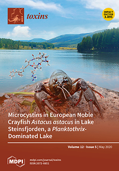
The microcystins are a large group of toxic cyclic heptapeptides produced by cyanobacteria such as Microcystis and Planktothrix spp. Many freshwater habitats experience regular cyanobacterial blooms caused by microcystin-producing Planktothrix spp. In Lake Steinsfjorden, Norway, noble crayfish (Astacus astacus) harvested for human consumption are exposed to the microcystins from the surrounding water and through their diet. What is the relationship between microcystins in the water and in the crayfish? What is the impact of microcystins on noble crayfish health? Is there a food safety concern, and are there steps that might be used to mitigate it? This study addresses these important questions. View this paper.
  • Issues are regarded as officially published after their release is announced to the table of contents alert mailing list.
  • You may sign up for e-mail alerts to receive table of contents of newly released issues.
  • PDF is the official format for papers published in both, html and pdf forms. To view the papers in pdf format, click on the "PDF Full-text" link, and use the free Adobe Reader to open them.
Order results
Result details
Section
Select all
Export citation of selected articles as:
13 pages, 1762 KiB  
Article
Prevalence and Genetic Diversity of Staphylococcal Enterotoxin (-Like) Genes sey, selw, selx, selz, sel26 and sel27 in Community-Acquired Methicillin-Resistant Staphylococcus aureus
by Meiji Soe Aung, Noriko Urushibara, Mitsuyo Kawaguchiya, Masahiko Ito, Satoshi Habadera and Nobumichi Kobayashi
Toxins 2020, 12(5), 347; https://doi.org/10.3390/toxins12050347 - 23 May 2020
Cited by 21 | Viewed by 3641
Abstract
Staphylococcal enterotoxins (SEs) are virulence factors of Staphylococcus aureus associated with various toxic diseases due to their emetic and superantigenic activities. Although at least 27 SE(-like) genes have been identified in S. aureus to date, the newly identified SE(-like) genes have not yet [...] Read more.
Staphylococcal enterotoxins (SEs) are virulence factors of Staphylococcus aureus associated with various toxic diseases due to their emetic and superantigenic activities. Although at least 27 SE(-like) genes have been identified in S. aureus to date, the newly identified SE(-like) genes have not yet been well characterized by their epidemiological features. In this study, the prevalence and genetic diversity of SE gene sey and SE-like genes selw, selx, selz, sel26, and sel27 were investigated for 624 clinical isolates of community-acquired methicillin-resistant S. aureus (CA-MRSA). The most prevalent SE(-like) gene was selw (92.9%), followed by selx (85.6%), sey (35.4%) and selz (5.6%), while sel26 and sel27 were not detected. Phylogenetically, sey, selw, selx, and selz were discriminated into 7, 10, 16, and 9 subtypes (groups), respectively. Among these subtypes, sey was the most conserved and showed the highest sequence identity (>98.8%), followed by selz and selx. The SE-like gene selw was the most divergent, and four out of ten genetic groups contained pseudogenes that may encode truncated product. Individual subtypes of SE(-like) genes were generally found in isolates with specific genotypes/lineages of S. aureus. This study revealed the putative ubiquity of selw and selx and the prevalence of sey and selz in some specific lineages (e.g., ST121) in CA-MRSA, suggesting a potential role of these newly described SEs(-like) in pathogenicity. Full article
(This article belongs to the Section Bacterial Toxins)
Show Figures

Figure 1

19 pages, 2196 KiB  
Article
Removal of Small Kernels Reduces the Content of Fusarium Mycotoxins in Oat Grain
by Guro Brodal, Heidi Udnes Aamot, Marit Almvik and Ingerd Skow Hofgaard
Toxins 2020, 12(5), 346; https://doi.org/10.3390/toxins12050346 - 23 May 2020
Cited by 12 | Viewed by 3758
Abstract
Cereal grain contaminated by Fusarium mycotoxins is undesirable in food and feed because of the harmful health effects of the mycotoxins in humans and animals. Reduction of mycotoxin content in grain by cleaning and size sorting has mainly been studied in wheat. We [...] Read more.
Cereal grain contaminated by Fusarium mycotoxins is undesirable in food and feed because of the harmful health effects of the mycotoxins in humans and animals. Reduction of mycotoxin content in grain by cleaning and size sorting has mainly been studied in wheat. We investigated whether the removal of small kernels by size sorting could be a method to reduce the content of mycotoxins in oat grain. Samples from 24 Norwegian mycotoxin-contaminated grain lots (14 from 2015 and 10 from 2018) were sorted by a laboratory sieve (sieve size 2.2 mm) into large and small kernel fractions and, in addition to unsorted grain samples, analyzed with LC-MS-MS for quantification of 10 mycotoxins. By removing the small kernel fraction (on average 15% and 21% of the weight of the samples from the two years, respectively), the mean concentrations of HT-2+T-2 toxins were reduced by 56% (from 745 to 328 µg/kg) in the 2015 samples and by 32% (from 178 to 121 µg/kg) in the 2018 samples. Deoxynivalenol (DON) was reduced by 24% (from 191 to 145 µg/kg) in the 2018 samples, and enniatin B (EnnB) by 44% (from 1059 to 594 µg/kg) in the 2015 samples. Despite low levels, our analyses showed a trend towards reduced content of DON, ADON, NIV, EnnA, EnnA1, EnnB1 and BEA after removing the small kernel fraction in samples from 2015. For several of the mycotoxins, the concentrations were considerably higher in the small kernel fraction compared to unsorted grain. Our results demonstrate that the level of mycotoxins in unprocessed oat grain can be reduced by removing small kernels. We assume that our study is the first report on the effect of size sorting on the content of enniatins (Enns), NIV and BEA in oat grains. Full article
(This article belongs to the Special Issue Mycotoxins in Feed and Food Chain: Present Status and Future Concerns)
Show Figures

Figure 1

52 pages, 3969 KiB  
Review
Molecular Biology of Escherichia coli Shiga Toxins’ Effects on Mammalian Cells
by Christian Menge
Toxins 2020, 12(5), 345; https://doi.org/10.3390/toxins12050345 - 23 May 2020
Cited by 37 | Viewed by 6371
Abstract
Shiga toxins (Stxs), syn. Vero(cyto)toxins, are potent bacterial exotoxins and the principal virulence factor of enterohemorrhagic Escherichia coli (EHEC), a subset of Shiga toxin-producing E. coli (STEC). EHEC strains, e.g., strains of serovars O157:H7 and O104:H4, may cause individual cases as well as [...] Read more.
Shiga toxins (Stxs), syn. Vero(cyto)toxins, are potent bacterial exotoxins and the principal virulence factor of enterohemorrhagic Escherichia coli (EHEC), a subset of Shiga toxin-producing E. coli (STEC). EHEC strains, e.g., strains of serovars O157:H7 and O104:H4, may cause individual cases as well as large outbreaks of life-threatening diseases in humans. Stxs primarily exert a ribotoxic activity in the eukaryotic target cells of the mammalian host resulting in rapid protein synthesis inhibition and cell death. Damage of endothelial cells in the kidneys and the central nervous system by Stxs is central in the pathogenesis of hemolytic uremic syndrome (HUS) in humans and edema disease in pigs. Probably even more important, the toxins also are capable of modulating a plethora of essential cellular functions, which eventually disturb intercellular communication. The review aims at providing a comprehensive overview of the current knowledge of the time course and the consecutive steps of Stx/cell interactions at the molecular level. Intervention measures deduced from an in-depth understanding of this molecular interplay may foster our basic understanding of cellular biology and microbial pathogenesis and pave the way to the creation of host-directed active compounds to mitigate the pathological conditions of STEC infections in the mammalian body. Full article
Show Figures

Figure 1

20 pages, 1862 KiB  
Review
Paralytic Shellfish Toxins (PST)-Transforming Enzymes: A Review
by Mariana I. C. Raposo, Maria Teresa S. R. Gomes, Maria João Botelho and Alisa Rudnitskaya
Toxins 2020, 12(5), 344; https://doi.org/10.3390/toxins12050344 - 22 May 2020
Cited by 36 | Viewed by 5940
Abstract
Paralytic shellfish toxins (PSTs) are a group of toxins that cause paralytic shellfish poisoning through blockage of voltage-gated sodium channels. PSTs are produced by prokaryotic freshwater cyanobacteria and eukaryotic marine dinoflagellates. Proliferation of toxic algae species can lead to harmful algal blooms, during [...] Read more.
Paralytic shellfish toxins (PSTs) are a group of toxins that cause paralytic shellfish poisoning through blockage of voltage-gated sodium channels. PSTs are produced by prokaryotic freshwater cyanobacteria and eukaryotic marine dinoflagellates. Proliferation of toxic algae species can lead to harmful algal blooms, during which seafood accumulate high levels of PSTs, posing a health threat to consumers. The existence of PST-transforming enzymes was first remarked due to the divergence of PST profiles and concentrations between contaminated bivalves and toxigenic organisms. Later, several enzymes involved in PST transformation, synthesis and elimination have been identified. The knowledge of PST-transforming enzymes is necessary for understanding the processes of toxin accumulation and depuration in mollusk bivalves. Furthermore, PST-transforming enzymes facilitate the obtainment of pure analogues of toxins as in natural sources they are present in a mixture. Pure compounds are of interest for the development of drug candidates and as analytical reference materials. PST-transforming enzymes can also be employed for the development of analytical tools for toxin detection. This review summarizes the PST-transforming enzymes identified so far in living organisms from bacteria to humans, with special emphasis on bivalves, cyanobacteria and dinoflagellates, and discusses enzymes’ biological functions and potential practical applications. Full article
(This article belongs to the Special Issue Bioactivity and Chemical Ecological Interactions of Marine Toxins)
Show Figures

Figure 1

17 pages, 4347 KiB  
Article
Temporary Membrane Permeabilization via the Pore-Forming Toxin Lysenin
by Nisha Shrestha, Christopher A. Thomas, Devon Richtsmeier, Andrew Bogard, Rebecca Hermann, Malyk Walker, Gamid Abatchev, Raquel J. Brown and Daniel Fologea
Toxins 2020, 12(5), 343; https://doi.org/10.3390/toxins12050343 - 22 May 2020
Cited by 5 | Viewed by 3805
Abstract
Pore-forming toxins are alluring tools for delivering biologically-active, impermeable cargoes to intracellular environments by introducing large conductance pathways into cell membranes. However, the lack of regulation often leads to the dissipation of electrical and chemical gradients, which might significantly affect the viability of [...] Read more.
Pore-forming toxins are alluring tools for delivering biologically-active, impermeable cargoes to intracellular environments by introducing large conductance pathways into cell membranes. However, the lack of regulation often leads to the dissipation of electrical and chemical gradients, which might significantly affect the viability of cells under scrutiny. To mitigate these problems, we explored the use of lysenin channels to reversibly control the barrier function of natural and artificial lipid membrane systems by controlling the lysenin’s transport properties. We employed artificial membranes and electrophysiology measurements in order to identify the influence of labels and media on the lysenin channel’s conductance. Two cell culture models: Jurkat cells in suspension and adherent ATDC5 cells were utilized to demonstrate that lysenin channels may provide temporary cytosol access to membrane non-permeant propidium iodide and phalloidin. Permeability and cell viability were assessed by fluorescence spectroscopy and microscopy. Membrane resealing by chitosan or specific media addition proved to be an effective way of maintaining cellular viability. In addition, we loaded non-permeant dyes into liposomes via lysenin channels by controlling their conducting state with multivalent metal cations. The improved control over membrane permeability might prove fruitful for a large variety of biological or biomedical applications that require only temporary, non-destructive access to the inner environment enclosed by natural and artificial membranes. Full article
(This article belongs to the Section Bacterial Toxins)
Show Figures

Figure 1

15 pages, 2335 KiB  
Review
Targeting the Early Endosome-to-Golgi Transport of Shiga Toxins as a Therapeutic Strategy
by Danyang Li, Andrey Selyunin and Somshuvra Mukhopadhyay
Toxins 2020, 12(5), 342; https://doi.org/10.3390/toxins12050342 - 22 May 2020
Cited by 9 | Viewed by 4992
Abstract
Shiga toxin (STx) produced by Shigella and closely related Shiga toxin 1 and 2 (STx1 and STx2) synthesized by Shiga toxin-producing Escherichia coli (STEC) are bacterial AB5 toxins. All three toxins target kidney cells and may cause life-threatening renal disease. While Shigella [...] Read more.
Shiga toxin (STx) produced by Shigella and closely related Shiga toxin 1 and 2 (STx1 and STx2) synthesized by Shiga toxin-producing Escherichia coli (STEC) are bacterial AB5 toxins. All three toxins target kidney cells and may cause life-threatening renal disease. While Shigella infections can be treated with antibiotics, resistance is increasing. Moreover, antibiotic therapy is contraindicated for STEC, and there are no definitive treatments for STEC-induced disease. To exert cellular toxicity, STx, STx1, and STx2 must undergo retrograde trafficking to reach their cytosolic target, ribosomes. Direct transport from early endosomes to the Golgi apparatus is an essential step that allows the toxins to bypass degradative late endosomes and lysosomes. The essentiality of this transport step also makes it an ideal target for the development of small-molecule inhibitors of toxin trafficking as potential therapeutics. Here, we review the recent advances in understanding the molecular mechanisms of the early endosome-to-Golgi transport of STx, STx1, and STx2, as well as the development of small-molecule inhibitors of toxin trafficking that act at the endosome/Golgi interface. Full article
(This article belongs to the Special Issue Inhibitors and Countermeasures against Bacterial and Plant Toxins)
Show Figures

Figure 1

13 pages, 1257 KiB  
Article
Monitoring Phenolic Compounds in Rice during the Growing Season in Relation to Fungal and Mycotoxin Contamination
by Paola Giorni, Silvia Rastelli, Sofia Fregonara and Terenzio Bertuzzi
Toxins 2020, 12(5), 341; https://doi.org/10.3390/toxins12050341 - 22 May 2020
Cited by 8 | Viewed by 3866
Abstract
Total phenolic content (TPC) and several phenolic acids present in rice grains were compared with fungal infection and mycotoxin presence throughout the growing season. Samples of 4 rice varieties were collected in 2018 and 2019 at 3 different plant phenological stages. Total fungal [...] Read more.
Total phenolic content (TPC) and several phenolic acids present in rice grains were compared with fungal infection and mycotoxin presence throughout the growing season. Samples of 4 rice varieties were collected in 2018 and 2019 at 3 different plant phenological stages. Total fungal and main mycotoxigenic fungi incidence were checked and mycotoxin content was analysed. On the same samples, TPC and the concentration of 8 main phenolic acids (chlorogenic acid, caffeic acid, syringic acid, 4-hydroxybenzoic acid (4-HBA), p-coumaric acid, ferulic acid, protocatecuic acid and gallic acid) were measured. The results showed significant differences between years for both fungal incidence and mycotoxin presence. In 2018 there was a lower fungal presence (42%) than in 2019 (57%) while, regarding mycotoxins, sterigmatocystin (STC) was found in almost all the samples and at all growing stages while deoxynivalenol (DON) was found particularly during ripening. An interesting relationship was found between fungal incidence and TPC, and some phenolic acids seemed to be more involved than others in the plant defense system. Ferulic acid and protocatecuic acid showed a different trend during the growing season depending on fungal incidence and resulted to be positively correlated with p-coumaric acid and 4-HBA that seem involved in mycotoxin containment in field. Full article
(This article belongs to the Special Issue Mycotoxins and Related Fungi in Crops)
Show Figures

Figure 1

20 pages, 1724 KiB  
Review
Monocytes in Uremia
by Matthias Girndt, Bogusz Trojanowicz and Christof Ulrich
Toxins 2020, 12(5), 340; https://doi.org/10.3390/toxins12050340 - 21 May 2020
Cited by 29 | Viewed by 3946
Abstract
Monocytes play an important role in both innate immunity and antigen presentation for specific cellular immune defense. In patients with chronic renal failure, as well as those treated with maintenance hemodialysis, these cells are largely dysregulated. There is a large body of literature [...] Read more.
Monocytes play an important role in both innate immunity and antigen presentation for specific cellular immune defense. In patients with chronic renal failure, as well as those treated with maintenance hemodialysis, these cells are largely dysregulated. There is a large body of literature on monocyte alterations in such patients. However, most of the publications report on small series, there is a vast spectrum of different methods and the heterogeneity of the data prevents any meta-analytic approach. Thus, a narrative review was performed to describe the current knowledge. Monocytes from patients with chronic renal failure differ from those of healthy individuals in the pattern of surface molecule expression, cytokine and mediator production, and function. If these findings can be summarized at all, they might be subsumed as showing chronic inflammation in resting cells together with limited activation upon immunologic challenge. The picture is complicated by the fact that monocytes fall into morphologically and functionally different populations and population shifts interact heavily with dysregulation of the individual cells. Severe complications of chronic renal failure such as impaired immune defense, inflammation, and atherosclerosis can be related to several aspects of monocyte dysfunction. Therefore, this review aims to provide an overview about the impairment and activation of monocytes by uremia and the resulting clinical consequences for renal failure patients. Full article
(This article belongs to the Special Issue Immune Dysfunction in Uremia)
Show Figures

Figure 1

16 pages, 3162 KiB  
Article
Diversity and Toxigenicity of Fungi that Cause Pineapple Fruitlet Core Rot
by Bastien Barral, Marc Chillet, Anna Doizy, Maeva Grassi, Laetitia Ragot, Mathieu Léchaudel, Noel Durand, Lindy Joy Rose, Altus Viljoen and Sabine Schorr-Galindo
Toxins 2020, 12(5), 339; https://doi.org/10.3390/toxins12050339 - 21 May 2020
Cited by 15 | Viewed by 5239
Abstract
The identity of the fungi responsible for fruitlet core rot (FCR) disease in pineapple has been the subject of investigation for some time. This study describes the diversity and toxigenic potential of fungal species causing FCR in La Reunion, an island in the [...] Read more.
The identity of the fungi responsible for fruitlet core rot (FCR) disease in pineapple has been the subject of investigation for some time. This study describes the diversity and toxigenic potential of fungal species causing FCR in La Reunion, an island in the Indian Ocean. One-hundred-and-fifty fungal isolates were obtained from infected and healthy fruitlets on Reunion Island and exclusively correspond to two genera of fungi: Fusarium and Talaromyces. The genus Fusarium made up 79% of the isolates, including 108 F. ananatum, 10 F. oxysporum, and one F. proliferatum. The genus Talaromyces accounted for 21% of the isolated fungi, which were all Talaromyces stollii. As the isolated fungal strains are potentially mycotoxigenic, identification and quantification of mycotoxins were carried out on naturally or artificially infected diseased fruits and under in vitro cultures of potential toxigenic isolates. Fumonisins B1 and B2 (FB1-FB2) and beauvericin (BEA) were found in infected fruitlets of pineapple and in the culture media of Fusarium species. Regarding the induction of mycotoxin in vitro, F. proliferatum produced 182 mg kg⁻1 of FB1 and F. oxysporum produced 192 mg kg⁻1 of BEA. These results provide a better understanding of the causal agents of FCR and their potential risk to pineapple consumers. Full article
(This article belongs to the Special Issue Phytopathogenic Fungi and Toxicity)
Show Figures

Graphical abstract

16 pages, 627 KiB  
Review
Human Botulism in France, 1875–2016
by Christine Rasetti-Escargueil, Emmanuel Lemichez and Michel R. Popoff
Toxins 2020, 12(5), 338; https://doi.org/10.3390/toxins12050338 - 21 May 2020
Cited by 23 | Viewed by 5567
Abstract
Botulism is a rare but severe disease which is characterized by paralysis and inhibition of secretions. Only a few cases had been reported at the end of the 19th century in France. The disease was frequent during the second world war, and then [...] Read more.
Botulism is a rare but severe disease which is characterized by paralysis and inhibition of secretions. Only a few cases had been reported at the end of the 19th century in France. The disease was frequent during the second world war, and then the incidence decreased progressively. However, human botulism is still present in France with 10–25 cases every year. Food-borne botulism was the main form of botulism in France, whereas infant botulism (17 cases between 2004 and 2016) was rare, and wound and inhalational botulism were exceptional. Type B was the prevalent botulism type and was mainly due to consumption of home-made or small-scale preparations of cured ham and to a lesser extent other pork meat products. In the recent period (2000–2016), a wider diversity of botulism types from various food origin including industrial foods was reported. Severe cases of type A and F botulism as well as type E botulism were more frequent. Albeit rare, the severity of botulism justifies its continued surveillance and recommendations to food industry and consumers regarding food hygiene and preservation practices. Full article
Show Figures

Figure 1

15 pages, 2832 KiB  
Article
Sensitive Aflatoxin B1 Detection Using Nanoparticle-Based Competitive Magnetic Immunodetection
by Jan Pietschmann, Holger Spiegel, Hans-Joachim Krause, Stefan Schillberg and Florian Schröper
Toxins 2020, 12(5), 337; https://doi.org/10.3390/toxins12050337 - 20 May 2020
Cited by 24 | Viewed by 4503
Abstract
Food and crop contaminations with mycotoxins are a severe health risk for consumers and cause high economic losses worldwide. Currently, different chromatographic- and immuno-based methods are used to detect mycotoxins within different sample matrices. There is a need for novel, highly sensitive detection [...] Read more.
Food and crop contaminations with mycotoxins are a severe health risk for consumers and cause high economic losses worldwide. Currently, different chromatographic- and immuno-based methods are used to detect mycotoxins within different sample matrices. There is a need for novel, highly sensitive detection technologies that avoid time-consuming procedures and expensive laboratory equipment but still provide sufficient sensitivity to achieve the mandated detection limit for mycotoxin content. Here we describe a novel, highly sensitive, and portable aflatoxin B1 detection approach using competitive magnetic immunodetection (cMID). As a reference method, a competitive ELISA optimized by checkerboard titration was established. For the novel cMID procedure, immunofiltration columns, coated with aflatoxin B1-BSA conjugate were used for competitive enrichment of biotinylated aflatoxin B1-specific antibodies. Subsequently, magnetic particles functionalized with streptavidin can be applied to magnetically label retained antibodies. By means of frequency mixing technology, particles were detected and quantified corresponding to the aflatoxin content in the sample. After the optimization of assay conditions, we successfully demonstrated the new competitive magnetic detection approach with a comparable detection limit of 1.1 ng aflatoxin B1 per mL sample to the cELISA reference method. Our results indicate that the cMID is a promising method reducing the risks of processing contaminated commodities. Full article
(This article belongs to the Special Issue Rapid Detection of Mycotoxin Contamination)
Show Figures

Figure 1

3 pages, 188 KiB  
Editorial
Introduction to the Toxins Special Issue on Identification and Functional Characterization of Novel Venom Components
by Steven D. Aird
Toxins 2020, 12(5), 336; https://doi.org/10.3390/toxins12050336 - 20 May 2020
Cited by 1 | Viewed by 2134
Abstract
Throughout most of the 20th century, the toxinological literature consisted largely of pharmacological and functional characterizations of crude venoms and venom constituents, often constituents that could not be identified unambiguously [...] Full article
16 pages, 1556 KiB  
Article
Delayed Release of Intracellular Microcystin Following Partial Oxidation of Cultured and Naturally Occurring Cyanobacteria
by Katherine E. Greenstein, Arash Zamyadi, Caitlin M. Glover, Craig Adams, Erik Rosenfeldt and Eric C. Wert
Toxins 2020, 12(5), 335; https://doi.org/10.3390/toxins12050335 - 20 May 2020
Cited by 15 | Viewed by 3516
Abstract
Oxidation processes can provide an effective barrier to eliminate cyanotoxins by damaging cyanobacteria cell membranes, releasing intracellular cyanotoxins, and subsequently oxidizing these toxins (now in extracellular form) based on published reaction kinetics. In this work, cyanobacteria cells from two natural blooms (from the [...] Read more.
Oxidation processes can provide an effective barrier to eliminate cyanotoxins by damaging cyanobacteria cell membranes, releasing intracellular cyanotoxins, and subsequently oxidizing these toxins (now in extracellular form) based on published reaction kinetics. In this work, cyanobacteria cells from two natural blooms (from the United States and Canada) and a laboratory-cultured Microcystis aeruginosa strain were treated with chlorine, monochloramine, chlorine dioxide, ozone, and potassium permanganate. The release of microcystin was measured immediately after oxidation (t ≤ 20 min), and following oxidant residual quenching (stagnation times = 96 or 168 h). Oxidant exposures (CT) were determined resulting in complete release of intracellular microcystin following chlorine (21 mg-min/L), chloramine (72 mg-min/L), chlorine dioxide (58 mg-min/L), ozone (4.1 mg-min/L), and permanganate (391 mg-min/L). Required oxidant exposures using indigenous cells were greater than lab-cultured Microcystis. Following partial oxidation of cells (oxidant exposures ≤ CT values cited above), additional intracellular microcystin and dissolved organic carbon (DOC) were released while the samples remained stagnant in the absence of an oxidant (>96 h after quenching). The delayed release of microcystin from partially oxidized cells has implications for drinking water treatment as these cells may be retained on a filter surface or in solids and continue to slowly release cyanotoxins and other metabolites into the finished water. Full article
(This article belongs to the Special Issue Removal of Cyanobacteria and Cyanotoxins in Waters)
Show Figures

Figure 1

9 pages, 1108 KiB  
Communication
Behavioral, Physiological, Demographic and Ecological Impacts of Hematophagous and Endoparasitic Insects on an Arctic Ungulate
by Kyle Joly, Ophélie Couriot, Matthew D. Cameron and Eliezer Gurarie
Toxins 2020, 12(5), 334; https://doi.org/10.3390/toxins12050334 - 20 May 2020
Cited by 15 | Viewed by 4024
Abstract
Animals that deliver a toxic secretion through a wound or to the body surface without a wound are considered venomous and toxungenous, respectively. Hematophagous insects, such as mosquitoes (Aedes spp.), meet the criteria for venomous, and some endoparasitic insects, such as warble [...] Read more.
Animals that deliver a toxic secretion through a wound or to the body surface without a wound are considered venomous and toxungenous, respectively. Hematophagous insects, such as mosquitoes (Aedes spp.), meet the criteria for venomous, and some endoparasitic insects, such as warble flies (Hypoderma tarandi), satisfy the definition for toxungenous. The impacts of these insects on their hosts are wide ranging. In the Arctic, their primary host is the most abundant ungulate, the caribou (Rangifer tarandus). The most conspicuous impacts of these insects on caribou are behavioral. Caribou increase their movements during peak insect harassment, evading and running away from these parasites. These behavioral responses scale up to physiological effects as caribou move to less productive habitats to reduce harassment which increases energetic costs due to locomotion, reduces nutrient intake due to less time spent foraging, and can lead to poorer physiological condition. Reduced physiological condition can lead to lower reproductive output and even higher mortality rates, with the potential to ultimately affect caribou demographics. Caribou affect all trophic levels in the Arctic and the processes that connect them, thus altering caribou demographics could impact the ecology of the region. Broadening the definitions of venomous and toxungenous animals to include hematophagous and endoparasitic insects should not only generate productive collaborations among toxinologists and parasitologists, but will also lead to a deeper understanding of the ecology of toxic secretions and their widespread influence. Full article
(This article belongs to the Special Issue The Behavioral Ecology of Venom)
Show Figures

Figure 1

20 pages, 7758 KiB  
Article
Classical and Alternative Activation of Rat Microglia Treated with Ultrapure Porphyromonas gingivalis Lipopolysaccharide In Vitro
by Zylfi Memedovski, Evan Czerwonka, Jin Han, Joshua Mayer, Margaret Luce, Lucas C. Klemm, Mary L. Hall and Alejandro M. S. Mayer
Toxins 2020, 12(5), 333; https://doi.org/10.3390/toxins12050333 - 19 May 2020
Cited by 13 | Viewed by 3440
Abstract
The possible relationship between periodontal disease resulting from the infection of gingival tissue by the Gram-negative bacterium Porphyromonas gingivalis (P. gingivalis) and the development of neuroinflammation remains under investigation. Recently, P. gingivalis lipopolysaccharide (LPS) was reported in the human brain, thus [...] Read more.
The possible relationship between periodontal disease resulting from the infection of gingival tissue by the Gram-negative bacterium Porphyromonas gingivalis (P. gingivalis) and the development of neuroinflammation remains under investigation. Recently, P. gingivalis lipopolysaccharide (LPS) was reported in the human brain, thus suggesting it might activate brain microglia, a cell type participating in neuroinflammation. We tested the hypothesis of whether in vitro exposure to ultrapure P. gingivalis LPS may result in classical and alternative activation phenotypes of rat microglia, with the concomitant release of cytokines and chemokines, as well as superoxide anion (O2), thromboxane B2 (TXB2), and matrix metalloprotease-9 (MMP-9). After an 18-h exposure of microglia to P. gingivalis LPS, the concentration-dependent responses were the following: 0.1–100 ng/mL P. gingivalis LPS increased O2 generation, with reduced inflammatory mediator generation; 1000–10,000 ng/mL P. gingivalis LPS generated MMP-9, macrophage inflammatory protein 1α (MIP-1α/CCL3), macrophage inflammatory protein-2 (MIP-2/CXCL2) release and significant O2 generation; 100,000 ng/mL P. gingivalis LPS sustained O2 production, maintained MMP-9, tumor necrosis factor-α (TNF-α), and interleukin-6 (IL-6) release, and triggered elevated levels of MIP-1α/CCL3, MIP-2/CXCL2, and cytokine-induced neutrophil chemoattractant 1 (CINC-1/CXCL-1), with a very low release of lactic dehydrogenase (LDH). Although P. gingivalis LPS was less potent than Escherichia coli (E. coli) LPS in stimulating TXB2, MMP-9, IL-6 and interleukin 10 (IL-10) generation, we observed that it appeared more efficacious in enhancing the release of O2, TNF-α, MIP-1α/CCL3, MIP-2/CXCL2 and CINC-1/CXCL-1. Our results provide support to our research hypothesis because an 18-h in vitro stimulation with ultrapure P. gingivalis LPS resulted in the classical and alternative activation of rat brain microglia and the concomitant release of cytokines and chemokines. Full article
(This article belongs to the Section Bacterial Toxins)
Show Figures

Figure 1

13 pages, 253 KiB  
Review
FDA Approvals and Consensus Guidelines for Botulinum Toxins in the Treatment of Dystonia
by Lauren L. Spiegel, Jill L. Ostrem and Ian O. Bledsoe
Toxins 2020, 12(5), 332; https://doi.org/10.3390/toxins12050332 - 17 May 2020
Cited by 29 | Viewed by 7059
Abstract
In 2016, the American Academy of Neurology (AAN) published practice guidelines for botulinum toxin (BoNT) in the treatment of blepharospasm, cervical dystonia, adult spasticity, and headache. This article, focusing on dystonia, provides context for these guidelines through literature review. Studies that led to [...] Read more.
In 2016, the American Academy of Neurology (AAN) published practice guidelines for botulinum toxin (BoNT) in the treatment of blepharospasm, cervical dystonia, adult spasticity, and headache. This article, focusing on dystonia, provides context for these guidelines through literature review. Studies that led to Food and Drug Administration (FDA) approval of each toxin for dystonia indications are reviewed, in addition to several studies highlighted by the AAN guidelines. The AAN guidelines for the use of BoNT in dystonia are compared with those of the European Federation of the Neurological Societies (EFNS), and common off-label uses for BoNT in dystonia are discussed. Toxins not currently FDA-approved for the treatment of dystonia are additionally reviewed. In the future, additional toxins may become FDA-approved for the treatment of dystonia given expanding research in this area. Full article
(This article belongs to the Special Issue Treatment of Dystonia with Botulinum Toxins)
17 pages, 1176 KiB  
Article
Aflatoxin Reduction in Maize by Industrial-Scale Cleaning Solutions
by Michelangelo Pascale, Antonio F. Logrieco, Matthias Graeber, Marina Hirschberger, Mareike Reichel, Vincenzo Lippolis, Annalisa De Girolamo, Veronica M.T. Lattanzio and Katarina Slettengren
Toxins 2020, 12(5), 331; https://doi.org/10.3390/toxins12050331 - 17 May 2020
Cited by 19 | Viewed by 4527
Abstract
Different batches of biomass/feed quality maize contaminated by aflatoxins were processed at the industrial scale (a continuous process and separate discontinuous steps) to evaluate the effect of different cleaning solutions on toxin reduction. The investigated cleaning solutions included: (i) mechanical size separation of [...] Read more.
Different batches of biomass/feed quality maize contaminated by aflatoxins were processed at the industrial scale (a continuous process and separate discontinuous steps) to evaluate the effect of different cleaning solutions on toxin reduction. The investigated cleaning solutions included: (i) mechanical size separation of coarse, small and broken kernels, (ii) removal of dust/fine particles through an aspiration channel, (iii) separation of kernels based on gravity and (iv) optical sorting of spatial and spectral kernel defects. Depending on the sampled fraction, dynamic or static sampling was performed according to the Commission Regulation No. 401/2006 along the entire cleaning process lines. Aflatoxin analyses of the water–slurry aggregate samples were performed according to the AOAC Official Method No. 2005.008 based on high-performance liquid chromatography and immunoaffinity column cleanup of the extracts. A significant reduction in aflatoxin content in the cleaned products, ranging from 65% to 84% with respect to the uncleaned products, was observed when continuous cleaning lines were used. Additionally, an overall aflatoxin reduction from 55% to 94% was obtained by combining results from separate cleaning steps. High levels of aflatoxins (up to 490 µg/kg) were found in the rejected fractions, with the highest levels in dust and in the rejected fractions from the aspirator and optical sorting. This study shows that a cleaning line combining both mechanical and optical sorting technologies provides an efficient solution for reducing aflatoxin contamination in maize. Full article
(This article belongs to the Collection Aflatoxins)
Show Figures

Figure 1

10 pages, 1255 KiB  
Communication
Neuroprotective Fragment C of Tetanus Toxin Modulates IL-6 in an ALS Mouse Model
by Laura Moreno-Martinez, Miriam de la Torre, María J. Muñoz, Pilar Zaragoza, José Aguilera, Ana C. Calvo and Rosario Osta
Toxins 2020, 12(5), 330; https://doi.org/10.3390/toxins12050330 - 17 May 2020
Cited by 8 | Viewed by 3320
Abstract
Neuroinflammation plays a significant role in amyotrophic lateral sclerosis (ALS) pathology, leading to the development of therapies targeting inflammation in recent years. Our group has studied the tetanus toxin C-terminal fragment (TTC) as a therapeutic molecule, showing neuroprotective properties in the SOD1G93A mouse [...] Read more.
Neuroinflammation plays a significant role in amyotrophic lateral sclerosis (ALS) pathology, leading to the development of therapies targeting inflammation in recent years. Our group has studied the tetanus toxin C-terminal fragment (TTC) as a therapeutic molecule, showing neuroprotective properties in the SOD1G93A mouse model. However, it is unknown whether TTC could have some effect on inflammation. The objective of this study was to assess the effect of TTC on the regulation of inflammatory mediators to elucidate its potential role in modulating inflammation occurring in ALS. After TTC treatment in SOD1G93A mice, levels of eotaxin-1, interleukin (IL)-2, IL-6 and macrophage inflammatory protein (MIP)-1 alpha (α) and galectin-1 were analyzed by immunoassays in plasma samples, whilst protein expression of caspase-1, IL-1β, IL-6 and NOD-, LRR- and pyrin domain-containing protein 3 (NLRP3) was measured in the spinal cord, extensor digitorum longus (EDL) muscle and soleus (SOL) muscle. The results showed reduced levels of IL-6 in spinal cord, EDL and SOL in treated SOD1G93A mice. In addition, TTC showed a different role in the modulation of NLRP3 and caspase-1 depending on the tissue analyzed. In conclusion, our results suggest that TTC could have a potential anti-inflammatory effect by reducing IL-6 levels in tissues drastically affected by the disease. However, further research is needed to study more in depth the anti-inflammatory effect of TTC in ALS. Full article
Show Figures

Figure 1

14 pages, 3783 KiB  
Article
Multi-Stress Induction of the Mycobacterium tuberculosis MbcTA Bactericidal Toxin-Antitoxin System
by Kanchiyaphat Ariyachaokun, Anna D. Grabowska, Claude Gutierrez and Olivier Neyrolles
Toxins 2020, 12(5), 329; https://doi.org/10.3390/toxins12050329 - 16 May 2020
Cited by 7 | Viewed by 4096
Abstract
MbcTA is a type II toxin/antitoxin (TA) system of Mycobacterium tuberculosis. The MbcT toxin triggers mycobacterial cell death in vitro and in vivo through the phosphorolysis of the essential metabolite NAD+ and its bactericidal activity is neutralized by physical interaction with [...] Read more.
MbcTA is a type II toxin/antitoxin (TA) system of Mycobacterium tuberculosis. The MbcT toxin triggers mycobacterial cell death in vitro and in vivo through the phosphorolysis of the essential metabolite NAD+ and its bactericidal activity is neutralized by physical interaction with its cognate antitoxin MbcA. Therefore, the MbcTA system appears as a promising target for the development of novel therapies against tuberculosis, through the identification of compounds able to antagonize or destabilize the MbcA antitoxin. Here, the expression of the mbcAT operon and its regulation were investigated. A dual fluorescent reporter system was developed, based on an integrative mycobacterial plasmid that encodes a constitutively expressed reporter, serving as an internal standard for monitoring mycobacterial gene expression, and an additional reporter, dependent on the promoter under investigation. This system was used both in M. tuberculosis and in the fast growing model species Mycobacterium smegmatis to: (i) assess the autoregulation of mbcAT; (ii) perform a genetic dissection of the mbcA promoter/operator region; and (iii) explore the regulation of mbcAT transcription from the mbcA promoter (PmbcA) in a variety of stress conditions, including in vivo in mice and in macrophages. Full article
(This article belongs to the Special Issue Toxin-Antitoxin Systems in Pathogenic Bacteria)
Show Figures

Figure 1

26 pages, 6700 KiB  
Article
Tetanus Toxin Synthesis is Under the Control of A Complex Network of Regulatory Genes in Clostridium tetani
by Diana Chapeton-Montes, Lucile Plourde, Cecile Deneve, Dominique Garnier, Fabien Barbirato, Vincent Colombié, Sandy Demay, Georges Haustant, Olivier Gorgette, Christine Schmitt, Catherine Thouvenot, Holger Brüggemann and Michel R. Popoff
Toxins 2020, 12(5), 328; https://doi.org/10.3390/toxins12050328 - 15 May 2020
Cited by 8 | Viewed by 8832
Abstract
Clostridium tetani produces a potent neurotoxin, the tetanus toxin (TeNT), which is responsible for an often-fatal neurological disease (tetanus) characterized by spastic paralysis. Prevention is efficiently acquired by vaccination with the TeNT toxoid, which is obtained by C. tetani fermentation and subsequent purification [...] Read more.
Clostridium tetani produces a potent neurotoxin, the tetanus toxin (TeNT), which is responsible for an often-fatal neurological disease (tetanus) characterized by spastic paralysis. Prevention is efficiently acquired by vaccination with the TeNT toxoid, which is obtained by C. tetani fermentation and subsequent purification and chemical inactivation. C. tetani synthesizes TeNT in a regulated manner. Indeed, the TeNT gene (tent) is mainly expressed in the late exponential and early stationary growth phases. The gene tetR (tetanus regulatory gene), located immediately upstream of tent, encodes an alternative sigma factor which was previously identified as a positive regulator of tent. In addition, the genome of C. tetani encodes more than 127 putative regulators, including 30 two-component systems (TCSs). Here, we investigated the impact of 12 regulators on TeNT synthesis which were selected based on their homology with related regulatory elements involved in toxin production in other clostridial species. Among nine TCSs tested, three of them impact TeNT production, including two positive regulators that indirectly stimulate tent and tetR transcription. One negative regulator was identified that interacts with both tent and tetR promoters. Two other TCSs showed a moderate effect: one binds to the tent promoter and weakly increases the extracellular TeNT level, and another one has a weak inverse effect. In addition, CodY (control of dciA (decoyinine induced operon) Y) but not Spo0A (sporulation stage 0) or the DNA repair protein Mfd (mutation frequency decline) positively controls TeNT synthesis by interacting with the tent promoter. Moreover, we found that inorganic phosphate and carbonate are among the environmental factors that control TeNT production. Our data show that TeNT synthesis is under the control of a complex network of regulators that are largely distinct from those involved in the control of toxin production in Clostridium botulinum or Clostridium difficile. Full article
Show Figures

Figure 1

24 pages, 7753 KiB  
Article
Diversity and Toxicity of the Genus Coolia Meunier in Brazil, and Detection of 44-methyl Gambierone in Coolia tropicalis
by Carlos Eduardo Junqueira de Azevedo Tibiriçá, Manoella Sibat, Luciano Felício Fernandes, Gwenaël Bilien, Nicolas Chomérat, Philipp Hess and Luiz L. Mafra Jr
Toxins 2020, 12(5), 327; https://doi.org/10.3390/toxins12050327 - 15 May 2020
Cited by 26 | Viewed by 4306
Abstract
Coolia is a genus of marine benthic dinoflagellates which is widely distributed in tropical and temperate zones. Toxicity has been reported in selected Coolia species, although the identity of causative compounds is still controversial. In this study, we investigated the taxonomical and toxicological [...] Read more.
Coolia is a genus of marine benthic dinoflagellates which is widely distributed in tropical and temperate zones. Toxicity has been reported in selected Coolia species, although the identity of causative compounds is still controversial. In this study, we investigated the taxonomical and toxicological aspects of Coolia species from Brazil. Since light- and electron microscopy-based morphology was not enough to distinguish small-celled species, ITS and LSU D1-D3 phylogenetic analyses were used for species definition. Cultures of Coolia palmyrensis and Coolia santacroce were established from samples collected along the northeastern Brazilian coast, the first record of both species in South Atlantic waters. Cultures of Coolia malayensis and Coolia tropicalis were also established and exhibited acute in vivo toxicity to adults of Artemia salina, while C. palmyrensis and C. santacroce were non-toxic. The presence of 30 yessotoxin analogues, 7 metabolites of Coolia and 44 Gambierdiscus metabolites was screened in 14 strains of Coolia. 44-methyl gambierone (formerly referred to as MTX3) and a new isomer of this compound were detected only in C. tropicalis, using both low- and high-resolution LC-MS/MS. To our knowledge, this is the first report of gambierone analogues in dinoflagellates other than Gambierdiscus; the role of C. tropicalis in ciguatera poisoning thus deserves to be considered in further investigations. Full article
(This article belongs to the Section Marine and Freshwater Toxins)
Show Figures

Graphical abstract

18 pages, 1062 KiB  
Review
Scorpion Toxins and Ion Channels: Potential Applications in Cancer Therapy
by Rosa Amalia Dueñas-Cuellar, Carlos José Correia Santana, Ana Carolina Martins Magalhães, Osmindo Rodrigues Pires, Jr., Wagner Fontes and Mariana S. Castro
Toxins 2020, 12(5), 326; https://doi.org/10.3390/toxins12050326 - 15 May 2020
Cited by 16 | Viewed by 6700
Abstract
Apoptosis, a genetically directed process of cell death, has been studied for many years, and the biochemical mechanisms that surround it are well known and described. There are at least three pathways by which apoptosis occurs, and each pathway depends on extra or [...] Read more.
Apoptosis, a genetically directed process of cell death, has been studied for many years, and the biochemical mechanisms that surround it are well known and described. There are at least three pathways by which apoptosis occurs, and each pathway depends on extra or intracellular processes for activation. Apoptosis is a vital process, but disturbances in proliferation and cell death rates can lead to the development of diseases like cancer. Several compounds, isolated from scorpion venoms, exhibit inhibitory effects on different cancer cells. Indeed, some of these compounds can differentiate between healthy and cancer cells within the same tissue. During the carcinogenic process, morphological, biochemical, and biological changes occur that enable these compounds to modulate cancer but not healthy cells. This review highlights cancer cell features that enable modulation by scorpion neurotoxins. The properties of the isolated scorpion neurotoxins in cancer cells and the potential uses of these compounds as alternative treatments for cancer are discussed. Full article
(This article belongs to the Section Animal Venoms)
Show Figures

Figure 1

12 pages, 4915 KiB  
Article
Screening Snake Venoms for Toxicity to Tetrahymena Pyriformis Revealed Anti-Protozoan Activity of Cobra Cytotoxins
by Olga N. Kuleshina, Elena V. Kruykova, Elena G. Cheremnykh, Leonid V. Kozlov, Tatyana V. Andreeva, Vladislav G. Starkov, Alexey V. Osipov, Rustam H. Ziganshin, Victor I. Tsetlin and Yuri N. Utkin
Toxins 2020, 12(5), 325; https://doi.org/10.3390/toxins12050325 - 15 May 2020
Cited by 8 | Viewed by 4221
Abstract
Snake venoms possess lethal activities against different organisms, ranging from bacteria to higher vertebrates. Several venoms were shown to be active against protozoa, however, data about the anti-protozoan activity of cobra and viper venoms are very scarce. We tested the effects of venoms [...] Read more.
Snake venoms possess lethal activities against different organisms, ranging from bacteria to higher vertebrates. Several venoms were shown to be active against protozoa, however, data about the anti-protozoan activity of cobra and viper venoms are very scarce. We tested the effects of venoms from several snake species on the ciliate Tetrahymena pyriformis. The venoms tested induced T. pyriformis immobilization, followed by death, the most pronounced effect being observed for cobra Naja sumatrana venom. The active polypeptides were isolated from this venom by a combination of gel-filtration, ion exchange and reversed-phase HPLC and analyzed by mass spectrometry. It was found that these were cytotoxins of the three-finger toxin family. The cytotoxins from several cobra species were tested and manifested toxicity for infusorians. Light microscopy revealed that, because of the cytotoxin action, the infusorians’ morphology was changed greatly, from teardrop-like to an almost spherical shape, this alteration being accompanied by a leakage of cell contents. Fluorescence microscopy showed that the fluorescently labelled cytotoxin 2 from cobra N. oxiana was localized mainly at the membrane of killed infusorians, indicating that cytotoxins may kill T. pyriformis by causing membrane rupture. This work is the first evidence of the antiprotozoal activity of cobra venom cytotoxins, as demonstrated by the example of the ciliate T. pyriformis. Full article
(This article belongs to the Special Issue Animal Venoms and Their Components: Molecular Mechanisms of Action)
Show Figures

Graphical abstract

30 pages, 4162 KiB  
Article
An Integrated Proteomic and Transcriptomic Analysis Reveals the Venom Complexity of the Bullet Ant Paraponera clavata
by Samira R. Aili, Axel Touchard, Regan Hayward, Samuel D. Robinson, Sandy S. Pineda, Hadrien Lalagüe, Mrinalini, Irina Vetter, Eivind A. B. Undheim, R. Manjunatha Kini, Pierre Escoubas, Matthew P. Padula, Garry S. A. Myers and Graham M. Nicholson
Toxins 2020, 12(5), 324; https://doi.org/10.3390/toxins12050324 - 14 May 2020
Cited by 17 | Viewed by 5259
Abstract
A critical hurdle in ant venom proteomic investigations is the lack of databases to comprehensively and specifically identify the sequence and function of venom proteins and peptides. To resolve this, we used venom gland transcriptomics to generate a sequence database that was used [...] Read more.
A critical hurdle in ant venom proteomic investigations is the lack of databases to comprehensively and specifically identify the sequence and function of venom proteins and peptides. To resolve this, we used venom gland transcriptomics to generate a sequence database that was used to assign the tandem mass spectrometry (MS) fragmentation spectra of venom peptides and proteins to specific transcripts. This was performed alongside a shotgun liquid chromatography–mass spectrometry (LC-MS/MS) analysis of the venom to confirm that these assigned transcripts were expressed as proteins. Through the combined transcriptomic and proteomic investigation of Paraponera clavata venom, we identified four times the number of proteins previously identified using 2D-PAGE alone. In addition to this, by mining the transcriptomic data, we identified several novel peptide sequences for future pharmacological investigations, some of which conform with inhibitor cysteine knot motifs. These types of peptides have the potential to be developed into pharmaceutical or bioinsecticide peptides. Full article
(This article belongs to the Special Issue Venom Proteomics and Transcriptomics)
Show Figures

Figure 1

15 pages, 2094 KiB  
Article
Shedding Light on the Venom Proteomes of the Allergy-Relevant Hymenoptera Polistes dominula (European Paper Wasp) and Vespula spp. (Yellow Jacket)
by Johannes Grosch, Christiane Hilger, Maria Beatrice Bilò, Stephanie Kler, Maximilian Schiener, Gunnar Dittmar, François Bernardin, Antoine Lesur, Markus Ollert, Carsten B. Schmidt-Weber and Simon Blank
Toxins 2020, 12(5), 323; https://doi.org/10.3390/toxins12050323 - 14 May 2020
Cited by 16 | Viewed by 4171
Abstract
Allergic reactions to stings of Hymenoptera species can have serious or even fatal consequences. If the identification of the culprit insect is possible, venom-specific immunotherapy effectively cures Hymenoptera venom allergies. Although component-resolved diagnostics has strongly evolved in recent years, the differentiation between allergies [...] Read more.
Allergic reactions to stings of Hymenoptera species can have serious or even fatal consequences. If the identification of the culprit insect is possible, venom-specific immunotherapy effectively cures Hymenoptera venom allergies. Although component-resolved diagnostics has strongly evolved in recent years, the differentiation between allergies to closely related species such as Polistes dominula and Vespula spp. is still challenging. In order to generate the basis for new diagnostic and therapeutic strategies, this study aims at resolving the venom proteomes (venomes) of these species. The venoms of P. dominula and Vespula spp. (V. germanica, V. vulgaris) were analyzed by liquid chromatography-mass spectrometry. Resulting proteins were characterized regarding their function, localization and biochemical properties. The analyses yielded 157 proteins in Vespula spp. and 100 in P. dominula venom; 48 proteins, including annotated allergens, were found in both samples. In addition to a variety of venom trace molecules, new allergen candidates such as icarapin-like protein and phospholipase A2 were identified. This study elucidates the venomes of closely related allergy-eliciting Hymenoptera species. The data indicates that relying on marker allergens to differentiate between P. dominula and Vespula spp. venom allergy is probably insufficient and that strategies using cross-reactive major allergens could be more promising. Full article
(This article belongs to the Section Animal Venoms)
Show Figures

Figure 1

18 pages, 3372 KiB  
Article
A Screening Tool for the Direct Analysis of Marine and Freshwater Phycotoxins in Organic SPATT Extracts from the Chesapeake Bay
by Michelle D. Onofrio, Claude R. Mallet, Allen R. Place and Juliette L. Smith
Toxins 2020, 12(5), 322; https://doi.org/10.3390/toxins12050322 - 13 May 2020
Cited by 15 | Viewed by 3497
Abstract
Many detection methods for phycotoxins, bioactive compounds produced by harmful algae, focus on one compound or a class of related compounds. Multiple harmful algal species often co-occur in the environment, however, emphasizing the need to analyze for the presence of multiple groups of [...] Read more.
Many detection methods for phycotoxins, bioactive compounds produced by harmful algae, focus on one compound or a class of related compounds. Multiple harmful algal species often co-occur in the environment, however, emphasizing the need to analyze for the presence of multiple groups of marine and freshwater phycotoxins in environmental samples, e.g., extracts from solid phase adsorption toxin tracking (SPATT). Two methods were developed to screen for 13 phycotoxins (microcystin-RR, -LR, -YR, azaspiracid-1, -2, karlotoxin 3, goniodomin A, brevetoxin-2, yessotoxin, pectenotoxin-2, dinophysistoxin-1, -2, and okadaic acid) in organic SPATT extracts using ultra-performance liquid chromatography–tandem mass spectrometry (UPLC-MS/MS) equipped with a trapping dimension (trap) and at-column dilution (ACD). The performance of each compound under 36 combinations of chromatographic conditions was characterized, and two final methods, acidic and basic, were selected based on peak shapes, signal intensities, resolution, and the separation in time of positive and negative MS ionization modes. Injection volumes of up to 1 mL were possible through trap/ACD technology, resulting in limits of detection between 0.001 and 0.05 µg/L across the analytes. Benefits highlighted in this study, beyond the improved detection limits and co-detection of multiple toxin groups, include the ability to inject samples of 100% organic solvent, ensuring analyte stability and streamlining workflow through the elimination of laborious sample preparation steps. Full article
(This article belongs to the Section Marine and Freshwater Toxins)
Show Figures

Figure 1

18 pages, 2190 KiB  
Article
Assessment of Ciguatera and Other Phycotoxin-Related Risks in Anaho Bay (Nuku Hiva Island, French Polynesia): Molecular, Toxicological, and Chemical Analyses of Passive Samplers
by Mélanie Roué, Kirsty F. Smith, Manoella Sibat, Jérôme Viallon, Kévin Henry, André Ung, Laura Biessy, Philipp Hess, Hélène Taiana Darius and Mireille Chinain
Toxins 2020, 12(5), 321; https://doi.org/10.3390/toxins12050321 - 13 May 2020
Cited by 14 | Viewed by 3446
Abstract
Ciguatera poisoning is a foodborne illness caused by the consumption of seafood contaminated with ciguatoxins (CTXs) produced by dinoflagellates from the genera Gambierdiscus and Fukuyoa. The suitability of Solid Phase Adsorption Toxin Tracking (SPATT) technology for the monitoring of dissolved CTXs in [...] Read more.
Ciguatera poisoning is a foodborne illness caused by the consumption of seafood contaminated with ciguatoxins (CTXs) produced by dinoflagellates from the genera Gambierdiscus and Fukuyoa. The suitability of Solid Phase Adsorption Toxin Tracking (SPATT) technology for the monitoring of dissolved CTXs in the marine environment has recently been demonstrated. To refine the use of this passive monitoring tool in ciguateric areas, the effects of deployment time and sampler format on the adsorption of CTXs by HP20 resin were assessed in Anaho Bay (Nuku Hiva Island, French Polynesia), a well-known ciguatera hotspot. Toxicity data assessed by means of the mouse neuroblastoma cell-based assay (CBA-N2a) showed that a 24 h deployment of 2.5 g of resin allowed concentrating quantifiable amounts of CTXs on SPATT samplers. The CTX levels varied with increasing deployment time, resin load, and surface area. In addition to CTXs, okadaic acid (OA) and dinophysistoxin-1 (DTX1) were also detected in SPATT extracts using liquid chromatography coupled to tandem mass spectrometry (LC-MS/MS), consistent with the presence of Gambierdiscus and Prorocentrum species in the environment, as assessed by quantitative polymerase chain reaction (qPCR) and high-throughput sequencing (HTS) metabarcoding analyses conducted on passive window screen (WS) artificial substrate samples. Although these preliminary findings await further confirmation in follow-up studies, they highlight the usefulness of SPATT samplers in the routine surveillance of CP risk on a temporal scale, and the monitoring of other phycotoxin-related risks in ciguatera-prone areas. Full article
(This article belongs to the Special Issue Ciguatoxins)
Show Figures

Figure 1

35 pages, 1913 KiB  
Review
Extracting and Analyzing Pyrrolizidine Alkaloids in Medicinal Plants: A Review
by Thomas Kopp, Mona Abdel-Tawab and Boris Mizaikoff
Toxins 2020, 12(5), 320; https://doi.org/10.3390/toxins12050320 - 13 May 2020
Cited by 39 | Viewed by 7050
Abstract
Pyrrolizidine alkaloids (PAs) are distributed in plant families of Asteraceae, Boraginaceae, and Fabaceae and serve in the chemical defense mechanism against herbivores. However, they became a matter of concern due to their toxicity associated with the high risk of intake within herbal [...] Read more.
Pyrrolizidine alkaloids (PAs) are distributed in plant families of Asteraceae, Boraginaceae, and Fabaceae and serve in the chemical defense mechanism against herbivores. However, they became a matter of concern due to their toxicity associated with the high risk of intake within herbal preparations, e.g., phytopharmaceutical formulations, medicinal teas, or other plant-derived drug products. In 1992, the German Federal Ministry of Health established the first limits of PA content for fourteen medicinal plants. Because of the toxic effects of PAs, the Federal Institute of Risk Assessment (BfR) established more stringent limits in 2011, whereby a daily intake <0.007 µg/kg body weight was recommended and valid until 2018. A threefold higher limit was then advised by BfR. To address consumer safety, there is the need for more efficient extraction procedures along with robust, selective, and sensitive analytical methods to address these concerns. With the increased prevalence of, e.g., phytopharmaceutical formulations, this timely review comprehensively focuses on the most relevant extraction and analysis strategies for each of those fourteen plant genera. While a variety of extraction procedures has been reported, differences in PA content of up to 1110 ppm (0.11% (w/w)) were obtained dependent on the nature of the solvent and the applied extraction technique. It is evident that the efficient extraction of PAs requires further improvements or at least standardization of the extraction conditions. Comparing the various analytical techniques applied regarding selectivity and sensitivity, LC-MS methods appear most suited. This review shows that both standardized extraction and sensitive determination of PAs is required for achieving appropriate safety levels concerning public health in future. Full article
(This article belongs to the Collection Toxicity of Natural Alkaloids)
Show Figures

Figure 1

17 pages, 2090 KiB  
Article
Nematicidal Activity of Stevia rebaudiana (Bertoni) Assisted by Phytochemical Analysis
by Nikoletta Ntalli, Konstantinos M. Kasiotis, Eirini Baira, Christos L. Stamatis and Kyriaki Machera
Toxins 2020, 12(5), 319; https://doi.org/10.3390/toxins12050319 - 12 May 2020
Cited by 8 | Viewed by 3370
Abstract
To date, there has been great demand for ecofriendly nematicides with beneficial properties to the nematode hosting plants. Great efforts are made towards the chemical characterization of botanical extracts exhibiting nematicidal activity against Meloidogyne spp., but only a small percentage of these [...] Read more.
To date, there has been great demand for ecofriendly nematicides with beneficial properties to the nematode hosting plants. Great efforts are made towards the chemical characterization of botanical extracts exhibiting nematicidal activity against Meloidogyne spp., but only a small percentage of these data are actually used by the chemical industry in order to develop new formulates. On the other hand, the ready to use farmer produced water extracts based on edible plants could be a sustainable and economic solution for low income countries. Herein, we evaluate the nematicidal potential of Stevia rebaudiana grown in Greece against Meloidogyne incognita and Meloidogyne javanica, two most notorious phytoparasitic nematode species causing great losses in tomato cultivation worldwide. In an effort to recycle the plant’s remnants, after leaves selection for commercial use, we use both leaves and wooden stems to test for activity. In vitro tests demonstrate significant paralysis activity of both plant parts’ water extracts against the second-stage juvenile (J2) of the parasites; while, in vivo bioassays demonstrated the substantial efficacy of leaves’ powder (95% at 1 g kg−1) followed by stems. Interestingly, the incorporation of up to 50 g powder/kg of soil is not phytotoxic, which demonstrates the ability to elevate the applied concentration of the nematicidal stevia powder under high inoculum level. Last but not least, the chemical composition analyses using cutting edge analytical methodologies, demonstrated amongst components molecules of already proven nematicidal activity, was exemplified by several flavonoids and essential oil components. Interestingly, and to our knowledge, for the flavonoids, morin and robinin, the anthocyanidin, keracyanin, and a napthalen-2-ol derivative is their first report in Stevia species. Full article
(This article belongs to the Special Issue Identification and Functional Characterization of Plant Toxins)
Show Figures

Figure 1

14 pages, 724 KiB  
Article
Orally Administered Fumonisins Affect Porcine Red Cell Membrane Sodium Pump Activity and Lipid Profile without Apparent Oxidative Damage
by András Szabó, Omeralfaroug Ali, Katalin Lóki, Krisztián Balogh, Miklós Mézes, Tibor Bartók, Levente Horváth and Melinda Kovács
Toxins 2020, 12(5), 318; https://doi.org/10.3390/toxins12050318 - 12 May 2020
Cited by 3 | Viewed by 2598
Abstract
Weaned piglets (n = 3 × 6) were fed 0, 15 and 30 mg/kg diet fumonisin (FB1, FB2 and FB3, i.e., FBs, a sphinganine analogue mycotoxin), from the age of 35 days for 21 days, to assess [...] Read more.
Weaned piglets (n = 3 × 6) were fed 0, 15 and 30 mg/kg diet fumonisin (FB1, FB2 and FB3, i.e., FBs, a sphinganine analogue mycotoxin), from the age of 35 days for 21 days, to assess mycotoxin induced, dose-dependent changes in the red cells’ membrane. Ouabain sensitive Na+/K+ ATPase activity was determined from lysed red cell membranes, membrane fatty acid (FA) profile was analysed, as well as antioxidant and lipid peroxidation endpoints. Final body weight was higher in the 30 mg/kg group (vs. control), even besides identical cumulative feed intake. After 3 weeks, there was a difference between control and the 30 mg/kg group in red cell membrane sodium pump activity; this change was dose-dependent (sig.: 0.036; R2 = 0.58). Membrane FA profile was strongly saturated with non-systematic inter-group differences; pooled data provided negative correlation with sodium pump activity (all individual membrane n6 FAs). Intracellular antioxidants (reduced glutathione and glutathione peroxidase) and lipid peroxidation indicators (conj. dienes, trienes and malondialdehyde) were non-responsive. We suppose a ceramide synthesis inhibitor (FB1) effect exerted onto the cell membrane, proven to be toxin dose-dependent and increasing sodium pump activity, with only indirect FA compositional correlations and lack of lipid peroxidation. Full article
(This article belongs to the Special Issue Effects of Feedborne Mycotoxins on Animal Health)
Show Figures

Figure 1

Previous Issue
Next Issue
Back to TopTop