
Fecal Calprotectin Elevations Associated with Food Intolerance/Malabsorption Are Significantly Reduced with Targeted Diets

Abstract
:1. Introduction
2. Methods
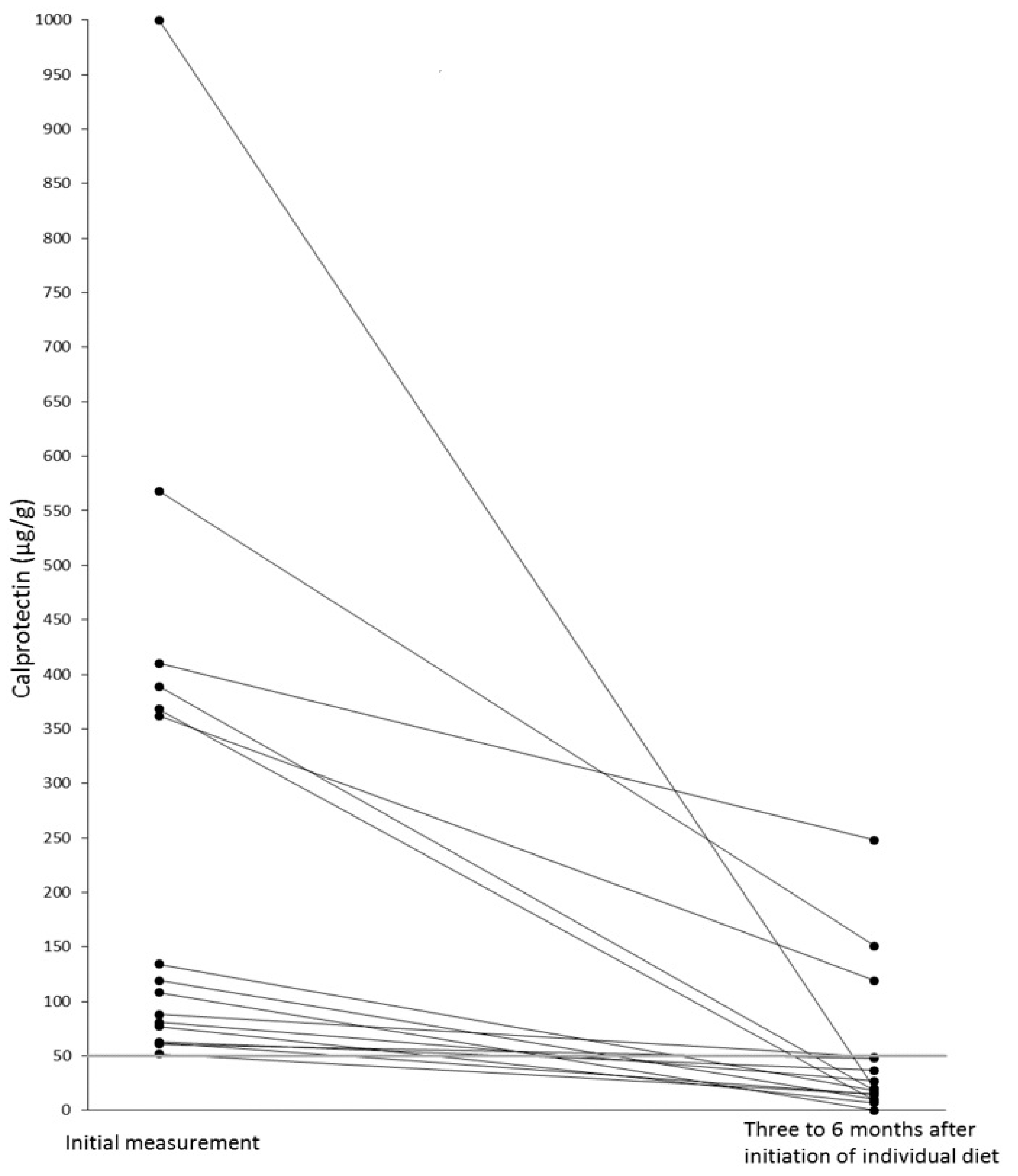
3. Results
4. Discussion
5. Conclusions
Author Contributions
Funding
Institutional Review Board Statement
Informed Consent Statement
Data Availability Statement
Acknowledgments
Conflicts of Interest
Abbreviations
References
- Deputy, M.; Devanaboina, R.; Al Bakir, I.; Burns, E.; Faiz, O. The role of faecal calprotectin in the diagnosis of inflammatory bowel disease. BMJ 2023, 380, e068947. [Google Scholar] [CrossRef] [PubMed]
- Adolph, T.; Meyer, M.; Schwärzler, J.; Mayr, L.; Grabherr, F.; Tilg, H. The metabolic nature of inflammatory bowel diseases. Nat. Rev. Gastroenterol. Hepatol. 2022, 19, 753–767. [Google Scholar] [CrossRef] [PubMed]
- Manceau, H.; Chicha-Cattoir, V.; Puy, H.; Peoc’h, K. Faecal calprotectin in inflammatory bowel diseases: Update and perspectives. Clin. Chem. Lab. Med. 2017, 55, 474–483. [Google Scholar] [CrossRef]
- Campbell, J.P.; Zierold, C.; Rode, A.; Blocki, F.A.; Vaughn, B.P. Clinical performance of a novel LIAISON fecal calprotectin assay for differentiation of inflammatory bowel disease from irritable bowel syndrome. J. Clin. Gastroenterol. 2021, 55, 239–243. [Google Scholar] [CrossRef] [PubMed]
- Camilleri, M. Diagnosis and treatment of irritable bowel syndrome. A review. JAMA 2021, 325, 865–877. [Google Scholar] [CrossRef]
- Schnedl, W.J.; Meier-Allard, N.; Lackner, S.; Enko, D.; Mangge, H.; Holasek, S.J. Increasing expiratory hydrogen in lactose intolerance is associated with additional food intolerance/malabsorption. Nutrients 2020, 12, 3690. [Google Scholar] [CrossRef]
- Schulz, C.; Kupčinskas, J. Review—Helicobacter pylori and non-malignant upper gastrointestinal diseases. Helicobacter 2020, 25 (Suppl. S1), e12738. [Google Scholar] [CrossRef] [PubMed]
- Storhaug, C.L.; Fosse, S.K.; Fadnes, L.T. Country, regional, and global estimates for lactose malabsorption in adults: A systematic review and meta-analysis. Lancet Gastroenterol. Hepatol. 2017, 2, 738–746. [Google Scholar] [CrossRef] [Green Version]
- Lasson, A.; Stotzer, P.O.; Öhman, L.; Isaksson, S.; Sapnara, M.; Strid, H. The intra-individual variability of faecal calprotectin: A prospective study in patients with active ulcerative colitis. J. Crohns Colitis 2015, 9, 26–32. [Google Scholar] [CrossRef] [Green Version]
- Wenzel, R.R.; Datz, C. Association of sprue-like enteropathy and angiotensin receptor-1 antagonists. Wien. Klin. Wochenschr. 2019, 131, 493–501. [Google Scholar] [CrossRef]
- Camilleri, M. Irritable bowel syndrome: Straightening the road from the Rome criteria. Neurogastroenterol. Motil. 2020, 32, e13957. [Google Scholar] [CrossRef] [PubMed]
- Gandhi, A.; Shah, A.; Jones, M.P.; Koloski, N.; Talley, N.J.; Morrison, M.; Holtmann, G. Methane positive small intestinal bacterial overgrowth in inflammatory bowel disease and irritable bowel syndrome: A systematic review and meta-analysis. Gut Microbes 2021, 13, 1933313. [Google Scholar] [CrossRef] [PubMed]
- Savarino, E.; Zingone, F.; Barberio, B.; Marasco, G.; Akyuz, F.; Akpinar, H.; Barboi, O.; Bodini, G.; Bor, S.; Chiarioni, G.; et al. Functional bowel disorders with diarrhoea: Clinical guidelines of the United European Gastroenterology and European Society for Neurogastroenterology and Motility. United Eur. Gastroenterol. J. 2022, 10, 556–584. [Google Scholar] [CrossRef] [PubMed]
- Carrasco-Labra, A.; Lytvyn, L.; Falck-Ytter, Y.; Surawicz, C.M.; Chey, W.D. AGA technical review on the evaluation of functional diarrhea and diarrhea-predominant irritable bowel syndrome in adults (IBS-D). Gastroenterology 2019, 157, 859–880. [Google Scholar] [CrossRef] [PubMed]
- Zollner, A.; Schmiderer, A.; Reider, S.; Oberhuber, G.; Pfister, A.; Texler, B.; Watschinger, C.; Koch, R.; Effenberger, M.; Raine, T.; et al. Faecal biomarkers in inflammatory bowel diseases: Calprotectin versus lipocalin-2-a comparative study. J. Crohns Colitis 2021, 15, 43–54. [Google Scholar] [CrossRef]
- Xiang, B.J.; Jiang, M.; Sun, M.J.; Dai, C. Optimal range of fecal calprotectin for predicting mucosal healing in patients with inflammatory bowel disease: A systematic review and meta-analysis. Visc. Med. 2021, 37, 338–348. [Google Scholar] [CrossRef] [PubMed]
- Jansson-Knodell, C.L.; Krajicek, E.J.; Ramakrishnan, M.; Rogers, N.A.; Siwiec, R.; Bohm, M.; Nowak, T.; Wo, J.; Lockett, C.; Xu, H.; et al. Relationships of intestinal lactase and the small intestinal microbiome with symptoms of lactose intolerance and intake in adults. Dig. Dis. Sci. 2022, 67, 5617–5627. [Google Scholar] [CrossRef]
- Sánchez-Pérez, S.; Comas-Basté, O.; Duelo, A.; Veciana-Nogués, M.T.; Berlanga, M.; Latorre-Moratalla, M.L.; Vidal-Carou, M.C. Intestinal dysbiosis in patients with histamine intolerance. Nutrients 2022, 14, 1774. [Google Scholar] [CrossRef]
- Basu, S.; Liu, C.; Zhou, X.K.; Nishiguchi, R.; Ha, T.; Chen, J.; Johncilla, M.; Yantiss, R.K.; Montrose, D.C.; Dannenberg, A.J. GLUT5 is a determinant of dietary fructose-mediated exacerbation of experimental colitis. Am. J. Physiol. Gastrointest. Liver Physiol. 2021, 321, G232–G242. [Google Scholar] [CrossRef]
- Perets, T.T.M.; Ben Simon, S.; Ashoro, O.; Hamouda, D.; Dickman, R.; Turjeman, S. Lasting effects of helicobacter pylori infection on the microbial communities of patients with and without small intestinal bacterial overgrowth. New Microbiol. 2022, 45, 193–198. [Google Scholar]
- Schnedl, W.J.; Meier-Allard, N.; Schenk, M.; Lackner, S.; Enko, D.; Mangge, H.; Holasek, S.J. Helicobacter pylori infection and lactose intolerance increase expiratory hydrogen. EXCLI J. 2022, 21, 426–435. [Google Scholar] [PubMed]
- Deng, Y.; Misselwitz, B.; Dai, N.; Fox, M. Lactose intolerance in adults: Biological mechanism and dietary management. Nutrients 2015, 7, 8020–8035. [Google Scholar] [CrossRef] [PubMed] [Green Version]
- Enko, D.; Meinitzer, A.; Mangge, H.; Kriegshäuser, G.; Halwachs-Baumann, G.; Reininghaus, E.Z.; Bengesser, S.A.; Schnedl, W.J. Concomitant prevalence of low serum diamine oxidase activity and carbohydrate malabsorption. Can. J. Gastroenterol. Hepatol. 2016, 2016, 4893501. [Google Scholar]
- Jukic, A.; Bakiri, L.; Wagner, E.F.; Tilg, H.; Adolph, T.E. Calprotectin: From biomarker to biological function. Gut 2021, 70, 1978–1988. [Google Scholar] [CrossRef]
- Dolovich, C.; Shafer, L.A.; Vagianos, K.; Witges, K.; Targownik, L.E.; Bernstein, C.N. The complex relationship between diet, symptoms, and intestinal inflammation in persons with inflammatory bowel disease: The Manitoba living with IBD study. JPEN J. Parenter. Enteral Nutr. 2022, 46, 867–877. [Google Scholar] [CrossRef] [PubMed]
- Burns, G.L.; Talley, N.J.; Keely, S. Immune responses in the irritable bowel syndromes: Time to consider the small intestine. BMC Med. 2022, 20, 115. [Google Scholar] [CrossRef]
- Forget, P.; Grandfils, C.; van Cutsem, J.L.; Dandrifosse, G. Diamine oxidase and disaccharidase activities in small intestinal biopsies of children. Pediatr. Res. 1984, 18, 647–649. [Google Scholar] [CrossRef] [Green Version]
- Schnedl, W.J.; Meier-Allard, N.; Michaelis, S.; Lackner, S.; Enko, D.; Mangge, H.; Holasek, S.J. Serum diamine oxidase values, indicating histamine intolerance, influence lactose tolerance breath test results. Nutrients 2022, 14, 2026. [Google Scholar] [CrossRef] [PubMed]
- Kerimi, A.; Gauer, J.S.; Crabbe, S.; Cheah, J.W.; Lau, J.; Walsh, R.; Cancalon, P.F.; Williamson, G. Effect of the flavonoid hesperidin on glucose and fructose transport, sucrase activity and glycaemic response to orange juice in a crossover trial on healthy volunteers. Br. J. Nutr. 2019, 121, 782–792. [Google Scholar] [CrossRef]


| Food Intolerance/Malabsorption | Number (n) of Patients | Percent (%) of Patients | Number (n) of Patients with High FCAL | Percent (%) of Patients with High FCAL |
|---|---|---|---|---|
| Number of patients | 228 | 100 | 39 | 17.1 |
| LIT-only | 49 | 21.5 | 14 | 28.6 |
| FM-only | 22 | 9.6 | 3 | 13.6 |
| HIT-only | 29 | 12.7 | 6 | 20.7 |
| H. pylori-only | 10 | 4.4 | 0 | 0 |
| LIT + FM | 18 | 7.9 | 2 | 11.1 |
| LIT + FM + HIT | 21 | 9.2 | 1 | 4.8 |
| LIT + HIT | 34 | 14.9 | 5 | 14.7 |
| LIT + H. pylori | 14 | 6.1 | 4 | 28.6 |
| LIT + HIT + H. pylori | 4 | 1.7 | 0 | 0 |
| LIT + FM + H. pylori | 5 | 2.2 | 0 | 0 |
| LIT + FM + HIT + H. pylori | 2 | 0.9 | 0 | 0 |
| FM + HIT | 11 | 4.8 | 1 | 9.1 |
| FM + HIT + H. pylori | 5 | 2.2 | 0 | 0 |
| FM + H. pylori | 1 | 0.4 | 1 | 100 |
| HIT + H. pylori | 2 | 0.9 | 1 | 50 |
| Candesartan | 1 | 0.4 | 1 | 100 |
Disclaimer/Publisher’s Note: The statements, opinions and data contained in all publications are solely those of the individual author(s) and contributor(s) and not of MDPI and/or the editor(s). MDPI and/or the editor(s) disclaim responsibility for any injury to people or property resulting from any ideas, methods, instructions or products referred to in the content. |
© 2023 by the authors. Licensee MDPI, Basel, Switzerland. This article is an open access article distributed under the terms and conditions of the Creative Commons Attribution (CC BY) license (https://creativecommons.org/licenses/by/4.0/).
Share and Cite
Schnedl, W.J.; Michaelis, S.; Enko, D.; Mangge, H. Fecal Calprotectin Elevations Associated with Food Intolerance/Malabsorption Are Significantly Reduced with Targeted Diets. Nutrients 2023, 15, 1179. https://doi.org/10.3390/nu15051179
Schnedl WJ, Michaelis S, Enko D, Mangge H. Fecal Calprotectin Elevations Associated with Food Intolerance/Malabsorption Are Significantly Reduced with Targeted Diets. Nutrients. 2023; 15(5):1179. https://doi.org/10.3390/nu15051179
Chicago/Turabian StyleSchnedl, Wolfgang J., Simon Michaelis, Dietmar Enko, and Harald Mangge. 2023. "Fecal Calprotectin Elevations Associated with Food Intolerance/Malabsorption Are Significantly Reduced with Targeted Diets" Nutrients 15, no. 5: 1179. https://doi.org/10.3390/nu15051179