Article

Peptidomic Characterization and Amino Acid Availability after Intake of Casein vs. a Casein Hydrolysate in a Pig Model

by
Pablo Jiménez-Barrios
1,
Laura Sánchez-Rivera
1,
Daniel Martínez-Maqueda
1,
Yann Le Gouar
2,
Didier Dupont
2,
Beatriz Miralles
1 and
Isidra Recio
1,*
1
Instituto de Investigación en Ciencias de la Alimentación, CIAL (CSIC-UAM, CEI UAM+CSIC), Nicolás Cabrera, 9, 28049 Madrid, Spain
2
Institut National de Recherche Pour l’Agriculture, l’Alimentation et l’Environnement (INRAE) STLO Agrocampus Ouest, 35042 Rennes, France
*
Author to whom correspondence should be addressed.
Nutrients 2023, 15(5), 1065; https://doi.org/10.3390/nu15051065
Submission received: 25 January 2023 / Revised: 16 February 2023 / Accepted: 17 February 2023 / Published: 21 February 2023

Abstract

:
It is known that casein hydrolysis accelerates gastrointestinal transit in comparison to intact casein, although the effect of the protein hydrolysis on the composition of the digests is not fully understood. The aim of this work is to characterize, at the peptidome level, duodenal digests from pigs, as a model of human digestion, fed with micellar casein and a previously described casein hydrolysate. In addition, in parallel experiments, plasma amino acid levels were quantified. A slower transit of nitrogen to the duodenum was found when the animals received micellar casein. Duodenal digests from casein contained a wider range of peptide sizes and a higher number of peptides above five amino acids long in comparison with the digests from the hydrolysate. The peptide profile was markedly different, and although β-casomorphin-7 precursors were also found in hydrolysate samples, other opioid sequences were more abundant in the casein digests. Within the same substrate, the evolution of the peptide pattern at different time points showed minimal changes, suggesting that the protein degradation rate relies more on the gastrointestinal location than on digestion time. Higher plasma concentrations of methionine, valine, lysine and amino acid metabolites were found in animals fed with the hydrolysate at short times (<200 min). The duodenal peptide profiles were evaluated with discriminant analysis tools specific for peptidomics to identify sequence differences between both substrates that can be used for future human physiological and metabolic studies.

1. Introduction

Milk is an excellent source of highly digestible proteins in human nutrition. It is well known that the speed of protein digestion and amino acid absorption is affected by the protein nature and the technological treatment applied, and protein digestion kinetics modulate the metabolic response. While the casein fraction of milk, so-called slow protein, coagulates in the stomach and promotes postprandial protein deposition, whey proteins induce a fast increase in plasma amino acids, protein synthesis, and oxidation [1]. Small changes in processing have also been found to affect the kinetics of protein digestion. For instance, dairy gels differing in the mode of coagulation (acidification vs. renneting) cause different compositions of porcine duodenal effluents and plasma amino acids [2].
Enzymatic hydrolysis is increasingly used in the dairy sector for the design of ingredients for infant formulas [3]. In addition, casein hydrolysates have demonstrated to stimulate insulin secretion and to show glycemic control properties in both animals and humans [4,5]. A diet containing hydrolyzed casein has been reported to enhance upper gastrointestinal transit and to increase ileal microbial carbohydrate metabolism in comparison with an intact casein-based diet [6]. However, the influence of the enzymatic hydrolysis of the casein fraction of milk on gastric emptying and composition of the gastrointestinal contents is not fully understood. A previous study with aged rats found that casein or whey hydrolysates do not alter gastrointestinal transit compared with their respective intact counterparts. The slowed gastrointestinal transit of casein was reversed with opioid inhibitors, suggesting the involvement of opioid receptors [7]. Similarly, protein hydrolysis has been described to reduce the gastrointestinal transit time in preterm infants [8]. In rats, this effect was also attributed to the reduction of casein-derived opioid receptor agonists [9].
The advances in peptidomic techniques based on mass spectrometry (MS) have allowed for study of the complex protein digestome at various regions of the gastrointestinal tract [10,11]. These techniques have widely been used to characterize the large variety of peptides generated by the gastrointestinal proteases and peptidases, mainly in vitro [12,13,14,15]. However, in recent years, the characterization of gastrointestinal effluents from human or pig, as an accepted model of human digestion, is building a valuable peptide repertoire, especially after oral administration of milk proteins, to be used in physiological and metabolic studies or to validate in vitro digestion protocols [16,17,18].
The aim of this work is to characterize the peptidome of duodenal digests obtained in cannulated pigs after the oral administration of micellar casein and a casein hydrolysate. Different time points from 5 to 150 min were considered to evaluate kinetics of protein digestion in the duodenum. In addition, in parallel experiments, quantitative analysis of plasma amino acids was performed to assess the impact of protein hydrolysis on amino acid availability. Because pig is recognized as an excellent model of the human gastrointestinal tract, the results offer new information about the faster digestion and quicker availability of amino acids of protein hydrolysates used in human nutrition.

2. Materials and Methods

2.1. Animal Experiment

All procedures were in accordance with the European Community guidelines for the use of laboratory animals (L358-86/609/EEC). The local committee for ethics in animal experimentation approved the study. The animals were 6 Large White × Landrace × Piétrain pigs of about 40 kg live weight. Two weeks before starting the trials, they were surgically fitted with a T-shaped cannula in the duodenum (10 cm downstream from the pylorus). Catheterization of the external jugular vein was also performed. Following the operation, the animals were housed in individual slatted pens within a ventilated room with controlled temperature (21 °C). During convalescence following the surgical procedure and also between the sampling days, pigs were fed with 800 g/d of a pig feed concentrate (Cooperl Arc Atlantique®, Pelstan, France) containing 16% proteins, 1% fat, 4% cellulose and 5% mineral matter and had free access to water.
The test meals were commercial micellar casein (Prodiet 85B, batch no. 141179) purchased from Ingredia S. A., Arras, France, and a pepsin hydrolysate from this casein. The hydrolysate was prepared with food grade pepsin (P389P/3 Biocatalyst 3000 U/mg) at 2% enzyme/substrate ratio (w/w), added at 0 and 3 h during a hydrolysis time of 6 h at 37 °C, as previously reported [19]. The nitrogen content of casein and the hydrolysate was 77.8 and 77.5%, respectively, as determined by Kjeldahl.
The collection of duodenal effluents and blood samples was performed in separate assays. In each one, both dairy products were randomly tested on each animal. Within a period (2 weeks), the days of sampling were separated by at least 2 day. Test meals, casein and the casein hydrolysate (129 g of powder, on the basis of protein content) were reconstituted in water (250 mL) and offered to overnight fasted pigs for 10 min. The pigs had no access to water until 4 h after the meal delivery. Duodenum effluents were collected in plastic bottles 15 min before and 5, 10, 15, 20, 30, 45, 60, 90, 120, and 150 min after ingestion of test meal. The sampling was stopped when 40 mL was collected or after a maximum of 3 min of sampling time. A protease inhibitor (Pefabloc® at 2.5 mM final concentration) was previously added to the sample tubes. Collected effluents were weighed, freeze-dried and stored at −20 °C.
Blood sampling was performed at 15 min before and 5, 20, 45 min, 1, 2, 3, 4 and 6 h after ingestion of the casein or its hydrolysate. Blood samples (5 mL) were collected in heparinized-lithium tubes where a protease inhibitor cocktail for plasma samples was previously added (1 tablet/10 mL blood) (Complete Mini, EDTA-free, Roche Farma, Madrid, Spain). Then, centrifugation at 3000× g 4 °C for 10 min was performed, and the supernatant was frozen with liquid nitrogen and stored at −80 °C. Amino acids and related metabolites in plasma were determined by ion-exchange chromatography with ninhydrine at the Diagnosis of Molecular Diseases Centre from the Autonomous University of Madrid. Briefly, 2 mL of plasma was deproteinized with 200 μL 10% trichloroacetic acid. After incubation for 15 min on ice, the samples were centrifuged for 10 min at 1500× g and 4 °C, and 150 μL of supernatant was collected. The samples were placed in an autosampler protected from light, and 20 μL was injected.

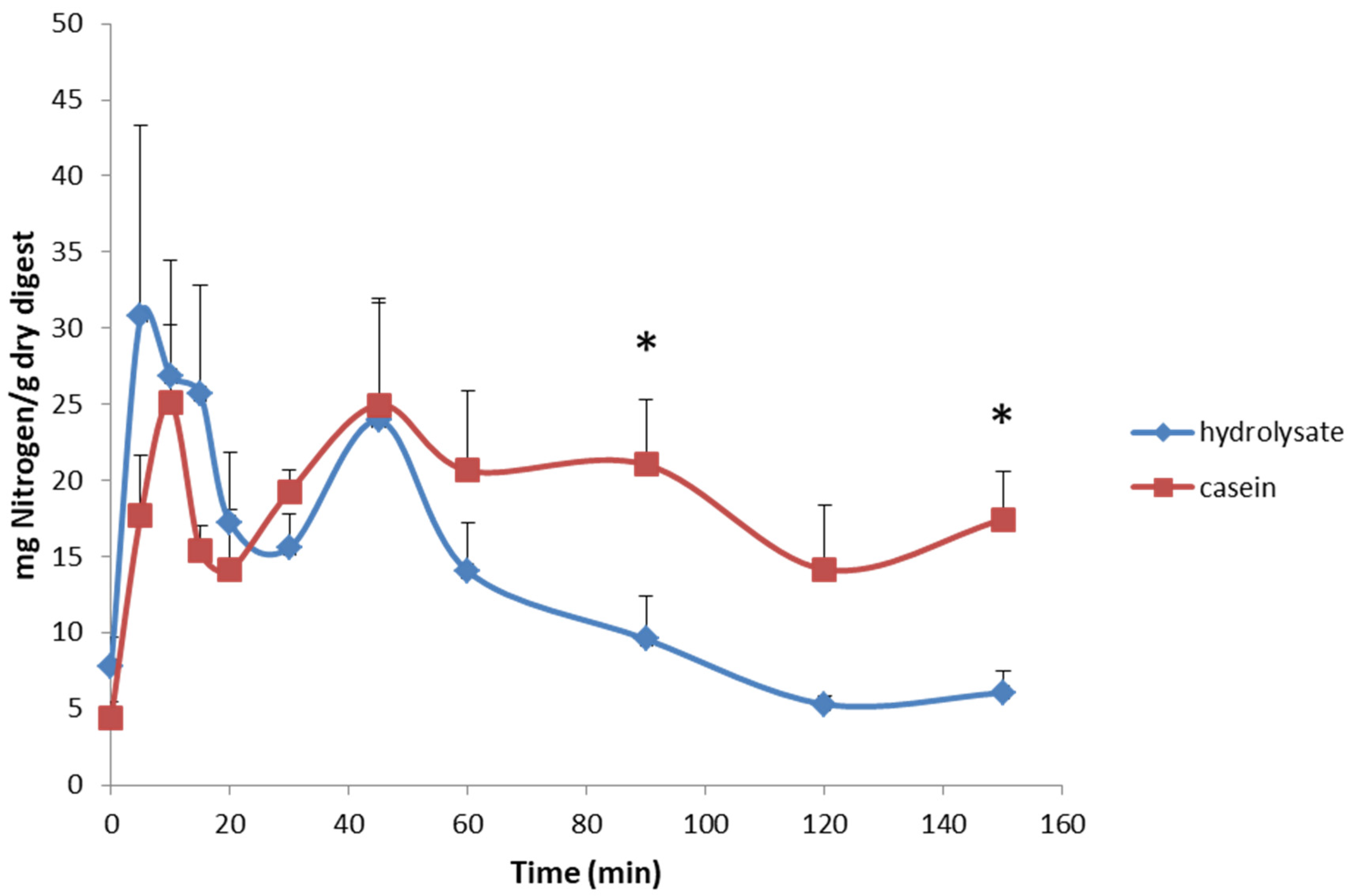
2.2. Nitrogen Analysis and SDS-PAGE

Nitrogen content of duodenal effluents was determined by elemental analysis in a LECO chns-932 analyzer at the Elemental analysis unit from the Interdepartmental Investigation Service from the Autonomous University of Madrid. Freeze-dried digests were adjusted to 1 mg of protein/mL of sample buffer (tris-HCL 0.05 M pH 6.8, SDS 1.6% w:v, glycerol 8% v:v, bromophenol blue 0.002% w:v, and β-mercaptoethanol 2% v:v) based on the nitrogen content, and the SDS-PAGE was conducted in Precast Criterion XT 12% Bis-Tris gels (Bio-Rad, Richmond, CA, USA) using the running buffer (19:1, v/v water/XT MES 20×; Bio-Rad Laboratories, Richmond, CA, USA). A molecular weight marker (Precision Plus ProteinTM Unstained standard, Bio-Rad Laboratories, Richmond, CA, USA) was used. A prior running at 100 V for 5 min was performed, and then, samples were run at 150 V for 45 min. Gel was rinsed with water, then stained with Coomasie Blue (Instant blue; Expedeon, Swavesey, UK) for 30 min, and rinsed again with water for 10 min.

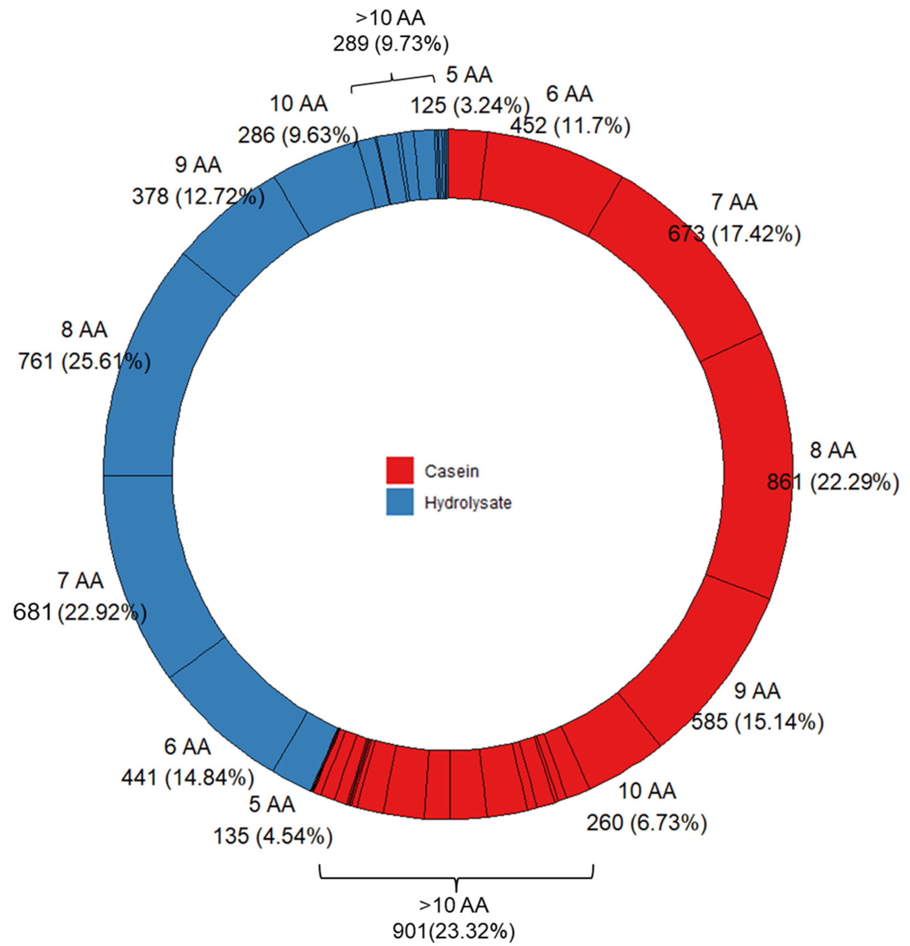
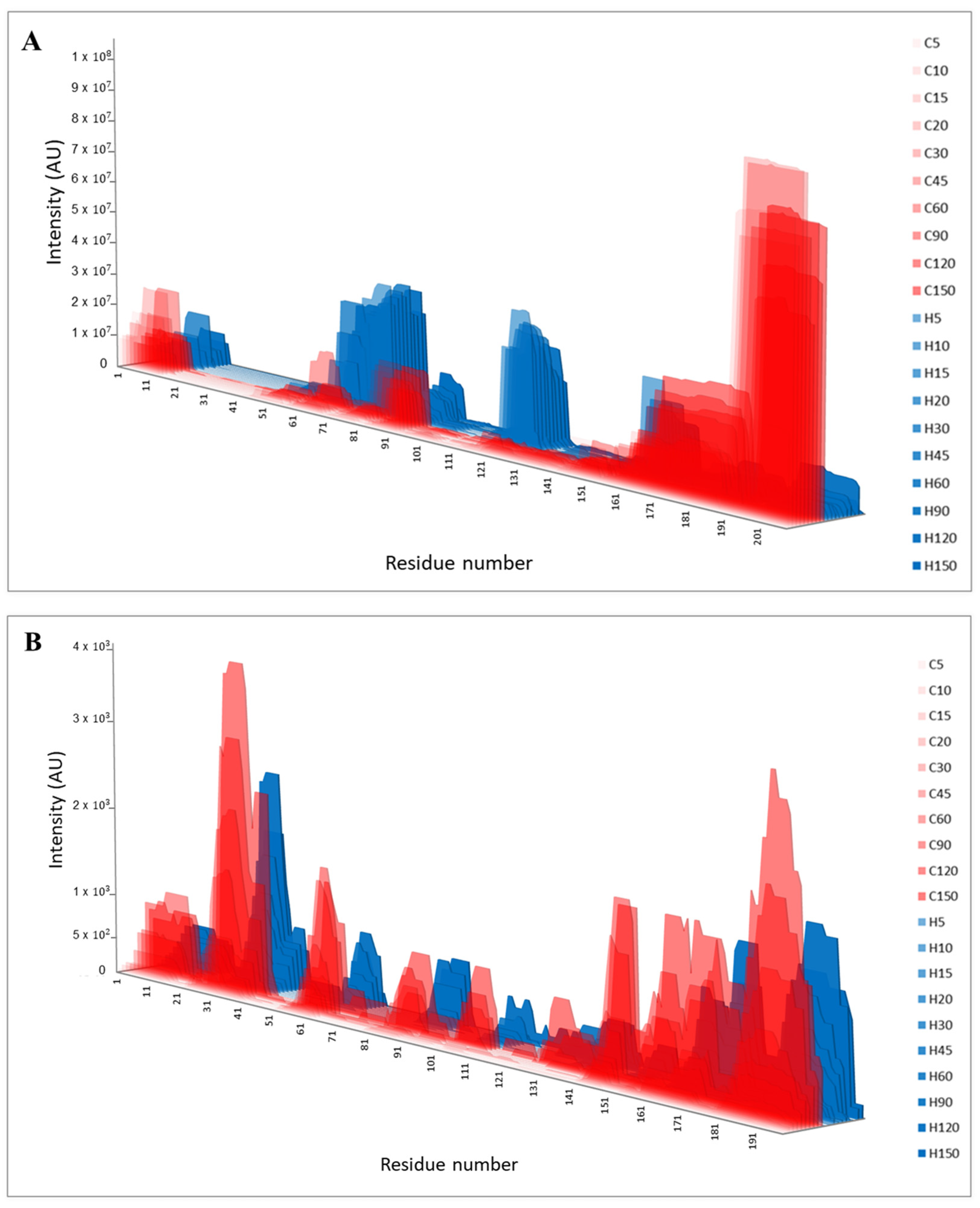
2.3. Peptidomic Analysis

Tandem mass spectrometry (HPLC-MS/MS) was conducted on an Agilent 1100 HPLC system (Agilent Technologies, Waldbron, Germany) coupled to an Esquire 3000 ion trap mass spectrometer (Bruker Daltonics, Bremen, Germany) as previously described [20]. A linear gradient from 0 to 45% solvent B (100:0.1, v/v acetonitrile:formic acid) for 120 min on a Mediterranean Sea C18 column, 150 × 2.1 mm (Teknokroma, Barcelona, Spain) was used at 0.2 mL/min. The freeze-dried digests were reconstituted in solvent A (100:0.1, v/v water:formic acid) and the supernatants after centrifugation at 13,000× g, for 10 min were analyzed. Spectra were recorded over the mass/charge (m/z) range 100–1200, and the target mass was set at 900 m/z. Data processing was conducted using the software Data Analysis, version 4.0 and Biotools, version 3.2 from Bruker Daltonics. MASCOT V2.4 (MatrixScience) was used for peptide identification with the use of homemade databases of major genetic variants of bovine caseins and porcine endogenous proteins. The parameters selected were: no enzyme, no post-translational modifications, and peptide tolerance 0.1% and 0.5 Da for MS and MS/MS, respectively. A manual revision for each identification was performed, regardless of the p value.
To carry out most peptidomic analysis steps, R scripts were used. R scripts are some lines of code using R programing language. Peptide size distribution of all samples was calculated. Peptide sequences were divided by substrate and then subdivided by size. Raw numbers and their percentages were shown using the R package “ggplot2” version 3. 4.0 [21]. Peptides previously grouped by substrate were analyzed using the R package “ggseqlogo” version 0.1 to obtain the SeqLogo figure [22]. The sequence logo shows the probability to find a particular amino acid at the N-terminal (1, 2, 3) or C-terminal (−3, −2, −1) end of the sequence. In the case of five-length peptides, the amino acid in the 3rd position at both terminal ends was the same.

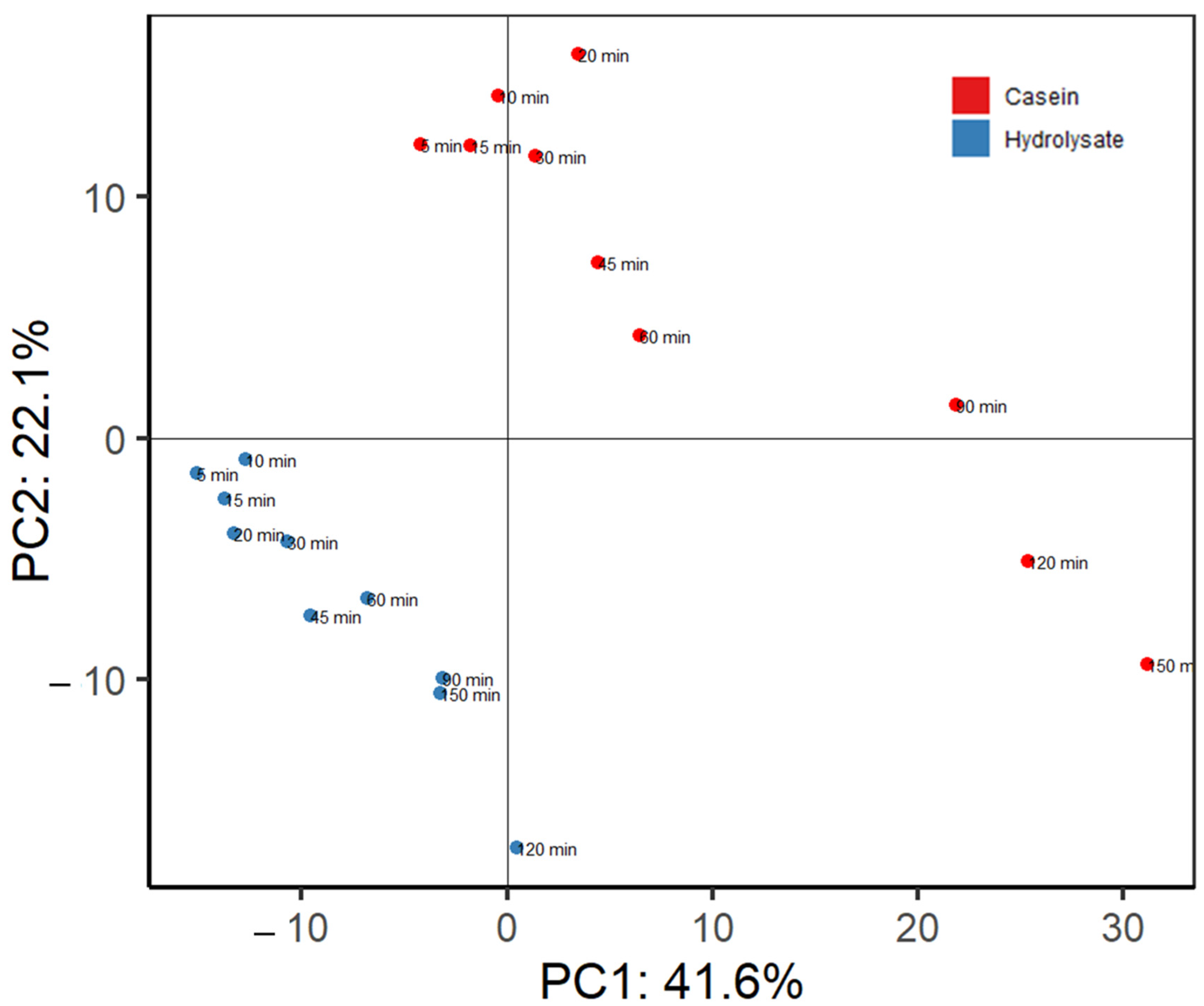
2.4. Statistical Analysis

Principal components analysis (PCA) was performed using the amino acids within the identified peptides as variables. For each amino acid and each time point, the value was calculated as the sum of intensities of all overlapping peptides. A hierarchical clustering tree of samples using the intensity of the peptides was built by PermutMatrix software version 1.9.3.0 [23]. Peptides present in at least two time points were used. Blood samples were analyzed using GraphPad Prism software version 8.0.2. Differences between substrates over time were found through a two-way ANOVA, followed by the Bonferroni test. The results were graphed with the R package “ggplot2”version 3.4.0 [21].

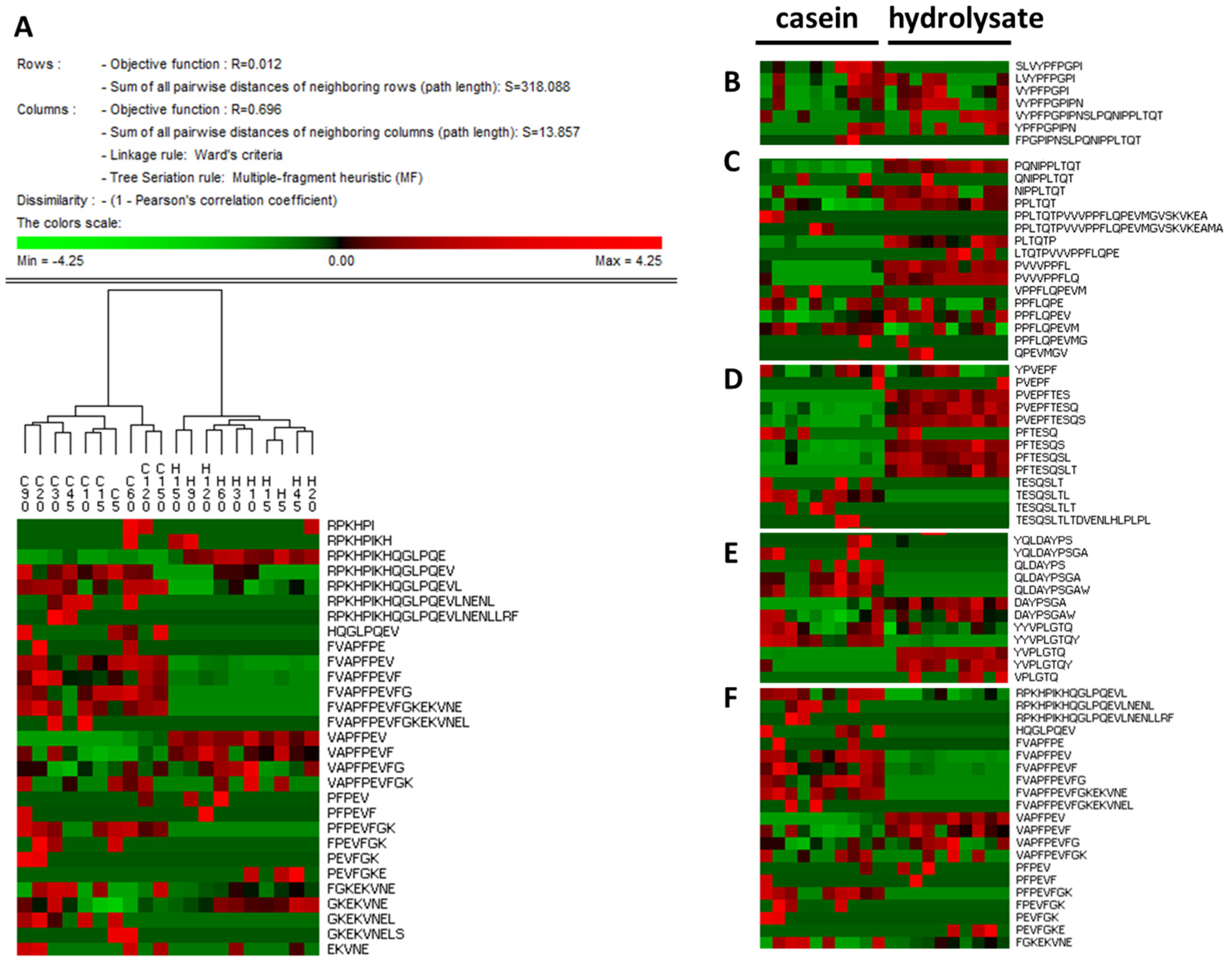
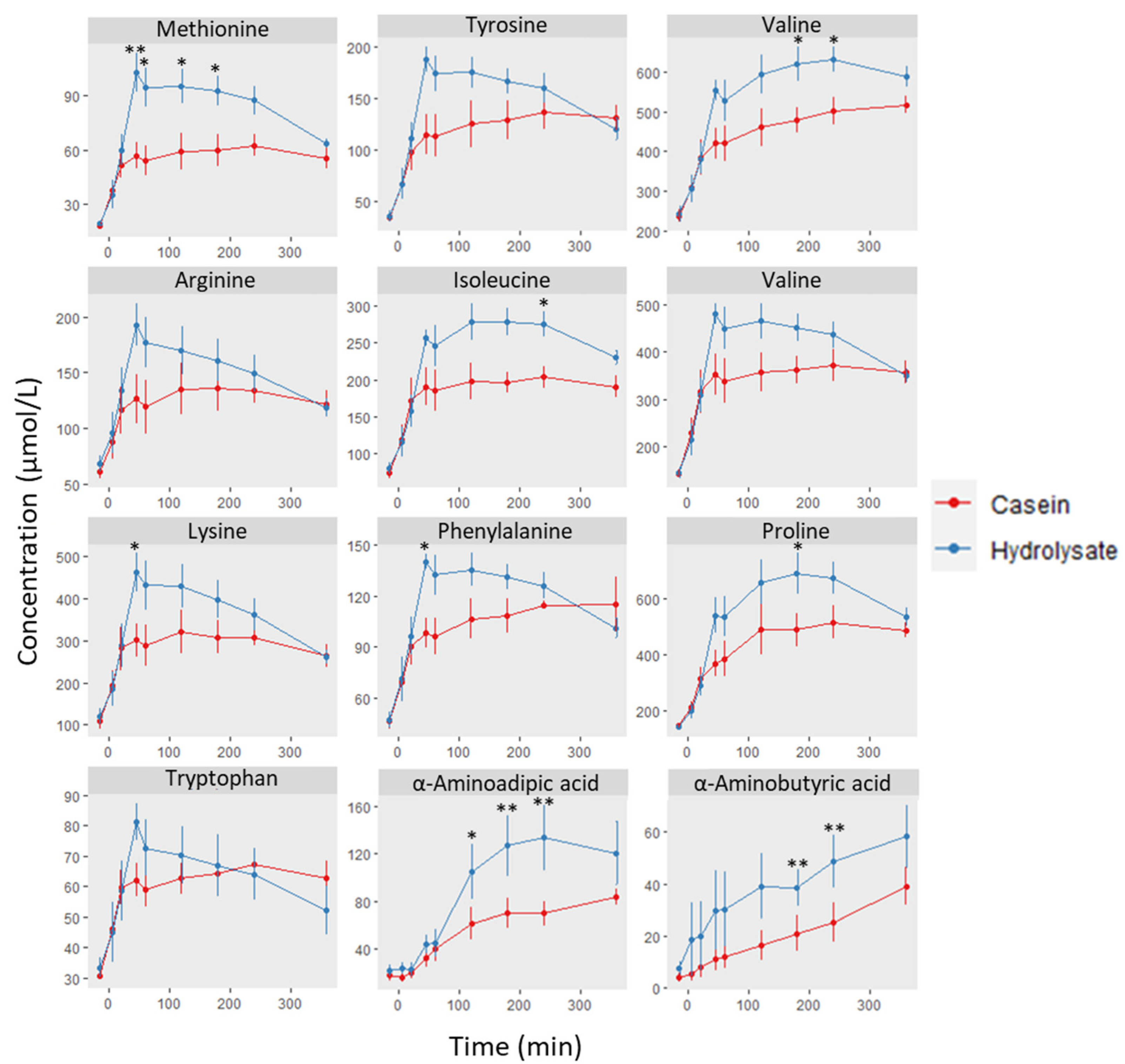
3. Results and Discussion

Duodenal contents after oral administration of casein and its hydrolysate were collected and freeze-dried until analysis, and the nitrogen content of the dried effluents is shown in Figure 1. Duodenal digests from casein had higher nitrogen content than those from the hydrolysate at 90 and 150 min, showing its emptying over a more extended period, compared to the hydrolysate. Although at short time points (<30 min), hydrolysate effluents showed higher protein content than casein ones, due to the high variability between animals, differences did not reach significance. Because the effluents were immediately frozen in liquid nitrogen and freeze-dried, flow rates of liquid effluents could not be measured, and differences in the nitrogen content between substrates could be diminished. Previous reports in aged rats have shown unaltered gastrointestinal transit with hydrolyzed casein compared with its intact counterpart [7]. Casein was emptied from the stomach mainly in the form of peptides, although in some time points (15, 45, 60 min), slight bands of intact casein fractions were visible by SDS-PAGE (Figure S1, Supplementary Material). In addition, because the casein substrate contained a small amount of whey proteins (β-lactoglobulin and α-lactalbumin), bands corresponding to these proteins were also detected up to 30 min after oral administration. Due to their high solubility, whey proteins are rapidly emptied from the stomach [1], and the net disappearance of β-lactoglobulin from human upper jejunum was reported to be faster than for casein [24]. Similarly, Barbé and co-workers found β-lactoglobulin in porcine duodenum after the administration of raw liquid milk [25]. In contrast to casein, the electrophoretic protein profile of the porcine duodenal effluents after the hydrolysate administration slightly varied with time, and all protein material detected was below 10 kDa.
The peptide fraction of the duodenal effluents was characterized by HPLC-MS/MS. A total of 3863 milk-derived peptides, around 400 peptides per time point, were identified in porcine duodenum after the administration of casein, and a lower number, ca. 300 per time point, was found after the administration of the hydrolysate (Table 1). This lower number in the case of the hydrolysate can be due to the limitation of our mass spectrometry conditions to identify peptides shorter than five amino acids. In addition, Table 1 shows the number of common peptides found in all animals (n = 6) to illustrate the inter-individual variability found. When using casein as feeding, the peptide profile showed a large variety of peptide sizes where ca. 25% were above 10 amino acids long. However, as expected, the peptides longer than 10 amino acids were below 10% when the protein was administered in the form of hydrolysate (Figure 2). This peptide size distribution matches with the information provided by SDS-PAGE (Figure S1, Supplementary Material). The most abundant peptides identified in both substrates corresponded to peptides between six and nine amino acids, but this can be caused by the selectivity of the MS method employed to detect peptides within this size range. Despite this, the peptide profile found in the duodenum after the administration of both substrates was notably different (Figure 3A,B). After casein administration, the most abundant β-casein-derived peptides belonged to the C-terminal domain of the protein, i.e., 193–209, while peptides in regions 58–80, and 115–126 from β-casein are specifically overrepresented in the duodenal effluents from the hydrolysate (Figure 3A). Other authors have also reported the resistance of the C-terminal region of β-casein to gastric pepsin both in simulated gastric digestions [26,27] and in pig duodenum [18]. The slower gastrointestinal transit of casein in comparison with soy has been attributed to the involvement of opioid peptides since it was reversed with opioid inhibitors [7]. Although some studies have found an accelerated gastrointestinal transport with hydrolyzed protein infant formula due to the reduction of opioid agonists [8,9], the presence of opioid sequences depends on the hydrolysis conditions (enzyme specificity, time…). Under our hydrolysis conditions, the regions corresponding to β-casein opioid peptides, i.e., β-casomorphin-7 (60YPFPGPI66) [28] and neocasomorphin-6 (114YPVEPF119) [29] were preserved, and these peptides or precursors thereof comprised highly overrepresented regions in the duodenal effluents from the hydrolysate. For the other main casein fraction, αs1-casein, differences in the duodenal peptide profile between substrates were less marked (Figure 3B), but the hydrolysate effluents showed a higher abundancy of regions 24–33, 166–172, and 180–196. The domains corresponding to previously reported opioid peptides (α-casomorphins), i.e., αs1-casein 90RYLGYLE96 [30] and 144YFYPEL149 [31], were more abundant in casein duodenal effluents than in hydrolysate ones. Other αs1-casein abundant domains, such as 24–33, were common for both substrates. The compared peptide profiles for αs2-casein and κ-casein are shown in Figure S2A,B, where clear differences between both substrates were also found.
In order to illustrate the evolution of the peptide profile with time, peptides were also aligned within the sequence of the main casein fractions, β-casein and αs1-casein, and represented in the form of heatmaps (Figure S3A–D, Supplementary material). These graphs are based in the appearance frequency of each amino acid without taking into account the peptide intensity. Again, the C-terminal region of β-casein corresponded to the domain where a higher number of peptides was identified after casein administration, but for hydrolysate effluents, regions 70–100 and 115–125 were especially rich in peptides. Within the same substrate, differences in the peptide profile with time were less relevant, concluding that protein digestion occurs along the gastrointestinal tract, and for a given part, protein degradation is similar. Similar results were previously reported in human jejunum [16,17] or in different regions of the porcine gastrointestinal tract [18]. In addition to milk-derived peptides, the HPLC-MS/MS analysis of the effluents allowed us to identify several peptides from endogenous proteins (Table S1, Supplementary Material). The main identified proteins corresponded to endogenous enzymes: pepsin, chymotrypsin, carboxypeptidases, dipeptidyl peptidase or lipases, but also other peptides belonged to the sequence of gastrointestinal hormones, mucins or ubiquitous proteins, such as albumin.
To compare the peptide composition of the effluents for both substrates at different time points in a holistic way, different techniques of discriminant analysis for peptidomics were used. Figure 4 shows the principal component analysis built with the peptide intensity values used in Figure 3 and Figure S3 (including β-, αs1-, αs2, and κ-casein). The first component explained 41.6% of the variance while PC2 reached 22.1%. The peptide intensity profiles at the different time points were clearly grouped per substrate, demonstrating the differences shown in Figure 3 and the Supplementary Material. In addition, effluents obtained at longer time points, i.e., 90, 120 and 150 min, were separated from those at shorter times, and this effect was observed in both substrates, although it was more notable for casein duodenal effluents.
The free software PermutMatrix was used to perform a graphical analysis of the peptide profiles based on sequence and intensity. The dendrogram in Figure 5 shows the clustering of samples per substrate, casein and hydrolysate. In addition, this analysis allows for the identification of the peptide differences in those regions overrepresented in each substrate. For instance, β-casomorphin precursors with valine or tyrosine at the N-terminal were highly detected in the effluents from the hydrolysate, but specific sequences, such as 57SLVYPFPGPI66 or 58LVYPFPGPI66, were more abundant in casein samples (Figure 5B). Similarly, β-casein regions containing the antihypertensive tri-peptides IPP and VPP [32] were overrepresented in the hydrolysate duodenal digests, but several related peptides found in the casein samples showed a different cleavage pattern with proline–proline at the N-terminal end (Figure 5C). The peptide known as neocasomorphin 114YPVEPF119 was found in duodenal digests from both casein and hydrolysate, although several peptides lacking the N-terminal tyrosine were especially abundant in the hydrolysate samples (Figure 5D). Because the presence of tyrosine at the N-terminal end is a structural requirement for opioid receptor agonists, the loss of this amino acid in the hydrolysate digests suggested a decrease in their opioid effect. Other regions where differences between substrates were notable corresponded to αs1-casein 154–173 (Figure 5E) and the αs1-casein N-terminal region (Figure 5F).
The analysis of the cleavage positions for both samples was carried out with ggseqlogo to visualize the most likely amino acid found at each of the three N- and C-terminal positions for both substrates (Figure S4, Supplementary Material). While for casein digests, the most probably N-terminal residue is a tyrosine, for hydrolysate digests, it is proline followed by tyrosine. Similarly, the most abundant C-terminal tripeptide is PPL and PQQ for casein and hydrolysate, respectively.
Plasma amino acids were also quantified over time of digestion. In addition to a baseline sample, blood was taken 5, 20, 45, 60, 120, 180, 240 and 360 min after test meal intake. As expected, a greater increase in total amino acids in plasma was observed with the hydrolysate from 45 to 240 min (Figure S5, Supplementary Material), but the values converged at 360 min. Previous reports in humans have described a faster rate of absorption with hydrolyzed casein than with intact casein, although similar whole-body postprandial retention of dietary nitrogen was observed [33]. Moreover, hydrolyzed casein did not enhance ileal endogenous protein losses in humans when compared with the parent protein [34]. When considering the dairy matrix of iso-protein products in the postprandial blood amino acid concentrations in human volunteers, the effect of the protein structure was evidenced as extended response curves with a micellar casein isolate as opposed to a whey protein isolate and yoghurt [35].
Figure 6 shows the determinations of selected amino where significant differences were shown at short times in the case of methionine, lysine or phenylalanine, peaking at 45 min. Other amino acids and metabolites are shown in the Supplementary Material Figure S6. Differences in the 2–3 h post-ingestion period were observed for valine, isoleucine, proline, and two metabolites: α-aminoadipic and α-aminobutyric acids. These values are in accordance with previous results in humans [33] where indispensable amino acids were significantly higher at short times after hydrolyzed casein intake compared to intact casein.

4. Conclusions

Our results confirm the emptying of intact casein over a more extended period, compared to casein hydrolysate. Discriminant analysis showed a markedly different peptide profile of duodenal digests for all casein fractions between both substrates. In the opioid peptide β-casomorphin-7, precursors with differing terminal ends were observed, with a prevalence of the VYPF- and YPF-ends in the hydrolysate, whereas some longer precursors were more abundant in the casein digests. For other opioid peptides, such as neocasomorphin 114YPVEPF119, an inactive form lacking the N-terminal tyrosine, prevailed in the hydrolysate samples. The peptide profile at different time points within the same substrate was similar, suggesting that the degree of protein degradation of the material reaching the duodenum is similar. Finally, the amino acid concentrations found in plasma confirmed a more rapid absorption in hydrolysate-fed animals, with several branched-chain and other hydrophobic amino acids peaking at shorter times than casein. These results can be applied to dairy ingredients formulated as hydrolysates or partly hydrolyzed products, such as infant formula or products used for immune, metabolic or hormonal effects.

Supplementary Materials

The following supporting information can be downloaded at: https://www.mdpi.com/article/10.3390/nu15051065/s1, Figure S1: Electrophoretic profile of pig duodenal effluents at different times of digestion after intake of casein and hydrolysate compared with the given substrate; Figure S2: Identified peptides in pig duodenal digests after intake of casein or hydrolysate over collection time represented by area graphs; Figure S3: Alignment of peptides identified in pig duodenal effluents collected at different times of digestion after intake of casein or hydrolysate and represented as heatmaps of β-casein and αS1-casein; Figure S4: SeqLogo analysis of terminal amino acid residues corresponding to the peptides identified in pig duodenal effluents at all times of digestion after intake of casein or hydrolysate; Figure S5: Total amino acids and metabolites determined in plasma after intake of casein or hydrolysate in the pig model; Figure S6: Concentration of amino acids in plasma of pigs (n = 6) at baseline (−15 min) and 5, 20, 45, 60, 120, 180, 240 and 360 min after intake of casein or hydrolysate. Table S1: Pig endogenous proteins identified in the analyzed duodenal effluents. Full PermutMatrix results are shown as an attached figure in .png format.

Author Contributions

Conceptualization, I.R. and B.M.; methodology, Y.L.G. and P.J.-B.; software, P.J.-B.; formal analysis, P.J.-B., L.S.-R. and D.M.-M.; investigation, P.J.-B., L.S.-R., D.D. and D.M.-M.; resources, D.D., Y.L.G. and I.R.; data curation, P.J.-B.; writing—original draft preparation, P.J.-B. and I.R.; writing—review and editing, P.J.-B., B.M. and I.R.; visualization, P.J.-B.; supervision, B.M. and I.R.; project administration, B.M. and I.R.; funding acquisition, I.R. All authors have read and agreed to the published version of the manuscript.

Funding

This work has received financial support from grants PID2019-107663RB-I00 and PDC2022-133489-I00 funded by MCIN/AEI/10.13039/501100011033.

Institutional Review Board Statement

The local Committee for Ethics in Animal Experimentation approved the study (CE15-10, Aubière, France).

Informed Consent Statement

Not applicable.

Data Availability Statement

Not applicable.

Conflicts of Interest

The authors declare no conflict of interest.

References

  1. Boirie, Y.; Dangin, M.; Gachon, P.; Vasson, M.-P.; Maubois, J.-L.; Beaufrère, B. Slow and Fast Dietary Proteins Differently Modulate Postprandial Protein Accretion. Proc. Natl. Acad. Sci. USA 1997, 94, 14930–14935. [Google Scholar] [CrossRef] [PubMed] [Green Version]
  2. Barbé, F.; Ménard, O.; Gouar, Y.L.; Buffière, C.; Famelart, M.-H.; Laroche, B.; Feunteun, S.L.; Rémond, D.; Dupont, D. Acid and Rennet Gels Exhibit Strong Differences in the Kinetics of Milk Protein Digestion and Amino Acid Bioavailability. Food Chem. 2014, 143, 1–8. [Google Scholar] [CrossRef] [PubMed]
  3. Vandenplas, Y.; Alarcon, P.; Fleischer, D.; Hernell, O.; Kolacek, S.; Laignelet, H.; Lönnerdal, B.; Raman, R.; Rigo, J.; Salvatore, S.; et al. Should Partial Hydrolysates Be Used as Starter Infant Formula? A Working Group Consensus. J. Pediatr. Gastroenterol. Nutr. 2016, 62, 22–35. [Google Scholar] [CrossRef] [PubMed] [Green Version]
  4. Jonker, J.T.; Wijngaarden, M.A.; Kloek, J.; Groeneveld, Y.; Gerhardt, C.; Brand, R.; Kies, A.K.; Romijn, J.A.; Smit, J.W.A. Effects of Low Doses of Casein Hydrolysate on Post-Challenge Glucose and Insulin Levels. Eur. J. Intern. Med. 2011, 22, 245–248. [Google Scholar] [CrossRef]
  5. Drummond, E.; Flynn, S.; Whelan, H.; Nongonierma, A.B.; Holton, T.A.; Robinson, A.; Egan, T.; Cagney, G.; Shields, D.C.; Gibney, E.R.; et al. Casein Hydrolysate with Glycemic Control Properties: Evidence from Cells, Animal Models, and Humans. J. Agric. Food Chem. 2018, 66, 4352–4363. [Google Scholar] [CrossRef]
  6. Shen, J.; Mu, C.; Wang, H.; Huang, Z.; Yu, K.; Zoetendal, E.G.; Zhu, W. Stimulation of Gastric Transit Function Driven by Hydrolyzed Casein Increases Small Intestinal Carbohydrate Availability and Its Microbial Metabolism. Mol. Nutr. Food Res. 2020, 64, 2000250. [Google Scholar] [CrossRef]
  7. Dalziel, J.; Young, W.; McKenzie, C.; Haggarty, N.; Roy, N. Gastric Emptying and Gastrointestinal Transit Compared among Native and Hydrolyzed Whey and Casein Milk Proteins in an Aged Rat Model. Nutrients 2017, 9, 1351. [Google Scholar] [CrossRef] [Green Version]
  8. Mihatsch, W.; Högel, J.; Pohlandt, F. Hydrolysed Protein Accelerates the Gastrointestinal Transport of Formula in Preterm Infants. Acta Paediatr. 2001, 90, 196–198. [Google Scholar] [CrossRef]
  9. Mihatsch, W.A.; Franz, A.R.; Kuhnt, B.; Högel, J.; Pohlandt, F. Hydrolysis of Casein Accelerates Gastrointestinal Transit via Reduction of Opioid Receptor Agonists Released from Casein in Rats. Neonatology 2005, 87, 160–163. [Google Scholar] [CrossRef]
  10. Picariello, G.; Mamone, G.; Nitride, C.; Addeo, F.; Ferranti, P. Protein Digestomics: Integrated Platforms to Study Food-Protein Digestion and Derived Functional and Active Peptides. TrAC Trends Anal. Chem. 2013, 52, 120–134. [Google Scholar] [CrossRef]
  11. Caira, S.; Picariello, G.; Renzone, G.; Arena, S.; Troise, A.D.; De Pascale, S.; Ciaravolo, V.; Pinto, G.; Addeo, F.; Scaloni, A. Recent Developments in Peptidomics for the Quali-Quantitative Analysis of Food-Derived Peptides in Human Body Fluids and Tissues. Trends Food Sci. Technol. 2022, 126, 41–60. [Google Scholar] [CrossRef]
  12. Basilicata, M.G.; Pepe, G.; Sommella, E.; Ostacolo, C.; Manfra, M.; Sosto, G.; Pagano, G.; Novellino, E.; Campiglia, P. Peptidome Profiles and Bioactivity Elucidation of Buffalo-Milk Dairy Products after Gastrointestinal Digestion. Food Res. Int. 2018, 105, 1003–1010. [Google Scholar] [CrossRef] [PubMed]
  13. Grosvenor, A.J.; Haigh, B.J.; Dyer, J.M. Digestion Proteomics: Tracking Lactoferrin Truncation and Peptide Release during Simulated Gastric Digestion. Food Funct. 2014, 5, 2699–2705. [Google Scholar] [CrossRef] [PubMed]
  14. Sánchez-Rivera, L.; Ménard, O.; Recio, I.; Dupont, D. Peptide Mapping during Dynamic Gastric Digestion of Heated and Unheated Skimmed Milk Powder. Food Res. Int. 2015, 77, 132–139. [Google Scholar] [CrossRef] [Green Version]
  15. Cattaneo, S.; Stuknytė, M.; Masotti, F.; De Noni, I. Protein Breakdown and Release of β-Casomorphins during in Vitro Gastro-Intestinal Digestion of Sterilised Model Systems of Liquid Infant Formula. Food Chem. 2017, 217, 476–482. [Google Scholar] [CrossRef] [PubMed]
  16. Boutrou, R.; Gaudichon, C.; Dupont, D.; Jardin, J.; Airinei, G.; Marsset-Baglieri, A.; Benamouzig, R.; Tomé, D.; Leonil, J. Sequential Release of Milk Protein Derived Bioactive Peptides in the Jejunum in Healthy Humans. Am. J. Clin. Nutr. 2013, 97, 1314–1323. [Google Scholar] [CrossRef] [Green Version]
  17. Sanchón, J.; Fernández-Tomé, S.; Miralles, B.; Hernández-Ledesma, B.; Tomé, D.; Gaudichon, C.; Recio, I. Protein Degradation and Peptide Release from Milk Proteins in Human Jejunum. Comparison with in Vitro Gastrointestinal Simulation. Food Chem. 2018, 239, 486–494. [Google Scholar] [CrossRef]
  18. Egger, L.; Schlegel, P.; Baumann, C.; Stoffers, H.; Guggisberg, D.; Brügger, C.; Dürr, D.; Stoll, P.; Vergères, G.; Portmann, R. Physiological Comparability of the Harmonized INFOGEST in Vitro Digestion Method to in Vivo Pig Digestion. Food Res. Int. 2017, 102, 567–574. [Google Scholar] [CrossRef]
  19. Contreras, M.d.M.; Sevilla, M.A.; Monroy-Ruiz, J.; Amigo, L.; Gómez-Sala, B.; Molina, E.; Ramos, M.; Recio, I. Food-Grade Production of an Antihypertensive Casein Hydrolysate and Resistance of Active Peptides to Drying and Storage. Int. Dairy J. 2011, 21, 470–476. [Google Scholar] [CrossRef]
  20. Miralles, B.; Sanchón, J.; Sánchez-Rivera, L.; Martínez-Maqueda, D.; Le Gouar, Y.; Dupont, D.; Amigo, L.; Recio, I. Digestion of Micellar Casein in Duodenum Cannulated Pigs. Correlation between in Vitro Simulated Gastric Digestion and in Vivo Data. Food Chem. 2021, 343, 128424. [Google Scholar] [CrossRef]
  21. Wickham, H. Ggplot2 Elegant Graphics for Data Analysis; Use R! Springer: New York, NY, USA, 2016; ISBN 978-3-319-24277-4. [Google Scholar]
  22. Wagih, O. Ggseqlogo: A Versatile R Package for Drawing Sequence Logos. Bioinformatics 2017, 33, 3645–3647. [Google Scholar] [CrossRef] [Green Version]
  23. Caraux, G.; Pinloche, S. PermutMatrix: A Graphical Environment to Arrange Gene Expression Profiles in Optimal Linear Order. Bioinformatics 2005, 21, 1280–1281. [Google Scholar] [CrossRef] [Green Version]
  24. Mahe, S.; Roos, N.; Benamouzig, R.; Rautureau, J.; Tome, D. Gastrojejunal Kinetics and the Digestion of [15N] F3- Lactoglobulin and Casein in Humans: The Influence of the Nature and Quantity of The. Am. J. Clin. Nutr. 1996, 63, 546–552. [Google Scholar] [CrossRef] [PubMed] [Green Version]
  25. Barbé, F.; Le Feunteun, S.; Rémond, D.; Ménard, O.; Jardin, J.; Henry, G.; Laroche, B.; Dupont, D. Tracking the in Vivo Release of Bioactive Peptides in the Gut during Digestion: Mass Spectrometry Peptidomic Characterization of Effluents Collected in the Gut of Dairy Matrix Fed Mini-Pigs. Food Res. Int. 2014, 63 Pt B, 147–156. [Google Scholar] [CrossRef]
  26. Picariello, G.; Miralles, B.; Mamone, G.; Sánchez-Rivera, L.; Recio, I.; Addeo, F.; Ferranti, P. Role of Intestinal Brush Border Peptidases in the Simulated Digestion of Milk Proteins. Mol. Nutr. Food Res. 2015, 59, 948–956. [Google Scholar] [CrossRef] [PubMed] [Green Version]
  27. Asledottir, T.; Le, T.T.; Petrat-Melin, B.; Devold, T.G.; Larsen, L.B.; Vegarud, G.E. Identification of Bioactive Peptides and Quantification of β-Casomorphin-7 from Bovine β-Casein A1, A2 and I after Ex Vivo Gastrointestinal Digestion. Int. Dairy J. 2017, 71, 98–106. [Google Scholar] [CrossRef]
  28. Brantl, V.; Teschemacher, H.; Henschen, A.; Lottspeich, F. Novel Opioid Peptides Derived from Casein (Beta-Casomorphins). I. Isolation from Bovine Casein Peptone. Hoppe Seylers Z Physiol. Chem. 1979, 360, 1211–1216. [Google Scholar] [CrossRef] [PubMed]
  29. Jinsmaa, Y.; Yoshikawa, M. Enzymatic Release of Neocasomorphin and [Beta]-Casomorphin from Bovine [Beta]-Casein. Peptides 1999, 20, 957–962. [Google Scholar] [CrossRef] [PubMed]
  30. Loukas, S.; Varoucha, D.; Zioudrou, C.; Streaty, R.A.; Klee, W.A. Opioid Activities and Structures of.Alpha.-Casein-Derived Exorphins. Biochemistry 1983, 22, 4567–4573. [Google Scholar] [CrossRef]
  31. Fernández-Tomé, S.; Martínez-Maqueda, D.; Girón, R.; Goicoechea, C.; Miralles, B.; Recio, I. Novel Peptides Derived from As1-Casein with Opioid Activity and Mucin Stimulatory Effect on HT29-MTX Cells. J. Funct. Foods 2016, 25, 466–476. [Google Scholar] [CrossRef] [Green Version]
  32. Nakamura, Y.; Yamamoto, N.; Sakai, K.; Takano, T. Antihypertensive Effect of Sour Milk and Peptides Isolated from It That Are Inhibitors to Angiotensin I-Converting Enzyme. J. Dairy Sci. 1995, 78, 1253–1257. [Google Scholar] [CrossRef] [PubMed]
  33. Deglaire, A.; Fromentin, C.; Fouillet, H.; Airinei, G.; Gaudichon, C.; Boutry, C.; Benamouzig, R.; Moughan, P.J.; Tomé, D.; Bos, C. Hydrolyzed Dietary Casein as Compared with the Intact Protein Reduces Postprandial Peripheral, but Not Whole-Body, Uptake of Nitrogen in Humans. Am. J. Clin. Nutr. 2009, 90, 1011–1022. [Google Scholar] [CrossRef] [PubMed] [Green Version]
  34. Deglaire, A.; Moughan, P.J.; Airinei, G.; Benamouzig, R.; Tomé, D. Intact and Hydrolyzed Casein Lead to Similar Ileal Endogenous Protein and Amino Acid Flows in Adult Humans. Am. J. Clin. Nutr. 2020, 111, 90–97. [Google Scholar] [CrossRef] [PubMed]
  35. Horstman, A.M.H.; Ganzevles, R.A.; Kudla, U.; Kardinaal, A.F.M.; van den Borne, J.J.G.C.; Huppertz, T. Postprandial Blood Amino Acid Concentrations in Older Adults after Consumption of Dairy Products: The Role of the Dairy Matrix. Int. Dairy J. 2021, 113, 104890. [Google Scholar] [CrossRef]
Figure 1. Average nitrogen content of pig duodenal digests (n = 6) over collection times at basal (−15 min), 5, 10, 15, 20, 30, 45, 60, 90, 120, and 150 min after intake of casein or hydrolysate. * indicates statistical difference (p < 0.05) by t test mean comparison.
Nutrients 15 01065 g001
Figure 2. Distribution by peptide size of numbers of identified peptides derived from αs1-, αs2-, β-, and κ-casein in pig duodenal digests (n = 6) at basal (−15 min), 5, 10, 15, 20, 30, 45, 60, 90, 120, and 150 min after intake of casein or hydrolysate.
Nutrients 15 01065 g002
Figure 3. Identified peptides in pig duodenal digests (n = 6) after intake of casein (C) or hydrolysate (H) over collection time represented by area graphs. The height (Y-axis) is proportional to the sum of the identified peptide intensities. Profile peptides from β-casein (A) and αs1-casein (B).
Nutrients 15 01065 g003
Figure 4. PCA analysis using, at each time point, the sum of intensities of all overlapping peptides identified in pig duodenal digests (n = 6) using each comprised amino acid as a variable.
Nutrients 15 01065 g004
Figure 5. (A) Heat map representation (fragment) of the hierarchical clustering analysis of peptides identified in pig duodenal digests (n = 6) at all collected times using the Permut software. Columns correspond to collected times for substrates casein (C) and hydrolysate (H). Selection of areas from the complete representation in panels (BF). The complete high-resolution image can be found in .png format in Supplementary Material.
Nutrients 15 01065 g005
Figure 6. Concentration of amino acids and metabolites in plasma of pigs (n = 6) after intake of casein or hydrolysate over time at basal (−15 min), 5, 20, 45, 60, 120, 180, 240, and 360 min. Differences between substrates at each time point and amino acid were found through a two-way ANOVA, followed by the Bonferroni test: ** p < 0.01; * p < 0.05.
Nutrients 15 01065 g006
Table 1. Total number of unique peptides derived from αs1-, αs2-, β-, and κ-casein identified in the duodenal effluents at each time point after intake of casein or hydrolysate, and those common among the assayed animals (n = 6).
TimeCaseinHydrolysate
Total
Peptide Number
Common
Peptide
Number 1
Total
Peptide Number
Common
Peptide
Number 1
544427025898
10369170294115
1531214023791
20419211281137
30303145299140
45312148274137
60418221283142
90427187290136
120425222338158
150434217258137
Total 23863193128121291
1 In all animals (n = 6). 2 Total number of peptides and common peptide number among all time points.
Disclaimer/Publisher’s Note: The statements, opinions and data contained in all publications are solely those of the individual author(s) and contributor(s) and not of MDPI and/or the editor(s). MDPI and/or the editor(s) disclaim responsibility for any injury to people or property resulting from any ideas, methods, instructions or products referred to in the content.

