Article

The Effect of Nerolidol Renal Dysfunction following Ischemia–Reperfusion Injury in the Rat

1
Department of Surgery, College of Medicine & Health Sciences, Al Ain P.O. Box 17666, United Arab Emirates
2
Department of Pathology, College of Medicine & Health Sciences, Al Ain P.O. Box 17666, United Arab Emirates
3
Department of Internal Medicine, College of Medicine & Health Sciences, Al Ain P.O. Box 17666, United Arab Emirates
4
School of Medicine, University of Jordan, Amman 11942, Jordan
*
Author to whom correspondence should be addressed.
Both authors share equal first authorship.
Nutrients 2023, 15(2), 455; https://doi.org/10.3390/nu15020455
Submission received: 16 November 2022 / Revised: 1 January 2023 / Accepted: 5 January 2023 / Published: 15 January 2023
(This article belongs to the Section Nutrition and Public Health)

Abstract

:
Efforts to decrease the deleterious effects of renal ischemia–reperfusion injury (IRI) are ongoing. Recently, there has been increasing interest in using natural phytochemical compounds as alternative remedies in several diseases. Nerolidol is a natural product extracted from plants with floral odors and has been proven to be effective for the treatment of some conditions. We investigated the effect of nerolidol in a rat model of renal IRI. Nerolidol was dissolved in a vehicle and administered orally as single daily dose of 200 mg/kg for 5 days prior to IRI and continued for 3 days post IRI. G-Sham (n = 10) underwent sham surgery, whereas G-IRI (n = 10) and G-IRI/NR (n = 10) underwent bilateral warm renal ischemia for 30 min and received the vehicle/nerolidol, respectively. Renal functions and histological changes were assessed before starting the medication, just prior to IRI and 3 days after IRI. Nerolidol significantly attenuated the alterations in serum creatinine and urea, creatinine clearance, urinary albumin and the urinary albumin–creatinine ratio. Nerolidol also significantly attenuated the alterations in markers of kidney injury; proinflammatory, profibrotic and apoptotic cytokines; oxidative stress markers; and histological changes. We conclude that nerolidol has a renoprotective effect on IRI-induced renal dysfunction. These findings might have clinical implications.

1. Introduction

Renal ischemia–reperfusion injury (IRI) occurs during many procedures and conditions such as renal transplantation, partial nephrectomy and recovery following systemic hypotension [1]. It leads to several renal alterations, including reduction in the glomerular filtration rate (GFR) [2]. Efforts to reduce the impact of IRI are ongoing, and several agents have been shown to attenuate this effect [3,4,5,6,7,8,9].
Nerolidol (3,7,11-trimethyl-1,6,10-dodecatrien-3-ol), also known as peruviol, is a naturally occurring sesquiterpene alcohol that is present in the essential oils of various plants with floral odors, including baccharis dracunculifolia [10,11]. Leaves are the main source of nerolidol, although it also is found in other parts of plants, such as flowers, seeds, fruits, roots and stems. Numerous methods have been used to extract nerolidol from plants, the most common of which is the hydrodistillation method using the Clevenger-type apparatus [12]. Nerolidol was shown to have protective effects under different conditions and in different organs. For instance, nerolidol has a protective effect against myocardial infarction in rats [13] Nerolidol was also found to be neuroprotective against Parkinson’s disease due to its antioxidant properties and lipid peroxidation inhibition activity [14,15]. Moreover, nerolidol exhibits antimicrobial activity against serval microbial agents, such as Staphylococcus Aureus, Candida Albicans and Escherichia Coli [16]. In the kidney, nerolidol was reported to have a renoprotective effect in lipopolysaccharide-induced acute kidney injury in rats, as it attenuated the rise in blood urea nitrogen (BUN) and creatinine and inhibited an increase in TNF-α and IL-1β [17]. In a thioacetamide-induced kidney injury model, nerolidol was also shown to ameliorate oxidative damage in kidney tissues [18]. However, the effect of nerolidol on renal dysfunction following IRI has not been studied yet; therefore, the aim of this study was to investigate this effect in a rat model of bilateral warm renal IRI.

2. Materials and Methods

Studies were performed on male Wistar rats weighing approximately 200 g at the time of IRI. Rats were fed a standard rat chow. Animals were fasted for 12 h before the experimental procedures but had access to water ad libitum. The experimental protocol was approved by the local ethics committee (ERA-2021-8383).

2.1. Ischemia–Reperfusion Injury

All surgical procedures were carried out under strict aseptic conditions as previously described [19]. In summary, animals were anesthetized by intraperitoneal injection of pentobarbitone (45 mg/kg). Through a midline abdominal incision, the left and right renal arteries were then exposed and dissected using a surgical microscope. Microsurgical non-traumatic bulldog clamps were applied simultaneously on the left and right renal arteries for 30 min, after which perfusion was restored by releasing the arterial clamps. This was followed by closure of the surgical incision in layers.

2.2. Experimental Protocol and Administration of Nerolidol and Vehicle

Nerolidol (Sigma-Aldrich) was dissolved in 0.5 ml of corn oil as a vehicle and administered by oral gavage immediately after preparation as a single daily dose of 200 mg/kg. The dose was similar to that used in other studies in rats [13,20]. As shown in Figure 1, the treatment started 7 days before the renal IRI surgery and continued for 3 days after the procedure. None of the treated animals showed any adverse effect.

2.3. Experimental Groups

The rats were assigned randomly to three groups:
G-Sham (n = 10): rats that underwent sham manipulation of both renal arteries and did not receive any medication;
G-IRI (n = 10): rats that underwent warm bilateral renal ischemia for 30 min and received only the vehicle (corn oil);
G-IRI/NR (n = 10): rats that underwent bilateral renal ischemia for 30 min and received nerolidol dissolved in corn oil.

2.4. Sample Collection and Analysis

Urine was collected using metabolic cages for 24 h at 3 different time points: just before the start of nerolidol/vehicle treatment for baseline pretreatment values (Basal), day 6 after nerolidol/vehicle treatment for pre-IRI values (Pre-IRI) and on day 3 after IRI (Post-IRI), and the volume of daily urine at these time points was calculated (Figure 1). Using the tail vein, blood was withdrawn at the same time as urine collection. All samples were frozen at −30 °C for later measurement of urea, albumin and creatinine levels. Seventy-two hours post IRI, the animals were anesthetized using barbiturate (60 mg/kg), and the kidneys were collected and stored in either liquid nitrogen then at −80 °C or in formalin for further assays.

2.5. Gene Expression Analysis

A wedge from the middle part of the left kidney containing both the cortex and medulla was excised. It was then snap-frozen in liquid nitrogen and stored at −80 °C for later measurement of gene expression of the following by reverse transcription polymerase chain reaction (RT-PCR):
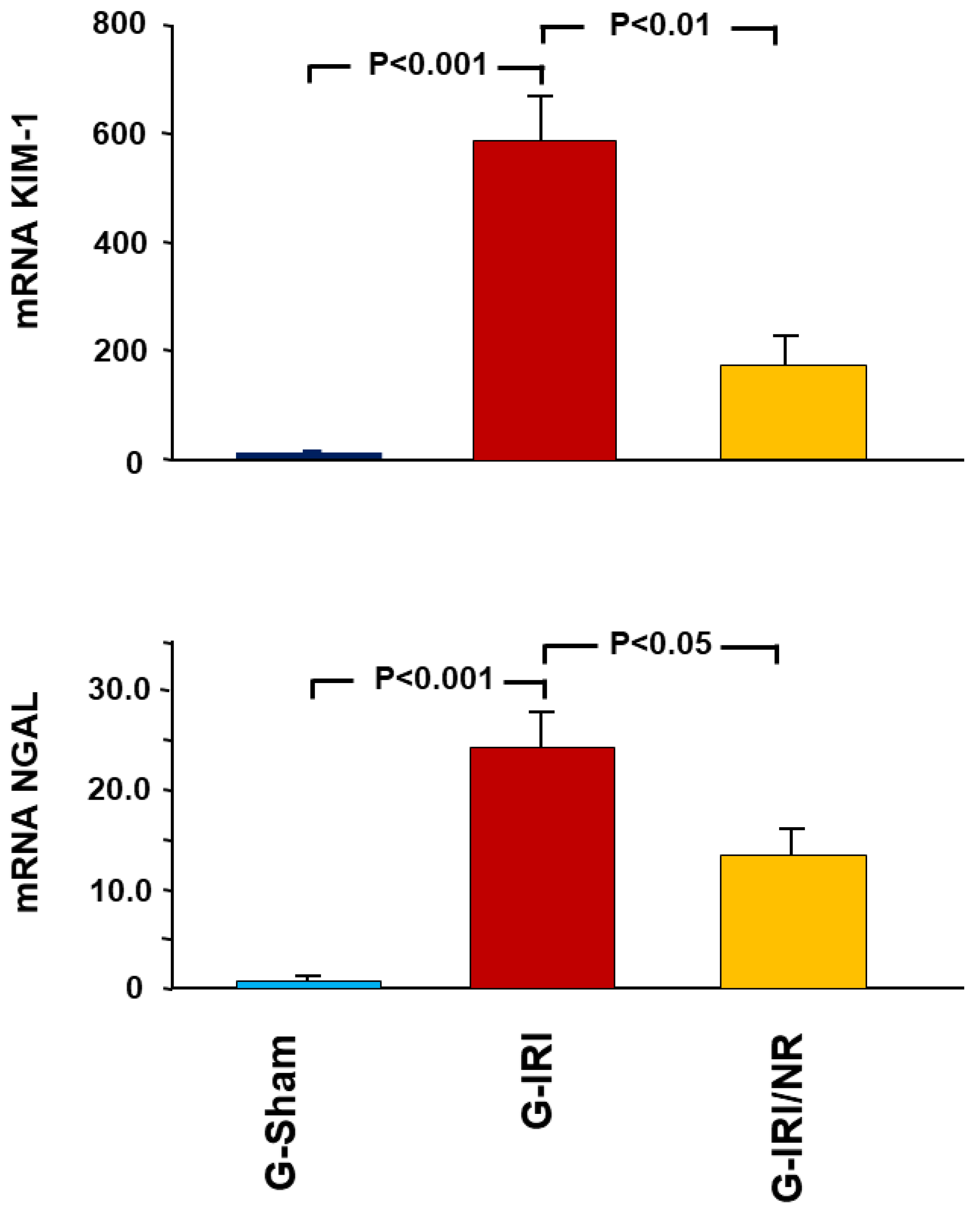
  • Acute kidney injury markers, i.e., kidney injury molecule-1 (KIM1) and neutrophil gelatinase-associated lipocalin (NGAL);
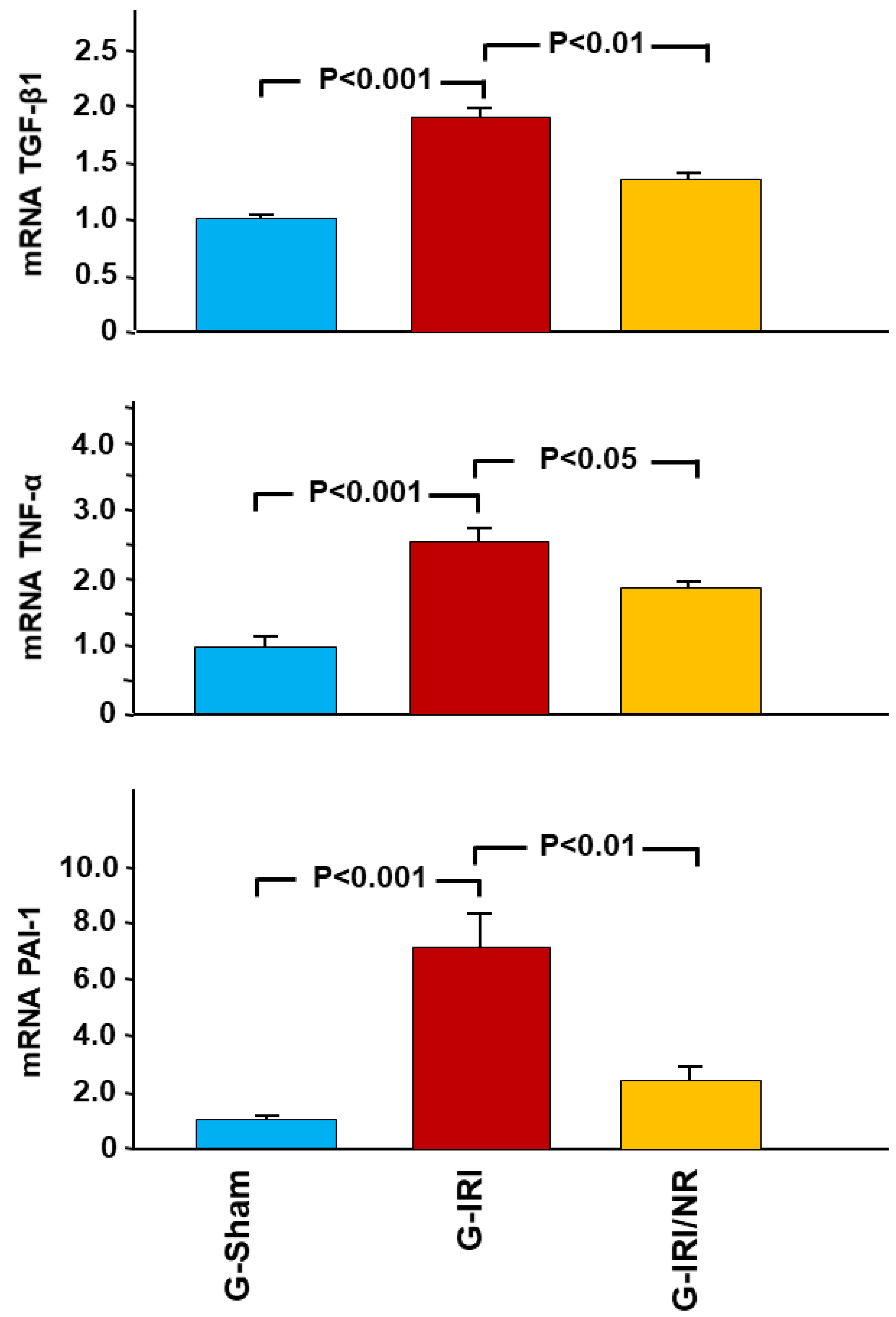
  • Cytokines involved in the inflammation and fibrosis, i.e., tumor necrosis factor-α (TNFα), transforming growth factor-β (TGF-β1), interleukin-6 (IL-6), interleukin-1 beta (IL-1β) and plasminogen activator inhibitor-1 (PAI-1);
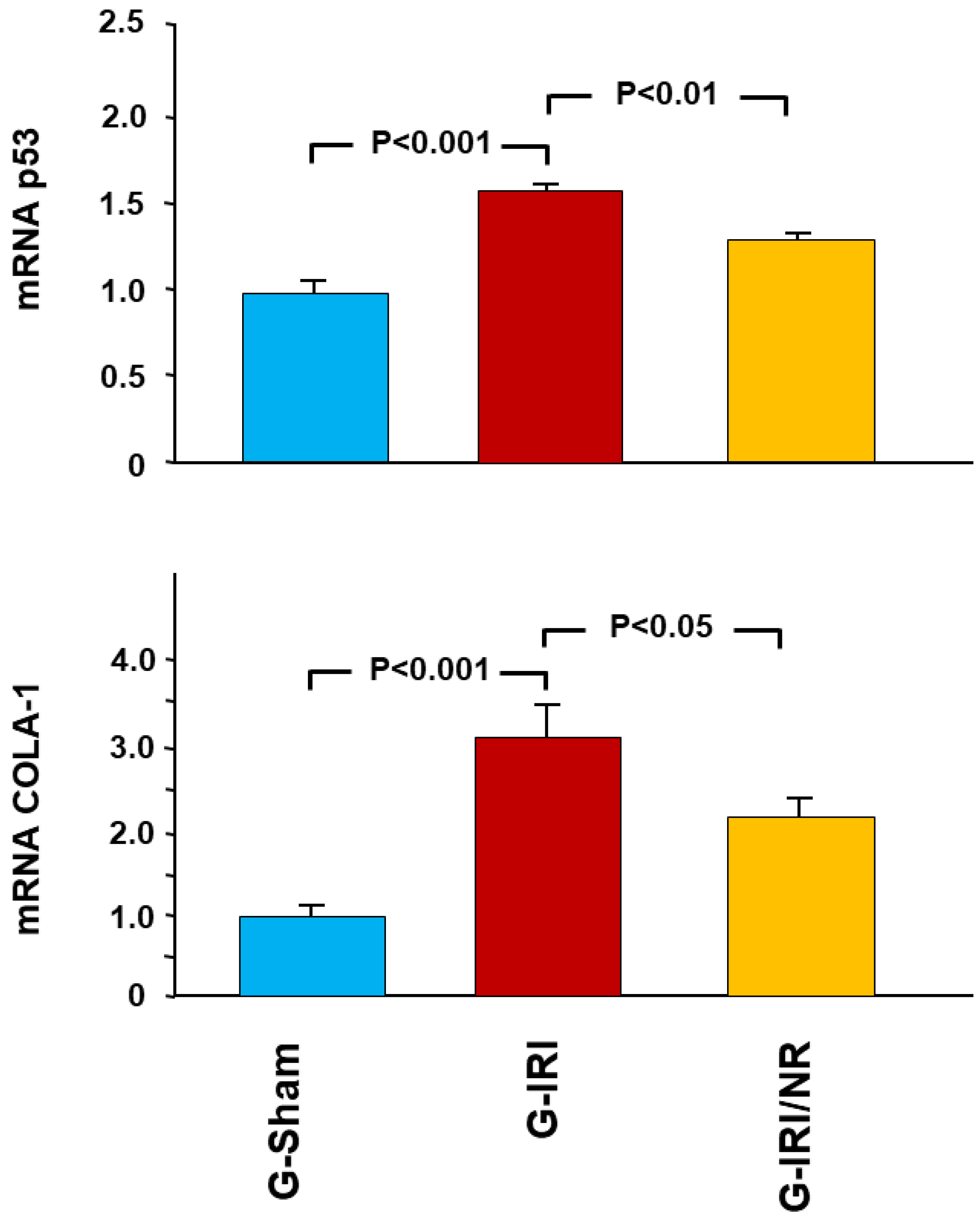
  • The proapoptotic gene p53.
  • Procollagen type-1 (COLA-1); and
  • The antioxidant enzymes glutathione peroxidase (GPX-1) and glutathione-disulfide reductase (GSR).
Extraction of total RNA from the frozen samples was performed using Qiazol Lysis reagent (Qiagen, Hilden, Germany) as per the manufacturer’s protocol. Estimation of the quantity and quality of the extracted RNA was performed using a NanoDrop 2000 spectrophotometer (Thermo Fisher Scientific, Inc., Wilmington, DE, USA).
Preparation of the strand complementary DNA (cDNA) in duplicates from 1.0 µg of extracted RNA was achieved using a QuantiTect® reverse transcription kit (Qiagen, Hilden, Germany) as per the manufacturer’s protocol. The protocol consisted of genomic DNA removal using the supplied gDNA wipeout buffer, ensuring elimination of interference by the genomic DNA. Subsequently, the prepared cDNA was used as a template for relative gene expression analysis using a TaqMan® hydrolysis probe chemistry kit. The reaction mixture contained 75 ng cDNA, TaqMan universal master mix (Thermo Fisher Scientific Inc., Wilmington, DE, USA), 0.5 µM of forward and reverse primers and 0.25 µM of fluorescent probe (Biosearch Technologies Inc., Petaluma, CA, USA). The probes were FAM-labeled.
Peptidylprolyl Isomerase A (PPIA) housekeeping gene was used for normalization. Its probe was labeled with Quasar 670, enabling multiplexing with the genes of interest. All samples were run in duplicates. At least one primer of all designed PCR primer sets was spanned the exon–exon junction to further exclude any interference of the genomic DNA. Table 1 shows the sequences of primers and probes. The calculated cycle threshold (CT) values were used to estimate the changes in gene expression of target genes using a delta–delta CT formula.
The results are expressed as the mean fold change of gene expression compared to G-Sham. The gene expression of the G-Sham animals was measured, and the average was calculated. This average was given a value of 1. All other groups, including the G-Sham animals were compared to this value.

2.6. Enzyme-Linked Immunosorbent Assay (ELISA)

A wedge from the middle part of the left kidney containing the cortex and medulla was excised. The renal tissue concentrations of total glutathione (GSH, Cayman Chemical, Ann Arbor, MI, USA) and malondialdehyde (MDA) (TBARS, Cayman Chemical, Ann Arbor, MI, USA) were measured using ELISA as per the manufacturer’s instructions. The levels were normalized to the total protein concentrations. The concentration was determined by interpolation from a standard curve. Both standards and samples were assayed in duplicate.

2.7. Histological Studies

The kidney tissue was washed with ice-cold saline, blotted using filter paper, cassetted and fixed directly in 10% neutral formalin for 24 h. This was followed by dehydration in increasing ethanol concentrations, clearing with xylene and embedding with paraffin. Next, 3 μm sections were prepared from paraffin blocks and stained with hematoxylin and eosin. The stained sections were evaluated blindly using light microscopy.
Microscopic scoring was performed by measuring the percentage of area that showed morphologic changes (tubular dilatation, tubular atrophy, interstitial fibrosis and mononuclear cellular infiltrate) in comparison to the total surface area in each sample. Measurement of the frequency of each histological abnormality was performed using Image J software (NIH, Milwaukee, WI, USA). The degree of abnormality was classified using the following scoring system: score 0, no abnormality; score 1, 1–25%; score 2, 26–50%; score 3, 51–75; score 4, 76–100%.

3. Statistical Analysis

Statistical analysis was performed using SPSS V16.0. Results are expressed as mean ± SEM. One-way factorial ANOVA was used for comparison of variables between groups and between different stages (Basal, Pre-IRI and Post-IRI) within each group. A p value less than 0.05 was considered statistically significant.

4. Results

As demonstrated in Table 2 and Table 3, the basal serum creatinine, serum urea, creatinine clearance, 24 h urinary albumin and albumin/creatinine ratio were similar in all groups (p > 0.05 for all variables). Similarly, there was no difference in any of these variables between the Pre-IRI and Basal values in any of the groups (p > 0.05 for all variables).
As expected, in the G-Sham, there was no difference in any variable post manipulation compared to the Pre-IRI value (p > 0.05 for all variables). In the G-IRI, there was a significant deterioration in renal parameters following IRI (Table 2). For instance, IRI caused an increase in serum creatinine from 0.32 ± 0.05 to 1.51 ± 0.48 mg/dL (p < 0.05). In contrast, creatinine clearance decreased to 33.5 ± 7.8 compared to 69.9 ± 6.6 mL/min before IRI (p < 0.01). In addition, IRI caused an increase in urinary albumin leakage. The 24 h urinary albumin and albumin/creatinine ratio increased to 0.783 ± 0.111 µg (vs. 0.079 ± 0.009, p < 0–001) and 166.1 ± 27.0 (vs. 13.7 ± 2.2, p < 0–001), respectively (Table 3).
As shown in Table 2 and Table 3, the administration of nerolidol significantly attenuated IRI-induced alterations in these parameters (p < 0.05 for all variables).

4.1. Gene Expression Analysis Results

As shown in Figure 2, IRI caused a significant increase in the gene expression of KIM-1 and NGAL, as demonstrated by comparing G-IRI to G-Sham (588.3 ± 92.6 vs. 1.0 ± 0.07 and 24.4 ± 3.5 vs. 1.0 ± 0.08, respectively; p < 0.001 for both). Nerolidol administration significantly attenuated this increase in the markers (173.4 ± 51.3 vs. 588.3 ± 92.6, p < 0.01 and 13.9 ± 3.1 vs. 24.4 ± 3.5, p < 0.05, respectively).
A similar effect was observed in relation to proinflammatory, profibrotic and proapoptotic cytokines (Figure 3, Figure 4 and Figure 5). For instance, IRI led to a significant increase in the gene expression of TGF-β1 in the G-IRI compared to G-Sham (1.88 ± 0.13 vs. 1.01 ± 0.06, p < 0.001), whereas nerolidol significantly attenuated this increase (1.46 ± 0.06 vs. 1.88 ± 0.13, p < 0.01). Similar effects were observed in the gene expression of TNF-α, PAI-1, IL-6, IL-1β and p53 (p < 0.05 for all these mediators).
The gene expression of procollagen type-1 (COLA-1) showed a similar trend, as the IRI caused a significant increase in the gene expression of COLA-1 in G-IRI compared to the G-Sham (3.10 ± 0.41 vs. 1.03 ± 0.12, p < 0.001) (Figure 5). Nerolidol significantly attenuated this rise (2.11 ± 0.20 vs. 3.10 ± 0.41, p < 0.05).
Nerolidol also significantly attenuated the change in the gene expression of some oxidative stress enzymes, such as GPX-1 and GSR (Figure 6). For instance, IRI led to a significant decrease in the gene expression of GSR (0.62 ± 0.05 vs. 1.02 ± 0.11, p < 0.01), and this was attenuated by nerolidol (0.83 ± 0.03 vs. 0.62 ± 0.05, p < 0.01).

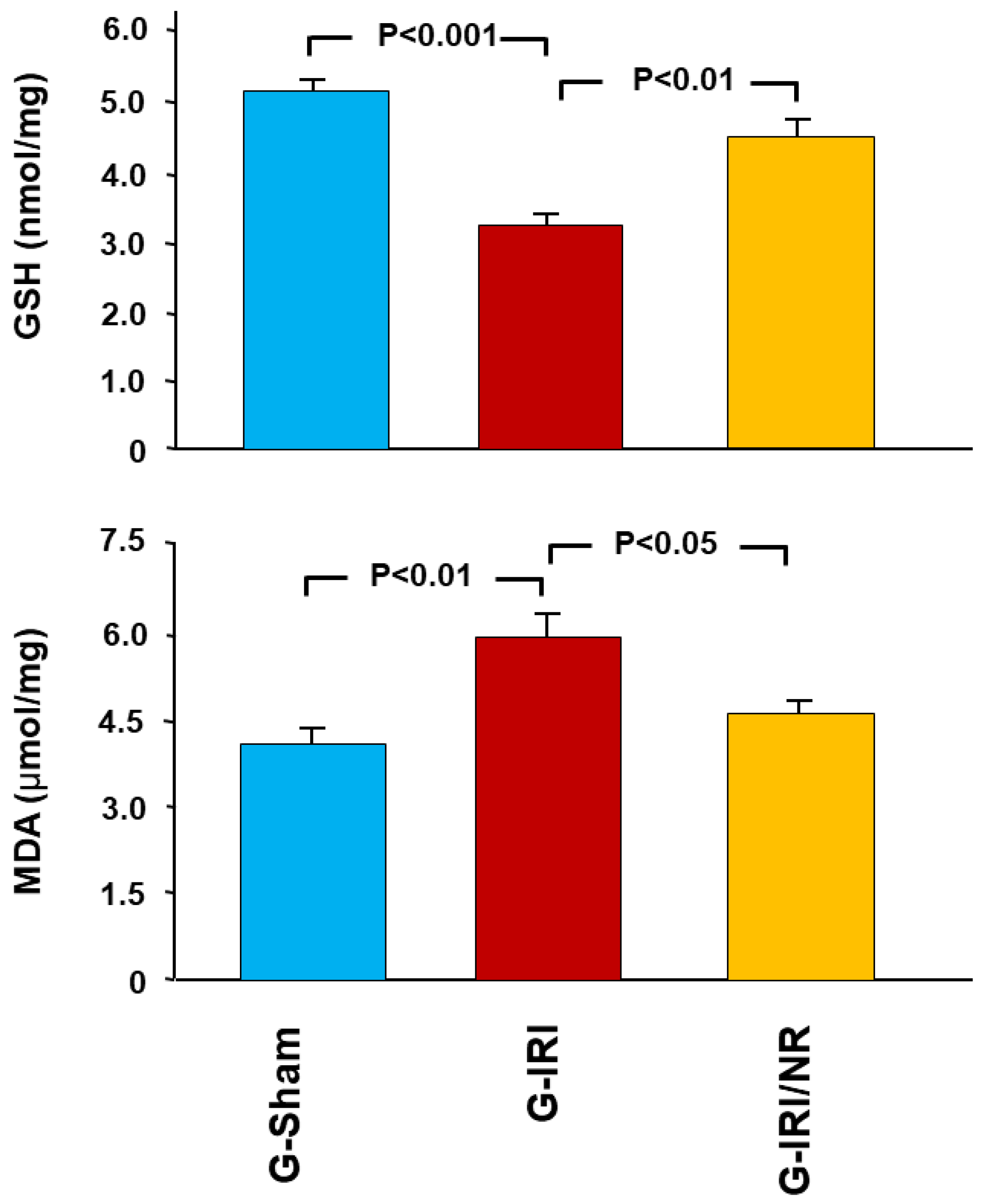
4.2. Enzyme-Linked Immunosorbent Assay (ELISA) Results

As shown in Figure 7, GSH tissue concentration was significantly lower in the G-IRI compared to the G-Sham (3.29 ± 0.12 vs. 5.15 ± 0.23, p < 0.001). Nerolidol attenuated this decrease (4.47 ± 0.29 vs. 3.29 ± 0.12, p < 0.01). On the other hand, IRI led to a significant increase in MDA tissue concentration (5.94 ± 0.73 vs. 4.09 ± 0.29, p < 0.01), and nerolidol resulted in a significant decrease in the MDA concentration (4.76 ± 0.21 vs. 5.94 ± 0.53, p < 0.05).

4.3. Histological Studies

As shown in Figure 8A,B, the kidneys in G-Sham had normal architecture and histology (score 0). IRI led to diffuse tubular injury and necrosis in 90.2 ± 1.9% (score 4) of the examined tissues (p < 0.001 compared to G-Sham). This was associated with the loss of brush border in proximal tubules, tubular dilatation, intratubular necrotic material and secretions and mild mixed inflammatory cell infiltration of the interstitium consisting mainly of lymphocytes (Figure 8C,D). As shown in Figure 8E,F, nerolidol attenuated the effect of IRI, as demonstrated by the fact that only 14.8 ± 4.8% (score 1) of the examined tissues in the G-IRI/NR showed focal cortical tubular injury with tubular dilatation, intratubular necrotic material and secretions and mild mixed inflammatory cell infiltration of the interstitium consisting mainly of lymphocytes (p < 0.001 compared to G-IRI).

5. Discussion

In the current study, we investigated, for the first time, the effect of nerolidol on renal dysfunction following IRI. We have shown that the administration of this agent prior to and soon after IRI can significantly attenuate IRI-induced alterations in renal functional parameters such as serum creatinine, creatinine clearance and urinary albumin leakage. It also significantly attenuated changes in the gene expression of markers of acute renal injury; proinflammatory, profibrotic and proapoptotic cytokines; and oxidative stress markers. Our data also show a significant improvement in the tissue level of oxidative stress markers and in histopathological changes.
IRI-induced renal damage is caused by both ischemia and reperfusion, leading to hypoxia and excessive production of reactive oxygen species, respectively. This damage is also associated with an inflammatory response characterized by increased production of several cytokines such as proinflammatory and profibrotic cytokines, including TNF-α, TGF-β1 and PAI [21,22,23,24,25,26].
In the current study, nerolidol was shown to have a significant antioxidant effect, as demonstrated by its effects on the gene expression of some oxidative stress enzymes, namely glutathione peroxidase and glutathione-disulfide reductase, as well as on the tissue concentration of glutathione and malondialdehyde. The antioxidant and free radical scavenging properties of nerolidol have been demonstrated in other models, such as memory impairment caused by Trypanosoma evansi in mice [27], thioacetamide-induced oxidative damage in heart and kidney tissue [18] and isoproterenol-induced myocardial damage [13]. The contradictory downregulation in the gene expression of the oxidative enzymes and in their tissue concentration observed in this study has been previously demonstrated by other studies in IRI [28,29] and other conditions [30,31] in which oxidative stress plays a major role in the pathophysiology of the disease process. This downregulation of mRNA has been attributed to the higher turnover of these enzymes at different levels [28]. Regardless of the exact reason, nerolidol led to a significant attenuation of this process in renal IRI.
In addition to its antioxidant properties, the current data demonstrate anti-inflammatory and antifibrotic properties of nerolidol, as shown by the attenuation of the IRI-induced rise in the gene expression of TNF-α, TGF-β1, PAI, IL-1 β and IL-6. TNF-α is a proinflammatory cytokine that is produced by several tissues including renal cells [32]. TGF-β is a profibrotic cytokine that stimulates renal cells to produce extracellular matrix proteins, leading to long-term glomerulosclerosis and tubulointerstitial fibrosis [26,33]. PAI-1 is also a profibrotic cytokine, as it is considered one of the major inhibitors of fibrinolysis [34,35]. All these cytokines are upregulated in the majority of renal diseases [23,24,26], and the effect of nerolidol on these cytokines in renal IRI appears to be similar to its effect in other conditions [17].
The anti-inflammatory and antioxidant properties of nerolidol observed in the current study led to an amelioration of IRI-associated renal functional damage, as shown by the significant attenuation in serum creatinine and creatinine clearance. The improvement in urinary albumin leakage in addition to these parameters indicates that nerolidol not only affects glomerular function but also renal tubular function. This is supported by its significant effects on the expression of some of the markers of acute renal injury, such as NGAL and KIM-1. The latter is strongly expressed and released by injured proximal tubular cells [36], whereas NGAL is synthesized in the thick ascending limb of Henle’s loop and collecting ducts [37]. Therefore, the current data indicate that nerolidol has an effect on different segments of renal tubules, accounting for improvements in renal functions, including urinary albumin leakage.
The current study is subject to some limitations. For instance, owing to the relatively short duration of follow-up after IRI in this study (3 days), it is difficult to assess the long-term histological effects. However, the significant attenuation of the inflammatory and fibrotic cytokines, as well as the attenuation in the gene expression of the proapoptotic gene and procollagen type-1 (COLA-1), indicates that it is likely that the use of nerolidol in IRI could lead to a milder degree of interstitial fibrosis in the long term. Another limitation of this study is the measurement of the gene expression of some cytokines and mediators and the lack of measurement of the protein expression of these factors, such as procollagen-1. Furthermore, in this study, we did not specifically test the tubular functions using electrolyte excretion. However, there are several indicators of an improvement in tubular functions in response to nerolidol administration, such as the improvement in histological features and the attenuation of KIM-1 and NGAL gene expression (vide supra). In addition, we have shown that there was an improvement in urinary albumin leakage under nerolidol treatment. In this regard, some authors believe that albuminuria is primarily caused by proximal tubular damage and impairment in the retrieval and degradation processes [38,39,40]; therefore, the improvement in albuminuria indicates an improvement in renal tubular functions. In addition to the abovementioned limitations, in the current study, we investigated the effect of nerolidol on the kidney but did not test its effect on other organs. Further studies are required to address this point.
The model used in the current study is similar to the clinical scenario of the renal ischemia encountered in conditions such as renal transplantation and partial nephrectomy. These procedures are performed worldwide at an increasing rate; hence, the protective effects of nerolidol demonstrated in the current study might be clinically relevant in such a way that these patients might benefit from taking this agent during the perioperative period. However, further clinical research is required to extrapolate these results to the clinical setup.
In conclusion, the administration of nerolidol before, during and after renal IRI appears to have ameliorated the IRI effects on the renal functional parameters, alterations in markers of kidney injury and histological features. The current data indicate that the protective effects are due to its anti-inflammatory, antifibrotic and antioxidative properties. These findings have potential clinical implications.

Author Contributions

This study was designed by F.T.H. and L.L., R.A., J.Y., J.A.R. and S.A.-S. were and responsible for data collection. The data in this research were prepared, analyzed and interpreted by F.T.H., S.A.-S., L.L., J.A.R. and A.F.H. The manuscript was prepared by F.T.H. All authors have read and agreed to the final manuscript.

Funding

This research was funded by the College of Medicine and Health Sciences, United Arab Emirates University, Grant #: 12M118.

Institutional Review Board Statement

This study was conducted according to the guidelines of the Declaration of Helsinki and approved by the College of Medicine and Health Sciences, UAEU Animal Ethics Committee (ERA-2021-8383).

Data Availability Statement

The data are available on request from the corresponding author.

Conflicts of Interest

None of the authors has any conflict of interest to declare.

References

  1. Weight, S.C.; Bell, P.R.; Nicholson, M.L. Renal ischaemia-reperfusion injury. Br. J. Surg. 1996, 83, 162–170. [Google Scholar] [PubMed]
  2. Myers, B.D.; Miller, D.C.; Mehigan, J.T.; Robertson, C.R.; Derby, G.; Spencer, R.; Friedman, S. Nature of the renal injury following total renal ischemia in man. J. Clin. Investig. 1984, 73, 329–341. [Google Scholar] [CrossRef] [Green Version]
  3. Hammad, F.T.; Davis, G.; Zhang, X.Y.; Wheatley, A.M. Endotelin ETA and ETB receptor antagonism during cold preservation in renal transplantation. Transplantation 2001, 71, 619–627. [Google Scholar] [CrossRef]
  4. Hammad, F.T.; Wheatley, A.M.; Davis, G. Role of endothelin ET(A) receptor antagonism in the post-transplant renal response to angiotensin II in the rat. Exp. Physiol. 2001, 86, 365–372. [Google Scholar] [CrossRef] [PubMed]
  5. Goes, N.; Urmson, J.; Ramassar, V.; Halloran, P. Ischemic acute tubular necrosis induces an extensive local cytokine response. Evidence for induction of interferon-gamma, transforming growth factor-beta 1, granulocyte-macrophage colony-stimulating factor, interleukin-2, and interleukin-10. Transplantation 1995, 59, 565–572. [Google Scholar] [CrossRef]
  6. Petersen, N.S.; Mitchell, H.K. Recovery of protein synthesis after heat shock: Prior heat treatment affects the ability of cells to translate mRNA. Proc. Natl. Acad. Sci. USA 1981, 78, 1708–1711. [Google Scholar] [CrossRef] [PubMed] [Green Version]
  7. Troppmann, C.; Gillingham, K.J.; Benedetti, E.; Almond, P.S.; Gruessner, R.W.; Najarian, J.S.; Matas, A.J. Delayed graft function, acute rejection, and outcome after cadaver renal transplantation. Transplantation 1995, 59, 962–968. [Google Scholar] [CrossRef]
  8. Unal, S.; Ozmen, S.; DemIr, Y.; Yavuzer, R.; LatIfoğlu, O.; Atabay, K.; Oguz, M. The Effect of Gradually Increased Blood Flow on Ischemia-Reperfusion Injury. Ann. Plast. Surg. 2001, 47, 412–416. [Google Scholar] [CrossRef]
  9. Yang, C.W.; Kim, B.S.; Kim, J.; Ahn, H.J.; Park, J.H.; Jin, D.C.; Kim, Y.S.; Bang, B.K. Preconditioning with sodium arsenite inhibits apoptotic cell death in rat kidney with ischemia/reperfusion or cyclosporine-induced Injuries. The possible role of heat-shock protein 70 as a mediator of ischemic tolerance. Nephron Exp. Nephrol. 2001, 9, 284–294. [Google Scholar] [CrossRef]
  10. Ferreira, F.M.; Palmeira, C.M.; Oliveira, M.M.; Santos, D.; Simões, A.M.; Rocha, S.M.; Coimbra, M.A.; Peixoto, F. Nerolidol effects on mitochondrial and cellular energetics. Toxicol. Vitr. 2012, 26, 189–196. [Google Scholar] [CrossRef]
  11. Lapczynski, A.; Bhatia, S.; Letizia, C.; Api, A. Fragrance material review on nerolidol (isomer unspecified). Food Chem. Toxicol. 2008, 46, S247–S250. [Google Scholar] [CrossRef] [PubMed]
  12. Bakkali, F.; Averbck, S.; Averbck, D.; Ldaomar, M. Biological effects of essential oils—A review. Food Chem. Toxicol. 2008, 46, 446–475. [Google Scholar] [CrossRef] [PubMed]
  13. Asaikumar, L.; Vennila, L.; Akila, P.; Sivasangari, S.; Kanimozhi, K.; Premalatha, V.; Sindhu, G. Preventive effect of nerolidol on isoproterenol induced myocardial damage in Wistar rats: Evidences from biochemical and histopathological studies. Drug Dev. Res. 2019, 80, 814–823. [Google Scholar] [CrossRef] [PubMed]
  14. Kaur, D.; Pahwa, P.; Goel, R.K. Protective Effect of Nerolidol Against Pentylenetetrazol-Induced Kindling, Oxidative Stress and Associated Behavioral Comorbidities in Mice. Neurochem. Res. 2016, 41, 2859–2867. [Google Scholar] [CrossRef] [PubMed]
  15. Neto, J.D.N.; De Almeida, A.A.C.; Oliveira, J.D.S.; Dos Santos, P.S.; De Sousa, D.P.; De Freitas, R.M. Antioxidant Effects of Nerolidol in Mice Hippocampus After Open Field Test. Neurochem. Res. 2013, 38, 1861–1870. [Google Scholar] [CrossRef]
  16. Krist, S.; Banovac, D.; Tabanca, N.; Wedge, D.E.; Gochev, V.K.; Wanner, J.; Schmidt, E.; Jirovetz, L. Antimicrobial Activity of Nerolidol and its Derivatives against Airborne Microbes and Further Biological Activities. Nat. Prod. Commun. 2015, 10, 143–148. [Google Scholar] [CrossRef] [PubMed]
  17. Zhang, L.; Sun, D.; Bao, Y.; Shi, Y.; Cui, Y.; Guo, M. Nerolidol Protects Against LPS-induced Acute Kidney Injury via Inhibiting TLR4/NF-κB Signaling. Phytother. Res. 2017, 31, 459–465. [Google Scholar] [CrossRef] [PubMed]
  18. Türkmen, N.B.; Yüce, H.; Taşlidere, A.; Şahin, Y.; Çiftçi, O. The Ameliorate Effects of Nerolidol on Thioasteamide-induced Oxidative Damage in Heart and Kidney Tissue. Turk. J. Pharm. Sci. 2022, 19, 1–8. [Google Scholar] [CrossRef]
  19. Hammad, F.T.; Al-Salam, S.; AlZaabi, S.S.; Alfalasi, M.M.; Hammad, A.F.; Yasin, J.; Lubbad, L. The effect of neprilysin and renin inhibition on the renal dysfunction following ischemia-reperfusion injury in the rat. Physiol. Rep. 2021, 9, e14723. [Google Scholar] [CrossRef]
  20. Klopell, F.C.; Lemos, M.; Sousa, J.P.B.; Comunello, E.; Maistro, E.L.; Bastos, J.K.; de Andrade, S.F. Nerolidol, an Antiulcer Constituent from the Essential Oil of Baccharis dracunculifolia DC (Asteraceae). Z. Nat. C J. Biosci. 2007, 62, 537–542. [Google Scholar] [CrossRef]
  21. Chatauret, N.; Badet, L.; Barrou, B.; Hauet, T. Ischemia-reperfusion: From cell biology to acute kidney injury. Front. Biosci. 2014, 24, S4–S12. [Google Scholar] [CrossRef] [PubMed]
  22. Furuichi, K.; Wada, T.; Kaneko, S.; Murphy, P.M. Roles of chemokines in renal ischemia/reperfusion injury. Front. Biosci. 2008, 13, 4021–4028. [Google Scholar] [CrossRef] [PubMed] [Green Version]
  23. Hammad, F.T.; Al-Salam, S.; Lubbad, L. Curcumin Provides Incomplete Protection of the Kidney in Ischemia Reperfusion Injury. Physiol. Res. 2012, 61, 503–511. [Google Scholar] [CrossRef]
  24. Hammad, F.T.; Al-Salam, S.; Lubbad, L. Does aliskiren protect the kidney following ischemia reperfusion injury? Physiol. Res 2013, 62, 681–690. [Google Scholar] [CrossRef]
  25. Lerman, L.; Textor, S.C. Pathophysiology of ischemic nephropathy. Urol. Clin. N. Am. 2001, 28, 793–803. [Google Scholar] [CrossRef]
  26. Spurgeon, K.R.; Donohoe, D.L.; Basile, D.P. Transforming growth factor-beta in acute renal failure: Receptor expression, effects on proliferation, cellularity, and vascularization after recovery from injury. Am. J. Physiol. Ren. Physiol. 2005, 288, F568–F577. [Google Scholar] [CrossRef]
  27. Baldissera, M.D.; Souza, C.F.; Grando, T.H.; Moreira, K.L.; Schafer, A.S.; Cossetin, L.F.; Da, S.A.; Da Veiga, M.L.; Da Rocha, M.I.U.; Stefani, L.M.; et al. Nerolidol-loaded nanospheres prevent behavioral impairment via ameliorating Na(+), K(+)-ATPase and AChE activities as well as reducing oxidative stress in the brain of Trypanosoma evansi-infected mice. Naunyn Schmiedebergs. Arch. Pharm. 2017, 390, 139–148. [Google Scholar] [CrossRef]
  28. Dobashi, K.; Ghosh, B.; Orak, J.; Singh, I.; Singh, A. Kidney ischemia-reperfusion: Modulation of antioxidant defenses. Mol. Cell. Biochem. 2000, 205, 1–11. [Google Scholar] [CrossRef]
  29. Jiang, G.P.; Liao, Y.J.; Huang, L.L.; Zeng, X.J.; Liao, X.H. Effects and molecular mechanism of pachymic acid on ferroptosis in renal ischemia reperfusion injury. Mol. Med. Rep. 2020, 23, 63. [Google Scholar] [CrossRef] [PubMed]
  30. AlAsmari, A.F.; Ali, N.; Alharbi, M.; Alqahtani, F.; Alasmari, F.; Almoqbel, D.; AlSwayyed, M.; Alshammari, A.; Alanazi, M.M.; Alhoshani, A.; et al. Geraniol Ameliorates Doxorubicin-Mediated Kidney Injury through Alteration of Antioxidant Status, Inflammation, and Apoptosis: Potential Roles of NF-κB and Nrf2/Ho-1. Nutrients 2022, 14, 1620. [Google Scholar] [CrossRef]
  31. Hammad, F.T.; Al Salam, S.; Nemmar, A.; Ali, M.; Lubbad, L. The Effect of Arabic Gum on Renal Function in Reversible Unilateral Ureteric Obstruction. Biomolecules 2019, 9, 25. [Google Scholar] [CrossRef] [Green Version]
  32. Speeckaert, M.M.; Speeckaert, R.; Laute, M.; Vanholder, R.; Delanghe, J.R. Tumor Necrosis Factor Receptors: Biology and Therapeutic Potential in Kidney Diseases. Am. J. Nephrol. 2012, 36, 261–270. [Google Scholar] [CrossRef]
  33. Ding, Y.; Choi, M.E. Regulation of autophagy by TGF-beta: Emerging role in kidney fibrosis. Semin. Nephrol 2014, 34, 62–71. [Google Scholar] [CrossRef] [PubMed] [Green Version]
  34. Chorostowska-Wynimko, J.; Swiercz, R.; Skrzypczak-Jankun, E.; Wojtowicz, A.; Selman, S.H.; Jankun, J. A novel form of the plasminogen activator inhibitor created by cysteine mutations extends its half-life: Relevance to cancer and angiogenesis. Mol. Cancer Ther. 2003, 2, 19–28. [Google Scholar]
  35. Ghosh, A.K.; Vaughan, D.E. PAI-1 in tissue fibrosis. J. Cell. Physiol. 2012, 227, 493–507. [Google Scholar] [CrossRef] [PubMed] [Green Version]
  36. Lim, A.I.; Tang, S.C.; Lai, K.N.; Leung, J.C. Kidney injury molecule-1: More than just an injury marker of tubular epithelial cells? J. Cell. Physiol. 2013, 228, 228–917. [Google Scholar] [CrossRef]
  37. Mishra, J.; Ma, Q.; Prada, A.; Mitsnefes, M.; Zahedi, K.; Yang, J.; Barasch, J.; Devarajan, P. Identification of Neutrophil Gelatinase-Associated Lipocalin as a Novel Early Urinary Biomarker for Ischemic Renal Injury. J. Am. Soc. Nephrol. 2003, 14, 2534–2543. [Google Scholar] [CrossRef] [PubMed] [Green Version]
  38. Comper, W.D.; Hilliard, L.M.; Nikolic-Paterson, D.J.; Russo, L.M. Disease-dependent mechanisms of albuminuria. Am. J. Physiol. Physiol. 2008, 295, F1589–F1600. [Google Scholar] [CrossRef] [Green Version]
  39. Eppel, G.A.; Osicka, T.M.; Pratt, L.M.; Jablonski, P.; Howden, B.O.; Glasgow, E.F.; Comper, W.D. The return of glomerular-filtered albumin to the rat renal vein. Kidney Int. 1999, 55, 1861–1870. [Google Scholar] [CrossRef] [Green Version]
  40. Osicka, T.M.; Strong, K.J.; Nikolic-Paterson, D.; Atkins, R.C.; Jerums, G.; Comper, W.D. Renal processing of serum proteins in an albumin-deficient environment: An in vivo study of glomerulonephritis in the Nagase analbuminaemic rat. Nephrol. Dial. Transplant. 2004, 19, 320–328. [Google Scholar] [CrossRef] [PubMed]
Figure 1. Schematic presentation of the study plan showing interventions in all groups.
Nutrients 15 00455 g001
Figure 2. Gene expression of two markers of acute renal injury (KIM-1 and NGAL) in all groups. Values represent mean ± SEM.
Nutrients 15 00455 g002
Figure 3. Gene expression of transforming growth factor-β (TGF-β1), tumor necrosis factor-α (TNF-α) and plasminogen activator inhibitor-1 (PAI-1) in all groups. Values represent mean ± SEM.
Nutrients 15 00455 g003
Figure 4. Gene expression of the proinflammatory cytokines interleukin 6 (IL-6) and interleukin 1 beta (IL-1β) in all groups. Values represent mean ± SEM.
Nutrients 15 00455 g004
Figure 5. Gene expression of the proapoptotic p53 gene and procollagen-1 (COLA-1) in all groups. Values represent mean ± SEM.
Nutrients 15 00455 g005
Figure 6. Gene expression of the antioxidant enzymes glutathione peroxidase (GPX-1) and glutathione-disulfide reductase (GSR) in all groups. Values represent mean ± SEM.
Nutrients 15 00455 g006
Figure 7. Kidney tissue concentration of gene expression of the glutathione (GSH) and malondialdehyde (MDA, TBARS) measured by ELISA. Values represent mean ± SEM.
Nutrients 15 00455 g007
Figure 8. Histological features in all the experimental groups. (A,B) The kidneys in G-Sham showed normal architecture and histology. (C,D) Histological features in the G-IRI with diffuse acute tubular necrosis (thin arrow) and intratubular necrotic material and secretions (arrowhead). (E,F) Histological features in G-IRI/NR with mild acute tubular necrosis and tubular dilation (thin arrow) and intratubular secretion (arrowhead).
Nutrients 15 00455 g008
Table 1. Forward and reverse primers and fluorogenic probe sequences used for real-time quantitative PCR analysis. KIM-1: kidney injury molecule-1; NGAL: neutrophil gelatinase-associated lipocalin, also called lipocalin 2 (Lcn2); TNF-α: tumor necrosis factor-alpha; TGF-β1: transforming growth factor-β; IL-1β: interleukin 1 beta; IL-6: interleukin 6; TGF-β1: transforming growth factor-β; PAI-1: plasminogen activator inhibitor-1; p53: proapoptotic gene p53; COL1A: procollagen type-1; GSR: glutathione-disulfide reductase; GPx-1: glutathione peroxidase 1; PPIA: peptidylprolyl isomerase A (housekeeping gene).
KIM-1
(NM_173149.2)
ForwardGCCTGGAATAATCACACTGTAAG
ReverseGCAACGGACATGCCAACATAG
Probed FAM-TCCCTTTGAGGAAGCCGCAGA-BHQ-1
Lipocalin 2 (LCN2)
(NM_130741.1)
ForwardCTGTTCCCACCGACCAATGC
ReverseCCACTGCACATCCCAGTCA
ProbeFAM-TGACAACTGAACAGACGGTGAGCG-BHQ-1
TNF-α
(NM_012675.3)
ForwardCTCACACTCAGATCATCTTCTC
ReverseCCGCTTGGTGGTTTGCTAC
ProbeFAM-CTCGAGTGACAAGCCCGTAGCC-BHQ-1
TGF-β1
NM_012620.1
ForwardGTGGCTGAACCAAGGAGACG
ReverseCGTGGAGTACATTATCTTTGCTGTC
ProbeFAM-ACAGGGCTTTCGCTTCAGTGCTC-BHQ-1
PAI-1
(NM_012620.1)
ForwardGGCACAATCCAACAGAGACAA
ReverseGGCTTCTCATCCCACTCTCAAG
ProbeFAM-CCTCTTCATGGGCCAGCTGATGG-BHQ-1
IL-6
(NM_012589.2)
ForwardTCACAGAGGATACCACCCACAACA
ReverseCACAAGTCCGGAGAGGAGAC
ProbeFAM-TCAGAATTGCCATTGCACAACTCT-BHQ-1
IL-1β
(NM_031512.2)
ForwardATGCCTCGTGCTGTCTGACC
ReverseGCTCATGGAGAATACCACTTGTTGG
ProbeFAM-AGCTGAAAGCTCTCCACCTCAATGGA-BHQ-1
p53
(NM_030989.3)
ForwardCGAGATGTTCCGAGAGCTGAATG
ReverseGTCTTCGGGTAGCTGGAGTG
ProbeFAM-CCTTGGAATTAAAGGATGCCCGTGC-BHQ-1
COL1A
(NM_053304.1)
ForwardCTGACTGGAAGAGCGGAGAGT
ReverseCCTGTCTCCATGTTGCAGTAGAC
ProbeFAM-ACTGGATCGACCCTAACCAAGGC-BHQ-1
GSR
NM_053906.2
ForwardCATCCCTACCGTGGTCTTCAG
ReverseATGGACGGCTTCATCTTCAGT
ProbeFAM-CCACCCGCCTATCGGGACAGT-BHQ-1
GPx-1
NM_030826.4
ForwardGTGCTGCTCATTGAGAATGTCG
ReverseTCATTCTTGCCATTCTCCTGATG
ProbeFAM-TCCCTCTGAGGCACCACGAC-BHQ-1
PPIA
(NM_017101.1)
ForwardGCGTCTGCTTCGAGCTGT
ReverseCACCCTGGCACATGAATCC
ProbeQuasar 670-TGCAGACAAAGTTCCAAAGACAGCA-BHQ-2
Table 2. Serum creatinine, serum urea and creatinine clearance in all groups before administration of nerolidol/vehicle (basal values), after administration and on the third day following IRI of the nerolidol/vehicle, just before ischemia–reperfusion injury (Pre-IRI) (Post-IRI). * Indicates statistical significance when compared to the Pre-IRI within the same group; $ indicates statistical significance compared to the G-IRI group. Values are expressed as mean ± SEM.
Group
G-ShamG-IRIG-IRI/NR
S. Creatinine
(mg/dL)
Basal0.31 ± 0.020.30 ± 0.020.32 ± 0.02
Pre-IRI0.29 ± 0.010.32 ± 0.050.33 ± 0.02
Post-IRI0.29 ± 0.021.51 ± 0.48 *0.37 ± 0.02 *,$
S. Urea
(mg/dL)
Basal28.9 ± 1.429.0 ± 1.431.1 ± 2.8
Pre-IRI26.6 ± 0.830.7 ± 2.828.8 ± 2.3
Post-IRI25.4 ± 0.682.2 ± 19.8 *31.7 ± 2.1 $
Creatinine Clearance
(mL/min)
Basal62.3 ± 4.364.7 ± 9.163.5 ± 5.7
Pre-IRI72.7 ± 3.469.9 ± 6.667.0 ± 5.1
Post-IRI76.4 ± 3.033.5 ± 7.8 *74.1 ± 2.8 $
(1)
Table 3. The 24 h urinary albumin and albumin/creatinine ratio in all groups before administration of the nerolidol/vehicle (basal values), after administration of the nerolidol/vehicle, just before the ischemia–reperfusion injury (Pre-IRI) and on the third day following IRI (Post-IRI). * Indicates statistical significance compared to the Pre-IRI within the same group; $ indicates statistical significance compared to the G-IRI group. Values are expressed as mean ± SEM.
$ Group
* G-ShamG-IRIG-IRI/NR
24-h Urinary Albumin
(µg)
Basal0.073 ± 0.0050.078 ± 0.0130.073 ± 0.009
Pre-IRI0.077 ± 0.0060.079 ± 0.0090.079 ± 0.006
Post-IRI0.069 ± 0.0050.783 ± 0.111 *0.570 ± 0.078 *,$
Albumin/Creatinine RatioBasal13.9 ± 1.113.1 ± 2.113.6 ± 1.7
Pre-IRI13.9 ± 0.913.7 ± 2.213.8 ± 0.8
Post-IRI12.4 ± 0.9166.1 ± 27.0 *88.8 ± 11.0 *,$
Disclaimer/Publisher’s Note: The statements, opinions and data contained in all publications are solely those of the individual author(s) and contributor(s) and not of MDPI and/or the editor(s). MDPI and/or the editor(s) disclaim responsibility for any injury to people or property resulting from any ideas, methods, instructions or products referred to in the content.

