Review

A Review of the Health Benefits of Food Enriched with Kynurenic Acid

by
Monika Turska
1,*,
Piotr Paluszkiewicz
2,
Waldemar A. Turski
3 and
Jolanta Parada-Turska
4
1
Department of Molecular Biology, The John Paul II Catholic University of Lublin, 20-708 Lublin, Poland
2
Department of General, Oncological and Metabolic Surgery, Institute of Hematology and Transfusion Medicine, 02-778 Warsaw, Poland
3
Department of Experimental and Clinical Pharmacology, Medical University of Lublin, 20-090 Lublin, Poland
4
Department of Rheumatology and Connective Tissue Diseases, Medical University of Lublin, 20-090 Lublin, Poland
*
Author to whom correspondence should be addressed.
Nutrients 2022, 14(19), 4182; https://doi.org/10.3390/nu14194182
Submission received: 14 September 2022 / Revised: 2 October 2022 / Accepted: 4 October 2022 / Published: 8 October 2022

Abstract

:
Kynurenic acid (KYNA), a metabolite of tryptophan, is an endogenous substance produced intracellularly by various human cells. In addition, KYNA can be synthesized by the gut microbiome and delivered in food. However, its content in food is very low and the total alimentary supply with food accounts for only 1–3% of daily KYNA excretion. The only known exception is chestnut honey, which has a higher KYNA content than other foods by at least two orders of magnitude. KYNA is readily absorbed from the gastrointestinal tract; it is not metabolized and is excreted mainly in urine. It possesses well-defined molecular targets, which allows the study and elucidation of KYNA’s role in various pathological conditions. Following a period of fascination with KYNA’s importance for the central nervous system, research into its role in the peripheral system has been expanding rapidly in recent years, bringing some exciting discoveries. KYNA does not penetrate from the peripheral circulation into the brain; hence, the following review summarizes knowledge on the peripheral consequences of KYNA administration, presents data on KYNA content in food products, in the context of its daily supply in diets, and systematizes the available pharmacokinetic data. Finally, it provides an analysis of the rationale behind enriching foods with KYNA for health-promoting effects.

Graphical Abstract

1. Introduction

In 2013, we published a review paper describing the potential role of kynurenic acid (KYNA) in the digestive system. It summarized the presence of KYNA in the lumen of the gastrointestinal tract, and its beneficial health effects in digestive diseases, as well as its presence in food, were summarized. At that time, we pointed out many gaps in the knowledge and understanding of KYNA’s effects outside the central nervous system. However, even then, we assumed that KYNA administration might have some therapeutic potential and, despite many knowledge gaps, we envisaged the relevance of its supplementation [1]. KYNA is a metabolite of tryptophan. It is synthesized endogenously in human and animal bodies and/or absorbed from the digestive system. Although KYNA’s deficiency symptoms have not been described to date, it is reasonable to investigate the benefits of its supplementation. Nowadays, after 10 years of unprecedented progress in the research on KYNA, we can deliberate whether it is legitimate to supplement our diet with KYNA. The current review focuses on the presence of KYNA in food, including estimations of the daily dietary intake of KYNA, its absorption, distribution, and excretion, and it summarizes the scientifically approved health benefits of KYNA administered via the alimentary route, which are not limited to the digestive system.

2. Molecular Targets of Kynurenic Acid

Over 40 years of research, several molecular targets on which KYNA acts have been identified (Figure 1). Further research is ongoing and it was recently proposed that KYNA may be a ligand for the hydroxycarboxylic acid receptor 3 (HCAR3) and adrenoceptor alpha-2B (ADRA2B) [2]. However, no direct evidence is available yet to support this hypothesis.
Glutamatergic receptors occur preferentially in the brain, and since KYNA does not cross the blood–brain barrier, their importance is not discussed in the context of alimentary supplementation. Data on this subject can be found in recent review publications [3]. Whether and how KYNA acts on the alpha-7 nicotinic receptor (α7nAChR) is still a matter of scientific debate [4]. Therefore, the effect of KYNA on the aryl hydrocarbon receptor (AhR) and the G protein-coupled receptor 35 (GPR35), which are abundant in peripheral tissues, is of greatest concern. It should, however, be noted that AhR is an intracellular receptor, and so far, no uptake of KYNA has been demonstrated. Thus, it is uncertain whether exogenously administered KYNA can effectively influence these receptors. In this context, studies on KYNA’s peripheral activity have focused mostly on its agonistic action on GPR35. It is worth noting that these receptors are particularly densely represented on both the immune cells and cells of the gastrointestinal tract mucosa [5,6].
GPR35 is a rhodopsin-like, 7-transmembrane class A G-protein coupled receptor firstly described in 1998 by O’Dowd et al. [7]. GPR35 gene transcript in humans can be alternatively spliced into variants GPR35a and GPR35b containing 309 and 340 amino-acids, respectively. The differences between these two variants were limited to GPR35 extracellular domain [8]. Six of seventy described single nucleotide polymorphisms in GPR35 gene were indicated as risk variants for inflammatory bowel disease, ankylosing spondylitis, psoriasis, lupus erythematosus, and primary sclerosing cholangitis [9].
Activation of GPR35 by endogenous ligands such as KYNA, lysophosphatidic acid (LPA), or mucosal chemokine CXCL17 resulted in internalization of GPR35 and activation of ERK and Rho phosphorylation signaling pathway. A basal activity of GPR35 was connected with Na/K-ATPase pump and induction of Src signaling in epithelial cells responsible for cell proliferation and neovascularization. Moreover, the presence and high expression of GPR35 in intestine together with high concentration of KYNA in its lumen, GPR35 coexpression with both cholecystokinin GPR65 receptor suggest that GPR35 may be a part of the gut-brain signal axis regulating energy balance [10].

3. Absorption of Kynurenic Acid from the Digestive Tract

3.1. Human Studies

The absorption of KYNA from the digestive tract in humans was demonstrated by Kaihara et al. (1956) and by Turska et al. (2019) [11,12]. In the study by Kaihara et al. (1956), synthetic KYNA was suspended in water and ingested by three human subjects. Different doses of KYNA were administered, i.e., 164, 410, and 820 μmole (equivalent to 31, 77.5, and 155 mg, respectively). The urinary excretion of KYNA was measured the day before and on days 1, 2, and 3 after KYNA ingestion. It was found that KYNA administration resulted in its enhanced excretion, especially on the first days after administration [12], which proves that KYNA is absorbed from the digestive tract after its oral ingestion. In the study by Turska et al. (2019), chestnut honey containing KYNA in an amount of 600 μg/g [13] was used. Honey was dissolved in water with a ratio of 1:1 (w:v). Then, 200 mL of the obtained solution was consumed over a 5 min period by both male and female healthy volunteers. All study participants refrained from eating any food and drank only mineral water for 6 h before the study. Venous blood samples were drawn from a peripheral venous catheter at specified times after the administration of the honey beverage: baseline (0), 15, 30, 60, 90, and 120 min. It was found that the mean KYNA concentration in the serum before the administration of chestnut honey was 0.052 ± 0.004 μM and 0.051 ± 0.014 μM in men and women, respectively. Thirty minutes after the ingestion of chestnut honey dissolved in water, the content of KYNA reached its maximum value of 237% and 308% vs. control in men and women, respectively, which proves that KYNA is absorbed from the digestive tract after its oral administration in food. Notably, after 2 h, the level of KYNA was almost back to normal [11].

3.2. Animal Studies

The absorption of KYNA from the digestive tract in animals was demonstrated by Kaihara et al. (1956) and by Turska et al. (2018) [12,14]. In the study by Kaihara et al. (1956), KYNA was suspended in water and administered to rats by a stomach tube at the dose of 164 μmole (equivalent to 31 mg). The measurement of the urinary excretion of KYNA was performed on the day before and on days 1, 2, and 3 after KYNA application. It was found that intragastric KYNA administration resulted in its enhanced excretion [12], which proves that KYNA is absorbed from the digestive tract after its alimentary administration in rats. In the study by Turska et al. (2018), labeled 5,7-3H-KYNA was dissolved in saline (1 mCi/mL) and administered intragastrically by oral gavage in a volume of 1 mL per 100 g of adult male Swiss mice body weight. Urine, blood, and tissue samples were collected 1, 3, 6, 12, and 24 h after KYNA administration. It was found that labeled KYNA was present in the blood, the spleen, and the liver, as well as in urine [14], proving that KYNA is absorbed from the digestive tract after its intragastric administration in mice.
KYNA administered orally or intragastrically is rapidly absorbed and is present in peripheral blood plasma several minutes after ingestion. The absorption dynamics suggest simple transmission of KYNA in the upper gastrointestinal tract.

4. Distribution of Kynurenic Acid

4.1. Human Studies

KYNA content in human tissues is shown in Figure 2, presented in Tables S1 and S2 in the Supplementary Materials, and described separately in the respective subsections below.

4.1.1. Serum

The presence of KYNA in human blood serum has been repeatedly reported (Table S1). The content of KYNA in the serum obtained from adult healthy humans ranges from 0.016 to 0.071 μM and is within the same limits in the serum obtained from children [15,16,17,18,19,20,21,22,23,24,25].

4.1.2. Saliva

The presence of KYNA in human saliva was disclosed by Kuc et al. (2006). In this study, the unstimulated saliva was collected from male adults who refrained from eating, drinking, smoking, and oral hygiene for 2 h before the collection. The presence of KYNA was evidenced in all these saliva samples. The mean concentration of KYNA in the saliva obtained from healthy subjects was 0.0034 μM [26] (Figure 2a).

4.1.3. Gastric Juice

The presence of KYNA in human gastric juice was reported by Paluszkiewicz et al. (2009). Included in this study were 23 adults (15 women and 8 men, mean age: 58 years) who were candidates for nonsteroid anti-inflammatory drugs therapy due to complaints of pain in the course of discopathia and arthrosis. Patients with signs of chronic gastrointestinal disorders or who had proton pump inhibitors were excluded. Upper gastrointestinal endoscopy was performed after 12 h of fasting. Gastric juice was aspirated via the suction channel. The presence of KYNA was determined in all these gastric juice samples. The mean KYNA content was 0.0099 μM. The level of KYNA was neither gender- nor age-dependent [27] (Figure 2a).

4.1.4. Bile

The presence of KYNA in human bile was disclosed by Paluszkiewicz et al. (2009). In this study, bile was obtained from patients with uncomplicated cholecystolithiasis. The group of 18 patients (12 women and 6 men, mean age: 55 years) underwent elective cholecystectomy due to bile stones localized in the gallbladder with uneventful outcomes. The bile samples were obtained extracorporally from the removed gallbladder. The presence of KYNA was determined in all these gallbladder and hepatic bile samples. The mean concentrations of KYNA in the bile obtained from patients with uncomplicated cholecystolithiasis and obstructive jaundice were 0.833 and 0.307 μM, respectively [27] (Figure 2a).

4.1.5. Intestinal Fluid

The presence of KYNA in the human intestinal fluid was reported by Walczak et al. (2011). In this study, 30 ambulatory adult patients scheduled for elective colonoscopy were enrolled. All patients underwent endoscopy after standard bowel cleansing with macrogol. The participants received a single dose of atropine sulphate immediately before the colonoscopy. The colonoscopies were performed under sedation using propofol. The samples (1–2 mL of mucus) were aspirated from the cecum or from the proximal part of the ascending colon. KYNA was detected in all samples obtained from the group of patients without pathological changes in the intestine. Its mean concentration was 0.0822 μM [28] (Figure 2a).

4.1.6. Synovial Fluid

Under physiological conditions, the amount of synovial fluid is too small to be collected for reliable testing. Therefore, the presence of KYNA was detected in the samples of synovial fluid obtained from patients with rheumatoid arthritis, inflammatory spondyloarthropathies, and osteoarthritis, with mean values of 0.0189, 0.0215, and 0.0305 µM, respectively [29].

4.1.7. Sweat

KYNA was detected in sweat samples. Since the density of sweat is very variable, the content of KYNA in sweat was expressed in relation to the sodium content. Two sampling methods were used: an absorbent patch was fixed to the skin for the entire exercise period, or sweat was absorbed at the end of this period by cotton ear tips. In nontrained subjects, the content of KYNA was 14.80 and 8.16 fmol/µg Na, respectively [30].

4.1.8. Cerebrospinal Fluid

The presence of KYNA in human cerebrospinal fluid has been repeatedly reported (Table S2). The content of KYNA in cerebrospinal fluid obtained from adult healthy humans ranges from 0.0009 to 0.005 μM [16,20,21,31,32,33,34,35,36,37]. The content of KYNA in cerebrospinal fluid obtained from children is within the same limits [38].

4.1.9. Brain

The presence of KYNA in human brain tissue was unequivocally proved by Turski and Schwarcz (1988) [39]. Shortly thereafter, this finding was supported by Moroni et al. (1988), by Connick et al. (1989), and by Swartz et al. (1990) [37,40,41]. In the study by Turski and Schwarcz (1988), human brain tissue was obtained post mortem from male subjects (age range: 50–71 years). KYNA was found in all studied brain samples. In this study, the content of KYNA in the human brain varied from 0.14 to 1.58 pmol/mg wet weight tissue (approximately 0.00014–0.00158 μM) in the cerebellum and the caudate nucleus, respectively [39]. Moroni et al. (1988) reported KYNA concentration in human cortex amounting to 0.15 pmol/mg wet weight tissue (approximately 0.00015 μM) [41]. In the study by Swartz et al. (1990), the content of KYNA in human cerebral cortex expressed per proteins amounted to 2.07 pmol/mg protein in the cerebral cortex, and to 3.38 pmol/mg protein in the putamen [37]. A similar range of KYNA content was reported by Baran et al. (2000) in the frontal cortex and the cerebellum, by Schwarcz et al. (2001) in the Brodmann area 9, 10, and 19, and by Baran et al. (2012) in the control frontal cortex and the cerebellum [42,43,44].

4.1.10. Other Organs

There are no data on the content of KYNA in human tissues other than the brain. Our previous review paper presented the content of KYNA in various tissues and organs of rats [1].

4.1.11. Breast Milk

The presence of KYNA in human milk was reported by O’Rourke et al. (2018) and by Milart et al. (2021) [45,46] (Figure 2a). In the paper by O’Rourke et al. (2018), 12 lactating mothers with term babies (>38 weeks) successfully completed the study. Two specimens of hindmilk were collected from all mothers postpartum, on day 7 and day 14 from their homes. The milk samples were manually expressed into sterile polypropylene containers and kept in the fridge until their collection by the researcher. At the laboratory, all samples were stored initially at −20 °C and then at −80 °C until assayed. It was found that the KYNA levels in breast milk were 0.057 and 0.221 μM on day 7 and day 14, respectively [45]. In the study by Milart et al. (2021), breast milk was obtained from 25 healthy breastfeeding women during the first 6 months after labor. The milk samples were collected six times: on day 3 and day 7, at week 2, and in the 1st, 3rd, 4th, 5th, and 6th months after the delivery. The women were instructed on how to collect their breast milk. The samples of human breast hindmilk, after the first breastfeeding of the day, were collected by means of breast pumps, in the amount of 5 mL, to plastic containers and stored in a fridge for no longer than 3 h. Then, placed in human tissue transport boxes, they were delivered to the laboratory, and transferred to glass sterile probes and stored. KYNA was found in all tested samples of human breast milk. The concentration of KYNA increased gradually from 0.021 μM on day 3 to 0.299 μM in the 6th month of breastfeeding [46].

4.1.12. Urine

The presence of KYNA in human urine obtained from adults and children has been repeatedly reported [47,48,49,50,51,52,53,54,55,56,57,58,59,60,61,62] (Figure 2b) (Table S3). The urine concentration of KYNA ranges between 4.04 and 22.18 μM in adults. Its concentration in children’s urine, as reported by Uberos et al., is 74.07 and 93.12 μM at night- and daytime, respectively [59].

4.2. Animal Studies

4.2.1. Distribution of Kynurenic Acid Administered via the Alimentary Route

The detailed distribution pattern of KYNA after its administration via the alimentary route came from an animal study by Turska et al. (2018). In this study, labeled 5,7-3H-KYNA was used. Adult male mice were fasted for 12 h before KYNA administration. KYNA dissolved in saline was applied intragastrically by oral gavage in a volume of 1 mL per 100 g of body weight. Urine, blood, and tissue samples were collected 1, 3, 6, 12, and 24 h after KYNA administration and radioactivity was analyzed. It was found that labeled KYNA was present in various parts of the digestive tract: the highest amounts were found in the stomach and the ileum, and lower in the cecum. An unexpectedly high amount of radioactivity was recorded in the intestinal content of the cecum, whereas no accumulation of KYNA was found in internal organs. As can be expected, as early as 1 h after labeled KYNA administration, a very high amount of radioactivity was found in urine [14].

4.2.2. Blood–Brain Barrier

There is a consensus that KYNA does not cross, or only poorly crosses, the blood–brain barrier under normal conditions [63,64,65]. Thus, it is not expected that KYNA administered orally can be distributed into the brain.

4.2.3. Blood–Placental Barrier

KYNA does not cross the placental barrier between the mother and the fetus, as evidenced in the pregnant mice by Goeden et al. [66]. KYNA (10 mg/kg) was administered orally on embryonic day 18. Although a slight but nonsignificant elevation of KYNA was noted, the final conclusion of the study was as follows: “no increase in KYNA levels was observed in the fetal plasma and brain after KYNA itself was given maternally, indicating that peripherally applied KYNA does not cross the placenta”. According to this statement, it is not to be expected that KYNA administered orally can be distributed into the fetus. Independently of the placental barrier function, the concentration of KYNA as high as 1.13 μM in last-trimester amniotic fluid was reported [23]. Amniotic fluid surrounded the fetus and filled the fetal respiratory and digestive lumen during pregnancy. This phenomenon indicated that fetal production of KYNA was initiated independently of the mother’s plasma concentration.
The tissue distribution of KYNA is organ- and system-selective. After peak concentration in blood plasma after ingestion, the highest concentration of KYNA was observed in bile, pancreatic juice, and intestinal lumen, gradually increasing along the intestine. Moreover, the elevation of KYNA administered by digestive route was not recorded in central nervous system and fetus. These findings indicate tight blood–brain and blood–placental barriers for KYNA administered by digestive route in normal conditions. It is thought-provoking that KYNA concentration in milk obtained from breastfeeding mothers gradually increases along breastfeeding time after delivery. This phenomenon suggests specific regulation of KYNA synthesis or excretion in breast tissue; however, data describing such mechanisms were not available to date.

5. Metabolism of Kynurenic Acid

5.1. Human Studies

Despite some previous research indicating that KYNA might be metabolized to quinaldic acid, it is widely accepted that KYNA is excreted unmetabolized. In 1955, Brown and Price reported the presence of quinaldic acid in amounts of 4.6–6.9 μmol/day in human urine. Moreover, it was found that after the ingestion of tryptophan (39.2 mmol), the content of urine quinaldic acid rose to 45 μmol/day in humans [67]. In 1956, Kaihara et al. showed that the ingestion of KYNA (164–820 μmol) resulted in a 4–8-fold increase in the quinaldic acid content in human urine [12].

5.2. Animal Studies

In 1955, Brown and Price reported the presence of quinaldic acid in amounts of 19 µmol in dog urine. Importantly, it was found that after tryptophan (9.8 mmole) ingestion, the content of urine quinaldic acid rose to 96 µmole in dogs. In cats and rats, the content of quinaldic acid was not confirmed [67]. In the study by Kaihara et al. (1956), KYNA was suspended in water and administered to rats by a stomach tube at the dose of 164 μmol (equivalent to 31 mg). It was found that KYNA administration resulted in enhanced excretion of quinaldic acid, which suggests that KYNA is metabolized to quinaldic acid after its alimentary administration [12]. On the other hand, in the study performed by Turski and Schwarcz (1988), labeled 3H-KYNA was used. Male adult rats were chronically implanted with a unilateral guide cannula, directed towards the dorsal hippocampus. Seven days after the surgery, intrahippocampal injections of the labeled KYNA were made in unanesthetized rats via an injection cannula. Brain and urine samples were obtained from the same animals. Urine was collected over a period of 30 or 120 min after completion of the intrahippocampal injection. A careful chromatographic analysis failed to reveal the presence of any metabolic product of KYNA, including quinolinic acid, in either the hippocampus or urine [68].
It is generally accepted that KYNA, despite its biologically receptor-related activity, is not metabolized and is described as an end product in tryptophan kynurenine pathway. To date, any metabolic route of KYNA inactivation in studied biological systems has not been identified.

6. Excretion of Kynurenic Acid

6.1. Human Studies

6.1.1. Urine

The excretion of KYNA in human urine has been repeatedly reported [47,48,49,50,51,52,53,54,55,56,57,58,59,60,61,62] (Table S3). It can be concluded that the excretion of KYNA in urine ranges from 1.14 to 6.29 mg/day/adult person. According to Uberos et al. (2010), in children, the urinary excretion of KYNA is even higher, ranging from 14.0–17.6 mg/day/person [59]. In another study performed by Molina-Carballo et al. (2021) on children aged 5–14 years, KYNA was found to be 5–7 μg per mg creatinine [60]. After mathematical conversion, the amount of the excreted KYNA can be estimated at the level of 2.64–3.30 mg/day/child. However, it must be considered that these are only two reports and that more detailed studies involving children of different ages are needed before any final conclusions can be drawn.

6.1.2. Feces

The presence of KYNA in the feces of adult humans was evidenced by Dong et al. (2020). In this study, the median and mean content of KYNA was 7.39 and 9.74 nmol/g, respectively. The range was wide, from 0.42 to 29.24 nmol/g [69]. A similar concentration of KYNA, 12.4 nmol/g, was reported in the feces of children by Shestopalov et al. (2020) [25]. Based on these results and an assumption that humans excrete an average of 128 g of fresh feces per person per day [70], it can be calculated that the excretion of KYNA in feces is approximately 0.18–0.23 mg/day/person (Table 1).

6.1.3. Sweat

Saran et al. (2021) reported that the content of KYNA in sweat ranged between 8.16 and 14.80 fmol/μg Na+ [30]. Based on these data and an assumption that humans produce 0.5–2 L of sweat/day [71] and excrete Na+ in the amount of 0.9 g/L [72], we calculated that the excretion of KYNA in sweat approximated 0.00069–0.00503 mg (Table 1).

6.1.4. Total Daily Excretion of Kynurenic Acid

The loss of KYNA in urine, feces, and sweat was taken into account in the estimation of the total excretion of KYNA (Table 1). The amount of KYNA excreted into the intestinal lumen as gastric juice, bile, and intestinal fluid was not considered separately, because KYNA can be absorbed from the gastrointestinal tract and, even if not all, the unabsorbed KYNA is excreted in the feces. All calculations were performed based on data from publications cited in appropriate sections of the paper. The results of our analyses are presented in Table 1. It can be concluded that the total estimated excretion of KYNA amounts to 1.15–7.0 mg/day/adult person. Most of that, 90–99% is excreted in urine. Feces account for 0.9–10% of KYNA excretion. The amount of KYNA removed in sweat is marginal, less than 0.1% (Table 1).
All available data indicate that KYNA is eliminated in unchanged form by bile, pancreatic juice, and urine. The elimination is observed rapidly after digestive absorption, which confirms that KYNA is only temporarily stored in tissues without any evidence of its accumulation.

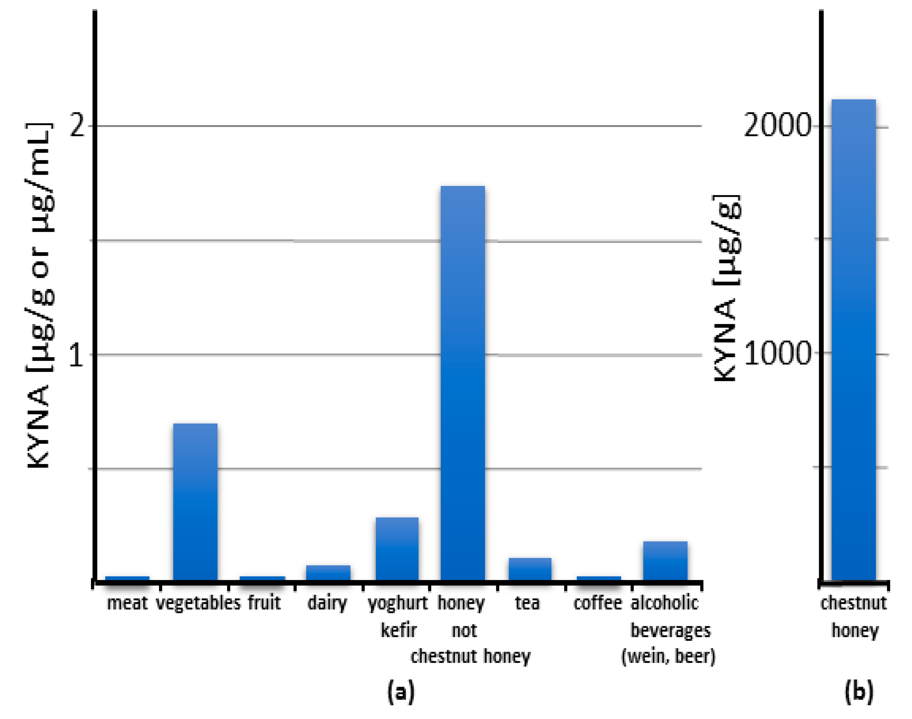
7. Kynurenic Acid in Food

7.1. Human Food

It has been repeatedly shown that KYNA is a natural component of food. Its content in food and food products varies within a wide range of concentrations, from trace amounts up to 2 mg/gram of chestnut honey [73,74,75,76,77,78,79,80] (Figure 3; Table S4).

7.1.1. Meat

It can be stated that the content of KYNA in meat is low: 0.0014, 0.0031, and 0.0037 μg/g wet weight in beef, pork, and fish, respectively. Even pig liver contains only slightly more KYNA—0.0091 μg/g wet weight. Since KYNA is rapidly excreted from the animal’s body (see Chapter 6), it does not seem possible to easily increase its amount in meat.

7.1.2. Vegetables

Vegetables are a richer source of KYNA (Figure 3; Table S4). Cauliflower, potato, and broccoli are some of the richest sources, containing KYNA in amounts of 0.0473, 0.1301, and 0.4184 μg/g wet weight, respectively [73]. However, it should be noted that large differences between varieties exist. The comparison of 16 different edible potato varieties grown under similar soil and climatic conditions showed up to 10-fold differences in KYNA content, from 0.04 to 0.65 μg/g wet weight [76]. Similarly, the comparison of yellow- and purple-fleshed potato cultivar Ismena and Provita revealed a threefold difference in KYNA content, 0.226 and 0.683 μg/g wet weight, respectively [81]. The origin of KYNA in plants is poorly understood. Both its synthesis from kynurenine and the absorption of KYNA from the soil were presented [82]. Since KYNA is found in the soil in varying amounts, and in extraordinary large amounts in manure [82], its content in the plant may depend on the site and cultivation method. This may also be the reason for significant differences in KYNA content even in the same type of vegetable. At the same time, it provides an opportunity to increase KYNA content in the plant by appropriate fertilization.

7.1.3. Fruit

The only fruit that has been studied is the apple, which contained 0.0023 μg of KYNA/g wet weight (Table S4) [73].

7.1.4. Spices and Herbs for Cooking

KYNA content in spices and herbs for cooking was determined in 19 different products (Table S4). The highest amount of KYNA was found in basil and thyme, 14.08 and 8.87 μg/g wet weight, respectively [77]. The numbers seem to be relatively high in comparison to plants. However, it must be stressed that, generally, commercially marketable spices are dried. Therefore, it is hard to accurately compare the concentration of KYNA in spices and fresh plants.

7.1.5. Honey

Unexpectedly, honey and other bee products contain relatively large amounts of KYNA (Figure 3; Table S4). An extremely high amount of KYNA was found in chestnut honey, up to 2114.9 μg/g [74]. Interestingly, this applies to chestnut honey obtained from different locations in Europe and Korea [13,74,80]. This is a phenomenon even among other types of honey. KYNA content in popular honey such as sunflower, multiflorous, buckwheat, acacia, and linden honey is 1.73, 0.877, 0.33, 0.181, and 0.179 μg/g, respectively. A high level of KYNA in chestnut honey seems to be related to the high KYNA content in male flowers of the chestnut tree [13].

7.1.6. Dairy

In commercially available dairy products, the content of KYNA is as follows: cow’s milk: 0.017 μg/mL, kefir: 0.2417 μg/mL, yoghurt: 0.2868 μg/mL, white cheese: 0.0766 μg/g, and hard cheese: 0.0084 [42,49] (Figure 3; Table S4) [73,78].

7.1.7. Fermented Food and Beverages

A relatively high KYNA content was found in fermented food products (Figure 3; Table S4). It was found in kefir and yoghurt in amounts of up to 0.242 and 0.287 μg/mL, respectively [73,78]. In addition, cocoa powder contains KYNA in the amount of 4.486 μg/g [78]. Interestingly, the alcoholic beverages wine and beer contain KYNA in a broad range of concentrations, up to 0.179 and 0.051 μg/mL, respectively [11,78,79]. These results indicate that the fermenting microorganisms produce KYNA and that this process may significantly increase the KYNA content in food and beverages.

7.1.8. Medicinal Herbs and Supplements

The presence of KYNA has been demonstrated in medicinal herbs and supplements (Table S5). The highest content expressed on a dry weight basis of the herbs was found in leaves of peppermint, nettle, birch, and horsetail, ranging from 2.27 to 3.82 μg/g dry weight [83]. The intake of KYNA in herbal infusions prepared according to manufacturer’s instructions were found to vary from 1.08 μg/day in the nettle root infusion to 32.5 and 32.6 μg/day in the nettle leaf and St. John’s wort infusion, respectively. Herbal supplements in the form of tablets also contain KYNA. KYNA delivery calculated in a maximum recommended dose of the supplement equals from 0.41 to 30.38 μg/g in chamomile and St. John’s wort tablets, respectively [82].

7.1.9. Baby Food

KYNA was found in human milk [45,46,73] (Table S6). Interestingly, the content of KYNA in human milk increases more than 14 times during the time of breastfeeding, starting from 0.004 μg/mL on day 3 after labor and reaching a value of 0.057 μg/mL in the 6th month of feeding [46]. KYNA was also found in all 46 artificial baby milk formulas studied. However, in comparison with human milk, in which its content naturally changed over time, the concentration of KYNA in artificial formulas was substantially lower and did not follow its physiological dynamics of changes [46]. In first-food formulas, KYNA content is clearly higher in products containing vegetables (0.0056–0.0148 µg/g) than in meat-based food (0.01 µg/g). In fruit and vegetable juice, its concentration is 0.0019 µg/g [73]. It should be noted that this estimation is based on single measurement results only, which are insufficient to draw definite conclusions.

7.2. Animal Food

The presence of KYNA was studied in animal feed for livestock, cats, dogs, and fish. It was shown that KYNA is present in animal feed in varying concentrations. The highest concentration of KYNA was found in feed for livestock, where it varied from 0.198 μg/g fresh weight to 0.414 μg/g fresh weight. Based on the measurement of KYNA content in feed ingredients, the authors concluded that the concentration of KYNA in animal feed was not controlled and deliberately set, but its final content depended on the ingredients used [84].

8. Kynurenic Acid Supplementation

8.1. Health Effects of Kynurenic Acid Supplementation

Since KYNA is formed endogenously in the body and can also be supplied in food, the question about the relevance of its supplementation is legitimate. No human studies devoted to health effects of exogenously administered KYNA have been conducted to date. An exception is a study in which a solution of chestnut honey was administered to young volunteers and pharmacokinetic parameters of KYNA were determined afterwards. No side effects were reported in this study [11]. Indirectly, the lack of KYNA toxicity can be inferred from studies in which tryptophan was administered to humans. It was found that tryptophan load resulted in an increase in kynurenines, including KYNA, in the blood. Very recently, Sathyasaikumar et al. (2022) reported no serious adverse events and no long-term changes in behavior and health in tryptophan-treated humans who had plasma KYNA levels that increased as much as 145-fold compared to pre-tryptophan values [85].
Further conclusions should be drawn based on the data obtained from studies on rodents. The data search performed showed that there are few publications describing KYNA administration via the alimentary route for the period lasting from 3 days to 2 months (Table 2).
Because KYNA is water-soluble, it was administered in drinking water in most studies. This mode of administration is very convenient because it does not cause stress to the animal. In addition, water was available ad libitum, which allowed KYNA to be taken in the most natural way according to the daily pattern of drinking. The concentrations used ranged from 2.5 to 250 mg/L, approximating a dose of 0.25 to 25 mg/kg of KYNA per 1 kg body weight/day, respectively. Studies have been conducted on both young and adult animals. Generally, no toxic effects of KYNA administration have been reported. When administered to young animals, KYNA did not interfere with their overall growth and development. However, a moderate reduction in the rate of weight gain was observed. This effect was evident in young, but not adult, animals. On the other hand, the reduction of weight gain rate was present in adult rats kept on a high-fat diet (Table 2). These observations allowed for hypothesizing antiobesogenic properties of KYNA during early development [46]. Interestingly, KYNA has been found to stimulate intestinal mucosal growth in young rats and to cause, inter alia, an increase in the intestinal surface area. However, this affects neither the body composition nor bone mineralization and endurance capacity (Table 2). Importantly, the supplementation of KYNA in drinking water to developing rats for 2 months did not impair their brain functions measured in adulthood [92].
In healthy adult mice, the alimentary administration of KYNA did not affect blood hematological parameters. However, experiments performed in vitro on leukocytes and splenocytes obtained from drug supplemented animals revealed that KYNA exerted antioxidant and immunomodulatory effects [87,88]. In spontaneously hypertensive adult rats, the administration of KYNA in drinking water for 3 weeks did not affect the mean arterial pressure, but it moderately reduced heart rate [89] (Table 2).
Another method of the alimentary administration of the drug was utilized by Li et al. (2021) [90]. In this study, KYNA was applied intragastrically to adult mice kept on a high-fat diet, once a day for 8 weeks, in a dose of 5 mg/kg/day. It was found that such a regimen resulted in declined body weight gain and reduced daily energy intake. Moreover, the following serum metabolic parameters were improved: triglyceride, serum high-density lipoprotein cholesterol, and low-density lipoprotein levels. Finally, both the atherosclerosis index and the coronary artery risk index were significantly decreased [90]. Similar metabolic effects of KYNA injected intraperitoneally at a dose of 5 mg/kg/day, once a day for 1 to 4 weeks, to mice on a high-fat diet were described [93]. Very recently, it was reported that KYNA administered intraperitoneally three times decreased the colonization of the intestine by fungi and ameliorated intestinal injury, i.e., inhibited inflammation, promoted the expression of intestinal tight junction proteins, and protected from intestinal barrier damage caused by invasive Candida albicans infection in mice [94] (Table 2). Other data indicating multiple health-promoting effects of KYNA come from studies in which the drug was acutely injected intraperitoneally (Table S7). This route of administration is preferred by researchers because it allows for the accurate dosage and precise determination of the time of action of the substance. Historically, the earliest data relate to the antiulcer effects of KYNA. Glavin and Pinsky, in 1989, showed that KYNA significantly blocked restraint-cold stress ulcers, ethanol ulcers, and basal nonstimulated gastric acid secretion in normal rats. The authors premised both peripheral and cerebral effects of KYNA [95]. Protective effects of injected KYNA on the liver and the pancreas, and in disorders of the lower gastrointestinal tract, were later described (Table S7). More recently, numerous reports performed on animals on the beneficial effect of KYNA on the conditions commonly referred to as metabolic diseases in humans were published. Antiobesity, cholesterol-lowering, glucose tolerance improvement, and antiatherosclerotic effects were evidenced in appropriate animal models (Table S7). It is surprising that many of these effects observed after KYNA administration in animals can be therapeutic targets for metabolic syndromes in humans (Figure 4).
Similar disturbances were described as obese-related multimorbidity, when a low-caloric KYNA-enriched functional diet should be considered as supportive care. The effect of KYNA on bone metabolism is also of interest. A recent publication by Shi et al. (2022) revealed that KYNA administered in a relatively low dose of 5 mg/kg/day for 4 weeks alleviated the postmenopausal osteoporosis and highlighted the involvement of the GPR35 receptor, a molecular target of KYNA, in this action [97] (Table S7).
In addition, it has been shown that KYNA may have beneficial health effects in life-threatening conditions (Table S7), and this issue requires a separate commentary. Moroni et al. (2012) were the first to communicate that KYNA administered subcutaneously at doses of 500 mg/kg (single injection) or 200 mg/kg three times at 0, 3, and 6 h after LPS dramatically reduced LPS-induced death in mice [98]. The Hungarian group confirmed that KYNA protects against LPS-induced sepsis in subsequent publications, in which a much lower KYNA dose of 30 mg/kg, i.p., was used [99,100]. Most recently, Wang et al. (2022) demonstrated that intraperitoneally administered KYNA (5 mg/kg; three times at days 3, 6, and 9) reduced the mortality of mice infected with Candida albicans [94]. Hsieh et al. (2011) reported that KYNA administered intravenously at doses ranging from 30–100 mg/kg attenuated multiorgan dysfunction in rats exposed to heatstroke [101]. Kaszaki et al. (2008) found a profound anti-inflammatory action of KYNA administered in intravenous infusion in experimental colon obstruction in dogs [102]. Similarly, Marciniak, in 2013, demonstrated that the intravenous infusion of KYNA alleviated symptoms of experimental acute pancreatitis in rats [103]. These results deserve special attention because they indicate the feasibility of using KYNA administered as a bolus or an intravenous infusion in life-threatening conditions.
The beneficial wound healing effects of KYNA (Table S7) after its external administration on the skin and cornea in rabbits are also worth mentioning [104,105]. It is worth noting that a clinical trial with the use of 0.5% KYNA dressing in people with skin scarring has already been successfully completed [106]. Furthermore, KYNA encapsulated in synthetic polymer microspheres implanted in a wound bed in rats was shown to reduce fibrotic tissue formation [107].
Detailed data on the health-promoting effects of KYNA administration in animals regardless of the route of administration are presented in Table S7 [26,87,90,93,94,95,97,98,99,100,101,102,103,104,105,106,108,109,110,111,112,113,114,115,116,117,118,119,120,121,122,123].
Since there are no substantial differences between the effects exerted by KYNA administered alimentary or infused by injection, it can be assumed that its supplementation by the oral route will produce similar effects as those described after infusion.

8.2. Clinical Trials

In the ClinicalTrials.gov database, only four registered studies were found in which KYNA was applied to humans. All studies were dedicated to examining the effect of the topical application of KYNA to skin (Table S8).

8.3. Patents

A survey of the PubChem database of patents in which the keyword KYNA appears revealed 23 patent applications dealing with the medical use of the drug (Table S9). Most of them relate to its oral or injectable administration and only a few assume the topical or local administration of KYNA. The most commonly claimed effect of KYNA is in digestive tract, liver, and pancreatic conditions. KYNA’s effects have also been recommended in cardiovascular pathologies, lipid metabolism disorders, obesity, and kidney dysfunction. In addition, its use in fibrotic diseases, eye diseases, and mental stress is mentioned. In general, this is in line with the data obtained from scientific publications. Some novelty derived from the patent descriptions is KYNA’s effect on skeletal muscles and its proposed use in sarcopenia or hangover control. In addition to the scientific content, it is worth noting that the number of patent applications on the medical use of KYNA has increased substantially since 2016.

9. Perspectives

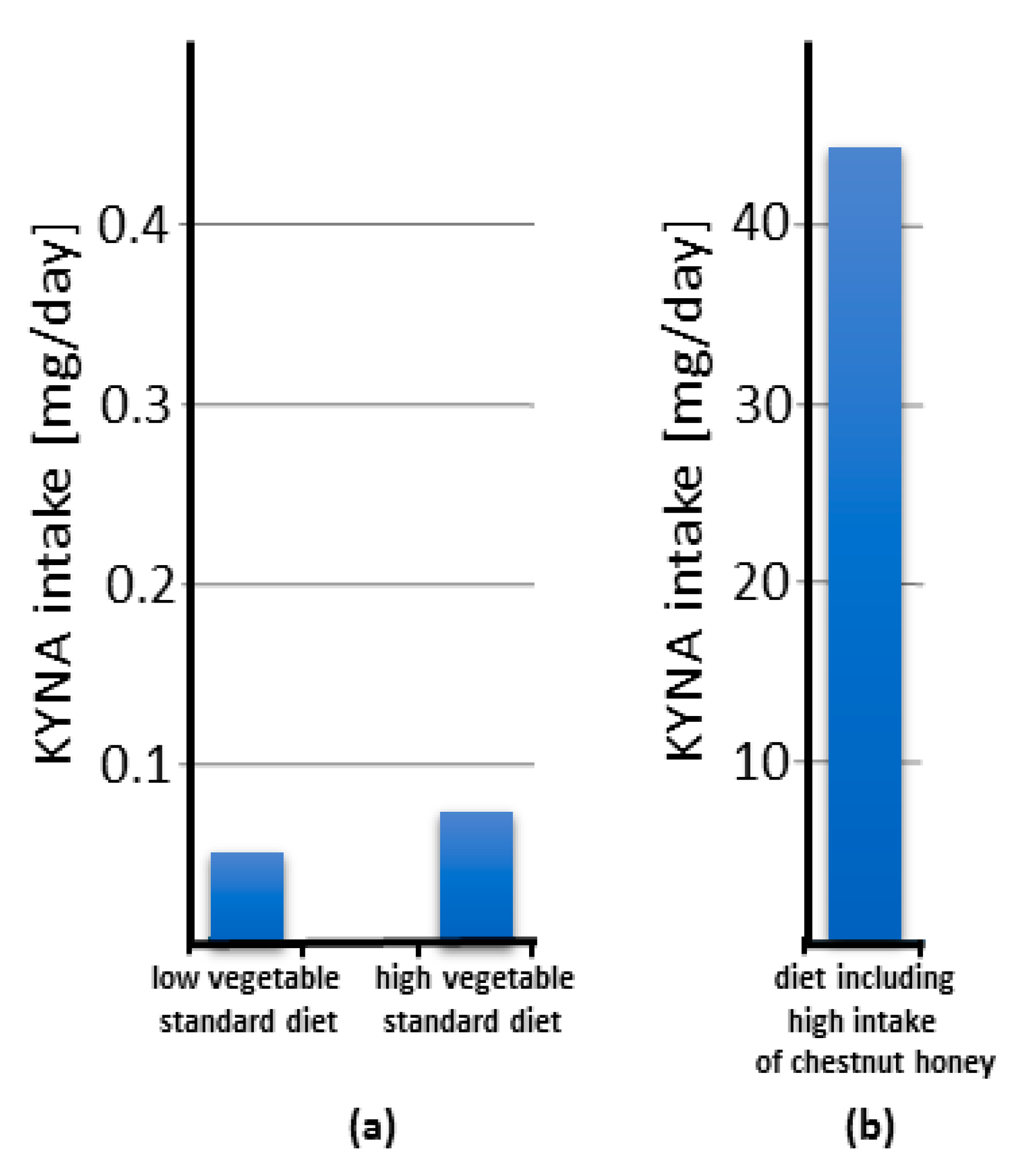
9.1. Food as a Dietary Source of Kynurenic Acid

Although the presence of KYNA in food is a topic that has been addressed several times in various publications, the knowledge of its content in food products is still insufficient. It should be underlined that most of the studies come from Eurasia. There are almost no data from other continents and regions where diets are based on other products than in Europe. Thus, it is advisable to continue the search for rich sources of KYNA among various food products from all over the world.
Based on the current knowledge, it can be concluded that KYNA content in food and food products is generally low (Figure 3; Table S4) and, according to our calculation (Tables S10–S13), its daily intake is very small (Figure 5a) and quite marginal compared to its excretion (Table 1). The calculation of food-related KYNA intake was based on simulation of a dish of cruciferous, yellow, green, and other vegetables mixed equally, assuming that a single serving weighs 80 g according to Bensley et al. (2003) [124]. The KYNA ingested in red meat, poultry, eggs, and other extensively analyzed nutrients is omitted due to trace of their contents. Low intake was established as a consumption of three or fewer servings per week. High intake was established as consumption of more than 10 servings per week.
The calculated mean daily intake of KYNA as a food constituent was estimated to be approximately 0.065 mg in a diet that does not contain honey. The mean daily intake of KYNA derived from food is notably higher when chestnut honey is consumed in significant quantities and may account for 20.81 mg. However, it should be noted that chestnut honey is produced by bees that collect pollen in areas where chestnut trees grow. These regions are limited due to soil and climate requirements as well as cultivation culture [125]. Thus, it can be concluded that, for most of the human population, the daily amount of food-derived KYNA lies well below 0.1 mg. Taking into account that the daily requirement for KYNA in adult humans, calculated on the basis of its daily excretion, varies from 1.2 to 7 mg (Table 1), it can be concluded that, for most people, the food-derived KYNA does not exceed 10% of the daily requirement. In contrast, in people consuming chestnut honey in their diet, the supplied KYNA, approximating the value of 20 mg, almost triples the daily need. The latter estimation points to the safety of the alimentary administration of KYNA in amounts of up to 20 mg/day.
KYNA content in meat is very low, while in vegetables it is considerably higher but there are large differences between individual types of vegetables and their varieties. Moreover, it seems to depend on where and how the plants are grown. For these reasons, it is difficult to make rational dietary recommendations yet. In terms of KYNA content, chestnut honey is a particular exception among food products including other types of honey. On the other hand, almost nothing is known about the KYNA content in fruit. Interestingly, fermented food contains a higher level of KYNA in comparison to unprocessed food. This knowledge can be used to increase KYNA content in the human diet.
It is noteworthy that KYNA is present in breast milk from the beginning of newborn feeding and its content considerably rises over time [46]. The importance of this phenomenon is still far from being understood, but it should be noted that KYNA content in baby food formulas does not parallel its physiological changes.

9.2. Chestnut Honey as a Dietary Source of Kynurenic Acid

Summarizing the data collected so far, it is evident that KYNA possesses potential health-promoting relevance (Table S7). Its presence in food has been repeatedly confirmed, but its amount is relatively small. The exception is chestnut honey, in which the content of this substance can be even 1000 times higher than in other food products (Figure 3; Table S4). The uniqueness of chestnut honey is reflected by the fact that the consumption of only 10 g of honey provides such an amount of KYNA that corresponds to its average daily excretion. Greater consumption of chestnut honey in the amounts recommended for use for medical purposes [126] can result in the delivery of more than 3000 times KYNA per day than in a standard diet (Figure 5b). This creates a unique opportunity to verify whether the consumption of chestnut honey brings health benefits to humans. In fact, there are scientific reports on the effects of chestnut honey on diseases modeled in laboratory animals [127,128,129,130,131] (Table S14). Interestingly, similar to KYNA, chestnut honey administered by the alimentary route counteracts the effects of a high-fat diet in mice and also has an antiulcer effect in the alcohol model of the disease in rats [127,128]. Moreover, it was found to enhance the healing of CCl4-induced liver damage in rats [129]. When administered topically, it improves skin wound healing and the healing of corneal alkali burns [130,131]. Other effects described based on in vitro studies and biochemical analyses are summarized in Table S14 [132,133,134,135,136,137,138,139,140,141]. It should be noted that many of the reported effects of chestnut honey parallel those of KYNA. Therefore, it would be interesting to see if a health-promoting effect has been recognized in population-based studies. Unfortunately, we found no relevant studies in the available literature. This seems to be due to the fact that chestnut honey is not identified in publications on the dietary effects on human health. Furthermore, honey is rarely seen as a separate category in such studies. Therefore, we propose to reinvestigate previous data or to conduct such studies including chestnut honey consumption as a separate item in the menu, due to its exceptionally high KYNA content [13]. Such a study may be facilitated by the fact that chestnut honey is not widely consumed worldwide. It is primarily used in areas where chestnut trees grow. In many countries, its consumption is marginal or practically nonexistent. This allows for the matching of appropriate study groups with a high consumption of chestnut honey and a control group that does not consume chestnut honey. A brief analysis of the chestnut cultivation area indicates that it is typical of Mediterranean European countries, but also of some Asian countries. It also appears that many so-called Blue Zones, regions of the world thought to have a higher than usual number of people who live much longer than average, are located in areas where chestnut trees grow.
Chestnut honey is indeed a very rich source of KYNA, but it has some drawbacks and there are significant limitations to its consumption. First of all, honey contains about 50% monosaccharides and, for this reason, its high consumption is not popular nowadays. However, it should be realized that chestnut honey attenuates postprandial glycemic response and, therefore, is classified as a food with a low or medium glycemic index and glycemic load [142,143,144]. Second, due to the presence of bee- and pollen-derived proteins, honey can be allergenic. Young children should not eat honey. Third, the amount of honey produced by bees is limited and may not be enough to cover increased demands. Therefore, other sources of KYNA should be searched for, and at best its safety should be assessed and the use of KYNA as a supplement allowed.

9.3. Other Sources of Kynurenic Acid

Recent publications indicate that yeast may be an alternative source of KYNA [79,145,146]. The production of KYNA by yeast can be carried out on an industrial scale and does not require either suitable soil or climatic conditions, as do chestnut cultivation and honey production. This is a major advantage. However, the disadvantages are the relatively low efficiency of the production process and the limited amount of yeast that can be used for feeding. Therefore, further work is needed to improve the process of KYNA production by microorganisms. There is also a lack of data on the amount of KYNA production by the gut microbiome and how this process can be intensified.

10. Concluding Remarks

KYNA appears to be a very promising molecule with potential health-promoting importance and beneficial pharmacodynamics. The fact that it is found in very large amounts in chestnut honey, which is consumed only in limited areas, creates the opportunity to trace its effects on the human body without the need for long-term prospective studies. The rationale for such studies appears to be obvious. It is also imperative not to forget to be cautious, and to perform appropriate studies and closely follow information about its potential side effects before KYNA enters widely into food supplementation. Thus, it seems that the question should be when, not if, KYNA will be used as a dietary supplement.

Supplementary Materials

The following supporting information can be downloaded at: https://www.mdpi.com/article/10.3390/nu14194182/s1, Table S1. Content of kynurenic acid in human serum; Table S2. Content of kynurenic acid in human cerebrospinal fluid; Table S3. Excretion of kynurenic acid in human urine; Table S4. Content of kynurenic acid in food, beverages, herbs, and spices for cooking; Table S5. Content of kynurenic acid in medicinal herbs and supplements; Table S6. Content of kynurenic acid in human milk and baby food; Table S7. Health-related effects of kynurenic acid (KYNA) administration; Table S8. Clinical trials designed to investigate the kynurenic acid (KYNA) effect registered in ClinicalTrials.gov database; Table S9. Patents and patent applications claiming medical uses of kynurenic acid (KYNA); Table S10. Estimated daily intake of kynurenic acid as an ingredient of food; Table S11. Intake of kynurenic acid in vegetables; Table S12. Intake of kynurenic acid in meat, dairy, egg, and beverages; Table S13. Intake of kynurenic acid in single and daily dose of honey; Table S14. Effects of chestnut honey established in animals, in vitro and biochemical studies.

Author Contributions

Conceptualization, M.T.; methodology, M.T. and P.P.; formal analysis, M.T., P.P. and W.A.T.; writing—original draft preparation, J.P.-T., M.T., P.P. and W.A.T.; writing—review and editing, M.T.; visualization, M.T. and W.A.T.; supervision, J.P.-T. and M.T.; project administration, W.A.T.; funding acquisition, J.P.-T. and W.A.T. All authors have read and agreed to the published version of the manuscript.

Funding

The publication was supported by grants DS 394 and DS 448 from the Medical University of Lublin, Poland.

Institutional Review Board Statement

Not applicable.

Informed Consent Statement

Not applicable.

Data Availability Statement

Not applicable.

Conflicts of Interest

The authors declare no conflict of interest.

References

  1. Turski, M.P.; Turska, M.; Paluszkiewicz, P.; Parada-Turska, J.; Oxenkrug, G.F. Kynurenic Acid in the Digestive System-New Facts, New Challenges. Int. J. Tryptophan Res. 2013, 6, 47–55. [Google Scholar] [CrossRef] [PubMed] [Green Version]
  2. Kapolka, N.J.; Taghon, G.J.; Rowe, J.B.; Morgan, W.M.; Enten, J.F.; Lambert, N.A.; Isom, D.G. DCyFIR: A High-Throughput CRISPR Platform for Multiplexed G Protein-Coupled Receptor Profiling and Ligand Discovery. Proc. Natl. Acad. Sci. USA 2020, 117, 13117–13126. [Google Scholar] [CrossRef]
  3. Collingridge, G.L.; Abraham, W.C. Glutamate Receptors and Synaptic Plasticity: The Impact of Evans and Watkins. Neuropharmacology 2022, 206, 108922. [Google Scholar] [CrossRef]
  4. Stone, T.W. Does Kynurenic Acid Act on Nicotinic Receptors? An Assessment of the Evidence. J. Neurochem. 2020, 152, 627–649. [Google Scholar] [CrossRef] [Green Version]
  5. Shore, D.M.; Reggio, P.H. The Therapeutic Potential of Orphan GPCRs, GPR35 and GPR55. Front. Pharm. 2015, 6, 69. [Google Scholar] [CrossRef] [PubMed] [Green Version]
  6. Wang, J.; Simonavicius, N.; Wu, X.; Swaminath, G.; Reagan, J.; Tian, H.; Ling, L. Kynurenic Acid as a Ligand for Orphan G Protein-Coupled Receptor GPR35. J. Biol. Chem. 2006, 281, 22021–22028. [Google Scholar] [CrossRef] [PubMed] [Green Version]
  7. O’Dowd, B.F.; Nguyen, T.; Marchese, A.; Cheng, R.; Lynch, K.R.; Heng, H.H.Q.; Kolakowski, L.F.; George, S.R. Discovery of Three Novel G-Protein-Coupled Receptor Genes. Genomics 1998, 47, 310–313. [Google Scholar] [CrossRef]
  8. Okumura, S.; Baba, H.; Kumada, T.; Nanmoku, K.; Nakajima, H.; Nakane, Y.; Hioki, K.; Ikenaka, K. Cloning of a G-Protein-Coupled Receptor That Shows an Activity to Transform NIH3T3 Cells and Is Expressed in Gastric Cancer Cells. Cancer Sci. 2004, 95, 131–135. [Google Scholar] [CrossRef] [Green Version]
  9. Kaya, B.; Melhem, H.; Niess, J.H. GPR35 in Intestinal Diseases: From Risk Gene to Function. Front. Immunol. 2021, 12, 717392. [Google Scholar] [CrossRef] [PubMed]
  10. Quon, T.; Lin, L.-C.; Ganguly, A.; Tobin, A.B.; Milligan, G. Therapeutic Opportunities and Challenges in Targeting the Orphan G Protein-Coupled Receptor GPR35. ACS Pharmacol. Transl. Sci. 2020, 3, 801–812. [Google Scholar] [CrossRef] [PubMed]
  11. Turska, M.; Rutyna, R.; Paluszkiewicz, M.; Terlecka, P.; Dobrowolski, A.; Pelak, J.; Turski, M.P.; Muszyńska, B.; Dabrowski, W.; Kocki, T.; et al. Presence of Kynurenic Acid in Alcoholic Beverages—Is This Good News, or Bad News? Med Hypotheses 2019, 122, 200–205. [Google Scholar] [CrossRef]
  12. Kaihara, M.; Price, J.M.; Takahashi, H. The Conversion of Kynurenic Acid to Quinaldic Acid by Humans and Rats. J Biol Chem 1956, 223, 705–708. [Google Scholar]
  13. Turski, M.P.; Chwil, S.; Turska, M.; Chwil, M.; Kocki, T.; Rajtar, G.; Parada-Turska, J. An Exceptionally High Content of Kynurenic Acid in Chestnut Honey and Flowers of Chestnut Tree. J. Food Compos. Anal. 2016, 48, 67–72. [Google Scholar] [CrossRef]
  14. Turska, M.; Pelak, J.; Turski, M.P.; Kocki, T.; Dukowski, P.; Plech, T.; Turski, W. Fate and Distribution of Kynurenic Acid Administered as Beverage. Pharm. Rep. 2018, 70, 1089–1096. [Google Scholar] [CrossRef]
  15. Li, P.; Zheng, J.; Bai, Y.; Wang, D.; Cui, Z.; Li, Y.; Zhang, J.; Wang, Y. Characterization of Kynurenine Pathway in Patients with Diarrhea-Predominant Irritable Bowel Syndrome. Eur. J. Histochem. 2020, 64. [Google Scholar] [CrossRef]
  16. Iłzecka, J.; Kocki, T.; Stelmasiak, Z.; Turski, W.A. Endogenous Protectant Kynurenic Acid in Amyotrophic Lateral Sclerosis. Acta Neurol. Scand. 2003, 107, 412–418. [Google Scholar] [CrossRef] [PubMed]
  17. Huang, J.; Tong, J.; Zhang, P.; Zhou, Y.; Cui, Y.; Tan, S.; Wang, Z.; Yang, F.; Kochunov, P.; Chiappelli, J.; et al. Effects of Neuroactive Metabolites of the Tryptophan Pathway on Working Memory and Cortical Thickness in Schizophrenia. Transl. Psychiatry 2021, 11, 198. [Google Scholar] [CrossRef]
  18. Hartai, Z.; Klivenyi, P.; Janaky, T.; Penke, B.; Dux, L.; Vecsei, L. Kynurenine Metabolism in Plasma and in Red Blood Cells in Parkinson’s Disease. J. Neurol. Sci. 2005, 239, 31–35. [Google Scholar] [CrossRef]
  19. Zhang, Z.; Zhang, M.; Luo, Y.; Ni, X.; Lu, H.; Wen, Y.; Fan, N. Preliminary Comparative Analysis of Kynurenine Pathway Metabolites in Chronic Ketamine Users, Schizophrenic Patients, and Healthy Controls. Hum. Psychopharmacol. 2020, 35, e2738. [Google Scholar] [CrossRef] [PubMed]
  20. Tömösi, F.; Kecskeméti, G.; Cseh, E.K.; Szabó, E.; Rajda, C.; Kormány, R.; Szabó, Z.; Vécsei, L.; Janáky, T. A Validated UHPLC-MS Method for Tryptophan Metabolites: Application in the Diagnosis of Multiple Sclerosis. J. Pharm. Biomed. Anal. 2020, 185, 113246. [Google Scholar] [CrossRef]
  21. Kepplinger, B.; Baran, H.; Kainz, A.; Ferraz-Leite, H.; Newcombe, J.; Kalina, P. Age-Related Increase of Kynurenic Acid in Human Cerebrospinal Fluid—IgG and Beta2-Microglobulin Changes. Neurosignals 2005, 14, 126–135. [Google Scholar] [CrossRef] [PubMed]
  22. Amirkhani, A.; Heldin, E.; Markides, K.E.; Bergquist, J. Quantitation of Tryptophan, Kynurenine and Kynurenic Acid in Human Plasma by Capillary Liquid Chromatography-Electrospray Ionization Tandem Mass Spectrometry. J. Chromatogr. B Anal. Technol. Biomed. Life Sci. 2002, 780, 381–387. [Google Scholar] [CrossRef]
  23. Milart, P.; Urbańska, E.M.; Turski, W.A.; Paszkowski, T.; Sikorski, R. Intrapartum Levels of Endogenous Glutamate Antagonist-Kynurenic Acid in Amniotic Fluid, Umbilical and Maternal Blood. Neurosci. Res. Comm. 1999, 24, 173–178. [Google Scholar] [CrossRef]
  24. Milart, P.; Sikorski, R. Kynurenic acid concentration in blood and urine during normal pregnancy. Ginekol. Pol. 1998, 69, 968–973. [Google Scholar] [PubMed]
  25. Shestopalov, A.V.; Shatova, O.P.; Gaponov, A.M.; Moskaleva, N.E.; Appolonova, S.A.; Tutelyan, A.V.; Makarov, V.V.; Yudin, S.M.; Rumyantsev, S.A. The study of tryptophan metabolite concentrations in blood serum and fecal extracts from obese children. Biomed. Khim. 2020, 66, 494–501. [Google Scholar] [CrossRef] [PubMed]
  26. Kuc, D.; Rahnama, M.; Tomaszewski, T.; Rzeski, W.; Wejksza, K.; Urbanik-Sypniewska, T.; Parada-Turska, J.; Wielosz, M.; Turski, W.A. Kynurenic Acid in Human Saliva--Does It Influence Oral Microflora? Pharm. Rep. 2006, 58, 393–398. [Google Scholar]
  27. Paluszkiewicz, P.; Zgrajka, W.; Saran, T.; Schabowski, J.; Piedra, J.L.V.; Fedkiv, O.; Rengman, S.; Pierzynowski, S.G.; Turski, W.A. High Concentration of Kynurenic Acid in Bile and Pancreatic Juice. Amino Acids 2009, 37, 637–641. [Google Scholar] [CrossRef] [PubMed]
  28. Walczak, K.; Dąbrowski, W.; Langner, E.; Zgrajka, W.; Piłat, J.; Kocki, T.; Rzeski, W.; Turski, W.A. Kynurenic Acid Synthesis and Kynurenine Aminotransferases Expression in Colon Derived Normal and Cancer Cells. Scand. J. Gastroenterol. 2011, 46, 903–912. [Google Scholar] [CrossRef] [PubMed]
  29. Parada-Turska, J.; Zgrajka, W.; Majdan, M. Kynurenic Acid in Synovial Fluid and Serum of Patients with Rheumatoid Arthritis, Spondyloarthropathy, and Osteoarthritis. J. Rheumatol. 2013, 40, 903–909. [Google Scholar] [CrossRef] [PubMed]
  30. Saran, T.; Turska, M.; Kocki, T.; Zawadka, M.; Zieliński, G.; Turski, W.A.; Gawda, P. Effect of 4-Week Physical Exercises on Tryptophan, Kynurenine and Kynurenic Acid Content in Human Sweat. Sci. Rep. 2021, 11, 11092. [Google Scholar] [CrossRef] [PubMed]
  31. Alarcan, H.; Chaumond, R.; Emond, P.; Benz-De Bretagne, I.; Lefèvre, A.; Bakkouche, S.-E.; Veyrat-Durebex, C.; Vourc’h, P.; Andres, C.; Corcia, P.; et al. Some CSF Kynurenine Pathway Intermediates Associated with Disease Evolution in Amyotrophic Lateral Sclerosis. Biomolecules 2021, 11, 691. [Google Scholar] [CrossRef] [PubMed]
  32. Rodrigues, F.B.; Byrne, L.M.; Lowe, A.J.; Tortelli, R.; Heins, M.; Flik, G.; Johnson, E.B.; De Vita, E.; Scahill, R.I.; Giorgini, F.; et al. Kynurenine Pathway Metabolites in Cerebrospinal Fluid and Blood as Potential Biomarkers in Huntington’s Disease. J. Neurochem. 2021, 158, 539–553. [Google Scholar] [CrossRef] [PubMed]
  33. Kepplinger, B.; Baran, H.; Kronsteiner, C.; Reuss, J. Increased Levels of Kynurenic Acid in the Cerebrospinal Fluid in Patients with Hydrocephalus. Neurosignals 2019, 27, 1–11. [Google Scholar] [CrossRef]
  34. Nilsson, L.K.; Linderholm, K.R.; Engberg, G.; Paulson, L.; Blennow, K.; Lindström, L.H.; Nordin, C.; Karanti, A.; Persson, P.; Erhardt, S. Elevated Levels of Kynurenic Acid in the Cerebrospinal Fluid of Male Patients with Schizophrenia. Schizophr. Res. 2005, 80, 315–322. [Google Scholar] [CrossRef] [PubMed] [Green Version]
  35. Erhardt, S.; Schwieler, L.; Engberg, G. Kynurenic Acid and Schizophrenia. Adv. Exp. Med. Biol. 2003, 527, 155–165. [Google Scholar] [CrossRef]
  36. Rejdak, K.; Bartosik-Psujek, H.; Dobosz, B.; Kocki, T.; Grieb, P.; Giovannoni, G.; Turski, W.A.; Stelmasiak, Z. Decreased Level of Kynurenic Acid in Cerebrospinal Fluid of Relapsing-Onset Multiple Sclerosis Patients. Neurosci. Lett. 2002, 331, 63–65. [Google Scholar] [CrossRef]
  37. Swartz, K.J.; Matson, W.R.; MacGarvey, U.; Ryan, E.A.; Beal, M.F. Measurement of Kynurenic Acid in Mammalian Brain Extracts and Cerebrospinal Fluid by High-Performance Liquid Chromatography with Fluorometric and Coulometric Electrode Array Detection. Anal. Biochem. 1990, 185, 363–376. [Google Scholar] [CrossRef]
  38. Yamamoto, H.; Murakami, H.; Horiguchi, K.; Egawa, B. Studies on Cerebrospinal Fluid Kynurenic Acid Concentrations in Epileptic Children. Brain Dev. 1995, 17, 327–329. [Google Scholar] [CrossRef]
  39. Turski, W.A.; Nakamura, M.; Todd, W.P.; Carpenter, B.K.; Whetsell, W.O.; Schwarcz, R. Identification and Quantification of Kynurenic Acid in Human Brain Tissue. Brain Res. 1988, 454, 164–169. [Google Scholar] [CrossRef]
  40. Connick, J.H.; Carlà, V.; Moroni, F.; Stone, T.W. Increase in Kynurenic Acid in Huntington’s Disease Motor Cortex. J. Neurochem. 1989, 52, 985–987. [Google Scholar] [CrossRef]
  41. Moroni, F.; Russi, P.; Lombardi, G.; Beni, M.; Carlà, V. Presence of Kynurenic Acid in the Mammalian Brain. J. Neurochem. 1988, 51, 177–180. [Google Scholar] [CrossRef] [PubMed]
  42. Baran, H.; Hainfellner, J.A.; Kepplinger, B.; Mazal, P.R.; Schmid, H.; Budka, H. Kynurenic Acid Metabolism in the Brain of HIV-1 Infected Patients. J. Neural. Transm. 2000, 107, 1127–1138. [Google Scholar] [CrossRef]
  43. Baran, H.; Hainfellner, J.A.; Kepplinger, B. Kynurenic Acid Metabolism in Various Types of Brain Pathology in HIV-1 Infected Patients. Int. J. Tryptophan Res. 2012, 5, 49–64. [Google Scholar] [CrossRef] [Green Version]
  44. Schwarcz, R.; Rassoulpour, A.; Wu, H.Q.; Medoff, D.; Tamminga, C.A.; Roberts, R.C. Increased Cortical Kynurenate Content in Schizophrenia. Biol. Psychiatry 2001, 50, 521–530. [Google Scholar] [CrossRef]
  45. O’Rourke, L.; Clarke, G.; Nolan, A.; Watkins, C.; Dinan, T.G.; Stanton, C.; Ross, R.P.; Ryan, C.A. Tryptophan Metabolic Profile in Term and Preterm Breast Milk: Implications for Health. J. Nutr. Sci. 2018, 7, e13. [Google Scholar] [CrossRef] [Green Version]
  46. Milart, P.; Paluszkiewicz, P.; Dobrowolski, P.; Tomaszewska, E.; Smolinska, K.; Debinska, I.; Gawel, K.; Walczak, K.; Bednarski, J.; Turska, M.; et al. Kynurenic Acid as the Neglected Ingredient of Commercial Baby Formulas. Sci. Rep. 2019, 9, 6108. [Google Scholar] [CrossRef] [Green Version]
  47. Oluwagbemigun, K.; Anesi, A.; Clarke, G.; Schmid, M.; Mattivi, F.; Nöthlings, U. An Investigation into the Temporal Reproducibility of Tryptophan Metabolite Networks Among Healthy Adolescents. Int. J. Tryptophan Res. 2021, 14, 11786469211041376. [Google Scholar] [CrossRef]
  48. Gomez-Gomez, A.; Marcos, J.; Aguilera, P.; To-Figueras, J.; Pozo, O.J. Comprehensive Analysis of the Tryptophan Metabolome in Urine of Patients with Acute Intermittent Porphyria. J. Chromatogr. B Anal. Technol. Biomed. Life Sci. 2017, 1060, 347–354. [Google Scholar] [CrossRef] [PubMed]
  49. Furlanetto, S.; Tognini, C.; Carpenedo, R.; La Porta, E.; Pinzauti, S. Set-up and Validation of an Adsorptive Stripping Voltammetric Method for Kynurenic Acid Determination in Human Urine. J. Pharm. Biomed. Anal. 1998, 18, 67–73. [Google Scholar] [CrossRef]
  50. Crow, B.; Bishop, M.; Paliakov, E.; Norton, D.; George, J.; Bralley, J.A. Analysis of Urinary Aromatic Acids by Liquid Chromatography Tandem Mass Spectrometry. Biomed. Chromatogr. 2008, 22, 1346–1353. [Google Scholar] [CrossRef] [PubMed]
  51. Mawatari, K.; Iinuma, F.; Watanabe, M. Fluorometric Determination of Urinary Kynurenic Acid by Flow Injection Analysis Equipped with a “Bypass Line” . Anal. Biochem. 1990, 190, 88–91. [Google Scholar] [CrossRef]
  52. Yan, J.; Kuzhiumparambil, U.; Bandodkar, S.; Solowij, N.; Fu, S. Development and Validation of a Simple, Rapid and Sensitive LC-MS/MS Method for the Measurement of Urinary Neurotransmitters and Their Metabolites. Anal. Bioanal. Chem. 2017, 409, 7191–7199. [Google Scholar] [CrossRef]
  53. Zhao, J.; Chen, H.; Ni, P.; Xu, B.; Luo, X.; Zhan, Y.; Gao, P.; Zhu, D. Simultaneous Determination of Urinary Tryptophan, Tryptophan-Related Metabolites and Creatinine by High Performance Liquid Chromatography with Ultraviolet and Fluorimetric Detection. J. Chromatogr. B Anal. Technol. Biomed. Life Sci. 2011, 879, 2720–2725. [Google Scholar] [CrossRef]
  54. Leklem, J.E. Quantitative Aspects of Tryptophan Metabolism in Humans and Other Species: A Review. Am. J. Clin. Nutr. 1971, 24, 659–672. [Google Scholar] [CrossRef] [PubMed]
  55. Shibata, K.; Hirose, J.; Fukuwatari, T. Method for Evaluation of the Requirements of B-Group Vitamins Using Tryptophan Metabolites in Human Urine. Int. J. Tryptophan Res. 2015, 8, 31–39. [Google Scholar] [CrossRef] [Green Version]
  56. Hiratsuka, C.; Fukuwatari, T.; Shibata, K. Fate of Dietary Tryptophan in Young Japanese Women. Int. J. Tryptophan Res. 2012, 5, 33–47. [Google Scholar] [CrossRef]
  57. Hiratsuka, C.; Fukuwatari, T.; Sano, M.; Saito, K.; Sasaki, S.; Shibata, K. Supplementing Healthy Women with up to 5.0 g/d of L-Tryptophan Has No Adverse Effects. J. Nutr. 2013, 143, 859–866. [Google Scholar] [CrossRef] [Green Version]
  58. Nadour, Z.; Simian, C.; Laprévote, O.; Loriot, M.-A.; Larabi, I.A.; Pallet, N. Validation of a Liquid Chromatography Coupled to Tandem Mass Spectrometry Method for Simultaneous Quantification of Tryptophan and 10 Key Metabolites of the Kynurenine Pathway in Plasma and Urine: Application to a Cohort of Acute Kidney Injury Patients. Clin. Chim. Acta 2022, 534, 115–127. [Google Scholar] [CrossRef]
  59. Uberos, J.; Romero, J.; Molina-Carballo, A.; Muñoz-Hoyos, A. Melatonin and Elimination of Kynurenines in Children with Down’s Syndrome. J. Pediatr. Endocrinol. Metab. 2010, 23, 277–282. [Google Scholar] [CrossRef]
  60. Molina-Carballo, A.; Cubero-Millán, I.; Fernández-López, L.; Checa-Ros, A.; Machado-Casas, I.; Jerez-Calero, A.; Blanca-Jover, E.; Cantarero-Malagón, A.-M.; Uberos, J.; Muñoz-Hoyos, A. Methylphenidate Ameliorates the Homeostatic Balance between Levels of Kynurenines in ADHD Children. Psychiatry Res. 2021, 303, 114060. [Google Scholar] [CrossRef]
  61. Muñoz-Hoyos, A.; Molina-Carballo, A.; Macías, M.; Rodríguez-Cabezas, T.; Martín-Medina, E.; Narbona-López, E.; Valenzuela-Ruiz, A.; Acuña-Castroviejo, D. Comparison between Tryptophan Methoxyindole and Kynurenine Metabolic Pathways in Normal and Preterm Neonates and in Neonates with Acute Fetal Distress. Eur. J. Endocrinol. 1998, 139, 89–95. [Google Scholar] [CrossRef] [Green Version]
  62. Muñóz-Hoyos, A.; Molina-Carballo, A.; Rodríguez-Cabezas, T.; Uberos-Fernández, J.; Ruiz-Cosano, C.; Acuña-Castroviejo, D. Relationships between Methoxyindole and Kynurenine Pathway Metabolites in Plasma and Urine in Children Suffering from Febrile and Epileptic Seizures. Clin. Endocrinol. 1997, 47, 667–677. [Google Scholar] [CrossRef]
  63. Fukui, S.; Schwarcz, R.; Rapoport, S.I.; Takada, Y.; Smith, Q.R. Blood-Brain Barrier Transport of Kynurenines: Implications for Brain Synthesis and Metabolism. J. Neurochem. 1991, 56, 2007–2017. [Google Scholar] [CrossRef]
  64. Füvesi, J.; Somlai, C.; Németh, H.; Varga, H.; Kis, Z.; Farkas, T.; Károly, N.; Dobszay, M.; Penke, Z.; Penke, B.; et al. Comparative Study on the Effects of Kynurenic Acid and Glucosamine-Kynurenic Acid. Pharm. Biochem. Behav. 2004, 77, 95–102. [Google Scholar] [CrossRef]
  65. Varga, N.; Csapó, E.; Majláth, Z.; Ilisz, I.; Krizbai, I.A.; Wilhelm, I.; Knapp, L.; Toldi, J.; Vécsei, L.; Dékány, I. Targeting of the Kynurenic Acid across the Blood-Brain Barrier by Core-Shell Nanoparticles. Eur. J. Pharm. Sci. 2016, 86, 67–74. [Google Scholar] [CrossRef] [PubMed] [Green Version]
  66. Goeden, N.; Notarangelo, F.M.; Pocivavsek, A.; Beggiato, S.; Bonnin, A.; Schwarcz, R. Prenatal Dynamics of Kynurenine Pathway Metabolism in Mice: Focus on Kynurenic Acid. Dev. Neurosci. 2017, 39, 519–528. [Google Scholar] [CrossRef] [PubMed]
  67. Brown, R.R.; Price, J.M. Quantitative Studies on Metabolites of Tryptophan in the Urine of the Dog, Cat, Rat, and Man. J. Biol. Chem. 1956, 219, 985–997. [Google Scholar] [CrossRef]
  68. Turski, W.A.; Schwarcz, R. On the Disposition of Intrahippocampally Injected Kynurenic Acid in the Rat. Exp. Brain Res. 1988, 71, 563–567. [Google Scholar] [CrossRef]
  69. Dong, F.; Hao, F.; Murray, I.A.; Smith, P.B.; Koo, I.; Tindall, A.M.; Kris-Etherton, P.M.; Gowda, K.; Amin, S.G.; Patterson, A.D.; et al. Intestinal Microbiota-Derived Tryptophan Metabolites Are Predictive of Ah Receptor Activity. Gut Microbes 2020, 12, 1–24. [Google Scholar] [CrossRef] [PubMed]
  70. Human Feces. Available online: https://en.wikipedia.org/wiki/Human_feces (accessed on 13 September 2022).
  71. Uttekar, P. How Much Does an Average Person Sweat in a Day? 2021. Available online: https://www.medicinenet.com/how_much_does_an_average_person_sweat_in_a_day/article.htm (accessed on 5 September 2022).
  72. Perspiration. Available online: https://en.wikipedia.org/wiki/Perspiration (accessed on 13 September 2022).
  73. Turski, M.P.; Turska, M.; Zgrajka, W.; Kuc, D.; Turski, W.A. Presence of Kynurenic Acid in Food and Honeybee Products. Amino Acids 2009, 36, 75–80. [Google Scholar] [CrossRef] [PubMed]
  74. Beretta, G.; Artali, R.; Caneva, E.; Orlandini, S.; Centini, M.; Facino, R.M. Quinoline Alkaloids in Honey: Further Analytical (HPLC-DAD-ESI-MS, Multidimensional Diffusion-Ordered NMR Spectroscopy), Theoretical and Chemometric Studies. J. Pharm. Biomed. Anal. 2009, 50, 432–439. [Google Scholar] [CrossRef]
  75. Muszynska, B.; Sutkowska-Ziaja, K.; Ekiert, H. Indole Compounds in Some Culinary-Medicinal Higher Basidiomycetes from Poland. Int. J. Med. Mushrooms 2011, 13, 449–454. [Google Scholar] [CrossRef]
  76. Turski, M.P.; Kamiński, P.; Zgrajka, W.; Turska, M.; Turski, W.A. Potato- an Important Source of Nutritional Kynurenic Acid. Plant Foods Hum. Nutr. 2012, 67, 17–23. [Google Scholar] [CrossRef] [Green Version]
  77. Turski, M.P.; Turska, M.; Kocki, T.; Turski, W.A.; Paluszkiewicz, P. Kynurenic Acid Content in Selected Culinary Herbs and Spices. J. Chem. 2015, 2015, 1–6. [Google Scholar] [CrossRef] [Green Version]
  78. Yılmaz, C.; Gökmen, V. Determination of Tryptophan Derivatives in Kynurenine Pathway in Fermented Foods Using Liquid Chromatography Tandem Mass Spectrometry. Food Chem. 2018, 243, 420–427. [Google Scholar] [CrossRef]
  79. Yılmaz, C.; Gökmen, V. Formation of Amino Acid Derivatives in White and Red Wines during Fermentation: Effects of Non-Saccharomyces Yeasts and Oenococcus Oeni. Food Chem. 2021, 343, 128415. [Google Scholar] [CrossRef]
  80. Kim, J.; Kim, D.; Lee, S. Quantitative Analysis of Kynurenic Acid in Chestnut Honey from Different Regions and Method Validation. Korean J. Pharmacogn. 2022, 53, 111–118. [Google Scholar] [CrossRef]
  81. Kita, A.; Kołodziejczyk, M.; Michalska-Ciechanowska, A.; Brzezowska, J.; Wicha-Komsta, K.; Turski, W. The Effect of Thermal Treatment on Selected Properties and Content of Biologically Active Compounds in Potato Crisps. Appl. Sci. 2022, 12, 555. [Google Scholar] [CrossRef]
  82. Turski, M.P.; Turska, M.; Zgrajka, W.; Bartnik, M.; Kocki, T.; Turski, W.A. Distribution, Synthesis, and Absorption of Kynurenic Acid in Plants. Planta Med 2011, 77, 858–864. [Google Scholar] [CrossRef]
  83. Zgrajka, W.; Turska, M.; Rajtar, G.; Majdan, M.; Parada-Turska, J. Kynurenic Acid Content in Anti-Rheumatic Herbs. Ann Agric Environ. Med. 2013, 20, 800–802. [Google Scholar]
  84. Turski, M.P.; Zgrajka, W.; Siwicki, A.K.; Paluszkiewicz, P. Presence and Content of Kynurenic Acid in Animal Feed. J. Anim. Physiol. Anim. Nutr. 2015, 99, 73–78. [Google Scholar] [CrossRef]
  85. Sathyasaikumar, K.V.; Notarangelo, F.M.; Kelly, D.L.; Rowland, L.M.; Hare, S.M.; Chen, S.; Mo, C.; Buchanan, R.W.; Schwarcz, R. Tryptophan Challenge in Healthy Controls and People with Schizophrenia: Acute Effects on Plasma Levels of Kynurenine, Kynurenic Acid and 5-Hydroxyindoleacetic Acid. Pharmaceuticals 2022, 15, 1003. [Google Scholar] [CrossRef]
  86. Turski, W.A.; Małaczewska, J.; Marciniak, S.; Bednarski, J.; Turski, M.P.; Jabłoński, M.; Siwicki, A.K. On the Toxicity of Kynurenic Acid in Vivo and in Vitro. Pharm. Rep. 2014, 66, 1127–1133. [Google Scholar] [CrossRef] [PubMed]
  87. Małaczewska, J.; Siwicki, A.K.; Wójcik, R.M.; Kaczorek, E.; Turski, W.A. Effect of Oral Administration of Kynurenic Acid on the Activity of the Peripheral Blood Leukocytes in Mice. Cent. Eur. J. Immunol. 2014, 39, 6–13. [Google Scholar] [CrossRef] [PubMed] [Green Version]
  88. Małaczewska, J.; Siwicki, A.K.; Wójcik, R.M.; Turski, W.A.; Kaczorek, E. The Effect of Kynurenic Acid on the Synthesis of Selected Cytokines by Murine Splenocytes—In Vitro and Ex Vivo Studies. Cent. Eur. J. Immunol. 2016, 41, 39–46. [Google Scholar] [CrossRef] [PubMed]
  89. Bądzyńska, B.; Zakrocka, I.; Turski, W.A.; Olszyński, K.H.; Sadowski, J.; Kompanowska-Jezierska, E. Kynurenic Acid Selectively Reduces Heart Rate in Spontaneously Hypertensive Rats. Naunyn. Schmiedebergs Arch. Pharm. 2020, 393, 673–679. [Google Scholar] [CrossRef]
  90. Li, J.; Zhang, Y.; Yang, S.; Lu, Z.; Li, G.; Wu, S.; Wu, D.-R.; Liu, J.; Zhou, B.; Wang, H.-M.D.; et al. The Beneficial Effects of Edible Kynurenic Acid from Marine Horseshoe Crab (Tachypleus Tridentatus) on Obesity, Hyperlipidemia, and Gut Microbiota in High-Fat Diet-Fed Mice. Oxid. Med. Cell. Longev. 2021, 2021, 8874503. [Google Scholar] [CrossRef]
  91. Tomaszewska, E.; Muszyński, S.; Kuc, D.; Dobrowolski, P.; Lamorski, K.; Smolińska, K.; Donaldson, J.; Świetlicka, I.; Mielnik-Błaszczak, M.; Paluszkiewicz, P.; et al. Chronic Dietary Supplementation with Kynurenic Acid, a Neuroactive Metabolite of Tryptophan, Decreased Body Weight without Negative Influence on Densitometry and Mandibular Bone Biomechanical Endurance in Young Rats. PLoS ONE 2019, 14, e0226205. [Google Scholar] [CrossRef] [Green Version]
  92. Kozlowska, M. Biochemical, Genetic and Behavioural Aspects of Dietary Supplementation with Kynurenic Acid in Rats. Doctoral Dissertation. Medical University of Lublin, Lublin, Poland, 2018. [Google Scholar]
  93. Agudelo, L.Z.; Ferreira, D.M.S.; Cervenka, I.; Bryzgalova, G.; Dadvar, S.; Jannig, P.R.; Pettersson-Klein, A.T.; Lakshmikanth, T.; Sustarsic, E.G.; Porsmyr-Palmertz, M.; et al. Kynurenic Acid and Gpr35 Regulate Adipose Tissue Energy Homeostasis and Inflammation. Cell Metab. 2018, 27, 378–392.e5. [Google Scholar] [CrossRef] [Green Version]
  94. Wang, Z.; Yin, L.; Qi, Y.; Zhang, J.; Zhu, H.; Tang, J. Intestinal Flora-Derived Kynurenic Acid Protects Against Intestinal Damage Caused by Candida Albicans Infection via Activation of Aryl Hydrocarbon Receptor. Front. Microbiol. 2022, 13, 934786. [Google Scholar] [CrossRef]
  95. Glavin, G.B.; Pinsky, C. Kynurenic Acid Attenuates Experimental Ulcer Formation and Basal Gastric Acid Secretion in Rats. Res. Commun. Chem. Pathol. Pharm. 1989, 64, 111–119. [Google Scholar]
  96. Dobrowolski, P.; Prejbisz, A.; Kuryłowicz, A.; Baska, A.; Burchard, P.; Chlebus, K.; Dzida, G.; Jankowski, P.; Jaroszewicz, J.; Jaworski, P.; et al. Zespół metaboliczny—Nowa definicja i postępowanie w praktyce. Lek. POZ 2022, 8, 147–170. [Google Scholar]
  97. Shi, T.; Shi, Y.; Gao, H.; Ma, Y.; Wang, Q.; Shen, S.; Shao, X.; Gong, W.; Chen, X.; Qin, J.; et al. Exercised Accelerated the Production of Muscle-Derived Kynurenic Acid in Skeletal Muscle and Alleviated the Postmenopausal Osteoporosis through the Gpr35/NFκB P65 Pathway. J. Orthop. Transl. 2022, 35, 1–12. [Google Scholar] [CrossRef] [PubMed]
  98. Moroni, F.; Cozzi, A.; Sili, M.; Mannaioni, G. Kynurenic Acid: A Metabolite with Multiple Actions and Multiple Targets in Brain and Periphery. J. Neural Transm. 2012, 119, 133–139. [Google Scholar] [CrossRef] [PubMed]
  99. Juhász, L.; Rutai, A.; Fejes, R.; Tallósy, S.P.; Poles, M.Z.; Szabó, A.; Szatmári, I.; Fülöp, F.; Vécsei, L.; Boros, M.; et al. Divergent Effects of the N-Methyl-D-Aspartate Receptor Antagonist Kynurenic Acid and the Synthetic Analog SZR-72 on Microcirculatory and Mitochondrial Dysfunction in Experimental Sepsis. Front. Med. 2020, 7, 566582. [Google Scholar] [CrossRef]
  100. Poles, M.Z.; Nászai, A.; Gulácsi, L.; Czakó, B.L.; Gál, K.G.; Glenz, R.J.; Dookhun, D.; Rutai, A.; Tallósy, S.P.; Szabó, A.; et al. Kynurenic Acid and Its Synthetic Derivatives Protect Against Sepsis-Associated Neutrophil Activation and Brain Mitochondrial Dysfunction in Rats. Front. Immunol. 2021, 12, 717157. [Google Scholar] [CrossRef]
  101. Hsieh, Y.; Chen, R.; Yeh, Y.; Lin, M.; Hsieh, J.; Chen, S. Kynurenic Acid Attenuates Multiorgan Dysfunction in Rats after Heatstroke. Acta Pharm. Sin. 2011, 32, 167–174. [Google Scholar] [CrossRef]
  102. Kaszaki, J.; Palásthy, Z.; Erczes, D.; Rácz, A.; Torday, C.; Varga, G.; Vécsei, L.; Boros, M. Kynurenic Acid Inhibits Intestinal Hypermotility and Xanthine Oxidase Activity during Experimental Colon Obstruction in Dogs. Neurogastroenterol. Motil. 2008, 20, 53–62. [Google Scholar] [CrossRef] [Green Version]
  103. Marciniak, A. Rola Kwasu Kynureninowego w Utrzymaniu Integralności Układu Zewnątrzwydzielniczego Trzustki w Doświadczalnym Ceruleinowym Ostrym Zapaleniu Trzustki. Habilitation Dissertation. Medical University of Lublin, Lublin, Poland, 2013. [Google Scholar]
  104. Poormasjedi-Meibod, M.-S.; Hartwell, R.; Kilani, R.T.; Ghahary, A. Anti-Scarring Properties of Different Tryptophan Derivatives. PLoS ONE 2014, 9, e91955. [Google Scholar] [CrossRef]
  105. Matysik-Woźniak, A.; Turski, W.A.; Turska, M.; Paduch, R.; Łańcut, M.; Piwowarczyk, P.; Czuczwar, M.; Rejdak, R. Kynurenic Acid Accelerates Healing of Corneal Epithelium In Vitro and In Vivo. Pharmaceuticals 2021, 14, 753. [Google Scholar] [CrossRef]
  106. Nestor, M.S.; Berman, B.; Fischer, D.L.; Han, H.; Gade, A.; Arnold, D.; Lawson, A. A Randomized, Double-Blind, Active- and Placebo-Controlled Trial Evaluating a Novel Topical Treatment for Keloid Scars. J. Drugs Derm. 2021, 20, 964–968. [Google Scholar] [CrossRef] [PubMed]
  107. Nabai, L.; Ghahary, A.; Jackson, J. Localized Controlled Release of Kynurenic Acid Encapsulated in Synthetic Polymer Reduces Implant—Induced Dermal Fibrosis. Pharmaceutics 2022, 14, 1546. [Google Scholar] [CrossRef] [PubMed]
  108. Wejksza, K.; Rzeski, W.; Turski, W.A. Kynurenic Acid Protects against the Homocysteine-Induced Impairment of Endothelial Cells. Pharm. Rep. 2009, 61, 751–756. [Google Scholar] [CrossRef]
  109. Zhao, C.; Wu, K.; Bao, L.; Chen, L.; Feng, L.; Liu, Z.; Wang, Y.; Fu, Y.; Zhang, N.; Hu, X. Kynurenic Acid Protects against Mastitis in Mice by Ameliorating Inflammatory Responses and Enhancing Blood-Milk Barrier Integrity. Mol. Immunol. 2021, 137, 134–144. [Google Scholar] [CrossRef]
  110. Csáti, A.; Edvinsson, L.; Vécsei, L.; Toldi, J.; Fülöp, F.; Tajti, J.; Warfvinge, K. Kynurenic Acid Modulates Experimentally Induced Inflammation in the Trigeminal Ganglion. J. Headache Pain 2015, 16, 99. [Google Scholar] [CrossRef]
  111. Varga, G.; Erces, D.; Fazekas, B.; Fülöp, M.; Kovács, T.; Kaszaki, J.; Fülöp, F.; Vécsei, L.; Boros, M. N-Methyl-D-Aspartate Receptor Antagonism Decreases Motility and Inflammatory Activation in the Early Phase of Acute Experimental Colitis in the Rat. Neurogastroenterol. Motil. 2010, 22, 217–225.e68. [Google Scholar] [CrossRef]
  112. Benbow, T.; Teja, F.; Sheikhi, A.; Exposto, F.G.; Svensson, P.; Cairns, B.E. Peripheral N-Methyl-D-Aspartate Receptor Activation Contributes to Monosodium Glutamate-Induced Headache but Not Nausea Behaviours in Rats. Sci. Rep. 2022, 12, 13894. [Google Scholar] [CrossRef]
  113. Körtési, T.; Tuka, B.; Tajti, J.; Bagoly, T.; Fülöp, F.; Helyes, Z.; Vécsei, L. Kynurenic Acid Inhibits the Electrical Stimulation Induced Elevated Pituitary Adenylate Cyclase-Activating Polypeptide Expression in the TNC. Front. Neurol. 2017, 8, 745. [Google Scholar] [CrossRef] [Green Version]
  114. Oláh, G.; Herédi, J.; Menyhárt, A.; Czinege, Z.; Nagy, D.; Fuzik, J.; Kocsis, K.; Knapp, L.; Krucsó, E.; Gellért, L.; et al. Unexpected Effects of Peripherally Administered Kynurenic Acid on Cortical Spreading Depression and Related Blood-Brain Barrier Permeability. Drug Des. Dev. 2013, 7, 981–987. [Google Scholar] [CrossRef] [Green Version]
  115. Knyihar-Csillik, E.; Mihaly, A.; Krisztin-Peva, B.; Robotka, H.; Szatmari, I.; Fulop, F.; Toldi, J.; Csillik, B.; Vecsei, L. The Kynurenate Analog SZR-72 Prevents the Nitroglycerol-Induced Increase of c-Fos Immunoreactivity in the Rat Caudal Trigeminal Nucleus: Comparative Studies of the Effects of SZR-72 and Kynurenic Acid. Neurosci. Res. 2008, 61, 429–432. [Google Scholar] [CrossRef] [Green Version]
  116. Ramírez Ortega, D.; Ugalde Muñiz, P.E.; Blanco Ayala, T.; Vázquez Cervantes, G.I.; Lugo Huitrón, R.; Pineda, B.; González Esquivel, D.F.; Pérez de la Cruz, G.; Pedraza Chaverrí, J.; Sánchez Chapul, L.; et al. On the Antioxidant Properties of L-Kynurenine: An Efficient ROS Scavenger and Enhancer of Rat Brain Antioxidant Defense. Antioxidants 2021, 11, 31. [Google Scholar] [CrossRef] [PubMed]
  117. Glavin, G.B.; Bose, R.; Pinsky, C. Kynurenic Acid Protects against Gastroduodenal Ulceration in Mice Injected with Extracts from Poisonous Atlantic Shellfish. Prog. Neuropsychopharmacol. Biol. Psychiatry 1989, 13, 569–572. [Google Scholar] [CrossRef]
  118. Mei, J.; Zhou, Y.; Yang, X.; Zhang, F.; Liu, X.; Yu, B. Active Components in Ephedra Sinica Stapf Disrupt the Interaction between ACE2 and SARS-CoV-2 RBD: Potent COVID-19 Therapeutic Agents. J. Ethnopharmacol. 2021, 278, 114303. [Google Scholar] [CrossRef] [PubMed]
  119. Lima, V.S.S.; Mariano, D.O.C.; Vigerelli, H.; Janussi, S.C.; Baptista, T.V.L.; Claudino, M.A.; Pimenta, D.C.; Sciani, J.M. Effects of Kynurenic Acid on the Rat Aorta Ischemia-Reperfusion Model: Pharmacological Characterization and Proteomic Profiling. Molecules 2021, 26, 2845. [Google Scholar] [CrossRef]
  120. Wyant, G.A.; Yu, W.; Doulamis, I.P.; Nomoto, R.S.; Saeed, M.Y.; Duignan, T.; McCully, J.D.; Kaelin, W.G. Mitochondrial Remodeling and Ischemic Protection by G Protein-Coupled Receptor 35 Agonists. Science 2022, 377, 621–629. [Google Scholar] [CrossRef] [PubMed]
  121. Marciniak, S.; Wnorowski, A.; Smolińska, K.; Walczyna, B.; Turski, W.; Kocki, T.; Paluszkiewicz, P.; Parada-Turska, J. Kynurenic Acid Protects against Thioacetamide-Induced Liver Injury in Rats. Anal. Cell. Pathol. 2018, 2018, 1270483. [Google Scholar] [CrossRef] [PubMed] [Green Version]
  122. Pyun, D.H.; Kim, T.J.; Kim, M.J.; Hong, S.A.; Abd El-Aty, A.M.; Jeong, J.H.; Jung, T.W. Endogenous Metabolite, Kynurenic Acid, Attenuates Nonalcoholic Fatty Liver Disease via AMPK/Autophagy- and AMPK/ORP150-Mediated Signaling. J. Cell Physiol. 2021, 236, 4902–4912. [Google Scholar] [CrossRef]
  123. Dolecka, J.; Urbanik-Sypniewska, T.; Skrzydło-Radomańska, B.; Parada-Turska, J. Effect of Kynurenic Acid on the Viability of Probiotics in Vitro. Pharm. Rep. 2011, 63, 548–551. [Google Scholar] [CrossRef]
  124. Bensley, L.; Van Eenwyk, J.; Bruemmer, B.A. Measuring Fruit and Vegetable Consumption: Providing Serving Size Information Doubles Estimated Percent Eating Five per Day. J. Am. Diet. Assoc. 2003, 103, 1530–1532. [Google Scholar] [CrossRef]
  125. Mellano, M.G.; Beccaro, G.L.; Donno, D.; Marinoni, D.T.; Boccacci, P.; Canterino, S.; Cerutti, A.K.; Bounous, G. Castanea Spp. Biodiversity Conservation: Collection and Characterization of the Genetic Diversity of an Endangered Species. Genet Resour Crop. Evol. 2012, 59, 1727–1741. [Google Scholar] [CrossRef]
  126. Honey. Available online: https://www.rxlist.com/honey/supplements.htm (accessed on 13 September 2022).
  127. Terzo, S.; Calvi, P.; Nuzzo, D.; Picone, P.; Galizzi, G.; Caruana, L.; Di Carlo, M.; Lentini, L.; Puleio, R.; Mulè, F.; et al. Preventive Impact of Long-Term Ingestion of Chestnut Honey on Glucose Disorders and Neurodegeneration in Obese Mice. Nutrients 2022, 14, 756. [Google Scholar] [CrossRef] [PubMed]
  128. Sahin, H.; Kaltalioglu, K.; Erisgin, Z.; Coskun-Cevher, S.; Kolayli, S. Protective Effects of Aqueous Extracts of Some Honeys against HCl/Ethanol-Induced Gastric Ulceration in Rats. J. Food Biochem. 2019, 43, e13054. [Google Scholar] [CrossRef] [PubMed]
  129. Saral, Ö.; Yildiz, O.; Aliyazicioğlu, R.; Yuluğ, E.; Canpolat, S.; Öztürk, F.; Kolayli, S. Apitherapy Products Enhance the Recovery of CCL4-Induced Hepatic Damages in Rats. Turk. J. Med. Sci. 2016, 46, 194–202. [Google Scholar] [CrossRef] [PubMed] [Green Version]
  130. Nisbet, H.O.; Nisbet, C.; Yarim, M.; Guler, A.; Ozak, A. Effects of Three Types of Honey on Cutaneous Wound Healing. Wounds 2010, 22, 275–283. [Google Scholar] [PubMed]
  131. Atalay, K.; Cabuk, K.S.; Kirgiz, A.; Caglar, A.K. Treatment of Corneal Alkali Burn with Chestnut Honey, Royal Jelly, and Chestnut Honey-Royal Jelly Mixture. Beyoglu. Eye J. 2019, 4, 196–201. [Google Scholar] [CrossRef]
  132. Seyhan, M.F.; Yılmaz, E.; Timirci-Kahraman, Ö.; Saygılı, N.; Kısakesen, H.İ.; Eronat, A.P.; Ceviz, A.B.; Bilgiç Gazioğlu, S.; Yılmaz-Aydoğan, H.; Öztürk, O. Anatolian Honey Is Not Only Sweet but Can Also Protect from Breast Cancer: Elixir for Women from Artemis to Present. IUBMB Life 2017, 69, 677–688. [Google Scholar] [CrossRef] [PubMed]
  133. Yildiz, O.; Karahalil, F.; Can, Z.; Sahin, H.; Kolayli, S. Total Monoamine Oxidase (MAO) Inhibition by Chestnut Honey, Pollen and Propolis. J. Enzym. Inhib. Med. Chem 2014, 29, 690–694. [Google Scholar] [CrossRef] [PubMed]
  134. Sahin, H. Honey as an Apitherapic Product: Its Inhibitory Effect on Urease and Xanthine Oxidase. J. Enzym. Inhib. Med. Chem. 2016, 31, 490–494. [Google Scholar] [CrossRef] [PubMed] [Green Version]
  135. Combarros-Fuertes, P.; M Estevinho, L.; Teixeira-Santos, R.; G Rodrigues, A.; Pina-Vaz, C.; Fresno, J.M.; Tornadijo, M.E. Antibacterial Action Mechanisms of Honey: Physiological Effects of Avocado, Chestnut, and Polyfloral Honey upon Staphylococcus Aureus and Escherichia Coli. Molecules 2020, 25, 1252. [Google Scholar] [CrossRef] [Green Version]
  136. Ronsisvalle, S.; Lissandrello, E.; Fuochi, V.; Petronio Petronio, G.; Straquadanio, C.; Crascì, L.; Panico, A.; Milito, M.; Cova, A.M.; Tempera, G.; et al. Antioxidant and Antimicrobial Properties of Casteanea Sativa Miller Chestnut Honey Produced on Mount Etna (Sicily). Nat. Prod. Res. 2019, 33, 843–850. [Google Scholar] [CrossRef] [PubMed]
  137. Pereira, C.; Barreira, J.C.M.; Calhelha, R.C.; Lopes, M.; Queiroz, M.J.R.P.; Vilas-Boas, M.; Barros, L.; Ferreira, I.C.F.R. Is Honey Able to Potentiate the Antioxidant and Cytotoxic Properties of Medicinal Plants Consumed as Infusions for Hepatoprotective Effects? Food Funct. 2015, 6, 1435–1442. [Google Scholar] [CrossRef] [Green Version]
  138. Kolayli, S.; Can, Z.; Yildiz, O.; Sahin, H.; Karaoglu, S.A. A Comparative Study of the Antihyaluronidase, Antiurease, Antioxidant, Antimicrobial and Physicochemical Properties of Different Unifloral Degrees of Chestnut (Castanea Sativa Mill.) Honeys. J. Enzym. Inhib. Med. Chem. 2016, 31, 96–104. [Google Scholar] [CrossRef] [Green Version]
  139. Can, Z.; Yildiz, O.; Sahin, H.; Akyuz Turumtay, E.; Silici, S.; Kolayli, S. An Investigation of Turkish Honeys: Their Physico-Chemical Properties, Antioxidant Capacities and Phenolic Profiles. Food Chem. 2015, 180, 133–141. [Google Scholar] [CrossRef]
  140. Truzzi, C.; Illuminati, S.; Annibaldia, A.; Finale, C.; Rossetti, M.; Scarponi, G. Physicochemical Properties of Honey from Marche, Central Italy: Classification of Unifloral and Multifloral Honeys by Multivariate Analysis. Nat. Prod. Commun. 2014, 9, 1595–1602. [Google Scholar] [CrossRef] [Green Version]
  141. Escuredo, O.; Míguez, M.; Fernández-González, M.; Carmen Seijo, M. Nutritional Value and Antioxidant Activity of Honeys Produced in a European Atlantic Area. Food Chem. 2013, 138, 851–856. [Google Scholar] [CrossRef]
  142. Atayoğlu, A.T.; Soylu, M.; Silici, S.; İnanç, N. Glycemic Index Values of Monofloral Turkish Honeys and the Effect Oftheir Consumption on Glucose Metabolism. Turk. J. Med. Sci. 2016, 46, 483–488. [Google Scholar] [CrossRef]
  143. Gourdomichali, T.; Papakonstantinou, E. Short-Term Effects of Six Greek Honey Varieties on Glycemic Response: A Randomized Clinical Trial in Healthy Subjects. Eur. J. Clin. Nutr. 2018, 72, 1709–1716. [Google Scholar] [CrossRef] [PubMed]
  144. Deibert, P.; König, D.; Kloock, B.; Groenefeld, M.; Berg, A. Glycaemic and Insulinaemic Properties of Some German Honey Varieties. Eur. J. Clin. Nutr. 2010, 64, 762–764. [Google Scholar] [CrossRef] [PubMed] [Green Version]
  145. Wróbel-Kwiatkowska, M.; Turski, W.; Kocki, T.; Rakicka-Pustułka, M.; Rymowicz, W. An Efficient Method for Production of Kynurenic Acid by Yarrowia Lipolytica. Yeast 2020, 37, 541–547. [Google Scholar] [CrossRef]
  146. Rakicka-Pustułka, M.; Ziuzia, P.; Pierwoła, J.; Szymański, K.; Wróbel-Kwiatkowska, M.; Lazar, Z. The Microbial Production of Kynurenic Acid Using Yarrowia Lipolytica Yeast Growing on Crude Glycerol and Soybean Molasses. Front. Bioeng. Biotechnol. 2022, 10, 936137. [Google Scholar] [CrossRef]
Figure 1. Graphical presentation of molecular targets of kynurenic acid. ADRA2B—adrenoceptor alpha 2B; AhR—aryl hydrocarbon receptor; GPR35—G protein-coupled receptor 35; GLU—glutamate receptor group; HCAR3—hydroxycarboxylic acid receptor 3; α7nAChR—alpha-7 nicotinic acetylcholine receptor; (+)—agonist; (−) antagonist; (?)—putative ligand.
Nutrients 14 04182 g001
Figure 2. Content of kynurenic acid (KYNA) in human brain and body fluids. Note that the scale on panel (b) is 25 times larger than on panel (a). CSF—cerebrospinal fluid.
Nutrients 14 04182 g002
Figure 3. Content of kynurenic acid (KYNA) in food categories: an overview. The columns reflect the value of the highest reported content of KYNA in each category. Note that the scale on panel (b) is 1000 times larger than on panel (a).
Nutrients 14 04182 g003
Figure 4. Graphic presentation of potential kynurenic acid (KYNA) targets in metabolic syndrome in humans. The effects of KYNA in specific pathological conditions drawn from animal studies are described in the text and are presented in Table S7. The graphic is based on [96]; however, only identified KYNA targets are presented.
Nutrients 14 04182 g004
Figure 5. Estimation of the daily intake of kynurenic acid (KYNA) in standard diets and diets rich in chestnut honey. The columns reflect the value of the highest reported content of KYNA (see Table S4 for detail). Note that the scale on panel (b) is 100 times larger than on panel (a). The daily intake of KYNA in the diet was calculated as follows. The calculation of food-related KYNA intake was based on simulation of a dish of cruciferous, yellow, green and other vegetables mixed equally assuming that a single serving weighs 80 g according to Bensley et al. 2003 [124]. The KYNA ingested in red meat, poultry, eggs and other extensively analyzed nutrients is omitted due to trace of their contents. Low intake was established as consumption of three or fewer servings per week. High intake was established as consumption of more than 10 servings per week.
Nutrients 14 04182 g005
Table 1. Estimation of daily excretion of kynurenic acid (KYNA) by adult human.
KYNA Excretion
Minimal Level (mg/day)Maximal Level (mg/day)
Urine1.146.29
Feces0.0100.707
Sweat0.000690.00503
Total1.157.00
See body text for references. Calculations of mean KYNA excretion in urine based on data presented in detail in Table S3.
Table 2. Consequences of kynurenic acid (KYNA) dietary supplementation in rodents.
SpeciesKYNA Treatment
(Dose, Schedule)
Effect/PropertiesReference
Adult animals
Rats, mice25 or 250 mg/L
in drinking water
for 3–21 days
  • Body weight gain/no effect.
  • Body composition/no effect.
[86]
Mice2.5, 25, or 250 mg/L
in drinking water
for 3, 7, 14, 28 days
  • Activity of peripheral blood leukocytes in vitro/immunomodulation; antioxidant properties.
[87]
Mice2.5, 25, or 250 mg/L
in drinking water
for 7–14 days
  • Hematological parameters/no effect.
  • Splenocytes in vitro/immunomodulatory effect on cytokine production.
[88]
Spontaneously hypertensive rats25 mg/kg/day
in drinking water
for 3 weeks
  • Heart rate/decrease.
  • Mean arterial pressure/no effect.
[89]
Mice5 mg/kg/day, intragastric;
once a day for 8 weeks
High-fat diet induced:
  • Increase of body weight gain/reduction.
  • Increase of daily energy intake/reduction.
  • Increase of serum triglyceride/decrease.
  • Decrease of serum high-density lipoprotein cholesterol/increase.
  • Increase of serum low-density lipoprotein cholesterol/inhibition.
  • Coronary artery risk index/reduction.
  • Atherosclerosis index/reduction.
  • Increase of the ratio of Firmicutes to Bacteroidetes/suppression.
[90]
Young animals
Rats25 or 250 mg/L
in drinking water; from PND 1 to PND 60
  • Body weight gain/attenuation.
  • Skeleton development/no effect on bone densitometry and biomechanical endurance.
[91]
Rats250 mg/L
in drinking water;
from PND 1 to PND 21
  • Body weight gain/attenuation.
  • Bone mineral density/no effect.
  • Morphological changes in jejunum/increase in both intestinal surface absorption area and mucosa thickness.
[46]
Rats25 mg/L
in drinking water;
from PND 21
until 9th week of life
  • Open field and locomotor activity tests/no effect.
  • Memory tests/no effect.
  • Depressive and anxiety tests/no effect.
  • Kynurenic acid content in blood and brain/no effect.
  • Kynurenine aminotransferases activity in brain tissue/no effect.
[92]
Publisher’s Note: MDPI stays neutral with regard to jurisdictional claims in published maps and institutional affiliations.

