Review

High Folate, Perturbed One-Carbon Metabolism and Gestational Diabetes Mellitus

by
Jessica M. Williamson
*,
Anya L. Arthurs
,
Melanie D. Smith
,
Claire T. Roberts
*,† and
Tanja Jankovic-Karasoulos
*,†
Pregnancy Health and Beyond Laboratory, Flinders Health and Medical Research Institute, Flinders University, Adelaide, SA 5042, Australia
*
Authors to whom correspondence should be addressed.
These authors have contributed equally to this work.
Nutrients 2022, 14(19), 3930; https://doi.org/10.3390/nu14193930
Submission received: 31 August 2022 / Revised: 12 September 2022 / Accepted: 20 September 2022 / Published: 22 September 2022
(This article belongs to the Special Issue Maternal Nutrition in Pregnancy)

Abstract

:
Folate is a dietary micronutrient essential to one-carbon metabolism. The World Health Organisation recommends folic acid (FA) supplementation pre-conception and in early pregnancy to reduce the risk of fetal neural tube defects (NTDs). Subsequently, many countries (~92) have mandatory FA fortification policies, as well as recommendations for periconceptional FA supplementation. Mandatory fortification initiatives have been largely successful in reducing the incidence of NTDs. However, humans have limited capacity to incorporate FA into the one-carbon metabolic pathway, resulting in the increasingly ubiquitous presence of circulating unmetabolised folic acid (uFA). Excess FA intake has emerged as a risk factor in gestational diabetes mellitus (GDM). Several other one-carbon metabolism components (vitamin B12, homocysteine and choline-derived betaine) are also closely entwined with GDM risk, suggesting a role for one-carbon metabolism in GDM pathogenesis. There is growing evidence from in vitro and animal studies suggesting a role for excess FA in dysregulation of one-carbon metabolism. Specifically, high levels of FA reduce methylenetetrahydrofolate reductase (MTHFR) activity, dysregulate the balance of thymidylate synthase (TS) and methionine synthase (MTR) activity, and elevate homocysteine. High homocysteine is associated with increased oxidative stress and trophoblast apoptosis and reduced human chorionic gonadotrophin (hCG) secretion and pancreatic β-cell function. While the relationship between high FA, perturbed one-carbon metabolism and GDM pathogenesis is not yet fully understood, here we summarise the current state of knowledge. Given rising rates of GDM, now estimated to be 14% globally, and widespread FA food fortification, further research is urgently needed to elucidate the mechanisms which underpin GDM pathogenesis.

1. Introduction

Gestational diabetes mellitus (GDM) is defined as glucose intolerance and subsequent hyperglycaemia diagnosed during pregnancy that is typically resolved post-delivery of the neonate [1]. Over the past two decades, the incidence of GDM has increased globally. In Australia, the incidence of GDM has risen dramatically, tripling from 5% in 2008–2009 to 16% in 2017–2018 [2]. Across the same time frame, the incidence of type 2 diabetes mellitus (T2DM) has remained constant (4.1% in 2008 compared to 4.4% in 2018 [3]), suggesting specific pregnancy adaptations contribute to development of glucose intolerance and subsequent hyperglycaemia. Considering multiple immediate and long-term health consequences to the mother and the baby, there is urgent need to identify factors that are driving the GDM rise, not just in Australia but globally. Over the past decade evidence is mounting on the association between GDM with high intake of folic acid (FA) and high circulating folate during pregnancy. Despite an increasing trend towards higher serum folate levels [4,5,6] and increasing evidence to suggest an association with GDM [7,8,9,10,11,12,13,14,15,16], the mechanistic link between excess folic acid intake and GDM development is unknown. This review will summarise the current knowledge on high FA intake and high circulating folate, the consequences for function of the one-carbon metabolic pathway and the implications for GDM pathogenesis.

2. Gestational Diabetes Mellitus (GDM): Health Significance

The risk of adverse perinatal and long-term events associated with GDM complicated pregnancies are well documented for both the mother and her baby. Perinatally this includes increased risk of maternal hypertension, preeclampsia, birth injury and caesarean section [17,18]. Long-term maternal health implications associated with having a GDM-complicated pregnancy include increased risk of developing metabolic syndrome [19,20,21], T2DM [22], and cardiovascular disease [23,24], specifically elevated blood pressure, serum triglycerides, and blood glucose, detected as early as 1 year post-partum [25].
In utero exposure to hyperglycaemia and GDM is associated with increased health risks for the baby, including preterm birth, macrosomia, large for gestational age, shoulder dystocia, neonatal respiratory complications and hypoglycaemia [17,18]. There is also increasing concern in the role of GDM exposure in utero on metabolic programming of the fetus, increasing risk of long-term metabolic dysfunction. A meta-analysis (3 studies, n = 4421) demonstrated that in utero exposure to GDM increased risk of metabolic syndrome in offspring (RR 2.07, 95% CI: 1.26–3.42) [21]. Another meta-analysis of 24 studies found offspring of GDM-complicated pregnancies had increased risk of markers of cardiovascular disease in childhood, including elevated systolic blood pressure, BMI and elevated fasting blood glucose [26]. In utero exposure to GDM-complicated pregnancy is also a risk factor for development of childhood diabetes (p < 0.001) [27]. Further, maternal glucose concentration during pregnancy had a significant inverse relationship with insulin sensitivity, and a positive relationship with static β-cell response in children aged 5–10 years [28]. A study of adults who were exposed to in utero hyperglycaemia found reduced insulin sensitivity and secretion (p < 0.005) [29]. There is increasing evidence that in utero exposure to GDM has long-term metabolic [21,29] and cardiovascular [26] effects on offspring health in later life.
Thus, the adverse maternal and neonatal outcomes associated with GDM are well known but the underlying mechanisms contributing to GDM, are less well understood.

3. Gestational Diabetes Mellitus: Risk Factors and the Role of Folic Acid in GDM Aetiology

In Australia, the incidence of GDM has tripled since 2008. Largely the dramatic rise in GDM has been attributed to changes in known maternal risk factors and diagnostic criteria. However, known GDM risk factors, such as maternal age [30], body mass index (BMI) [31] and ethnicity [32], fail to completely explain the rise in GDM incidence in Australia. Advanced maternal age (≥35 years) is a well-known risk factor for GDM [30]. However, the proportion of women giving birth aged ≥35 remained stable from 2008 (22.9% of women giving birth) until 2016 (22.8%), [33] by which time the incidence of GDM had increased to 13.8% [2]. BMI is also a well-established risk factor for GDM, but BMI alone also fails to explain this dramatic rise [31]. While overall BMI is increasing, modest trends are observed amongst women who gave birth. In 2012, 26.5% and 20.7% of women giving birth were overweight or obese, respectively, rising to 26.8% and 21.8% in 2018 [34]. Unfortunately, collection of uniform maternal BMI national data began in 2012, and therefore no pre-FA fortification data exist. It is possible that the modest rise in maternal BMI may contribute to GDM incidence, but it is not likely to explain the three-fold increase observed over the past 12 years. Similarly, the change in diagnostic criteria with the adoption of the International Association of Diabetes in Pregnancy Study Groups guidelines from 2014, also fails to account for the increase, given GDM cases had already more than doubled by 2014 (12%) and have continued to rise since [2,35]. Population ethnicity, maternal age, BMI and changing diagnostic criteria likely contribute to rising GDM, but even combined are unlikely to explain a three-fold increase in GDM across the past decade. Interestingly, the rise in GDM closely follows the introduction of mandatory FA food fortification in Australia, implemented in 2009, suggesting a potential mechanistic role of FA in GDM pathogenesis.

3.1. High FA and GDM Risk

Emerging evidence increasingly links high FA intake with GDM (Table 1). A 2016 prospective cohort study (n = 1938) found an increased risk of GDM associated with daily FA supplementation in the first trimester (aOR: 2.25 95% CI: 1.35–3.76) noting an additive effect of pre-pregnancy BMI ≥ 25 kg/m2 on further increasing GDM risk (OR: 5.63 95% CI: 2.77–11.46) [7]. More recently, the Shanghai Preconception Cohort Study found FA supplementation increased GDM risk (aOR: 1.73 95% CI: 1.19–2.53, p = 0.004) [8]. The authors further showed that RBC folate exceeding 600 ng/mL (1360 nmol/L) is associated with an increased GDM risk (aOR: 1.58 95% CI: 1.03–2.41, p = 0.033). Duration of FA supplementation has also been implicated with GDM. FA supplementation for ≥3 months prior to pregnancy is associated with an increased risk of GDM, compared to supplementation < 3 months (aRR: 1.72 95% CI: 1.17–2.53, p < 0.01) [9] and <2 months (aOR: 3.45 95% CI: 1.01–11.8, p < 0.05) [10].
Higher red blood cell (RBC) folate was detected in GDM pregnancies (n = 392) compared to controls (n = 1890) in the second trimester across each quintile (p trend = 0.012) [11]. This is consistent with a 2021 study of pregnant women that found RBC folate prior to 12 week’s gestation was indicative of increased GDM risk (aOR: 2.473 95% CI: 1.013–6.037, p = 0.047) [12]. Similarly, a study of women between 24–28 weeks’ (n = 406) found increased serum folate was positively associated with increased GDM risk (OR 1.98 95% CI: 1.00–3.90, p = 0.049) [14]. Concordantly, a prospective study (n = 4746) found increased serum folate prior to 16 weeks’ gestation was associated with an 11% higher relative risk of GDM after adjusting for confounding variables (aRR: 1.11 95% CI: 1.036–1.182, p = 0.002) [15]. A nested case–control cohort similarly found that maternal serum folate levels at 15 weeks’ gestation were higher in women who developed GDM (n = 33, mean ± SD: 37.6 ± 8 nmol/L) compared to uncomplicated pregnancies (n = 111, 31.9 ± 11.2, p = 0.007) but statistical significance was lost after adjusting for maternal age, BMI and smoking status [16].
Collectively, there is increasing evidence for a role for FA in GDM epidemiology. To understand how changes to maternal FA intake and circulating folate can contribute to GDM it is imperative to understand the role of folate and its synthetic derivative FA in pregnancy health.

3.2. High FA and Metabolic Dysfunction

The mechanistic relationship between high FA and GDM risk is poorly elucidated. A growing body of research suggests high FA exposure in utero may program metabolic function in offspring, though the maternal effects are less well studied. In human studies, higher maternal folate has also been associated with insulin resistance [36,37], and adiposity [37] in offspring. This is consistent with findings from murine models, which suggest maternal FA intake is associated with adverse metabolic outcomes in the offspring, specifically adipocyte morphology [38], glucose intolerance and insulin resistance [39], and impaired insulin synthesis and fat metabolism [40]. The underlying molecular pathways for altered adiposity and increased insulin resistance due to high FA are currently unclear, although altered DNA methylation, upregulation of lipogenesis pathway genes and downregulation of glucose transporter 4 (GLUT4) in adipose and muscle tissues have been implicated by these studies. Other studies have also found maternal high fat and high folate diets interact to exacerbate disturbed lipid metabolism [41,42] and insulin signalling [42] in offspring. There is significant evidence for a role of high FA in metabolic programming in the offspring but a role in maternal metabolism is yet to be demonstrated. One study assessed metabolic function in a study of male Sprague Dawley rats fed either high fat diet or low-fat diet with either excess FA (7.5 mg FA/kg) or control FA (0.75 mg/kg) for 12 weeks [43]. In high fat-treated rats, excess FA resulted in increased adipocyte size and induction of several lipogenic genes in adipose tissue and impaired glucose tolerance [43]. Interestingly, in low fat diet-treated mice, excess FA did not alter body weight or composition, nor glucose tolerance [43]. Theoretically, high FA may have adverse maternal metabolic effects, either in isolation or in combination with a high fat diet. A potential relationship between concomitant high fat and high FA diets has significant implications in pregnancy health, particularly in explaining a causal relationship with GDM. However, further research is needed.

3.3. High FA and β-Cell Dysfunction

High FA has been shown to alter β-cell function via the folate receptor α (FOLRα) signalling [44,45]. One study reports that an optimal dose of FA (0.1 μM) promotes differentiation of porcine pancreatic stem cells into insulin-secreting cells, as well as increasing cell viability and proliferative capacity. However, the highest FA dose (1 μM) reduced cell viability and proliferation compared to the optimal dose [44]. The authors propose the mechanism of FA action in porcine pancreatic stem cells occurs via FA-FOLRα binding. Interestingly, FOLRα expression was also upregulated in response to 0.1 μM FA and downregulated in response to 1 μM treatment. This research suggests a mechanistic role for high dose FA in β-cell dysfunction in a mammalian model. However, further research is needed to confirm conservation to human models, specifically with reference to the adaptive β-cell response that is necessary for pregnancy [46] and implicated in GDM pathogenesis [47,48].

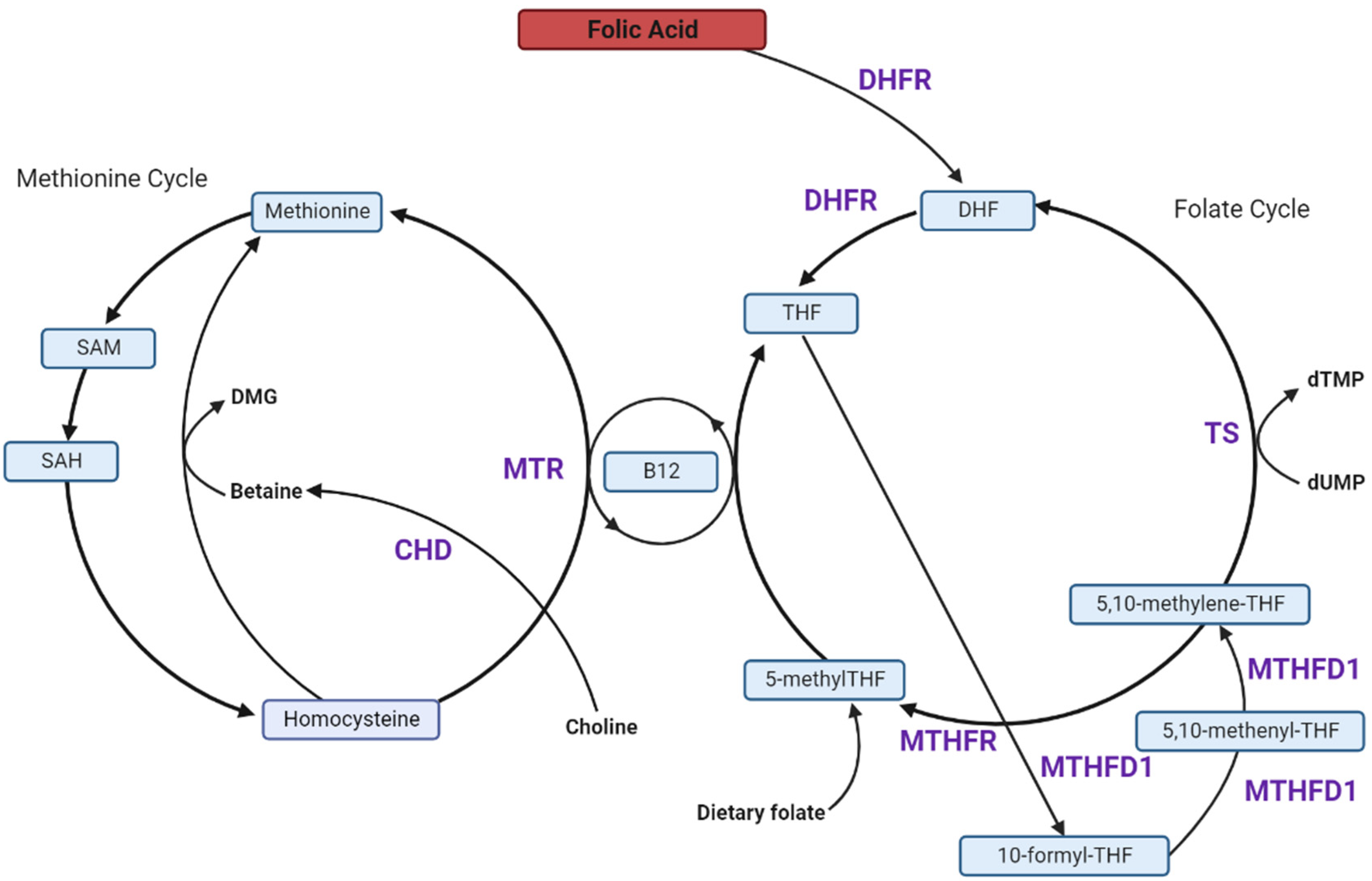
4. Folate and One-Carbon Metabolism in Pregnancy

Folate is a dietary micronutrient essential to one-carbon metabolism, a universal process comprised of multiple interconnected pathways; specifically, the folate cycle, the methionine cycle, and the trans-sulphuration pathway (Figure 1) [49,50]. One-carbon metabolism is critical for the biosynthesis of purines, thymidylate and re-methylation homocysteine to methionine. Homocysteine is a proinflammatory amino acid and high levels can be detrimental to health, including pregnancy [51]. Thus, perturbations to one-carbon metabolism can impact maternal and neonatal health [52,53,54,55]. Humans require dietary folate to maintain different functional aspects of the one-carbon metabolic pathway, whether in the form of non-synthetic folates, obtained from food such as dark leafy greens, eggs, and liver or acquired from FA, a synthetic form, obtained from fortified foods or supplements [56,57]. Unlike non-synthetic folate, FA requires reduction via dihydrofolate reductase (DHFR) to dihydrofolate (DHF) and sequentially tetrahydrofolate (THF) [58]. THF is then interconverted to intermediate metabolites 10-formylTHF, 5,10-methenylTHF and 5,10-methyleneTHF, before incorporation into the folate cycle as the most reduced folate form 5-methylTHF [59]. Expression of one-carbon metabolic enzymes is shown in all human adult tissues suggesting that all tissues should be able to generate de novo one-carbon units. Folates carrying one-carbon units do not freely transport across intracellular membranes. Thus, a complete set of one-carbon enzymes exist in both the mitochondria and the cytosol to synthesize 5,10-methylene-THF in both compartments. It is believed that the one-carbon metabolic pathway is localized in the mitochondria to uncouple this pathway from glycolysis [60,61].
Physiologically, folate has essential functions in growth, differentiation and repair, and thus adequate supply is highly critical during fetal and placental development [62,63]. The human newborn has been estimated to have grown from one to 1.25 × 10¹² cells during gestation with an additional 0.25 × 10¹² cells in the umbilical cord, placenta and fetal membranes [64,65]. Folate plays critical physiological roles in pregnancy to support uterine and placental growth and development, sustained cell division necessary for fetal growth, and specifically in prevention of neural tube defects (NTDs) [66,67,68]. Further, preconception folic acid intake has also been positively associated with fetal growth and reduced risk for small-for-gestational-age (SGA) [69] and reduced risk of spontaneous preterm birth (sPTB) [70]. There is also some evidence for a modest protective effect of folic acid supplementation in preeclampsia risk [71]. In pregnancy, folate requirements increase 5- to 10-fold, to accommodate the needs of the growing feto-placental unit, as well as increasing maternal metabolic demands [66]. Currently, guidelines recommend 400 μg daily FA intake to prevent NTDs [72]. As dietary folate is often inadequate [73], many countries (~92) have mandatory FA food fortification policies, as well as recommendations of periconceptional FA supplementation [74].
Mandatory FA food interventions have significantly reduced folate deficiency and the occurrence of neural tube defects, most recently reviewed by Wilson & O’Connor (2021) [75]. Indeed, in countries with FA food fortification and FA supplementation guidelines for pregnancy, folate excess is more common than folate deficiency. A systematic review confirms that in the context of mandatory FA fortification, most women preconception, and through pregnancy, are exceeding the upper tolerable limit (~1000 μg/d) [76]. While direct consequences of nutrient deficiencies have been well documented [77,78,79], the effects of excess FA intake are less well elucidated and are more likely to have direct effects on one-carbon metabolism and ultimately complicated and indirect effects on maternal and fetal health, and pregnancy outcome.

5. High FA Intake and Unmetabolized FA (uFA)

High intake of FA is known to increase circulating unmetabolized FA (uFA), and this is becoming increasingly prevalent, particularly in the context of food fortification. uFA has been detected in the serum in non-pregnant [6] and pregnant [80] populations, umbilical cord blood [81,82,83], neonatal plasma [84], and in breast milk [85,86,87]. uFA has also been detected in maternal and neonatal plasma [88], serum [82] and umbilical cord blood [82] in a voluntary fortified population.
Accumulation of uFA itself may be detrimental, though there is limited research regarding the adverse effects of circulating uFA in pregnancy. Studies of allergic disease [89] and neurodevelopment [90], have found no adverse effects associated with uFA. However, some research indicates that higher maternal folate is associated with insulin resistance [36,37] and adiposity [37] in offspring. A direct role for circulating uFA in impairing maternal metabolism or in GDM pathogenesis remains to be elucidated. While there is growing evidence for the presence of uFA, the extent to which maternal uFA may be harmful is not well established, nor has a relationship between uFA and pregnancy outcome, including GDM, been described.
The presence of circulating uFA is largely attributed to excess intake, limited enzyme metabolic capacity and a low saturation threshold [91,92]. Understanding the relationship of uFA and one-carbon metabolism remains a significant area for future research, particularly considering the increasing prevalence of uFA, and mounting evidence to suggest a relationship between high FA and GDM.

6. Effects of Excess FA on the Players in the 1C-Metabolism Pathway

6.1. Excess FA Saturates Limited DHFR Capacity

The human gastrointestinal tract can efficiently convert non-synthetic folates to 5-methyltetrahydrofolate (5-methylTHF), the active form for methylation pathways, but has a reduced capacity to convert FA [91,93]. Unlike naturally occurring folate, which is converted to THF without requiring the action of DHFR, FA is inactive until DHFR action reduces it sequentially to DHF and then activated THF in a two-step enzymatic process (Figure 2) [94]. DHFR activity in humans is limited and variable [5]. Oral doses of 260–280 μg FA are sufficient to result in unmetabolized circulating FA (uFA) due to saturated DHFR capacity [95]. Mandatory fortification initiatives and periconceptional FA supplementation have resulted in high FA intake exceeding the upper tolerable limit (~1000 μg/d), and thus, high circulating folate levels in pregnancy [96].

6.2. Excess FA Reduces MTHFR Protein, Causing a Pseudo-MTHFR Deficiency

After conversion of FA to THF, THF is converted by the methylenetetrahydrofolate reductase (MTHFR) enzyme to 5-methylTHF (Figure 1 and Figure 2), the bioavailable form that acts as an essential co-substrate in homocysteine re-methylation. MTHFR is therefore essential in regulating folate bioavailability.
In murine studies, high dietary FA has been shown to induce MTHFR dysfunction. Dietary FA exceeding recommendations (2 mg/kg mouse chow [97]) by 10-fold (10x-FA, 20 mg FA/kg mouse chow), reduced MTHFR protein in the maternal liver (p < 0.05) [98]. Furthermore, dietary FA exceeding recommendation five-fold (5x-FA, 10 mg FA/kg) reduced MTHFR protein [99,100] and activity [100] in the maternal liver, effectively inducing a pseudo-MTHFR deficiency. In addition to FA, DHF has also been characterised as an inhibitor of MTHFR activity in vitro studies [101]. An important caveat is species-specific differences in folate metabolism. Specifically, hepatic DHFR is significantly lower in humans compared to other animals [93,102]. The metabolic rate of DHFR activity in human liver is proposed to be 56 times slower than rat liver [93]. Thus, limited conclusions can be drawn from animal studies. Evidence for MTHFR-deficiency in response to excess FA is well established in mouse models. A pseudo-MTHFR syndrome stemming from excess intake, in the absence of MTHFR polymorphisms, has been documented clinically [103]. Whether this is widely applicable to pregnant women, and its biological significance, is not yet established.

6.3. FA Reduces Methionine Synthase (MTR) Activity and Favours the Thymidylate Synthase (TS) Cycle

Paradoxically, FA excess has been implicated in inhibition of folate-dependent pathways, particularly the methionine synthase (MTR) cycle [104,105], instead promoting thymidylate synthase (TS) cycle activity [104]. MTR activity, required for homocysteine re-methylation, is dependent on adequate vitamin B12 availability. The active form of folate, 5-methylTHF, is converted from 5,10-methyleneTHF, however, 5,10-methyleneTHF can alternatively be used as a one-carbon donor in the conversion of deoxyuridine monophosphate (dUMP) to deoxythymidine monophosphate (dTMP) via thymidylate synthase (Figure 1) with key functions in pyrimidine biosynthesis. In a study of Caenorhabditis elegans (C. elegans), 100 mM synthetic FA decreased methylenetetrahydrofolate reductase (mthf-1) and methionine synthase (metr-1) mRNA but increased thymidylate synthase mRNA [104]. An earlier study of C. elegans also demonstrated reduced enzymatic activity of methionine synthase by ~40% and reduced expression of methionine synthase reductase (mtrr-1) and mthf-1 mRNA in high-FA conditions (8.8 μmol/plate) [105]. Taken together, there is emerging evidence that conditions of excess FA favour TS metabolism, facilitating DNA synthesis and cellular growth, at the expense of MTR, effectively reducing MTR action required to support methylation processes [104,105]. Given a secondary action of TS activity is DHF generation, this may further potentiate DHFR saturation induced by increased FA intake
The implications of these perturbations to the one-carbon metabolic pathway are not yet known. An important caveat is that this research is limited to C. elegans and has not been assessed in in vivo in humans, ex vivo in tissue, nor in placental cell lines. Favouring TS activity at the expense of cobalamin-dependent MTR activity due to excess FA, becomes increasingly of interest considering a proposed high folate/low B12 interaction in GDM pathogenesis. Whether this phenomenon occurs in the human placenta and whether this may contribute to pregnancy complications, is an interesting area for future research.

6.4. Dietary FA Alters Choline and Betaine Metabolism

Disruption of folate metabolism results in use of choline-derived betaine, an alternative methyl donor in methionine synthesis as described above (Figure 1) [106]. This process works in a folate-independent manner to facilitate generation of S-adenosylmethionine (SAM), the universal methyl donor [107]. Following methylation of DNA and other molecules, SAM is converted to S-adenosylhomocysteine (SAH). Therefore, decreases in SAM:SAH ratio indicate reduced methylation potential, whereas reductions in betaine concentrations suggest alternate utilisation as a methionine cycle methyl donor. In a murine study dietary FA exceeding the recommended dietary FA by five-fold, decreased maternal plasma betaine (p = 0.043) and increased SAM (p = 0.009) but reduced placental SAM:SAH (p = 0.039) and SAM (p = 0.021) with the latter further exacerbated in male-bearing pregnancies (p = 0.006) [100]. This study suggests that high FA may disrupt the maternal methionine pathway, but further research is needed to validate this finding. The consequences of disrupting this pathway, particularly in early gestation hypomethylated placenta, for pregnancy health and outcome, including GDM pathogenesis, need to be established. [108]. In the context of human pregnancy this may promote placental and fetal growth and perturb spatio-temporal methylation of DNA and other molecules important in key developmental steps.

7. Association between One-Carbon Metabolism Players and GDM

7.1. High FA and Low Vitamin B12 Are Associated with Increased GDM Risk

Beyond adequate folate intake, one-carbon metabolism is also dependent on adequate supply of vitamin B12. Vitamin B12, or cobalamin, is a water-soluble B-vitamin that is an essential cofactor in one-carbon metabolism, and is largely obtained from animal products, making vegetarianism a risk factor for its deficiency [109,110]. There is growing evidence suggesting a role for vitamin B12 insufficiency in GDM development, either in isolation or in conjunction with excess FA intake, summarised in Table 2 [14,15,111,112,113,114]. In a UK-based retrospective case–control study (n = 344), women with B12 deficiency (<150 pmol/L) in 2nd and 3rd trimesters had an increased likelihood of GDM diagnosis compared to B12-replete women (aOR: 2.59 95% CI: 1.35–4.98, p = 0.004), after adjustment for serum folate, age, parity, smoking status, and ethnicity. Interestingly, folate level did not differ between GDM and non-GDM pregnancies but only folate deficiency, not excess, was reported [115]. The relationship between vitamin B12 deficiency and GDM diagnosis was still present, but attenuated, after adjustment for maternal BMI (aOR: 2.05, 95% CI: 1.03–4.10, p = 0.04).
Similarly, a prospective cohort study, found an inverse relationship between vitamin B12 and GDM risk (aOR: 0.856; 95% CI 0.786–0.933; p = 0.0004) although this was mediated in part by BMI [15]. Vitamin B12 deficiency (<220 pmol/L in serum) also increased GDM risk (aRR: 1.383, 95% CI: 1.157–1.652, p = 0.0004). However, low vitamin B12/high folate was associated with the greatest increase in GDM risk (aRR: 1.742 95% CI: 1.226–2.437, p = 0.003) [15]. Interestingly, high folate remained associated with increased risk of GDM after adjustment for vitamin B12 status (aRR 1.11 95% CI: 1.036–1.182, p = 0.002) [15]. Similarly, a prospective study at 26 weeks’ gestation (n = 913) found the greatest risk of GDM was associated with B12 insufficiency (<150 pmol/L) paired with high folate levels (OR: 1.97 95.3831% CI: 1.05–3.68) [112]. A cross-sectional study of women (n = 406, 24–28 weeks’ gestation) found higher folate:B12 ratio was associated with increased GDM risk (OR: 3.08 95% CI: 1.63–5.83) [14]. A small study of vitamin B12-deficient women at 30 weeks’ gestation (n = 29) found GDM incidence increased across folate tertiles after adjustment (5.4% (n =  7), 10.5% (n = 12), 10.9% (n = 10), from lowest to highest tertile, p =  0.04) [111]. Given vitamin B12 is an essential cofactor in one-carbon metabolism, and the increasing evidence for a role of folate in GDM pathogenesis, it is unsurprising there is also a role for vitamin B12 deficiency in GDM. This is potentially concerning given only food fortification with FA, but not vitamin B12, is mandatory in many countries, especially in the context of widespread vegetarianism, for example in India. The mechanism by which high folate and low vitamin B12 interact to confer GDM risk requires further research. One possible explanation could be the shift from methionine to folate pathways which favour DNA synthesis over methylation and epigenetic programming, leading to not only altered gene expression but increased homocysteine and associated systemic inflammation and insulin resistance.

7.2. Circulating Homocysteine Is Elevated in GDM-Complicated Pregnancies

Homocysteine is a proinflammatory, methionine-derived amino acid, essential in one-carbon metabolism [117]. Homocysteine can undergo transulphuration or can be re-methylated to methionine for cellular methylation assuming sufficient folate is available [117]. The state of increased inflammation, such as that associated with hyperhomocysteinemia, is associated with increased insulin resistance [118]. Homocysteine can impair the translocation of certain glucose transporters to the plasma membrane reducing systemic glucose uptake. Thus, high FA intake which can result in impaired methionine cycle activity mimicking folate deficiency, as mentioned above, can lead to hyperhomocysteinemia, increased insulin resistance and reduced glucose uptake.
A systematic review and meta-analysis (12 studies, GDM n = 712, control n = 1277) indicates homocysteine is significantly elevated in women with GDM-complicated pregnancies, compared to healthy controls (Standard mean difference (SMD) = 0.55; 95% CI: 0.25–0.85, p = 0.0003) [119]. Interestingly, two studies stratified participants into three groups: normoglycemic (normal response to 1 h glucose challenge, no oral glucose tolerance test (oGTT) follow-up), glucose intolerant (a positive response to 1 h glucose challenge but normal oGTT results) and GDM [120,121]. Of note, each study had slightly different thresholds for oGTT follow-up, Tarim et al. required 1 h-post challenge blood glucose exceeding >7.5 nmol/L, and Guven et al. required blood glucose exceeding 7.8 nmol/L. Tarim et al. found homocysteine was elevated in women with glucose intolerance and with diagnosed GDM (p < 0.001) compared to women with a normoglycemic pregnancy [120]. Guven et al. found a significant (p < 0.01) increase in serum homocysteine between women with GDM-complicated pregnancies and normoglycemic pregnancy, but no statistically significant differences between any other group [121]. Interestingly, neither study observed differences in serum vitamin B12 nor folate. Several studies which did not stratify for glucose intolerance have also observed increased serum homocysteine in women with GDM-complicated pregnancies compared to non-GDM pregnancies [122,123,124,125,126].
Conversely, several studies have found no difference in homocysteine concentrations in women with GDM-complicated pregnancies compared to controls. Idzior-Waluś et al. noted no difference in serum homocysteine between women with GDM-complicated pregnancies (n = 44, 8 ± 2.0 μmol/L) and non-GDM pregnancies (n = 17, 7.4 ± 1.1 μmol/L) between 26 and 32 weeks’ gestation [127]. However, all patients were recruited from an outpatient diabetic clinic, and had all been referred based on positive response to 50 g glucose load; thus, there is likely to be some degree of glucose intolerance within the non-GDM group. Similarly, in a prospective study of pregnant women between 24–28 weeks’ gestation, serum homocysteine did not differ between GDM-complicated pregnancies (n = 60, 7.41 μmol/L ± 2.61) and uncomplicated controls (n = 19, 8.02 μmol/L ± 2.27) [113]. It is important to note that neither of these studies stratified for glucose intolerance below the threshold for GDM diagnosis, both have very few non-GDM controls, and show homocysteine levels that are higher than is characteristic of non-diabetic pregnancy at a similar gestation [128]. Interestingly, one study at ~34 weeks’ gestation, found reduced total plasma homocysteine in women with glucose intolerance (n = 18, 5.0 μmol/L ± 1.7) compared to normoglycemic women (n = 190, 6.6 ± 2.0, p = 0.024), but no difference between GDM-complicated (n = 17, 6.8 ± 2.7) and normoglycemic pregnancies. The authors note that all women with GDM were treated with a low glycaemic diet, which is likely to explain this observation [129]. Similarly, Akturk et al. found no differences in homocysteine levels in late pregnancy (32–39 weeks’), though all women diagnosed with GDM (n = 54, according to American Diabetes Association criteria, at 24–28 weeks’ gestation [130]) were treated following GDM diagnosis (n = 48 diet only, n = 6 diet and insulin) [131].
Together, these studies (Summarised in Table 3) suggest that homocysteine levels are elevated in GDM-complicated pregnancies in mid-late gestation. Furthermore, two studies which found no differences in homocysteine in late pregnancy also involve treatment, suggesting elevated circulating homocysteine in GDM are modifiable [129,131]. Predominantly, these studies assess homocysteine in the second and third trimester of pregnancy, after GDM onset, making it difficult to assess causation between homocysteine and GDM pathogenesis. Only one study measured fasting serum homocysteine in early gestation (8–12 weeks’ gestation), finding no difference in homocysteine levels between normoglycemic (n = 83, 14.41 ± 7.98 μmol/L) and GDM-complicated pregnancies (n = 7, 15.66 ± 7.61, p = 0.6312) [132]. However, the low statistical power of this study and very high homocysteine levels of this cohort, mean that limited conclusions can be drawn. The high homocysteine levels in this cohort may be somewhat explained by widespread vegetarianism and associated vitamin B12 deficiency, which is common in the study country, India [133]. However, the authors do not provide patient data to confirm this speculation. Furthermore, vitamin B12 and folate status that are known to influence homocysteine were not assessed. It remains unclear whether homocysteine contributes to GDM aetiology or is a consequence of hyperglycaemia. In addition, whether elevated homocysteine reflects perturbation of other one-carbon metabolites or whether high folate and high homocysteine act independently in GDM pathophysiology remain unknown. Further research in early pregnancy is needed to elucidate the nature of the homocysteine-GDM relationship.
There are several mechanisms by which high homocysteine may contribute to GDM:
Via pancreatic β-cells: Pancreatic β-cells secrete insulin to maintain glucose homeostasis [134]. During pregnancy, there is an adaptive increase in β-cell mass to increase insulin release, accommodating the increased insulin resistance of pregnancy [46,48,135]. Insufficient β-cell adaptation is associated with hyperglycaemia and GDM diagnosis [136,137]. In vitro, homocysteine has been shown to inhibit glucose-induced insulin secretion by β-cells in a dose-dependent manner at moderate (5.6 mM, p < 0.001) and stimulatory (16.7 mM, p < 0.001) glucose concentrations [138,139]. Serum homocysteine was inversely associated with several parameters of pancreatic islet β-cell function, in T2DM patients but a similar effect in GDM has not yet been characterised [140]. Further research is needed to clarify the relationship between elevated homocysteine and β-cell function in GDM pathogenesis.
Via the placenta to induce oxidative stress: Within the placenta, oxidative stress is necessary for angiogenesis, immunoregulation and vasoactivity [141]. However, an imbalance of reactive oxygen species and antioxidant activity can result in elevated oxidative stress and lipid peroxidation, and subsequent cellular damage [142,143]. The role of homocysteine as a pro-oxidant is well-established [144,145,146,147], as is the role of oxidative stress in insulin resistance [118,148] and disruption of insulin signalling [149,150].
In C. elegans, malondialdehyde (MDA), a marker of lipid peroxidation, and hydrogen peroxide (H2O2), an activator of oxidative stress, were increased after high dose-FA induced elevated homocysteine [105]. Other research indicates that supraphysiological FA can exacerbate lipid peroxidation in oxidative stress conditions [151]. In BeWo cells (choriocarcinoma cell line) oxidative stress was induced with tert-butylhydroperoxide (TBH) treatment under deficient (1 nM), physiological (20 nM) and supraphysiological (2.3 μM) FA treatment. The latter increased MDA content compared to physiological FA, both in response to 100 μM and 300 μM TBH treatment [151]. However, the interaction between FA, homocysteine and oxidative stress requires further research. Both oxidative stress [152,153,154,155] and lipid peroxidation [156,157] markers, have been shown to be associated with pregnancies complicated by GDM. In addition, there may also be a role for excess FA in inducing or exacerbating existing oxidative stress, either through elevated homocysteine or an alternate pathway. Further research is warranted to elucidate the mechanisms by which elevated homocysteine, oxidative stress, and potentially uFA contribute to GDM pathogenesis.
Via the placenta to induce apoptosis: Homocysteine can also cause placental dysfunction by promoting cellular apoptosis. In vitro homocysteine treatment can induce trophoblast apoptosis in primary human placental trophoblasts (36 weeks’ gestation) [158]. Interestingly, a follow-up study found that treatment with FA (20 nmol/L, a plasma concentration considered healthy), alleviated homocysteine-induced apoptosis [159]. Similarly, in primary cultured human trophoblasts collected at term, homocysteine-thiolactone, a homocysteine oxidation product, induced apoptosis in a dose-dependent manner [160]. In contrast, treatment with folate (10 μmol/L, representing a supraphysiological dose) did not alleviate homocysteine-induced apoptosis
Despite some evidence that homocysteine induces trophoblast apoptosis, a role for apoptosis in GDM remains controversial. There is both evidence for an increased [161,162] and decreased [163] trophoblast apoptotic index in placentae from GDM-complicated pregnancies, compared to those from uncomplicated pregnancies. Further research to definitively demonstrate the role of trophoblast apoptosis in GDM is needed, with an emphasis on potential effects of homocysteine and FA.
Via dysregulated hCG secretion: A fourth mechanism by which high homocysteine levels may contribute to GDM is through altering hormone profiles, specifically human chorionic gonadotrophin (hCG). hCG is a hormone produced by placental syncytiotrophoblasts and is critical to pregnancy maintenance [164]. A systematic review and meta-analysis concluded that first trimester β-hCG is reduced in women subsequently diagnosed with GDM, compared to women who remain normoglycemic [165]. In placental explants collected from uncomplicated pregnancies, in vitro homocysteine treatment significantly reduced hCG secretion at 20, 40 and 80 μmol/L (50–80% reduction, p < 0.004) [158]. Similarly, in placental villous trophoblasts, isolated from uncomplicated term placentae, a 43% reduction of hCG secretion under 20 μmol/L homocysteine treatment (p < 0.02) was observed. FA treatment (40 nmol/L) was able to restore hCG secretion (p < 0.05) [159], though the effects of excess FA was not evaluated. Ahmed et al., observed no differences in hCG secretion in placental explants nor in BeWo villous cytotrophoblast, under FA deficient (2 ng/mL), physiological (20 ng/mL), elevated (200 ng/mL) and supraphysiological (2000 ng/mL) conditions [166]. This suggests FA may not directly alter trophoblastic hCG secretion, instead its’ action may be mediated through elevating homocysteine. A caveat of the existing work is the use of term rather than early gestation placentae, which makes it difficult to ascertain a causal relationship in GDM pathogenesis. Current knowledge indicates that homocysteine reduces hCG secretion and reduced hCG has been previously implicated in GDM-complicated pregnancies [165]. Further research is needed to clarify whether elevated homocysteine contributes to GDM pathophysiology or is just an indicator of perturbed one-carbon metabolism.

7.3. Interactions between Excess Folate, Vitamin B12, Homocysteine and Risk for GDM

Given folate, vitamin B12 and homocysteine have all been previously implicated in GDM risk, and each is necessary for one-carbon metabolic function, the complex interplay of these metabolites becomes increasingly relevant. Remethylation of homocysteine to methionine occurs via cobalamin-dependent enzyme MTR, with 5-methylTHF as a necessary cofactor in MTR activity [53]. Given Vitamin B12 is essential in 5-methylTHF uptake, insufficient B12 can result in folate becoming trapped in this form [56,167]. Thus, the methionine pathway is both highly dependent on adequate folate availability and B12 uptake, as well as MTR action [56,105]. Generally, homocysteine has an inverse relationship with folate, with elevated homocysteine is considered to be a sensitive marker of folate deficiency [168,169]. Given both high homocysteine and high FA are associated with GDM [11,13,14,15,16] it is plausible the homocysteine remethylation pathway is perturbed in both deficient and high FA conditions, but the nature of this relationship requires further investigation. Some evidence suggests excess FA may directly induce elevated homocysteine. In both BeWo and JEG3 human choriocarcinoma cell lines, increased homocysteine was observed under supraphysiological FA (2000 ng/mL) conditions compared to normal physiological (20 ng/mL) treatment [170]. High dose FA supplementation in C. elegans has been shown to induce high homocysteine [105]. However, there is limited in vivo human research to verify the effect of supraphysiological FA and further investigation is required.
Evidence suggests the FA-homocysteine relationship may be mediated through vitamin B12 deficiency. In a study of healthy adults, homocysteine concentrations only decreased across increasing serum folate categories when vitamin B12 was >148 pmol/L (p < 0.001), and this was not observed when vitamin B12 was <148 pmol/L [171]. Beyond the vitamin B12-folate interaction observed clinically, there is also in vitro evidence that vitamin B12 sufficiency is necessary to reduce elevated homocysteine levels. In BeWo and JEG3 choriocarcinoma cell lines, treatment with various forms of vitamin B12, including cobalamin and combined methylcobalamin (MeCBl) and adenosylcobalamin (AdCbl), lowered homocysteine levels, which were elevated by supraphysiological FA treatment [170]. This suggests that rather than a direct role of high FA intake in perturbing homocysteine remethylation, vitamin B12 instead acts as the limiting factor. However, FA may have a direct, concomitant role in interfering with vitamin B12-dependent metabolism through reduced MTR action [104,105], which is exacerbated in cases where vitamin B12 is already limited.

7.4. Choline-Derived Betaine Is Associated with Decreased GDM Risk

Choline is an essential nutrient, that can be synthesized endogenously in the human liver but obtaining additional dietary choline from sources such as red meat, poultry, fish, eggs and soybeans, is necessary to achieve adequate levels in circulation [172]. Dietary choline can be either water-soluble, which is metabolized in the liver, or lipid-soluble and transported through the lymphatic system. While choline acts in multiple pathways, in one-carbon metabolism choline is converted to betaine for DNA methylation, phospholipid synthesis and fetal neurodevelopment [173].
When folate or vitamin B12 supply are limited, choline-derived betaine is used as a methyl donor for homocysteine re-methylation and global methylation processes [174] (Figure 1). The role of choline and betaine in GDM development has been understudied, a summary of existing research is provided in Table 4. Two studies report a protective role of maternal betaine in GDM [175,176]. In a prospective cohort study (n = 486), maternal plasma betaine (≤200 nmol/mL) in early pregnancy (median: 10 weeks’ gestation, IQR: 9–11) was associated with increased GDM risk (OR: 5.00 95% CI: 2.76–9.07) [175]. Similarly, in a cohort study of dichorionic twin pregnancies (n = 187), plasma betaine had an inverse relationship with GDM risk (p trend = 0.015) [176].
Conversely, one study found no association between maternal circulating betaine and GDM risk [177]. However, plasma betaine was decreased in cord blood of neonates of GDM-complicated pregnancies (18.5 ± 3.9 μmol/L) compared to uncomplicated (21.2 ± 4.7 μmol/L, p = 0.02). Given there were no differences in maternal betaine observed, the authors proposed that GDM may alter the transfer of betaine to the fetus and/or that women with a GDM-complicated pregnancy utilise betaine for maternal metabolism, resulting in depleted neonatal cord concentrations [177]. Collectively, there is emerging evidence that choline-derived betaine may play a protective role in GDM risk. Further research is needed to determine the relationship of choline and betaine in GDM pathogenesis, particularly with reference to other one-carbon metabolites.
While it has been established that choline can modulate important markers of placental function [178], the mechanism by which insufficient choline can increase GDM risk is not well understood. The oxidation of choline to betaine occurs in the mitochondria, and is catalysed by choline dehydrogenase (CHD) which, based on animal studies, is presumed to be located on the inner mitochondrial membrane [179].The necessity of effective mitochondria in betaine conversion suggests perturbed mitochondrial function may be the mechanistic link. Reduced mitochondria content [180] and alterations to placental mitochondrial dynamics and metabolism, specifically increased mitochondrial fusion [181], have been observed in the placenta of GDM-complicated pregnancies. Further, Abbade et al. propose hyperinsulinemia mediates the observed mitochondrial dysfunction using JEG-3 cells as an in vitro model. The relationship between mitochondrial function and GDM needs further clarification to establish whether mitochondrial modifications underpin GDM pathogenesis, or are a consequence of the GDM state, specifically the accompanying hyperinsulinemia. Further establishing the interplay between choline-betaine conversion in the pathway is also of significant interest, given the proposed protective role of choline in GDM pathology.

8. Conclusions

Several one-carbon metabolites have been implicated in risk for GDM. Specifically, increasing concentrations of circulating folate [7,11,14,15,16] and homocysteine [113,120,121,122,127], and decreasing vitamin B12 [14,15,111,112,115] and betaine [175,176,177] associate with increased GDM risk. Excess intake of FA has emerged as a key risk factor in GDM development. While the mechanism is largely unknown, some research suggests high FA may have a direct effect on pancreatic β-cell signalling [44,45]. There is growing evidence that dysregulation of several aspects of the one-carbon metabolic pathway may be at play (Summarised in Figure 3). Proposed interactions include a limited capacity to incorporate excess FA into the one-carbon metabolic pathway which can affect downstream transcription and methylation events, perturbations to the function and activity of one-carbon metabolism enzymes, notably MTR, TS and MTHFR. Alternatively, the relationship between excess FA and GDM risk may not be mediated by one-carbon metabolism and may instead result from direct adverse effects of circulating uFA which appears in circulation in response to saturated DHFR capacity. However, this is not well-established in the literature. Currently, the role of excess FA in dysregulation of aspects of one-carbon metabolism relies largely on limited in vitro research warranting further studies. Nevertheless, one-carbon metabolism is closely entwined with GDM risk. Given rising rates of GDM around the world and very widespread global FA food fortification, in the absence of vitamin B12 fortification, further research is urgently needed to elucidate the mechanisms by which perturbations of one-carbon metabolism, including high circulating uFA, contribute to GDM pathogenesis.

Author Contributions

Writing—original draft preparation, J.M.W.; writing—review and editing, J.M.W., A.L.A., M.D.S., C.T.R. and T.J.-K.; supervision, A.L.A., C.T.R. and T.J.-K.; funding acquisition, C.T.R. and T.J.-K. All authors have read and agreed to the published version of the manuscript.

Funding

J.M.W. was supported by a Flinders Health and Medical Research Institute (FHMRI) Scholarship. C.T.R. was supported by a National Health and Medical Research Council (NHMRC) Investigator grant (GNT1174971) and a Matthew Flinders Professorial Fellowship funded by Flinders University. This research was funded by NHMRC (GNT1161079) awarded to C.T.R. and a Flinders Foundation Health Seed Grant (2021) awarded to T.J.K.

Institutional Review Board Statement

Not applicable.

Informed Consent Statement

Not applicable.

Data Availability Statement

Not applicable.

Acknowledgments

Figures were created with Biorender.com.

Conflicts of Interest

The authors declare no conflict of interest.

References

  1. Chiefari, E.; Arcidiacono, B.; Foti, D.; Brunetti, A. Gestational diabetes mellitus: An updated overview. J. Endocrinol. Investig. 2017, 40, 899–909. [Google Scholar] [CrossRef] [PubMed]
  2. Australian Institute of Health and Welfare. Incidence of Gestational Diabetes in Australia, Cat. No: CVD 85; AIHW: Canberra, Australia, 2019. [Google Scholar]
  3. Australian Institute of Health and Welfare. Diabetes, Cat. No: CVD 82; AIHW: Canberra, Australia, 2020. [Google Scholar]
  4. Palchetti, C.Z.; Paniz, C.; de Carli, E.; Marchioni, D.M.; Colli, C.; Steluti, J.; Pfeiffer, C.M.; Fazili, Z.; Guerra-Shinohara, E.M. Association between Serum Unmetabolized Folic Acid Concentrations and Folic Acid from Fortified Foods. J. Am. Coll. Nutr. 2017, 36, 572–578. [Google Scholar] [CrossRef]
  5. Wright, A.J.A.; Dainty, J.R.; Finglas, P.M. Folic acid metabolism in human subjects revisited: Potential implications for proposed mandatory folic acid fortification in the UK. Br. J. Nutr. 2007, 98, 667–675. [Google Scholar] [CrossRef]
  6. Pfeiffer, C.M.; Sternberg, M.R.; Fazili, Z.; Yetley, E.A.; Lacher, D.A.; Bailey, R.L.; Johnson, C.L. Unmetabolized folic acid is detected in nearly all serum samples from US children, adolescents, and adults. J. Nutr. 2015, 145, 520–531. [Google Scholar] [CrossRef] [PubMed]
  7. Zhu, B.; Ge, X.; Huang, K.; Mao, L.; Yan, S.; Xu, Y.; Huang, S.; Hao, J.; Zhu, P.; Niu, Y.; et al. Folic Acid Supplement Intake in Early Pregnancy Increases Risk of Gestational Diabetes Mellitus: Evidence from a Prospective Cohort Study. Diabetes Care 2016, 39, e36–e37. [Google Scholar] [CrossRef] [PubMed]
  8. Chen, X.; Zhang, Y.; Chen, H.; Jiang, Y.; Wang, Y.; Wang, D.; Li, M.; Dou, Y.; Sun, X.; Huang, G.; et al. Association of Maternal Folate and Vitamin B12 in Early Pregnancy with Gestational Diabetes Mellitus: A Prospective Cohort Study. Diabetes Care 2021, 44, 217–223. [Google Scholar] [CrossRef]
  9. Cheng, G.; Sha, T.; Gao, X.; He, Q.; Wu, X.; Tian, Q.; Yang, F.; Tang, C.; Wu, X.; Xie, Q.; et al. The Associations between the Duration of Folic Acid Supplementation, Gestational Diabetes Mellitus, and Adverse Birth Outcomes based on a Birth Cohort. Int. J. Environ. Res. Public Health 2019, 16, 4511. [Google Scholar] [CrossRef]
  10. Huang, L.; Yu, X.; Li, L.; Chen, Y.; Yang, Y.; Yang, Y.; Hu, Y.; Zhao, Y.; Tang, H.; Xu, D.; et al. Duration of periconceptional folic acid supplementation and risk of gestational diabetes mellitus. Asia Pac. J. Clin. Nutr. 2019, 28, 321–329. [Google Scholar] [CrossRef]
  11. Xie, K.; Xu, P.; Fu, Z.; Gu, X.; Li, H.; Cui, X.; You, L.; Zhu, L.; Ji, C.; Guo, X. Association of maternal folate status in the second trimester of pregnancy with the risk of gestational diabetes mellitus. Food Sci. Nutr. 2019, 7, 3759–3765. [Google Scholar] [CrossRef]
  12. Liu, P.J.; Liu, Y.; Ma, L.; Yao, A.M.; Chen, X.Y.; Hou, Y.X.; Wu, L.P.; Xia, L.Y. Associations Between Gestational Diabetes Mellitus Risk and Folate Status in Early Pregnancy and MTHFR C677T Polymorphisms in Chinese Women. Diabetes Metab. Syndr. Obes. 2020, 13, 1499–1507. [Google Scholar] [CrossRef]
  13. Li, N.; Jiang, J.; Guo, L. Effects of maternal folate and vitamin B12 on gestational diabetes mellitus: A dose-response meta-analysis of observational studies. Eur. J. Clin. Nutr. 2022; in press. [Google Scholar] [CrossRef]
  14. Li, S.; Hou, Y.; Yan, X.; Wang, Y.; Shi, C.; Wu, X.; Liu, H.; Zhang, L.; Zhang, X.; Liu, J.; et al. Joint effects of folate and vitamin B(12) imbalance with maternal characteristics on gestational diabetes mellitus. J. Diabetes 2019, 11, 744–751. [Google Scholar] [CrossRef] [PubMed]
  15. Saravanan, P.; Sukumar, N.; Adaikalakoteswari, A.; Goljan, I.; Venkataraman, H.; Gopinath, A.; Bagias, C.; Yajnik, C.S.; Stallard, N.; Ghebremichael-Weldeselassie, Y.; et al. Association of maternal vitamin B12 and folate levels in early pregnancy with gestational diabetes: A prospective UK cohort study (PRiDE study). Diabetologia 2021, 64, 2170–2182. [Google Scholar] [CrossRef] [PubMed]
  16. Jankovic-Karasoulos, T.; Furness, D.L.; Leemaqz, S.Y.; Dekker, G.A.; Grzeskowiak, L.E.; Grieger, J.A.; Andraweera, P.H.; McCullough, D.; McAninch, D.; McCowan, L.M.; et al. Maternal folate, one-carbon metabolism and pregnancy outcomes. Matern. Child Nutr. 2021, 17, e13064. [Google Scholar] [CrossRef] [PubMed]
  17. Lowe, W.L., Jr.; Scholtens, D.M.; Lowe, L.P.; Kuang, A.; Nodzenski, M.; Talbot, O.; Catalano, P.M.; Linder, B.; Brickman, W.J.; Clayton, P.; et al. Association of Gestational Diabetes with Maternal Disorders of Glucose Metabolism and Childhood Adiposity. JAMA 2018, 320, 1005–1016. [Google Scholar] [CrossRef] [PubMed]
  18. Metzger, B.E.; Lowe, L.P.; Dyer, A.R.; Trimble, E.R.; Chaovarindr, U.; Coustan, D.R.; Hadden, D.R.; McCance, D.R.; Hod, M.; McIntyre, H.D.; et al. Hyperglycemia and adverse pregnancy outcomes. N. Engl. J. Med. 2008, 358, 1991–2002. [Google Scholar] [CrossRef] [PubMed]
  19. Shen, Y.; Li, W.; Leng, J.; Zhang, S.; Liu, H.; Li, W.; Wang, L.; Tian, H.; Chen, J.; Qi, L.; et al. High risk of metabolic syndrome after delivery in pregnancies complicated by gestational diabetes. Diabetes Res. Clin. Pract. 2019, 150, 219–226. [Google Scholar] [CrossRef]
  20. Kaiser, K.; Nielsen, M.F.; Kallfa, E.; Dubietyte, G.; Lauszus, F.F. Metabolic syndrome in women with previous gestational diabetes. Sci. Rep. 2021, 11, 11558. [Google Scholar] [CrossRef] [PubMed]
  21. Pathirana, M.M.; Lassi, Z.S.; Ali, A.; Arstall, M.A.; Roberts, C.T.; Andraweera, P.H. Association between metabolic syndrome and gestational diabetes mellitus in women and their children: A systematic review and meta-analysis. Endocrine 2021, 71, 310–320. [Google Scholar] [CrossRef]
  22. Bellamy, L.; Casas, J.-P.; Hingorani, A.D.; Williams, D. Type 2 diabetes mellitus after gestational diabetes: A systematic review and meta-analysis. Lancet 2009, 373, 1773–1779. [Google Scholar] [CrossRef]
  23. Goueslard, K.; Cottenet, J.; Mariet, A.-S.; Giroud, M.; Cottin, Y.; Petit, J.-M.; Quantin, C. Early cardiovascular events in women with a history of gestational diabetes mellitus. Cardiovasc. Diabetol. 2016, 15, 15. [Google Scholar] [CrossRef]
  24. Kessous, R.; Shoham-Vardi, I.; Pariente, G.; Sherf, M.; Sheiner, E. An association between gestational diabetes mellitus and long-term maternal cardiovascular morbidity. Heart 2013, 99, 1118. [Google Scholar] [CrossRef]
  25. Pathirana, M.M.; Lassi, Z.; Ali, A.; Arstall, M.; Roberts, C.T.; Andraweera, P.H. Cardiovascular risk factors in women with previous gestational diabetes mellitus: A systematic review and meta-analysis. Rev. Endocr. Metab. Disord. 2021, 22, 729–761. [Google Scholar] [CrossRef] [PubMed]
  26. Pathirana, M.M.; Lassi, Z.S.; Roberts, C.T.; Andraweera, P.H. Cardiovascular risk factors in offspring exposed to gestational diabetes mellitus in utero: Systematic review and meta-analysis. J. Dev. Orig. Health Dis. 2020, 11, 599–616. [Google Scholar] [CrossRef] [PubMed]
  27. Abokaf, H.; Shoham-Vardi, I.; Sergienko, R.; Landau, D.; Sheiner, E. In utero exposure to gestational diabetes mellitus and long term endocrine morbidity of the offspring. Diabetes Res. Clin. Pract. 2018, 144, 231–235. [Google Scholar] [CrossRef]
  28. Bush, N.C.; Chandler-Laney, P.C.; Rouse, D.J.; Granger, W.M.; Oster, R.A.; Gower, B.A. Higher maternal gestational glucose concentration is associated with lower offspring insulin sensitivity and altered beta-cell function. J. Clin. Endocrinol. Metab. 2011, 96, E803–E809. [Google Scholar] [CrossRef]
  29. Kelstrup, L.; Damm, P.; Mathiesen, E.R.; Hansen, T.; Vaag, A.A.; Pedersen, O.; Clausen, T.D. Insulin Resistance and Impaired Pancreatic β-Cell Function in Adult Offspring of Women with Diabetes in Pregnancy. J. Clin. Endocrinol. Metab. 2013, 98, 3793–3801. [Google Scholar] [CrossRef] [PubMed]
  30. Lean, S.C.; Derricott, H.; Jones, R.L.; Heazell, A.E.P. Advanced maternal age and adverse pregnancy outcomes: A systematic review and meta-analysis. PLoS ONE 2017, 12, e0186287. [Google Scholar] [CrossRef]
  31. Giannakou, K.; Evangelou, E.; Yiallouros, P.; Christophi, C.A.; Middleton, N.; Papatheodorou, E.; Papatheodorou, S.I. Risk factors for gestational diabetes: An umbrella review of meta-analyses of observational studies. PLoS ONE 2019, 14, e0215372. [Google Scholar] [CrossRef]
  32. Caughey, A.B.; Cheng, Y.W.; Stotland, N.E.; Washington, A.E.; Escobar, G.J. Maternal and paternal race/ethnicity are both associated with gestational diabetes. Am. J. Obstet. Gynecol. 2010, 202, 616.e1–616.e5. [Google Scholar] [CrossRef]
  33. Australian Institute of Health and Welfare. Australia’s Mothers and Babies 2018—In Brief, Cat. No: PER 108; AIHW: Canberra, Australia, 2020. [Google Scholar]
  34. Australian Institute of Health and Welfare. Australia’s Mothers and Babies, Cat. No: PER 101; AIHW: Canberra, Australia, 2021. [Google Scholar]
  35. Metzger, B.E.; Gabbe, S.G.; Persson, B.; Buchanan, T.A.; Catalano, P.A.; Damm, P.; Dyer, A.R.; Leiva, A.; Hod, M.; Kitzmiler, J.L.; et al. International association of diabetes and pregnancy study groups recommendations on the diagnosis and classification of hyperglycemia in pregnancy. Diabetes Care 2010, 33, 676–682. [Google Scholar] [CrossRef]
  36. Krishnaveni, G.V.; Veena, S.R.; Karat, S.C.; Yajnik, C.S.; Fall, C.H.D. Association between maternal folate concentrations during pregnancy and insulin resistance in Indian children. Diabetologia 2014, 57, 110–121. [Google Scholar] [CrossRef]
  37. Yajnik, C.S.; Deshpande, S.S.; Jackson, A.A.; Refsum, H.; Rao, S.; Fisher, D.J.; Bhat, D.S.; Naik, S.S.; Coyaji, K.J.; Joglekar, C.V.; et al. Vitamin B12 and folate concentrations during pregnancy and insulin resistance in the offspring: The Pune Maternal Nutrition Study. Diabetologia 2008, 51, 29–38. [Google Scholar] [CrossRef]
  38. Tojal, A.; Neves, C.; Veiga, H.; Ferreira, S.; Rodrigues, I.; Martel, F.; Calhau, C.; Negrão, R.; Keating, E. Perigestational high folic acid: Impact on offspring’s peripheral metabolic response. Food Funct. 2019, 10, 7216–7226. [Google Scholar] [CrossRef]
  39. Huang, Y.; He, Y.; Sun, X.; He, Y.; Li, Y.; Sun, C. Maternal High Folic Acid Supplement Promotes Glucose Intolerance and Insulin Resistance in Male Mouse Offspring Fed a High-Fat Diet. Int. J. Mol. Sci. 2014, 15, 6298–6313. [Google Scholar] [CrossRef] [PubMed]
  40. Kintaka, Y.; Wada, N.; Shioda, S.; Nakamura, S.; Yamazaki, Y.; Mochizuki, K. Excessive folic acid supplementation in pregnant mice impairs insulin secretion and induces the expression of genes associated with fatty liver in their offspring. Heliyon 2020, 6, e03597. [Google Scholar] [CrossRef]
  41. Yang, X.; Huang, Y.; Sun, C.; Li, J. Maternal Prenatal Folic Acid Supplementation Programs Offspring Lipid Metabolism by Aberrant DNA Methylation in Hepatic ATGL and Adipose LPL in Rats. Nutrients 2017, 9, 935. [Google Scholar] [CrossRef]
  42. Cuthbert, C.E.; Foster, J.E.; Ramdath, D.D. A maternal high-fat, high-sucrose diet alters insulin sensitivity and expression of insulin signalling and lipid metabolism genes and proteins in male rat offspring: Effect of folic acid supplementation. Br. J. Nutr. 2017, 118, 580–588. [Google Scholar] [CrossRef]
  43. Kelly, K.B.; Kennelly, J.P.; Ordonez, M.; Nelson, R.; Leonard, K.; Stabler, S.; Gomez-Muñoz, A.; Field, C.J.; Jacobs, R.L. Excess Folic Acid Increases Lipid Storage, Weight Gain, and Adipose Tissue Inflammation in High Fat Diet-Fed Rats. Nutrients 2016, 8, 594. [Google Scholar] [CrossRef] [PubMed]
  44. Yang, H.; Qin, D.; Xu, S.; He, C.; Sun, J.; Hua, J.; Peng, S. Folic acid promotes proliferation and differentiation of porcine pancreatic stem cells into insulin-secreting cells through canonical Wnt and ERK signaling pathway. J. Steroid Biochem. Mol. Biol. 2021, 205, 105772. [Google Scholar] [CrossRef]
  45. Karampelias, C.; Rezanejad, H.; Rosko, M.; Duan, L.; Lu, J.; Pazzagli, L.; Bertolino, P.; Cesta, C.E.; Liu, X.; Korbutt, G.S.; et al. Reinforcing one-carbon metabolism via folic acid/Folr1 promotes β-cell differentiation. Nat. Commun. 2021, 12, 3362. [Google Scholar] [CrossRef]
  46. Baeyens, L.; Hindi, S.; Sorenson, R.L.; German, M.S. β-Cell adaptation in pregnancy. Diabetes Obes. Metab. 2016, 18 (Suppl. S1), 63–70. [Google Scholar] [CrossRef]
  47. Butler, A.E.; Cao-Minh, L.; Galasso, R.; Rizza, R.A.; Corradin, A.; Cobelli, C.; Butler, P.C. Adaptive changes in pancreatic beta cell fractional area and beta cell turnover in human pregnancy. Diabetologia 2010, 53, 2167–2176. [Google Scholar] [CrossRef]
  48. Moyce, B.L.; Dolinsky, V.W. Maternal β-Cell Adaptations in Pregnancy and Placental Signalling: Implications for Gestational Diabetes. Int. J. Mol. Sci. 2018, 19, 3467. [Google Scholar] [CrossRef] [PubMed]
  49. Shulpekova, Y.; Nechaev, V.; Kardasheva, S.; Sedova, A.; Kurbatova, A.; Bueverova, E.; Kopylov, A.; Malsagova, K.; Dlamini, J.C.; Ivashkin, V. The Concept of Folic Acid in Health and Disease. Molecules 2021, 26, 3731. [Google Scholar] [CrossRef]
  50. Selhub, J. Folate, vitamin B12 and vitamin B6 and one carbon metabolism. J. Nutr. Health Aging 2002, 6, 39–42. [Google Scholar]
  51. Dai, C.; Fei, Y.; Li, J.; Shi, Y.; Yang, X. A Novel Review of Homocysteine and Pregnancy Complications. BioMed Res. Int. 2021, 2021, 6652231. [Google Scholar] [CrossRef]
  52. Kalhan, S.C. One carbon metabolism in pregnancy: Impact on maternal, fetal and neonatal health. Mol. Cell. Endocrinol. 2016, 435, 48–60. [Google Scholar] [CrossRef]
  53. Lan, X.; Field, M.S.; Stover, P.J. Cell cycle regulation of folate-mediated one-carbon metabolism. Wiley Interdiscip. Rev. Syst. Biol. Med. 2018, 10, e1426. [Google Scholar] [CrossRef]
  54. Crider, K.S.; Yang, T.P.; Berry, R.J.; Bailey, L.B. Folate and DNA methylation: A review of molecular mechanisms and the evidence for folate’s role. Adv. Nutr. 2012, 3, 21–38. [Google Scholar] [CrossRef]
  55. Korsmo, H.W.; Jiang, X. One carbon metabolism and early development: A diet-dependent destiny. Trends Endocrinol. Metab. 2021, 32, 579–593. [Google Scholar] [CrossRef]
  56. Scaglione, F.; Panzavolta, G. Folate, folic acid and 5-methyltetrahydrofolate are not the same thing. Xenobiotica 2014, 44, 480–488. [Google Scholar] [CrossRef] [PubMed]
  57. Beal, T.; Ortenzi, F. Priority Micronutrient Density in Foods. Front. Nutr. 2022, 9, 806566. [Google Scholar] [CrossRef] [PubMed]
  58. Darcy-Vrillon, B.; Selhub, J.; Rosenberg, I.H. Analysis of sequential events in intestinal absorption of folylpolyglutamate. Am. J. Physiol. Gastrointest. Liver Physiol. 1988, 255, G361–G366. [Google Scholar] [CrossRef] [PubMed]
  59. Zhao, L.N.; Kaldis, P. Pairing structural reconstruction with catalytic competence to evaluate the mechanisms of key enzymes in the folate-mediated one-carbon pathway. FEBS J. 2022; in press. [Google Scholar] [CrossRef]
  60. Ducker, G.S.; Rabinowitz, J.D. One-Carbon Metabolism in Health and Disease. Cell Metab. 2017, 25, 27–42. [Google Scholar] [CrossRef]
  61. Tibbetts, A.S.; Appling, D.R. Compartmentalization of Mammalian folate-mediated one-carbon metabolism. Annu. Rev. Nutr. 2010, 30, 57–81. [Google Scholar] [CrossRef] [PubMed]
  62. Goh, Y.I.; Koren, G. Folic acid in pregnancy and fetal outcomes. J. Obstet. Gynaecol. 2008, 28, 3–13. [Google Scholar] [CrossRef] [PubMed]
  63. Baker, B.C.; Hayes, D.J.; Jones, R.L. Effects of micronutrients on placental function: Evidence from clinical studies to animal models. Reproduction 2018, 156, R69–R82. [Google Scholar] [CrossRef] [PubMed]
  64. Osgood, E.E. Development and growth of hematopoietic tissues; with a clinically practical method of growth analysis. Pediatrics 1955, 15, 733–751. [Google Scholar] [CrossRef]
  65. Hirsch, H.R. The dynamics of repetitive asymmetric cell division. Mech. Ageing Dev. 1977, 6, 319–332. [Google Scholar] [CrossRef]
  66. Antony, A.C. In utero physiology: Role of folic acid in nutrient delivery and fetal development. Am. J. Clin. Nutr. 2007, 85, 598S–603S. [Google Scholar] [CrossRef]
  67. Fekete, K.; Berti, C.; Cetin, I.; Hermoso, M.; Koletzko, B.V.; Decsi, T. Perinatal folate supply: Relevance in health outcome parameters. Matern. Child Nutr. 2010, 6, 23–38. [Google Scholar] [CrossRef] [PubMed]
  68. Tamura, T.; Picciano, M.F. Folate and human reproduction. Am. J. Clin. Nutr. 2006, 83, 993–1016. [Google Scholar] [CrossRef] [PubMed]
  69. Bulloch, R.E.; Wall, C.R.; Thompson, J.M.D.; Taylor, R.S.; Poston, L.; Roberts, C.T.; Dekker, G.A.; Kenny, L.C.; Simpson, N.A.B.; Myers, J.E.; et al. Folic acid supplementation is associated with size at birth in the Screening for Pregnancy Endpoints (SCOPE) international prospective cohort study. Early Hum. Dev. 2020, 147, 105058. [Google Scholar] [CrossRef] [PubMed]
  70. Bukowski, R.; Malone, F.D.; Porter, F.T.; Nyberg, D.A.; Comstock, C.H.; Hankins, G.D.; Eddleman, K.; Gross, S.J.; Dugoff, L.; Craigo, S.D.; et al. Preconceptional folate supplementation and the risk of spontaneous preterm birth: A cohort study. PLoS Med. 2009, 6, e1000061. [Google Scholar] [CrossRef]
  71. Bulloch, R.E.; Lovell, A.L.; Jordan, V.M.B.; McCowan, L.M.E.; Thompson, J.M.D.; Wall, C.R. Maternal folic acid supplementation for the prevention of preeclampsia: A systematic review and meta-analysis. Paediatr. Perinat. Epidemiol. 2018, 32, 346–357. [Google Scholar] [CrossRef] [PubMed]
  72. Caro, R.; Fast, J. Pregnancy Myths and Practical Tips. Am. Fam. Physician 2020, 102, 420–426. [Google Scholar]
  73. Berti, C.; Biesalski, H.K.; Gärtner, R.; Lapillonne, A.; Pietrzik, K.; Poston, L.; Redman, C.; Koletzko, B.; Cetin, I. Micronutrients in pregnancy: Current knowledge and unresolved questions. Clin. Nutr. 2011, 30, 689–701. [Google Scholar] [CrossRef]
  74. Wilson, R.D.; O’Connor, D.L. Guideline No. 427: Folic Acid and Multivitamin Supplementation for Prevention of Folic Acid-Sensitive Congenital Anomalies. J. Obstet. Gynaecol. Can. 2022, 44, 707–719.e1. [Google Scholar] [CrossRef]
  75. Wilson, R.D.; O’Connor, D.L. Maternal folic acid and multivitamin supplementation: International clinical evidence with considerations for the prevention of folate-sensitive birth defects. Prev. Med. Rep. 2021, 24, 101617. [Google Scholar] [CrossRef]
  76. Ledowsky, C.; Mahimbo, A.; Scarf, V.; Steel, A. Women Taking a Folic Acid Supplement in Countries with Mandatory Food Fortification Programs May Be Exceeding the Upper Tolerable Limit of Folic Acid: A Systematic Review. Nutrients 2022, 14, 2715. [Google Scholar] [CrossRef]
  77. Gernand, A.D.; Schulze, K.J.; Stewart, C.P.; West, K.P.; Christian, P. Micronutrient deficiencies in pregnancy worldwide: Health effects and prevention. Nat. Rev. Endocrinol. 2016, 12, 274–289. [Google Scholar] [CrossRef]
  78. Mousa, A.; Naqash, A.; Lim, S. Macronutrient and Micronutrient Intake during Pregnancy: An Overview of Recent Evidence. Nutrients 2019, 11, 443. [Google Scholar] [CrossRef]
  79. Belkacemi, L.; Nelson, D.M.; Desai, M.; Ross, M.G. Maternal Undernutrition Influences Placental-Fetal Development1. Biol. Reprod. 2010, 83, 325–331. [Google Scholar] [CrossRef] [PubMed]
  80. Murphy, M.S.Q.; Muldoon, K.A.; Sheyholislami, H.; Behan, N.; Lamers, Y.; Rybak, N.; White, R.R.; Harvey, A.L.J.; Gaudet, L.M.; Smith, G.N.; et al. Impact of high-dose folic acid supplementation in pregnancy on biomarkers of folate status and 1-carbon metabolism: An ancillary study of the Folic Acid Clinical Trial (FACT). Am. J. Clin. Nutr. 2021, 113, 1361–1371. [Google Scholar] [CrossRef]
  81. Plumptre, L.; Masih, S.P.; Ly, A.; Aufreiter, S.; Sohn, K.-J.; Croxford, R.; Lausman, A.Y.; Berger, H.; O’Connor, D.L.; Kim, Y.-I. High concentrations of folate and unmetabolized folic acid in a cohort of pregnant Canadian women and umbilical cord blood. Am. J. Clin. Nutr. 2015, 102, 848–857. [Google Scholar] [CrossRef]
  82. Sweeney, M.R.; McPartlin, J.; Weir, D.G.; Daly, S.; Pentieva, K.; Daly, L.; Scott, J.M. Evidence of unmetabolised folic acid in cord blood of newborn and serum of 4-day-old infants. Br. J. Nutr. 2005, 94, 727–730. [Google Scholar] [CrossRef]
  83. Raghavan, R.; Selhub, J.; Paul, L.; Ji, Y.; Wang, G.; Hong, X.; Zuckerman, B.; Fallin, M.D.; Wang, X. A prospective birth cohort study on cord blood folate subtypes and risk of autism spectrum disorder. Am. J. Clin. Nutr. 2020, 112, 1304–1317. [Google Scholar] [CrossRef]
  84. McGowan, E.C.; Hong, X.; Selhub, J.; Paul, L.; Wood, R.A.; Matsui, E.C.; Keet, C.A.; Wang, X. Association between Folate Metabolites and the Development of Food Allergy in Children. J. Allergy Clin. Immunol. Pract. 2020, 8, 132–140.e5. [Google Scholar] [CrossRef]
  85. Page, R.; Robichaud, A.; Arbuckle, T.E.; Fraser, W.D.; MacFarlane, A.J. Total folate and unmetabolized folic acid in the breast milk of a cross-section of Canadian women. Am. J. Clin. Nutr. 2017, 105, 1101–1109. [Google Scholar] [CrossRef] [PubMed]
  86. Page, R.; Wong, A.; Arbuckle, T.E.; MacFarlane, A.J. The MTHFR 677C>T polymorphism is associated with unmetabolized folic acid in breast milk in a cohort of Canadian women. Am. J. Clin. Nutr. 2019, 110, 401–409. [Google Scholar] [CrossRef] [Green Version]
  87. Houghton, L.A.; Yang, J.; O’Connor, D.L. Unmetabolized folic acid and total folate concentrations in breast milk are unaffected by low-dose folate supplements. Am. J. Clin. Nutr. 2009, 89, 216–220. [Google Scholar] [CrossRef] [PubMed]
  88. Sweeney, M.R.; Staines, A.; Daly, L.; Traynor, A.; Daly, S.; Bailey, S.W.; Alverson, P.B.; Ayling, J.E.; Scott, J.M. Persistent circulating unmetabolised folic acid in a setting of liberal voluntary folic acid fortification. Implications for further mandatory fortification? BMC Public Health 2009, 9, 295. [Google Scholar] [CrossRef] [PubMed]
  89. Best, K.P.; Green, T.J.; Sulistyoningrum, D.C.; Sullivan, T.R.; Aufreiter, S.; Prescott, S.L.; Makrides, M.; Skubisz, M.; O’Connor, D.L.; Palmer, D.J. Maternal Late-Pregnancy Serum Unmetabolized Folic Acid Concentrations Are not Associated with Infant Allergic Disease: A Prospective Cohort Study. J. Nutr. 2021, 151, 1553–1560. [Google Scholar] [CrossRef]
  90. Husebye, E.S.N.; Wendel, A.W.K.; Gilhus, N.E.; Riedel, B.; Bjørk, M.H. Plasma unmetabolized folic acid in pregnancy and risk of autistic traits and language impairment in antiseizure medication-exposed children of women with epilepsy. Am. J. Clin. Nutr. 2022, 115, 1432–1440. [Google Scholar] [CrossRef] [PubMed]
  91. Patanwala, I.; King, M.J.; Barrett, D.A.; Rose, J.; Jackson, R.; Hudson, M.; Philo, M.; Dainty, J.R.; Wright, A.J.; Finglas, P.M.; et al. Folic acid handling by the human gut: Implications for food fortification and supplementation. Am. J. Clin. Nutr. 2014, 100, 593–599. [Google Scholar] [CrossRef] [PubMed]
  92. Hu, J.; Wang, B.; Sahyoun, N.R. Application of the Key Events Dose-response Framework to Folate Metabolism. Crit. Rev. Food Sci. Nutr. 2016, 56, 1325–1333. [Google Scholar] [CrossRef]
  93. Bailey, S.W.; Ayling, J.E. The extremely slow and variable activity of dihydrofolate reductase in human liver and its implications for high folic acid intake. Proc. Natl. Acad. Sci. USA 2009, 106, 15424–15429. [Google Scholar] [CrossRef]
  94. Blakley, R.L.; Benkovic, S.J.; Whitehead, V.M. Folates and Pterins, Chemistry and Biochemistry of Folates; Wiley-Interscience: Hoboken, NJ, USA, 1984; Volume 1. [Google Scholar]
  95. Kelly, P.; McPartlin, J.; Goggins, M.; Weir, D.G.; Scott, J.M. Unmetabolized folic acid in serum: Acute studies in subjects consuming fortified food and supplements. Am. J. Clin. Nutr. 1997, 65, 1790–1795. [Google Scholar] [CrossRef]
  96. Reynolds, E.H. What is the safe upper intake level of folic acid for the nervous system? Implications for folic acid fortification policies. Eur. J. Clin. Nutr. 2016, 70, 537–540. [Google Scholar] [CrossRef]
  97. Reeves, P.G. Components of the AIN-93 diets as improvements in the AIN-76A diet. J. Nutr. 1997, 127, 838S–841S. [Google Scholar] [CrossRef]
  98. Bahous, R.H.; Jadavji, N.M.; Deng, L.; Cosín-Tomás, M.; Lu, J.; Malysheva, O.; Leung, K.-Y.; Ho, M.-K.; Pallàs, M.; Kaliman, P.; et al. High dietary folate in pregnant mice leads to pseudo-MTHFR deficiency and altered methyl metabolism, with embryonic growth delay and short-term memory impairment in offspring. Hum. Mol. Genet. 2017, 26, 888–900. [Google Scholar] [CrossRef] [PubMed]
  99. Cosín-Tomás, M.; Luan, Y.; Leclerc, D.; Malysheva, O.V.; Lauzon, N.; Bahous, R.H.; Christensen, K.E.; Caudill, M.A.; Rozen, R. Moderate Folic Acid Supplementation in Pregnant Mice Results in Behavioral Alterations in Offspring with Sex-Specific Changes in Methyl Metabolism. Nutrients 2020, 12, 1716. [Google Scholar] [CrossRef]
  100. Luan, Y.; Leclerc, D.; Cosín-Tomás, M.; Malysheva, O.V.; Wasek, B.; Bottiglieri, T.; Caudill, M.A.; Rozen, R. Moderate Folic Acid Supplementation in Pregnant Mice Results in Altered Methyl Metabolism and in Sex-Specific Placental Transcription Changes. Mol. Nutr. Food Res. 2021, 65, e2100197. [Google Scholar] [CrossRef] [PubMed]
  101. Matthews, R.G.; Daubner, S.C. Modulation of methylenetetrahydrofolate reductase activity by S-adenosylmethionine and by dihydrofolate and its polyglutamate analogues. Adv. Enzym. Regul. 1982, 20, 123–131. [Google Scholar] [CrossRef]
  102. Whitehead, V.M.; Kamen, B.A.; Beaulieu, D. Levels of Dihydrofolate Reductase in Livers of Birds, Animals, Primates, and Man. Cancer Drug Deliv. 1987, 4, 185–189. [Google Scholar] [CrossRef] [PubMed]
  103. Cornet, D.; Clement, A.; Clement, P.; Menezo, Y. High doses of folic acid induce a pseudo-methylenetetrahydrofolate syndrome. SAGE Open Med. Case Rep. 2019, 7, 2050313X19850435. [Google Scholar] [CrossRef]
  104. Ortbauer, M.; Ripper, D.; Fuhrmann, T.; Lassi, M.; Auernigg-Haselmaier, S.; Stiegler, C.; König, J. Folate deficiency and over-supplementation causes impaired folate metabolism: Regulation and adaptation mechanisms in Caenorhabditis elegans. Mol. Nutr. Food Res. 2016, 60, 949–956. [Google Scholar] [CrossRef]
  105. Koseki, K.; Maekawa, Y.; Bito, T.; Yabuta, Y.; Watanabe, F. High-dose folic acid supplementation results in significant accumulation of unmetabolized homocysteine, leading to severe oxidative stress in Caenorhabditis elegans. Redox Biol. 2020, 37, 101724. [Google Scholar] [CrossRef]
  106. Ueland, P.M.; Holm, P.I.; Hustad, S. Betaine: A key modulator of one-carbon metabolism and homocysteine status. Clin. Chem. Lab. Med. 2005, 43, 1069–1075. [Google Scholar] [CrossRef]
  107. Brosnan, J.T.; Jacobs, R.L.; Stead, L.M.; Brosnan, M.E. Methylation demand: A key determinant of homocysteine metabolism. Acta Biochim. Pol. 2004, 51, 405–413. [Google Scholar] [CrossRef] [Green Version]
  108. Sauer, J.; Mason, J.B.; Choi, S.-W. Too much folate: A risk factor for cancer and cardiovascular disease? Curr. Opin. Clin. Nutr. Metab. Care 2009, 12, 30–36. [Google Scholar] [CrossRef] [PubMed]
  109. Pawlak, R.; Parrott, S.J.; Raj, S.; Cullum-Dugan, D.; Lucus, D. How prevalent is vitamin B12 deficiency among vegetarians? Nutr. Rev. 2013, 71, 110–117. [Google Scholar] [CrossRef] [PubMed]
  110. Watanabe, F. Vitamin B12 sources and bioavailability. Exp. Biol. Med. 2007, 232, 1266–1274. [Google Scholar] [CrossRef] [PubMed]
  111. Krishnaveni, G.V.; Hill, J.C.; Veena, S.R.; Bhat, D.S.; Wills, A.K.; Karat, C.L.S.; Yajnik, C.S.; Fall, C.H.D. Low plasma vitamin B12 in pregnancy is associated with gestational ‘diabesity’ and later diabetes. Diabetologia 2009, 52, 2350–2358. [Google Scholar] [CrossRef]
  112. Lai, J.S.; Pang, W.W.; Cai, S.; Lee, Y.S.; Chan, J.K.Y.; Shek, L.P.C.; Yap, F.K.P.; Tan, K.H.; Godfrey, K.M.; van Dam, R.M.; et al. High folate and low vitamin B12 status during pregnancy is associated with gestational diabetes mellitus. Clin. Nutr. 2018, 37, 940–947. [Google Scholar] [CrossRef]
  113. Radzicka, S.; Ziolkowska, K.; Zaborowski, M.; Brazert, J.; Pietryga, M. Serum homocysteine and vitamin B12 levels in women with gestational diabetes mellitus. Ginekologia Polska 2019, 90, 381–387. [Google Scholar] [CrossRef]
  114. Paul, L.; Selhub, J. Interaction between excess folate and low vitamin B12 status. Mol. Asp. Med. 2017, 53, 43–47. [Google Scholar] [CrossRef]
  115. Sukumar, N.; Venkataraman, H.; Wilson, S.; Goljan, I.; Selvamoni, S.; Patel, V.; Saravanan, P. Vitamin B12 Status among Pregnant Women in the UK and Its Association with Obesity and Gestational Diabetes. Nutrients 2016, 8, 768. [Google Scholar] [CrossRef]
  116. Russell, M.A.; Carpenter, M.W.; Coustan, D.R. Screening and diagnosis of gestational diabetes mellitus. Clin. Obstet. Gynecol. 2007, 50, 949–958. [Google Scholar] [CrossRef]
  117. Hermann, A.; Sitdikova, G. Homocysteine: Biochemistry, Molecular Biology and Role in Disease. Biomolecules 2021, 11, 737. [Google Scholar] [CrossRef]
  118. van der Windt, M.; Schoenmakers, S.; van Rijn, B.; Galjaard, S.; Steegers-Theunissen, R.; van Rossem, L. Epidemiology and (Patho)Physiology of Folic Acid Supplement Use in Obese Women before and during Pregnancy. Nutrients 2021, 13, 331. [Google Scholar] [CrossRef] [PubMed]
  119. Zheng, Y.; Deng, H.Y.; Qiao, Z.Y.; Gong, F.X. Homocysteine level and gestational diabetes mellitus: A systematic review and meta-analysis. Gynecol. Endocrinol. 2021, 37, 987–994. [Google Scholar] [CrossRef] [PubMed]
  120. Tarim, E.; Bagis, T.; Kilicdag, E.; Erkanli, S.; Aslan, E.; Sezgin, N.; Kuscu, E. Elevated plasma homocysteine levels in gestational diabetes mellitus. Acta Obstet. Gynecol. Scand. 2004, 83, 543–547. [Google Scholar] [CrossRef]
  121. Guven, M.A.; Kilinc, M.; Batukan, C.; Ekerbicer, H.C.; Aksu, T. Elevated second trimester serum homocysteine levels in women with gestational diabetes mellitus. Arch. Gynecol. Obstet. 2006, 274, 333–337. [Google Scholar] [CrossRef] [PubMed]
  122. Seghieri, G.; Breschi, M.C.; Anichini, R.; De Bellis, A.; Alviggi, L.; Maida, I.; Franconi, F. Serum homocysteine levels are increased in women with gestational diabetes mellitus. Metabolism 2003, 52, 720–723. [Google Scholar] [CrossRef]
  123. Tarim, E.; Yigit, F.; Kilicdag, E.; Bagis, T.; Demircan, S.; Simsek, E.; Haydardedeoglu, B.; Yanik, F. Early onset of subclinical atherosclerosis in women with gestational diabetes mellitus. Ultrasound Obstet. Gynecol. 2006, 27, 177–182. [Google Scholar] [CrossRef] [PubMed]
  124. Davari-Tanha, F.; Khan-Mohamadi, F.; Kaveh, M.; Shariat, M. Homocysteine in gestational diabetes and normal pregnancy plus effects of folic acid. Iran. J. Public Health 2008, 37, 118–126. [Google Scholar]
  125. Atay, A.E.; Simsek, H.; Demir, B.; Sakar, M.N.; Kaya, M.; Pasa, S.; Demir, S.; Sit, D. Noninvasive assessment of subclinical atherosclerosis in normotensive gravidae with gestational diabetes. Herz 2014, 39, 627–632. [Google Scholar] [CrossRef]
  126. Deng, M.; Zhou, J.; Tang, Z.; Xiang, J.; Yi, J.; Peng, Y.; Di, L.; Zhai, X.; Yang, M.; Du, Y. The correlation between plasma total homocysteine level and gestational diabetes mellitus in a Chinese Han population. Sci. Rep. 2020, 10, 18679. [Google Scholar] [CrossRef]
  127. Idzior-Waluś, B.; Cyganek, K.; Sztefko, K.; Seghieri, G.; Breschi, M.C.; Waluś-Miarka, M.; Kawalec, E.; Seretny, M.; Sieradzki, J. Total plasma homocysteine correlates in women with gestational diabetes. Arch. Gynecol. Obstet. 2008, 278, 309–313. [Google Scholar] [CrossRef]
  128. Walker, M.C.; Smith, G.N.; Perkins, S.L.; Keely, E.J.; Garner, P.R. Changes in homocysteine levels during normal pregnancy. Am. J. Obstet. Gynecol. 1999, 180, 660–664. [Google Scholar] [CrossRef]
  129. López-Quesada, E.; Antònia Vilaseca, M.; Gómez, E.; Lailla, J.M. Are plasma total homocysteine and other amino acids associated with glucose intolerance in uncomplicated pregnancies and preeclampsia? Eur. J. Obstet. Gynecol. Reprod. Biol. 2005, 119, 36–41. [Google Scholar] [CrossRef]
  130. Metzger, B.E.; Buchanan, T.A.; Coustan, D.R.; de Leiva, A.; Dunger, D.B.; Hadden, D.R.; Hod, M.; Kitzmiller, J.L.; Kjos, S.L.; Oats, J.N.; et al. Summary and Recommendations of the Fifth International Workshop-Conference on Gestational Diabetes Mellitus. Diabetes Care 2007, 30, S251–S260. [Google Scholar] [CrossRef] [PubMed]
  131. Akturk, M.; Altinova, A.; Mert, I.; Dincel, A.; Sargin, A.; Buyukkagnici, U.; Arslan, M.; Danisman, N. Asymmetric dimethylarginine concentrations are elevated in women with gestational diabetes. Endocrine 2010, 38, 134–141. [Google Scholar] [CrossRef]
  132. Mascarenhas, M.; Habeebullah, S.; Sridhar, M.G. Revisiting the role of first trimester homocysteine as an index of maternal and fetal outcome. J. Pregnancy 2014, 2014, 123024. [Google Scholar] [CrossRef] [PubMed]
  133. Singla, R.; Garg, A.; Surana, V.; Aggarwal, S.; Gupta, G.; Singla, S. Vitamin B12 Deficiency is Endemic in Indian Population: A Perspective from North India. Indian J. Endocrinol. Metab. 2019, 23, 211–214. [Google Scholar] [CrossRef]
  134. Campbell, J.E.; Newgard, C.B. Mechanisms controlling pancreatic islet cell function in insulin secretion. Nat. Rev. Mol. Cell Biol. 2021, 22, 142–158. [Google Scholar] [CrossRef]
  135. Lain, K.Y.; Catalano, P.M. Metabolic changes in pregnancy. Clin. Obstet. Gynecol. 2007, 50, 938–948. [Google Scholar] [CrossRef]
  136. Hill, D.J. Placental control of metabolic adaptations in the mother for an optimal pregnancy outcome. What goes wrong in gestational diabetes? Placenta 2018, 69, 162–168. [Google Scholar] [CrossRef]
  137. Simpson, S.; Smith, L.; Bowe, J. Placental peptides regulating islet adaptation to pregnancy: Clinical potential in gestational diabetes mellitus. Curr. Opin. Pharmacol. 2018, 43, 59–65. [Google Scholar] [CrossRef]
  138. Patterson, S.; Flatt, P.R.; Brennan, L.; Newsholme, P.; McClenaghan, N.H. Detrimental actions of metabolic syndrome risk factor, homocysteine, on pancreatic β-cell glucose metabolism and insulin secretion. J. Endocrinol. 2006, 189, 301–310. [Google Scholar] [CrossRef]
  139. Patterson, S.; Scullion, S.M.J.; McCluskey, J.T.; Flatt, P.R.; McClenaghan, N.H. Prolonged exposure to homocysteine results in diminished but reversible pancreatic β-cell responsiveness to insulinotropic agents. Diabetes Metab. Res. Rev. 2007, 23, 324–334. [Google Scholar] [CrossRef] [PubMed]
  140. Yuan, X.; Ding, S.; Zhou, L.; Wen, S.; Du, A.; Diao, J. Association between plasma homocysteine levels and pancreatic islet beta-cell function in the patients with type 2 diabetes mellitus: A cross-sectional study from China. Ann. Palliat. Med. 2021, 10, 8169–8179. [Google Scholar] [CrossRef] [PubMed]
  141. Wu, F.; Tian, F.-J.; Lin, Y.; Xu, W.-M. Oxidative Stress: Placenta Function and Dysfunction. Am. J. Reprod. Immunol. 2016, 76, 258–271. [Google Scholar] [CrossRef] [PubMed]
  142. Burton, G.J.; Jauniaux, E. Oxidative stress. Best Pract. Res. Clin. Obstet. Gynaecol. 2011, 25, 287–299. [Google Scholar] [CrossRef] [PubMed]
  143. Schoots, M.H.; Gordijn, S.J.; Scherjon, S.A.; van Goor, H.; Hillebrands, J.L. Oxidative stress in placental pathology. Placenta 2018, 69, 153–161. [Google Scholar] [CrossRef] [PubMed]
  144. Feng, P.N.; Liang, Y.R.; Lin, W.B.; Yao, Z.R.; Chen, D.B.; Chen, P.S.; Ouyang, J. Homocysteine induced oxidative stress in human umbilical vein endothelial cells via regulating methylation of SORBS1. Eur. Rev. Med. Pharmacol. Sci. 2018, 22, 6948–6958. [Google Scholar] [CrossRef]
  145. Topal, G.; Brunet, A.; Millanvoye, E.; Boucher, J.L.; Rendu, F.; Devynck, M.A.; David-Dufilho, M. Homocysteine induces oxidative stress by uncoupling of NO synthase activity through reduction of tetrahydrobiopterin. Free. Radic. Biol. Med. 2004, 36, 1532–1541. [Google Scholar] [CrossRef]
  146. Currò, M.; Gugliandolo, A.; Gangemi, C.; Risitano, R.; Ientile, R.; Caccamo, D. Toxic Effects of Mildly Elevated Homocysteine Concentrations in Neuronal-Like Cells. Neurochem. Res. 2014, 39, 1485–1495. [Google Scholar] [CrossRef]
  147. Perna, A.F.; Ingrosso, D.; De Santo, N.G. Homocysteine and oxidative stress. Amino Acids 2003, 25, 409–417. [Google Scholar] [CrossRef]
  148. Yaribeygi, H.; Farrokhi, F.R.; Butler, A.E.; Sahebkar, A. Insulin resistance: Review of the underlying molecular mechanisms. J. Cell. Physiol. 2019, 234, 8152–8161. [Google Scholar] [CrossRef]
  149. Hotamisligil, G.S.; Davis, R.J. Cell Signaling and Stress Responses. Cold Spring Harb. Perspect. Biol. 2016, 8, a006072. [Google Scholar] [CrossRef] [PubMed]
  150. Rains, J.L.; Jain, S.K. Oxidative stress, insulin signaling, and diabetes. Free Radic. Biol. Med. 2011, 50, 567–575. [Google Scholar] [CrossRef] [PubMed]
  151. Carletti, J.V.; Correia-Branco, A.; Silva, C.R.; Andrade, N.; Silva, L.O.P.; Martel, F. The effect of oxidative stress induced by tert-butylhydroperoxide under distinct folic acid conditions: An in vitro study using cultured human trophoblast-derived cells. Reprod. Toxicol. 2018, 77, 33–42. [Google Scholar] [CrossRef]
  152. Jaya, B.; Renuka Devi, M.R.; Karthikeyan, E.; Saikumar, P. A comparative study of oxidative stress among gestational diabetics and normal pregnancy. Natl. J. Physiol. Pharm. Pharmacol. 2019, 9, 86–89. [Google Scholar] [CrossRef]
  153. Karacay, Ö.; Sepici-Dincel, A.; Karcaaltincaba, D.; Sahin, D.; Yalvaç, S.; Akyol, M.; Kandemir, Ö.; Altan, N. A quantitative evaluation of total antioxidant status and oxidative stress markers in preeclampsia and gestational diabetic patients in 24–36 weeks of gestation. Diabetes Res. Clin. Pract. 2010, 89, 231–238. [Google Scholar] [CrossRef] [PubMed]
  154. Rueangdetnarong, H.; Sekararithi, R.; Jaiwongkam, T.; Kumfu, S.; Chattipakorn, N.; Tongsong, T.; Jatavan, P. Comparisons of the oxidative stress biomarkers levels in gestational diabetes mellitus (GDM) and non-GDM among Thai population: Cohort study. Endocr. Connect. 2018, 7, 681–687. [Google Scholar] [CrossRef] [PubMed]
  155. Zhu, C.; Yang, H.; Geng, Q.; Ma, Q.; Long, Y.; Zhou, C.; Chen, M. Association of oxidative stress biomarkers with gestational diabetes mellitus in pregnant women: A case-control study. PLoS ONE 2015, 10, e0126490. [Google Scholar] [CrossRef]
  156. Zhang, B.; Zhang, T.; Hu, S.; Sun, L. Association of serum lipid peroxidation and glutathione peroxidase 4 levels with clinical outcomes and metabolic abnormalities among patients with gestational diabetes mellitus: A case-control study in the Chinese population. Front. Biosci. 2022, 27, 68. [Google Scholar] [CrossRef]
  157. Lewandowski, K.C.; Stojanovic, N.; Press, M.; Tuck, S.; Lewiński, A.; Karbownik-Lewińska, M. Raised concentrations of lipid peroxidation products (LPO) in pregnant women with impaired glucose tolerance. Ann. Agric. Environ. Med. 2014, 21, 429–434. [Google Scholar] [CrossRef]
  158. Di Simone, N.; Maggiano, N.; Caliandro, D.; Riccardi, P.; Evangelista, A.; Carducci, B.; Caruso, A. Homocysteine Induces Trophoblast Cell Death with Apoptotic Features1. Biol. Reprod. 2003, 69, 1129–1134. [Google Scholar] [CrossRef] [PubMed]
  159. Di Simone, N.; Riccardi, P.; Maggiano, N.; Piacentani, A.; D’Asta, M.; Capelli, A.; Caruso, A. Effect of folic acid on homocysteine-induced trophoblast apoptosis. Mol. Hum. Reprod. 2004, 10, 665–669. [Google Scholar] [CrossRef] [PubMed]
  160. Kamudhamas, A.; Pang, L.; Smith, S.D.; Sadovsky, Y.; Nelson, D.M. Homocysteine thiolactone induces apoptosis in cultured human trophoblasts: A mechanism for homocysteine-mediated placental dysfunction? Am. J. Obstet. Gynecol. 2004, 191, 563–571. [Google Scholar] [CrossRef] [PubMed]
  161. Sgarbosa, F.; Barbisan, L.F.; Brasil, M.A.; Costa, E.; Calderon, I.M.; Gonçalves, C.R.; Bevilacqua, E.; Rudge, M.V. Changes in apoptosis and Bcl-2 expression in human hyperglycemic, term placental trophoblast. Diabetes Res. Clin. Pract. 2006, 73, 143–149. [Google Scholar] [CrossRef]
  162. Araújo, J.R.; Correia-Branco, A.; Ramalho, C.; Keating, E.; Martel, F. Gestational diabetes mellitus decreases placental uptake of long-chain polyunsaturated fatty acids: Involvement of long-chain acyl-CoA synthetase. J. Nutr. Biochem. 2013, 24, 1741–1750. [Google Scholar] [CrossRef]
  163. Belkacemi, L.; Kjos, S.; Nelson, D.M.; Desai, M.; Ross, M.G. Reduced apoptosis in term placentas from gestational diabetic pregnancies. J. Dev. Orig. Health Dis. 2013, 4, 256–265. [Google Scholar] [CrossRef]
  164. Nwabuobi, C.; Arlier, S.; Schatz, F.; Guzeloglu-Kayisli, O.; Lockwood, C.J.; Kayisli, U.A. hCG: Biological Functions and Clinical Applications. Int J. Mol. Sci 2017, 18, 2037. [Google Scholar] [CrossRef]
  165. Donovan, B.M.; Nidey, N.L.; Jasper, E.A.; Robinson, J.G.; Bao, W.; Saftlas, A.F.; Ryckman, K.K. First trimester prenatal screening biomarkers and gestational diabetes mellitus: A systematic review and meta-analysis. PLoS ONE 2018, 13, e0201319. [Google Scholar] [CrossRef]
  166. Ahmed, T.; Fellus, I.; Gaudet, J.; MacFarlane, A.J.; Fontaine-Bisson, B.; Bainbridge, S.A. Effect of folic acid on human trophoblast health and function in vitro. Placenta 2016, 37, 7–15. [Google Scholar] [CrossRef]
  167. Rush, E.C.; Katre, P.; Yajnik, C.S. Vitamin B12: One carbon metabolism, fetal growth and programming for chronic disease. Eur. J. Clin. Nutr. 2014, 68, 2–7. [Google Scholar] [CrossRef]
  168. Sobczyńska-Malefora, A.; Harrington, D.J. Laboratory assessment of folate (vitamin B9) status. J. Clin. Pathol. 2018, 71, 949. [Google Scholar] [CrossRef] [PubMed]
  169. McMullin, M.F.; Young, P.B.; Bailie, K.E.M.; Savage, G.A.; Lappin, T.R.J.; White, R. Homocysteine and methylmalonic acid as indicators of folate and vitamin B12 deficiency in pregnancy. Clin. Lab. Haematol. 2001, 23, 161–165. [Google Scholar] [CrossRef]
  170. Shah, T.; Joshi, K.; Mishra, S.; Otiv, S.; Kumbar, V. Molecular and cellular effects of vitamin B12 forms on human trophoblast cells in presence of excessive folate. Biomed. Pharmacother. 2016, 84, 526–534. [Google Scholar] [CrossRef]
  171. Selhub, J.; Morris, M.S.; Jacques, P.F. In vitamin B12 deficiency, higher serum folate is associated with increased total homocysteine and methylmalonic acid concentrations. Proc. Natl. Acad. Sci. USA 2007, 104, 19995–20000. [Google Scholar] [CrossRef]
  172. Wiedeman, A.M.; Barr, S.I.; Green, T.J.; Xu, Z.; Innis, S.M.; Kitts, D.D. Dietary Choline Intake: Current State of Knowledge across the Life Cycle. Nutrients 2018, 10, 1513. [Google Scholar] [CrossRef]
  173. Friso, S.; Udali, S.; De Santis, D.; Choi, S.-W. One-carbon metabolism and epigenetics. Mol. Asp. Med. 2017, 54, 28–36. [Google Scholar] [CrossRef]
  174. Ganz, A.B.; Shields, K.; Fomin, V.G.; Lopez, Y.S.; Mohan, S.; Lovesky, J.; Chuang, J.C.; Ganti, A.; Carrier, B.; Yan, J.; et al. Genetic impairments in folate enzymes increase dependence on dietary choline for phosphatidylcholine production at the expense of betaine synthesis. FASEB J. 2016, 30, 3321–3333. [Google Scholar] [CrossRef]
  175. Huo, X.; Li, J.; Cao, Y.-F.; Li, S.-N.; Shao, P.; Leng, J.; Li, W.; Liu, J.; Yang, K.; Ma, R.C.W.; et al. Trimethylamine N-Oxide Metabolites in Early Pregnancy and Risk of Gestational Diabetes: A Nested Case-Control Study. J. Clin. Endocrinol. Metab. 2019, 104, 5529–5539. [Google Scholar] [CrossRef] [PubMed]
  176. Gong, X.; Du, Y.; Li, X.; Yang, J.; Zhang, X.; Wei, Y.; Zhao, Y. Maternal Plasma Betaine in Middle Pregnancy Was Associated with Decreased Risk of GDM in Twin Pregnancy: A Cohort Study. Diabetes Metab. Syndr. Obes. 2021, 14, 2495–2504. [Google Scholar] [CrossRef] [PubMed]
  177. Barzilay, E.; Moon, A.; Plumptre, L.; Masih, S.P.; Sohn, K.-J.; Visentin, C.E.; Ly, A.; Malysheva, O.; Croxford, R.; Caudill, M.A.; et al. Fetal one-carbon nutrient concentrations may be affected by gestational diabetes. Nutr. Res. 2018, 55, 57–64. [Google Scholar] [CrossRef]
  178. King, J.H.; Kwan, S.T.C.; Yan, J.; Jiang, X.; Fomin, V.G.; Levine, S.P.; Wei, E.; Roberson, M.S.; Caudill, M.A. Maternal Choline Supplementation Modulates Placental Markers of Inflammation, Angiogenesis, and Apoptosis in a Mouse Model of Placental Insufficiency. Nutrients 2019, 11, 374. [Google Scholar] [CrossRef] [PubMed] [Green Version]
  179. Salvi, F.; Gadda, G. Human choline dehydrogenase: Medical promises and biochemical challenges. Arch. Biochem. Biophys. 2013, 537, 243–252. [Google Scholar] [CrossRef] [PubMed]
  180. Das, M.; Sauceda, C.; Webster, N.J.G. Mitochondrial Dysfunction in Obesity and Reproduction. Endocrinology. 2021, 162, bqaa158. [Google Scholar] [CrossRef] [PubMed]
  181. Abbade, J.; Klemetti, M.M.; Farrell, A.; Ermini, L.; Gillmore, T.; Sallais, J.; Tagliaferro, A.; Post, M.; Caniggia, I. Increased placental mitochondrial fusion in gestational diabetes mellitus: An adaptive mechanism to optimize feto-placental metabolic homeostasis? BMJ DRC. 2020, 8, e000923. [Google Scholar] [CrossRef] [PubMed]
Figure 1. Overview of one-carbon metabolism. Folic acid (FA) is reduced via dihydrofolate reductase (DHFR) to dihydrofolate (DHF) and sequentially tetrahydrofolate (THF) [58]. THF is interconverted to intermediate metabolites 10-formyltetrahydrofolate (10-formylTHF), 5,10-methenyltetrahydrofolate (5,10-methenylTHF and 5,10-methylenetetrahydrofolate (5,10-methyleneTHF), Methylenetetrahydrofolate dehydrogenase (MTHFD1) regulates conversion of THF. After conversion of THF to 5,10-methyleneTHF), a substrate of methylenetetrahydrofolate reductase (MTHFR), 5,10-methyleneTHF can be used in the conversion of deoxyuridine monophosphate (dUMP) to deoxythymidine monophosphate (dTMP) via thymidylate synthase (TS). Alternatively, MTFHR converts 5,10-methyleneTHF to 5-methylTHF. 5-methylTHF is used for homocysteine re-methylation to methionine and is reliant on vitamin B12 (B12)-dependent methionine synthase (MTR). Methionine is converted to S-adenosylmethionine (SAM), a methyl donor in methylation reactions, and sequentially to S-adenosylhomocysteine (SAH), a substrate of homocysteine re-methylation. Alternatively, betaine derived from choline catalysed by choline dehydrogenase (CHD), can be used as a methyl donor in homocysteine re-methylation in a folate-independent manner. After donating a methyl group, betaine becomes dimethylglycine (DMG) Adapted from [16].
Nutrients 14 03930 g001
Figure 2. Folic Acid Metabolism. Folic acid (FA) requires reduction by dihydrofolate reductase (DHFR) to dihydrofolate (DHF) and sequentially tetrahydrofolate (THF). THF is then converted to 5,10-methyleneTHF and 5-methylTHF, dependent on methylenetetrahydrofolate dehydrogenase 1 (MTHDF1) and methylenetetrahydrofolate reductase (MTHFR) function.
Nutrients 14 03930 g002
Figure 3. Overview of the effects of supraphysiological FA on one-carbon metabolism. Excess FA has demonstrated reduced methylenetetrahydrofolate reductase (MTHFR) activity in humans and mice favouring the thymidylate synthase (TS) cycle at the expense of methionine synthase (MTR) activity in C. elegans and elevated homocysteine in C. elegans, human choriocarcinoma BeWo and JEG3 cells.
Nutrients 14 03930 g003
Table 1. Summary of research on the relationship between folate and GDM.
AuthorsStudy CountryPregnancy Status (n)Weeks’ GestationMeasureResults
Zhu et al., 2016 [7]ChinaNon-GDM (1689) vs. GDM (249)<12FA supplementationaOR: 2.25 95% CI: 1.35–3.76
Cheng et al., 2019 [9]ChinaNon-GDM (853) vs. GDM (97)≥3 months pre-conceptionFA supplementationaRR: 1.72 95% CI: 1.17–2.53, p < 0.01
Huang et al., 2019 [10]ChinaNon-GDM (293) vs. GDM (33)16–18FA supplementationaOR: 3.45 95% CI: 1.01–11.8, p < 0.05
Chen et al., 2021 [8]ChinaNon-GDM (878) vs. GDM (180)9–13FA supplementationaOR: 1.73 95% CI: 1.19–2.53, p = 0.004
RBC FolateaOR: 1.58 95% CI: 1.03–2.41, p = 0.033
Xie et al., 2019 [11]ChinaUncomplicated (1890) vs. GDM (392)19–24RBC folateRR per 1-SD increase: 1.16 95% CI 1.03–1.30, p = 0.012
Liu et al., 2020 [12]ChinaNon-GDM (299) vs. GDM (67<12RBC FolateaOR: 2.473 95% CI: 1.013–6.037, p = 0.047
Li et al., 2019 [14]ChinaUncomplicated (316) vs. GDM (90)24–28Serum folateOR: 1.98 95% CI: 1.00–3.90, p = 0.049
Saravanan et al., 2021 [15]UKUncomplicated (3702) vs. GDM (526)12.5 ± 1.4Serum folateaRR: 1.11 95% CI: 1.036–1.182, p = 0.002
Jankovic-Karasoulos et al., 2021 [16]Australia and New ZealandUncomplicated (111) vs. GDM (33)15 ± 1Serum folatemean ± SD (nmol/L): 31.9 ± 11.2 vs. 37.6 ± 8.0, p = 0.007)
aOR: 1.22 (0.93–1.59), p = 0.149
Table 2. Summary of research on the relationship between circulating vitamin B12 and GDM.
AuthorsStudy CountryPregnancy Status (n)Weeks’ GestationMeasureResults
Sukumar et al., 2016 [115]UKB12-deficient < 150 pmol/L (90) vs. B12-replete > 150 pmol/L (254)26.9 ± 5.3WHO 1999 GDM criteriaOR: 2.59 95% CI 1.35–4.98, p = 0.004.
aOR: 2.05, 95% CI: 1.03–4.10, p = 0.04
Saravan et al., 2021 [15]UKB12-deficient < 220 pmol/L (1790) vs. B12-replete > 220 pmol/L (2530)12.5 ± 1.4IADPSG-GDMaRR: 1.383, 95% CI 1.157–1.652, p = 0.0004
Uncomplicated (3687) vs. GDM (633)12.5 ± 1.4 Serum B12aRR: 0.856, 95% CI: 0.786–0.933, p = 0.0004
B12 tertile 1 + folate tertile 3aRR: 1.742 95% CI: 1.226–2.437, p = 0.003
Li et al., 2019
[14]
ChinaUncomplicated (110) vs. GDM (27)24–28Serum folate:B12 ratio 26.67–41.03aOR: 1.53 95% CI: 0.79–2.97, p = 0.211
Uncomplicated (93) vs. GDM (43)24–28Serum folate:B12 ratio ≥ 41.03aOR: 3.08 95% CI: 1.63–5.83, p = 0.001
Lai et al., 2018 [112]SingaporeFolate Tertile 1 (Ref) (193) vs. Folate Tertile 2 (164)26–28WHO 1999 GDM criteriaaOR: 1.94 95% CI: 1.04–3.62, p = 0.036
Folate Tertile 1 (Ref) (193) vs. Folate Tertile 3 (156)26–28WHO 1999 GDM criteriaaOR: 1.97 95% CI: 1.05–3.68, p = 0.034
Krishnaveni et al., 2009
[111]
IndiaFolate ≤ 21.3 nmol/L (129) vs. Folate > 21.3–45.4 nmol/L (114) and Folate > 45.4 nmol/L (91)30
GDM Carpenter–Coustan criteria [116]5.4%, 10.5%, 10.9% (Tertile 1, 2, and 3, respectively), p = 0.04
WHO, World Health Organisation; IADPSG, International Association of the Diabetes and Pregnancy Study Groups; GDM, gestational diabetes mellitus; aOR, adjusted odds ratio; CI, confidence interval aRR, adjusted risk ratio.
Table 3. Summary of research on the relationship between circulating homocysteine and GDM.
AuthorsStudy CountryPregnancy Status (n)Weeks’ GestationHcy MeasureResults
Tarim et al., 2004 [120]TurkeyNormoglycemic ≤ 7.5 nmol/L, 1 h-50 g glucose (210) vs. glucose intolerant, >7.5 nmol/L glucose challenge, normal oGTT (66) vs. GDM (28)24–28Plasma
8 h fasting
Mean ± SD (μmol/L)
Group 1: 4.80 ± 0.98
Group 2: 5.51 ± 1.08
Group 3: 5.70 ± 0.90
(p < 0.001)
Guven et al., 2006 [121]TurkeyNormoglycemic ≤ 7.8 nmol/L, 1 h-50 g glucose (147) vs. glucose intolerant > 7.8 nmol/L glucose challenge, normal oGTT (46) vs. GDM (30)24–28SerumMean ± SD (μmol/L)
Group 1: 7.4 ± 1.6
Group 2: 8.1 ± 2.5
Group 3: 9.0 ± 3.1,
p < 0.01
Seghieri et al., 2003 [122]ItalyNon-GDM (78) vs. GDM (15)24–28SerumMean ± SD (μmol/L)
Control: 4.45 ± 1.52
GDM: 5.88 ± 2.26,
p = 0.003
Tarim et al., 2006 [123]TurkeyNon-GDM (40) vs. GDM (30)24–28PlasmaMean ± SD (μmol/L) Control: 5.03 ± 0.91
GDM: 5.96 ± 1.70
p = 0.027
Davari-Tanha et al., 2008 [124]IranNon-GDM (40) vs. GDM (40)24–28Plasma 8 h fastingMean ± SD (μmol/L)
Control: 5.05 ± 1.1
GDM: 7.8 ± 1.6
p < 0.0001
Atay et al., 2014 [125]NSUncomplicated (38) vs. GDM (37)24–28Serum 12 h fastingMean ± SD mmol/l)
Control: 5.91 ± 3.87
GDM: 9.57 ± 4.46
p <  0.001
Deng et al., 2020 [126]ChinaNon-GDM (350) vs. GDM (346)24–28PlasmaMean ± SD (μmol/L)
Control: 6.17 ± 1.29 GDM: 6.61 ± 1.32
p = 0.001
Idzior-Waluś et al., 2008 [127]PolandNon-GDM (17) vs. GDM (44)26–32SerumMean ± SD (μmol/L)
Control: 7.4 ± 1.1 GDM 8 ± 2.0
NS
Radzicka et al., 2019
[113]
PolandUncomplicated (19) vs. GDM (60)24–28SerumMean ± IQR (μmol/L)
Control: 8.02 ± 2.27
GDM: 7.41 ± 2.61 (NS)
López-Quesada et al., 2005 [129]SpainNormoglycemic ≤ 7.8 nmol/L, 1 h-50 g glucose (190) vs. Glucose intolerant (18) > 7.8 nmol/L glucose challenge, normal oGTT vs. GDM (17)34Plasma fastingMedian ± SD (μmol/L)
Group 1: 6.6 ± 2.0
Group 2: 5.0 ± 1.7
Group 3 6.8 ± 2.7
Akturk et al., 2010
[131]
TurkeyNormoglycemic (69) vs. GDM (54)32–39PlasmaMean ± SEM (μmol/L)
Control: 5.62 ± 0.34
GDM: 5.20 ± 0.30
Mascarenhas et al.
2014
[132]
IndiaNormoglycemic (83) vs. GDM (7)8–12Serum overnight fastingMean (μmol/L)
Control: 14.41 ± 7.98
GDM: 15.66 ± 7.61
p = 0.6312
oGTT, oral glucose tolerance test; GDM, gestational diabetes mellitus; NS, not stated; SD, standard deviation; IQR, interquartile range; SEM, standard error of mean.
Table 4. Summary of research on the relationship between betaine and GDM.
AuthorsStudy CountryPregnancy Status (n)Weeks’ GestationMeasureResults
Huo et al., 2019 [175]ChinaUncomplicated (243) vs. GDM (243)Median: 10 (IQR: 9–11)Serum betaineMean (IQR) (nmol/mL)
Control: 290.4 (244.2–378.8) GDM: 229.7 (195.6–279.9), p < 0.0001
Betaine ≤ 200 nmol/mL (90) vs.
Betaine > 200 nmol/mL (396)
Median: 10 (IQR: 9–11)GDM (WHO 2013 criteria)OR: 5.00 95% CI: 2.76–9.07, p < 0.0001
aOR: 4.88 95% CI 2.51–9.50, p < 0.0001
Gong et al., 2021 [176]ChinaBetaine Tertile 1 (62) vs.
Betaine Tertile 2 (63) vs.
Betaine Tertile 3 (62)
5.4–11.4IADPSG-GDMaRR: 0.41 (95% CI: 0.19– 0.86, p-trend = 0.015
Barzilay et al., 2018 [177]CanadaUncomplicated (296) vs. GDM (18)12–16Plasma betaineMean ± SD (μmol/L): 13.4 ± 4.1 vs. 12.1 ± 2.4, p = 0.15
Uncomplicated (278) vs. GDM (16)37–42Plasma betaineMean ± SD (μmol/L): 10.4 ± 2.8 vs. 10.3 ± 2.2, p = 0.92
Uncomplicated (252) vs. GDM (14)28–42Cord blood plasma betaineMean ± SD (μmol/L): 21.2 ± 4.7 vs. 18.5 ± 3.9, p = 0.02
GDM, gestational diabetes mellitus; IQR, interquartile range; WHO, World Health Organisation; aOR, adjusted odds ratio; CI, confidence interval; IADPSG, International Association of the Diabetes and Pregnancy Study Groups; aRR, adjusted risk ratio; SD, standard deviation; RBC, red blood cell.
Publisher’s Note: MDPI stays neutral with regard to jurisdictional claims in published maps and institutional affiliations.

