Article

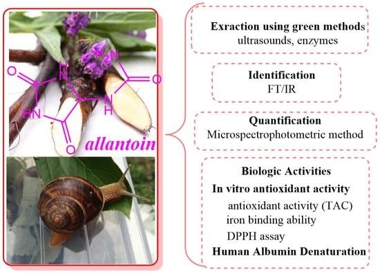
Allantoin from Valuable Romanian Animal and Plant Sources with Promising Anti-Inflammatory Activity as a Nutricosmetic Ingredient

by
Rodica Mihaela Dinica
1,
Cristina Sandu
1,2,
Andreea Veronica Dediu Botezatu
1,
Anna Cazanevscaia Busuioc
1,
Fanica Balanescu
1,2,
Maria Daniela Ionica Mihaila
1,
Caterina Nela Dumitru
2,
Bianca Furdui
1,* and
Alina Viorica Iancu
2,*
1
Department of Chemistry Physical and Environment, Faculty of Sciences and Environment, “Dunarea de Jos” University of Galati, 800201 Galati, Romania
2
Faculty of Medicine and Pharmacy, “Dunarea de Jos” University of Galati, 800010 Galati, Romania
*
Authors to whom correspondence should be addressed.
Sustainability 2021, 13(18), 10170; https://doi.org/10.3390/su131810170
Submission received: 6 August 2021 / Revised: 28 August 2021 / Accepted: 7 September 2021 / Published: 11 September 2021
(This article belongs to the Collection Advances in Biomass Waste Valorization)

Abstract

:
Helix aspersa (HA), Helix pomatia (HP) and Symphytum officinale are common organisms in Romania’s biosphere, widely known for their allantoin content and their therapeutic properties. Herein, the allantoin was separated and quantified from the aqueous extracts of Romanian comfrey root and the secretions of HA and HP snails. This study also focused on determining the antioxidant and anti-inflammatory activities of these Romanian allantoin-rich samples. The plant extracts were obtained through two methods: ultrasonic extraction and enzymatic ultrasonic extraction. A microplate method was used for the quantitative determination of allantoin content. The antioxidant activity was measured by using the DPPH radical scavenging method. The antioxidant capacity of the samples was studied in order to observe the type of interactions generated by the chemical complex present in their composition. High concentrations of allantoin were obtained by enzymatic ultrasonic extraction method (EUE—102 ± 0.74 μg/mL), and also in the water-soluble fraction of the snail secretion (FS1—22.051 μg/mL). The antioxidant screening suggests that Symphytum officinale and snail mucus extracts could be used as promising natural substitutes for synthetic antioxidants in products used for therapeutic purposes. The evaluation of anti-inflammatory activity was also investigated, allantoin-rich samples showing a promising action (FS1—81.87 ± 2.34%). In future, the inclusion of allantoin-rich extracts in various novel pharmaceutical forms for new therapeutic applications could be achieved. The study will continue with the formulation of a nutricosmetic product with snail mucus and Symphytum officinale extract as principal bioactive ingredients.

Graphical Abstract

1. Introduction

Uric acid is converted into allantoin through enzymatic or chemical oxidation, [1]. As a result of cellular metabolism, allantoin is found in a variety of organisms, from bacteria and plants, to invertebrates and vertebrates. The main therapeutic effects of allantoin (Figure 1) are stimulation of cell growth, regeneration of tissues and rebuilding of the granulation tissue [2,3,4,5,6,7]. Nutricosmetics are considered natural ingredients and products that act as nutritional supplements for the care of the beauty of hair, nails and skin, new innovative products being launched worldwide every year [4]. Allantoin is an active principle with keratoplastic and healing actions, soothing and moisturizing effects [8], commonly encountered in cosmetic and pharmaceutical products for treating burns, skin ulcers and wounds [9,10]. The mechanism of action involves stimulating fibroblast proliferation, elastin and collagen synthesis and the inhibition of the migration of inflammatory cells to the injury [11]. Recent studies have shown that allantoin has a beneficial effect in the treatment of gastritis [6], and it has also antihypertensive properties [12]. With a metformin-like chemical structure [13], both being guanidine derivatives, is has been proved that allantoin has a hypoglycemic effect [14]. FDA recognizes that allantoin acts as a skin protector in concentrations between 0.5 to 2% [15]. An ancient beauty secret, the snail’s mucus possesses many qualities, and it is considered a traditional remedy for various conditions [10]. The useful effect of mollusk secretions on wrinkled or burnt skin has been recently described [16]. According to a recent study, snail secretion has a potential antimelanogenic effect and can be used as a metastasis inhibitor [17]. Some studies showed that mucus secretion of snails can be used as a wound dressing due to its healing potential [18]. Mollusk secretion showed antimicrobial [19] and antioxidant properties [20]. The treatment with slime offered protection against colon inflammation, therefore it can be used in therapy of inflammatory bowel diseases [21]. Helix aspersa (HA) and Helix pomatia (HP), from the Helicidae family, are species of land snail native to the Mediterranean region. Snail mucus is a complex matrix, and it owes its properties to the high content of allantoin, collagen, elastine, glycolic acid, vitamins, as well as other compounds [3]. Symphytum officinale (comfrey) is a common wild plant, member of Boraginaceae family. It contains compounds like allantoin, flavonoids, tannins, amino acids, saponins and large quantities of alkaloids [22,23]. Its topical uses are based on its anti-inflammatory [24], analgesic and astringent effects [25], being used for the treatment of osteoarthritis, bruises, back pain [26] or skin irritation [7]. It has also been reported that the extract of comfrey roots has a proliferative effect and stimulates tissue regeneration [27]. Allantoin and rosemary acid are crucial for their pharmacodynamic properties. Symphytum officinale extract can be used in concentration of 0.5–4% as an emollient and cleanser for the skin [28]. The biologically active compound common to both samples, comfrey root and snail mucus, is allantoin. Over time, different methods have been developed for allantoin dosing: alkaline titration, TLC [29], HPLC or spectrophotometry [5,30]. Nowadays, there is a special interest for the discovery of new antioxidant bioactive compounds including polyphenols [31,32,33,34], polysaccharides [35,36], but also naturally derived peptides [37]. Recently, the antioxidant activity was analyzed on different samples obtained from comfrey roots [5,22,26] and snail mucus [4,17,18]. The innovation of using natural products, mainly from medicinal plants, but also animal products, was reported in different scientific studies [17,29].
The present study was designed to identify and quantify the allantoin content and to evaluate the antioxidant and anti-inflammatory properties of Romanian Symphytum officinale root extract and of the secretion from HA and HP snails, both containing allantoin [38]. Another aim of our study was to explore, in parallel, the in vitro iron and molybdenum binding and anti-inflammatory capacities of Romanian Symphytum officinale root extracts and snail slime with the aim of gaining more information about their biological activities, which could be useful in the development of new drugs or new pharmaceutical treatments, as they could be valuable active ingredients in a wide range of products, with benefits especially for skin problems.
To the best of our knowledge, we present here the first study concerning the quantification of two important Romanian plant and animal sources of allantoin and their antioxidant and anti-inflammatory potential, with future purpose of using them as principal ingredients in nutricosmetic products.

2. Materials and Methods

2.1. Chemicals and Materials

All the reagents and organic solvents were purchased from Sigma-Aldrich (Milan, Italy). The reagents were prepared freshly with distilled water. Allantoin 93791, used as standard, was supplied from Sigma-Aldrich Chemical Company (St. Louis, MO, USA). Human serum albumin (30% solution in 20 mM Tris-HCl, 0.1% NaN₃, pH 6–8, Calbiochem®, Merck KGaA, Darmstadt, Germany) was used for this study. Symphytum officinale roots were obtained from a local producer. The vegetal material was grinded mechanically in order to get a fine powder. Land snails were collected from a garden in Berești village, Galați county, Romania. The morphological characteristics, in particular the color of the shell, general dark color, allowed their recognition; they belong to the Helicidae family, species Helix aspersa (with four bands, the second and third diffuse) and Helix pomatia (numerous brown and white bands). A total of 68 snails were selected and kept indoors in a large plastic box. Before mucus collection, they were subjected to fasting for one day to avoid contamination of the mucus with their own excretion during extraction.

2.1.1. Mucus Allantoin Rich Extraction

To obtain fresh pure mucus by the Mane et al. method with slight modifications [39], 68 snails from two Romanian species (Figure 2), were washed with distilled water gently and quickly to avoid the loss of slime. The mucus collection was performed by taking each individual and stimulating with a sterile plastic stick the pedal glands located inside the front end of the snail’s foot [39]. The snails were released back into their natural habitat after extraction.
The secretion was kept in a sterile container for 12 h at 4 °C to reduce the mucilaginous effect and allow measurement of the amount of mucus obtained. To ensure a proper preservation of the sample, the mucus was lyophilized. The fractional extraction of the mucus was carried out based on the method described by Uivaroșan et al. [40]. Lyophilized snail secretion (0.1 g) was dissolved in 100 mL of ultrapure water. Mucus solution was subjected to ultrasound treatment for 60 min in an ultrasonic bath, then the mixture was centrifuged at 5000 RCF, 60 min at 4 °C with a Universal 320 R Hettich centrifuge (Andreas Hettich GmbH & Co. KG, Tuttlingen, Germany). After filtration, the supernatant was further considered as water soluble fraction (FS1) and the precipitate was collected as water insoluble fraction (PP1). The supernatant was further diluted with ethanol in a ratio of 1:3 (mL). The precipitation was then performed at −18 °C for 12 h. The precipitate, named as hydroalcoholic mucous insoluble fraction (FM), was collected after the centrifugation at 2900 RCF, for 30 min and 4 °C. The supernatant was referenced as FS2 (hydroalcoholic supernatant).

2.1.2. Comfrey Root Allantoin Rich Aqueous Extraction

The extracts were obtained through two methods: ultrasonic extraction and enzymatic ultrasonic extraction (Bandelin Sonorex Digitech, 60 Hz, Berlin Germany). Triturated Symphytum officinale roots (20 g) were subjected to aqueous extraction in 100 mL distilled water, according to the method used by Wang et al. [41]. In the EUE method, 1% papain was used with the same quantity of triturated Symphytum officinale roots in 100mL distilled water, which was carried out at 50 °C for 70 min [42]. The extracts obtained were centrifuged at 5000 RCF for 15 min at 5 °C and the precipitate was removed. Further, the aqueous phase of each sample was concentrated to 10 mL and used in analyzes as EU (aqueous comfrey root extract obtained by ultrasonic extraction) and EUE (aqueous comfrey root extract obtained by enzymatic ultrasonic extraction) samples. In the case of the sample extracted by the enzymatic method, the blank sample for each further analysis was represented by a solution prepared according to the same method without Symphytum officinale root.

2.1.3. Ultrasonic Methanolic Extraction

Symphytum officinale powdered roots (20 g), were used in ultrasonic methanolic extraction with 100 mL methanol at 50 °C for 1 h. Next, the mixture was filtered and concentrated at 50 °C to a volume of 20 mL by using a Rotary evaporator BUCHI Rotavapor™ R-100. For allantoin to precipitate, the concentrated solution was cooled to 4 °C and collected by filtration. The previous step was repeated for the filtered solution to precipitate the remaining allantoin, which was collected by filtration and further labelled as UME.

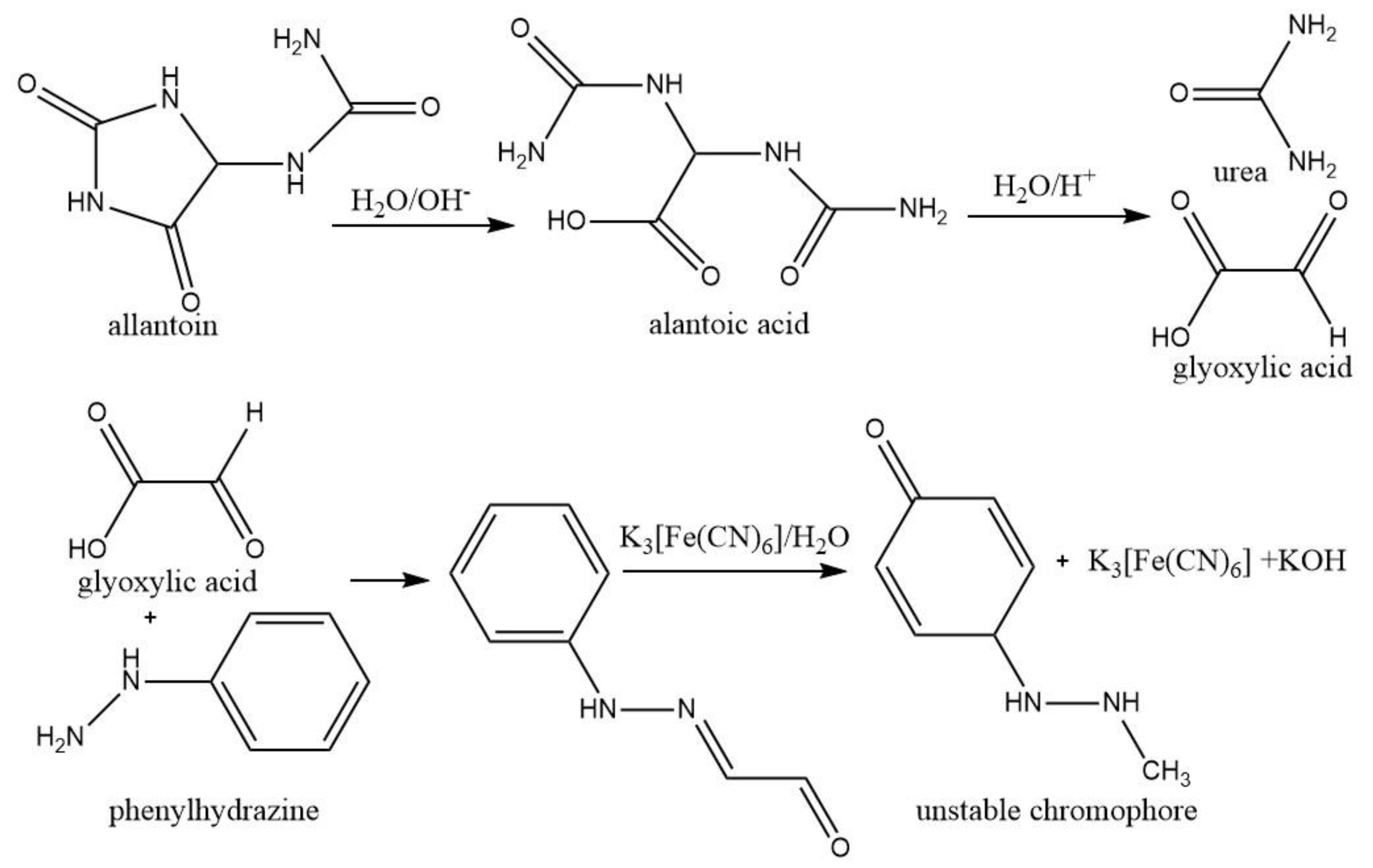
2.2. Quantification of Allantoin from the Aqueous Extracts

The quantification of allantoin in the obtained fractions was analyzed by using a microplate method as previously described [43], with slight modifications. The quantification method is based on the formation of the orange color produced by the interaction of allantoin with potassium ferricyanide (Figure 3) [22].
Aliquots of 10 μL of each sample were added to 100 μL distilled water, were treated with 20 μL NaOH (0.5 M), sonicated in an ultrasonic bath for one minute, and then maintained in a boiling bath for 7 min (for allantoin hydrolysis in allantoic acid). A blank sample was prepared by using 10 μL of distilled water instead of samples. The samples were cooled in cold water bath, then 20 μL HCl (0.5 M) was added (for the occurrence of the allantoic acid hydrolysis to urea and glyoxylic acid). A total of 20 μL solution of 0.023 M phenylhydrazine hydrochloride freshly prepared was added, then the samples were sonicated for one minute in an ultrasonic bath, afterwards they were maintained in a boiling bath for 7 min. After that, the samples were placed in an ice water bath and kept at −20 °C for 10 min. A total of 60 μL of concentrated HCl and 20 μL of 0.05 M potassium ferricyanide solution, freshly prepared before use, were mixed thoroughly till an orange to a red brick colored solution appeared. The calibration curve was obtained by using various concentrations of pure allantoin (Sigma-Aldrich, St. Louis, MO, USA). A stock solution of 100 mg/L pure allantoin was used for the quantitative analysis. The allantoin concentration was calculated by using the linear regression equation (y = 0.0039x + 0.0354) based on allantoin standard calibration curve in the range of concentrations 4–100 μg/mL. A blank sample was prepared by using distilled water instead of allantoin solution. Each test was performed in triplicate. The samples were read at 522 nm with a multiplate reader (iTecan Pro 200, Tecan Trading AG, Männedorf, Switzerland).

2.3. FT-IR Analysis of Samples

The infrared spectra were collected using a Nicolet iS50 FT-IR spectrometer (Thermo Scientific) equipped with a built-in ATR accessory. A total of 32 scans were co-added over the range of 4000–400 cm−1 with a resolution of 4 cm−1. Air was taken as the reference for the background spectrum before each sample. After each spectrum, the ATR plate was cleaned with ethanol solution. In order to verify that no residue from the previous sample remained, a background spectrum was collected each time and compared to the previous background spectrum. The FT-IR spectrometer was placed in a room that was air conditioned with controlled temperature (21 ± 1 °C).

2.3.1. In Vitro Antioxidant Activity of Allantoin Rich Samples by DPPH Assay

Determination of DPPH radical scavenging activity was done by the discoloration of purple colored DPPH solution. DPPH assay was evaluated based on a literature procedure with small modifications [44]. Briefly, a volume of 100 μL from each sample at various concentrations was added to 100 μL of DPPH solution. The mixtures were kept at room temperature in dark conditions, for 60 min, at room temperature and the absorbance was determined with Tecan iPro 200 multiplate reader at 517 nm. The absorbance was measured after 15, 30 and 60 min. The control contained only DPPH and the solvent. Inhibition of free radical DPPH was evaluated by using the following formula:
Inhibition (%) = (Acontrol − Asample)/Acontrol × 100
where Acontrol was the absorbance of the control group (including all the reagents, except the tested samples), and Asample was the absorbance of the samples.

2.3.2. In Vitro Evaluation of Total Antioxidant Activity (TAC)

This assay is based on formation of a blue Mo (V)-complex at acidic pH from the reduction of Mo (VI) to Mo (V) [45]. For total antioxidant capacity assay, 0.1 mL test samples were mixed in Eppendorf tubes with 1 mL of reagent solution (0.6 M sulfuric acid, 28 mM sodium phosphate, and 4 mM ammonium molybdate). Then, they were incubated at 95 °C for 90 min. After the mixture had cooled to room temperature, the absorbance of each solution was measured at 695 nm. All the experiments were done in triplicate. The total antioxidant activity was expressed as the absorbance of the sample at 695 nm. The higher absorbance value indicated higher antioxidant activity [46].

2.3.3. In Vitro Determination of Iron Binding Ability

The iron (II) binding capacity of the chelators was determined according to the method described in the literature [46] by an analysis of 96-well plates with microplate reader (Tecan Infinite Pro 200, Tecan Trading AG, Männedorf, Switzerland). Briefly, an amount of 225 μL sample was mixed with 10 μL ferrous sulphate (2 mM), then the mixture was left at room temperature for 5 min and after 15 μL ferrozine (0.2 mM) was added to start the reaction. A blank was prepared by using solvent instead of ferrozine. The resulting mixture was mixed and stored for 10 min at room temperature and then the absorbance was measured at 562 nm. Na2EDTA represented the positive control standard. All the experiments were done in triplicate. The iron binding capacity was evaluated by using the following formula:
Iron binding ability (%) = (Acontrol − Asample)/Acontrol × 100
where Acontrol is the absorbance of the control and Asample is the absorbance of the samples.

2.4. Inhibition of Human Albumin Denaturation

The present study was carried out to evaluate the anti-inflammatory property of the allantoin containing samples by human albumin denaturation assay. The purpose of this study was to investigate whether the tested samples could inhibit human serum albumin (HSA) thermal denaturation, which is the most abundant plasma protein [47,48]. Human serum albumin (30% solution in 20 mM Tris-HCl, 0.1% NaN₃, pH 6–8, Merck KGaA, Darmstadt, Germany) was used for this study. PBS (phosphate buffer saline, pH 6.3) served as dilution solvent [49]. The assay was done by Saso et al. method [50], with slight modifications. Briefly, in Eppendorf tubes were added 600 μL of reaction mixture, which was comprised of 200 μL of phosphate buffered saline, 200 μL of various concentrations of samples and 200 μL of 2% (w/v) human albumin. PBS buffer and human albumin served as control sample. Then the mixture was incubated at 37 °C in a heating shaker with stirring at 200 rpm (Thermoshaker Biosan, TS100, Riga, Latvia) for 15 min, and then heated at 70 °C for 30 min. After cooling, their absorbance was measured at 595 nm with Tecan microplate reader. Diclofenac sodium (standard drug) was used as reference drug. The percentage inhibition of protein denaturation was calculated as follows:
%Inhibition = 1 − [(Ax, 70 °C − Ax blank, 25 °C)/(Acontrol, 70 °C − Acontrol blank, 25 °C)] * 100
where Ax, 70 °C was the absorbance of the sample x at the concentration c after heating at 70 °C, Ablank was the absorbance of the same sample before heating. Control represented denaturized protein. Each sample was done in triplicate.

3. Results

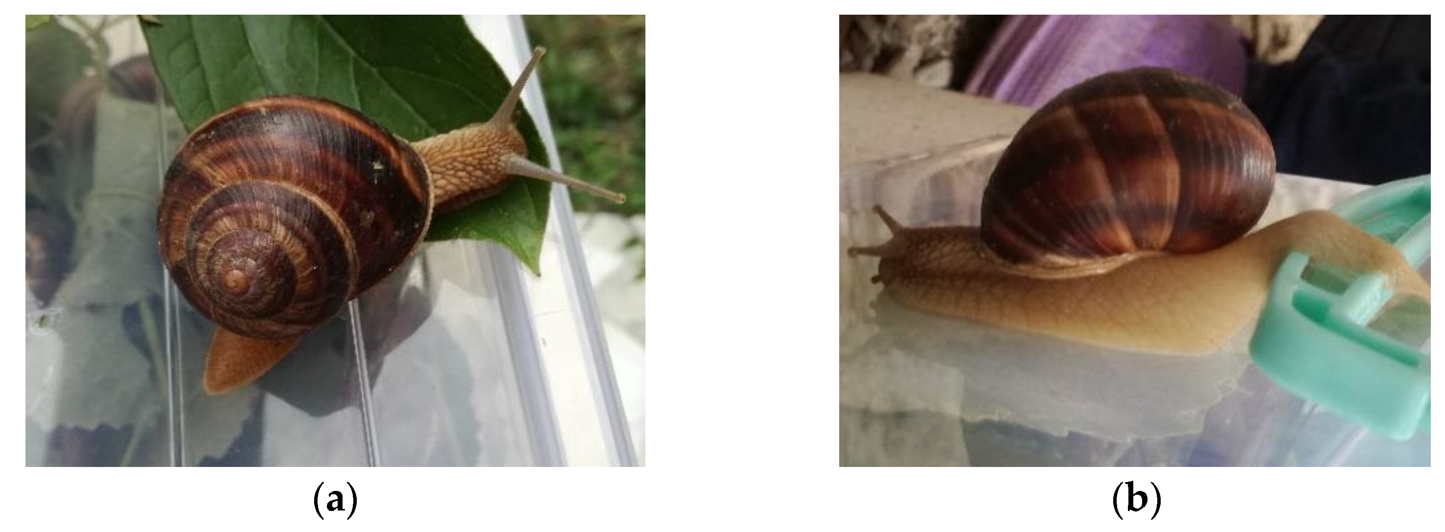
3.1. Characterization of Snail’s Secretion

A total of 74 g of crude mucus were obtained from 68 snails from two Romanian species by the Mane et al. modified method [39]. Given that, to our knowledge, these two native Romanian species of snails selected for our study were not studied for the same purpose to identify allantoin in their mucus, our goal was to highlight the allantoin content and properties of mucus in both species of snails that frequently spread in Romanian gardens and are not sufficiently valued for therapeutic purposes. Other studies show that the two species of garden snails produce about the same percentage of mucus [51] and that is why we chose not to collect the mucus separately.

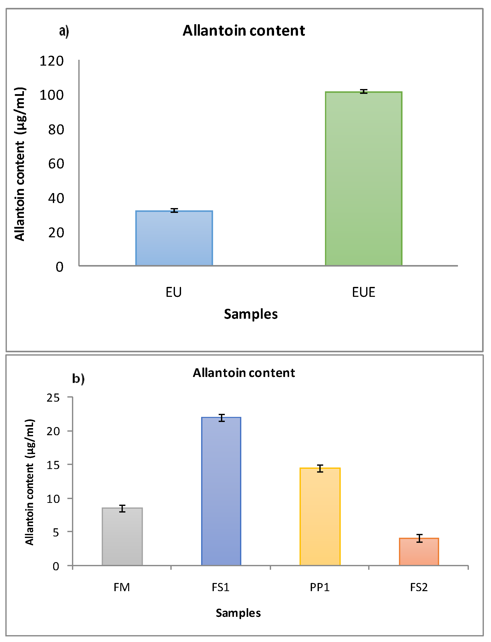
3.2. Allantoin Quantification

One of the leading objectives of this study was the determination of the characteristic bioactive compound of the snail secretion and comfrey root, allantoin. A stock solution of 100 μg/mL was used for the quantitative analysis of allantoin, and the diluted working standards used for achieving of standards value graph were in the range of concentrations 4–100 μg/mL. The allantoin concentration was calculated by using the linear regression equation (y = 0.0039x + 0.0354) based on allantoin standard calibration curve (the concentrations used were: 4.00, 8.00, 10.00, 20.00, 30.00, 40.00, 50.00, 80.00, 90.00, 100.00 μg/mL).
The results regarding the amount of allantoin from the obtained fractions are presented in the Figure 4. It was observed that the allantoin concentration vary according to the studied species, so that the level of allantoin in comfrey root extract is higher than that of snail mucus. Symphytum officinale EUE extract showed a high allantoin content (102 ± 0.74 μg/mL) which is four time greater than the allantoin content of Symphytum officinale EU extract (32.465 ± 0.45 μg/mL). By comparing the values from Figure 4a, it can be noticed that the presence of papain in extracts obtained by EUE increased the allantoin extraction and concentration.
Allantoin concentration and distribution in mucus fractions varied greatly. Higher allantoin content was obtained in FS1 (22.051 ± 0.64 μg/mL) compared to the further processed fraction FS2 (4.102 ± 0.08 μg/mL). A significant difference between allantoin levels of these four fractions of snail mucus was remarked. Higher concentrations were found in the aqueous phases obtained from the secretion of snails, FS1 (22.051 ± 0.59 μg/mL) and PP1 (14.50 ± 0.62 μg/mL), while in the hydroalcoholic phases, FM (8.54 ± 0.65 μg/mL) and FS2 (4.102 ± 0.49 μg/mL) was found the least allantoin content. Of all the fractions analyzed, the richest in allantoin was FS1 and Symphytum officinale EUE extract.
The results regarding the amount of allantoin from the aqueous fractions obtained are presented in Figure 4. It was observed that the allantoin concentration vary according to the studied extraction methods, so that the level of allantoin in comfrey root by EUE method is higher than that in EU method. Therefore, it can be noticed that the presence of papain in extracts obtained by EUE method increased the allantoin extraction.
Allantoin from snail secretion was obtained in a yield of 22 ± 0.85 mg/g snail secretion and from Symphytum officinale roots was obtained in a yield of 3.29 mg/g root.
Snail mucus has been used in various directions in medicine since ancient times to relieve lesions and various diseases. Research over the years on the secretions of Helix aspersa and Helix pomatia snails has confirmed that mucus contains a combination of ingredients with beneficial and therapeutic qualities, such as allantoin. For the determination of allantoin, several analytical methods have been developed and published for snail mucus, biological samples and for cosmetics and pharmaceuticals, by alkaline titration methods and infrared spectrometry, as well as determinations by HPLC [15,52]. The literature data concerning the allantoin content are reported only for the crude extract. Thus, Laneri isolated 0.41 g/L allantoin from the mucus of H. aspersa [4] and El Mubarak et al. isolated allantoin between 0.4 and 0.16 mg/mL, from the mucus of H. aspersa too [15]. Fu et al. reported a concentration of allantoin in snail mucus of 47–52 mg/L (HA) [10]. In Symphytum officinale root, the allantoin content reported by Kimel et al. was of 0.94–2.10% in raw material in alcoholic extracts [53].

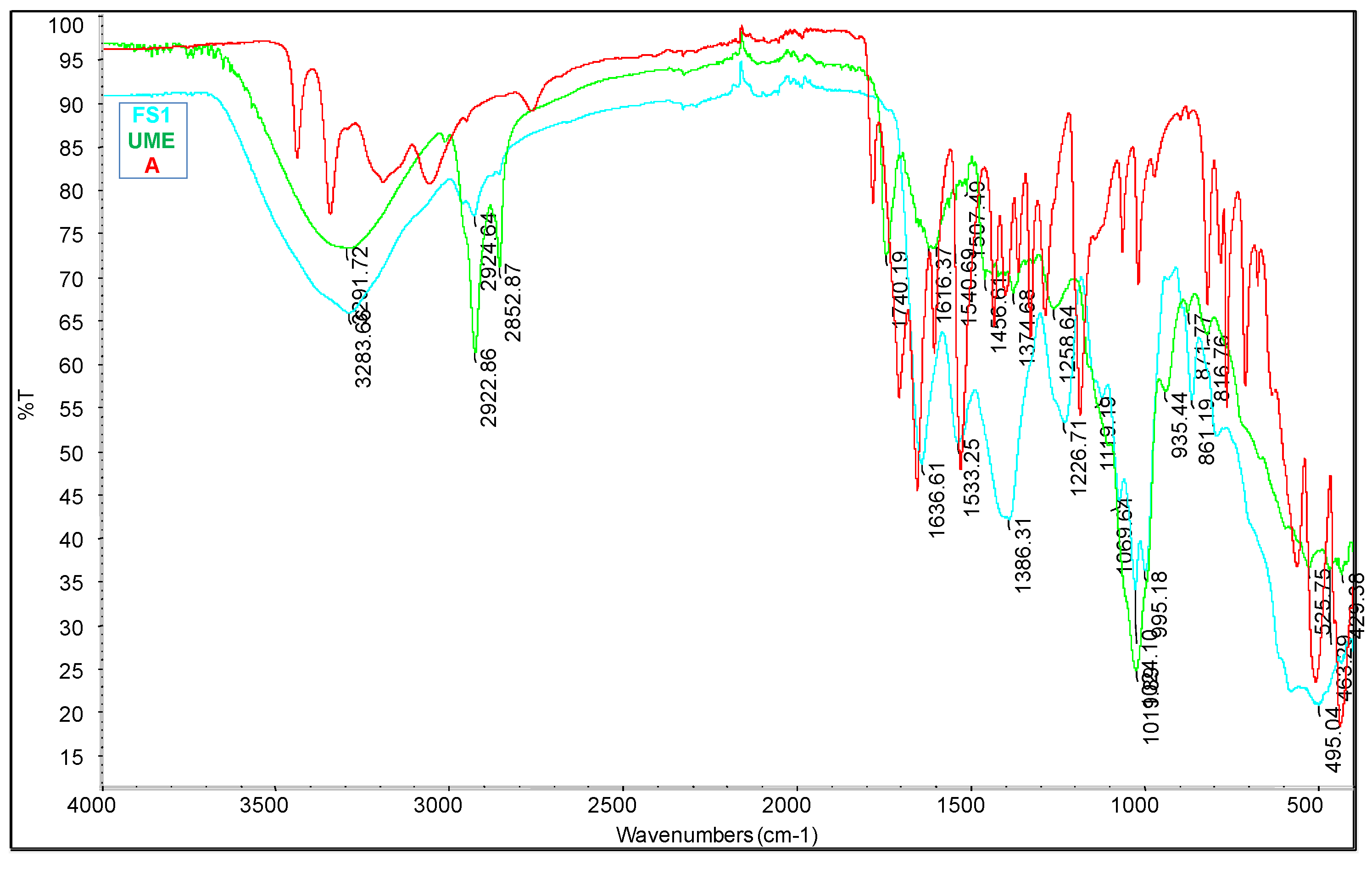
3.3. FT-IR Analysis of Samples

The characterization of the active compounds based on their functional groups is possible through Fourier transform infrared spectroscopy (FT-IR). The ATR-FTIR spectrum, in the range 4000 to 400 cm−1, of pure allantoin and extracted allantoin from the samples are shown in Figure 5.
All the samples show the IR absorption regions characteristic to allantoin functional groups, as indicated in Table 1.
Bands at 3500–3350 cm−1 and 3450–3150 cm−1 related to the asymmetric and symmetric stretching vibrations of amino groups could be identified, whereas the bands related to the vibration of H-N-C groups were identified at 1550–1500 cm−1. The absorptions bands at about 1636–1640 cm−1 could be due to stretching vibrations of C = O and C = C groups, associated to amidic groups. The NH2 scissoring vibration is identified at 1600–1615 cm−1 with medium intensity. The stretching vibration with weak intensity at 1120–910 cm−1 occurred because of the C-NH2, aliphatic amine group. The bands at 860–510 cm−1 and also at 450 cm−1 are due to N-H out of plane bending vibration. The bands at 1840–1640 cm−1 and also 780–760 cm−1 could be related to the C = O vibration. The bands identified in each sample showed deviations from the functional groups characteristic bands of pure allantoin, which could be associated with inter-molecular and intra-molecular interactions between allantoin and other compounds in the samples analyzed from snail mucus and comfrey roots [54].

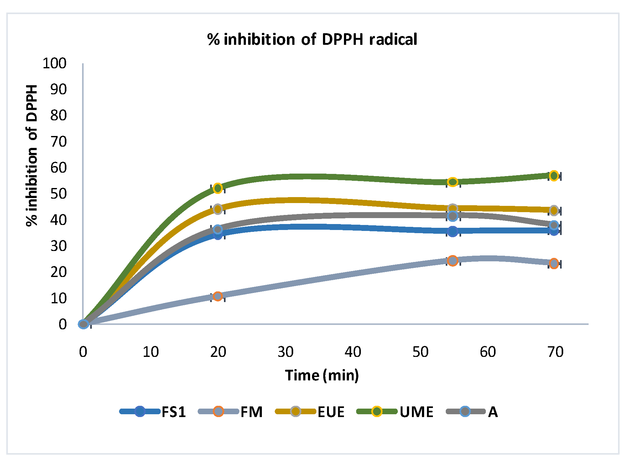
3.3.1. DPPH Antioxidant Activity

In the next step, the antioxidant effect of the preselected fractions, FS (1 mg/mL) and EUE (100 mg/mL) was evaluated. The DPPH scavenging activity of different allantoin-containing species has been demonstrated [7,28]. Nevertheless, there are no reports about comparative analysis of Helix aspersa, Helix pomatia secretion and Symphytum officinale extracts on their antioxidant behavior. To appreciate the antioxidant potential of selected samples and to study the interactions of the extracts with free radicals, their activity was tested in vitro (DPPH). In addition, the allantoin rich samples were analyzed. The samples manifested various levels of antioxidant potential (Figure 6).
Figure 6 shows the DPPH’s radical inhibitory percentage of all the samples; both of allantoin plant and animal sources revealed antioxidant activity, but the water-soluble fraction of snail secretion showed lower activity than Symphytum officinale root extracts obtained by methanolic (UME) and aqueous enzymatic (EUE) ultrasonic extraction. However, FM showed the lowest antioxidant activity compared to the antioxidant properties of other samples. According to other studies from the literature, both snail mucus and Symphytum officinale roots contain a considerable amount of allantoin, being one of the main natural compounds to which this important antioxidant activity can be attributed [5,20]. Therefore, considering that the highest amount of allantoin was found in the ultrasonic methanolic extract of S. officinale as well as in the water-soluble fraction of the snail mucus solution, FS1, the highest antioxidant activity for these two samples can also be attributed to this valuable allantoin content.

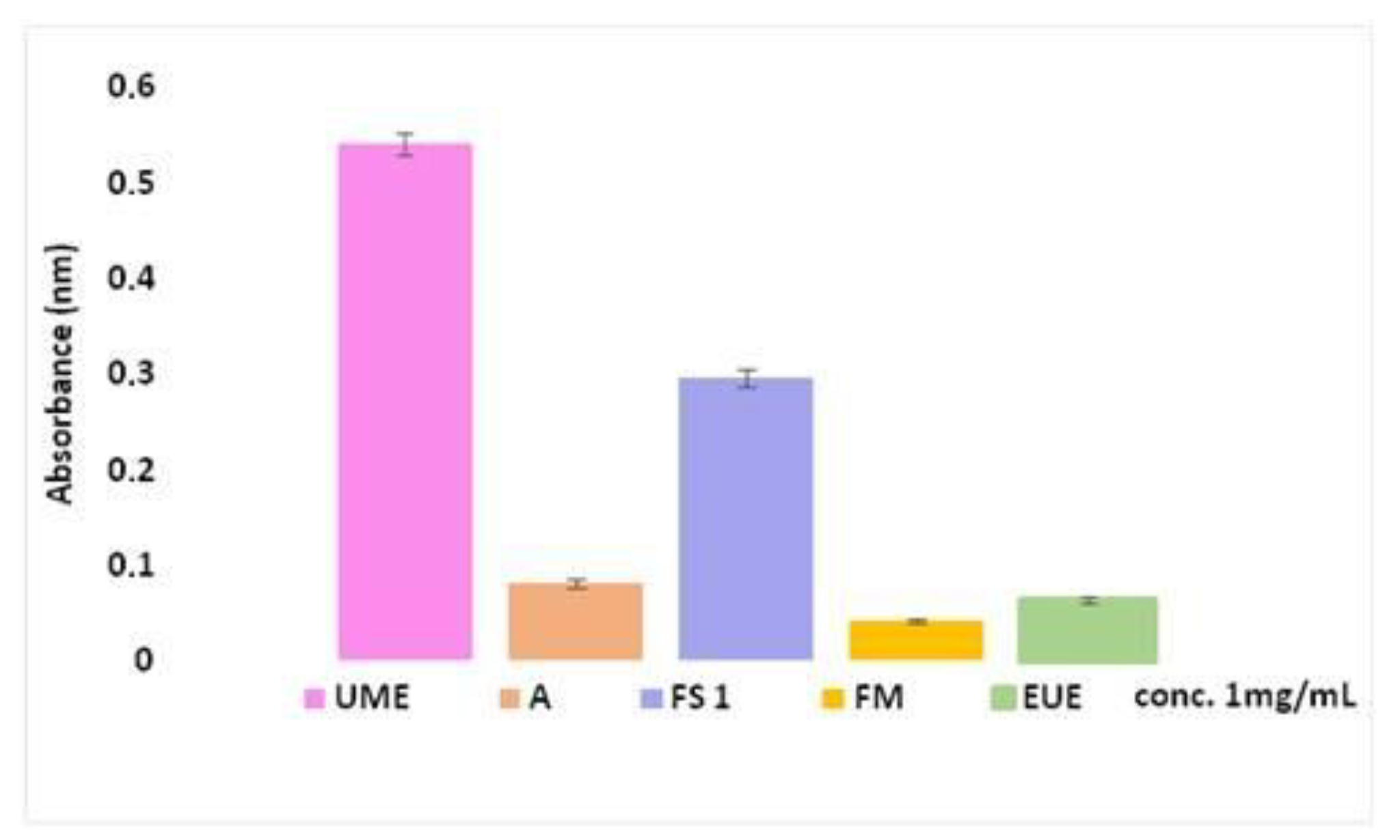
3.3.2. Evaluation of Total Antioxidant Activity

The phosphomolybdenum method has been used to evaluate the total antioxidant capacity of the samples. In the presence of the extracts, Mo (VI) is reduced to Mo (V) and forms a green–blue colored phosphomolybdenum V complex, which shows a maximum absorbance at 695 nm. Considering the fact that the higher absorbance of the samples means a higher total antioxidant activity, it was observed that the ultrasonic methanolic extract (UME) from the root and the water-soluble fraction of the snail mucus (FS1) have the highest total antioxidant capacity (Figure 7).
The best results were identified for UME plant extracts and FS1 water soluble snail fraction. These samples showed high antioxidant activities due to the fact that in the ultrasound methanolic extract (UME) from the roots as well as in the water-soluble fraction of snail mucus (FS1) were observed a considerable amount of allantoin but also the presence of other bioactive compounds, these results agreeing with the data from the literature [7,20,24,53]. Figure 7 shows that the antioxidant capacity is lower for A, EUE and FM samples. Therefore, in future studies UME and FS1 samples will be used as active principles in nutricosmetic products developed in our laboratory (ongoing research, unpublished), due to their good antioxidant capacity which may help human beings to treat various conditions caused by oxidative stress.

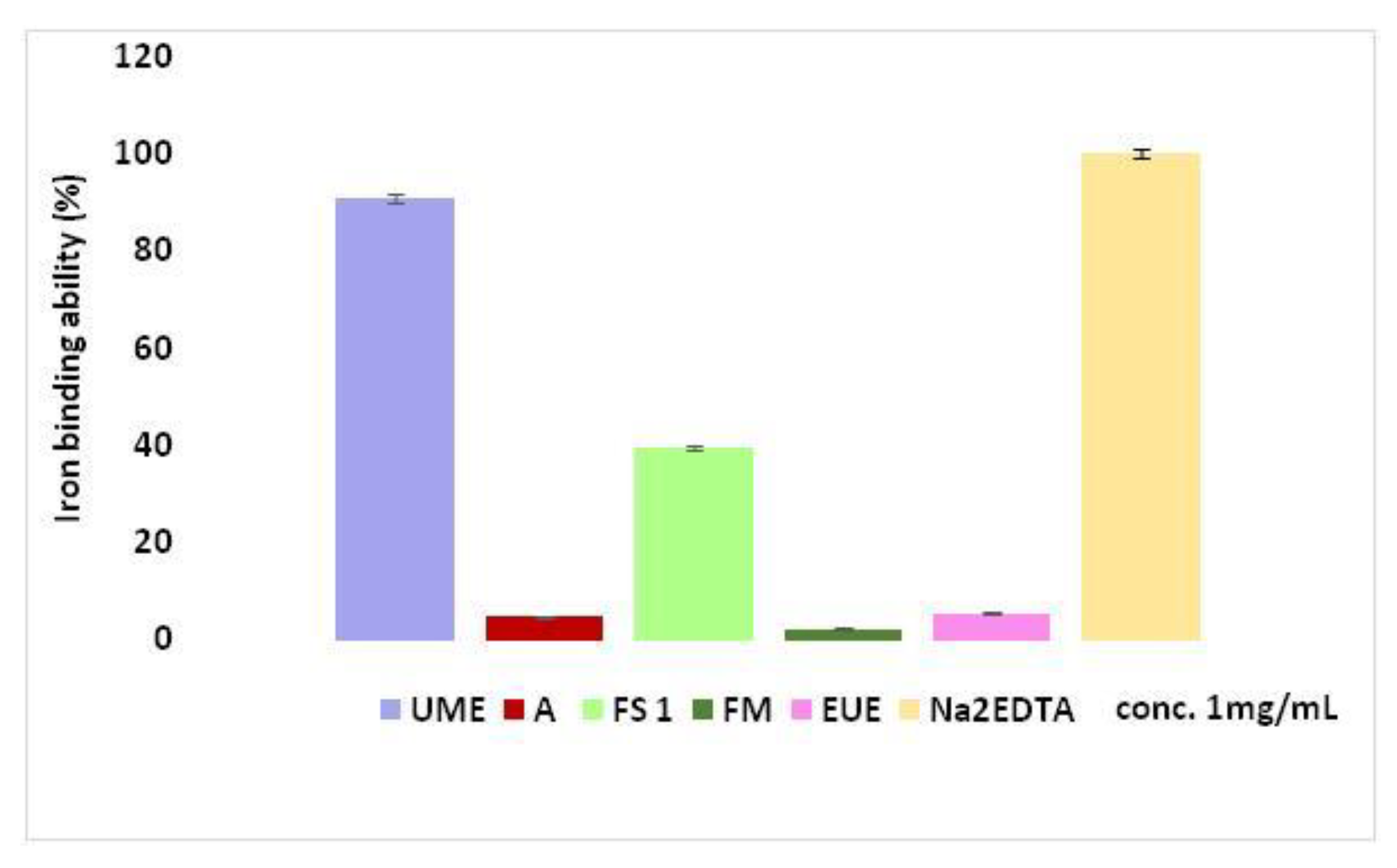
3.3.3. Determination of Iron Binding Ability

Free radicals’ accumulation in human body can lead to various abnormalities and diseases. Iron is known to have significance in metabolic disorders, the study of the relationship between iron deficiency and skin diseases being in progress. This assay indicates that not all test samples could chelate transition metal ions, only FS1 from snail and UME from S. officinale extracts have small iron blinding abilities, which could be connected to the chemical structure of the compounds in their composition. In our study, the results regarding the iron chelating abilities of all the samples were compared to those of a known strong iron chelator, Na2EDTA (Figure 8), the methanolic ultrasonic extract (UME) having the most promising activity in order to be selected as an active ingredient in a nutricosmetic product in future research.
The extraction methods from plants and snails mucus both in our study and in other studies in the literature [5,22,53], led to the extraction of allantoin but also of other bioactive compounds, with a higher content of them in the samples extracted with methanol from S. officinale roots (UME) and the water-soluble fraction of snail mucus (FS1), therefore, this may be the reason why the two samples showed the best antioxidant properties in the iron ion chelating method.

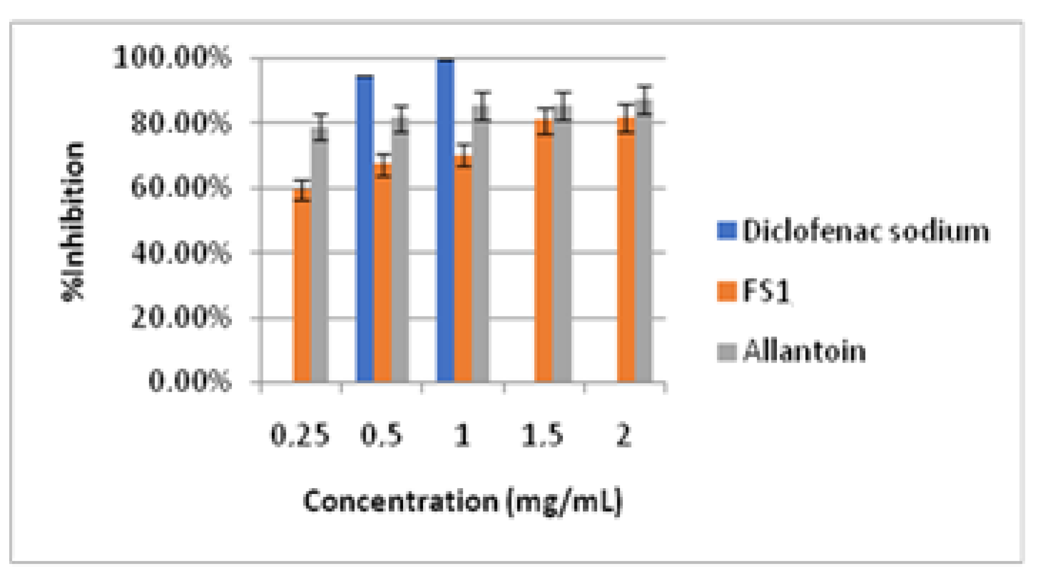
3.3.4. Inhibition of Human Albumin Denaturation
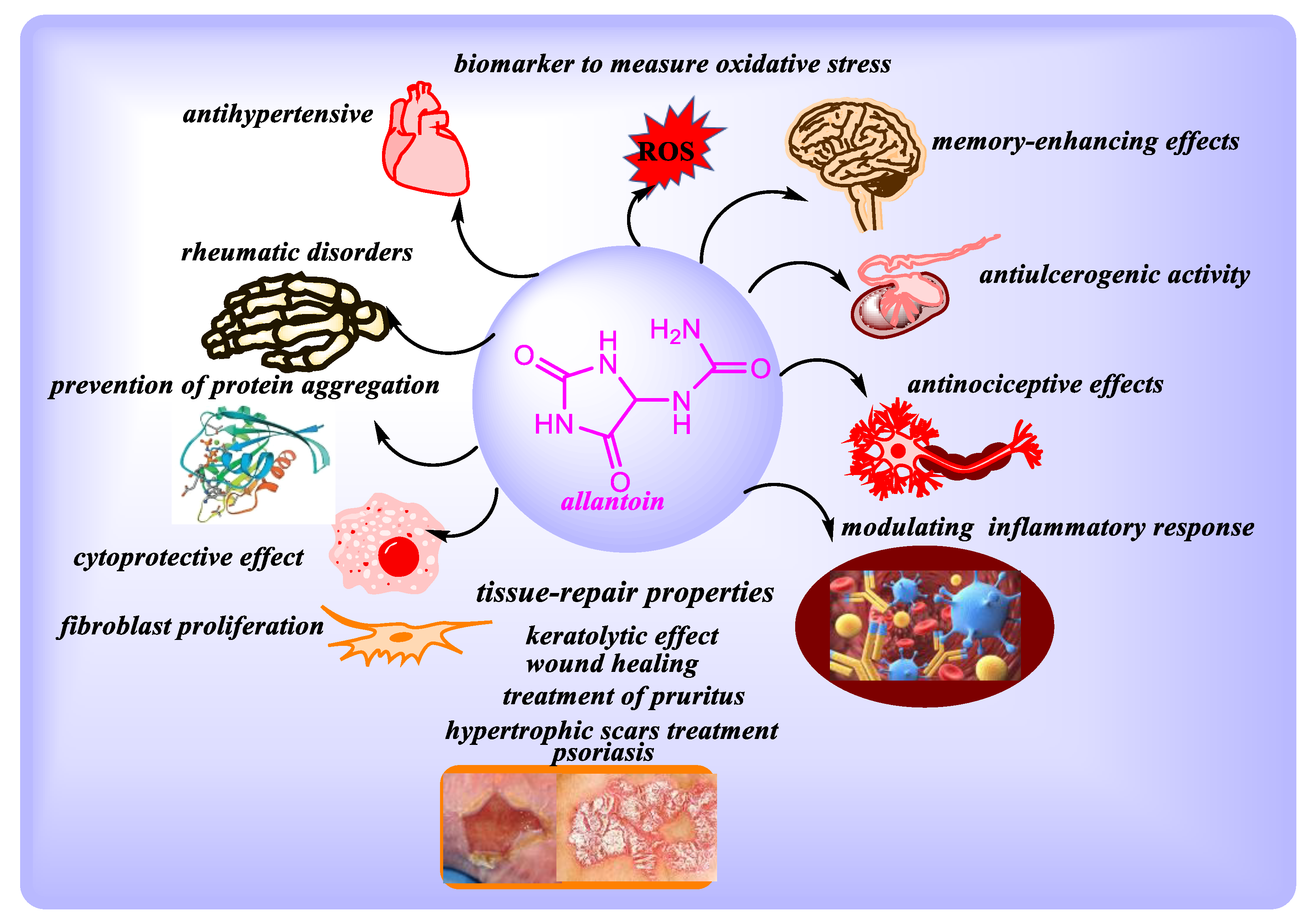
In this study, an anti-inflammatory method was adapted to evaluate the anti-arthritic activity of allantoin from different sources, meaning that the treatment with different extracts could lead to the release of various bioactive substances that could play a role in manifesting a particular pharmacological activity. Human albumin denaturation test was carried out for this purpose for the selected allantoin rich samples (Figure 9).
This study concluded that, in the range of concentrations tested from 0.25 to 2 mg/mL, the pure allantoin and the water-soluble fraction of snail secretion (FS1) (Figure 9) showed very good inhibitory activity of the thermal denaturation of albumin ranging from 59.55 ± 2.45% to 81.87 ± 2.34% for FS1 and from 78.78 ± 1.83% to 87.20 ± 2.11% for pure allantoin, when compared to the activity of the diclofenac sodium standard drug (94.64 ± 0.35% for 0.5 mg/mL and 99.85 ± 0.24% for 1 mg/mL). The ultrasonic aqueous extracts EUE and EU (1 ÷ 10 mg/mL), were also tested but this extracts from S. officinale roots are colored and their inhibitory thermal denaturation of albumin could not be properly evaluated at the wavelength used in this method. Therefore, another method of testing for anti-inflammatory activity will be performed in future studies. According to the results from the literature, the secondary metabolites of S. Oficinale show anti-inflammatory activity both by their in vitro action on biomolecules and also in vivo, thus motivating our interest for this type of extracts from this plant not sufficiently valued in Romania.

4. Discussion

The most common techniques of obtaining extracts are maceration and heat reflux with solvents, but these are very simple extraction methods with the disadvantage of long extraction time and low extraction efficiency compared to modern extraction methods such as ultrasound or enzymatic ultrasound extraction [55]. Ultrasounds produce a cavitation effect by collapsing of ultrasonic bubbles, releasing energy, destroying the cell structure and improving mass transfer, so the extractability of biological compounds increases [56]. Ultrasound extraction has the advantage of increasing the extraction yield and reducing the extraction time [57]. Therefore, the methods selected for this study were ultrasound assisted (EU) and enzymatic ultrasound assisted extractions (EUE). To identify the presence of allantoin in the samples, the UV-Vis spectroscopy, TLC and ATR-FTIR analysis were used. According to FTIR spectra, similar bands specific to allantoin were observed in all the samples and they suggest the presence of the important functional groups which are also shown in pure allantoin and in snail extracted allantoin. The characteristic IR (ATR, cm−1) bands identified for carbonyl groups, amino, and also amide moieties are arguments for the presence of allantoin in the analyzed samples: 3500–3250 cm−1 (υNH2); 3350–3150 cm−1 (υN-H); 1550–1500 cm−1 (υH-N-C); 1840–1640 cm−1, 1636–1640 cm−1 (υC = O); 1600–1615 cm−1 (υN-H); 1120–910 cm−1 (υ C-NH2); 860–510 cm−1 (υN-H); 780–760 cm−1 (υC = O) [54].
To quantify allantoin in snail secretion, samples were prepared and analyzed by using the modified method after Young and Conway [43]. The method was also applied for assessing the allantoin in Symphytum officinale root extract, as it is an important active constituent of the species. The modified methods presented in this research offer many advantages such as less time and less cost when a large number of samples are analyzed, as well as requiring a small quantity of samples and reagents and also enabling accurate and fast reading of multiple samples. A considerable difference between allantoin contents of the analyzed samples and sources was observed. This difference is due to the source of the extract (vegetal or animal), the type of extraction and the affinity for the solvent used for extraction. In the present study, the concentration of allantoin in Symphytum officinale extracts (EUE—102 ± 0.74 μg/mL and EU—32.465 ± 0.45 μg/mL) was superior to that from the snail secretion (FS1—22.051 μg/mL and PP1—14.50 μg/mL). The explanation could be connected to the different chemical composition of the species. The higher concentration of allantoin in the aqueous phases obtained from the secretion of Helix aspersa and Helix pomatia snails compared to ethanolic fractions is due to the solubility of allantoin in water. The difference between the allantoin content of the two extracts obtained from comfrey root indicates that the extraction yield is significantly influenced by the extraction method. Among the two extraction techniques, EUE exhibited the highest content of allantoin, the reason being due to the presence of enzyme (papain). Papain can degrade the cell wall of plant materials and promote the release of intracellular constituents [42]. In addition, papain helps break proteins down into peptides and amino acids, and finally up to uric acid, which can be converted to allantoin [42]. Therefore, the association of papain and the ultrasound seems to be beneficial, acting synergistically and increasing the allantoin extraction through the effects of cavitation and enzymatic hydrolysis.
The scavenging activity of DPPH radicals was performed for screening the antioxidant properties. The tested fractions were capable of reducing the stable free radical DPPH to the yellow colored 1,1-diphenyl-2-picrylhydrazyl. In the present study, comfrey root extracts UME and EUE, and the water-soluble fractions, FS1, of snail secretion showed the highest antioxidant activity. Given the allantoin content, along with the rest of the complex composition of the Symphytum officinale roots and the snails secretions, it is highly probable that any interactions that may occur between biologically active compounds will influence the activity either positively (synergy) or negatively (antagonism). Indeed, interaction among different species may promote changes in overall antioxidant capacity, which are difficult to predict on the basis of their individual antioxidant capacity [58].
The anti-inflammatory activity of snail mucus samples and also of comfrey root extracts was analyzed by the inhibitory assay of human serum albumin thermal denaturation. In other researches, the mucus obtained from giant African snail, Lissachatina fulica, has shown also promising anti-inflammatory potential both by in vivo tests and by in vitro tests [59]. Following the analysis performed, Sri Harti et al. concluded that improving an inflammatory status with a preparation based on the combination of snail mucus and chitosan may be an alternative to synthetic compounds [60]. There are many clinical studies proving the efficacy of comfrey root extracts against pro-inflammatory factors [61]. Seigner et al. investigated the anti-inflammatory properties of a hydroalcoholic comfrey root extract in an in vitro model of inflammation and the results obtained agree that the constituents of comfrey root can interfere with key pro-inflammatory molecules [24]. Therefore, the optimizations of extraction methods and the development of new nutricosmetic products are of real interest nowadays and future research in our laboratory will be based on the development and analysis of such products based on extracts from plant and animal sources rich in allantoin.

5. Conclusions

Quantitative determination of allantoin, a chemical constituent characteristic of comfrey root and snail secretion, was performed by a spectrophotometric method adapted in microplate format. High concentrations of allantoin were found in the methanolic extracts of comfrey roots and also in the extracts obtained by ultrasound-assisted enzymatic extraction, as well as in the water-soluble fraction of snail secretion. The combination of ultrasound technology in the extraction process and the use of enzymes is a preferable alternative to traditional methods of extracting plant products. The results indicate that allantoin-rich extracts, UME, FS1 and EUE can be used as promising natural antioxidants. The water-soluble snail mucus fraction, FS1, showed a promising inhibitory activity of thermal denaturation of human serum albumin, comparable to that of diclofenac, a well-known anti-inflammatory compound. The main constituent of these species, allantoin, as well as its bioactive properties, stimulate us to develop new research directions in order to design and develop methods to obtain diversified nutricosmetic products that harness the antioxidant and anti-inflammatory potential of the analyzed samples.

Author Contributions

Conceptualization, R.M.D., B.F. and A.V.I.; data curation, A.V.D.B., A.C.B., M.D.I.M. and B.F.; funding acquisition, R.M.D., A.C.B. and B.F.; investigation, C.S., A.V.D.B., A.C.B., F.B. and C.N.D.; methodology, R.M.D., C.S., A.V.D.B., A.C.B., B.F. and A.V.I.; project administration, R.M.D. and A.C.B.; resources, R.M.D., F.B., B.F. and A.V.I.; software, C.S., A.V.D.B. and A.C.B.; supervision, R.M.D., B.F. and A.V.I.; visualization, A.V.D.B., A.C.B., F.B. and C.N.D.; writing—original draft, R.M.D., C.S., A.V.D.B., A.C.B. and M.D.I.M.; writing—review and editing, R.M.D., A.V.D.B., A.C.B. and B.F. All authors have read and agreed to the published version of the manuscript.

Funding

This research was supported by the project “ANTREPRENORDOC”, Contract No. 36355/23.05.2019, financed by The Human Capital Operational Program 2014–2020 (POCU).

Acknowledgments

The results of this work were also presented to the 9th edition of the Scientific Conference organized by the Doctoral Schools of “Dunărea de Jos” University of Galati (SCDS-UDJG) http://www.cssd-udjg.ugal.ro/ (accessed on 3 June 2021) that was held on 10th and 11th of June 2021, in Galati, Romania.

Conflicts of Interest

The authors declare no conflict of interest.

References

  1. Ramazzina, I.; Folli, C.; Secchi, A.; Berni, R.; Percudani, R. Completing the uric acid degradation pathway through phylogenetic comparison of whole genomes. Nat. Chem. Biol. 2006, 2, 144–148. [Google Scholar] [CrossRef] [PubMed]
  2. Arakawa, T.; Maluf, N.K. The effects of allantoin, arginine and NaCl on thermal melting and aggregation of ribonuclease, bovine serum albumin and lysozyme. Int. J. Biol. Macromol. 2018, 107, 1692–1696. [Google Scholar] [CrossRef]
  3. Trapella, C.; Rizzo, R.; Gallo, S.; Alogna, A.; Bortolotti, D.; Casciano, F.; Zauli, G.; Secchiero, P.; Voltan, R. HelixComplex snail mucus exhibits pro-survival, proliferative and pro-migration effects on mammalian fibroblasts. Sci. Rep. 2018, 8, 17665. [Google Scholar] [CrossRef] [PubMed]
  4. Laneri, S.; Di Lorenzo, R.; Sacchi, A.; Dini, I. Dosage of bioactive molecules in the nutricosmeceutical Helix aspersa muller mucus and formulation of new cosmetic cream with moisturizing effect. Nat. Prod. Commun. 2019, 14, 1934578X19868606. [Google Scholar] [CrossRef] [Green Version]
  5. Sowa, I.; Paduch, R.; Strzemski, M.; Zielińska, S.; Rydzik-Strzemska, E.; Sawicki, J.; Kocjan, R.; Polkowski, J.; Matkowski, A.; Latalski, M.; et al. Proliferative and antioxidant activity of Symphytum officinale root extract. Nat. Prod. Res. 2018, 32, 605–609. [Google Scholar] [CrossRef] [PubMed]
  6. Eslami-Farsani, M.; Moslehi, A.; Hatami-shahmir, A. Allantoin improves histopathological evaluations in a rat model of gastritis. Physiol. Int. 2018, 105, 309–324. [Google Scholar] [CrossRef] [PubMed]
  7. Savic, V.L.; Nikolic, V.D.; Arsic, I.A.; Stanojevic, L.P.; Najman, S.J.; Stojanovic, S.; Mladenovic-Ranisavljevic, I.I. Comparative Study of the Biological Activity of Allantoin and Aqueous Extract of the Comfrey Root. Phyther. Res. 2015, 29, 1117–1122. [Google Scholar] [CrossRef]
  8. Selamoglu, Z.; Dusgun, C.; Akgul, H.; Gulhan, M.F. In-vitro antioxidant activities of the ethanolic extracts of some contained-allantoin plants. Iran. J. Pharm. Res. 2017, 16, 92–98. [Google Scholar]
  9. Becker, L.C.; Bergfeld, W.F.; Belsito, D.V.; Klaassen, C.D.; Marks, J.G.; Shank, R.C.; Slaga, T.J.; Snyder, P.W.; Andersen, F.A. Final report of the safety assessment of allantoin and its related complexes. Int. J. Toxicol. 2010, 29, 84S–97S. [Google Scholar] [CrossRef]
  10. Fu, Y.C.; Ferng, L.H.A.; Huang, P.Y. Quantitative analysis of allantoin and allantoic acid in yam tuber, mucilage, skin and bulbil of the Dioscorea species. Food Chem. 2006, 94, 541–549. [Google Scholar] [CrossRef]
  11. Araújo, L.U.; Grabe-Guimarães, A.; Mosqueira, V.C.F.; Carneiro, C.M.; Silva-Barcellos, N.M. Profile of wound healing process induced by allantoin1. Acta Cir. Bras. 2010, 25, 460–466. [Google Scholar] [CrossRef]
  12. Chen, M.F.; Tsai, J.T.; Chen, L.J.; Wu, T.P.; Yang, J.J.; Yin, L.T.; Yang, Y.L.; Chiang, T.A.; Lu, H.L.; Wu, M.C. Antihypertensive action of allantoin in animals. Biomed. Res. Int. 2014, 2014, 690135. [Google Scholar] [CrossRef]
  13. Lin, K.C.; Yeh, L.R.; Chen, L.J.; Wen, Y.J.; Cheng, K.C.; Cheng, J.T. Plasma glucose-lowering action of allantoin is induced by activation of imidazoline I-2 receptors in streptozotocin-induced diabetic rats. Horm. Metab. Res. 2012, 44, 41–46. [Google Scholar] [CrossRef]
  14. Go, H.K.; Rahman, M.; Kim, G.B.; Na, C.S.; Song, C.H.; Kim, J.S.; Kim, S.J.; Kang, H.S. Antidiabetic effects of yam (Dioscorea batatas) and its active constituent, allantoin, in a rat model of streptozotocin-induced diabetes. Nutrients 2015, 7, 8532–8544. [Google Scholar] [CrossRef] [PubMed]
  15. El Mubarak, M.A.S.; Lamari, F.N.; Kontoyannis, C. Simultaneous determination of allantoin and glycolic acid in snail mucus and cosmetic creams with high performance liquid chromatography and ultraviolet detection. J. Chromatogr. A 2013, 1322, 49–53. [Google Scholar] [CrossRef]
  16. Noothuan, N.; Apitanyasai, K.; Panha, S.; Tassanakajon, A. Snail mucus from the mantle and foot of two land snails, Lissachatina fulica and Hemiplecta distincta, exhibits different protein profile and biological activity. BMC Res. Notes 2021, 14, 138. [Google Scholar] [CrossRef] [PubMed]
  17. Ellijimi, C.; Ben Hammouda, M.; Othman, H.; Moslah, W.; Jebali, J.; Mabrouk, H.B.; Morjen, M.; Haoues, M.; Luis, J.; Marrakchi, N.; et al. Helix aspersa maxima mucus exhibits antimelanogenic and antitumoral effects against melanoma cells. Biomed. Pharmacother. 2018, 101, 871–880. [Google Scholar] [CrossRef] [PubMed]
  18. Santana, W.A.; de Melo, C.M.; Cardoso, J.C.; Pereira-Filho, R.N.; Rabelo, A.S.; Reis, F.P.; de Albuquerque-Júnior, R.L.C. Assessment of Antimicrobial Activity and Healing Potential of Mucous Secretion of Achatina fulica. Int. J. Morphol. 2012, 30, 365–373. [Google Scholar] [CrossRef] [Green Version]
  19. Cilia, G.; Fratini, F. Antimicrobial properties of terrestrial snail and slug mucus. J. Complement. Integr. Med. 2018, 15, 1–10. [Google Scholar] [CrossRef]
  20. Ulagesan, S.; Kuppusamy, A.; Kim, H.J. Antimicrobial and antioxidant activities of protein hydrolysate from terrestrial snail Cryptozona bistrialis. J. Appl. Pharm. Sci. 2018, 8, 12–19. [Google Scholar]
  21. Hatuikulipi, T.N.; Kouachi, M.; Bouchetob, L.E.; Naimi, D. Preventive effect of Helix aspersa slime against experimentally chemo-induced colitis in rat. Der Pharm. Lett. 2016, 8, 200–206. [Google Scholar]
  22. Neagu, E.; Pǎun, G.; Radu, L.G. Phytochemical study of some Symphytum officinalis extracts concentrated by membranous procedures. UPB Sci. Bull. Ser. B Chem. Mater. Sci. 2011, 73, 65–74. [Google Scholar]
  23. Al-Nimer, M.S.M.; Wahbee, Z. Ultraviolet light assisted extraction of flavonoids and allantoin from aqueous and alcoholic extracts of Symphytum officinale. J. Intercult. Ethnopharmacol. 2017, 6, 280–283. [Google Scholar] [CrossRef] [Green Version]
  24. Seigner, J.; Junker-Samek, M.; Plaza, A.; D’Urso, G.; Masullo, M.; Piacente, S.; Holper-Schichl, Y.M.; De Martin, R. A symphytum officinale root extract exerts anti-inflammatory properties by affecting two distinct steps of NF-κB signaling. Front. Pharmacol. 2019, 10, 289. [Google Scholar] [CrossRef] [PubMed]
  25. Corciovă, A.; Matei, D.; Ivănescu, B. Medicinal herbs as possible sources of anti-inflammatory products. Balneo Res. J. 2017, 8, 231–241. [Google Scholar] [CrossRef]
  26. Staiger, C. Comfrey root: From tradition to modern clinical trials. Wien. Med. Wochenschr. 2013, 163, 58–64. [Google Scholar] [CrossRef] [PubMed] [Green Version]
  27. Ustun Alkan, F.; Anlas, C.; Ustuner, O.; Bakırel, T.; Bilge Sari, A. Antioxidant and proliferative effects of aqueous and ethanolic extracts of Symphytum officinale on 3T3 Swiss albino mouse fibroblast cell line. Pelagia Res. Libr. Asian J. Plant Sci. Res. 2014, 4, 62–68. [Google Scholar]
  28. Salehi, B.; Sharopov, F.; Tumer, T.B.; Ozleyen, A.; Rodríguez-Pérez, C.; Ezzat, S.M.; Azzini, E.; Hosseinabadi, T.; Butnariu, M.; Sarac, I.; et al. Symphytum species: A comprehensive review on chemical composition, food applications and phytopharmacology. Molecules 2019, 24, 2272. [Google Scholar] [CrossRef] [Green Version]
  29. Chen, X.B.; Matuszewski, W.; Kowalczyk, J. Determination of allantoin in biological, cosmetic, and pharmaceutical samples. J. AOAC Int. 1996, 79, 628–635. [Google Scholar] [CrossRef] [Green Version]
  30. Haghi, G.; Arshi, R.; Safaei, A. Improved high-performance liquid chromatography (HPLC) method for qualitative and quantitative analysis of allantoin in Zea mays. J. Agric. Food Chem. 2008, 56, 1205–1209. [Google Scholar] [CrossRef]
  31. Cudalbeanu, M.; Furdui, B.; Cârâc, G.; Barbu, V.; Iancu, A.V.; Marques, F.; Leitão, J.H.; Sousa, S.A.; Dinica, R.M. Antifungal, antitumoral and antioxidant potential of the danube delta nymphaea alba extracts. Antibiotics 2020, 9, 7. [Google Scholar] [CrossRef] [PubMed] [Green Version]
  32. Kicel, A.; Owczarek, A.; Kapusta, P.; Kolodziejczyk-Czepas, J.; Olszewska, M.A. Contribution of individual polyphenols to antioxidant activity of cotoneaster bullatus and Cotoneaster zabelii leaves—Structural relationships, synergy effects and application for quality control. Antioxidants 2020, 9, 69. [Google Scholar] [CrossRef] [PubMed] [Green Version]
  33. Cudalbeanu, M.; Ghinea, I.O.; Furdui, B.; Dah-nouvlessounon, D.; Raclea, R.; Id, T.C.; Cucolea, I.E.; Urlan, F.; Dinica, R.M. Exploring New Antioxidant and Mineral Compounds from Nymphaea alba Wild-Grown in Danube Delta Biosphere. Molecules 2018, 23, 1247. [Google Scholar] [CrossRef] [Green Version]
  34. Balanescu, F.; Mihaila, M.D.I.; Cârâc, G.; Furdui, B.; Vînătoru, C.; Avramescu, S.M.; Lisa, E.L.; Cudalbeanu, M.; Dinica, R.M. Flavonoid profiles of two new approved romanian ocimum hybrids. Molecules 2020, 25, 4573. [Google Scholar] [CrossRef]
  35. Chen, S.; Shang, H.; Yang, J.; Li, R.; Wu, H. Effects of different extraction techniques on physicochemical properties and activities of polysaccharides from comfrey (Symphytum officinale L.) root. Ind. Crops Prod. 2018, 121, 18–25. [Google Scholar] [CrossRef]
  36. Li, X.; Wang, L. Effect of extraction method on structure and antioxidant activity of Hohenbuehelia serotina polysaccharides. Int. J. Biol. Macromol. 2016, 83, 270–276. [Google Scholar] [CrossRef]
  37. Kostadinova, N.; Voynikov, Y.; Dolashki, A.; Krumova, E.; Abrashev, R.; Kowalewski, D.; Stevanovic, S.; Velkova, L.; Velikova, R.; Dolashka, P. Antioxidative screening of fractions from the mucus of garden snail Cornu aspersum. Bulg. Chem. Commun. 2018, 50, 176–183. [Google Scholar]
  38. Kedare, S.B.; Singh, R.P. Genesis and development of DPPH method of antioxidant assay. J. Food Sci. Technol. 2011, 48, 412–422. [Google Scholar] [CrossRef] [Green Version]
  39. Mane, P.C.; Sayyed, S.A.R.; Kadam, D.D.; D.Shinde, M.; Fatehmulla, A.; Aldhafiri, A.M.; Alghamdi, E.A.; Amalnerkar, D.P.; Chaudhari, R.D. Terrestrial snail-mucus mediated green synthesis of silver nanoparticles and in vitro investigations on their antimicrobial and anticancer activities. Sci. Rep. 2021, 11, 1–16. [Google Scholar]
  40. Gabriel, U.I.; Mirela, S.; Ionel, J. Quantification of mucoproteins (glycoproteins) from snails mucus, Helix aspersa and Helix Pomatia. J. Agroaliment. Process. Technol. 2011, 17, 410–413. [Google Scholar]
  41. Wang, Y.; Mao, F.; Wei, X. Characterization and antioxidant activities of polysaccharides from leaves, flowers and seeds of green tea. Carbohydr. Polym. 2012, 88, 146–153. [Google Scholar] [CrossRef]
  42. Febrianto, N.A.; Wahyudi, T. Optimization study of ultrasound-assisted polyphenol extraction from cocoa powder and utilization of its grounds using papain solution. Pelita Perkeb. 2016, 32, 22–33. [Google Scholar] [CrossRef] [Green Version]
  43. Young, E.G.; Conway, C.F. On the Estimation of Allantoin By the Rimini-Schryver Reaction. J. Biol. Chem. 1942, 142, 839–853. [Google Scholar] [CrossRef]
  44. Mihăilă, B.; Dinică, R.M.; Tatu, A.L.; Buzia, O.D. New insights in vitiligo treatments using bioactive compounds from piper nigrum. Exp. Ther. Med. 2019, 17, 1039–1044. [Google Scholar] [CrossRef] [PubMed] [Green Version]
  45. Wan, C.; Yu, Y.; Zhou, S.; Liu, W.; Tian, S.; Cao, S. Antioxidant activity and free radical-scavenging capacity of Gynura divaricata leaf extracts at different temperatures. Pharmacogn. Mag. 2011, 7, 40–45. [Google Scholar] [PubMed] [Green Version]
  46. Busuioc, A.C.; Botezatu, A.V.D.; Furdui, B.; Vinatoru, C.; Maggi, F.; Caprioli, G.; Dinica, R.M. Comparative study of the chemical compositions and antioxidant activities of fresh juices from romanian cucurbitaceae varieties. Molecules 2020, 25, 5468. [Google Scholar] [CrossRef] [PubMed]
  47. Vlasova, I.M.; Saletsky, A.M. Study of the Denaturation of Human Serum Albumin. J. Appl. Spectrosc. 2009, 76, 536–541. [Google Scholar] [CrossRef]
  48. Goy-López, S.; Juárez, J.; Alatorre-Meda, M.; Casals, E.; Puntes, V.F.; Taboada, P.; Mosquera, V. Physicochemical characteristics of protein-NP bioconjugates: The role of particle curvature and solution conditions on human serum albumin conformation and fibrillogenesis inhibition. Langmuir 2012, 28, 9113–9126. [Google Scholar] [CrossRef]
  49. Wetzel, R.; Becker, M.; Behlke, J.; Billwitz, H.; Böhm, S.; Ebert, B.; Hamann, H.; Krumbiegel, J.; Lassmann, G. Temperature Behaviour of Human Serum Albumin. Eur. J. Biochem. 1980, 104, 469–478. [Google Scholar] [CrossRef]
  50. Saso, L.; Valentini, G.; Casini, M.L.; Mattei, E.; Braghiroli, L.; Mazzanti, G.; Panzironi, C.; Grippa, E.; Silvestrini, B. Inhibition of protein denaturation by fatty acids, bile salts and other natural substances: A new hypothesis for the mechanism of action of fish oil in rheumatic diseases. Jpn. J. Pharmacol. 1999, 79, 89–99. [Google Scholar] [CrossRef] [Green Version]
  51. Gomot, A. Biochemical composition of Helix snails: Influence of genetic and physiological factors. J. Molluscan Stud. 1998, 64, 173–181. [Google Scholar] [CrossRef] [Green Version]
  52. Chung, W.Y.; Benzie, I.F.F. Plasma allantoin measurement by isocratic liquid chromatography with tandem mass spectrometry: Method evaluation and application in oxidative stress biomonitoring. Clin. Chim. Acta 2013, 424, 237–244. [Google Scholar] [CrossRef] [PubMed]
  53. Kimel, K.; Zienkiewicz, M.; Sparzak-Stefanowska, B.; Krauze-Baranowska, M. TLC-densitometric analysis of allantoin in Symphytum officinale L. roots. Acta Pharm. 2019, 70, 101–110. [Google Scholar] [CrossRef] [Green Version]
  54. Alam, M.J.; Ahmad, S. FTIR, FT-Raman, UV-Visible spectra and quantum chemical calculations of allantoin molecule and its hydrogen bonded dimers. Spectrochim. Acta–Part. A Mol. Biomol. Spectrosc. 2015, 136, 961–978. [Google Scholar] [CrossRef]
  55. Zhang, Q.W.; Lin, L.G.; Ye, W.C. Techniques for extraction and isolation of natural products: A comprehensive review. Chin. Med. 2018, 13, 1–26. [Google Scholar] [CrossRef] [Green Version]
  56. Chokki, M.; Cudalbeanu, M.; Zongo, C.; Dah-Nouvlessounon, D.; Ghinea, I.O.; Furdui, B.; Raclea, R.; Savadogo, A.; Baba-Moussa, L.; Avamescu, S.M.; et al. Exploring antioxidant and enzymes (A-Amylase and B-Glucosidase) inhibitory activity of Morinda lucida and Momordica charantia leaves from benin. Foods 2020, 9, 434. [Google Scholar] [CrossRef] [Green Version]
  57. Jovanović, A.A.; Đorđević, V.B.; Zdunić, G.M.; Pljevljakušić, D.S.; Šavikin, K.P.; Gođevac, D.M.; Bugarski, B.M. Optimization of the extraction process of polyphenols from Thymus serpyllum L. herb using maceration, heat- and ultrasound-assisted techniques. Sep. Purif. Technol. 2017, 179, 369–380. [Google Scholar] [CrossRef] [Green Version]
  58. Pinelo, M.; Manzocco, L.; Nuñez, M.J.; Nicoli, M.C. Interaction among Phenols in Food Fortification: Negative Synergism on Antioxidant Capacity. J. Agric. Food Chem. 2004, 52, 1177–1180. [Google Scholar] [CrossRef] [PubMed]
  59. Wiya, C.; Nantarat, N.; Saenphet, K. Antiinflammatory activity of slime extract from Giant African Snail (Lissachatina fulica). Indian J. Pharm. Sci. 2020, 82, 499–505. [Google Scholar] [CrossRef]
  60. Harti, A.S.; Murharyati, A.; Dwi Sulisetyawati, S.; Oktariani, M. The effectiveness of snail mucus (Achatina fulica) and chitosan toward limfosit proliferation in vitro. Asian J. Pharm. Clin. Res. 2018, 11, 85–88. [Google Scholar] [CrossRef]
  61. Koll, R.; Buhr, M.; Dieter, R.; Pabst, H.; Predel, H.G.; Petrowicz, O.; Giannetti, B.; Klingenburg, S.; Staiger, C. Efficacy and tolerance of a comfrey root extract (Extr. Rad. Symphyti) in the treatment of ankle distorsions: Results of a multicenter, randomized, placebo-controlled, double-blind study. Phytomedicine 2004, 11, 470–477. [Google Scholar] [CrossRef] [PubMed]
Figure 1. The main bioactive properties of allantoin.
Sustainability 13 10170 g001
Figure 2. Dorsal images of snail species used in this study: (a) Helix aspersa, (b) Helix pomatia.
Sustainability 13 10170 g002
Figure 3. The reaction of allantoin with the main reagents provided in the principle of the quantification method [43].
Sustainability 13 10170 g003
Figure 4. Experimental values of allantoin content (μg/mL) in (a) comfrey root extract and (b) in the fractions of snail mucus; where: EU—ultrasonic aqueous extract of comfrey root, EUE—enzymatic ultrasonic aqueous extract of comfrey root, FM—hydroalcoholic mucous insoluble fraction, FS1—water soluble fraction of mucus solution, PP1—water insoluble fraction of mucus solution, FS2—hydroalcoholic fraction. The error bars represent ± standard deviation of three replicates.
Sustainability 13 10170 g004
Figure 5. FTIR spectra for pure allantoin and snail and comfrey extracts; red: pure allantoin (A); blue: water soluble fraction of mucus solution (FS1), green: comfrey ultrasonic methanolic extract (UME).
Sustainability 13 10170 g005
Figure 6. Antiradical scavenging activities of samples: FS1—water soluble fraction of mucus solution, FM—hydroalcoholic mucous insoluble fraction, EUE—enzymatic ultrasonic aqueous extract of comfrey root, UME—comfrey root ultrasonic methanolic extract, A—pure allantoin (Sigma-Aldrich, St. Louis, MO, USA).
Sustainability 13 10170 g006
Figure 7. Absorbance evaluation of total antioxidant activity by phosphomolybdenum method, where: EUE—enzymatic ultrasonic aqueous extract of comfrey root, FM—hydroalcoholic mucous insoluble fraction, FS1—water soluble fraction of mucus solution, UME—ultrasonic methanolic extract of comfrey root, A—pure allantoin. The error bars represent ± standard deviation of three replicates.
Sustainability 13 10170 g007
Figure 8. Iron binding ability of the samples, where: EUE—enzymatic ultrasonic aqueous extract of comfrey root, FM—hydroalcoholic mucous insoluble fraction, FS1—water soluble fraction of mucus solution, UME—ultrasonic methanolic extract of comfrey root, A—pure allantoin. Values are means of three replicates ± SD.
Sustainability 13 10170 g008
Figure 9. Percentage of inhibition of albumin denaturation, where FS1 represents the water-soluble fraction of snail mucus. Error bars represent ± standard deviation of three replicates.
Sustainability 13 10170 g009
Table 1. The experimental IR-ATR bands (cm−1) and their probable functional groups assignments.
AUMEFS1Corresponding Functional Groups [54]
IR (ATR, cm−1) Bands
3338.77s3290.01s3283.66sυ NH2
3187.08m3156.28m3263.25mυ N-H
1600.91s1604.82w1606.04m
1525.26vs1540.69s1533.25sυ H-N-C
669.67w664.79w652.28w
1702.12vs1740.01vs1712.41mυ C = O
1651.53vs1616.44s1636.61vs
777.13m786.41w788.07w
1013.91m1019.89vs1024.10mυ C-NH2
Abbreviation used: A—pure allantoin (Sigma-Aldrich, St. Louis, MO, USA), UME—methanolic ultrasonic extract of comfrey roots, FS1—water soluble fraction of snail mucus; w—weak, m—medium, s—strong, vs—very strong.
Publisher’s Note: MDPI stays neutral with regard to jurisdictional claims in published maps and institutional affiliations.

