Article

Potential Bioinoculants for Sustainable Agriculture Prospected from Ferruginous Caves of the Iron Quadrangle/Brazil

by
Camila G. C. Lemes
1,
Isabella F. Cordeiro
1,
Camila H. de Paula
1,
Ana K. Silva
1,
Flávio F. do Carmo
2,
Luciana H. Y. Kamino
2,
Flávia M. S. Carvalho
3,
Juan C. Caicedo
4,
Jesus A. Ferro
3 and
Leandro M. Moreira
1,5,*
1
Programa de Pós-Graduação em Biotecnologia, Núcleo de Pesquisas em Ciências Biológicas, Universidade Federal de Ouro Preto, Ouro Preto 35400-000, MG, Brazil
2
Instituto Prístino, Belo Horizonte 30642-180, MG, Brazil
3
Departamento de Tecnologia, Faculdade de Ciências Agrárias e Veterinárias de Jaboticabal (FCAV), Universidade Estadual Paulista (UNESP), Jaboticabal 14884-900, SP, Brazil
4
Faculty of Natural Sciences, Universidad de Santander, Bucaramanga 680003, Colombia
5
Departamento de Ciências Biológicas, Instituto de Ciências Exatas e Biológicas, Universidade Federal de Ouro Preto, Ouro Preto 35400-000, MG, Brazil
*
Author to whom correspondence should be addressed.
Sustainability 2021, 13(16), 9354; https://doi.org/10.3390/su13169354
Submission received: 29 June 2021 / Revised: 5 August 2021 / Accepted: 10 August 2021 / Published: 20 August 2021
(This article belongs to the Special Issue Beneficial Microbes for Sustainable Agriculture)

Abstract

:
Biocontrol and plant growth-promoting bacteria (PGPB) are important agricultural bioinoculants. This study aimed to prospect new potential bioinoculants for a more sustainable agriculture from ferruginous caves of the Brazilian Iron Quadrangle. Culturable bacteria, from seven caves and one canga soil sample, were evaluated for biocontroller activity of the phytopathogens Xanthomonas citri subsp. CitriXcc306 (citrus canker), Fusarium oxysporumFo (fusariosis), and Colletotrichum lindemuthianumCl89 (bean anthracnose). The ability of the superior candidates to solubilize inorganic phosphate, fix nitrogen, and produce hydrolytic enzymes and siderophores was then analyzed. Out of 563 isolates, 47 inhibited the growth of Xcc306 in vitro, of which 9 reduced citrus canker up to 68% when co-inoculated with the pathogen on host plants. Twenty of the 47 inhibited Fo growth directly by 51–73%, and 15 indirectly by 75–81%. These 15 inhibited Cl89 growth in vitro (up to 93% directly and 100% indirectly), fixed nitrogen, produced proteases and siderophores, showed motility ability, produced biofilm, and all but one solubilized inorganic phosphate. Therefore, 15 (2.66%) bacterial isolates, from the genera Serratia, Nissabacter, and Dickeya, act simultaneously as biocontrollers and PGPBs, and could be important candidates for future investigations in planta as an alternative to minimize the use of pesticides and chemical fertilizers through sustainable agricultural management practices.

1. Introduction

The Iron Quadrangle (IQ) is located in the central-south of the state of Minas Gerais (MG), Brazil (Figure 1A) and presents broad economic interest due to the large amount of iron, gold, topaz, and emerald in its soils [1].
Therefore, this region is considered one of the most important mineral extraction areas in the world, where intense mining activity has been established since 1960 [2] (Figure 1B). Despite its importance, this activity has often jeopardized one of the least studied Brazilian ecosystems that has one of the highest rates of plant endemism in the country [3], the so-called rusty grasslands that are located in an area considered key to plant biodiversity (Figure 1B). In this confrontation between mineral extractivism and environmental conservation, the IQ has endured irrecoverable damage to its landscape and, consequently, to the associated biodiversity. Recent examples of this confrontation have been directly linked to the collapse of the Fundão tailings dam in Mariana in 2015 [4,5] and, more recently, in Brumadinho in 2019 [6], which culminated in the loss of human lives and caused an environmental liability unprecedented in Brazil’s history.
In addition to its outstanding biodiversity, the IQ has a large number of natural cavities. There are approximately 711 cavities with genesis associated with banded iron formations (BIF) recorded in the IQ region, and hundreds more yet to be catalogued [7]. In Brazil, the natural underground caves registered with ferruginous lithotypes have a total of 2517 records, which is equivalent to 13.7% of the patrimony identified in the country [8] (Figure 1B).
Despite this number, unlike the carbonate and quartz cavities, the cavities in ferruginous lithotypes generally do not exceed 100 m of horizontal projection [9] and have entrances of small diameters. They are mainly located on the edges of the canga (ferruginous outcrops) because of erosive processes, with a minority originating from small vertical openings caused by the collapse of the canga mantle [10]. Internally, these cavities have ducts completely devoid of light and tend to have stable environmental conditions, such as temperature and humidity, thus providing a selective environment for life [11].
Similar to other caves whose biotic and abiotic factors are unique, little explored, or difficult to access, these environments are given a certain notoriety in a series of research studies that seek new discoveries [12,13,14,15], especially regarding the presence and potential use of microorganisms [16,17,18,19,20]. An example of these studies is the work of Cheeptham et al. who reported actinomycetes in a volcanic cave in Canada able to act against honey bee pathogens [21]. Other research has revealed and identified bacteria producing enzymes of industrial interest [22,23] as well as producers of new antibiotics and other therapeutic molecules [24,25,26]. Despite advances in the applicability and use of cave-associated microbiota, currently only one investigation has proposed to analyze the potential of these microorganisms to act as bioinoculants of agricultural interest [27].
The agricultural scenario is considered a booming sector and one of the most promising for the use of solutions containing microorganisms alone or in consortium [28,29]. This is directly related to the increased demand for food [30,31], the production of which is less dependent on the use of agrochemicals [31] that impact environmental health and, consequently, human and animal health [30,31].
Given this scenario, through large-scale sequencing analysis of the 16S rRNA gene of prokaryotes, we identified bacterial genera associated with the production of biomolecules of biotechnological interest in floor and ceiling samples of ferruginous caves of the IQ Lemes [32]. With the aim of continuing these initial discoveries, the objectives of this study were to isolate and evaluate, for the first time, the capacity of culturable bacteria, obtained from ferruginous caves of the IQ, to act as potential biocontrol agents of agricultural pathogens and as plant growth promoters in sustainable agriculture. This is based on the premise that the expansion of the root system observed inside the ferruginous caves (Figure 1C), originating from plants above the ceiling, may be influenced by the microbiota associated with this environment. The results of this research will contribute to future field investigations with the objective of verifying the possibility of implementing bioinoculants as an eco-friendly alternative to reduce the use of chemical fertilizers and pesticides. This work also highlights, for the first time, new possibilities for scientific investigations, allowing the genetic heritage associated with cave environments to be better understood and investigated in various other sectors of the economy, thus attributing importance to the ecosystem services associated with the cave environment.

2. Materials and Methods

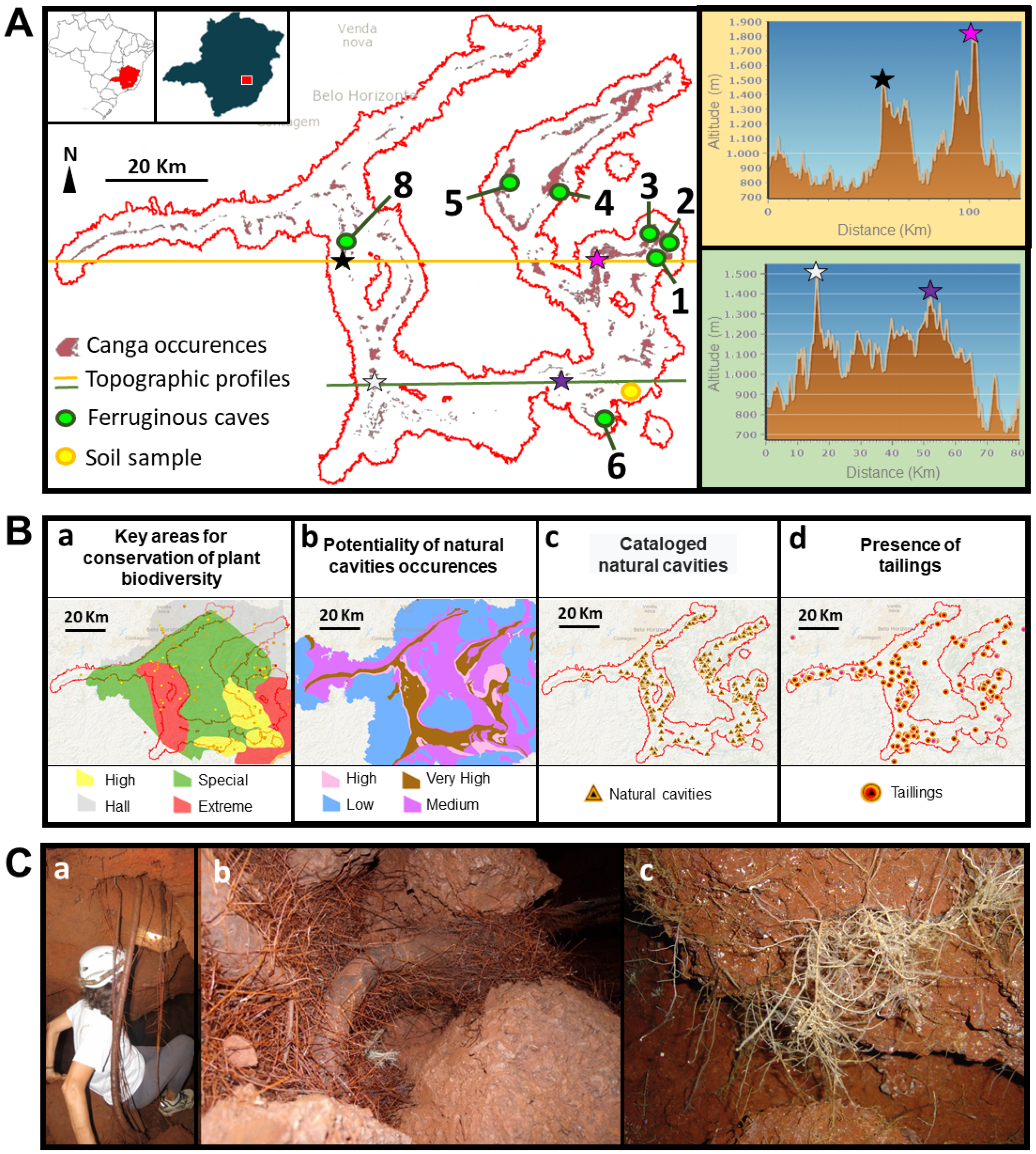
2.1. Cave Selection and Collection of Sample Material

The selection of caves investigated in this study was previously described by Lemes et al. [32]. Samples were obtained to investigate the abundance and prokaryotic diversity previously established in this study, while a portion of the samples was directed to the study of the culturable microbiota described below. Samples were collected from March to October 2016 in seven of the eight caves previously reported, excluding only the samples from the quartzite cave (Cave 7) [32] (Figure 1A). All sampling permits were approved by the responsible environmental agencies. Additionally, a sample of canga-covered soil was collected at the premises of the Federal University of Ouro Preto (UFOP), which was used to obtain bacterial isolates as an external control for the cave environment.

2.2. Isolation and Preservation of Culturable Bacterial Isolates

For the isolation of culturable bacteria, each sample (0.5 g) was diluted in a 50 mL falcon tube containing 10 mL of previously sterilized tap water. This was further diluted by factors of 1:10 and 1:100 using the same solvent. From each solution, 100 µL was applied to individual Petri dishes (90 × 15 mm) containing Luria-Bertani (LB) medium with pH adjusted to 7.0, and spread with a sterile Drigalski loop, adapting to the protocols previously described [33,34]. The plates were then labeled and held at 28 °C for 3 d. The grown bacterial colonies were streaked using the depletion technique in new Petri plates (90 × 15 mm) containing LB medium using sterile wooden toothpicks, and subsequently kept in the same conditions. After 2 d of growth, the isolates were collected using a platinum loop and transferred individually into properly labeled cryogenic tubes containing 700 µL of liquid LB and incubated in a shaker at 28 °C with agitation at 180 rpm. After 2 d, bacterial growth was checked by visual turbidity analysis, and 300 µL of 50% glycerol was added to the initial volume. The samples were stored in a freezer at −20 °C to perform the assays.

2.3. In Vitro Inhibition Assays against Xanthomonas citri subsp. citri (Xcc306)

All previously obtained culturable bacterial isolates were thawed and, using autoclaved wooden sticks, were transferred to 96-well U-bottom plates containing 200 µL of liquid LB medium and incubated for 48 h at 28 °C. Simultaneously, Xcc306 was grown in liquid LB medium for 3 d at 28 °C. Then, 15 µL of this pre-inoculum (OD600nm = 1.0) were transferred to Petri dishes (150 × 15 mm) containing LB medium. Using a Drigalski loop, a mat was created for the homogeneous growth of the phytopathogen. The bacterial isolates previously grown on 96-well plates were then repotted with the aid of a 96-tip colony multiinoculator onto the Xcc306 mat. A plate containing only the homogeneous phytopathogen mat was used as a control. The Petri plates were then incubated at 28 °C for 2 d, and the diameter of the inhibition halo and the colony were measured using a digital caliper (Western Dc). The assays were performed in duplicate.

2.4. In Vivo Inhibition Assay against Xcc306

This assay was performed only with bacterial isolates that showed positive results in the in vitro inhibition assays of Xcc306. We used young, same-aged plants of Rangpur lime (Citrus limonia), growing in a growth chamber at 27 °C with a 16 h photoperiod as compatible hosts. Infection of the plants was performed using the leaf infiltration method previously described by Caicedo et al. [35]. Different isolates were inoculated onto each plant using three independent leaves for each isolate. The pre-inoculum of the isolates that induced antagonism to Xcc306 consisted of a final concentration of 108 CFU/mL with MgCl2 solution (10 mM). On the abaxial side, to the left side of the central leaf vein, the antagonist isolate and Xcc306 were inoculated from a pre-inoculum mixture. To the right side of the central vein only Xcc306 was inoculated as a positive control for canker severity. Both inoculations were performed in duplicate. As negative controls, closer to the underside of the leaf, MgCl2 solution (10 mM) was inoculated to the right of the central vein, and only the antagonist isolate was inoculated to the left of the central vein. The infiltrated leaves were photographed 3, 7, 14, and 21 d after infiltration (DAI), and lesions were quantified from the damaged areas in the plant tissue using ImageJ software (v. 1.49) [36].

2.5. In Vitro Inhibition Test against Fusarium oxysporum (Fo)

In this assay, only isolates that were able to inhibit the in vitro growth of Xcc306 were evaluated because of financial limitations. To verify Fo inhibition, direct, indirect, and volatile compound-mediated inhibition assays were performed. For all assays, isolates were grown on LB medium for 48 h at 28 °C and diluted to an optical density (OD600nm) of 0.13. Fo was grown on synthetic potato dextrose agar (PDA) medium and incubated for 7 d at 28 °C [37].
For the direct inhibition assay, 10 µL of the solution containing the isolates were transferred to Petri dishes (90 × 15 mm) using a platinum loop, making a 3 cm edge square from the mycelium of the fungus, which was previously placed in the center of the Petri dish with the help of a 6 mm diameter sterile straw. The plates were incubated in a BOD incubator at 28 °C. Fungal growth (mm) was measured using a digital pachymeter (Western Dc) when the mycelial structure of the fungus on the control plate reached 30 mm in diameter [33].
A cellophane overlay technique was used for the indirect inhibition assay [38]. In this assay, a previously sterilized 12 cm diameter semi-permeable cellophane membrane (P45) was placed over a Petri dish (90 × 15 mm) containing synthetic PDA medium. On the cellophane, 200 µL of each bacterial isolate was added, and the solution was spread with a Drigalski loop over the entire surface of the paper, preventing samples of the bacteria from coming into direct contact with the culture medium. The plates were then kept in BOD at 28 °C and after 24 h of bacterial growth, the cellophane was removed and discarded, and the mycelium of the fungus was then inserted in the center of the plate with the help of a 6 mm diameter sterile straw. The Petri dishes were incubated again in the BOD under the same conditions as previously identified, and growth evaluation measurements were performed when the mycelium of the fungus in the control plate reached 30 mm in diameter using a digital caliper (Western Dc).
To verify inhibition by volatile compounds, septated Petri dishes (90 × 15 mm) containing synthetic PDA medium were used in all three chambers. In one chamber of the plate, a mycelial disk of the fungus was placed using a sterile 6 mm diameter straw, and in the other chambers 10 µL of the bacterial isolate was inoculated with a platinum loop. The Petri dishes were incubated in BOD at 28 °C, and the evaluation measurements followed the same pattern as the previous assays. In all assays, a fungal growth control plate was prepared. All assays were performed in triplicate.

2.6. In Vitro Inhibition Assay against Colletotrichum lindemuthianum Strain 89 (Cl89)

This assay was performed only with the isolates that presented inhibition above 75% of the mycelial growth of Fo. For the development of this assay, the fungus Cl89 was previously grown in natural PDA medium (20 g/L glucose; 200 g/L potato (Solanum tuberosum), 17 g/L agar), poured into Petri plates (90 × 15 mm), and incubated in BOD at 25 °C [39] for 30 d. The experimental methodology to evaluate the biocontrol of Cl89 followed the same procedures adopted for direct, indirect, and volatile inhibition of Fo. All experiments were performed in triplicate, evaluations were based on mycelial size (mm) measurement, and inhibition percentages were calculated.

2.7. Calculation of the Inhibition Rate of Phytopathogenic Fungi

The inhibition rate was calculated using the following equation: Inhibition rate % (I) = (Ø1 − Ø2)/Ø1 − 100 [38], with Ø1 being the mycelial disc diameter of the target pathogen under the control condition, and Ø2 denoting the mycelial disc diameter of the pathogen in the presence of the antagonist or its diffusible compounds.

2.8. Plant Growth Promotion Assays

All plant growth promotion assays were performed only with the isolates that showed promising results in the phytopathogen biocontrol assays used in this study. For all assays, the bacterial isolates were grown on liquid LB medium for 2 d, and at the time of the assay, the OD600nm was set to 0.13.

2.9. Calcium Phosphate Solubilization Assay

The National Botanical Research Institute Phosphate (NBRIP) protocol was used to verify the in vitro phosphate solubilization potential [40]. The assays were performed in triplicate, and the solubilization efficiency was determined based on the formula established by Nguyen, et al. [41].

2.10. Siderophore Production Assay

The production of siderophores was verified by inoculating 2 µL of each bacterial isolate into Petri dishes (90 × 15 mm) containing chromeazurol agar assay (CAS) medium according to a previously described method [42]. The results were performed in duplicate, and a positive result was determined by the presence of a yellow-orange halo around the colony.

2.11. Nitrogen Fixation Assay

The isolates (20 µL) were inoculated into microtubes containing 1.0 mL of semi-solid BAz medium, without cycloheximide, supplemented with 5 g/L mannitol and 5 g/L sucrose, adapted from [43]. The assays were performed in triplicate, and positive results were identified by measuring a film formed on the surface of the inoculated medium.

2.12. Hydrolase Production Assay

Amylase production. The isolates (2 µL) were inoculated onto Petri dishes (90 × 15 mm) containing starch agar medium. After 24 h of growth at 28 °C, the result was revealed by adding Lugol’s iodine and reacting for 5 min [44]. Cellulase production. The isolates (2 µL) were inoculated in Petri dishes (90 × 15 mm) containing carboxymethyl cellulose (CMC) agar medium. After 48 h of incubation, the result was verified by adding 5 mL of Congo red solution, and after 15 min, the solution was discarded, and 5 mL of 1M NaCl was added for another 15 min reaction time [45]. Protease production. The isolates (2 µL) were grown in Petri dishes (90 × 15 mm) containing a specific solid medium (pH 5.0) for 4 d at 28 °C [44].
Evaluation of the enzymatic assays. To determine the enzymatic activity of the isolates, the enzymatic index (EI) was calculated [46] using the ratio between the average diameter of the degradation halo and the average colony diameter (halo diameter/colony diameter), and for consideration of the enzyme production in solid medium, the EI was ≥ 2.0 [47,48]. All tests were performed in triplicate.

2.13. Bacterial Motility Assay

The swimming phenotype (flagellum-dependent motility of bacterial isolates) was verified by inoculating 5 µL of each bacterial isolate (OD600nm = 0.1) into the center of Petri dishes (60 × 15 mm) containing semi-solid LB medium with 0.3% agar [49]. The plates were incubated in BOD at 28 °C for 24 h and the results were photographed. The assays were performed in duplicate.

2.14. Test of Biofilm Production and Bacterial Self-Aggregation Capacity

Biofilm production was carried out based on O’Toole [50] with adaptations. To verify the self-aggregation capacity, the bacterial isolates were grown in liquid LB at 28 °C, and followed the methods proposed by Muzzolón [51] and adapted by Lima [52].

2.15. Molecular Identification of Potential Bioinoculants

The bacterial isolates were subjected to DNA extraction using the NucleoSpin® Tissue kit (Macherey-Nagel, Düren, Germany) following the manufacturer’s recommendations. The DNA samples were quantified, and purity was evaluated using a NanoDrop™ spectrophotometer (Thermo Fisher Scientific, Waltham, MA, USA). Electrophoresis was performed in 0.8% agarose gel to confirm the quality of the extracted DNA (data not shown). For partial amplification of the 16S rRNA gene, oligonucleotides 515F and 926R were used [53]. PCR was prepared to a final volume of 30 µL, and amplification was performed in a Biocycler® thermal cycler using the reagents and parameters established previously [33], and changing the annealing temperature to 57 °C. Amplification products were visualized on a 0.8% agarose gel, and 22 µL of the PCR product were purified using the GFX PCR DNA and Gel Band Purification Kit (GE Healthcare, Chicago, IL, USA), following the manufacturer’s instructions. The samples were sent to the Center for Biological Resources and Genomic Biology (CREBIO) at the Universidade Estadual Paulista “Júlio de Mesquita Filho”, Jaboticabal Campus, where sequencing [35] was performed. The DNA sequences were then verified and edited using BioEdit Sequence Alignment Editor, software version 7.0.5.3 [54], and analyzed using BLASTn (Basic Local Alignment Search Tool) [55].

2.16. Statistical Analysis

First, the data were analyzed for homogeneity of variance and normality using Levene and Kolmogorov-Smirnov tests, respectively. Once these assumptions were met, the data were subjected to an analysis of variance (ANOVA). For the in vivo inhibition assay of Xcc306, data were subjected to Tukey’s test (p < 0.05) using Prism 5 software. Data for direct and indirect in vitro inhibition of Fo, N fixation, hydrolytic enzyme production, self-aggregating capacity, and biofilm formation were subjected to the Skott-Knott test (p < 0.05), while the data of direct and indirect inhibition of Cl89 and phosphate solubilization were subjected to the least significant difference (LSD, p < 0.05) test to test the significant differences in the means of the treatments. Statistical analyses were performed using SAS 9.4 software [56]. Data are presented as means ± standard deviation (SD).

3. Results

A total of 28 floor, ceiling, and biofilm samples were obtained from among the seven canga caves and one superficial soil sample from the canga outside the cave environment (Figure 2).
From this sample set, 712 cultivable bacterial isolates were collected, of which 563 (79.0%) remained stable after being kept at −20 °C for up to 3 years (Figure 2), thus being characterized as stable to mild storage conditions.
In the direct in vitro inhibition assay against Xcc306, 47 bacterial isolates (8.3%) were positive, as demonstrated by the formation of a halo of inhibition around the isolates (Figure 3A).
Most of these (n = 42, 89.4%) were obtained from samples from Cave 6, whereas a single isolate was found from Caves 2 and 5, and three isolates from Cave 8 (Figure 3B). No isolates from superficial soil outside the cave showed positive inhibition against Xcc306.
To understand if the in vitro inhibition would also be observed in vivo, these 47 isolates were co-inoculated with the phytopathogen onto wild lemon plants. Of these, nine isolates (19.1%) (D9, A8, D11, A9, D3, A10, A11, A7, and B7) showed a significant reduction in the lesion caused by the phytopathogen, ranging from 38% to 68% of necrotic symptoms when compared to the control condition (Figure 3C), without apparent induction of hypersensitivity response when inoculated alone into plant tissue (Figure 3D). In addition, another five isolates (10.6 %) (B8, B11, C1, D7, and E3), although not able to significantly reduce the lesion, showed the ability to delay the injury process (Figure 3D).
From the 47 isolates capable of inhibiting Xcc306 in vitro, 36 were evaluated for their ability to inhibit Fo growth. Of these, 20 were effective in inhibiting mycelial disk growth by over 50% in the direct inhibition assays (Figure 4A).
In the indirect inhibition assays, 15 isolates (A2, A3, A4, A5, A6, A8, A10, B1, B4, B6, B9, C2, C3, C7, and D3) out of the 36 evaluated showed inhibition capacities ranging from 76.2% (A5) to 81.4% (C3) of mycelial growth (Figure 3B). In the inhibition assays mediated by volatile compounds, no isolate showed a positive result against Fo (data not shown). The 15 isolates that showed indirect inhibition above 75% for Fo constituted the third screening of biocontrol activities, this time against Cl89. An additional isolate was incorporated in this step, identified in this study as A1, which showed a low indirect inhibition rate to Fo, and whose colony coloration in the culture medium was white, in contrast to the other 15 that present colony coloration in different shades of red-orange. The results showed that for direct inhibition of Cl89, the 16 isolates were able to inhibit over 80% of the mycelial disc of the pathogen (Figure 5A).
In the indirect inhibition assays, the 15 red isolates were able to completely prevent the growth of Cl89 (100% inhibition), in contrast to the white isolate A1, which showed little inhibitory capacity for the mycelial disk growth of Cl89. Similar to Fo, there was no inhibition mediated by volatile compounds for Cl89 (data not shown).
The same 16 isolates selected for the Cl89 indirect inhibition assays were then evaluated for their ability to act as plant growth promoters, and the results are compiled in Table 1.
In the calcium phosphate solubilization assays, 14 isolates showed promise, with the lowest solubilization efficiency at 20.4% (B9) and the highest at 301.8% (A6) and 260.4% (B4), revealing a significant difference among isolates (Figure 6).
The isolates A1 (blank) and A4 were not able to form a halo; therefore, they were considered incapable of solubilizing calcium phosphate. As for the ability to produce siderophores, the 15 isolates with red colonies were able to produce some iron chelator, while isolate A1 did not show any positive results for this assay. In the nitrogen fixation assays, the same 15 red isolates showed a positive result of diazotrophic action, indicated by the formation of a film of bacterial growth on the surface of the BAz culture medium, which was not observed for isolate A1. In the assays of hydrolytic enzyme production, the 16 isolates verified were able to produce protease, with higher production identified for isolates A1 and A8, and only isolate A1 was able to produce amylase and cellulase. All 16 isolates were positive for swimming-type motility and biofilm formation, with isolates A1, A2, B9, and C2 showing the highest capacity to produce this attribute. In the self-aggregation assays, isolate A1, although not statistically different from A2, A3, A4, A5, A8, A10, B1, B4, B6, and B9 showed the highest percentage of self-aggregation, whereas isolates A6, C2, C3, C7, and D3 did not show any capacity for this attribute. Finally, the 16 isolates with the superior results had the V4–V5 region of the 16S rRNA gene partially sequenced for molecular identification (Table 1), and were identified as Dickeya, Nissabacter, and Serratia.

4. Discussion

Agricultural production plays an important role in global economic growth [28]. For example, in 2018, it was responsible for 4% of the global gross domestic product (GDP), and in some developing countries it could account for more than 25% of GDP [57]. In Brazil, one of the largest agricultural producers, agribusiness was responsible for 26.6% in 2020, and the agricultural sector alone contributed 18.5% to the national GDP [58].
Despite these expressive rates, much of the success is due to the use of defensives and fertilizers [59,60,61], both of which are adopted as recurrent practices in the management of large-scale monocultures. The indiscriminate use of these agrochemicals has caused severe environmental damage, harm to human and animal health, and is responsible for inducing resistance in pathogens and pests [62,63,64]. At the same time, such compounds affect the soil ecology, decreasing the area available for agricultural cultivation and reducing the effectiveness of the product itself [59,65,66].
Trade barriers have been imposed by some countries on the commercialization of products on the basis of chemical management [67] to help combat these issues, concomitant with the pressure exerted by a more conscious society, which has led to the desire to find natural products that can reduce or replace the use of these chemicals [30,68]. The use of biofertilizers and biopesticides has been highlighted in this scenario of changing the standards for productive management [69,70,71,72,73,74,75,76]. Nevertheless, there are many difficulties associated with the production of bioinoculants, ranging from the selection of these microorganisms to the validation of their beneficial effects in the field, which makes production costly [77], resulting in little diversity of these products in such a competitive market as the global agricultural market. In addition to biocontrol and biofertilization, microorganisms are highlighted in the areas of bioremediation and phytoremediation [78,79].
The discovery of new potential bioinoculants is emerging, and success involves prospecting unexplored environments, such as deserts, coal mines [80,81,82], and caves. Studies have highlighted the importance of cave microbiota [83,84]. The search for new biocontrollers and plant growth promoters from cave environments is still incipient, and only Venkadesaperumal et al. have presented data from this approach with significant results sampling volcanic mud and limestone caves from the Andaman and Nicobar Islands region (Gulf of Bengal) [27].
The proposal of the present work was to extend this approach to the possibility of finding biocontrollers from the ferruginous caves of the Brazilian IQ region. However, in contrast to the work of Venkadesaperumal et al., complex root systems were found within the ferruginous caves of the IQ, and these findings motivated this proposal (Figure 1C). The premise was that the expansion of root systems within the cave structures, composed of very rigid material, may be directly related to the role played by the microbiota present in this environment (Figure 1). This premise is supported by the fact that the soils above the ferruginous caves are mainly formed by itabirite, which is not very porous, with low water retention capacity for flora maintenance; therefore, the plants of this environment are highly specialized, justifying the high plant endemism observed [3].
Based on this premise, and considering that the most widely studied agents of biological control and plant growth promotion are bacteria, the objective of this study was to evaluate these capabilities in the largest possible number of culturable bacteria obtained from floor and ceiling samples of ferruginous caves of the IQ. From 29 samples derived from seven caves, located at different points in the IQ, and from the top soil of the region, 712 culturable bacteria were initially obtained. However, after storage at −20 °C, 563 isolates remained viable after re-activation in culture medium. The number of isolates per cave was not related in this study to diversity, only to the fact that it has a direct relation to the number of samples prospected per cave (Figure 2).
The assays were performed from these 563 isolates; however, given the financial and practical limitations of prospecting all the proposed biological functions for this set of isolates, we chose to perform serial and success-dependent steps in the previous step. This instituted a selection filter, thus allowing a smaller number of isolates to be evaluated for all the proposed attributes (Figure 7).
Initially, the 563 isolates were evaluated for their ability to inhibit in vitro Xanthomonas citri subsp. citri strain 306 (Xcc306), which is considered the most virulent strain causing citrus canker. A total of 47 (8.35%) isolates showed some antagonism against Xcc306, with emphasis on isolates from Cave 6, which accounted for 42 of these 47 isolates. To understand if this antagonistic action was also observed in vivo, an analysis of this effect was then performed in wild lemon plants. All 47 isolates were evaluated, and 9 (D9, A8, D11, A9, D3, A10, A11, A7, and B7) showed 36% to 68% reduction in virulence phenotypes when co-inoculated with Xcc306, without inducing any hypersensitivity response in the plant when inoculated alone. This is a good indication for the development of an efficient bacterial inoculant. Although no isolate was completely efficient in controlling the pathogen, a comparison with biological control data of Xcc306 described in other works allows us to conclude that the rates found are comparable. Kalita et al. [85] isolated bacteria of the genera Bacillus subtilis, Bacillus polymyxa, and Pseudomonas fluorescens, which were able to inhibit Xcc306 in vitro. Bacillus subtilis, in particular, was able to reduce the incidence of the disease by 61.9% under controlled conditions and when co-inoculated with Xcc306. In addition, Bacillus spp. are described as efficient biocontrollers for other crops [86], and other studies using antagonistic bacterial isolates have demonstrated success in reducing the severity and decreasing the incidence of this pathogen in citrus species [35,87,88,89,90] in similar proportions as found in the cave isolates.
In another study on the biocontrol of Xcc306, 10 bacterial isolates capable of forming a halo of inhibition against Xcc were found, all identified from the genus Bacillus. When co-inoculated with citrus plants, two of these bacterial isolates showed no symptoms of citrus canker within three weeks of inoculation [91]. In addition to the 9 isolates in the present study that contributed to the reduction in phenotypes, another set of 5 isolates was able to delay the effects of the disease, B8, B11, C1, D7, and E3 (Figure 3D). Although preliminary, these isolates present themselves as future sustainable alternatives to the use of copper-based substances in the management of Xcc306, whose recent discoveries have led to the development of metal-tolerant strains which are more difficult to control in the field [92,93].
In a subsequent step, 36 of the isolates most antagonistic to Xcc306 were evaluated for their ability to inhibit the growth of Fusarium oxysporum. The genus Fusarium is composed of more than 100 host-specific strains, many of which show pandistribution [94] and affect a wide variety of crops. Once in contact with the host, the fungus penetrates the roots, enters the vascular system, and uses the xylematic vessels, which are then obstructed by mycelium and spores, which promote vascular and leaf discoloration, low plant stature, yellowing of older leaves, wilting, and often plant death [95]. Several strategies are used to combat Fo, such as the use of chemical control to prevent infection. Besides presenting doubtful efficiency, they can cause damage to the microbiota associated with the plant and accumulate in the tissues, eventually being transferred to other trophic levels [96,97]. Another widely used strategy to restrict Fo-induced damage is the use of resistant plant varieties [97]. However, this resistance is not stable over the long term [98], and because of the problems caused by such approaches, the use of bacterial isolates that act as antagonists to Fo as sustainable alternatives [76,99,100,101], and the data from our work corroborate this perspective.
A total of 15 isolates (A2, A3, A4, A5, A6, A8, A10, B1, B4, B6, B9, C2, C3, C7, and D3) showed some efficiency in Fo antagonism. When we evaluated the performance of cave isolates against isolates obtained in other studies, we observed that the biocontrol efficiency was comparable and, in some cases, superior.
Zain et al. and Yan reported the efficiency of bacterial isolates in reducing approximately 65% of the mycelial disc growth of Fo in vitro [102,103]. Among the cave isolates evaluated for this function, 15 obtained inhibition rates higher than 75%, highlighting the possibility of their application in field trials. To understand if this antifungal potential extends to another phytopathogenic fungus, C. lindemuthianum (Cl) was selected as a secondary target. This fungus is the causal agent of anthracnose, the main disease affecting bean crops (Phaseolus vulgaris L.) [104]. The symptoms of anthracnose can be observed throughout the plant; however, typically dark brown necrotic lesions on the abaxial nerves of the leaves and on the stems, in addition to decreased leaf photosynthesis activity are noted [105,106]. Among the most used Cl control strategies are the use of resistant cultivars [107], the use of chemical controllers sprayed on the aerial part of the plant [108], seed treatment [109] and crop rotation [110]. Similarly, the use of biological control agents has been shown to be promising against Cl [111,112,113,114] and bacteria have played a prominent role in this approach [115,116].
All 15 isolates capable of inhibiting Fo were also capable of directly inhibiting more than 75 % and indirectly inhibiting 100% of the mycelial growth of Cl89. These data demonstrate that the isolates can be used as direct biocontrollers, which can be employed in commercial compositions, keeping the microorganisms alive and active, or in the production of these diffusible biomolecules with inhibitory capacity as new inputs for more sustainable food production. When we compared our results to those previously described in the literature, we noted the superior efficiency of the cave isolates. Gholami, et al. [117], for example, isolated 103 endophytic bacteria from roots of the Fabaceae family, and of these, 8 showed inhibition of C. lindemuthianum in vitro with values between 75% and 96%. Pandey et al. [118], using the same model as a target, evaluated strains of Pseudomonas fluorescens and Bacillus cereus, and obtained mycelium growth inhibition of 52% and 31%, respectively. In this study, we demonstrated that the cave isolates were able to inhibit more than 80% of direct growth and 100% by indirect inhibition. After identifying the isolates capable of presenting biocontrol action against the three phytopathogens investigated, the next step was to identify the potential for plant growth promotion, which could characterize a dual function of these potential bioinoculants. Of the 15 isolates with biocontrol potential, 14 showed in vitro inorganic phosphate solubilization capacity. Considering that phosphorus (P) is one of the most important macronutrients for plants, the availability of soluble phosphate favors plant growth [119,120], and most of the P present in the soil is in insoluble form [121]; therefore, phosphate-solubilizing bacteria play a key role in this relationship [122]. Previous studies have determined that the profitability of some crop production is significantly increased when these microorganisms are used as inoculants [123,124]. With the proposed isolation and characterization of these bacteria, a number of studies have established prospecting proposals. Peréz-Rodriguez et al. [125] found 33 isolates, with phosphate solubilization efficiencies ranging from 120% to 360%, and demonstrated that these isolates increased seed quality relative to the control when inoculated into tomato seeds.
Regarding the production of siderophores and the diazotrophic capacity, it was observed that all 15 isolates produced some of these metal chelating molecules and were also able to fix nitrogen. Siderophores are microbial biomolecules produced by acting as chelators of metals, especially iron, and the presence of these biomolecules in the bacterial-plant interrelationship favors plant growth, thus allowing them to increase their capacity to acquire iron, an important micronutrient [126], while triggering plant defense responses against potential pathogens [127], thus increasing the plant’s immune response. Bioinoculants that present such capacity are desired and have been continuously investigated, along with the search for diazotrophic organisms. These, in turn, are capable of fixing atmospheric nitrogen gas into ammonium ions in the soil, to be transformed into nitrite and nitrate by other microbial species, which is then available to the plant roots. This capacity is well developed in Rhizobiaceae, and it has been shown that non-symbiotic diazotrophic bacteria can be widely used as bioinoculants in plant growth [128]. It is important to highlight that there are other more robust techniques that allow a better assessment of the capacity of bacteria to solubilize phosphate and fix nitrogen. The techniques used in this work only suggest an indication of this potential, thus qualitatively contributing to the function of promoting plant growth by the bacterial isolates investigated.
Despite the ability to produce siderophores, solubilize phosphate, and fix nitrogen, none of the 15 biocontrol isolates produced the hydrolytic enzymes cellulase and amylase. Proteases secreted into the rhizosphere environment can facilitate the acquisition of organic nitrogen by plant roots [129], and protease-secreting bacteria have been shown to contribute to plant growth by inhibiting the growth of phytopathogens [128]. Together, these three activities observed by the selected bacterial isolates make them excellent candidates for plant growth promotion, while also showing biocontrol efficiency.
Finally, in addition to acting as biocontrollers and biofertilizers, bioinoculants could be able to produce biofilms, which facilitate the adherence and attachment of the bacteria to the roots and plant tissues, thus allowing them to develop their role as inoculants [130,131]. This is due to the sessile cell form, which adheres to biofilms and is better able to resist unfavorable environmental conditions, including stresses caused by external stimuli [132]. Although self-aggregation is a positive factor in the large-scale production of bacteria-based inoculants [130], the isolates analyzed were not efficient in this activity.
Once the potentials were identified, the next step was to determine the genus to which the 15 bacterial isolates belonged by partial sequencing of the 16S rRNA gene. Isolates A10, B1, B4, B6, B9, C2, C3, C7, and D3 were found to belong to the genus Serratia, and it has been reported that bacteria of this genus are capable of producing a secondary compound called prodigiosin, which is reddish in color, corroborating the phenotype of the colonies in vitro. Serratia species show antagonistic activity against citrus pathogens [133,134], Fusarium oxysporum [135,136], and Colletotrichum sp. [137]. Considering that bacteria of the Serratia genus are producers of prodigiosins and that the evaluated isolates present a reddish colony (an indication of the production of these pigments), it is possible to infer that such inhibition may be due to such biological action, which opens a new perspective of functional and structural evaluation of these molecules [138].
Isolate A8 was identified as belonging to the genus Dickeya. However, we cannot corroborate the inhibitory and growth-promoting effects of other Dickeya species, as these are classically reported as plant pathogens [139]. Nevertheless, the phenotypic characteristics of this genus resemble those observed for this isolate in vitro. Finally, isolates A3, A4, A5, and A6 were identified as belonging to the genus Nissabacter, whose only documented species is Nissabacter archeti [140]. However, unlike what was observed for Dickeya, the phenotypic characteristics found do not corroborate the data described in the literature.

5. Conclusions

From 563 cultivable bacteria obtained from floor and ceiling samples of seven ferruginous caves, it was possible to identify a total of 15 bacteria efficient in acting as biocontrollers of three important phytopathogens that affect diverse agricultural crops of great economic interest. These bacteria were also able to act in vitro as plant growth promoters, which places them as important candidates for in planta biocontrol and biopromoter action trials, thus evidencing potential microbiological services as an alternative to minimize the use of pesticides and chemical fertilizers through sustainable agricultural management practices. Indirectly, these results also highlight, for the first time, that the ferruginous caves of the IQ are characterized as important hotspots of genetic heritage that are fundamental for scientific development and the discovery of diverse potentials, requiring special attention in light of the extreme anthropic activities they have been enduring.

Author Contributions

Data curation, C.G.C.L., F.F.d.C., L.H.Y.K., F.M.S.C., J.C.C. and J.A.F.; Formal analysis, C.G.C.L. and L.M.M.; Funding acquisition, L.M.M.; Investigation, C.G.C.L., C.H.d.P., A.K.S., J.A.F. and L.M.M.; Methodology, C.G.C.L., I.F.C., C.H.d.P., A.K.S., F.F.d.C., L.H.Y.K., F.F.d.C. and J.C.C.; Project administration, L.M.M.; Supervision, L.M.M.; Validation, C.G.C.L., I.F.C. and C.H.d.P.; Writing—original draft, C.G.C.L. and L.M.M.; Writing—review & editing, C.G.C.L., I.F.C., C.H.d.P., A.K.S., F.F.d.C., L.H.Y.K., F.M.S.C., J.C.C., J.A.F. and L.M.M. All authors have read and agreed to the published version of the manuscript.

Funding

This study was financed in part by the Coordenação de Aperfeiçoamento de Pessoal de Nível Superior—Brazil (CAPES)–Finance Code 001—(the BIGA Project, CFP 51/2013, process 3385/2013), Conselho Nacional de Desenvolvimento Científico e Tecnológico (CNPq Process 481226/2013-3), and Fundação de Amparo à Pesquisa do Estado de Minas Gerais—FAPEMIG (process APQ-02387-14 and process APQ-02357-17). LMM and JAF have a research fellowship from CNPq. LMM has UFOP grants. The funders had no role in study design, data collection and analysis, decision to publish, or preparation of the manuscript.

Institutional Review Board Statement

Not applicable.

Informed Consent Statement

Not applicable.

Data Availability Statement

The present research was registered in the National System for the Management of Genetic Heritage and Associated Traditional Knowledge (SISGEN) platform with the access registration number ADC7A7C. The collections were authorized by the Authorization and Information System on Biodiversity (SISBIO) under number 54015-1.

Acknowledgments

To the members of the Plant Laboratory of the Technology Department of the College of Agricultural and Veterinary Sciences of the Universidade Estadual Paulista—Júlio de Mesquita Filho, Jaboticabal, SP and of the DNA and Bacteria-Environment Integration Laboratory of the Federal University of Ouro Preto. Special thanks to Marisa Queiroz for providing a sample of Colletotrichum lindemuthianum strain 89 for the investigations.

Conflicts of Interest

The authors declare no conflict of interest.

References

  1. Roeser, H.; Roeser, P. O Quadrilátero Ferrífero-MG, Brasil: Aspectos sobre sua história, seus recursos minerais e problemas ambientais relacionados. Rev. Geonomos 2013, 18, 33–37. [Google Scholar] [CrossRef] [Green Version]
  2. Azevedo, Ú.; Machado, M. Geological and mining heritage of iron quadrangle, Minas Gerais—Characterization and strategies for education and geotourism. Bol. Parana. Geosci. 2013, 70, 120–136. [Google Scholar]
  3. Jacobi, C.M.; Carmo, F.F. The contribution of ironstone outcrops to plant diversity in the Iron Quadrangle, a threatened Brazilian landscape. Ambio 2008, 37, 324–326. [Google Scholar] [CrossRef]
  4. Fernandes, G.W.; Goulart, F.F.; Ranieri, B.D.; Coelho, M.S.; Dales, K.; Boesche, N.; Bustamante, M.; Carvalho, F.A.; Carvalho, D.C.; Dirzo, R.; et al. Deep into the mud: Ecological and socio-economic impacts of the dam breach in Mariana, Brazil. Nat. Conserv. 2016, 14, 35–45. [Google Scholar] [CrossRef]
  5. Carmo, F.F.D.; Kamino, L.H.Y.; Junior, R.T.; Campos, I.C.D.; Carmo, F.F.D.; Silvino, G.; Castro, K.J.D.S.X.D.; Mauro, M.L.; Rodrigues, N.U.A.; Miranda, M.P.D.S.; et al. Fundão tailings dam failures: The environment tragedy of the largest technological disaster of Brazilian mining in global context. Perspect. Ecol. Conserv. 2017, 15, 145–151. [Google Scholar] [CrossRef]
  6. Cionek, V.M.; Alves, G.H.Z.; Tófoli, R.M.; Rodrigues-Filho, J.L.; Dias, R.M. Brazil in the mud again: Lessons not learned from Mariana dam collapse. Biodivers. Conserv. 2019, 28, 1935–1938. [Google Scholar] [CrossRef]
  7. Pereira, M.C.; Ruchkys, Ú.; Pereira, E.O. Panorama do Patrimônio Espeleológico do Quadrilátero Ferrífero. In Patrimônio Espeleológico em Rochas Ferruginosas: Propostas Para sua Conservação no Quadrilátero Ferrífero, Minas Gerais; Ruchkys, Ú., Travassos, L.E.P., Rasteiro, M.A., Faria, L.E., Eds.; Sociedade Brasileira de Espeleologia: Campinas, Brasil, 2015; Volume 1, pp. 276–287. [Google Scholar]
  8. CECAV. Anuário Estatístico do Patrimônio Espeleológico Brasileiro. Available online: http://www.icmbio.gov.br/cecav/ (accessed on 4 July 2021).
  9. Auler, A.S.; Piló, L.B. Aspectos morfológicos e evolução de cavernas em minério de ferro no Quadrilátero Ferrífero, Minas Gerais. In Proceedings of the Encontro Brasileiro de Estudos do Instituto de Geociências; IG-USP: São Paulo, Brazil, 2007; p. 8. [Google Scholar]
  10. Simmons, G.C. Canga Caves in the Quadrilátero Ferrífero, Minas Gerais, Brazil. Bull. Natl. Speleol. Soc. 1963, 22, 66–72. [Google Scholar]
  11. Culver, D.C. Cave Life: Evolution and Ecology; Harvard University Press: Cambridge, MA, USA, 1982; p. 189. [Google Scholar]
  12. Davila-Ramos, S.; Castelan-Sanchez, H.G.; Martinez-Avila, L.; Sanchez-Carbente, M.D.R.; Peralta, R.; Hernandez-Mendoza, A.; Dobson, A.D.W.; Gonzalez, R.A.; Pastor, N.; Batista-Garcia, R.A. A Review on Viral Metagenomics in Extreme Environments. Front. Microbiol. 2019, 10, 2403. [Google Scholar] [CrossRef] [Green Version]
  13. Orellana, R.; Macaya, C.; Bravo, G.; Dorochesi, F.; Cumsille, A.; Valencia, R.; Rojas, C.; Seeger, M. Living at the Frontiers of Life: Extremophiles in Chile and Their Potential for Bioremediation. Front. Microbiol. 2018, 9, 2309. [Google Scholar] [CrossRef]
  14. Nunez-Montero, K.; Barrientos, L. Advances in Antarctic Research for Antimicrobial Discovery: A Comprehensive Narrative Review of Bacteria from Antarctic Environments as Potential Sources of Novel Antibiotic Compounds Against Human Pathogens and Microorganisms of Industrial Importance. Antibiotics 2018, 7, 90. [Google Scholar] [CrossRef] [Green Version]
  15. Orcutt, B.N.; Larowe, D.E.; Biddle, J.F.; Colwell, F.S.; Glazer, B.T.; Reese, B.K.; Kirkpatrick, J.B.; Lapham, L.L.; Mills, H.J.; Sylvan, J.B.; et al. Microbial activity in the marine deep biosphere: Progress and prospects. Front. Microbiol. 2013, 4, 189. [Google Scholar] [CrossRef] [PubMed] [Green Version]
  16. Rooney, D.; Hutchens, E.; Clipson, N.; Baldini, J.; McDermott, F. Microbial Community Diversity of Moonmilk Deposits at Ballynamintra Cave, Co. Waterford, Ireland. Microb. Ecol. 2010, 60, 753–761. [Google Scholar] [CrossRef] [PubMed]
  17. Yasir, M. Analysis of bacterial communities and characterization of antimicrobial strains from cave microbiota. Braz. J. Microbiol. 2018, 49, 248–257. [Google Scholar] [CrossRef] [PubMed]
  18. Gabriel, C.; Northup, D. Microbial Ecology: Caves as an Extreme Habitat; Springer: New York, NY, USA, 2013; Volume 1, pp. 85–108. [Google Scholar]
  19. Hershey, O.S.; Barton, H.A. The Microbial Diversity of Caves. In Cave Ecology; Moldovan, O.T., Kováč, Ľ., Halse, S., Eds.; Springer International Publishing: Cham, Switzerland, 2018; pp. 69–90. [Google Scholar]
  20. Rangseekaew, P.; Pathomaree, W. Cave Actinobacteria as Producers of Bioactive Metabolites. Front. Microbiol. 2019, 10, 387. [Google Scholar] [CrossRef] [PubMed] [Green Version]
  21. Cheeptham, N.; Kay, S.; Pathom-aree, W. Screening of Volcanic Cave Actinomycetes for Antimicrobial Activity Against Paenibacillus larvae, A Honey Bee Pathogen. Chiang Mai J. Sci. 2012, 40, 26. [Google Scholar]
  22. Riquelme, C.; Enes Dapkevicius, M.L.; Miller, A.Z.; Charlop-Powers, Z.; Brady, S.; Mason, C.; Cheeptham, N. Biotechnological potential of Actinobacteria from Canadian and Azorean volcanic caves. Appl. Microbiol. Biotechnol. 2017, 101, 843–857. [Google Scholar] [CrossRef] [PubMed] [Green Version]
  23. Ghosh, S.; Kuisiene, N.; Cheeptham, N. The cave microbiome as a source for drug discovery: Reality or pipe dream? Biochem. Pharmacol. 2017, 134, 18–34. [Google Scholar] [CrossRef]
  24. Pawlowski, A.C.; Wang, W.; Koteva, K.; Barton, H.A.; McArthur, A.G.; Wright, G.D. A diverse intrinsic antibiotic resistome from a cave bacterium. Nat. Commun. 2016, 7, 13803. [Google Scholar] [CrossRef] [Green Version]
  25. Bhullar, K.; Waglechner, N.; Pawlowski, A.; Koteva, K.; Banks, E.D.; Johnston, M.D.; Barton, H.A.; Wright, G.D. Antibiotic Resistance Is Prevalent in an Isolated Cave Microbiome. PLoS ONE 2012, 7, e34953. [Google Scholar] [CrossRef]
  26. Montano, E.; Henderson, L. Studies of Antibiotic Production by Cave Bacteria. In Cave Microbiomes: A Novel Resource for Drug Discovery; SpringerBriefs in Microbiology; Cheeptham, N., Ed.; Springer: New York, NY, USA, 2013; pp. 109–130. [Google Scholar] [CrossRef]
  27. Venkadesaperumal, G.; Amaresan, N.; Kumar, K. Plant growth promoting capability and genetic diversity of bacteria isolated from mud volcano and lime cave of Andaman and Nicobar Islands. Braz. J. Microbiol. 2014, 45, 1271–1281. [Google Scholar] [CrossRef] [Green Version]
  28. Sessitsch, A.; Brader, G.; Pfaffenbichler, N.; Gusenbauer, D.; Mitter, B. The contribution of plant microbiota to economy growth. Microb. Biotechnol. 2018, 11, 801–805. [Google Scholar] [CrossRef]
  29. Diguță, C.F.; Nițoi, G.D.; Matei, F.; Luță, G.; Cornea, C.P. The Biotechnological Potential of Pediococcus spp. Isolated from Kombucha Microbial Consortium. Foods 2020, 9, 1780. [Google Scholar] [CrossRef]
  30. Ramakrishna, W.; Yadav, R.; Li, K. Plant growth promoting bacteria in agriculture: Two sides of a coin. Appl. Soil Ecol. 2019, 138, 10–18. [Google Scholar] [CrossRef]
  31. Compant, S.; Duffy, B.; Nowak, J.; Clement, C.; Barka, E.A. Use of plant growth-promoting bacteria for biocontrol of plant diseases: Principles, mechanisms of action, and future prospects. Appl. Environ. Microbiol. 2005, 71, 4951–4959. [Google Scholar] [CrossRef] [PubMed] [Green Version]
  32. Lemes, C.G.C. Diversidade Metagenômica e Potencial Biotecnológico de Cavernas de Canga do Quadrilátero Ferrífero. Master’s Thesis, Universidade Federal de Ouro Preto, Ouro Preto, Brazil, 2018. [Google Scholar]
  33. Felestrino, E.B.; Santiago, I.F.; Freitas, L.D.; Rosa, L.H.; Ribeiro, S.P.; Moreira, L.M. Plant Growth Promoting Bacteria Associated with Langsdorffia hypogaea-Rhizosphere-Host Biological Interface: A Neglected Model of Bacterial Prospection. Front. Microbiol. 2017, 8, 172. [Google Scholar] [CrossRef] [PubMed] [Green Version]
  34. Caneschi, W.L.; Felestrino, É.B.; Fonseca, N.P.; Villa, M.M.; Lemes, C.G.C.; Cordeiro, I.F.; Assis, R.D.A.B.; Sanchez, A.B.; Vieira, I.T.; Kamino, L.H.Y.; et al. Brazilian Ironstone Plant Communities as Reservoirs of Culturable Bacteria With Diverse Biotechnological Potential. Front. Microbiol. 2018, 9, 1638. [Google Scholar] [CrossRef] [PubMed] [Green Version]
  35. Caicedo, J.C.; Villamizar, S.; Ferro, M.I.T.; Kupper, K.C.; Ferro, J.A. Bacteria from the citrus phylloplane can disrupt cell–cell signalling in Xanthomonas citri and reduce citrus canker disease severity. Plant Pathol. 2016, 65, 782–791. [Google Scholar] [CrossRef]
  36. Schneider, C.A.; Rasband, W.S.; Eliceiri, K.W. NIH Image to ImageJ: 25 years of image analysis. Nat. Methods 2012, 9, 671–675. [Google Scholar] [CrossRef]
  37. Cruz, D.R.; Leandro, L.F.S.; Munkvold, G.P. Effects of Temperature and pH on Fusarium oxysporum and Soybean Seedling Disease. Plant Dis. 2019, 103, 3234–3243. [Google Scholar] [CrossRef] [PubMed]
  38. Nourozian, J.; Etebarian, H.R.; Khodakaramian, G. Biological control of Fusarium graminearum on wheat by antagonistic bacteria. Songklanakarin J. Sci. Technol. 2006, 28, 29–38. [Google Scholar]
  39. Tozze-Júnior, H.J.; Firmino, A.C.; Fischer, I.H.; Furtado, E.L.; Júnior, N.S.M. Caracterização de isolados de Colletotrichum spp. associados às frutíferas no Estado de São Paulo. Summa Phytopathol. 2015, 41, 270–280. [Google Scholar] [CrossRef]
  40. Nautiyal, C.S. An efficient microbiological growth medium for screening phosphate solubilizing microorganisms. FEMS Microbiol. Lett. 1999, 170, 265–270. [Google Scholar] [CrossRef] [PubMed]
  41. Nguyen, C.; Yan, W.; Tacon, F.L.; Lapeyrie, F. Genetic variability of phosphate solubilizing activity by monocaryotic and dicaryotic mycelia of the ectomycorrhizal fungus Laccaria bicolor (Maire) P.D. Orton. Plant Soil 1992, 143, 193–199. [Google Scholar] [CrossRef]
  42. Schwyn, B.; Neilands, J.B. Universal chemical assay for the detection and determination of siderophores. Anal. Biochem. 1987, 160, 47–56. [Google Scholar] [CrossRef]
  43. Estrada-De Los Santos, P.; Bustillos-Cristales, R.; Caballero-Mellado, J. Burkholderia, a genus rich in plant-associated nitrogen fixers with wide environmental and geographic distribution. Appl. Environ. Microbiol. 2001, 67, 2790–2798. [Google Scholar] [CrossRef] [PubMed] [Green Version]
  44. Strauss, M.L.; Jolly, N.P.; Lambrechts, M.G.; van Rensburg, P. Screening for the production of extracellular hydrolytic enzymes by non-Saccharomyces wine yeasts. J. Appl. Microbiol. 2001, 91, 182–190. [Google Scholar] [CrossRef] [PubMed]
  45. Menegatto, M.B.D.S. Avaliação do Potencial Celulolítico e Fermentativo de Bactérias do Gênero Clostridium e da Microbiota Autóctone na Fermentação do Bagaço de Cana Bruto. Master’s Thesis, Universidade Federal de Ouro Preto, Ouro Preto, Brazil, 2019. [Google Scholar]
  46. Hankin, L.; Anagnostakis, S.L. The Use of Solid Media for Detection of Enzyme Production by Fungi. Mycologia 1975, 67, 597–607. [Google Scholar] [CrossRef]
  47. Montenegro, S.; Araújo, J.; Stamford, N. Atividade enzimática de microrganismos isolados do jacatupé (Pachyrhizus erosus L. Urban). Ciênc. Tecnol. Aliment. 1998, 18, 382–385. [Google Scholar] [CrossRef]
  48. Lealem, F.; Gashe, B.A. Amylase production by a Gram-positive bacterium isolated from fermenting tef (Eragrostis tef). J. Appl. Bacteriol. 1994, 77, 348–352. [Google Scholar] [CrossRef]
  49. Park, S.Y.; Pontes, M.H.; Groisman, E.A. Flagella-independent surface motility in Salmonella enterica serovar Typhimurium. Proc. Natl. Acad. Sci. USA 2015, 112, 1850–1855. [Google Scholar] [CrossRef] [Green Version]
  50. O’Toole, G.A. Microtiter dish biofilm formation assay. J. Vis. Exp. 2011, e2437. [Google Scholar] [CrossRef] [PubMed]
  51. Muzzolón, J. Uso de Bacterias Lácticas y/o Levaduras en la Prevención de Aflatoxicosis en Animales de Compañía. Monografia. Universidad Nacional de Río Cuarto, Río Cuarto, Brazil, 2010. [Google Scholar]
  52. Lima, C.E. Potencial Probiótico de Bactérias Ácido-Láticas Isoladas de Ração de Peixe. Master’s Thesis, Universidade Federal do Piauí, Teresina, Brazil, 2016. [Google Scholar]
  53. Fouhy, F.; Clooney, A.G.; Stanton, C.; Claesson, M.J.; Cotter, P.D. 16S rRNA gene sequencing of mock microbial populations- impact of DNA extraction method, primer choice and sequencing platform. BMC Microbiol. 2016, 16, 123. [Google Scholar] [CrossRef] [PubMed]
  54. Hall, T.A. BioEdit: A user-friendly biological sequence alignment editor and analysis program for Windows 95/98/NT. Nucl. Acids. Symp. Ser. 1999, 41, 95–98. [Google Scholar]
  55. Altschul, S.F.; Gish, W.; Miller, W.; Myers, E.W.; Lipman, D.J. Basic local alignment search tool. J. Mol. Biol. 1990, 215, 403–410. [Google Scholar] [CrossRef]
  56. Sas, I. SAS® 9.4 ODS Graphics: Procedures Guide, 4th ed.; SAS Institute Inc.: Cary, NC, USA, 2014. [Google Scholar]
  57. Bank, T.W. Agriculture and Food. Available online: https://www.worldbank.org/en/topic/agriculture/overview (accessed on 30 March 2021).
  58. CEPEA; CNA. PIB do Agronegócio Alcança Participação de 26.6% no PIB Brasileiro em 2020; Centro de Estudos Avançados em Economia Aplicada (CEPEA), Confederação da Agricultura e Pecuária do Brasil (CNA): Piracicaba, Brazil, 2021; p. 18. [Google Scholar]
  59. Shang, Y.; Hasan, M.K.; Ahammed, G.J.; Li, M.; Yin, H.; Zhou, J. Applications of Nanotechnology in Plant Growth and Crop Protection: A Review. Molecules 2019, 24, 2558. [Google Scholar] [CrossRef] [Green Version]
  60. Chen, P.; Du, Q.; Liu, X.; Zhou, L.; Hussain, S.; Lei, L.; Song, C.; Wang, X.; Liu, W.; Yang, F.; et al. Effects of reduced nitrogen inputs on crop yield and nitrogen use efficiency in a long-term maize-soybean relay strip intercropping system. PLoS ONE 2017, 12, e0184503. [Google Scholar] [CrossRef] [Green Version]
  61. Cooper, J.; Dobson, H. The benefits of pesticides to mankind and the environment. Crop. Prot. 2007, 26, 1337–1348. [Google Scholar] [CrossRef]
  62. Rosell, G.; Quero, C.; Coll, J.; Guerrero, A. Biorational insecticides in pest management. J. Pestic. Sci. 2008, 33, 103–121. [Google Scholar] [CrossRef] [Green Version]
  63. Liu, Y.; Li, S.; Ni, Z.; Qu, M.; Zhong, D.; Ye, C.; Tang, F. Pesticides in persimmons, jujubes and soil from China: Residue levels, risk assessment and relationship between fruits and soils. Sci. Total Environ. 2016, 542, 620–628. [Google Scholar] [CrossRef]
  64. Tilman, D.; Cassman, K.G.; Matson, P.A.; Naylor, R.; Polasky, S. Agricultural sustainability and intensive production practices. Nature 2002, 418, 671–677. [Google Scholar] [CrossRef]
  65. Ma, L.; Ma, W.Q.; Velthof, G.L.; Wang, F.H.; Qin, W.; Zhang, F.S.; Oenema, O. Modeling nutrient flows in the food chain of China. J. Environ. Quality 2010, 39, 1279–1289. [Google Scholar] [CrossRef] [PubMed]
  66. Zahid, M.; Abbasi, M.K.; Hameed, S.; Rahim, N. Isolation and identification of indigenous plant growth promoting rhizobacteria from Himalayan region of Kashmir and their effect on improving growth and nutrient contents of maize (Zea mays L.). Front. Microbiol. 2015, 6, 207. [Google Scholar] [CrossRef]
  67. Sarkar, S.; Gil, J.D.B.; Keeley, J.; Möhring, N.; Jansen, K. The Use of Pesticides in Developing Countries and Their Impact on Health and the Right to Food; Oxford Analytica Europe: Brussels, Belgium, 2021; p. 44. ISBN 978-92-846-7673-6. [Google Scholar]
  68. Singh, D.P.; Singh, H.B.; Prabha, R. Microbial Inoculants in Sustainable Agricultural Productivity; Singh, D.P., Singh, H.B., Prabha, R., Eds.; Springer: New Delhi, India, 2016; Volume 2, p. 308. [Google Scholar]
  69. Malusà, E.; Pinzari, F.; Canfora, L. Efficacy of Biofertilizers: Challenges to Improve Crop Production. In Microbial Inoculants in Sustainable Agricultural Productivity: Vol. 2: Functional Applications; Singh, D.P., Singh, H.B., Prabha, R., Eds.; Springer: New Delhi, India, 2016; pp. 17–40. [Google Scholar]
  70. Dong, L.; Li, Y.; Xu, J.; Yang, J.; Wei, G.; Shen, L.; Ding, W.; Chen, S. Biofertilizers regulate the soil microbial community and enhance Panax ginseng yields. Chin. Med. 2019, 14, 20. [Google Scholar] [CrossRef] [Green Version]
  71. Bhardwaj, D.; Ansari, M.W.; Sahoo, R.K.; Tuteja, N. Biofertilizers function as key player in sustainable agriculture by improving soil fertility, plant tolerance and crop productivity. Microb. Cell Factories 2014, 13, 66. [Google Scholar] [CrossRef] [PubMed] [Green Version]
  72. Dal Cortivo, C.; Ferrari, M.; Visioli, G.; Lauro, M.; Fornasier, F.; Barion, G.; Panozzo, A.; Vamerali, T. Effects of Seed-Applied Biofertilizers on Rhizosphere Biodiversity and Growth of Common Wheat (Triticum aestivum L.) in the Field. Front. Plant Sci. 2020, 11, 72. [Google Scholar] [CrossRef] [PubMed] [Green Version]
  73. Soumare, A.; Boubekri, K.; Lyamlouli, K.; Hafidi, M.; Ouhdouch, Y.; Kouisni, L. From Isolation of Phosphate Solubilizing Microbes to Their Formulation and Use as Biofertilizers: Status and Needs. Front. Bioeng. Biotechnol. 2019, 7, 425. [Google Scholar] [CrossRef] [PubMed]
  74. Newitt, J.T.; Prudence, S.M.M.; Hutchings, M.I.; Worsley, S.F. Biocontrol of Cereal Crop Diseases Using Streptomycetes. Pathogens 2019, 8, 78. [Google Scholar] [CrossRef] [Green Version]
  75. Dagher, F.; Olishevska, S.; Philion, V.; Zheng, J.; Deziel, E. Development of a novel biological control agent targeting the phytopathogen Erwinia amylovora. Heliyon 2020, 6, e05222. [Google Scholar] [CrossRef]
  76. Bubici, G.; Kaushal, M.; Prigigallo, M.I.; Gómez-Lama Cabanás, C.; Mercado-Blanco, J. Biological Control Agents Against Fusarium Wilt of Banana. Front. Microbiol. 2019, 10, 616. [Google Scholar] [CrossRef] [Green Version]
  77. Sethi, S.K.; Adhikary, S.P. Cost effective pilot scale production of biofertilizer using Rhizobium and Azotobacter. Afr. J. Biotechnol. 2012, 11, 13490–13493. [Google Scholar] [CrossRef]
  78. Ortega, H.E.; Torres-Mendoza, D.; Cubilla-Rios, L. Patents on Endophytic Fungi for Agriculture and Bio- and Phytoremediation Applications. Microorganisms 2020, 8, 1237. [Google Scholar] [CrossRef]
  79. Shahzad, R.; Bilal, S.; Imran, M.; Khan, A.L.; Alosaimi, A.A.; Al-Shwyeh, H.A.; Almahasheer, H.; Rehman, S.; Lee, I.-J. Amelioration of heavy metal stress by endophytic Bacillus amyloliquefaciens RWL-1 in rice by regulating metabolic changes: Potential for bacterial bioremediation. Biochem. J. 2019, 476, 3385–3400. [Google Scholar] [CrossRef]
  80. Khan, N.; Martinez-Hidalgo, P.; Humm, E.A.; Maymon, M.; Kaplan, D.; Hirsch, A.M. Inoculation With a Microbe Isolated From the Negev Desert Enhances Corn Growth. Front. Microbiol. 2020, 11, 1149. [Google Scholar] [CrossRef]
  81. Alsharif, W.; Saad, M.M.; Hirt, H. Desert Microbes for Boosting Sustainable Agriculture in Extreme Environments. Front. Microbiol. 2020, 11, 1666. [Google Scholar] [CrossRef] [PubMed]
  82. Pranaw, K.; Pidlisnyuk, V.; Trogl, J.; Malinska, H. Bioprospecting of a Novel Plant Growth-Promoting Bacterium Bacillus altitudinis KP-14 for Enhancing Miscanthus x giganteus Growth in Metals Contaminated Soil. Biology 2020, 9, 305. [Google Scholar] [CrossRef]
  83. Portillo, M.C.; Saiz-Jimenez, C.; Gonzalez, J.M. Molecular characterization of total and metabolically active bacterial communities of “white colonizations” in the Altamira Cave, Spain. Res. Microbiol. 2009, 160, 41–47. [Google Scholar] [CrossRef]
  84. Gonzalez, J.M.; Portillo, M.C.; Saiz-Jimenez, C. Metabolically active Crenarchaeota in Altamira Cave. Naturwissenschaften 2006, 93, 42–45. [Google Scholar] [CrossRef]
  85. Kalita, P.; Bora, L.C.; Bhagabati, K.N. Phylloplane microflora of citrus and their role in management of citrus canker. Indian Phytopathol. 1996, 49, 234–237. [Google Scholar]
  86. Grosu, A.I.; Sicuia, O.-A.; Dobre, A.; Voaideş, C.; Cornea, C.P. Evaluation of Some Bacillus spp. Strains for the Biocontrol of Fusarium graminearum and F. culmorum in Wheat. Agric. Agric. Sci. Procedia 2015, 6, 559–566. [Google Scholar] [CrossRef] [Green Version]
  87. Das, R.; Mondal, B.; Mondal, P.; Khatua, D.C.; Mukherjee, N. Biological management of citrus canker on acid lime through Bacillus subtilis (S-12) in West Bengal, India. J. Biopestic. 2014, 7, 38–41. [Google Scholar]
  88. Islam, M.N.; Ali, M.S.; Choi, S.J.; Hyun, J.W.; Baek, K.H. Biocontrol of Citrus Canker Disease Caused by Xanthomonas citri subsp. citri Using an Endophytic Bacillus thuringiensis. Plant Pathol. J. 2019, 35, 486–497. [Google Scholar] [CrossRef]
  89. Villamizar, S.; Ferro, J.A.; Caicedo, J.C.; Alves, L.M.C. Bactericidal Effect of Entomopathogenic Bacterium Pseudomonas entomophila Against Xanthomonas citri Reduces Citrus Canker Disease Severity. Front. Microbiol. 2020, 11, 1431. [Google Scholar] [CrossRef]
  90. Rabbee, M.F.; Ali, M.S.; Baek, K.-H. Endophyte Bacillus velezensis Isolated from Citrus spp. Controls Streptomycin-Resistant Xanthomonas citri subsp. citri That Causes Citrus Bacterial Canker. Agronomy 2019, 9, 470. [Google Scholar] [CrossRef] [Green Version]
  91. Daungfu, O.; Youpensuk, S.; Lumyong, S. Endophytic Bacteria Isolated from Citrus Plants for Biological Control of Citrus Canker in Lime Plants. Trop. Life Sci. Res. 2019, 30, 73–88. [Google Scholar] [CrossRef]
  92. Roach, R.; Mann, R.; Gambley, C.G.; Shivas, R.G.; Chapman, T.; Rodoni, B. Pathogenicity and copper tolerance in Australian Xanthomonas species associated with bacterial leaf spot. Crop. Prot. 2020, 127, 104923. [Google Scholar] [CrossRef]
  93. Marin, T.G.S.; Galvanin, A.L.; Lanza, F.E.; Behlau, F. Description of copper tolerant Xanthomonas citri subsp. citri and genotypic comparison with sensitive and resistant strains. Plant Pathol. 2019, 68, 1088–1098. [Google Scholar] [CrossRef]
  94. Gordon, T.R. Fusarium oxysporum and the Fusarium Wilt Syndrome. Annu. Rev. Phytopathol. 2017, 55, 23–39. [Google Scholar] [CrossRef]
  95. McGovern, R.J. Management of tomato diseases caused by Fusarium oxysporum. Crop. Prot. 2015, 73, 78–92. [Google Scholar] [CrossRef]
  96. López-Aranda, J.M.; Domínguez, P.; Miranda, L.; de los Santos, B.; Talavera, M.; Daugovish, O.; Soria, C.; Chamorro, M.; Medina, J.J. Fumigant Use for Strawberry Production in Europe: The Current Landscape and Solutions. Int. J. Fruit Sci. 2016, 16, 1–15. [Google Scholar] [CrossRef]
  97. de Lamo, F.J.; Takken, F.L.W. Biocontrol by Fusarium oxysporum Using Endophyte-Mediated Resistance. Front. Plant Sci. 2020, 11, 37. [Google Scholar] [CrossRef] [PubMed] [Green Version]
  98. Takken, F.; Rep, M. The arms race between tomato and Fusarium oxysporum. Mol. Plant Pathol. 2010, 11, 309–314. [Google Scholar] [CrossRef]
  99. Tehrani, A.; Ramezani, M. Biological control of Fusarium oxysporum, the causal agent of onion wilt by antagonistic bacteria. Commun. Agric. Appl. Biol. Sci. 2003, 68, 543–547. [Google Scholar]
  100. Moreno-Velandia, C.A.; Izquierdo-García, L.F.; Ongena, M.; Kloepper, J.W.; Cotes, A.M. Soil sterilization, pathogen and antagonist concentration affect biological control of Fusarium wilt of cape gooseberry by Bacillus velezensis Bs006. Plant Soil 2019, 435, 39–55. [Google Scholar] [CrossRef] [Green Version]
  101. Malathi, S. Biological control of onion basal rot caused by Fusarium oxysporum f. sp. cepae. Asian J. Bio Sci. 2015, 10, 21–26. [Google Scholar] [CrossRef]
  102. Zain, M.; Yasmin, S.; Hafeez, F.Y. Isolation and Characterization of Plant Growth Promoting Antagonistic Bacteria from Cotton and Sugarcane Plants for Suppression of Phytopathogenic Fusarium Species. Iran. J. Biotechnol. 2019, 17, e1974. [Google Scholar] [CrossRef] [PubMed]
  103. Yang, W. Components of rhizospheric bacterial communities of barley and their potential for plant growth promotion and biocontrol of Fusarium wilt of watermelon. Braz. J. Microbiol. 2019, 50, 749–757. [Google Scholar] [CrossRef] [PubMed]
  104. Pastor-Corrales, M.A. Enfermedades del frijol causadas por hongos. In Frijol: Investigación y Producción. Programa de las Naciones Unidas (PNUD); Marceliano, L.G., Fernando, O., Fernández, O., Schoonhoven, A., Eds.; Centro Internacional de Agricultura Tropical (CIAT): Cali, Colombia, 1985; pp. 169–206. [Google Scholar]
  105. Bianchini, A.; Maringoni, A.C.; Carneiro, B.S.M.T.G. Doenças do feijoeiro (Phaseolus vulgaris L.). In Manual de Fitopatologia. Doenças das Plantas Cultivadas; Agronômica Ceres: São Paulo, Brazil, 1997; Volume 3, pp. 376–399. [Google Scholar]
  106. Bassanezi, R.B.; Amorim, L.; Filho, A.B.; Hau, B.; Berger, R.D. Accounting for photosynthetic efficiency of bean leaves with rust, angular leaf spot and anthracnose to assess crop damage. Plant Pathol. 2001, 50, 443–452. [Google Scholar] [CrossRef]
  107. Pereira, A.C.; Paula, R.G.D.; Júnior, T.J.D.P.; Silva, L.C.D.; Carneiro, J.E.D.S.; Carneiro, P.C.S. Selection of anthracnose resistant common beans using detached leaves in partially controlled environment. Rev. Ceres 2014, 61, 518–522. [Google Scholar] [CrossRef] [Green Version]
  108. Amin, M.; Fitsum, S.; Selvaraj, T.; Mulugeta, N. Field Management of Anthracnose (Colletotrichum lindemuthianum) in Common Bean through Fungicides and Bioagents. Adv. Crop Sci. Technol. 2014, 2, 1000124. [Google Scholar] [CrossRef] [Green Version]
  109. Gillard, C.L.; Ranatunga, N.K.; Conner, R.L. The control of dry bean anthracnose through seed treatment and the correct application timing of foliar fungicides. Crop. Prot. 2012, 37, 81–90. [Google Scholar] [CrossRef]
  110. Schwartz, H.F.; Steadman, J.R.; Hall, R.; Forster, R.L. Compendium of Bean Diseases; APS Press: St. Paul, Brazil, 2005; p. 109. [Google Scholar]
  111. Dildey, O.D.F. Indução de Resistência à Antracnose do Feijoeiro por Frações de Filtrado de Cultura e Extrato de Micélio de Trichoderma longibrachiatum. Ph.D. Thesis, Universidade Estadual do Oeste do Paraná, Marechal Cândido Rondon, Brazil, 2017. [Google Scholar]
  112. Martins, S.J.; Faria, A.F.; Pedroso, M.P.; Cunha, M.G.; Rocha, M.R.; Medeiros, F.H.V. Microbial volatiles organic compounds control anthracnose (Colletotrichum lindemuthianum) in common bean (Phaseolus vulgaris L.). Biol. Control 2019, 131, 36–42. [Google Scholar] [CrossRef]
  113. Christmann, P.; Dalla Pria, M.; Hennipman, H.; Godoy, A. In vitro control of Colletotrichum lindemuthianum by Trichoderma spp. and in vivo with Alternative Products. Int. J. Adv. Eng. Res. Sci. 2019, 6, 74–80. [Google Scholar] [CrossRef]
  114. Pedro, E.A.D.S.; Harakava, R.; Lucon, C.M.M.; Guzzo, S.D. Promoção do crescimento do feijoeiro e controle da antracnose por Trichoderma spp. Pesqui. Agropecuária Bras. 2012, 47, 1589–1595. [Google Scholar] [CrossRef] [Green Version]
  115. Garcia, F.A.D.O.; Romeiro, R.D.S. Biocontrole da mancha-angular do feijoeiro por antagonistas bacterianos. Pesqui. Agropecuária Bras. 2011, 46, 1603–1608. [Google Scholar] [CrossRef]
  116. Corrêa, B.; Andrea, M.; Denardin, N.; Soares, V.; Schäfer, J.; Ludwig, J. Influência da microbiolização de sementes de feijão sobre a transmissão de Colletrichum lindemuthianum (Saac e Magn). Rev. Bras. Sementes 2008, 30, 156–163. [Google Scholar] [CrossRef]
  117. Gholami, M.; Khakvar, R.; Aliasgharzad, N. Application of endophytic bacteria for controlling anthracnose disease (Colletotrichum lindemuthianum) on bean plants. Arch. Phytopathol. Plant Prot. 2013, 46, 1831–1838. [Google Scholar] [CrossRef]
  118. Pandey, N.; Ravi, S.; Gusain, V.M. In vitro efficacy of biocontrol agents and fungicides against anthracnose of french bean caused by Colletotrichum lindemuthianum (Sacc. and Magn.). Int. J. Chem. Stud. 2019, 7, 3266–3269. [Google Scholar]
  119. Zhang, Y.; Chen, F.S.; Wu, X.Q.; Luan, F.G.; Zhang, L.P.; Fang, X.M.; Wan, S.Z.; Hu, X.F.; Ye, J.R. Isolation and characterization of two phosphate-solubilizing fungi from rhizosphere soil of moso bamboo and their functional capacities when exposed to different phosphorus sources and pH environments. PLoS ONE 2018, 13, e0199625. [Google Scholar] [CrossRef] [PubMed] [Green Version]
  120. Adnan, M.; Shah, Z.; Fahad, S.; Arif, M.; Alam, M.; Khan, I.A.; Mian, I.A.; Basir, A.; Ullah, H.; Arshad, M.; et al. Phosphate-Solubilizing Bacteria Nullify the Antagonistic Effect of Soil Calcification on Bioavailability of Phosphorus in Alkaline Soils. Sci. Rep. 2017, 7, 16131. [Google Scholar] [CrossRef]
  121. Turan, M.; Ataoğlu, N.; Sahin, F. Effects of Bacillus FS3 on growth of tomato (Lycopersicon esculentum L.) plants and availability of phosphorus in soil. Plant Soil Environ. 2007, 53, 58–64. [Google Scholar] [CrossRef] [Green Version]
  122. Alori, E.T.; Glick, B.R.; Babalola, O.O. Microbial Phosphorus Solubilization and Its Potential for Use in Sustainable Agriculture. Front. Microbiol. 2017, 8, 971. [Google Scholar] [CrossRef] [Green Version]
  123. Ali, H.; Ahmad, M.I. Agronomic efficiency and profitability of cotton on integrated use of phosphorus and plant microbes. Braz. J. Biol. 2021, 81, 484–494. [Google Scholar] [CrossRef] [PubMed]
  124. Munda, S.; Shivakumar, B.G.; Rana, D.S.; Gangaiah, B.; Manjaiah, K.M.; Dass, A.; Layek, J.; Lakshman, K. Inorganic phosphorus along with biofertilizers improves profitability and sustainability in soybean (Glycine max)–potato (Solanum tuberosum) cropping system. J. Saudi Soc. Agric. Sci. 2018, 17, 107–113. [Google Scholar] [CrossRef]
  125. Perez-Rodriguez, M.M.; Piccoli, P.; Anzuay, M.S.; Baraldi, R.; Neri, L.; Taurian, T.; Lobato Ureche, M.A.; Segura, D.M.; Cohen, A.C. Native bacteria isolated from roots and rhizosphere of Solanum lycopersicum L. increase tomato seedling growth under a reduced fertilization regime. Sci. Rep. 2020, 10, 15642. [Google Scholar] [CrossRef] [PubMed]
  126. Saha, M.; Sarkar, S.; Sarkar, B.; Sharma, B.K.; Bhattacharjee, S.; Tribedi, P. Microbial siderophores and their potential applications: A review. Environ. Sci. Pollut. Res. Int. 2016, 23, 3984–3999. [Google Scholar] [CrossRef]
  127. Aznar, A.; Dellagi, A. New insights into the role of siderophores as triggers of plant immunity: What can we learn from animals? J. Exp. Bot. 2015, 66, 3001–3010. [Google Scholar] [CrossRef] [PubMed] [Green Version]
  128. Ahmad, F.; Ahmad, I.; Aqil, F.; Khan, M.S.; Hayat, S. Diversity and Potential of Nonsymbiotic Diazotrophic Bacteria in Promoting Plant Growth, Chapter 5. In Plant-Bacteria Interactions; Wiley: Hoboken, NJ, USA, 2008; pp. 81–109. [Google Scholar] [CrossRef]
  129. Greenfield, L.M.; Hill, P.W.; Paterson, E.; Baggs, E.M.; Jones, D.L. Do plants use root-derived proteases to promote the uptake of soil organic nitrogen? Plant Soil 2020, 456, 355–367. [Google Scholar] [CrossRef] [PubMed]
  130. Bogino, P.C.; Oliva Mde, L.; Sorroche, F.G.; Giordano, W. The role of bacterial biofilms and surface components in plant-bacterial associations. Int. J. Mol. Sci. 2013, 14, 15838–15859. [Google Scholar] [CrossRef] [Green Version]
  131. Seneviratne, G.; Weerasekara, M.; Seneviratne, C.; Ismail, J.; Kecskés, M.; Kennedy, I. Importance of Biofilm Formation in Plant Growth Promoting Rhizobacterial Action. In Plant Growth and Health Promoting Bacteria; Microbiology Monographs; Springer: Berlin/Heidelberg, Germany, 2010; Volume 18, pp. 81–95. [Google Scholar] [CrossRef]
  132. Herrera, J.J.; Cabo, M.L.; Gonzalez, A.; Pazos, I.; Pastoriza, L. Adhesion and detachment kinetics of several strains of Staphylococcus aureus subsp. aureus under three different experimental conditions. Food Microbiol. 2007, 24, 585–591. [Google Scholar] [CrossRef] [PubMed]
  133. Meziane, H.; Gavriel, S.; Ismailov, Z.; Chet, I.; Chernin, L.; Höfte, M. Control of green and blue mould on orange fruit by Serratia plymuthica strains IC14 and IC1270 and putative modes of action. Postharvest Biol. Technol. 2006, 39, 125–133. [Google Scholar] [CrossRef]
  134. Queiroz, B.P.V.D.; Melo, I.S.D. Antagonism of Serratia marcescens towards Phytophthora parasitica and its effects in promoting the growth of citrus. Braz. J. Microbiol. 2006, 37, 448–450. [Google Scholar] [CrossRef] [Green Version]
  135. Kar, N.; Gharbi, S.; Mebrouk, K.; Hen, J. Biological Control of Fusarium oxysporum f.p. lycopersici Isolated from Algerian Tomato by Pseudomonas fluorescens, Bacillus cereus, Serratia marcescens and Trichoderma harzianum. Res. J. Agron. 2010, 4, 31–34. [Google Scholar] [CrossRef]
  136. Singh, G.; Bhalla, A.; Hoondal, G. Effect of Environmental Factors on Chitinase Production by Serratia Marcescens GG5. IUP J. Life Sci. 2010, 4, 16–24. [Google Scholar]
  137. Jamsari, A.; Kamelia, R.; Syukur, S.; Syukriani, L.; Ferita, I. Biochemical and pathogenic potential characterization of Serratia plymuthica UBCR_12 as promising biological agents for controlling Colletotrichum gloeosporioides. Asian J. Agric. Biol. 2018, 6, 95–102. [Google Scholar]
  138. Wei, G. Induced Systemic Resistance to Cucumber Diseases and Increased Plant Growth by Plant Growth-Promoting Rhizobacteria Under Field Conditions. Phytopathology 1996, 86, 221. [Google Scholar] [CrossRef]
  139. Samson, R.; Legendre, J.B.; Christen, R.; Saux, M.F.-L.; Achouak, W.; Gardan, L. Transfer of Pectobacterium chrysanthemi (Burkholder et al., 1953) Brenner et al. 1973 and Brenneria paradisiaca to the genus Dickeya gen. nov. as Dickeya chrysanthemi comb. nov. and Dickeya paradisiaca comb. nov. and delineation of four novel species, Dickeya dadantii sp. nov., Dickeya dianthicola sp. nov., Dickeya dieffenbachiae sp. nov. and Dickeya zeae sp. nov. Int. J. Syst. Evol. Microbiol. 2005, 55, 1415–1427. [Google Scholar] [CrossRef]
  140. Mlaga, K.D.; Lotte, R.; Montaudie, H.; Rolain, J.M.; Ruimy, R. ‘Nissabacter archeti’ gen. nov., sp. nov., a new member of Enterobacteriaceae family, isolated from human sample at Archet 2 Hospital, Nice, France. New Microbes New Infect. 2017, 17, 81–83. [Google Scholar] [CrossRef] [PubMed]
Figure 1. (A) Map of the IQ location in Minas Gerais State, Brazil, summarizing the occurrence of reed beds and the topology in two cross sections. (B) Highlighting of other important features and information associated with the IQ that make it particular. Note that the IQ is located in key areas for the conservation of plant biodiversity (a); the entire region presents a high potential for the occurrence of natural cavities (b), many of which have already been catalogued (c); which, in turn, share space with mining activity (d). (C) Highlighting some of the internal characteristics of the cavities. In all the caves investigated, the conduits were small and the ceiling composed of reed, while the floor could be formed of reed or itabirite (a). Numerous roots and root outcroppings were found inside the cavities, which served as hypothesis indicators for the selection study of plant growth-promoting bacteria (b,c).
Sustainability 13 09354 g001
Figure 2. Total number of bacterial isolates obtained from the samples collected in the investigated caves (1–8), plus the topsoil sample. The samples from cave 7 were not incorporated in this study because they are samples from a quartzite Lemes [31].
Sustainability 13 09354 g002
Figure 3. In vitro and in vivo inhibition assay of Xcc306. (A) Example Petri dishes (150 × 15mm) containing some bacterial isolates obtained from the reed caves that are able to inhibit the phytopathogen, result represented by the presence of a transparent halo. (B) Isolates that inhibit Xcc306 in vitro, each dot represents an isolate able to inhibit the phytopathogen according to the size of the inhibition halo (mm). The colors represent the origin of each isolate. No isolate taken from the canga soil showed positive results for this assay. (C) Statistical analysis of isolates able to reduce Xcc306 injury in vivo * p < 0.05, ** p < 0.01 and *** p < 0.001 compared to isolate D9. # p < 0.05, ## p < 0.01 and ### p < 0.001 compared to isolate A8. ++ p < 0.01, +++ p < 0.001 and ++++ p < 0.0001 compared to control isolate. (D) In the upper part, example of the abaxial side of citrus leaves with co-inoculation of the phytopathogen with isolate A8 capable of lesion reduction. On the upper left side, the phytopathogen was co-inoculated two spots with the cave isolate, on the upper right two spots with the phytopathogen alone. On the lower left side only the cave isolate was inoculated and on the lower right MgCl2 (used as blank) was inoculated. (D) At the bottom, Xcc306 in co-inoculation with isolate C1, capable of slowing down the disease severity process caused by Xcc306. The images represent the evolution of the leaf damage process evaluated at days 7, 14 and 21 after inoculation, respectively.
Sustainability 13 09354 g003
Figure 4. Inhibition of Fusarium oxysporum (Fo). (A) Direct inhibition (%) of the phytopathogen by caves isolates and demonstration of control and some inhibition in Petri dish (90 × 15 mm). Different letters between isolates differ by confidence interval by Skott-Knott test (p < 0.05). (B) Indirect inhibition (%) of phytopathogen by caves isolates and demonstration of the control and some inhibitions in Petri dish (90 × 15 mm). Different letters between isolates differ by confidence interval by Skott-Knott test (p < 0.05). Data were x0.75 transformed.
Sustainability 13 09354 g004
Figure 5. Inhibition of Colletotrichum lindemuthianum (Cl89). (A) Direct inhibition (%) of the phytopathogen by the caves isolates and demonstration of control and some inhibition in Petri dish (90 × 15 mm). Different letters between isolates differ by confidence interval by test LSD test (p < 0.05). (B) Indirect inhibition (%) of phytopathogen by the cave isolates and demonstration of the control and some inhibitions in Petri dish (90 × 15 mm). With # data without variance, different letters between isolates differ by confidence interval by test LSD test (p < 0.05).
Sustainability 13 09354 g005
Figure 6. Phosphate solubilization. (A) Calcium phosphate solubilization efficiency (%). * Data without variance. Different letters between isolates differ by LSD test (p < 0.05). (B) Demonstration of the control plate and some examples in Petri dish (90 × 15 mm) containing NBRIP medium.
Sustainability 13 09354 g006
Figure 7. Flowchart shows the experimental design of this study. Xcc: Xanthomonas citri subsp. citri Scheme 306. Fo: Fusarium oxysporum; Cl: Colletotrichum lindemuthianum strain 89; PGPB: Plant growth-promoting bacteria; D: Direct inhibition; I: Indirect inhibition. Black numbers: evaluated isolates; Red numbers: isolates that tested positive for the respective assay evaluated.
Sustainability 13 09354 g007
Table 1. General characteristics and identification of the 15 best bacterial isolates likely to be used as biocontrollers and plant growth promoters.
IsolateSourceCCBiocontrol Activity (%)PGP ActivityMolecular Identification
CaveSamplesXcc306
(iVt) (mm)
Xcc306
(iVv)
Fo
(DI iVt)
Fo
(II iVt)
Cl89
(DI iVt)
Cl89
(II iVt)
PSNF
(mm)
P/C/ASidGenusAccessionIdent. (%)Cov. (%)NCBI Ref
A12CeilingWhite21.7836.423.72.783.110.300− + +Bacillus sp.MZ46859896.09%97%NR_075005.2
A26FloorRed20.18−12.655.079.498.3100.082.93.5+ − −+NI
A36FloorRed22.074.656.776.995.0100.049.35.5+ − −+Nissabacter sp.MZ46859994.42%95%NR_147393.1
A46FloorRed18.60 54.979.698.2100.0 5.5+ − −+Nissabacter sp.MZ46860096.50%924%NR_147393.1
A56FloorRed17.1935.056.176.299.3100.0157.97.5+ − −+Nissabacter sp.MZ46860198.78%93%NR_147393.1
A66FloorRed20.5640.756.477.997.3100.0301.86.5+ − −+Nissabacter sp.MZ46860299.39%94%NR_147393.1
A86FloorRed16.6567.853.979.597.7100.0151.57+ − −+Dickeya sp.MZ46860398.47%98%NR_151914.1
A106FloorRed18.3644.257.677.399.0100.024.97+ − −+Serratia sp.MZ46860499.28%95%NR_114157.1
B16FloorRed22.860.4656.179.899.3100.041.56+ − −+Serratia sp.MZ46860599.64%89%NR_114157.1
B46CeilingRed22.6917.258.479.597.8100.0260.43.5+ − −+Serratia sp.MZ46860698.48%87%NR_114043.1
B66CeilingRed23.3928.057.478.197.5100.049.35.5+ − −+Serratia sp.MZ46860798.20%95%NR_114157.1
B96CeilingRed18.3820.567.078.099.0100.020.44.5+ − −+Serratia sp.MZ46860897.93%94%NR_114157.1
C26CeilingRed21.66−2.162.780,098.8100.065.55+ − −+Serratia sp.MZ46860999.64%97%NR_114157.1
C36CeilingRed19.8840.866.081.483.7100.046.95.5+ − −+Serratia sp.MZ46861098.93%90%NR_114157.1
C76CeilingRed22.02−8.067.079.599.0100.052.84+ − −+Serratia sp.MZ46861199.64%94%NR_114157.1
D38CeilingRed20.0746.068.480.698.9100.039.04+ − −+Serratia sp.MZ46861299.65%95%NR_114157.1
CC: Colony color; Xcc: Xanthomonas citri subsp. citri strain 306; Fo: Fusarium oxysporum; Cl: Colletotrichum lindemuthianum strain 89; DI: Direct inhibition; II: Indirect inhibition; iVt: in vitro assay; iVv: in vivo assay; PGP: Plant growth-promoting; PS: Phosphate solubilizing; NF: Nitrogen fixing; P/C/A: Protease, Celullase, Amylase assays; Sid: Siderophores production; Ident.: Identity; Cov.: Coverage; NI: Non-identified. + (positive) and − (negative) results for biochemical assays.
Publisher’s Note: MDPI stays neutral with regard to jurisdictional claims in published maps and institutional affiliations.

